Embed Size (px)
Citation preview

IPA3
MODULE
TOPIC 1. Excretion System
Asri Widowati, M.Pd
Faculty of Mathematics & Nature Science
Yogyakarta State University
2010

EEEExcretion Systemxcretion Systemxcretion Systemxcretion System
Learning objectives:Learning objectives:Learning objectives:Learning objectives:
After this lesson, students are expected: 1. Identify and explain functions organs of human excretion system. 2. Explain the urine formation. 3.Explain disorder and disfunction excretion system
OverviewOverviewOverviewOverview
m
ExExExExcretion in Human cretion in Human cretion in Human cretion in Human
The nitrogenous waste materials produced in the animal body due to metabolic
reactions are of no use to the cell. These waste materials if allowed to accumulate in the
body, may become toxic. Therefore, they must be removed from the body. The process of
elimination of metabolic waster products from the animal body to regulate the composition
of the body fluids and tissues is called excretion.
What we're dealing with here are the organs in the body that have a role in removing
metabolic wastes. . Metabolic wastes are by-products of metabolism. metabolism is
the sum total of the chemical reactions that keep an organism alive.
Examples of these "chemical reactions" would be things like synthesis, respiration,
hydrolysis, & neutralization reactions. Each of them have a role in keeping a living
thing ticking; and in so doing, each produces certain waste products. These waste
products are referred to as metabolic wastes.
The excretory systemexcretory systemexcretory systemexcretory system is a biological system that removes excess,
unnecessary or dangerous materials from an organism.

Excretory FunctionExcretory FunctionExcretory FunctionExcretory Function
The Excretory System removes waste or garbage and maintains the internal stability
of the cell. Excretion may occur by diffusion through the cell membrane or by means of
specialized structures and system.
Excretion OrgansExcretion OrgansExcretion OrgansExcretion Organs
The kidneys are kidney bean-shaped organs located on either side of the vertebral
column or spine near the small of the back (the beans were so named because they
resembled small kidneys). The left kidney usually sits slightly higher than the right one.
The size of an adult kidney is approximately 4 inches (10 centimeters) long, 2.5 inches (6
centimeters) wide, and 1 inch (2.5 centimeters) thick.
Structure of Kidneys
The kidneys are kidney bean-shaped
organs located on either side of the vertebral
column or spine near the small of the back
(the beans were so named because they
resembled small kidneys). The left kidney
usually sits slightly higher than the right one.
The size of an adult kidney is approximately 4
inches (10 centimeters) long, 2.5 inches (6
centimeters) wide, and 1 inch (2.5
centimeters) thick.
The kidney itself is divided into three regions.
The outermost region is called the renal
cortex, which has a somewhat granular
appearance. To the inside of the cortex is the renal medulla. The medulla is divided into
the cone-shaped renal pyramids, each of which terminates as a renal papilla. Extending
down between the pyramids are the renal columns. The central cream-colored region of
the kidney is the renal pelvis. The outer portions of the pelvis divide into short tubes, the
KidneysKidneysKidneysKidneys
FigureFigureFigureFigure Structure Anatomy of KidneyStructure Anatomy of KidneyStructure Anatomy of KidneyStructure Anatomy of Kidney
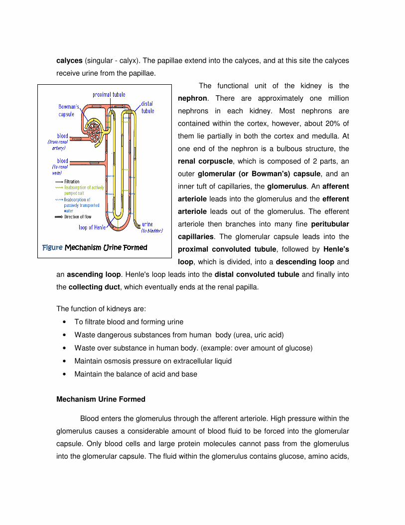
calyces (singular - calyx). The papillae extend into the calyces, and at this site the calyces
receive urine from the papillae.
The functional unit of the kidney is the
nephron. There are approximately one million
nephrons in each kidney. Most nephrons are
contained within the cortex, however, about 20% of
them lie partially in both the cortex and medulla. At
one end of the nephron is a bulbous structure, the
renal corpuscle, which is composed of 2 parts, an
outer glomerular (or Bowman's) capsule, and an
inner tuft of capillaries, the glomerulus. An afferent
arteriole leads into the glomerulus and the efferent
arteriole leads out of the glomerulus. The efferent
arteriole then branches into many fine peritubular
capillaries. The glomerular capsule leads into the
proximal convoluted tubule, followed by Henle's
loop, which is divided, into a descending loop and
an ascending loop. Henle's loop leads into the distal convoluted tubule and finally into
the collecting duct, which eventually ends at the renal papilla.
The function of kidneys are:
• To filtrate blood and forming urine
• Waste dangerous substances from human body (urea, uric acid)
• Waste over substance in human body. (example: over amount of glucose)
• Maintain osmosis pressure on extracellular liquid
• Maintain the balance of acid and base
Mechanism Urine Formed
Blood enters the glomerulus through the afferent arteriole. High pressure within the
glomerulus causes a considerable amount of blood fluid to be forced into the glomerular
capsule. Only blood cells and large protein molecules cannot pass from the glomerulus
into the glomerular capsule. The fluid within the glomerulus contains glucose, amino acids,
Figure Figure Figure Figure Mechanism Urine FormedMechanism Urine FormedMechanism Urine FormedMechanism Urine Formed

urea, salts, and a large amount of water. While we
want to eliminate urea from the body, we cannot
afford to lose large quantities of the other substances.
The challenge for the nephron is to eliminate the urea
while reclaiming the other substances. As the filtrate
passes into the proximal convoluted tubule,
essentially all the glucose and most of the water,
salts, and amino acids are reabsorbed. However, we
are still losing too much water and sodium. The
descending loop of Henle is very permeable to water
but not very permeable to sodium. Consequently
water moves out of the tubule, but sodium remains in the tubule. The fluid in the tubule
becomes more concentrated as it reaches the bottom of the loop. The ascending loop is
relatively impermeable to water, but sodium is actively pumped out of the tubule.
As the fluid moves up the ascending loop, it becomes less concentrated, and by the time it
reaches the distal convoluted tubule, the composition of the fluid is little different from the
fluid that entered the descending loop. However, considerable amounts of water and
sodium have been reclaimed as the fluid passes through Henle’s loop.
After Henle's loop, the fluid enters the distal convoluted tubule. Water reabsorption
here is facilitated by antidiuretic hormone (ADH), which is released by the pituitary
gland. The more ADH released from the pituitary, the greater the water reabsorption in the
distal tubule, and the more concentrated the urine. The amount of ADH released is
dependent on the hydration state of the individual.
If you have been eating large amounts of watermelon, little ADH would be released,
less water would be reabsorbed in the distal tubule, and a dilute urine would be produced.
If you are dehydrated due to bouncing your buns off in aerobics class, more ADH would be
released, more water would be reabsorbed in the distal tubule, and a more concentrated
urine would be produced. Following the distal convoluted tubule, the fluid enters the
collecting duct, and eventually leaves the nephron at the renal papillae.
Excretory systems are diverse, but nearly all produce fluid waste urine in process
involving several steps:
Figure Figure Figure Figure Structure Structure Structure Structure of Neuronof Neuronof Neuronof Neuron

� Filtration: Body fluid (blood, coelomic fluid, or hemolymph) is collected involving
filtration through selectively permeable membranes. Hydrostatic pressure (blood
pressure) forces water and small solutes into excretory system. Fluid = filtrate.
� Selective Reabsorption: excretory systems use active transport to reabsorb valuable
solutes from the filtrate and returns them to body fluids.
� Secretion: Other substances like toxins or excess ions are extracted from body fluids
and added to filtrate using active transport
� Excretion: filtrate leaves system and body as urine
Kidneys Disorder
• Cystitis : Inflammation of the urinary bladder caused by a bacterial infection
• Glomerulonephritis : Inflammation of the glomeruli in the renal corpuscles of the
kidneys.
• Kidney cancer develops when cells in certain tissues in the kidneys become
abnormal and grow uncontrollably, forming tumors.
• Kidney stones: Large accumulations of calcium salt crystals from urine that may
form in the kidneys.
• Pyelonephritis : Inflammation of the kidneys caused by a bacterial infection.
• Urethritis : Inflammation of the urethra caused by a bacterial infection.
• Urinary incontinence: Involuntary and unintentional passage or urine.
• Albuminuria : showed by albumin or protein molecules in urine
• Glukosaria : showed by glucose molecules in urine
• Hematuria showed by erythrocytes in urine.
The human lungs are the organs of respiration and excretion humans. Humans
have two lungs, with the left being divided into two lobes and the right into three lobes.
Lungs

Oxygen (O2) from air breathed in goes into
the red blood cell via alveoli. Carbon dioxide
(CO2) goes from the red blood cell into alveoli
and breathed out. As respiration occurs
carbon dioxide is produced as a waste
product. As the carbon dioxide
accumulates in body cells, it eventually
diffuses out of the cells & into the
bloodstream, which eventually circulates to
the lungs. And here, in the alveoli of the
lungs, carbon dioxide diffuses from the
blood, into the lung tissue, and then leaves
the body every time we exhale. We should
note that some water vapor also exits the body during exhalation. Functions of
Lungs are: (1) supply oxygen to the cells and tissues of the body; (2) dispose of the
carbon dioxide.
Disfunctions of Lungs are:
• TBC
Tuberculosis (TB) is an infectious disease caused by bacteria whose scientific
name is Mycobacterium tuberculosis.
• Pneumonia
Pneumonia is an inflammation of the lung, usually caused by an infection.
Three common causes are bacteria, viruses and fungi.
• Lungs cancer caused by smoke, gases, cigarette.
• Emphysema the lungs Frozen because contain of air
The skin acts as an organ of excretion by removing water and small amounts of
urea and salts (as sweat). Function of Skin are: (1) produce sweat; (2) protect our
Skin
FigureFigureFigureFigure Structure of Lungs

body; (3)save fat surplus; (4) control body temperature; (5) place where vitamin D
is made; (6) sense of touch.
Structure of Skin
Figure Figure Figure Figure Structure Anatomy of SkinStructure Anatomy of SkinStructure Anatomy of SkinStructure Anatomy of Skin
The skin has a slightly acidic coating of oil at the surface. This coating protects the
skin against some bacteria. Below the surface is a complex of sweat and oil glands, hair
follicles, blood vessels, nerves, and muscle tissue. These are held together by a tough
connective tissue called collagen. Below the collagen is a layer of fat and muscle, which
provides some contour and acts as a cushion and as insulation.
The skin has three layers. The inner most layer is known as the lower dermis, the middle
layer is called the dermis, and the outer layer is known as the epidermis.
(1) Lower Dermis
The various glands such as the oil and sweat glands originate in the lower
dermis. From here, they rise to the surface of the skin to eliminate waste matter. Lower
dermis also acts as a cushion for the rest of the skin. It contains the finely distributed
muscles of the skin which regulate body temperature.
(2) Dermis
The dermis is the layer that lies underneath the epidermis, and it is composed
entirely of living cells. It consists of bundles of tough fibers which give your skin its

elasticity, firmness and strength. There are also blood vessels, which feed vital
nutrients to these areas.
The most important function of dermis is respiration. The countless tiny blood
vessels, or capillaries end here in finely-drawn networks, from where they feed the
outer skin layer. Dermis also determines the tone of the skin.
(3) Epidermis
This is the top layer of skin and the one you can actually see. It protects your
body from invasion and infection and helps to seal in moisture. It's built up of several
layers of living cells which are then topped by sheets of dead cells. It's constantly
growing, with new cells being produced at its base. They quickly die, and are pushed
up to the surface by the arrival of new ones, These dead cells eventually flake away,
which means that every new layer of skin is another chance to have a soft, glowing
complexion.
The lower levels of living cells are fed by the blood supply from
underneath,whereas the upper dead cells only need water to ensure they're kept
plump and smooth. The epidermis is responsible for your coloring, as it holds the skin's
pigment. It ranges in thickness from l/20-th of an inch on the palms and soles, to 1/200-
th of an inch on the face.
The skin contains the following specialized organs: (1) Sebaceous glands are tiny
organs which usually open into hair follicles on the surface of your skin. They produce an
oily secretion, called sebum, which is your skin's natural lubricant; (2) The sebaceous
glands are most concentrated on the scalp and face - particularly around the nose,
cheeks, chin and forehead, which is why these are usually the most oily areas of your skin;
(3) Sweat glands are all over your body. There are millions of them and their main
function is to regulate your body temperature. When sweat evaporates on the skin's
surface, the temperature of your skin drops; (4) Hairs grow from the hair follicles. They
can help keep your body warm by trapping air underneath them. There are no hairs on the
soles of your feet and palms of your hands.

How does sweat produce?
The liver is a reddish brown organ with four lobes of unequal size and
shape. It is the largest gland in the body . It is located in the right upper quadrant
of the abdominal cavity, resting just below the diaphragm.
Functions of Liver
Liver regulates glycogen storage, plasma protein synthesis, and drug
detoxification. The liver secretes bile, a base used for breaking down fats. It helps
get rid of unneeded wastes in the body. It changes toxic ammonia, which is a
poisonous gas , to urea, a harmless fluid. Bile pigments (biliverdin and bilirubin)
are produced as a result of break down of haemoglobin in the worn out red blood
cells in the liver and excreted with the bile into the duodenum and removed along
with the faeces. Liver also excretes cholesterol, various inactivated products of
steroid hormones and harmful products like alcohol, nicotine and several drugs.
LiverLiverLiverLiver

The liver is also responsible for converting ammonia produced by deamination into
less toxic urea by combining it with carbon dioxide.
AssignmentAssignmentAssignmentAssignment
Cross of ExcressionCross of ExcressionCross of ExcressionCross of Excression

Reference
Champbel, Reece, & Mitchell. 2004. Biologi Jilid I, II, & III [terjemahan]. Jakarta:
Gelora Aksara Pratama
Guyton, A. 1996. Fisiologi Manusia dan Mekanisme Penyakit [terjemahan] .
Jakarta: EGC Kedokteran.
Solomon, Berg, & Diana Martin. 2006. Biology Seventh edition. Canada: Thomson
Learning, Inc.