Embed Size (px)
Citation preview
© 2018 Indian Journal of Plastic Surgery | Published by Wolters Kluwer ‑ Medknow 70
INTRODUCTION
Pressure sore is a common surgical condition often referred to plastic surgeons for management. In general, approximately 9% of all hospitalised
patients develop pressure sore, its occurrence in acute care setting being around 11%.[1] Pressure sore can be managed conservatively or surgically with a flap depending on stage, size, involvement of bone and general condition of
Infragluteal fasciocutaneous flap for management of recurrent ischial pressure sore
Umesh Kumar, Pradeep JainDepartment of Plastic Surgery, Institute of Medical Sciences, Banaras Hindu University, Varanasi, Uttar Pradesh, India
Address for correspondence: Dr. Umesh Kumar, 25 Brij Enclave Extension–1, Bazardiha (PO), Varanasi ‑ 221 109, Uttar Pradesh, India. E‑mail: [email protected]
ABSTRACT
Objective: The objective of the study was to determine the feasibility of infragluteal fasciocutaneous flap in recurrent ischial pressure sore. Materials and Methods: In our study, from 2015 to 2017, nine patients suffering from recurrent ischial sore with scars of previous surgery were managed with infragluteal fasciocutaneous flap. Wound bed was prepared by surgical debridement and negative pressure wound therapy in each case. In two cases, gracilis muscle flap was used as adjuvant to fill up the residual cavity. Donor area of flap was primarily closed. Results: Infragluteal fasciocutaneous flap was used in all nine cases. Superficial distal congestion was present in two cases. Haematoma (1) and infection (1) at flap donor site occurred. Recurrence of ulcer was observed in two cases which were managed by bursectomy and advancement of the bridge segment of the original infragluteal fasciocutaneous flap. All flaps survived without any major complication. Discussion: Ischial pressure sores have a tendency of recurrence after conservative or flap surgery. Scars due to previous surgeries adjacent to the pressure sore preclude the use of local skin or muscle flap. Infragluteal fasciocutaneous flap is a thick reliable fasciocutaneous flap that can be used for resurfacing recurrent ischial pressure sore. This flap has an axial pattern blood supply along with rich subfascial and fascial plexus supplied by various perforators. Conclusion: Infragluteal fasciocutaneous flap is reliable option for managing recurrent ischial sore as it transposes well‑vascularised thick fasciocutaneous flap from adjacent posterior thigh and its bridge segment can be further used in case of recurrence.
KEYWORDS
Infragluteal fasciocutaneous flap; ischial pressure sore; perforator plus flap; recurrent pressure ulcer
Access this article onlineQuick Response Code:
Website:
www.ijps.org
DOI:
10.4103/ijps.IJPS_15_18How to cite this article: Kumar U, Jain P. Infragluteal fasciocutaneous flap for management of recurrent ischial pressure sore. Indian J Plast Surg 2018;51:70‑6.
This is an open access journal, and articles are distributed under the terms of the Creative Commons Attribution‑NonCommercial‑ShareAlike 4.0 License, which allows others to remix, tweak, and build upon the work non‑commercially, as long as appropriate credit is given and the new creations are licensed under the identical terms.
For reprints contact: [email protected]
Original Article
Published online: 2019-07-26
Indian Journal of Plastic Surgery Volume 51 Issue 1 January‑April 201871
Kumar and Jain: Infragluteal fasciocutaneous flap
the patient . Long‑term follow up studies on patients with pressure sore document‑wide range of recurrence rates varying from 3%–6% to 33%–100%. Conservative therapy of recurrent ischial ulcer is not routinely successful. Out of various techniques described for ischial pressure sore, we preferred fasciocutaneous flap over musculocutaneous flap. Homma et al. reported using a posteromedial thigh fasciocutaneous flap based on the perforators of either gracilis or the adductor magnus muscle in the treatment of ischial pressure sore.[2] Tissue from the infragluteal region that is posterior thigh was transferred as a free fasciocutaneous flap based on descending branch of inferior gluteal artery. It was first described as an alternative for autologous breast reconstruction in thin patients by Papp et al. in 2007. It was successfully used in 28 breast reconstructions with disadvantages of insufficient volume in two and variable sensory problems at donor site.[3] The posterior thigh skin can be transferred based on perforating vessels from descending branch of the inferior gluteal artery that accompanies the posterior femoral cutaneous nerve. The flap can be used repeatedly in the event of recurrence.[4] Even if the vessel is not present due to anatomical variation, the flap survives on superiorly based random fasciocutaneous flap supplied by multiple perforators from the cruciate anastomosis of the fascial plexus.[5] The perforator plus techniques combine the advantage by providing additional blood supply and preventing distal congestion of peninsular flap by safeguarding venous return.[6]
AnatomyThe inferior gluteal artery is usually a terminal branch of the anterior trunk of the internal iliac but
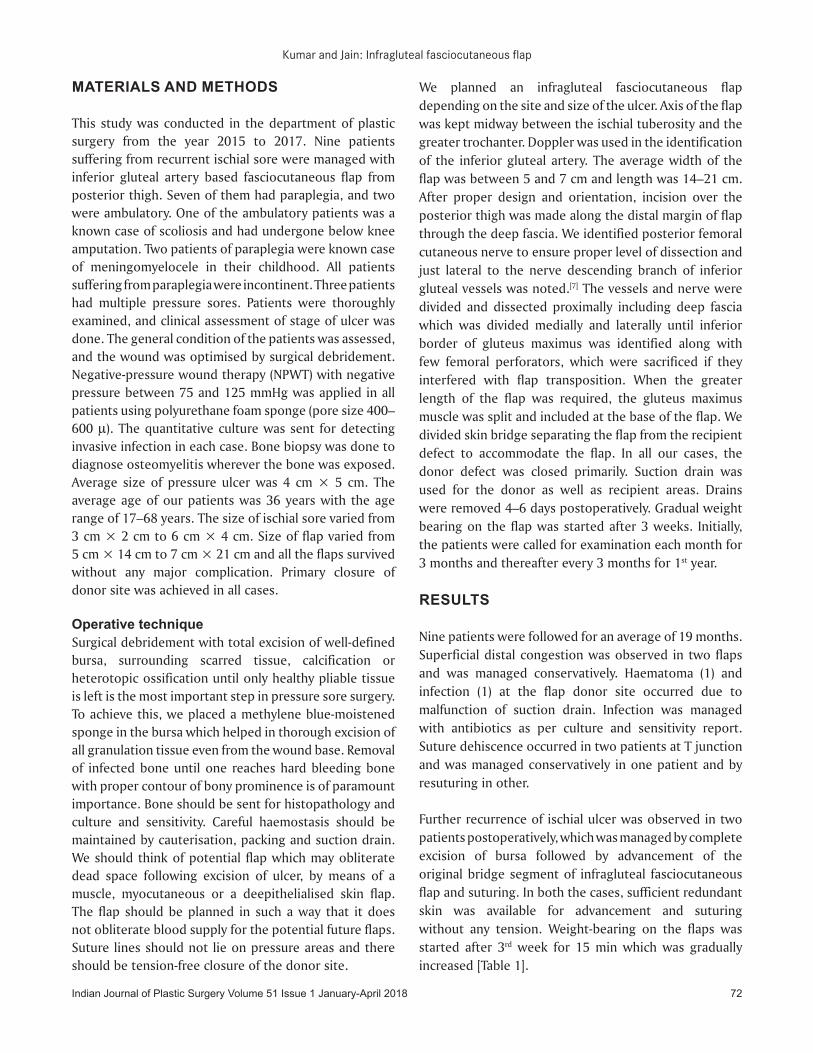
may arise with the superior gluteal from a common trunk. It enters the gluteal region through the greater sciatic notch below the piriformis muscle. It measures 3.5 mm in diameter and can be marked on the surface by a point halfway along a line drawn from the posterior superior iliac spine to the ischial tuberosity. The inferior gluteal artery divides into two main branches medial and lateral, which supplies lower two‑third of gluteus maximus muscle and its overlying skin through multiple perforators. Proximal to the inferior margin of gluteus maximus muscle the artery anastomoses with obturator and medial femoral circumflex arteries and sends several deep branches to the hamstring muscle. The descending branch of inferior gluteal artery accompanies the posterior cutaneous nerve and is lateral in the subcutaneous plane just below the fascia lata, superficial to the bicep femoris muscle. The cutaneous territory of the posterior thigh probably has dual supply, a subfascial plexus created by the descending branch of the inferior gluteal artery and a fascial plexus lying above the deep fascia supplied by musculocutaneous perforators of femoral and obturator artery or the inferior gluteal artery. To a variable degree, both vascular systems feed a plexus of vessel just superficial to deep fascia. Venous drainage parallels its arterial supply. The traditionally described gluteal thigh flap can be raised as a musculocutaneous flap based on descending branch of the inferior gluteal artery or as its fasciocutaneous extension if longer length of flap is required. We hereby describe the clinical application of infragluteal fasciocutaneous flap [Figure 1].
Figure 1: (a) Flap anatomy showing inferior gluteal artery and its descending branch with posterior cutaneous nerve and gluteus maximus muscle. (b) Preoperative view of a 17 years old male paraplegic (operated meningomyelocele) with right ischial pressure sore. (c) Sore after bursectomy and partial
ischiectomy. (d) Gluteal thigh with gracilis muscle flap. (e) Two weeks’ post‑operative view after reconstruction. (f) Three months’ post‑operative view
d
cb
f
a
e
Indian Journal of Plastic Surgery Volume 51 Issue 1 January‑April 2018 72
Kumar and Jain: Infragluteal fasciocutaneous flap
MATERIALS AND METHODS
This study was conducted in the department of plastic surgery from the year 2015 to 2017. Nine patients suffering from recurrent ischial sore were managed with inferior gluteal artery based fasciocutaneous flap from posterior thigh. Seven of them had paraplegia, and two were ambulatory. One of the ambulatory patients was a known case of scoliosis and had undergone below knee amputation. Two patients of paraplegia were known case of meningomyelocele in their childhood. All patients suffering from paraplegia were incontinent. Three patients had multiple pressure sores. Patients were thoroughly examined, and clinical assessment of stage of ulcer was done. The general condition of the patients was assessed, and the wound was optimised by surgical debridement. Negative‑pressure wound therapy (NPWT) with negative pressure between 75 and 125 mmHg was applied in all patients using polyurethane foam sponge (pore size 400–600 μ). The quantitative culture was sent for detecting invasive infection in each case. Bone biopsy was done to diagnose osteomyelitis wherever the bone was exposed. Average size of pressure ulcer was 4 cm × 5 cm. The average age of our patients was 36 years with the age range of 17–68 years. The size of ischial sore varied from 3 cm × 2 cm to 6 cm × 4 cm. Size of flap varied from 5 cm × 14 cm to 7 cm × 21 cm and all the flaps survived without any major complication. Primary closure of donor site was achieved in all cases.
Operative techniqueSurgical debridement with total excision of well‑defined bursa, surrounding scarred tissue, calcification or heterotopic ossification until only healthy pliable tissue is left is the most important step in pressure sore surgery. To achieve this, we placed a methylene blue‑moistened sponge in the bursa which helped in thorough excision of all granulation tissue even from the wound base. Removal of infected bone until one reaches hard bleeding bone with proper contour of bony prominence is of paramount importance. Bone should be sent for histopathology and culture and sensitivity. Careful haemostasis should be maintained by cauterisation, packing and suction drain. We should think of potential flap which may obliterate dead space following excision of ulcer, by means of a muscle, myocutaneous or a deepithelialised skin flap. The flap should be planned in such a way that it does not obliterate blood supply for the potential future flaps. Suture lines should not lie on pressure areas and there should be tension‑free closure of the donor site.
We planned an infragluteal fasciocutaneous flap depending on the site and size of the ulcer. Axis of the flap was kept midway between the ischial tuberosity and the greater trochanter. Doppler was used in the identification of the inferior gluteal artery. The average width of the flap was between 5 and 7 cm and length was 14–21 cm. After proper design and orientation, incision over the posterior thigh was made along the distal margin of flap through the deep fascia. We identified posterior femoral cutaneous nerve to ensure proper level of dissection and just lateral to the nerve descending branch of inferior gluteal vessels was noted.[7] The vessels and nerve were divided and dissected proximally including deep fascia which was divided medially and laterally until inferior border of gluteus maximus was identified along with few femoral perforators, which were sacrificed if they interfered with flap transposition. When the greater length of the flap was required, the gluteus maximus muscle was split and included at the base of the flap. We divided skin bridge separating the flap from the recipient defect to accommodate the flap. In all our cases, the donor defect was closed primarily. Suction drain was used for the donor as well as recipient areas. Drains were removed 4–6 days postoperatively. Gradual weight bearing on the flap was started after 3 weeks. Initially, the patients were called for examination each month for 3 months and thereafter every 3 months for 1st year.
RESULTS
Nine patients were followed for an average of 19 months. Superficial distal congestion was observed in two flaps and was managed conservatively. Haematoma (1) and infection (1) at the flap donor site occurred due to malfunction of suction drain. Infection was managed with antibiotics as per culture and sensitivity report. Suture dehiscence occurred in two patients at T junction and was managed conservatively in one patient and by resuturing in other.
Further recurrence of ischial ulcer was observed in two patients postoperatively, which was managed by complete excision of bursa followed by advancement of the original bridge segment of infragluteal fasciocutaneous flap and suturing. In both the cases, sufficient redundant skin was available for advancement and suturing without any tension. Weight‑bearing on the flaps was started after 3rd week for 15 min which was gradually increased [Table 1].
Indian Journal of Plastic Surgery Volume 51 Issue 1 January‑April 201873
Kumar and Jain: Infragluteal fasciocutaneous flap
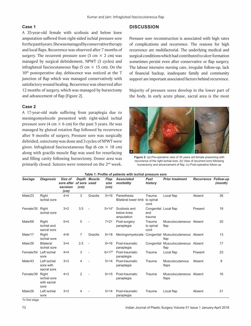
Case 1A 35‑year‑old female with scoliosis and below knee amputation suffered from right‑sided ischial pressure sore for the past 6 years. She was managed by conservative therapy and local flaps. Recurrence was observed after 7 months of surgery. The recurrent pressure sore (3 cm × 2 cm) was managed by surgical debridement, NPWT (3 cycles) and infragluteal fasciocutaneous flap (5 cm × 15 cm). On the 10th postoperative day, dehiscence was noticed at the T junction of flap which was managed conservatively with satisfactory wound healing. Recurrence was observed after 12 months of surgery, which was managed by bursectomy and advancement of flap [Figure 2].
Case 2A 17‑year‑old male suffering from paraplegia due to meningomyelocele presented with right‑sided ischial pressure sore (4 cm × 6 cm) for the past 5 years. He was managed by gluteal rotation flap followed by recurrence after 9 months of surgery. Pressure sore was surgically debrided, ostectomy was done and 3 cycles of NPWT were given. Infragluteal fasciocutaneous flap (6 cm × 18 cm) along with gracilis muscle flap was used for resurfacing and filling cavity following bursectomy. Donor area was primarily closed. Sutures were removed on the 2nd week.
DISCUSSION
Pressure sore reconstruction is associated with high rates of complications and recurrence. The reasons for high recurrence are multifactorial. The underlying medical and surgical conditions which had contributed to ulcer formation sometimes persist even after conservative or flap surgery. The labour intensive nursing care, irregular follow‑up, lack of financial backup, inadequate family and community support are important associated factors behind recurrence.
Majority of pressure sores develop in the lower part of the body. In early acute phase, sacral area is the most
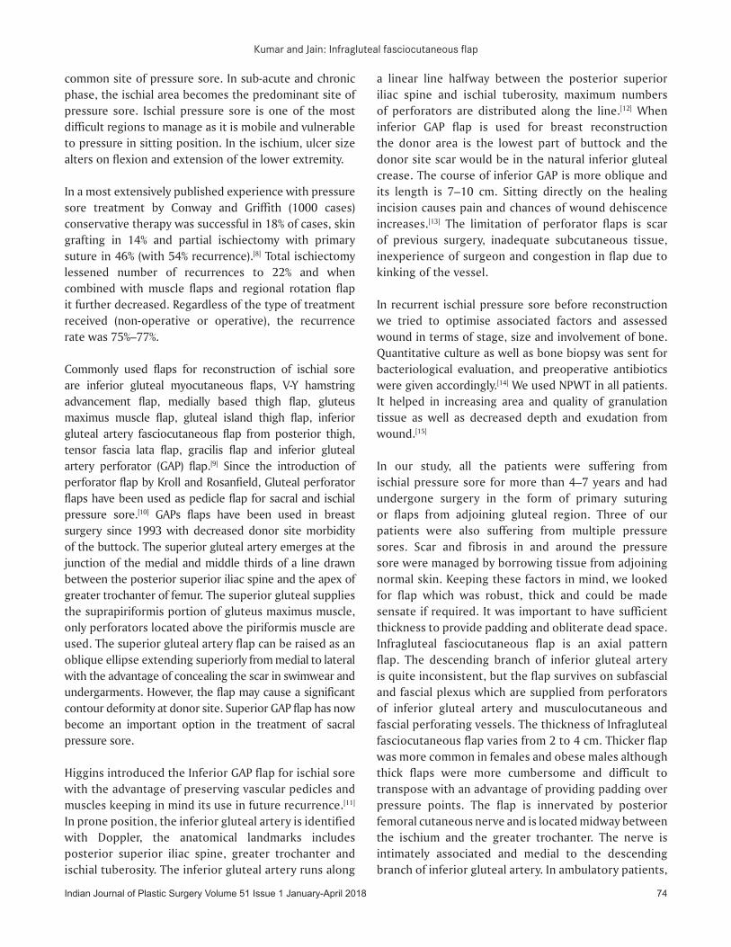
Table 1: Profile of patients with ischial pressure soreSex/age Diagnosis Size of
sore after excision
(cm)
Depth of sore
(cm)
Muscle used
Flap size (cm)
Associated morbidity
Past history
Prior treatment Recurrence Follow‑up (month)
Male/23 Right ischial sore
4×4 3 Gracilis 5×16 Paresthesia Bilateral lower limb
Trauma to spinal cord
Local flap Absent 36
Female/35 Right ischial sore
3×2 3.5 ‑ 5×14* Scoliosis and below knee amputation
Congenital and trauma
Local flap Present 18
Male/68 Right ischial and sacral sore
5×4 5 ‑ 7×21 Post‑surgery paraplegia
Trauma to spinal cord
Musculocutaneous flap
Absent 20
Male/17 Right ischial sore
4×6 7 Gracilis 6×18 Meningomyelocele Congenital Musculocutaneous flap
Absent 13
Male/26 Bilateral ischial sore
3×4 2.5 ‑ 6×16 Post‑traumatic paraplegia
Congenital Musculocutaneous flap
Absent 17
Female/54 Left ischial sore
4×4 3 ‑ 6×17* Post‑traumatic paraplegia
Trauma Local flap Present 23
Male/43 Left ischial sore with sacral sore
3×3 4 ‑ 5×14 Post‑traumatic paraplegia
Trauma Musculocutanous flaps
Absent 8
Female/39 Right ischial sore with sacral sore
4×3 2 ‑ 6×15 Post‑traumatic paraplegia
Trauma Musculocutaneous flaps
Absent 16
Male/26 Left ischial sore
3×3 4 ‑ 5×14 Post‑traumatic paraplegia
Trauma Local flap Absent 21
*In first stage
Figure 2: (a) Pre‑operative view of 35 years old female presenting with recurrence of the right ischial sore. (b) View of recurrent sore following
bursectomy and advancement of flap. (c) Post‑operative follow‑up
cba
Indian Journal of Plastic Surgery Volume 51 Issue 1 January‑April 2018 74
Kumar and Jain: Infragluteal fasciocutaneous flap
common site of pressure sore. In sub‑acute and chronic phase, the ischial area becomes the predominant site of pressure sore. Ischial pressure sore is one of the most difficult regions to manage as it is mobile and vulnerable to pressure in sitting position. In the ischium, ulcer size alters on flexion and extension of the lower extremity.
In a most extensively published experience with pressure sore treatment by Conway and Griffith (1000 cases) conservative therapy was successful in 18% of cases, skin grafting in 14% and partial ischiectomy with primary suture in 46% (with 54% recurrence).[8] Total ischiectomy lessened number of recurrences to 22% and when combined with muscle flaps and regional rotation flap it further decreased. Regardless of the type of treatment received (non‑operative or operative), the recurrence rate was 75%–77%.
Commonly used flaps for reconstruction of ischial sore are inferior gluteal myocutaneous flaps, V‑Y hamstring advancement flap, medially based thigh flap, gluteus maximus muscle flap, gluteal island thigh flap, inferior gluteal artery fasciocutaneous flap from posterior thigh, tensor fascia lata flap, gracilis flap and inferior gluteal artery perforator (GAP) flap.[9] Since the introduction of perforator flap by Kroll and Rosanfield, Gluteal perforator flaps have been used as pedicle flap for sacral and ischial pressure sore.[10] GAPs flaps have been used in breast surgery since 1993 with decreased donor site morbidity of the buttock. The superior gluteal artery emerges at the junction of the medial and middle thirds of a line drawn between the posterior superior iliac spine and the apex of greater trochanter of femur. The superior gluteal supplies the suprapiriformis portion of gluteus maximus muscle, only perforators located above the piriformis muscle are used. The superior gluteal artery flap can be raised as an oblique ellipse extending superiorly from medial to lateral with the advantage of concealing the scar in swimwear and undergarments. However, the flap may cause a significant contour deformity at donor site. Superior GAP flap has now become an important option in the treatment of sacral pressure sore.
Higgins introduced the Inferior GAP flap for ischial sore with the advantage of preserving vascular pedicles and muscles keeping in mind its use in future recurrence.[11] In prone position, the inferior gluteal artery is identified with Doppler, the anatomical landmarks includes posterior superior iliac spine, greater trochanter and ischial tuberosity. The inferior gluteal artery runs along
a linear line halfway between the posterior superior iliac spine and ischial tuberosity, maximum numbers of perforators are distributed along the line.[12] When inferior GAP flap is used for breast reconstruction the donor area is the lowest part of buttock and the donor site scar would be in the natural inferior gluteal crease. The course of inferior GAP is more oblique and its length is 7–10 cm. Sitting directly on the healing incision causes pain and chances of wound dehiscence increases.[13] The limitation of perforator flaps is scar of previous surgery, inadequate subcutaneous tissue, inexperience of surgeon and congestion in flap due to kinking of the vessel.
In recurrent ischial pressure sore before reconstruction we tried to optimise associated factors and assessed wound in terms of stage, size and involvement of bone. Quantitative culture as well as bone biopsy was sent for bacteriological evaluation, and preoperative antibiotics were given accordingly.[14] We used NPWT in all patients. It helped in increasing area and quality of granulation tissue as well as decreased depth and exudation from wound.[15]
In our study, all the patients were suffering from ischial pressure sore for more than 4–7 years and had undergone surgery in the form of primary suturing or flaps from adjoining gluteal region. Three of our patients were also suffering from multiple pressure sores. Scar and fibrosis in and around the pressure sore were managed by borrowing tissue from adjoining normal skin. Keeping these factors in mind, we looked for flap which was robust, thick and could be made sensate if required. It was important to have sufficient thickness to provide padding and obliterate dead space. Infragluteal fasciocutaneous flap is an axial pattern flap. The descending branch of inferior gluteal artery is quite inconsistent, but the flap survives on subfascial and fascial plexus which are supplied from perforators of inferior gluteal artery and musculocutaneous and fascial perforating vessels. The thickness of Infragluteal fasciocutaneous flap varies from 2 to 4 cm. Thicker flap was more common in females and obese males although thick flaps were more cumbersome and difficult to transpose with an advantage of providing padding over pressure points. The flap is innervated by posterior femoral cutaneous nerve and is located midway between the ischium and the greater trochanter. The nerve is intimately associated and medial to the descending branch of inferior gluteal artery. In ambulatory patients,
Indian Journal of Plastic Surgery Volume 51 Issue 1 January‑April 201875
Kumar and Jain: Infragluteal fasciocutaneous flap
sacrifice of nerve did not cause any major sensory deficit except numbness.[16] The point of rotation of flap varies in peninsular flaps, the cone of rotation absorbs one‑third of flap length. When we incorporated gluteus maximus, the arc of rotation was extended to the sciatic foramen. Then, the raised flap can be called as inferior gluteal artery myocutaneous flap (Hurwitz).[17]
We kept flap width between 5 and 7 cm and could achieve primary closure of the donor site. In two cases of paraplegia, we used gracilis muscle to fill the cavity following bursectomy and partial ischiectomy. One of the advantages of infragluteal fasciocutaneous flap is that the flap can be used repeatedly in the event of recurrence. Foster et al. reviewed ischial coverage from 1979 to 1995 in which 139 ischial pressure sores were treated. In his study, the inferior gluteus maximus island flaps and inferior gluteal thigh flap had the highest success rates 94% and 93%, respectively.[18] While the V‑Y hamstring flap and tensor fascia lata flap had the poorest healing rates 58% and 50%, respectively. Ahluwalia et al. reviewed 72 ischial wounds and found a complication rate of 16% and recurrence rate of 7%.[19]
There are a few limitations of this flap; thicker flaps are cumbersome and difficult to transpose. In peninsular flap, the cone of rotation absorbs up to one‑third of the flap length. To resurface small defects, large flap has to be dissected. If flap width exceeds 6–7 cm the donor area requires split thickness graft. While using this flap in 9 patients, we encountered only two recurrences. Both patients were ambulatory. On counselling, patients revealed lack of care of the ischial region as the primary cause. Recurrence was managed by bursectomy and advancement of the flap. None of our flaps failed.
CONCLUSION
The infragluteal fasciocutaneous flap is a reliable option for managing recurrent ischial sore as it transposes well‑vascularised thick fasciocutaneous flap from the adjacent posterior thigh region to obliterate the dead space following bursectomy and ischiectomy. It can also be used in cases of recurrence by advancing the same flap taking advantage of loose tissue in bridge segment.
Declaration of patient consentThe authors certify that they have obtained all appropriate patient consent forms. In the form the patient(s) has/have
given his/her/their consent for his/her/their images and other clinical information to be reported in the journal. The patients understand that their names and initials will not be published and due efforts will be made to conceal their identity, but anonymity cannot be guaranteed.
Financial support and sponsorshipNil.
Conflicts of interestThere are no conflicts of interest.
REFERENCES
1. John BD, John MS, Linda PG. Pressure sore. In: Chales TH, editor. Grabb’s and Smith’s Plastic Surgery. 6th ed. Philadelphia: Lippincott Williams & Wilkins and Wolters Kluwer Business; 2007. p. 722.
2. Homma K, Murakami G, Fujioka H, Fujita T, Imai A, Ezoe K, et al. Treatment of ischial pressure ulcers with a posteromedial thigh fasciocutaneous flap. Plast Reconstr Surg 2001;108:1990‑6.
3. Papp C, Windhofer C, Gruber S. Breast reconstruction with the fasciocutaneous infragluteal free flap (FCI). Ann Plast Surg 2007;58:131‑6.
4. Robert K, Jeffrey JE. Pressure sores. In: Peter NC, David SH, editors. Plastic Surgery. Vol. 4. London: Elsevier Saunders; 2013. p. 352.
5. Song WC, Bae SM, Han SH, Koh KS. Anatomical and radiological study of the superior and inferior gluteal arteries in the gluteus maximus muscle for musculocutaneous flap in Koreans. J Plast reconstr Aesthet Surg 2006;59(9):935‑41.
6. Sharma RK. Perforator flap: Evoluation of the concept and its place in plastic surgery repertoire. Indian J Plast Surg 2010;43:148‑50.
7. Walton RL, Hurwitz DJ, Bunkis J. Gluteal thigh flap for reconstruction of perineal defects. In: Strauch B, Vasconez LO, Hall‑Findlay EJ, editors. Grabb’s Encyclopedia of Flaps. 2nd ed., Vol. 3. Philadelphia: Lippincott Raven Publishers; 1998. p. 1499.
8. Conway H, Griffith BH. Plastic surgery for closure of decubitus ulcers in patients with paraplegia; based on experience with 1,000 cases. Am J Surg 1956;91:946‑75.
9. Foster RD, Anthony JP, Mathes SJ, Hoffman WY. Ischial pressure sore coverage: A rationale for flap selection. Br J Plast Surg 1997;50(5):374‑9.
10. Kim YS, Lew DH, Roh TS, Yoo WM, Lee WJ, Tark KC, et al. Inferior gluteal artery perforator flap: A viable alternative for ischial pressure sores. J Plast Reconstr Aesthet Surg 2009;62:1347‑54.
11. Higgins JP, Orlando GS, Blondeel PN. Ischial pressure sore reconstruction using an inferior gluteal artery perforator (IGAP) flap. Br J Plast Surg 2002;55:83‑5.
12. Beshlian KM, Paige KT. Inferior gluteal artery perforator flap breast reconstruction. Am J Surg 2008;195:651‑3.
13. Allen RJ, Levine JL, Granzow JW. The in‑the‑crease inferior gluteal artery perforator flap for breast reconstruction. Plast Reconstr Surg 2006;118:333‑9.
14. Stal S, Serure A, Donovan W, Spira M. The perioperative management of the patient with pressure sores. Ann Plast Surg 1983;11:347‑56.
15. Greer SE, Duthie E, Cartolano B, Koehler KM,

Indian Journal of Plastic Surgery Volume 51 Issue 1 January‑April 2018 76
Kumar and Jain: Infragluteal fasciocutaneous flap
Maydick‑Youngberg D, Longaker MT, et al. Techniques for applying subatmospheric pressure dressing to wounds in difficult regions of anatomy. J Wound Ostomy Continence Nurs 1999;26:250‑3.
16. Hurwitz DJ, Swartz WM, Mathes SJ. The gluteal thigh flap: A reliable, sensate flap for the closure of buttock and perineal wounds. Plast Reconstr Surg 1981;68:521‑32.
17. Hurwitz DJ. Closure of a large defect of the pelvic cavity by an
extended compound myocutaneous flap based on the inferior gluteal artery. Br J Plast Surg 1980;33:256‑61.
18. Foster RD. Pressure sore. In: Mathes SJ, Hentz VR, editor. Plastic Surgery. 2nd ed., Vol. 6. Philadelphia: Saunders/Elsevier; 2006. p. 1321.
19. Ahluwalia R, Martin D, Mahoney JL. The operative treatment of pressure wounds: A 10‑year experience in flap selection. Int Wound J 2010;7:103‑6.