• An Example of Fibrinoheterophilic and Lymphocytic Inflammation– Diffuse fibrinoheterophilic– Multifocal Lymphocytic
• Broilers– 56 Days– Company Experiencing Increases in Mortality as Birds Reach Processing Age
– Increased Condemnations
Presenter
Presentation Notes
This case is an example to illustrate fibrinoheterophilic inflammation with multifocal collections of lymphocytes (lymphoid cells) around blood vessels. The morphologic diagnosis is cellulitis because the inflammation is located in the dermis and underlying connective tissue. Broilers with this condition are condemned at processing with the meat inspection classification of infectious process. The next few images will show the gross and microscopic lesions. Look for the components that make this a fibrinoheterophilic and lymphocytic cellulitis.
Infectious Process
Presenter
Presentation Notes
This is a gross lesion of cellulitis or infectious process. The small arrow points to an area of old hemorrhage and ulceration of the skin and the wide arrow points to the accumulation of caseous exudate in the subcutaneous tissue. The microscopic features of this condition are illustrated in the following two slides.
Dermatitis ‐ Cellulitis
A B
C D
Presenter
Presentation Notes
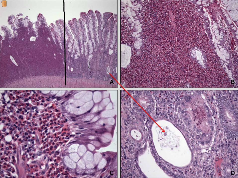
Image A is a low power view showing the diffuse nature of the inflammation. The wide arrow points to the epithelial surface of the skin. The black bar is over subcutaneous tissue that is expanded by the accumulation of a fibrinoheterophilic exudate and collections of lymphocyte, within the circle, around blood vessels. Image B is a higher magnification that includes the epithelium ( on the left side) and the subcutaneous connective tissue that is expanded by a fibrinoheterophilic and lymphocytic exudate. The accumulation of darkly stained (basophilic) cells is a focal collection of lymphocytes. See slide 5 (the next PowerPoint image in the component) to note that lymphocytes are accumulating around blood vessels. Image C is an area of the lesion that is composed of fibrin represented by the acellular eosinophilic material, and heterophils. Image D is a higher magnification of fibrin and heterophils. Other cell types shown include histiocytes and immature fibroblasts. It is not necessary to identify all of the cell types, just the predominant ones. In this case (see A and B) heterophils and lymphocytes predominate.
A B
C
Presenter
Presentation Notes
Image A is a high power magnification of fibrin that contains a few scattered heterophils. Image B shows the accumulation of lymphocytes around blood vessels and the expanded connective tissue. Image C shows congested blood vessels that contain some lymphocytes and heterophils, in addition to erythrocytes. Note the expanded connective tissue and the mixed population of cells. It is not necessary to identify all of these cells. They are a mixture of heterophils, histiocytes (macrophages), and fibroblasts.
Eosinophils
• Enteritis in Brown Egg Layer Breeders
• Cause of Disease is Unknown
• Eosinophilic Enteritis
• Also Have a Chronic Granulomatous Response– Focal Caseous Necrosis
• Hypertrophy of Enterocytes and Hyperplasia of Mucous‐Producing Cells
Presenter
Presentation Notes
This is a case that illustrates inflammation with large numbers of eosinophils. Also illustrated are hypertrophy of goblet cells and necrosis of crypt epithelial cells with formation of cysts. In addition to eosinophils, mast cells and/or basophils are shown. Because the lesion involves the lamina propria of the small intestine, the morphologic diagnosis is eosinophilic enteritis.
A B
C D
Presenter
Presentation Notes
Image A shows an expanded lamina propria with a sharp division or separation (identified by the black vertical line) between the region of granulocytic cell (eosinophil) accumulation and a more chronic lesion with expansion of lamina propria and cystic changes in crypts. It is difficult to identify the granulocytes as being eosinophils since at this magnification they could be heterophils. Image B is a higher magnification showing a diffuse infiltration of granulocytes. Enterocytes are distended by accumulation of mucus (goblet cell hypertrophy). Image C is an even higher magnification showing granulocytes in the lamina propria and enterocytes distended by mucus. Image D is a higher magnification of the region on the right of the black vertical line in A. An arrow connects the cystic crypt in A to a higher magnification in D. Note the granuloma characterized by central caseous necrosis surrounded by giant cells just above and to the right of the cystic crypt.
Eosinophils
A
B C
Presenter
Presentation Notes
Image A is a Giemsa stain that reveals the granules of the eosinophils. The blue staining cells are mast cells. Image B is a higher magnification showing eosinophils and mast cells. Image C is an even higher magnification showing the eosinophilic spherical granules of eosinophils and the blue stained mast cell granules.
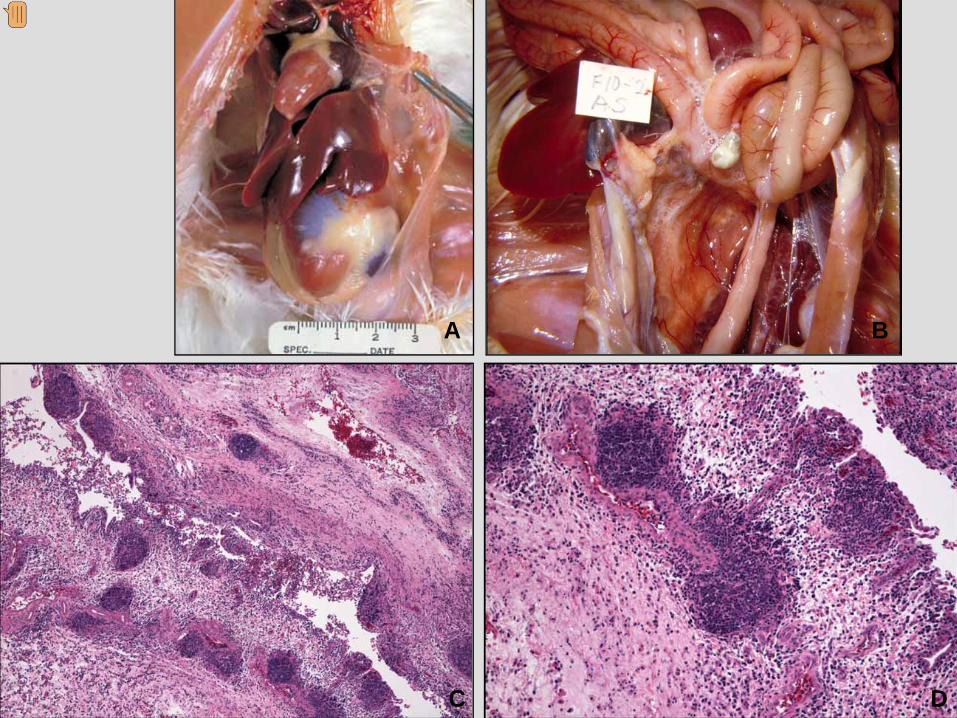
This case is from a broiler experimentally infected with Mycoplasma gallisepticum. The lymphofollicular reaction shown is typical in mycoplasma infections. There also is a fibrinoheterophilic inflammation – in this case airsacculitis. The diffuse inflammation causes significant expansion of the air sac.
A B
C D
Presenter
Presentation Notes
Images A and B are gross lesions of airsacculitis. In image A the air sac (located between the tip of the forceps and the liver and gizzard) is cloudy and thicker than normal. Note the foamy exudate in B. Image C shows a greatly expanded air sac with numerous collections of darkly-stained lymphocytes and a diffuse fibrinoheterophilic inflammation. Exudate is on the surface epithelial cells that are hyperplastic. Image D is a higher magnification showing the nodular collections of lymphocytes and the expansion of the air sac wall.
Granulomatous Pneumonia
• Broilers
• 28 Days
• Aspergillosis
• Giant Cells and Caseous Necrosis
Presenter
Presentation Notes
This case from 28 day-old broilers with Aspergillosis illustrates granulomatous pneumonia.
A
B C
Presenter
Presentation Notes
A is a gross lesion showing congested lungs with multiple nodules throughout. B shows a granuloma replacing lung tissue. Giant cells are numerous. C shows a multinucleated giant cell. Pale, poorly stained structures within the cytoplasm of this giant cell are fragments of fungal hyphae.