Embed Size (px)
Citation preview

Template for Reporting Results of Biomarker Testing of Specimens From Patients With Chronic Lymphocytic Leukemia/Small Lymphocytic Lymphoma Template web posting date: July 2015 Authors Eric Duncavage, MD, FCAP Department of Pathology and Immunology, Washington University School of Medicine, Saint Louis,
Missouri Ranjana H. Advani, MD
Oncology Division, Stanford University Medical Center, Stanford, California Steven Agosti, MD, FCAP Pathology & Laboratory Medicine Service, James A. Haley Veterans Affairs Medical Center, Tampa,
Florida Randa Alsabeh, MD, FCAP Department of Pathology, Kaiser Permanente Medical Center, Los Angeles, California Philip Foulis, MD, FCAP Pathology & Laboratory Medicine Service, James A. Haley Veterans Affairs Medical Center, Tampa,
Florida Christine Gibson, CTR Moffitt Cancer Center, Tampa, Florida Loveleen Kang, MD, FCAP Pathology & Laboratory Medicine Service, James A. Haley Veterans Affairs Medical Center, Tampa,
Florida Joseph D Khoury, MD, FCAP Department Hematopathology, Division of Pathology and Laboratory Medicine, The University of Texas
MD Anderson Cancer Center, Houston, Texas L. Jeffrey Medeiros, MD, FCAP Department of Hematopathology, Division of Pathology and Laboratory Medicine, The University of Texas
MD Anderson Cancer Center, Houston, Texas Robert S. Ohgami, MD, PhD, FCAP Department of Pathology, Stanford Hospital and Clinics, Stanford, California Dennis P. O'Malley, MD, FCAP Department of Pathology, Clarient Pathology Services, Aliso Viejo, California Keyur P. Patel, MD, PhD, FCAP Department of Hematopathology, Division of Pathology and Laboratory Medicine, The University of Texas
MD Anderson Cancer Center, Houston, Texas Carla Wilson, MD, PhD, FCAP Department of Hematopathology, University of New Mexico Health Sciences Center, Albuquerque, New
Mexico For the Members of the Cancer Biomarker Reporting Committee, College of American Pathologists
Chronic Lymphocytic Leukemia • Biomarkers CLL_Biomarkers 1.0.0.1
2
© 2015 College of American Pathologists (CAP). All rights reserved. The College does not permit reproduction of any substantial portion of these templates without its written authorization. The College hereby authorizes use of these templates by physicians and other health care providers in reporting results of biomarker testing on patient specimens, in teaching, and in carrying out medical research for nonprofit purposes. This authorization does not extend to reproduction or other use of any substantial portion of these templates for commercial purposes without the written consent of the College. The CAP also authorizes physicians and other health care practitioners to make modified versions of the templates solely for their individual use in reporting results of biomarker testing for individual patients, teaching, and carrying out medical research for non-profit purposes. The CAP further authorizes the following uses by physicians and other health care practitioners, in reporting on surgical specimens for individual patients, in teaching, and in carrying out medical research for non-profit purposes: (1) Dictation from the original or modified templates for the purposes of creating a text-based patient record on paper, or in a word processing document; (2) Copying from the original or modified templates into a text-based patient record on paper, or in a word processing document; (3) The use of a computerized system for items (1) and (2), provided that the template data is stored intact as a single text-based document, and is not stored as multiple discrete data fields. Other than uses (1), (2), and (3) above, the CAP does not authorize any use of the templates in electronic medical records systems, pathology informatics systems, cancer registry computer systems, computerized databases, mappings between coding works, or any computerized system without a written license from the CAP. Any public dissemination of the original or modified templates is prohibited without a written license from the CAP. The College of American Pathologists offers these templates to assist pathologists in providing clinically useful and relevant information when reporting results of biomarker testing. The College regards the reporting elements in the templates as important elements of the biomarker test report, but the manner in which these elements are reported is at the discretion of each specific pathologist, taking into account clinician preferences, institutional policies, and individual practice. The College developed these templates as educational tools to assist pathologists in the useful reporting of relevant information. It did not issue them for use in litigation, reimbursement, or other contexts. Nevertheless, the College recognizes that the templates might be used by hospitals, attorneys, payers, and others. The College cautions that use of the templates other than for their intended educational purpose may involve additional considerations that are beyond the scope of this document. The inclusion of a product name or service in a CAP publication should not be construed as an endorsement of such product or service, nor is failure to include the name of a product or service to be construed as disapproval.
Chronic Lymphocytic Leukemia • Biomarkers CLL_Biomarkers 1.0.0.1
3
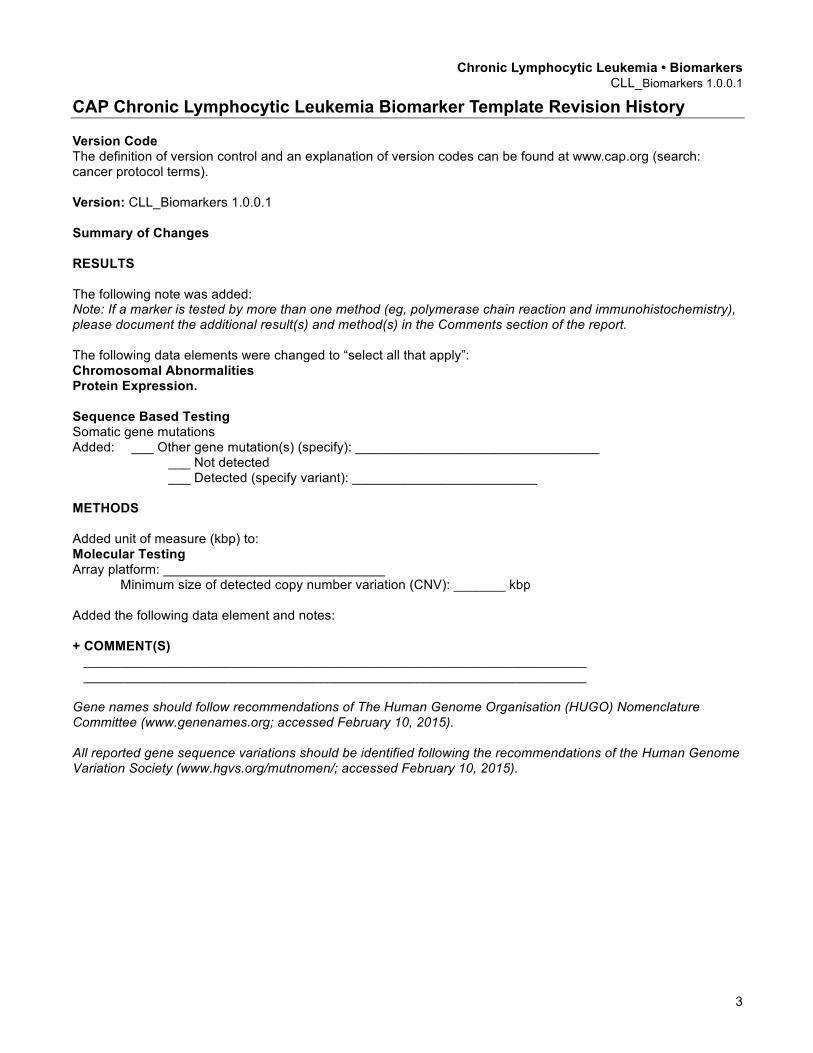
CAP Chronic Lymphocytic Leukemia Biomarker Template Revision History Version Code The definition of version control and an explanation of version codes can be found at www.cap.org (search: cancer protocol terms). Version: CLL_Biomarkers 1.0.0.1 Summary of Changes RESULTS The following note was added: Note: If a marker is tested by more than one method (eg, polymerase chain reaction and immunohistochemistry), please document the additional result(s) and method(s) in the Comments section of the report. The following data elements were changed to “select all that apply”: Chromosomal Abnormalities Protein Expression. Sequence Based Testing Somatic gene mutations Added: ___ Other gene mutation(s) (specify): _________________________________
___ Not detected ___ Detected (specify variant): _________________________ METHODS Added unit of measure (kbp) to: Molecular Testing Array platform: ______________________________
Minimum size of detected copy number variation (CNV): _______ kbp Added the following data element and notes: + COMMENT(S) ____________________________________________________________________ ____________________________________________________________________ Gene names should follow recommendations of The Human Genome Organisation (HUGO) Nomenclature Committee (www.genenames.org; accessed February 10, 2015). All reported gene sequence variations should be identified following the recommendations of the Human Genome Variation Society (www.hgvs.org/mutnomen/; accessed February 10, 2015).

CAP Approved Chronic Lymphocytic Leukemia • Biomarkers CLL_Biomarkers 1.0.0.1
4 + Data elements preceded by this symbol are not required.
Chronic Lymphocytic Leukemia Biomarker Reporting Template Template web posting date: July 2015 Completion of the template is the responsibility of the laboratory performing the biomarker testing and/or providing the interpretation. When both testing and interpretation are performed elsewhere (eg, a reference laboratory), synoptic reporting of the results by the laboratory submitting the tissue for testing is also encouraged to ensure that all information is included in the patient’s medical record and thus readily available to the treating clinical team.
CHRONIC LYMPHOCYTIC LEUKEMIA/SMALL LYMPHOCYTIC LYMPHOMA (CLL) Select a single response unless otherwise indicated. Note: Use of this template is optional. + SPECIMEN TYPE + ___ Peripheral blood + ___ Bone marrow + ___ Lymph node (specify site): ___________________ + ___ Other (specify): ____________________ + RESULTS Note: If a marker is tested by more than one method (eg, polymerase chain reaction and immunohistochemistry), please document the additional result(s) and method(s) in the Comments section of the report. + Chromosomal Abnormalities (Note B) (select all that apply) + ___ 13q deletion
+ ___ Not detected + ___ Detected + ___Other abnormal signal patterns (specify): _______________
+ ___ Trisomy 12 + ___ Not detected + ___ Detected + ___ Other abnormal signal patterns (specify): _______________
+ ___ 11q deletion + ___ Not detected + ___ Detected + ___ Other abnormal signal patterns (specify): _______________
+ ___ 17p deletion + ___ Not detected + ___ Detected + ___ Other abnormal signal patterns (specify): _______________
+ Other probes tested + Specify probe: ____________________ + Specify results: ____________________
+ Additional copy number variations noted + Gains (specify regions): _____________________________________________________ + Losses (specify regions): _____________________________________________________
CAP Approved Chronic Lymphocytic Leukemia • Biomarkers CLL_Biomarkers 1.0.0.1
5 + Data elements preceded by this symbol are not required.
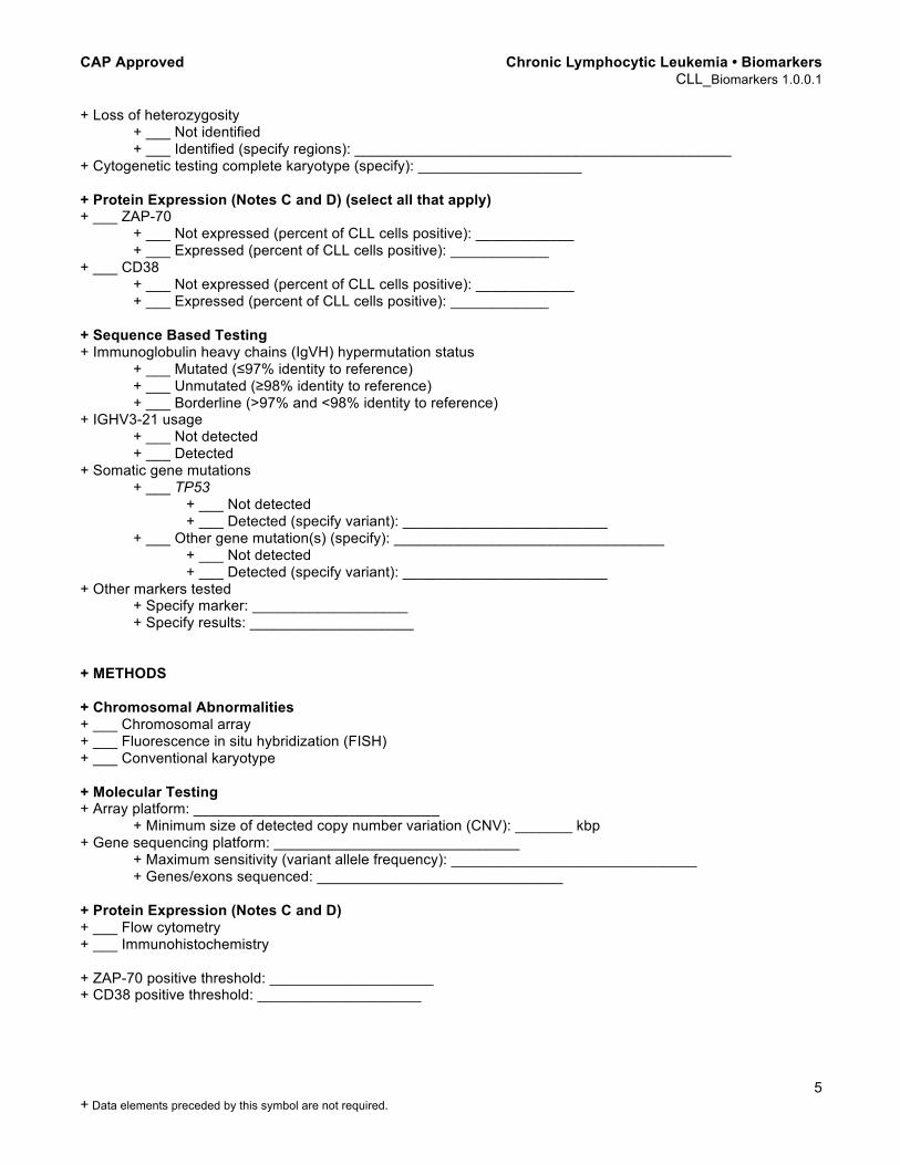
+ Loss of heterozygosity + ___ Not identified + ___ Identified (specify regions): ______________________________________________
+ Cytogenetic testing complete karyotype (specify): ____________________
+ Protein Expression (Notes C and D) (select all that apply) + ___ ZAP-70
+ ___ Not expressed (percent of CLL cells positive): ____________ + ___ Expressed (percent of CLL cells positive): ____________
+ ___ CD38 + ___ Not expressed (percent of CLL cells positive): ____________ + ___ Expressed (percent of CLL cells positive): ____________
+ Sequence Based Testing + Immunoglobulin heavy chains (IgVH) hypermutation status + ___ Mutated (≤97% identity to reference) + ___ Unmutated (≥98% identity to reference) + ___ Borderline (>97% and <98% identity to reference) + IGHV3-21 usage
+ ___ Not detected + ___ Detected + Somatic gene mutations + ___ TP53 + ___ Not detected + ___ Detected (specify variant): _________________________ + ___ Other gene mutation(s) (specify): _________________________________
+ ___ Not detected + ___ Detected (specify variant): _________________________ + Other markers tested + Specify marker: ___________________ + Specify results: ____________________ + METHODS + Chromosomal Abnormalities + ___ Chromosomal array + ___ Fluorescence in situ hybridization (FISH) + ___ Conventional karyotype + Molecular Testing + Array platform: ______________________________
+ Minimum size of detected copy number variation (CNV): _______ kbp + Gene sequencing platform: ______________________________
+ Maximum sensitivity (variant allele frequency): ______________________________ + Genes/exons sequenced: ______________________________
+ Protein Expression (Notes C and D) + ___ Flow cytometry + ___ Immunohistochemistry + ZAP-70 positive threshold: ____________________ + CD38 positive threshold: ____________________
CAP Approved Chronic Lymphocytic Leukemia • Biomarkers CLL_Biomarkers 1.0.0.1
6 + Data elements preceded by this symbol are not required.
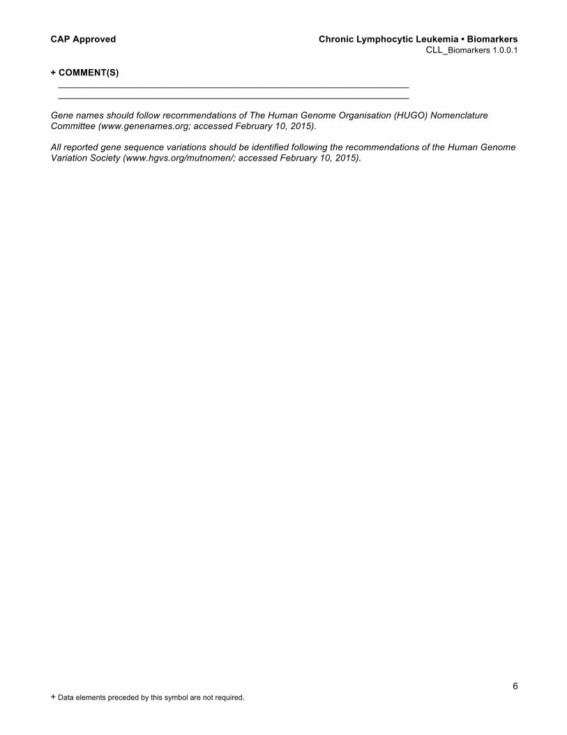
+ COMMENT(S) ____________________________________________________________________ ____________________________________________________________________ Gene names should follow recommendations of The Human Genome Organisation (HUGO) Nomenclature Committee (www.genenames.org; accessed February 10, 2015). All reported gene sequence variations should be identified following the recommendations of the Human Genome Variation Society (www.hgvs.org/mutnomen/; accessed February 10, 2015).
Background Documentation Chronic Lymphocytic Leukemia • Biomarkers CLL_Biomarkers 1.0.0.1
7
Explanatory Notes A. Introduction Somatic mutation in the rearranged variable regions of immunoglobulin heavy chains (IgVH) has been reported to be of prognostic importance since 1999.1,2 Patients with IgVH unmutated genes have a more aggressive disease and are more resistant to therapy than those with mutated IgVH genes. Most researchers defined unmutated IgVH based on 98% or more homology to reference and mutated IgVH with less than or equal to 97% homology to reference.3 Determining IgVH mutations requires specific equipment and is laborious, expensive, and time-consuming. Due to all these limitations, surrogate markers including CD38 and ZAP-70, with the similar prognostic value as IgVH mutation status are more widely used. Detection of immunoglobulin VH3-21 usage by sequencing of IgH rearrangements has been associated with poor outcome in CLL and should be reported when detected by IgH sequencing. B. Prognosis in CLL FISH Del 11q contains several tumor suppressor genes including ATM.4,5 This gene is associated with cell cycle regulation and p53 pathway activation. BIRC3, which is also in the deleted region of interest, is a candidate gene that may also play a role in CLL pathobiology. Del 11q is associated with younger age and poor prognosis. Del 13q is often seen as a sole abnormality in CLL.4,5 It is associated with a favorable prognosis. Several genes and micro-RNAs (mRNA) have been suggested as candidate genes in these cases of CLL. Del 17p is thought to affect the TP53 gene, a key regulator of cell cycle.4,5 Other deleted genes may also play a role. Patients with del17p will often have other genetic abnormalities and other poor prognosis markers. Trisomy 12 (+12) affects CLL by an unknown mechanism.4,5 Patients with trisomy 12 have a good response to treatment. Some additional trisomies (+19, +19) are seen in association with trisomy 12. Marker Approx. Frequency Prognosis Notes Del 13q14 35%-45% Low risk Trisomy 12 11%-16% Intermediate-High risk Del 11q22-23 (ATM; BIRC3)
10%-17% Intermediate-High risk Bulky disease, aggressive clinical course, shorter survival
Del 17p (TP53) 3%-7% High risk Frequently no response to therapy or relapse after therapy
No abnormalities by FISH
Low-Intermediate risk
C. CD38 Expression CD38 is a 45 KDa transmembrane glycoprotein that was the first maker found to correlate with IgVH mutation.2
Patients with CD38-positive cells have unmutated IgVH genes and higher need for chemotherapy as well as shorter overall survival. However, subsequent studies showed that association between mutation status and CD38 expression level was not absolute and that CD38 expression should be considered as an independent prognostic marker in CLL.6 CD38 expression is determined by flow cytometry. A 30% cutoff level is generally used empirically to classify CD38-positive and CD38-negative patients; individual laboratories should determine their own criteria for calling CD38-positive and CD38-negative cases, and specific laboratory cut-offs should be described in the methods section above.1,6
CD38 expression may vary over time and may show a bimodal
expression profile.4
D. ZAP-70 Expression Comparative microarray studies performed on cases of CLL with mutated and unmutated IgVH genes showed differential expression of gene encoding for zeta-associated protein of 70 kDa (ZAP-70).7 Zap-70 is normally
Background Documentation Chronic Lymphocytic Leukemia • Biomarkers CLL_Biomarkers 1.0.0.1
8
expressed in T cells and NK cells. The majority of the CLL cases with mutated IgVH are ZAP-70 negative, while cases with unmutated IgVH are ZAP-70 positive. ZAP-70 expression in CLL cells can be determined by various methods including western blotting, quantitative reverse transcription polymerase chain reaction (RT-PCR), immunohistochemistry, and flow cytometry. However, flow cytometry is the preferred technique for assessing ZAP-70 expression in CLL cells.8 Flow cytometry allows simultaneous evaluation of ZAP-70 protein expression in CLL cells and normal lymphocytes. A 20% cutoff threshold is commonly used to separate ZAP-70-negative from ZAP-70-positive CLL cases; however, this threshold may vary significantly from laboratory to laboratory depending on how negative controls are defined. There is inherent laboratory-to-laboratory variability in ZAP-70 testing due to the following: different antibody clones used (variable antigen affinity), different conjugated fluorochromes (variable intensity), variable methods of cell permeabilization (for intracellular staining), variable staining procedures, variable gating procedures, and variable reporting methods. Moreover, ZAP-70 is a labile protein; most consensus guidelines recommend ZAP-70 testing within 24 hours of sample collection. Laboratories should establish firm gating criteria for sample collection and determine reference populations at the point of method validation of their assay to ensure optimal interassay precision. Different gating strategies are discussed extensively in a prior multicenter international harmonization study. 9 References 1. Hamblin TJ, Davis Z, Gardiner A, Oscier DG, Stevenson FK. Unmutated IgV(H) genes are associated with a
more aggressive form of chronic lymphocytic leukemia. Blood. 1999;94(6):1848-1854. 2. Damie RN, Wasil T, Fais F, et al. Ig VH gene mutation status and CD38 expression as novel prognostic
indicators in chronic lymphocytic leukemia. Blood. 1999;94(6):1840-1847. 3. Bockstaele FV, Verhasselt B, Philippe J. Prognostic markers in chronic lymphocytic leukemia: a
comprehensive review. Blood Reviews. 2009;23:2-47. 4. Rosenquist R, Cortese D, Bhoi S, Gunnarsson R. Prognostic markers and their clinical applicability in chronic
lymphocytic leukemia: where do we stand? Leuk Lymphoma. 2013;54(11):2351-2364. 5. Chiorazzi N. Implications of new prognostic markers in chronic lymphocytic leukemia. Hematology Am Soc
Hematol Educ Program. 2012;2012:76-87. 6. Hamblin TJ, Orchard JA, Ibboston RE, et al. CD38 expression and immunoglobulin variable region mutations
are independent prognostic variables in chronic lymphocytic leukemia, but CD38 expression may vary during the course of the disease. Blood. 2002;99(3):1023-1029.
7. Rosenwald A, Alizadeh AA, Widhopf G, et al. Relation of gene expression phenotype to immunoglobulin mutation genotype in B cell chronic lymphocytic leukemia. J Exp Med. 2001;194(11):1639-1647.
8. Crespo M, Bosch F, Villamor N, et al. ZAP-70 expression as a surrogate for immunoglobulin-variable-region mutations in chronic lymphocytic leukemia. N Engl J Med. 2003;348(18):1764-1775.
9. Letestu R, Rawstron A, Ghia P, et al. Evaluation of ZAP-70 expression by flow cytometry in chronic lymphocytic leukemia: a multicentric international harmonization process. Cytometry B Clin Cytom. 2006;70(4):309-314.


![ReviewArticle Lenalidomide and Chronic Lymphocytic Leukemia · ReviewArticle Lenalidomide and Chronic Lymphocytic Leukemia AnaPilarGonzález-Rodríguez,1 AngelR.Payer,1 ... Ferrajoli[7]](https://img.dokumen.tips/doc/110x75/5acf388a7f8b9ad24f8c2cdd/reviewarticle-lenalidomide-and-chronic-lymphocytic-leukemia-lenalidomide-and-chronic.jpg)