Embed Size (px)
Citation preview

In vitro, in silico and in vivo studychallenges the impact of bronchialthermoplasty on acute airway smoothmuscle mass loss
Igor L. Chernyavsky 1,10, Richard J. Russell2,10, Ruth M. Saunders2,10,Gavin E. Morris3, Rachid Berair2, Amisha Singapuri2, Latifa Chachi2,Adel H. Mansur4, Peter H. Howarth5, Patrick Dennison5, Rekha Chaudhuri6,7,Stephen Bicknell6, Felicity R.A.J. Rose8, Salman Siddiqui2, Bindi S. Brook9,11
and Christopher E. Brightling2,11
@ERSpublicationsBronchial thermoplasty treatment for asthma has unexpected possible mechanisms of actionhttp://ow.ly/ZcuE30jsaSa
Cite this article as: Chernyavsky IL, Russell RJ, Saunders RM, et al. In vitro, in silico and in vivo studychallenges the impact of bronchial thermoplasty on acute airway smooth muscle mass loss. Eur Respir J2018; 51: 1701680 [https://doi.org/10.1183/13993003.01680-2017].
ABSTRACT Bronchial thermoplasty is a treatment for asthma. It is currently unclear whether itshistopathological impact is sufficiently explained by the proportion of airway wall that is exposed totemperatures necessary to affect cell survival.
Airway smooth muscle and bronchial epithelial cells were exposed to media (37–70°C) for 10 s tomimic thermoplasty. In silico we developed a mathematical model of airway heat distribution post-thermoplasty. In vivo we determined airway smooth muscle mass and epithelial integrity pre- and post-thermoplasty in 14 patients with severe asthma.
In vitro airway smooth muscle and epithelial cell number decreased significantly following the additionof media heated to ⩾65°C. In silico simulations showed a heterogeneous heat distribution that wasamplified in larger airways, with <10% of the airway wall heated to >60°C in airways with an inner radiusof ∼4 mm. In vivo at 6 weeks post-thermoplasty, there was an improvement in asthma control (measuredvia Asthma Control Questionnaire-6; mean difference 0.7, 95% CI 0.1–1.3; p=0.03), airway smooth musclemass decreased (absolute median reduction 5%, interquartile range (IQR) 0–10; p=0.03) and epithelialintegrity increased (14%, IQR 6–29; p=0.007). Neither of the latter two outcomes was related to improvedasthma control.
Integrated in vitro and in silico modelling suggest that the reduction in airway smooth muscle post-thermoplasty cannot be fully explained by acute heating, and nor did this reduction confer a greaterimprovement in asthma control.
This article has supplementary material available from erj.ersjournals.com
Received: Aug 17 2017 | Accepted after revision: March 29 2018
Copyright ©ERS 2018. This ERJ Open article is open access and distributed under the terms of the Creative CommonsAttribution Licence 4.0.
https://doi.org/10.1183/13993003.01680-2017 Eur Respir J 2018; 51: 1701680
| ORIGINAL ARTICLEASTHMA

Affiliations: 1School of Mathematics, University of Manchester, Manchester, UK. 2Dept of Infection, Immunityand Inflammation, Institute for Lung Health, NIHR Leicester Biomedical Research Centre, University ofLeicester, Leicester, UK. 3Dept of Cardiovascular Sciences, University of Leicester, Leicester, UK. 4Heart ofEngland NHS Trust, Birmingham, UK. 5Clinical and Experimental Sciences, University of Southampton,Southampton NIHR Respiratory Biomedical Research Unit, University Hospital Southampton NHS Trust,Southampton, UK. 6Gartnavel General Hospital, Glasgow, UK. 7Institute of Infection, Immunity andInflammation, University of Glasgow, Glasgow, UK. 8Centre for Biomolecular Sciences, University ofNottingham, Nottingham, UK. 9School of Mathematical Sciences, University of Nottingham, Nottingham, UK.10These authors contributed equally to the study. 11Co-senior authors.
Correspondence: Igor L. Chernyavsky, School of Mathematics, University of Manchester, Oxford Road,Manchester M13 9PL, UK. E-mail: [email protected]
IntroductionBronchial thermoplasty (BT) is a non-pharmacological therapy for treating severe asthma by selectivelyheating conductive airways (3–10 mm in diameter) from within the lumen with a low-power electricalcurrent [1, 2]. During the BT procedure, thermal energy is delivered to the airway wall via abronchoscope-inserted catheter with a distal basket of four electrodes that expand to make contact withthe airway wall, aiming to reach a target temperature of 65°C for 10 s.
The primary target of BT is the airway smooth muscle (ASM), a key contributor to airway remodelling,particularly in severe asthma [2–7]. Previous animal studies demonstrated a reduction in airwayhyper-responsiveness and an altered ASM histological appearance following BT [8]. Subsequent clinicaltrials showed improved quality of life and reduced frequency of severe exacerbations in those receiving BTversus a sham procedure, but found no significant difference in lung function as a result of the treatment[9–12]. In uncontrolled observational studies, BT has been associated with an ∼50–80% relative loss ofASM mass determined by bronchial biopsies [13–16], which were typically obtained 1–3 months aftercompletion of the BT procedures. Although thermal ablation by radiofrequency energy is commonly usedin surgical practice [17, 18], there is a paucity of theoretical [19] and in vitro [20] studies in humans thatassess the direct effect of supra-febrile temperatures on ASM cells’ survival and function, or the earlyeffects of BT upon ASM mass and epithelial integrity.
We hypothesised that during BT the proportion of the airway exposed to temperatures necessary to affectASM and epithelial cell survival, determined from in vitro experiments, is sufficient to explain the impactof BT. To test our hypothesis we employed in vitro, in silico and in vivo methodologies to define the acuteimpact of BT on ASM and epithelial cells.
MethodsDetailed methods are included in the supplementary material.
In vitro heating of human primary ASM, epithelial cells and bronchial epithelial cellsPrimary ASM and epithelial cells were cultured as described previously [21]. The study was approved bythe Leicestershire Research Ethics Committee (REC 08/H0406/189). Informed consent was obtained fromall subjects. The human bronchial epithelial cells (hBECs) were obtained from LGC Standards (Middlesex,UK).
Cells were grown to confluence in 6- or 24-well plates, then exposed to heated media for 10 s using theprotocol described in the supplementary methods. Heat loss over the 10-s period was measured andshowed that following the addition of media heated to 65°C, cells were exposed to a mean temperature of58–59°C in both 6- and 24-well plates (supplementary table S4).
The number of remaining adherent viable cells up to 2 weeks post-heating was assessed using PrestoBlue®(Thermo Fisher Scientific, Warrington, UK) according to manufacturer’s instructions and confirmed atspecified times by cell counts. The percentage of the remaining adherent cell population undergoingapoptosis or necrosis 24 h post-heating was determined using the Alexa Fluor® 488 Annexin V/Dead CellApoptosis Kit (Thermo Fisher Scientific) as described previously [22]. Fluorescence emission was collectedat 530 nm (Annexin V) and >575 nm (propidium iodide) on a flow cytometer and the percentage ofapoptotic and necrotic cells derived respectively using WEASEL™ software (Frank Battye Flow CytometryConsulting, Melbourne, Australia).
In silico bioheat mathematical modellingBecause it is not currently possible to measure the heat transfer within the airway wall during BT in vivo,a two-dimensional mathematical model was developed that couples Joule heating due to the electricalcurrent generated by the BT electrodes (with integrated temperature control feedback, similar to [19]) with
https://doi.org/10.1183/13993003.01680-2017 2
ASTHMA | I.L. CHERNYAVSKY ET AL.

the bioheat transfer in the airway wall and surrounding parenchymal tissue. The material properties of thelung were applied to the model as shown in supplementary table S1. The coupled model was implementedusing the finite element-based modelling framework of COMSOL Multiphysics® 5.2 (Stockholm, Sweden).Heat maps were integrated over the airway wall to characterise heating pattern heterogeneity.
Local sensitivity analysis was conducted to identify key geometric, physiological and equipmentparameters, and to test the robustness of model predictions. Mathematical model formulation and furthertechnical details can be found in the supplementary material. The mathematical model was validated usingthe in vitro data on the cooling of heated media in multi-well plates (see the in vitro methods above),which showed good agreement (supplementary figure S3).
The predicted distribution of temperatures in the airway wall, in combination with the thermaldose-dependent response of ASM and bronchial epithelial cells in vitro, was used to estimate the overall acuteimpact of BT on the bronchial wall (the modelling framework is illustrated in supplementary figure S1).
In vivo response to BTBronchial biopsies were obtained from 14 subjects before and after BT. All subjects had severe asthma asdefined by American Thoracic Society/European Respiratory Society guidelines, and underwent BT as partof their clinical care.
BT was performed as per manufacturer’s guidelines over three staged treatment sessions in the followingorder: right lower lobe, left lower lobe, and both right and left upper lobes. The right middle lobe was nottreated owing to the risk of airway collapse and “right middle lobe syndrome” [23]. Biopsies were obtainedfrom the untreated right upper lobe at the first BT session and the treated right lower lobe segmental andsubsegmental airways at the second BT session.
Biopsies were embedded in paraffin and 4-μm sections stained with haematoxylin and eosin oranti-α-smooth muscle actin (α-SMA, clone 1A4; Dako, Stockport, UK). Biopsies were assessed by a singleobserver (RJR) blinded to clinical characteristics to determine 1) the ASM content as a percentage of totalbiopsy area; 2) the epithelial integrity by measuring and expressing the length of intact, damaged anddenuded epithelium as a percentage of reticular basement membrane length; and 3) the number ofmyofibroblasts (isolated α-SMA-positive stained cells in the lamina propria that were neither located as partof the ASM bundle nor as vascular smooth muscle cells adjacent to vessels) per mm2 of lamina propria.
Statistical analysisThe statistical analysis is discussed in more detail in the supplementary methods. Briefly, data wereanalysed in GraphPad Prism® 7.0 and R project 3.2.4, using parametric and non-parametric tests asappropriate. Confidence intervals for the medians of cell counts were estimated using the bootstrappercentile method (R boot package). A p-value <0.05 was considered statistically significant.
ResultsIn vitro apoptosis and necrosis of ASM and hBEC cellsA metabolic assay demonstrated that the addition of media heated to 65°C or 70°C for 10 s, but not 45–60°C, resulted in a significant reduction in the number of ASM and hBEC cells remaining adherent after 24 hcompared to 37°C (figure 1a, b). This reduction in the number of viable (metabolically active) adherent cellspersisted over 2 weeks after the addition of media heated to 65°C or 70°C for ASM and to 70°C for hBECcells compared to 37°C. The number of viable hBEC cells recovered, such that 10 days after the addition ofmedia heated to 65°C their numbers were not significantly reduced versus 37°C (figure 1a–d). The resultspresented in figure 1a–d were confirmed at various times using cell counts (figure 1e, f), which supported theresults of the metabolic assay. There was a significant reduction in ASM and hBEC cell counts after theaddition of media heated to 65°C at 24–48 h (data not shown), which persisted for 1 week (figure 1e) and2 weeks (data not shown) for ASM, with a recovery of hBEC cell number after 1 week (figure 1f). In additionto the reduction in ASM and hBEC cell number post-addition of media heated to 65°C or 70°C versus 37°C,there was a significant increase in the percentage of cells undergoing necrosis, but not apoptosis (figure 1g, h)in the remaining adherent cell population after 24 h. The median of the relative reduction in viable ASM cellnumber after the addition of media heated to 65°C at 24 h was 60% (95% bootstrap CI 40–80%).
In silico heating heterogeneity profilesOur computational finite element-based model implementing the BT protocol showed a high degree oftemperature variation over an airway wall. An example with reference model geometry (inner and outerwall radii of 2.2 and 3.3 mm, respectively) is shown in figure 2. To assess the impact each of theparameters in the mathematical model had on heat distribution, we undertook a local sensitivity analysis(supplementary material). This demonstrated that the model predictions were relatively insensitive to the
https://doi.org/10.1183/13993003.01680-2017 3
ASTHMA | I.L. CHERNYAVSKY ET AL.

a) ASM b) hBEC
0.0
0.4
0.8
1.2
1.6c)
Via
ble
ce
lls R
FU
Time days
0 1
***
******
******
****
*** *** *** ***
*****
**
***********
***
*
3 5 7 10 14
0.0
0.4
0.8
1.2
1.6d)
Via
ble
ce
lls R
FU
Time days
0 1 3 5 7 10 14
37°C45°C50°C55°C60°C65°C70°C
Day 0 Day 1 Day 14 Day 0 Day 1 Day 14
0.0
0.5
1.0
1.5e)
No
rma
lise
d c
ell
co
un
ts
Temperature °C
45 50 55 60 65
0.0
0.5
1.0
1.5f)
No
rma
lise
d c
ell
co
un
ts
Temperature °C
45 50 55 60 65
*
0
20
40
80 ApoptosisNecrosis
*
*
* *
60
g)
Po
pu
lati
on
%
Temperature °C
37 45 50 55 60 65 70
0
20
40
60h)
Po
pu
lati
on
%
Temperature °C
37 45 50 55 60 65 70
FIGURE 1 Response of in vitro heated airway smooth muscle (ASM) and human bronchial epithelial (hBEC) cells. a, b) Representative cellmorphology for cultures following addition of media heated to 65°C; note the incomplete recovery of ASM (a) compared to hBEC (b) cells over2 weeks. Scale bar, 0.1 mm. c, d) Longitudinal viability of ASM (c) and hBEC (d) cells following addition of media heated to specified temperatures(mean±SE). e, f ) Total cell count relative to 37°C-matched control 1 week after the addition of heated media for ASM (e) and hBEC (f ) cells. g, h)Proportion of apoptotic and necrotic ASM (g) and hBEC (h) cells determined by flow cytometry 24 h after the addition of media heated to specifiedtemperatures (mean, 95% CI). *p<0.05, **p<0.01, ***p<0.001 versus 37°C controls.
https://doi.org/10.1183/13993003.01680-2017 4
ASTHMA | I.L. CHERNYAVSKY ET AL.

tissue material properties and heating control parameters (supplementary table S3), but stronglydependent on the airway calibre and wall thickness (supplementary table S3 and figure S2). Thisamplification of temperature variation in the larger airways is shown in figure 3, demonstrating only asmall fraction of the wall heated to 65°C (figure 3 and table 1).
Although energy transfer is more efficient in the smallest airways accessible to a BT catheter (figure 3a),suboptimal heating is possible in the case of an occluded airway with reduced luminal cooling (figure 3b),and heating heterogeneity is strongly exacerbated in larger airways (figure 3c). Our model suggests that<5% of a typical airway (internal radius ∼4 mm) treated with BT is exposed to temperatures >65°C, and<10% to temperatures >60°C (table 1). Post-BT thermal equilibration does not improve the extent ofheating in the upper temperature range, with no portion of the wall experiencing temperatures >65°C inthe 1–2 s after the end of energy delivery (figure 3d, e), even when there is no volumetric cooling due totissue perfusion and alveolar moisture evaporation. Therefore, only a small percentage of the area ofairways treated with BT are likely to be exposed to temperatures >60°C, except for the smallest treatedairways. Owing to the accessibility of the airways with a bronchoscope, those airways biopsied wereproximal to the smallest airways treated with BT.
In vivo BT impact on ASM mass and epithelial integrityThe baseline and follow-up clinical characteristics of the 14 subjects are shown in table 2. Nine subjectswere receiving treatment with regular systemic corticosteroids (Global Initiative for Asthma (GINA)step 5) and the remaining subjects were receiving GINA step 4 treatment. Six weeks after the last BT
6666.1
52.8
°C
64
62
60
58
56
54
20
30
40
50
60
70
80
c) d)
a) b)
Time s
20 4 6 8 10
Electrode temperature °CMidpoint temperature °C
Electrode voltage V
0
0.02
0.04
0.06
0.08
He
ate
d w
all
fra
cti
on
Temperature °C
504540 55 60 65 70
Parenchyma
Airway wall
Electrodes
FIGURE 2 Characterisation of bronchial thermoplasty (BT) heating patterns. a) Reference model geometry(inner wall radius of 2.2 mm, outer radius of 3.3 mm). b) Heat map at the end of a single BT activation (10 s).c) Temporal dynamics of the applied voltage (red), electrode temperature (solid blue) and temperature at themidpoint between two electrodes (dashed green; marked by a white dot in b). d) Distribution of heated wallarea fractions, corresponding to b.
https://doi.org/10.1183/13993003.01680-2017 5
ASTHMA | I.L. CHERNYAVSKY ET AL.

intervention there was no change in lung function, whereas scores for both the Asthma ControlQuestionnaire-6 (ACQ6) and Asthma Quality of Life Questionnaire (AQLQ) significantly improved bymore than the clinically important difference of 0.5 (ACQ6 mean difference −0.7, 95% CI −1.3 to −0.1,p=0.03; AQLQ mean difference 0.8, 95% CI 0.1 to 1.5, p=0.03) (table 2).
The median time between baseline and follow-up biopsies was 28 days (range 14–56 days). Arepresentative bronchial biopsy is shown in figure 4a. There was a reduction in median ASM mass from12% (interquartile range (IQR) 6–17) pre-BT to 6% (IQR 1–10) post-BT (median difference 5%, IQR0–10, Wilcoxon p=0.03; figure 4b). The median relative reduction in ASM mass was 58% (IQR 6–90).
There was also a significant improvement in median epithelial integrity from 29% (IQR 15–40) pre-BT to46% (IQR 25–56) post-BT (median improvement 14%, IQR 6–29, p=0.007; figure 4c, d). The medianrelative increase in epithelial integrity was 56% (IQR 19–120).
a)
0
0.05
0.1
0.15
He
ate
d w
all
fra
cti
on
Temperature °C
504540 55 60 65 70
d)
35
40
45
50
55
60
65
70
Tem
pe
ratu
re °
C
Time s
ElectrodeMidpointElectrode (no cooling)Midpoint (no cooling)
20100 30 40 50 60
e) f)
0
0.05
0.1
He
ate
d w
all
fra
cti
on
Temperature °C
70 656055500
0.05
0.1
0.15
He
ate
d w
all
fra
cti
on
Temperature °C
t=10 s t=12 s
70 65605550
b)
0
0.05
0.1
0.15
He
ate
d w
all
fra
cti
on
0.2
Temperature °C
504540 55 60 65 70
c)
0
0.02
0.04
0.06
He
ate
d w
all
fra
cti
on
0.08
Temperature °C
504540 55 60 65 70
FIGURE 3 Airway temperature heterogeneity across bronchial generations and heating scenarios. Heating patterns a) at the lowest end ofbronchial thermoplasty (BT) applicability (luminal radius of 1.5 mm); b) for a midrange airway (luminal radius of 2.2 mm, corresponding to figure2b, d) with impeded luminal evaporative cooling (e.g. occluded with a bronchoscope); and c) for a larger airway (luminal radius of 4.4 mm).d) Thermal dynamics of an airway wall after the end of a single BT activation (marked by vertical dashed line) for the reference case of figure 2(solid and dashed) and for the case of absent tissue perfusion and evaporative cooling (dotted lines). e, f ) Temperature distributions at 10 s (e)and 12 s (f ), corresponding to the case of absent volumetric tissue cooling.
TABLE 1 Quantification of simulated thermal impact of bronchial thermoplasty at the end of anactivation cycle
Intermediate conducting airway# Large conducting airway
Baseline Occluded (no evaporativecooling)
Mean wall temperature °C 59 59 50Wall area fraction heated >65°C % 3 2 1Wall area fraction heated >60°C % 43 35 7Wall area fraction heated >55°C % 93 100 25
#: inner radius 2.2 mm, outer radius 3.5 mm; ¶: inner radius 4.4 mm, outer radius 5.7 mm.
https://doi.org/10.1183/13993003.01680-2017 6
ASTHMA | I.L. CHERNYAVSKY ET AL.
There was a numerical but nonsignificant reduction in the number of sub-epithelial myofibroblasts(pre-BT median 25 cells·mm−2, IQR 7–47 versus post-BT median 13 cells·mm−2, IQR 6–21; p=0.17).There was a significant inverse correlation between the change in ASM mass and myofibroblast count inthe lamina propria following BT (Spearman r=−0.55, p=0.046; figure 4e). Change in epithelial integritydid not correlate with change in myofibroblast count nor ASM mass following BT (data not shown).
Improvements in AQLQ score did not correlate with pre-BT epithelial integrity, ASM mass or myofibroblastnumber in the lamina propria nor with post-BT change (data not shown). Only three subjects had animprovement ⩾1.0 thus no responder analysis was undertaken. Pre-BT epithelial integrity, ASM mass andmyofibroblast number in the lamina propria were not related to change in ACQ6 score (data not shown).The change in ACQ6 score was inversely related to the change in ASM mass (Spearman r=−0.67, p=0.018),but not with epithelial integrity (r=−0.03, p=0.09) or myofibroblast number in the lamina propria (r=0.41,p=0.18). Five subjects had an improvement in ACQ6 score ⩾1.0. Compared with subjects not showing thisimprovement in ACQ6, these five subjects had a small increase in ASM mass post-BT (median 2%, IQR −2to 8 versus median −10%, IQR −8 to −12; p=0.003). This was in contrast to a higher pre-BT myofibroblastnumber in the lamina propria (59 cells·mm−2, IQR 36–84 versus 7 cells·mm−2, IQR 3–43; p=0.03) andgreater decrease post-BT (37 cells·mm−2, IQR 30–51 versus −8 cells·mm−2, IQR 3 to −27; p=0.048). Thosewith both an increase in ASM mass and a decrease in myofibroblast number in the lamina propria had thegreatest improvement in ACQ6 score compared with those that either had a decrease in both or a decreasein ASM mass and increase in myofibroblast number (figure 4e).
DiscussionWe have developed an integrated in vitro and in silico framework to model the acute effects of BT on ASMand epithelial cells. In this framework, the in silico mathematical model serves as a “bridge” between the invitro and in vivo thermal effects, which are inaccessible by other means. The in vitro model identified asharp threshold in the response of both hBEC and ASM cells to heating. In vitro hBEC and ASM cellnumber decreased significantly after the addition of media heated to ⩾65°C. Importantly, taking intoaccount the heat loss over 10 s in the in vitro experiments, these cells were exposed to a mean temperatureof 58–59°C. The mathematical model predicted a localised and highly heterogeneous heating pattern thatis very sensitive to airway calibre, with a relatively small fraction of the bronchial wall heated to >60°C inall but the smallest airways (see also [25]). The integrated in vitro and in silico model predictions weretested against in vivo bronchial biopsies taken pre- and post-BT. The biopsy samples showed an increase inepithelial integrity and a reduction in ASM mass after BT.
Although greater than predicted based on our mathematical model, the observed post-BT relative medianreduction in ASM mass of 58% was consistent with previous clinical studies [13–16]. To explain this levelof ASM reduction by acute thermal injury alone, most of the airway wall in the calibre of airways sampledat bronchoscopy would need to be heated to ⩾60°C. Thus, if the in vitro and in silico predictions arecorrect, the difference must be due to an alternative biological mechanism triggered in response to BT,such as the active thermal bystander effect [26].
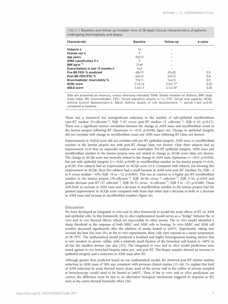
TABLE 2 Baseline and follow-up (median time of 28 days) clinical characteristics of patientsundergoing thermoplasty and biopsy
Characteristic Baseline Follow-up p-value
Subjects n 14 –
Female sex n 9 –Age years 52±13 –
GINA classification 5 n 9 –BMI kg·m−2 31±8 –Exacerbations in last 12 months n 4±3 –
Pre-BD FEV1 % predicted 68±19 67±20 0.5Post-BD FEV1/FVC % 63±12 63±12 0.8Bronchodilator reversibility % 19±11 16±12 0.5ACQ6 score 3.1±1.6 2.5±1.7* 0.03AQLQ score 3.4±1.7 4.1±1.8* 0.03
Data are presented as mean±SD, unless otherwise indicated. GINA: Global Initiative for Asthma; BMI: bodymass index; BD: bronchodilator; FEV1: forced expiratory volume in 1 s; FVC: forced vital capacity; ACQ6:Asthma Control Questionnaire-6; AQLQ: Asthma Quality of Life Questionnaire. *: paired t-test p<0.05compared to baseline.
https://doi.org/10.1183/13993003.01680-2017 7
ASTHMA | I.L. CHERNYAVSKY ET AL.

Our in vivo biopsies showed significantly improved epithelial integrity after BT, which likely reflectsepithelial repair in response to thermal injury. Indeed, our in vitro data show evidence of acute phaseepithelial repair in response to heat. Others studies have not found changes in the epithelial phenotype inbiopsies obtained 3 months after completion of BT treatment, but have reported other effects uponcollagen deposition and bronchial nerves [16]. Although in this small study a reduction in ASM mass didnot correlate with increased epithelial integrity, whether epithelial repair might impact on other changes inairway wall structure, including ASM mass, following BT warrants further investigation.
Myofibroblast numbers are increased in the lamina propria in patients with asthma and they traffic to sitesof injury, differentiate and promote wound repair [27–29]. We considered whether BT might affect thenumber of myofibroblast cells in the lamina propria, and whether the prevalence of these cells might relateto changes observed in the epithelium and ASM. We identified a numerical but nonsignificant reductionin the overall number of myofibroblasts in the lamina propria in response to BT. However, there was asignificant inverse correlation between the change in ASM mass and myofibroblast number after BT. Thismight represent a dynamic relationship between myofibroblasts and the ASM bundle, with migration ofmyofibroblasts to and from the ASM bundle. Despite the small number of subjects in our study, we wereable to explore the relationship between the effects of BT on ASM mass, epithelial integrity andmyofibroblast numbers in the lamina propria and asthma-related symptoms assessed 6 weeks after the lastBT intervention. Surprisingly we found that improvement in asthma control was inversely related topost-BT change in ASM mass, and that improvement in asthma control was greatest in those with anincrease in ASM mass and a reduction in myofibroblast number. The observed epithelial repair was notassociated with improved asthma control, but whether epithelial repair contributes to the reducedexacerbation rates observed in larger studies after BT needs to be further investigated.
Our study had a number of potential limitations. The in vitro studies could not fully recapitulate thebehaviour of the ASM cells and bronchial epithelial cells in vivo because the heated media was addeddirectly to specific cell types in isolation. They also did not take into account asthmatic versus
b)a)E
ELP
ASM
ASM
G
0
40 p=0.03 p=0.007
30
20
10
Air
wa
y sm
oo
th
mu
scle
ma
ss %
Baseline Post-treatment
c)
0
80
60
40
20Ep
ith
eli
al
inte
gri
ty %
100
Baseline Post-treatment
d)
0
100
50
Tota
l e
pit
he
liu
m %
e)
–30
–20
–10
0
10
20
Δ A
SM
%
Baseline
29
53
19
44
44
12
Post-treatment
DenudedDamagedIntact
40200–20
Δ Myofibroblasts per mm2
Δ ACQ –0.4 (–2 to 0.9)
Δ ACQ –1.7 (–2 to –1.2)
Δ ACQ –0.3 (–0.5 to 0.2)
–40–60
FIGURE 4 Histology analysis of airway smooth muscle (ASM) content and epithelial integrity in bronchial biopsies (at baseline and at about1 month post-bronchial thermoplasty (BT)). a) Example endobronchial biopsy stained for α-smooth muscle actin (E: epithelium; LP: laminapropria; G: gland). b) ASM mass % pre- and post-BT (p<0.05). c) Epithelial integrity pre- and post-BT (p<0.01). In b and c, the horizontal linerepresents the median, the box represents the interquartile range (IQR) and the whiskers represent the minimum and maximum. d) Detailedbreakdown of epithelial structure at baseline and post-BT (mean). The total percentage for the baseline does not equal 100% owing to rounding.e) Change in ASM mass versus the number of myofibroblasts per mm2 of lamina propria following BT (Spearman’s rank correlation r=−0.55,p=0.046), with change in Asthma Control Questionnaire-6 (ACQ6) score (mean (IQR)) reported for each response subgroup (quadrants).
https://doi.org/10.1183/13993003.01680-2017 8
ASTHMA | I.L. CHERNYAVSKY ET AL.

non-asthmatic cellular phenotype, cell–cell interactions or the presence of submucosal tissue. In addition,there was a small amount of heat loss over the 10-s exposure in the model system. However, our in vitrodata were very consistent across different methodological approaches and between two centres.
The mathematical model involved a number of simplifying assumptions. We modelled an airway andsurrounding parenchyma as a cross-section and neglected three-dimensional effects. This approximation isjustified by the relatively long (∼5 mm) length of the electrode compared to the airway wall thickness(∼1 mm). The computational model also used averaged homogeneous electrothermal material propertiesof the airway wall and parenchymal tissues and did not account for possible anatomo-physiologicalvariations within and between treated individuals. There was also the possibility of inherent operatorvariability, patient lung movement and complex automatic controller safeguards incorporated into the BTprotocol, which were not included in the model. We have, however, tested the robustness of the in silicomodel and quantified the uncertainty associated with model predictions via 1) appropriate meshconvergence tests, 2) application of random spatial perturbations in tissue material properties (results notshown) and 3) a comprehensive parameter sensitivity analysis. Indeed, the sensitivity analyses showed thatmodel predictions remained unaffected by a moderate level of variability in tissue material properties,whereas airway wall and luminal morphometry had the greatest impact on the model. Nonetheless, thedeveloped mathematical modelling framework is intended to provide a qualitative rather than quantitativeinsight into the impact of BT. Thus, notwithstanding the limitations of the in vitro and in silico modelling,we are confident that these data do not support the concept of substantial acute loss of ASM mass as adirect response to the heating effect of BT in the airways sampled at bronchoscopy.
There were also shortcomings in the BT in vivo clinical trial. First, the in vivo bronchial biopsies weretaken from the right upper lobe at baseline and the right lower lobe following BT, and the ASM mass atbaseline was lower than we have previously reported [24]. It is possible that some of the changesdemonstrated were due to variability in baseline remodelling between lung lobes, and variability in subjectselection in this cohort compared to others. We also observed a high degree of inter-patient variability inthe ASM response to BT. However, despite these factors, the observed overall magnitude of ASM massreduction was similar to that given in previous reports, giving confidence to the likelihood of the observedchanges being genuine. Finally, our BT in vivo study was too small to determine whether the observedchanges in airway remodelling relate to future clinical risk such as exacerbations. Determining this requireseither a large prospective study or a meta-analysis of the reported BT biopsy studies.
In conclusion, our in vivo data support a reduction in ASM mass in bronchial biopsies obtained post-BTbut our combined in vitro and in silico modelling suggest that the extent of this reduction in ASM masscannot be entirely explained by a direct acute effect of thermal injury on ASM following BT. Although wecannot exclude the possibility that peri-procedural prednisolone contributed to remodelling, prednisolonewas administered prior to all procedures and, importantly, its effects on the epithelium are inconsistentand no effects on the ASM mass have previously been reported [30]. Our data therefore challenge thecurrent concepts of the potential mechanisms of BT, indicating that an alternative mechanism(s) besidesdirect thermal injury may contribute to this process. Epithelial integrity was also shown to increase inresponse to BT, and post-BT myofibroblast number in the lamina propria was inversely related to ASMmass. Whether epithelial repair in response to thermal injury and/or the dynamic interaction between theASM and myofibroblasts have consequent effects upon BT-associated reduced ASM mass remains to beconfirmed. Whether the BT protocol can be optimised to target specific airways or elements of airwayremodelling, perhaps in combination with patient-specific modelling to facilitate precision medicine, needsto be studied.
Acknowledgements: The authors would like to thank Oliver Jensen, Ian Jones and Timothy Waite for their advice andcritical review of the manuscript; all AirPROM Consortium members for stimulating discussions at various stages of thestudy; and Davinder Kaur and Michael Biddle for assistance with the in vitro experiments.
Author contributions: I.L. Chernyavsky and B.S. Brook took part in mathematical model design; I.L. Chernyavskyperformed numerical simulations; I.L. Chernyavsky, R.M. Saunders, G.E. Morris, F.R.A.J. Rose, S. Siddiqui, B.S. Brookand C.E. Brightling conceived the experimental model; R.M. Saunders and G.E. Morris conducted in vitro experiments;I.L. Chernyavsky, R.J. Russell and R.M. Saunders performed statistical data analysis; R. Berair, A. Singapuri, A.H.Mansur, P.H. Howarth, P. Dennison, R. Chaudhuri, S. Bicknell, S. Siddiqui and C.E. Brightling coordinated the clinicaltrial and undertook procedures; R.J. Russell, R. Berair and L. Chachi analysed patient biopsy data; I.L. Chernyavsky, R.J.Russell, R.M. Saunders, S. Siddiqui, B.S. Brook and C.E. Brightling interpreted the results and prepared the manuscript.All authors read and approved the final manuscript.
Support statement: The work was part supported by AirPROM 7th EU Framework grant 270194 (all authors), MedicalResearch Council (MRC) grant MR/N011538/1 (I.L. Chernyavsky), MRC grant MR/M004643/1 (B.S. Brook), WellcomeTrust Senior Fellowship WT082265 (C.E. Brightling) and by National Institute for Health (NIHR) Leicester Biomedical
https://doi.org/10.1183/13993003.01680-2017 9
ASTHMA | I.L. CHERNYAVSKY ET AL.

Research Centre. The views expressed are those of the authors and not necessarily those of the NHS, the NIHR or theDepartment of Health. Funding information for this article has been deposited with the Crossref Funder Registry.
Conflict of interest: R.M. Saunders reports grants from 7th EU Framework, Wellcome Trust and National Institute forHealth Research, during the conduct of the study. A.H. Mansur has received an educational grant for service supportfrom AstraZeneca Pharmaceuticals, and received fees for talks and advisory group contribution and conferenceattendance from Novartis, GlaxoSmithKline, AstraZeneca, Napp Pharmaceuticals, Boehringer Ingelheim and others,outside the submitted work. P.H. Howarth reports grants from the European Union (AirPROM collaborative grant),during the conduct of the study. R. Chaudhuri reports being an advisory board member for GlaxoSmithKline,AstraZeneca, Teva Pharmaceutical Industries and Novartis and receiving educational grants for her institute fromNovartis; receiving fees for speaking at meetings organised by GlaxoSmithKline, AstraZeneca, Chiesi and for attendinginternational conferences sponsored by Novartis, Teva Pharmaceutical Industries, AstraZeneca and BoehringerIngelheim. S. Siddiqui reports personal fees for advisory board participation from AstraZeneca and BoehringerIngelheim, personal fees for advisory/consulting from Owlstone Nanotech and Mundipharma, speaker fees fromNovartis, grants for imaging research in asthma from Napp Pharmaceuticals, and speaker fees from the EuropeanRespiratory Society, outside the submitted work. C.E. Brightling has received, paid to his institution, grants andconsultancy fees from GlaxoSmithKline, Novartis, Chiesi, MedImmune/AstraZeneca, Boehringer Ingelheim, MSDPharmaceuticals, PrEP Biopharm, Vectura, Teva Pharmaceutical Industries, Sanofi, Regeneron and Roche/Genentech.I.L. Chernyavsky reports research fellowship support from the European Commission (FP7 AirPROM) and a grant fromMedical Research Council UK (New Investigator Research Grant), during the conduct of the study.
References1 US FDA. Alair Bronchial Thermoplasty System. Summary of safety and effectiveness data. 2010. www.accessdata.
fda.gov/cdrh_docs/pdf8/P080032b.pdf Date last accessed: April 24, 2018.2 Wilhelm CP, Chipps BE. Bronchial thermoplasty: a review of the evidence. Ann Allergy Asthma Immunol 2016;
116: 92–98.3 Seow CY, Fredberg JJ. Historical perspective on airway smooth muscle: the saga of a frustrated cell. J Appl Physiol
2001; 91: 938–952.4 Mitzner W. Airway smooth muscle: the appendix of the lung. Am J Respir Crit Care Med 2004; 169: 787–790.5 Zuyderduyn S, Sukkar MB, Fust A, et al. Treating asthma means treating airway smooth muscle cells. Eur Respir J
2008; 32: 265–274.6 Janssen LJ. Airway smooth muscle as a target in asthma and the beneficial effects of bronchial thermoplasty.
J Allergy 2012; 2012: 593784.7 Laxmanan B, Hogarth DK. Bronchial thermoplasty in asthma: current perspectives. J Asthma Allergy 2015; 8:
39–49.8 Danek CJ, Lombard CM, Dungworth DL, et al. Reduction in airway hyperresponsiveness to methacholine by the
application of RF energy in dogs. J Appl Physiol 2004; 97: 1946–1953.9 Cox G, Miller JD, McWilliams A, et al. Bronchial thermoplasty for asthma. Am J Respir Crit Care Med 2006; 173:
965–969.10 Pavord ID, Cox G, Thomson NC, et al. Safety and efficacy of bronchial thermoplasty in symptomatic, severe
asthma. Am J Respir Crit Care Med 2007; 176: 1185–1191.11 Castro M, Rubin AS, Laviolette M, et al. Effectiveness and safety of bronchial thermoplasty in the treatment of
severe asthma. Am J Respir Crit Care Med 2010; 181: 116–124.12 Bellanti JA, Settipane RA. Bronchial thermoplasty: quo vadis? Allergy Asthma Proc 2015; 36: 240–240.13 Pretolani M, Dombret M-C, Thabut G, et al. Reduction of airway smooth muscle mass by bronchial thermoplasty
in patients with severe asthma. Am J Respir Crit Care Med 2014; 190: 1452–1454.14 Chakir J, Haj-Salem I, Gras D, et al. Effects of bronchial thermoplasty on airway smooth muscle and collagen
deposition in asthma. Ann Am Thorac Soc 2015; 12: 1612–1618.15 Denner DR, Doeing DC, Hogarth DK, et al. Airway inflammation after bronchial thermoplasty for severe asthma.
Ann Am Thorac Soc 2015; 12: 1302–1309.16 Pretolani M, Bergqvist A, Thabut G, et al. Effectiveness of bronchial thermoplasty in patients with severe
refractory asthma: clinical and histopathologic correlations. J Allergy Clin Immunol 2017; 139: 1176–1185.17 Berjano EJ Theoretical modeling for radiofrequency ablation: state-of-the-art and challenges for the future. Biomed
Eng Online 2006; 5: 24.18 Slebos D-J, Klooster K, Koegelenberg CFN, et al. Targeted lung denervation for moderate to severe COPD: a pilot
study. Thorax 2015; 70: 411–419.19 Jarrard J, Wizeman B, Brown R, et al. A theoretical model of the application of RF energy to the airway wall and
its experimental validation. Biomed Eng Online 2010; 9: 81.20 Dyrda P, Tazzeo T, DoHarris L, et al. Acute response of airway muscle to extreme temperature includes disruption
of actin–myosin interaction. Am J Respir Cell Mol Biol 2011; 44: 213–221.21 Kaur D, Gomez E, Doe C, et al. IL-33 drives airway hyper-responsiveness through IL-13-mediated mast cell:
airway smooth muscle crosstalk. Allergy 2015; 70: 556–567.22 Hollins F, Kaur D, Yang W, et al. Human airway smooth muscle promotes human lung mast cell survival,
proliferation, and constitutive activation: cooperative roles for CADM1, stem cell factor, and IL-6. J Immunol2008; 181: 2772–2780.
23 Dombret M-C, Alagha K, Boulet LP, et al. Bronchial thermoplasty: a new therapeutic option for the treatment ofsevere, uncontrolled asthma in adults. Eur Respir Rev 2014; 23: 510–518.
24 Berair R, Hartley R, Mistry V, et al. Associations in asthma between quantitative computed tomography andbronchial biopsy-derived airway remodelling. Eur Respir J 2017; 49: 1601507.
25 Boulet L-P, Laviolette M. Acute effects of bronchial thermoplasty: a matter of concern or an indicator of possiblebenefit to small airways? Eur Respir J 2017; 49: 1700029.
26 Purschke M, Laubach H-J, Anderson RR, et al. Thermal injury causes DNA damage and lethality in unheatedsurrounding cells: active thermal bystander effect. J Investig Dermatol 2010; 130: 86–92.
https://doi.org/10.1183/13993003.01680-2017 10
ASTHMA | I.L. CHERNYAVSKY ET AL.

27 Kaur D, Saunders R, Berger P, et al. Airway smooth muscle and mast cell-derived CC chemokine ligand 19mediate airway smooth muscle migration in asthma. Am J Respir Crit Care Med 2006; 174: 1179–1188.
28 Hinz B. The role of myofibroblasts in wound healing. Curr Res Transl Med 2016; 64: 171–177.29 Gerarduzzi C, Di Battista JA. Myofibroblast repair mechanisms post-inflammatory response: a fibrotic perspective.
Inflamm Res 2017; 66: 451–465.30 Berair R, Brightling CE. Asthma therapy and its effect on airway remodelling. Drugs 2014; 74: 1345–1369.
https://doi.org/10.1183/13993003.01680-2017 11
ASTHMA | I.L. CHERNYAVSKY ET AL.