Embed Size (px)
Citation preview

International Journal of Science and Research (IJSR) ISSN (Online): 2319-7064
Index Copernicus Value (2013): 6.14 | Impact Factor (2013): 4.438
Volume 4 Issue 4, April 2015
www.ijsr.net Licensed Under Creative Commons Attribution CC BY
In Silico and in Vitro Reverse Genetics of yebV of
Escherchia Coli
R. Mythili1, N. Vijayan
2
Abstract: Out of 4300 protein coding genes, 850 still have no attributed function. yebV is one such gene that is highly conserved in
bacterial domain and not been characterized yet. It may be due to gene’s varying expression levels in different phases or its small size or
its non essentiality character. In this paper, we have studied different physiological nature of cell mutated in yebV gene, also tried to
model this protein in insilico methods.
Keywords: Escherchia coli, yebV, Bioinformatical tools and Physiological experiments.
1. Introduction
Contemporary strategy announced the systematical approach
for cataloging the phenotypes of all the mutation expressed,
primed the Escherichia coli gene module and transcript.
Influence of all genetic loci or products drag the way
towards a new extremity in the biology of miniature
organisms. Potent mechanism for this exertion is emanating
and intention to standardize the Escherichia coli community
is initiated. The predictable comfort is an operative model of
a bacterial cell.
The first complete genomic sequence of Escherichia coli
(strain k-12, MG1655 derivative) was published in 1997
(Lawrence and Ochman 1998, Merlin, McAteer et al. 2002).
Escherichia coli can serve the whole genomic and proteomic
network, its genome still consist of cryptic, pseudo and
unknown function genes. It has an ability to transfer the
genetic information horizontally (Ochman, Lawrence et al.
2000). This enhances its speciation by introducing novel
genes and thus permits exploitation of competitive
environments.
The approach of unknown function of the gene is being
encountered by reverse genetics. Through deleting target
gene to detect the phenotypic characterization which direct
towards its role in bacterial physiology. The deleted strains
were obtained from Escherichia coli Genetic Stock Center,
Yale University. Though the gene yebV is well conserved in
bacterial domain, it is also disclosed in domain archaea
under families like euryarchaeota and thaumarchaeota.
Further bacterial domain is conserved in more than one
phylum such as eubacteria, firmicutes, actinobacteria and
bacteroidetes.
yebV is 237 base pairs long nucleotide sequence encoding a
protein of 78 amino acids with molecular weight of 8.75
kDa (Medigue, Viari et al. 1993, Hu, Sherlock et al. 2014).
Its isoelectric point is 4.25 indicating it as a mild acidic
protein (regulonDB) belongs to putative conserved domains
of superfamily DUF (Domain of Unknown Function) 1480.
Bioinformatical tools like Multalign and Clustal ω were
used to align YebV protein in evolutionary tree. With the
help of aligning, 20 highly structurally or functionally
conserved residues were spotted (consurf). Being a
consensus sequence its structure was adumbrated using
homology modeling tool – Modeller 9.12 (Yang, Roy et al.
2013, Yang, Roy et al. 2013). Based on the Bioinformatical
results, physiological experiments were designed and carried
out. Experiments like fitness assay (Sezonov, Joseleau-Petit
et al. 2007, Sakamoto, Terui et al. 2012, Terui, Akiyama et
al. 2012), sub - MIC antibiotic inhibition, growth curve with
antibiotics, and stress experiments such as heat, osmotic and
pH shocks were carried out. These physiological
experiments were carried out on the basis of microarray
results curated from gene expression omnibus (Chuang,
Daniels et al. 1993, Zhou and Yang 2006, Hu, Janga et al.
2009).
2. Materials and Methods
Bioinformatical tools: Bacterial linkage conservation was
determined by BLAST. Prior the nucleotide and protein
sequences were obtained using Colibri, a database exclusive
for escherhia coli. Comparitive studies of conserved residues
in the protein sequence and its multiple sequence alignment
was performed using Multalign and Consurf. Moddeling of
yebv protein was executed using moddeler 9.12. the
template was considered by blasting proteining against pdb.
The optimum model obtained was conscripted using
procheck-online tool and used to assist its appropriate
ligand. The various ligands available are interacted to
estimate the protein function. The process of docking was
facilitated using an online tool COACH. The conscript
model was uploaded to obtain the ligand information and
docked structure of protein.
Physiological Experiments:
Bacterial Strains: Parent strain BW25113 from Keio
collections having ∆(araD-araB)567 Formally termed as
∆araBADAH33. This deletion extends from ~25 bp upstream
of the araB start codon to ~8 bp into the beginning of the
araD gene, ∆lacZ4787(::rrnB-3) Formally termed as
∆lacZWJ16 or ::rrnB-4. 4 tandem copies of the rrnB
transcriptional terminator inserted by gene replacement into
the region extending from near the SacII site near N-
terminus of lacZ through the promoter, λ-, rph-1 is a 1 bp
deletion that consequncing in frameshift over last 15 codons
and has polar effect on pyre leading to suboptimal
pyrimidine levels on minimal medium. (Jensen 1993 JBact.
175:3401), ∆(rhaD-rhaB)568, hsdR514. Mutant strain
∆yebV having ∆(araD-araB)567, ∆lacZ4787(::rrnB-3), λ-,
∆yebV761::kan, rph-1, ∆(rhaD-rhaB)568, hsdR514. The
Paper ID: SUB153457 1806

International Journal of Science and Research (IJSR) ISSN (Online): 2319-7064
Index Copernicus Value (2013): 6.14 | Impact Factor (2013): 4.438
Volume 4 Issue 4, April 2015
www.ijsr.net Licensed Under Creative Commons Attribution CC BY
primers used to knockout ∆yebV761::kan were :
CTGAAAAATGGTGCGATCGATCGGACTGGTCGTAC
CACAATCGGCAGCTAAATGATTCCGGGGATCCGT
CGACC and
ACCCGCCATGCCGACGGGTTCTTTTTGGATCAGGCA
AGACGCATAATCCATGTAGGCTGGAGCTGCTTCG
. The bolded sequence matches the pKD13 template (Baba,
Ara et al. 2006).
Media used: Luria – Bertani (LB) medium – From Hi-
media (1% Tryptone, 0.5% Yeast extract, 1% Sodium
chloride) (Sezonov, Joseleau-Petit et al. 2007). M9 Medium
(M9) – Minimal 9 salt (6g disodium hydrogen phosphate, 3g
potassium dihydrogen phosphate, 0.5g sodium chloride, 1g
ammonium chloride per liter for 1X), 2mM magnesium
sulphate, 0.1mM calcium chloride and 0.2% dextrose were
autoclaved separately and mixed as per the requirement
(Biriukova, Krylov et al. 2010). Minimal A Medium (MMA)
– Minimal A salt (10.5g dipotassium hydrogen phosphate,
4.5g potassium dihydrogen phosphate, 1g ammonium
sulphate, 0.5g sodium citrate.2H2O per liter for 1X) 2mM
magnesium sulphate and 0.2% dextrose were autoclaved
separately and mixed as per the requirement (Marr 1991).
24 hours CFU in different media: The overnight cultures
were diluted 1000 fold in triplicates into fresh tubes
containing 3mL of different media (LB, M9 and MMA). The
tubes were incubated for 24hours in shaker incubator at
37°C with 180rpm. The samples were serially diluted in
0.9% sodium chloride and 100µL of it was spread plated on
LB agar plates. The plates were incubated overnight at 37°C
and colonies were counted (de Pedro, Llamas et al. 1975,
Marr 1991).
Antibiotic susceptibility test: The overnight cultures were
diluted 10 fold into fresh tubes of 3mL LB. 100μL of the
diluted culture was spread plated on LB agar plates and kept
undisturbed for 15-20. Antibiotics were tested for sensitivity
namely tetracycline, ampicillin, rifampicin, ciprofloxacin,
erythromycin, chloramphenicol and nalidixic acid with
varying concentrations. Discs were placed on the inoculated
agar plates and kept for overnight incubation (Kronvall and
Ringertz 1991). The zone of inhibition formed around each
antibiotic disc was measured. A graph was plotted
comparing the mutant strain with the wild type.
Biofilm assay: Overnight culture was diluted 100 fold and
loaded into a 96-well microtiter plate. The plate was
incubated for 24 hours in non shaking condition at 37◦C. The
culture from the plate was then discarded and washed with
PBS buffer to remove any remaining floating cells. 125µL
of 0.1% crystal violet dye was added to each well and
incubated for 20 minutes for staining at room temperature.
The dye was then discarded and the plate was read at a
wavelength of 595nm in ELISA plate reader. The plate was
washed twice with water to remove any dye residues and
then washed by 90% ethanol to solubilise any dye remaining
in the wells. The plate was emptied properly and again read
at a wavelength of 595nm.
Heat shock assay: Overnight cultures were diluted 100 fold
in 25mL LB flasks and incubated till it reaches midlog (OD
0.45 at 450 nm wavelength) at 37°C in shaker incubator with
180 rpm. The flasks were then kept in waterbath preset at
50°C for 20 minutes. At every 5 minutes samples were taken
and plated. The percentage of cells surviving the heat
treatment was calculated as the number of CFU/ml
remaining after the heat treatment divided by the initial
CFU/ml at time zero.
pH assay: Overnight cultures were diluted 100 fold to grow
till midlog (OD 0.45 at 450 nm wavelength) in LB flasks at
37◦C with 180 rpm. 1 mL midlog culture was then
centrifuged and the pellet was resuspended in same volume
of three different pH buffer medium of sodium acetate and
acetic acid buffer (pH 5), PBS buffer (pH 7) and glycine and
sodium hydroxide buffer (pH 8.7). The tube was incubated
for one hour and then serially diluted and spread plated on
LB agar plates. The plates were incubated overnight at 37°C
and colonies were counted.
Growth curve assay: LB with 0.5% glucose was used to
prepare different concentrations of antibiotics (1μg/μL,
2μg/μL and 4μg/μL) and to dilute the overnight cultures
1000 fold. The prepared samples were loaded into a 96 -
well microtiter plate. One full row was added with LB, to
serve as blank. A wavelength of 450nm was used in the
ELISA plate reader to measure the optical density of the
samples at every 1hour interval to check for growth. The
plate was replaced in shaker every time after reading
throughout the experiment. A graph is plotted with optical
density against time.
3. Results
a) Bioinformatical Outcomes
1) Determination of conservation in bacterial lineage:
After selecting the gene, it is checked for conservancy and
proved with help of blast results. Both yebV nucleotide and
protein sequences were blasted against Escherichia coli and
other organism. When nucleotide BLAST was performed, it
was found that the gene yebV is well conserved in
Enterobacteriacae family. On blasting protein YebV
sequence, its occurance was also observed in many genus
under Enterobactriaceae family. This protein YebV belongs
to DUF (Domain of Unoknow Function) 1480 superfamily.
Further when the blast was carried out after excluding
Enterobactriaceae family, the gene is also present in
Enterococcus, Streptomyces sp., Bacteroides, Peptoniphilus
sps. and also apart from bacterial kingdom it was also
spotted in archeal kingdom in Halosimplex carlsbadense,
Methanosarcina barkeri, Candidatus Nitrososphaera
gargensis, Haloferax mucosum, Halophilic archaeon
J07HX5 and Haloarcula vallismortis.
2) Conserved amino acid residues:
After knowing its wide range of occurrence with help of
blast, to know its conserved residues, multiple sequence
alignment (MSA) was done. The level of conservity is
differentiated with darker colour changes.
Paper ID: SUB153457 1807
International Journal of Science and Research (IJSR) ISSN (Online): 2319-7064
Index Copernicus Value (2013): 6.14 | Impact Factor (2013): 4.438
Volume 4 Issue 4, April 2015
www.ijsr.net Licensed Under Creative Commons Attribution CC BY
Figure 1: MSA showing conserved residues in the
sequence.
On aligning the protein sequence nearly 20 amino acids was
reported as 9th
level of conservation. Those were M1, I7,
D13, I28, P29, C30, S32, D33, Q38, L40, D41, D43, S49,
P50, A51, L61, Y66, D67, W73 and M75.
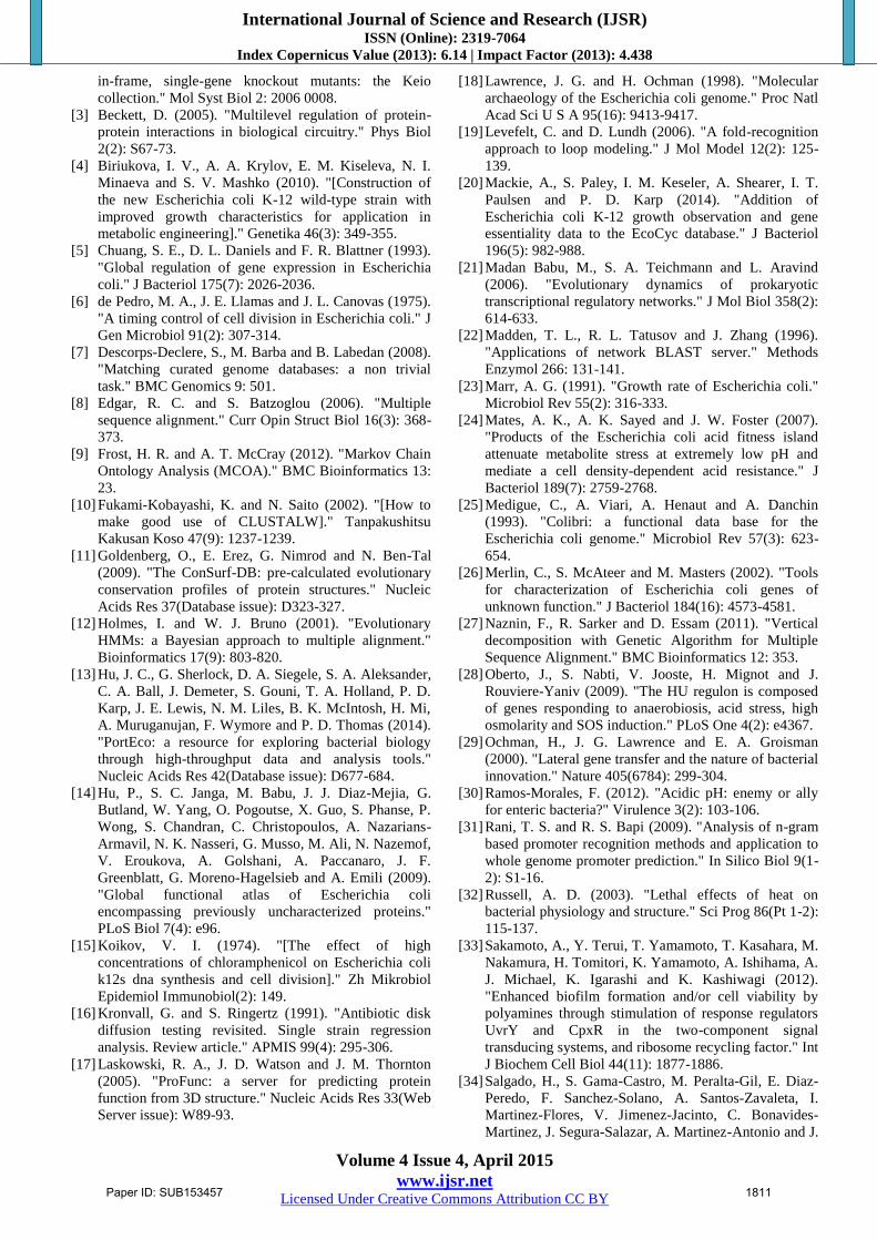
3) Molecular Modelling
Modeling of protein was done using Modeller 9.12. The
main aim for modelling protein was to analyze the protein
knowing its native (tertiary protein) structure, its stability
and its interacting capabilities.
Figure 2: Modeled 3D structure of YebV protein
Four model structures were generated using structure of
malate dehydrogenase of Escherichia coli (PDB Id: 2PWZ)
as a template. This protein has four different chains in it, out
of which ‘A’ chain was used as template. This had a Q score
of 0.81 out of 1 and RMSD of 0.60 angstrom, when checked
for matching folds.
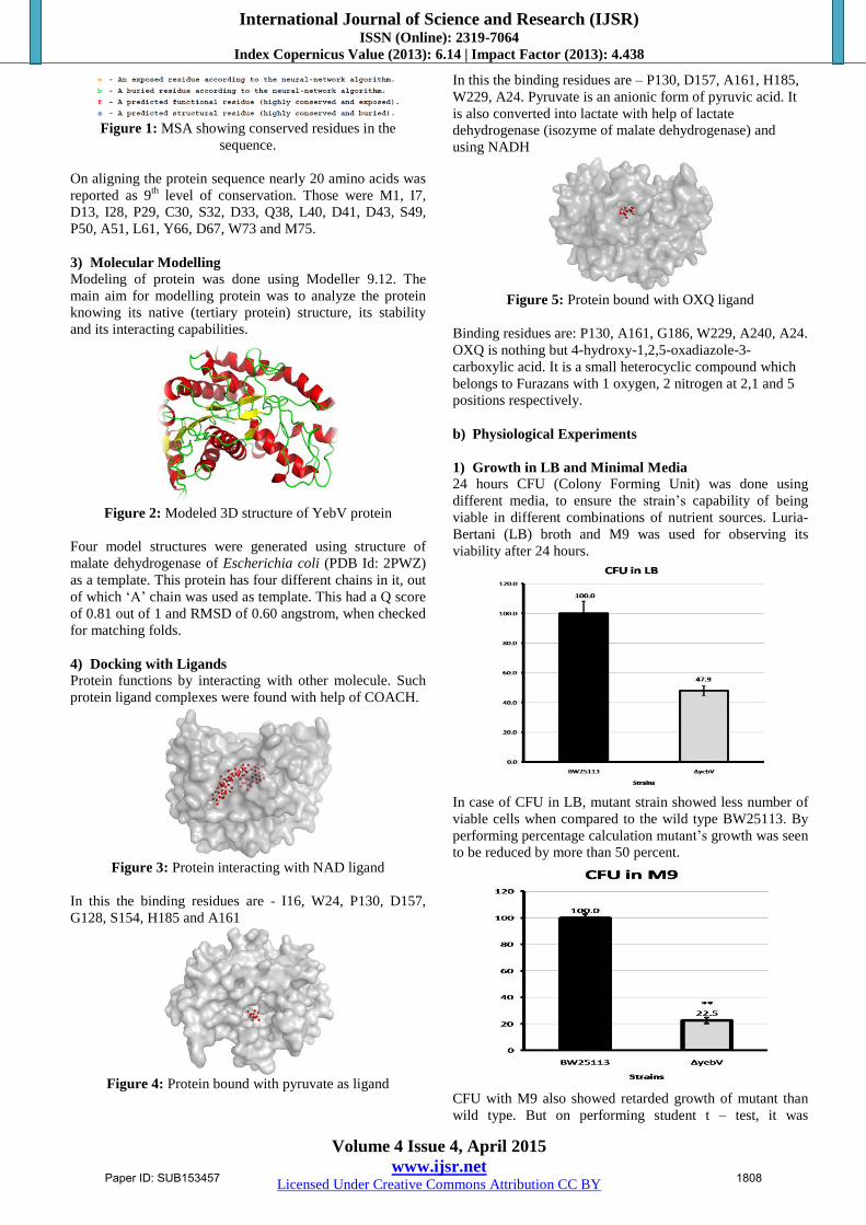
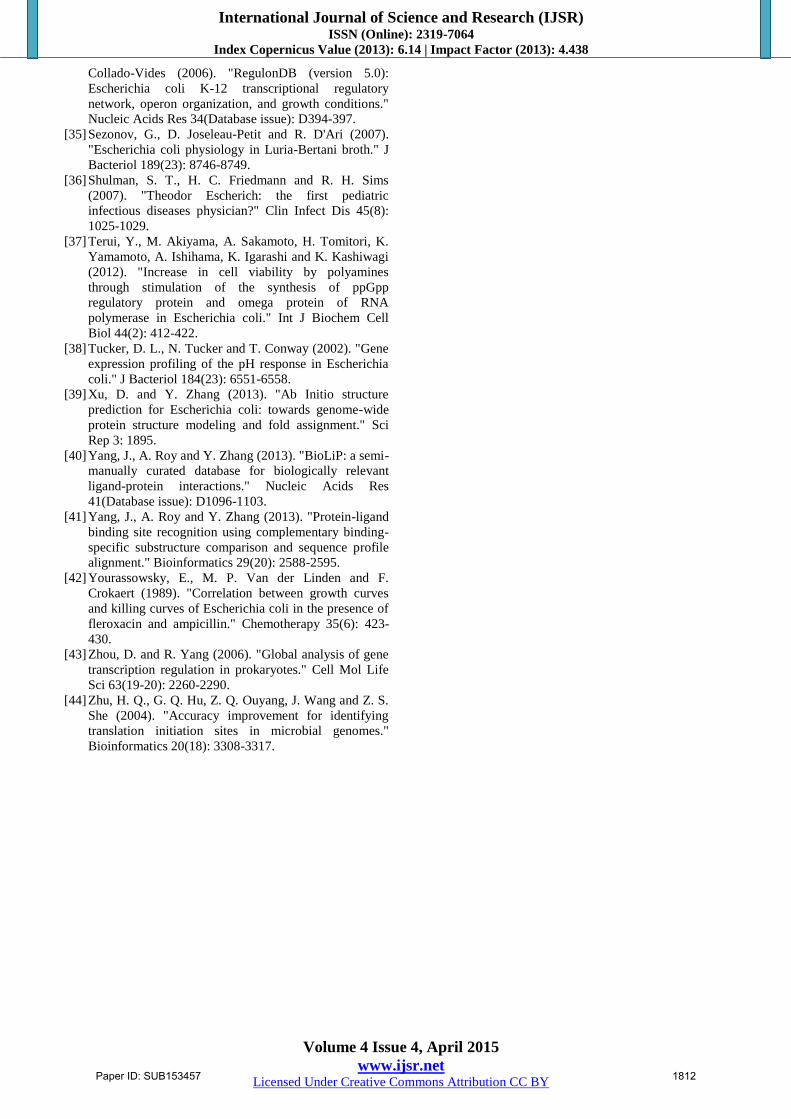
4) Docking with Ligands Protein functions by interacting with other molecule. Such
protein ligand complexes were found with help of COACH.
Figure 3: Protein interacting with NAD ligand
In this the binding residues are - I16, W24, P130, D157,
G128, S154, H185 and A161
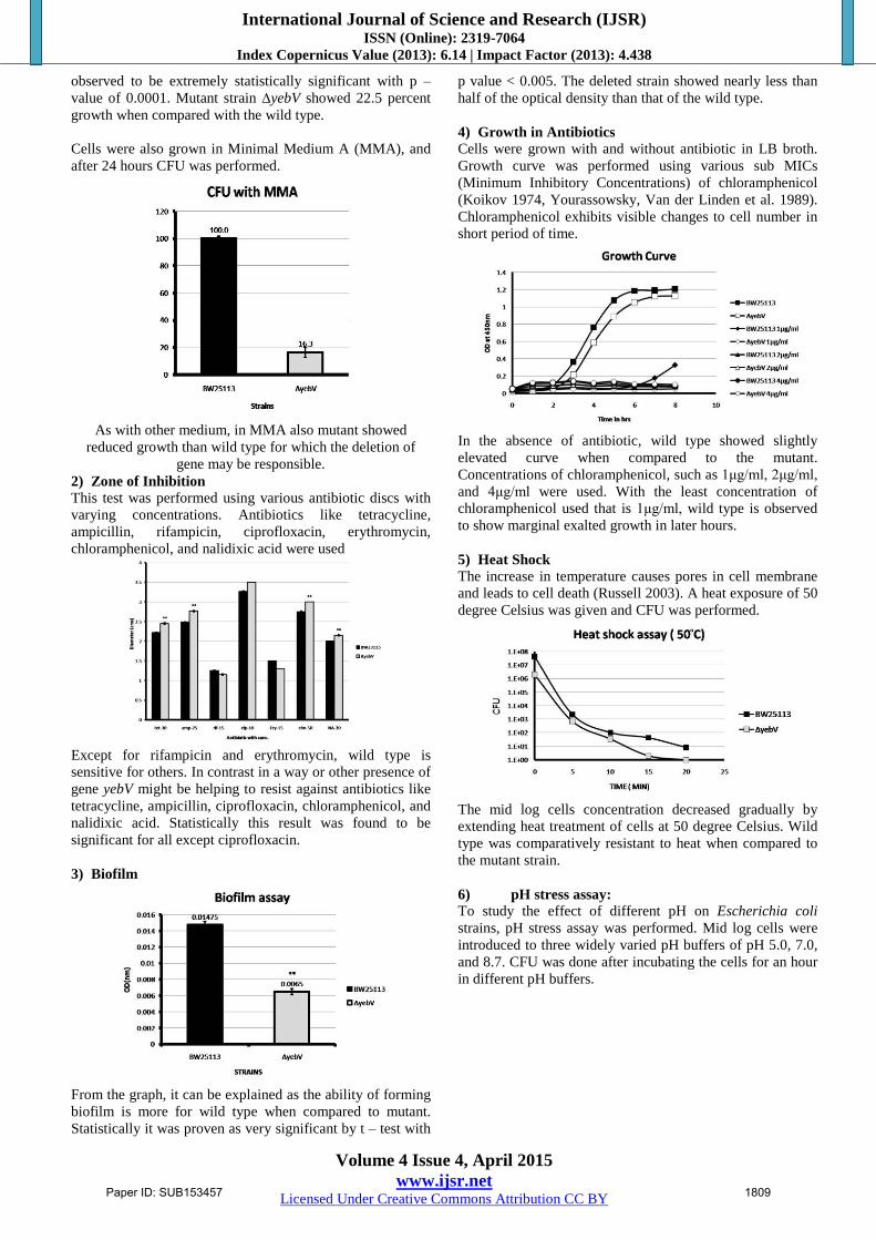
Figure 4: Protein bound with pyruvate as ligand
In this the binding residues are – P130, D157, A161, H185,
W229, A24. Pyruvate is an anionic form of pyruvic acid. It
is also converted into lactate with help of lactate
dehydrogenase (isozyme of malate dehydrogenase) and
using NADH
Figure 5: Protein bound with OXQ ligand
Binding residues are: P130, A161, G186, W229, A240, A24.
OXQ is nothing but 4-hydroxy-1,2,5-oxadiazole-3-
carboxylic acid. It is a small heterocyclic compound which
belongs to Furazans with 1 oxygen, 2 nitrogen at 2,1 and 5
positions respectively.
b) Physiological Experiments
1) Growth in LB and Minimal Media
24 hours CFU (Colony Forming Unit) was done using
different media, to ensure the strain’s capability of being
viable in different combinations of nutrient sources. Luria-
Bertani (LB) broth and M9 was used for observing its
viability after 24 hours.
In case of CFU in LB, mutant strain showed less number of
viable cells when compared to the wild type BW25113. By
performing percentage calculation mutant’s growth was seen
to be reduced by more than 50 percent.
CFU with M9 also showed retarded growth of mutant than
wild type. But on performing student t – test, it was
Paper ID: SUB153457 1808
International Journal of Science and Research (IJSR) ISSN (Online): 2319-7064
Index Copernicus Value (2013): 6.14 | Impact Factor (2013): 4.438
Volume 4 Issue 4, April 2015
www.ijsr.net Licensed Under Creative Commons Attribution CC BY
observed to be extremely statistically significant with p –
value of 0.0001. Mutant strain ∆yebV showed 22.5 percent
growth when compared with the wild type.
Cells were also grown in Minimal Medium A (MMA), and
after 24 hours CFU was performed.
As with other medium, in MMA also mutant showed
reduced growth than wild type for which the deletion of
gene may be responsible.
2) Zone of Inhibition
This test was performed using various antibiotic discs with
varying concentrations. Antibiotics like tetracycline,
ampicillin, rifampicin, ciprofloxacin, erythromycin,
chloramphenicol, and nalidixic acid were used
Except for rifampicin and erythromycin, wild type is
sensitive for others. In contrast in a way or other presence of
gene yebV might be helping to resist against antibiotics like
tetracycline, ampicillin, ciprofloxacin, chloramphenicol, and
nalidixic acid. Statistically this result was found to be
significant for all except ciprofloxacin.
3) Biofilm
From the graph, it can be explained as the ability of forming
biofilm is more for wild type when compared to mutant.
Statistically it was proven as very significant by t – test with
p value < 0.005. The deleted strain showed nearly less than
half of the optical density than that of the wild type.
4) Growth in Antibiotics
Cells were grown with and without antibiotic in LB broth.
Growth curve was performed using various sub MICs
(Minimum Inhibitory Concentrations) of chloramphenicol
(Koikov 1974, Yourassowsky, Van der Linden et al. 1989).
Chloramphenicol exhibits visible changes to cell number in
short period of time.
In the absence of antibiotic, wild type showed slightly
elevated curve when compared to the mutant.
Concentrations of chloramphenicol, such as 1μg/ml, 2μg/ml,
and 4μg/ml were used. With the least concentration of
chloramphenicol used that is 1μg/ml, wild type is observed
to show marginal exalted growth in later hours.
5) Heat Shock
The increase in temperature causes pores in cell membrane
and leads to cell death (Russell 2003). A heat exposure of 50
degree Celsius was given and CFU was performed.
The mid log cells concentration decreased gradually by
extending heat treatment of cells at 50 degree Celsius. Wild
type was comparatively resistant to heat when compared to
the mutant strain.
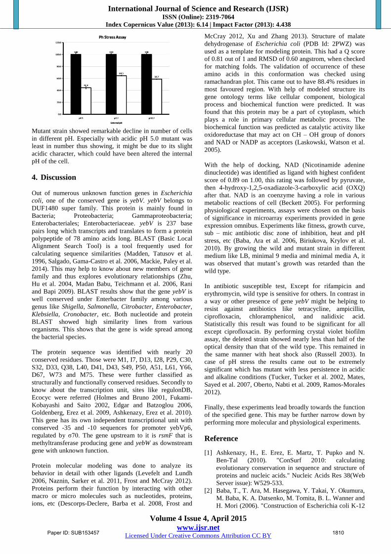
6) pH stress assay:
To study the effect of different pH on Escherichia coli
strains, pH stress assay was performed. Mid log cells were
introduced to three widely varied pH buffers of pH 5.0, 7.0,
and 8.7. CFU was done after incubating the cells for an hour
in different pH buffers.
Paper ID: SUB153457 1809
International Journal of Science and Research (IJSR) ISSN (Online): 2319-7064
Index Copernicus Value (2013): 6.14 | Impact Factor (2013): 4.438
Volume 4 Issue 4, April 2015
www.ijsr.net Licensed Under Creative Commons Attribution CC BY
Mutant strain showed remarkable decline in number of cells
in different pH. Especially with acidic pH 5.0 mutant was
least in number thus showing, it might be due to its slight
acidic character, which could have been altered the internal
pH of the cell.
4. Discussion
Out of numerous unknown function genes in Escherichia
coli, one of the conserved gene is yebV. yebV belongs to
DUF1480 super family. This protein is mainly found in
Bacteria; Proteobacteria; Gammaproteobacteria;
Enterobacteriales; Enterobacteriaceae. yebV is 237 base
pairs long which transcripts and translates to form a protein
polypeptide of 78 amino acids long. BLAST (Basic Local
Alignment Search Tool) is a tool frequently used for
calculating sequence similarities (Madden, Tatusov et al.
1996, Salgado, Gama-Castro et al. 2006, Mackie, Paley et al.
2014). This may help to know about new members of gene
family and thus explores evolutionary relationships (Zhu,
Hu et al. 2004, Madan Babu, Teichmann et al. 2006, Rani
and Bapi 2009). BLAST results show that the gene yebV is
well conserved under Enterbacter family among various
genus like Shigella, Salmonella, Citrobacter, Enterobacter,
Klebsiella, Cronobacter, etc. Both nucleotide and protein
BLAST showed high similarity lines from various
organisms. This shows that the gene is wide spread among
the bacterial species.
The protein sequence was identified with nearly 20
conserved residues. Those were M1, I7, D13, I28, P29, C30,
S32, D33, Q38, L40, D41, D43, S49, P50, A51, L61, Y66,
D67, W73 and M75. These were further classified as
structurally and functionally conserved residues. Secondly to
know about the transcription unit, sites like regulonDB,
Ecocyc were referred (Holmes and Bruno 2001, Fukami-
Kobayashi and Saito 2002, Edgar and Batzoglou 2006,
Goldenberg, Erez et al. 2009, Ashkenazy, Erez et al. 2010).
This gene has its own independent transcriptional unit with
conserved -35 and -10 sequences for promoter yebVp6,
regulated by σ70. The gene upstream to it is rsmF that is
methyltransferase producing gene and yebW as downstream
gene with unknown function.
Protein molecular modeling was done to analyze its
behavior in detail with other ligands (Levefelt and Lundh
2006, Naznin, Sarker et al. 2011, Frost and McCray 2012).
Proteins perform their function by interacting with other
macro or micro molecules such as nucleotides, proteins,
ions, etc (Descorps-Declere, Barba et al. 2008, Frost and
McCray 2012, Xu and Zhang 2013). Structure of malate
dehydrogenase of Escherichia coli (PDB Id: 2PWZ) was
used as a template for modeling protein. This had a Q score
of 0.81 out of 1 and RMSD of 0.60 angstrom, when checked
for matching folds. The validation of occurrence of these
amino acids in this conformation was checked using
ramachandran plot. This came out to have 88.4% residues in
most favoured region. With help of modeled structure its
gene ontology terms like cellular component, biological
process and biochemical function were predicted. It was
found that this protein may be a part of cytoplasm, which
plays a role in primary cellular metabolic process. The
biochemical function was predicted as catalytic activity like
oxidoreductase that may act on CH – OH group of donors
and NAD or NADP as acceptors (Laskowski, Watson et al.
2005).
With the help of docking, NAD (Nicotinamide adenine
dinucleotide) was identified as ligand with highest confident
score of 0.89 on 1.00, this rating was followed by pyruvate,
then 4-hydroxy-1,2,5-oxadiazole-3-carboxylic acid (OXQ)
after that. NAD is an coenzyme having a role in various
metabolic reactions of cell (Beckett 2005). For performing
physiological experiments, assays were chosen on the basis
of significance in microarray experiments provided in gene
expression omnibus. Experiments like fitness, growth curve,
sub – mic antibiotic disc zone of inhibition, heat and pH
stress, etc (Baba, Ara et al. 2006, Biriukova, Krylov et al.
2010). By growing the wild and mutant strain in different
medium like LB, minimal 9 media and minimal media A, it
was observed that mutant’s growth was retarded than the
wild type.
In antibiotic susceptible test, Except for rifampicin and
erythromycin, wild type is sensitive for others. In contrast in
a way or other presence of gene yebV might be helping to
resist against antibiotics like tetracycline, ampicillin,
ciprofloxacin, chloramphenicol, and nalidixic acid.
Statistically this result was found to be significant for all
except ciprofloxacin. By performing crystal violet biofilm
assay, the deleted strain showed nearly less than half of the
optical density than that of the wild type. This remained in
the same manner with heat shock also (Russell 2003). In
case of pH stress the results came out to be extremely
significant which has mutant with less persistence in acidic
and alkaline conditions (Tucker, Tucker et al. 2002, Mates,
Sayed et al. 2007, Oberto, Nabti et al. 2009, Ramos-Morales
2012).
Finally, these experiments lead broadly towards the function
of the specified gene. This may be further narrow down by
performing more molecular and physiological experiments.
Reference
[1] Ashkenazy, H., E. Erez, E. Martz, T. Pupko and N.
Ben-Tal (2010). "ConSurf 2010: calculating
evolutionary conservation in sequence and structure of
proteins and nucleic acids." Nucleic Acids Res 38(Web
Server issue): W529-533.
[2] Baba, T., T. Ara, M. Hasegawa, Y. Takai, Y. Okumura,
M. Baba, K. A. Datsenko, M. Tomita, B. L. Wanner and
H. Mori (2006). "Construction of Escherichia coli K-12
Paper ID: SUB153457 1810
International Journal of Science and Research (IJSR) ISSN (Online): 2319-7064
Index Copernicus Value (2013): 6.14 | Impact Factor (2013): 4.438
Volume 4 Issue 4, April 2015
www.ijsr.net Licensed Under Creative Commons Attribution CC BY
in-frame, single-gene knockout mutants: the Keio
collection." Mol Syst Biol 2: 2006 0008.
[3] Beckett, D. (2005). "Multilevel regulation of protein-
protein interactions in biological circuitry." Phys Biol
2(2): S67-73.
[4] Biriukova, I. V., A. A. Krylov, E. M. Kiseleva, N. I.
Minaeva and S. V. Mashko (2010). "[Construction of
the new Escherichia coli K-12 wild-type strain with
improved growth characteristics for application in
metabolic engineering]." Genetika 46(3): 349-355.
[5] Chuang, S. E., D. L. Daniels and F. R. Blattner (1993).
"Global regulation of gene expression in Escherichia
coli." J Bacteriol 175(7): 2026-2036.
[6] de Pedro, M. A., J. E. Llamas and J. L. Canovas (1975).
"A timing control of cell division in Escherichia coli." J
Gen Microbiol 91(2): 307-314.
[7] Descorps-Declere, S., M. Barba and B. Labedan (2008).
"Matching curated genome databases: a non trivial
task." BMC Genomics 9: 501.
[8] Edgar, R. C. and S. Batzoglou (2006). "Multiple
sequence alignment." Curr Opin Struct Biol 16(3): 368-
373.
[9] Frost, H. R. and A. T. McCray (2012). "Markov Chain
Ontology Analysis (MCOA)." BMC Bioinformatics 13:
23.
[10] Fukami-Kobayashi, K. and N. Saito (2002). "[How to
make good use of CLUSTALW]." Tanpakushitsu
Kakusan Koso 47(9): 1237-1239.
[11] Goldenberg, O., E. Erez, G. Nimrod and N. Ben-Tal
(2009). "The ConSurf-DB: pre-calculated evolutionary
conservation profiles of protein structures." Nucleic
Acids Res 37(Database issue): D323-327.
[12] Holmes, I. and W. J. Bruno (2001). "Evolutionary
HMMs: a Bayesian approach to multiple alignment."
Bioinformatics 17(9): 803-820.
[13] Hu, J. C., G. Sherlock, D. A. Siegele, S. A. Aleksander,
C. A. Ball, J. Demeter, S. Gouni, T. A. Holland, P. D.
Karp, J. E. Lewis, N. M. Liles, B. K. McIntosh, H. Mi,
A. Muruganujan, F. Wymore and P. D. Thomas (2014).
"PortEco: a resource for exploring bacterial biology
through high-throughput data and analysis tools."
Nucleic Acids Res 42(Database issue): D677-684.
[14] Hu, P., S. C. Janga, M. Babu, J. J. Diaz-Mejia, G.
Butland, W. Yang, O. Pogoutse, X. Guo, S. Phanse, P.
Wong, S. Chandran, C. Christopoulos, A. Nazarians-
Armavil, N. K. Nasseri, G. Musso, M. Ali, N. Nazemof,
V. Eroukova, A. Golshani, A. Paccanaro, J. F.
Greenblatt, G. Moreno-Hagelsieb and A. Emili (2009).
"Global functional atlas of Escherichia coli
encompassing previously uncharacterized proteins."
PLoS Biol 7(4): e96.
[15] Koikov, V. I. (1974). "[The effect of high
concentrations of chloramphenicol on Escherichia coli
k12s dna synthesis and cell division]." Zh Mikrobiol
Epidemiol Immunobiol(2): 149.
[16] Kronvall, G. and S. Ringertz (1991). "Antibiotic disk
diffusion testing revisited. Single strain regression
analysis. Review article." APMIS 99(4): 295-306.
[17] Laskowski, R. A., J. D. Watson and J. M. Thornton
(2005). "ProFunc: a server for predicting protein
function from 3D structure." Nucleic Acids Res 33(Web
Server issue): W89-93.
[18] Lawrence, J. G. and H. Ochman (1998). "Molecular
archaeology of the Escherichia coli genome." Proc Natl
Acad Sci U S A 95(16): 9413-9417.
[19] Levefelt, C. and D. Lundh (2006). "A fold-recognition
approach to loop modeling." J Mol Model 12(2): 125-
139.
[20] Mackie, A., S. Paley, I. M. Keseler, A. Shearer, I. T.
Paulsen and P. D. Karp (2014). "Addition of
Escherichia coli K-12 growth observation and gene
essentiality data to the EcoCyc database." J Bacteriol
196(5): 982-988.
[21] Madan Babu, M., S. A. Teichmann and L. Aravind
(2006). "Evolutionary dynamics of prokaryotic
transcriptional regulatory networks." J Mol Biol 358(2):
614-633.
[22] Madden, T. L., R. L. Tatusov and J. Zhang (1996).
"Applications of network BLAST server." Methods
Enzymol 266: 131-141.
[23] Marr, A. G. (1991). "Growth rate of Escherichia coli."
Microbiol Rev 55(2): 316-333.
[24] Mates, A. K., A. K. Sayed and J. W. Foster (2007).
"Products of the Escherichia coli acid fitness island
attenuate metabolite stress at extremely low pH and
mediate a cell density-dependent acid resistance." J
Bacteriol 189(7): 2759-2768.
[25] Medigue, C., A. Viari, A. Henaut and A. Danchin
(1993). "Colibri: a functional data base for the
Escherichia coli genome." Microbiol Rev 57(3): 623-
654.
[26] Merlin, C., S. McAteer and M. Masters (2002). "Tools
for characterization of Escherichia coli genes of
unknown function." J Bacteriol 184(16): 4573-4581.
[27] Naznin, F., R. Sarker and D. Essam (2011). "Vertical
decomposition with Genetic Algorithm for Multiple
Sequence Alignment." BMC Bioinformatics 12: 353.
[28] Oberto, J., S. Nabti, V. Jooste, H. Mignot and J.
Rouviere-Yaniv (2009). "The HU regulon is composed
of genes responding to anaerobiosis, acid stress, high
osmolarity and SOS induction." PLoS One 4(2): e4367.
[29] Ochman, H., J. G. Lawrence and E. A. Groisman
(2000). "Lateral gene transfer and the nature of bacterial
innovation." Nature 405(6784): 299-304.
[30] Ramos-Morales, F. (2012). "Acidic pH: enemy or ally
for enteric bacteria?" Virulence 3(2): 103-106.
[31] Rani, T. S. and R. S. Bapi (2009). "Analysis of n-gram
based promoter recognition methods and application to
whole genome promoter prediction." In Silico Biol 9(1-
2): S1-16.
[32] Russell, A. D. (2003). "Lethal effects of heat on
bacterial physiology and structure." Sci Prog 86(Pt 1-2):
115-137.
[33] Sakamoto, A., Y. Terui, T. Yamamoto, T. Kasahara, M.
Nakamura, H. Tomitori, K. Yamamoto, A. Ishihama, A.
J. Michael, K. Igarashi and K. Kashiwagi (2012).
"Enhanced biofilm formation and/or cell viability by
polyamines through stimulation of response regulators
UvrY and CpxR in the two-component signal
transducing systems, and ribosome recycling factor." Int
J Biochem Cell Biol 44(11): 1877-1886.
[34] Salgado, H., S. Gama-Castro, M. Peralta-Gil, E. Diaz-
Peredo, F. Sanchez-Solano, A. Santos-Zavaleta, I.
Martinez-Flores, V. Jimenez-Jacinto, C. Bonavides-
Martinez, J. Segura-Salazar, A. Martinez-Antonio and J.
Paper ID: SUB153457 1811
International Journal of Science and Research (IJSR) ISSN (Online): 2319-7064
Index Copernicus Value (2013): 6.14 | Impact Factor (2013): 4.438
Volume 4 Issue 4, April 2015
www.ijsr.net Licensed Under Creative Commons Attribution CC BY
Collado-Vides (2006). "RegulonDB (version 5.0):
Escherichia coli K-12 transcriptional regulatory
network, operon organization, and growth conditions."
Nucleic Acids Res 34(Database issue): D394-397.
[35] Sezonov, G., D. Joseleau-Petit and R. D'Ari (2007).
"Escherichia coli physiology in Luria-Bertani broth." J
Bacteriol 189(23): 8746-8749.
[36] Shulman, S. T., H. C. Friedmann and R. H. Sims
(2007). "Theodor Escherich: the first pediatric
infectious diseases physician?" Clin Infect Dis 45(8):
1025-1029.
[37] Terui, Y., M. Akiyama, A. Sakamoto, H. Tomitori, K.
Yamamoto, A. Ishihama, K. Igarashi and K. Kashiwagi
(2012). "Increase in cell viability by polyamines
through stimulation of the synthesis of ppGpp
regulatory protein and omega protein of RNA
polymerase in Escherichia coli." Int J Biochem Cell
Biol 44(2): 412-422.
[38] Tucker, D. L., N. Tucker and T. Conway (2002). "Gene
expression profiling of the pH response in Escherichia
coli." J Bacteriol 184(23): 6551-6558.
[39] Xu, D. and Y. Zhang (2013). "Ab Initio structure
prediction for Escherichia coli: towards genome-wide
protein structure modeling and fold assignment." Sci
Rep 3: 1895.
[40] Yang, J., A. Roy and Y. Zhang (2013). "BioLiP: a semi-
manually curated database for biologically relevant
ligand-protein interactions." Nucleic Acids Res
41(Database issue): D1096-1103.
[41] Yang, J., A. Roy and Y. Zhang (2013). "Protein-ligand
binding site recognition using complementary binding-
specific substructure comparison and sequence profile
alignment." Bioinformatics 29(20): 2588-2595.
[42] Yourassowsky, E., M. P. Van der Linden and F.
Crokaert (1989). "Correlation between growth curves
and killing curves of Escherichia coli in the presence of
fleroxacin and ampicillin." Chemotherapy 35(6): 423-
430.
[43] Zhou, D. and R. Yang (2006). "Global analysis of gene
transcription regulation in prokaryotes." Cell Mol Life
Sci 63(19-20): 2260-2290.
[44] Zhu, H. Q., G. Q. Hu, Z. Q. Ouyang, J. Wang and Z. S.
She (2004). "Accuracy improvement for identifying
translation initiation sites in microbial genomes."
Bioinformatics 20(18): 3308-3317.
Paper ID: SUB153457 1812