Embed Size (px)
Citation preview

Immune-Related Response Criteria:
Variations in I-O Response Patterns
and Implications for Treatment
Lee Schwartzberg, MD, FACP
Director, The West Clinic
Memphis, TN
7.14.15
12 – 1 pm, EST
ICLIO e-Course 01
Objectives
By the end of this e-course, participants will be able to:
– Understand why RECIST criteria have been adapted in assessing tumor response to immuno-oncologic agents
– Understand the differences between RECIST and Immune-related Response Criteria (irRC)
– Understand irRC use in evaluating tumor response to immuno-oncologic agents
– Understand the diversity of potential tumor responses to IO agents and the direction of treatment planning in the practice setting
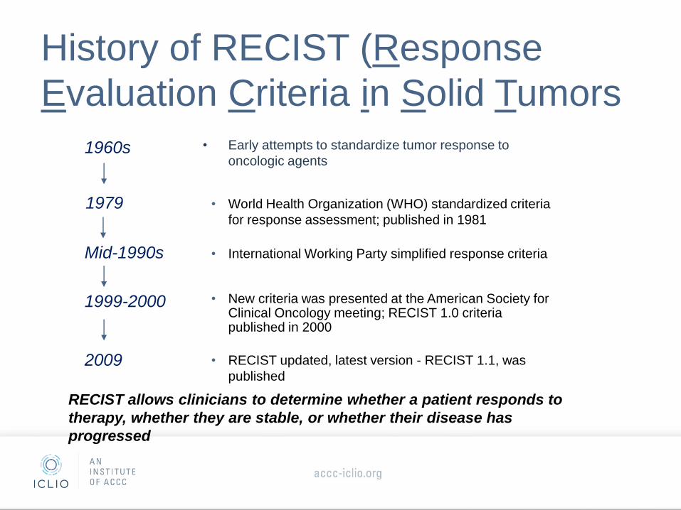
History of RECIST (Response
Evaluation Criteria in Solid Tumors • Early attempts to standardize tumor response to
oncologic agents 1960s
1979
Mid-1990s
1999-2000
2009
• World Health Organization (WHO) standardized criteria
for response assessment; published in 1981
• International Working Party simplified response criteria
• New criteria was presented at the American Society for Clinical Oncology meeting; RECIST 1.0 criteria published in 2000
• RECIST updated, latest version - RECIST 1.1, was
published
RECIST allows clinicians to determine whether a patient responds to
therapy, whether they are stable, or whether their disease has
progressed

RECIST 1.1 - Response Criteria Target Lesions - includes all
measurable lesions*; max 2 per
organ, 5 lesions total
Evaluation of
Target Lesions
RECIST Guideline
CR Disappearance of all target
lesions; confirmed at > 4
weeks
PR > 30% decrease of SoD
from baseline, confirmed at
> 4 weeks
PD > 20% increase from
smallest sum of diameters
recorded and 5 mm
absolute increase over
lowest sum
SD Neither PR or PD
Evaluation of
non-target
lesions
RECIST Guideline
CR Disappearance of all non-
target lesions; normalization of
tumor markers
PD Appearance of > 1 new lesions
and/or progression of existing
non-target lesions
SD Persistence of > 1 non-target
lesion; tumor marker level
above normal
CR (Complete Response); PR (Partial Response); PD (Progressive Disease); SD (Stable Disease)
*measurable lesion = > 10 mm in longest diameter by CT Scan; > 20 mm in longest diameter by x-ray
sources: Eisenhauer et al., 2009; Nishino et al, 2010; and RECIST, Applying the Rules, National Cancer Institute, https://ccrod.cancer.gov/confluence/download/attachments/71041052/RECIST6.pdf?version=1&modificationDate=1317305352430
Non-Target Lesions – all other
lesions not classified as a target lesion
or sites of disease
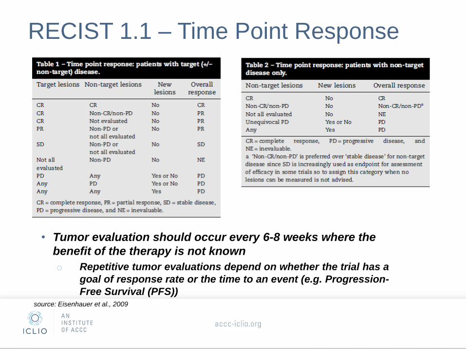
RECIST 1.1 – Time Point Response
source: Eisenhauer et al., 2009
• Tumor evaluation should occur every 6-8 weeks where the
benefit of the therapy is not known
o Repetitive tumor evaluations depend on whether the trial has a
goal of response rate or the time to an event (e.g. Progression-
Free Survival (PFS))
RECIST for determining tumor response
is applicable to cytotoxic agents
• Cytotoxic agents directly kill a tumor cell or prevent tumor cells
from dividing (e.g. chemotherapy); therefore, response of
cytotoxic agents can be easily measured from the start of
therapy
• Early increase in tumor burden and/or an early increase in
tumor size signifies progressive disease
– Once progression is detected, drug cessation is recommended
Response after initial treatment of a cytotoxic
agent can often predict remission and survival
Immuno-oncology agents differ from cytotoxic
agents in that they stimulate an innate immune
response against the tumor
• Vaccines: trigger the immune system to initiate an anti-tumor response against an existing cancer
• Monoclonal Antibodies: antibodies directed against tumor cells; they can block signaling pathways needed for tumor growth and trigger an immune-mediated cytotoxic response
• Checkpoint inhibitors: tumors escape detection by the immune system through expression of “checkpoint” proteins on their cell surface. CTLA-4 and PD-1 receptors are examples of “checkpoint” receptors; targeted inhibition towards these receptors enhances T cell response towards the tumor
• Cytokines: stimulates a broad-based immune response (e.g. interleukin-2 and interferon-α)
http://www.fightcancerwithimmunotherapy.com/ImmunotherapyAndCancer/TypesOfCancerImmunotherapy.aspx
Source:
http://www.fightcancerwithimmunotherapy.com/immunotherapyandcancer/typesofcancerimmunotherapy.aspx

The unique mechanism of action of immuno-
oncology agents requires modified tumor response
criteria
– Anti-tumor response to immunotherapy may take longer compared to
cytotoxic agent response
– Clinical response to immune therapies can manifest after
conventional progressive disease (PD) – “pseudoprogression”
– Discontinuation of immune therapy may not be appropriate in some
cases, unless PD is confirmed
– Allowance for “clinically insignificant” PD (e.g., small new lesions in
the presence of other responsive lesions) is recommended
– Durable stable disease may represent antitumor activity
source: Wolchock et al., 2009
RECIST may not provide a complete
assessment of immunotherapeutics:

Differing mechanism of
immunotherapy
Ipilimumab – clinical observations and evaluation
of a novel set of response criteria
• Ipilimumab: human, monoclonal antibody that binds to the cytotoxic T-lymphocyte-associated antigen 4 (CTLA-4) on T cells. Blocking CTLA-4 from interacting with its ligands augments a T cell immune response to tumor cells
• Ipilimumab is indicated for the treatment of unresectable or metastatic melanoma
• Ipilimumab was studied in three multicenter phase II trials evaluating 487 patients with unresectable stage III or IV melanoma
• Activity was categorized using a novel set of criteria – Tumor assessments carried out at week 12 following the end of
the induction dosing period (ipilimumab 10 mg/kg every three weeks times x4)
source: Wolchock et al., 2009

Four patterns of response were observed in
patients treated with ipilimumab
• Overall, ~30% of patients had disease control (CR, PR, or SD)
• Of the 4 patterns of response observed two met conventional
criteria for tumor response:
Response in baseline lesions “stable disease” with slow, steady
decline in total tumor volume
SPD = sum of the product of perpendicular diameters
Triangles = ipilimumab dosing time points source: Wolchock et al., 2009
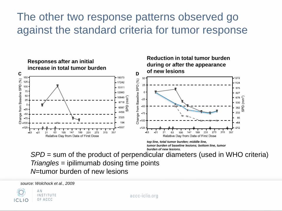
The other two response patterns observed go
against the standard criteria for tumor response
Responses after an initial
increase in total tumor burden
Reduction in total tumor burden
during or after the appearance
of new lesions
SPD = sum of the product of perpendicular diameters (used in WHO criteria)
Triangles = ipilimumab dosing time points
N=tumor burden of new lesions
top line, total tumor burden; middle line,
tumor burden of baseline lesions; bottom line, tumor
burden of new lesions.
source: Wolchock et al., 2009

A number of ipilimumab treated patients initially
characterized as PD, are considered PR or SD
using the irRC Guideline
source: Wolchock et al., 2009
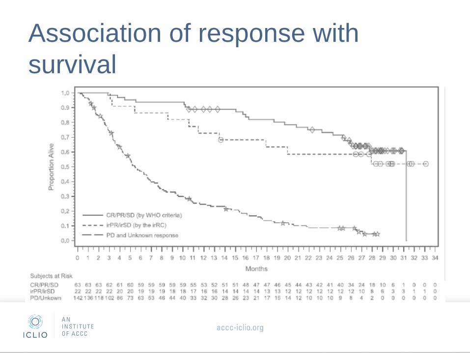
Association of response with
survival

Clinical trials utilizing both irRC and RECIST 1.1 to
measure tumor response
http://meetinglibrary.asco.org/content/134449-144 • 411 pts, 192 were on MK-3475 (pembrolizumab) > 28
weeks
• 215 patients had either a CR, PR, or SD by RECIST and
irRC
• 51 patients had PD by RECIST, but had either a CR, PR,
or SD by irRC
Authors concluded:
“conventional criteria such as RECIST may underestimate the
benefit of MK-3475 in approximately 10% of treated pts.”
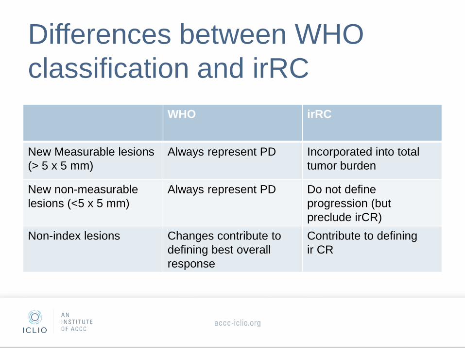
Differences between WHO
classification and irRC
WHO irRC
New Measurable lesions
(> 5 x 5 mm)
Always represent PD Incorporated into total
tumor burden
New non-measurable
lesions (<5 x 5 mm)
Always represent PD Do not define
progression (but
preclude irCR)
Non-index lesions Changes contribute to
defining best overall
response
Contribute to defining
ir CR
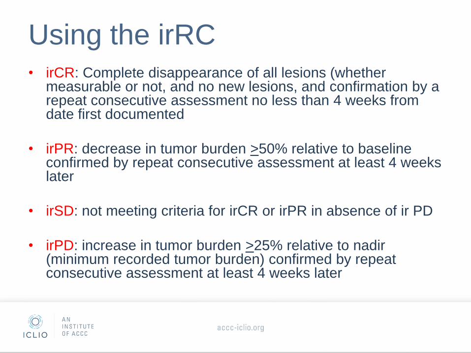
Using the irRC • irCR: Complete disappearance of all lesions (whether
measurable or not, and no new lesions, and confirmation by a repeat consecutive assessment no less than 4 weeks from date first documented
• irPR: decrease in tumor burden >50% relative to baseline confirmed by repeat consecutive assessment at least 4 weeks later
• irSD: not meeting criteria for irCR or irPR in absence of ir PD
• irPD: increase in tumor burden >25% relative to nadir (minimum recorded tumor burden) confirmed by repeat consecutive assessment at least 4 weeks later

Real World Case Examples: Case Study 1
• 52 yo male
• Thyroid nodule: low grade papillary cancer
• Referred to Dr. Portnoy, West Clinic
• CT neck: extensive lymphadenopathy and multiple pulmonary nodules
• PET/CT: 2.5 cm left upper lobe mass, multiple nodules in lungs, subcutaneous met in inferior R axilla, L adrenal mass, 5 cm mass in the gluteus maximus, bony lesions in L iliac bone and R hip
• Pain in R hip, weight loss, fatigue
• Jehovah’s Witness
• Anemia with Hemoglobin 7 gm/dl; Creatinine 2.2
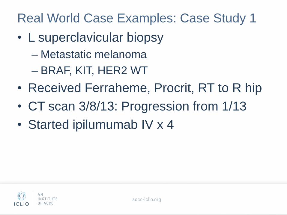
Real World Case Examples: Case Study 1
• L superclavicular biopsy
– Metastatic melanoma
– BRAF, KIT, HER2 WT
• Received Ferraheme, Procrit, RT to R hip
• CT scan 3/8/13: Progression from 1/13
• Started ipilumumab IV x 4

3/8/2013 CT Pt #1
Real World Case Examples: Case Study 1
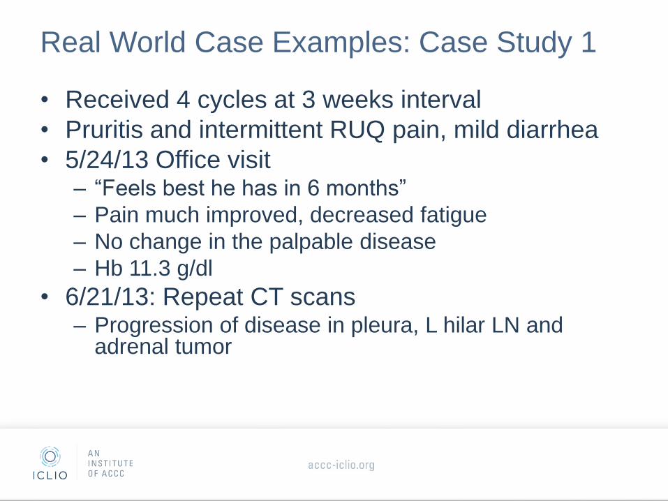
• Received 4 cycles at 3 weeks interval
• Pruritis and intermittent RUQ pain, mild diarrhea
• 5/24/13 Office visit – “Feels best he has in 6 months”
– Pain much improved, decreased fatigue
– No change in the palpable disease
– Hb 11.3 g/dl
• 6/21/13: Repeat CT scans – Progression of disease in pleura, L hilar LN and
adrenal tumor

6/21/13 3/8/13
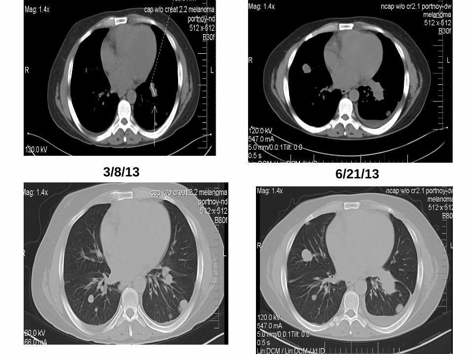
6/21/13 3/8/13
Real World Case Examples: Case Study 1
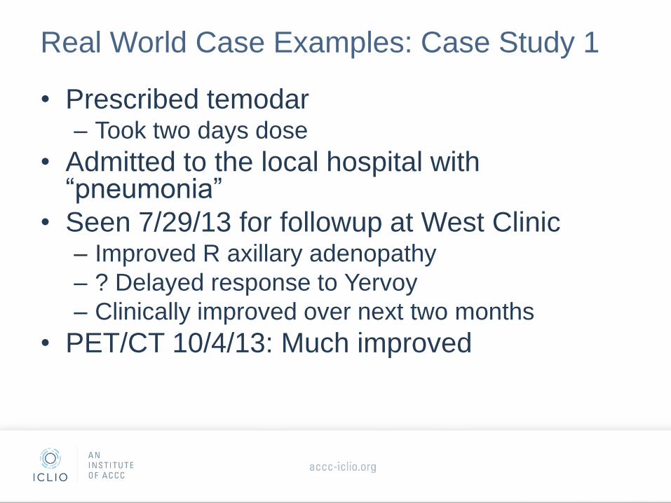
• Prescribed temodar – Took two days dose
• Admitted to the local hospital with “pneumonia”
• Seen 7/29/13 for followup at West Clinic – Improved R axillary adenopathy
– ? Delayed response to Yervoy
– Clinically improved over next two months
• PET/CT 10/4/13: Much improved
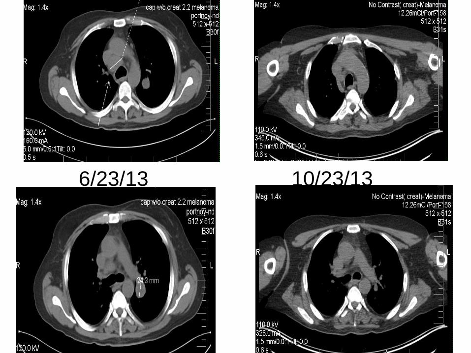
6/23/13 10/23/13
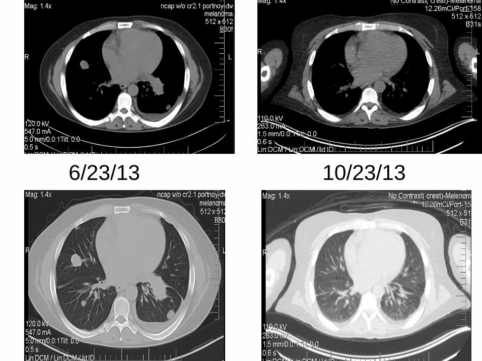
6/23/13 10/23/13
Real World Case Examples: Case Study 1
• 4/13/14: CT scan
– No pulmonary nodules
– Sclerotic bone metastases
– No adrenal metastases
• 4/14/15: CT scan
– No evidence of disease
Real World Case Examples: Case Study 2
• 66 year old female
• Nausea, cough, weight loss of 40 lbs
• Referred to Dr. Somer, West Clinic
• CT 5/23/13: RUL mass, multiple nodules in both lungs, bilateral hilar and mediastinal lymph nodes, ground glass opacities, confirmed by PET/CT
• CT guided biopsy R lung:
– moderately differentiated adenocarcinoma
– Molecular profiling: EGFR WT, ROS and ALK without rearrangement
Real World Case Examples: Case Study 2
• Started treatment with Carboplatin/pemetrexed
• Received 4 cycles
• CT 8/13: Good response to therapy
• Stable PR 12/13
• CT 5/14: – POD with new consolidation/mass RML, multiple
bilateral nodules, large RUL nodule
– Symptomatically worse, on chronic O2
– Started 2nd line erlotinib, after Veristrat good molecular signature
• 11/6/14: CT showed POD in lungs
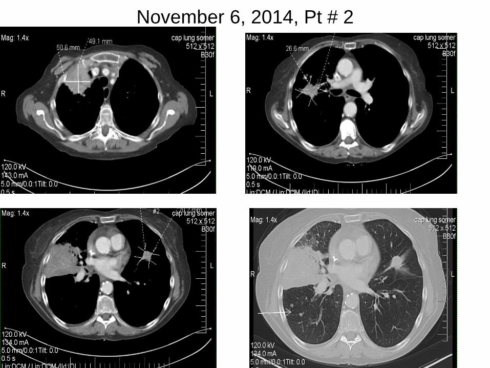
November 6, 2014, Pt # 2
Real World Case Examples: Case Study 2
• Evaluated for Nivolumab trial
• Initiated 11/21/14
• One week later, admitted to hospital with
increased SOB, nausea and diarrhea
• Treated with aggressive pulmonary
measures, O2, and antibiotics for VRE in
urine
• Improved symptomatically and was able to
resume nivolumab
• Re-evaluated 1/3/15 with CT
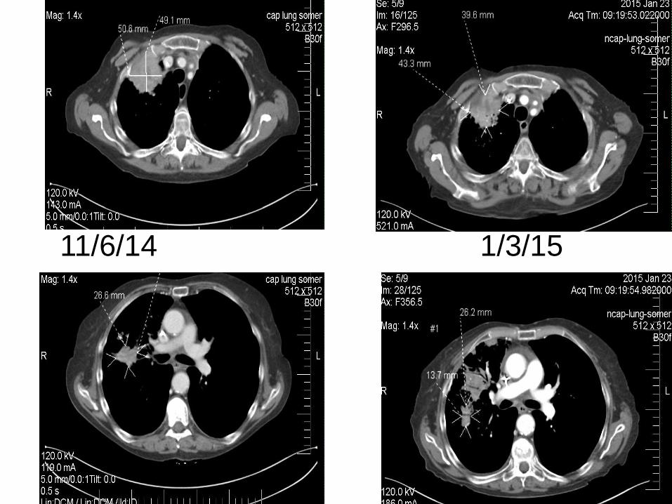
11/6/14 1/3/15
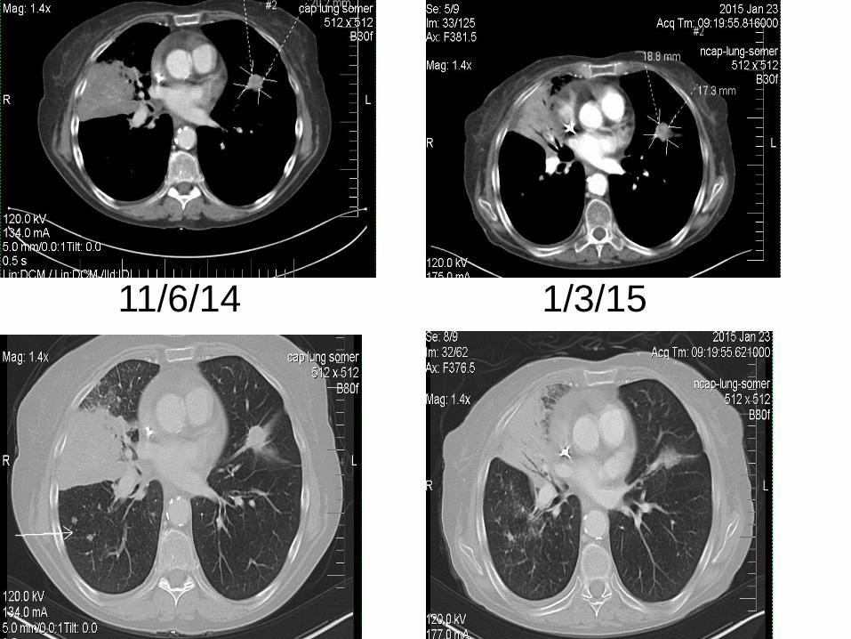
11/6/14 1/3/15
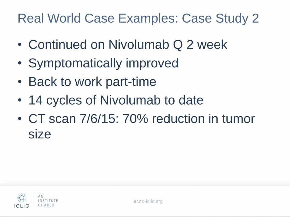
Real World Case Examples: Case Study 2
• Continued on Nivolumab Q 2 week
• Symptomatically improved
• Back to work part-time
• 14 cycles of Nivolumab to date
• CT scan 7/6/15: 70% reduction in tumor
size
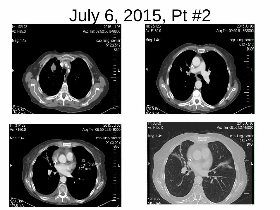
July 6, 2015, Pt #2

Summary
• Immune mediated drugs (such as CTLA-4 inhibitors and PD (L)-1 inhibitors are finding use in many cancer types
• Immuno-oncology drugs work differently than traditional cytotoxics but stimulating the immune system
• With immunotherapy, imaging studies may show initial worsening of lesions in terms of size and even new lesions during initial therapy evaluation
• A new response system, the irRC, was developed and is in use now for patients treated with immuno-oncology agentsd
• In absence of clinical progression, pseudo-progression on scans should be strongly considered and patients re-evaluated carefully

Questions?

References Eisenhauer, E.A, et al. New response evaluation criteria in solid tumours: Revised RECIST guideline (version 1.1). European Journal of Cancer 2009; 45:228-247
Garon, E.B., et al. Antitumor activity of pembrolizumab (Pembro; MK-3475) and correlation with programmed death ligand 1 (PD-L1) expression in a pooled analysis of patients (pts) with advanced Non–Small Cell Lung Carcinoma (NSCLC). Annals of Oncology 2014; 25: 1-41.
Hodi, S.F., et al. Evaluation of immune-related response criteria (irRC) in patients (pts) with advanced melanoma (MEL) treated with the anti-PD-1 monoclonal antibody MK-3475. Journal of Clinical Oncology 2014; 32:5s
Nishino, M., et al. Revised RECIST Guideline Version 1.1: What Oncologists Want to Know and What Radiologists Need to Know. American Journal of Roentgenology 2010; 195:281-289
Wolchock et al. Guidelines for the Evaluation of Immune Therapy Activity in Solid Tumors: Immune Therapy Activity in Solid Tumors: Immune-Related Response Criteria. Clinical Cancer Research 2009; 15:7412-7420
RECIST, Applying the Rules, National Cancer Institute https://ccrod.cancer.gov/confluence/download/attachments/71041052/RECIST6.pdf?version=1&modificationDate=1317305352430
Immunotherapy: Fight Cancer with Immunotherapy http://www.fightcancerwithimmunotherapy.com/ImmunotherapyAndCancer/TypesOfCancerImmunotherapy.aspx