Embed Size (px)
Citation preview

1 www.brain101.info
III. NEURO-HISTOLOGY
SANTIAGO RAMON Y CAJAL (Winner of the 1906 Nobel Prize in Medicine) cameup with evidence that nerve fibers are not continuous, but rather contiguous, and thatsynapses separate them.
o He asserted the polarity of cells, with dendrites receiving and axonsdelivering.
o Also, he established the neuronal growth occurs at the proximal stump(nearest the cell body) during development, and not at the distal stump.
NEURON
CONTENTS OF THE NEURON SOMA: Only those things beyond the obvious.
NISSL BODIES: Clusters of basophilic Rough ER, found in abundance in the neuron soma. Phagosomes: Waste-containing vacuoles that fuse with primary lysosomes to form secondary
lysosomes. DENSE BODY: A tertiary lysosome, or a lysosomes that has already degraded much of its contents,
but has non-digestible materials remaining.o Dense bodies contain Lipofuscin, which tends to accumulate with age.
DENDRITES
They generally develop after the axons. No Golgi apparatus, and Nissl Bodies (i.e. Rough ER) diminish as you get away from the soma. Microtubules: The orientation of microtubules in dendrites is mixed, both plus to minus and minus to
plus.
AXONS
Axon Hillock is the initial segment of the axon, as it narrows down from the soma.o Nissl substances and Golgi can still be found at the hillock, but diminish as you move down the axon.
Myelin Segments:o Node of Ranvier: The region of saltatory conduction where there is no myelin.o Internode: The myelinated regions between nodes of Ranvier.o Paranode: The region right next to a node of Ranvier.
Microtubules and Neurofilaments: Histologically, they predominate throughout the axon.o For Microtubules, the plus-end points away from the cell body.o Mitochondria and smooth ER can also be found in axon. Other organelles are absent.
AXOLEMMA: Membrane of the axon.

2 www.brain101.info
AXONAL TRANSPORT
Anterograde Transport: Movement away from the soma, toward the axon terminal.o Fast Anterograde Transport: 200-400 mm / day. This is the majority of standard protein transport, of
stuff from the Rough ER.o Slow Anterograde Transport: Proteins transported by this mechanism are synthesized on free polysomes in the soma. Dynamin is the name of the microtubule motor protein that functions in slow-component A. Proteins include actin, spectrin, clathrin, and others. Motor mechanism may utilize actin/myosin. Calcium-dependent proteases function to disassemble the structure so that proteins can be utilized
at their destination. Retrograde Transport: Movement toward the soma.o TWO FUNCTIONS:o Molecules transported: NGF, neurotoxins, viruseso Horseradish peroxidase = a retrograde tracer molecule.o Dynein moves things from the plus to the minus end of microtubules and is therefore responsible for
retrograde transport.
NEUROPIL: The interconnected and interwoven processes of dendrites, axons, and glia. (The neuronalenvironment).
Distinguishing Axons and Dendrites Histologically:
Dendrites have a homogenous collection of microtubules, while axons have them in clumps. Axons may be myelinated. Dendrites aren’t. The presence of synaptic vesicle indicates that it is an axon.
TYPES / CLASSIFICATIONS OF NEURONS
According to function: Sensory / Motoro Sensory: The majority of neurons.o Motor: (The cell bodies are) found in three discrete places.o Associative: Interneurons, which make connection between other neurons.
According to function: Mode of Actiono Excitatoryo Inhibitoryo Both Excitatory and Inhibitory: Release neurotransmitters that excite some neurons but inhibit
others. According to lengtho Golgi Type I: Long axons, as in PNS.o Golgi Type II: Short axons, as in CNS.
According to number of processes
a: Axon d: Dendrite
o (1) Unipolar—have a single process that may give rise many branches.o (2) Bipolar: Having a single axon and single dendrite coming out of the cell body.o (3) Pseudounipolar: Having the single process come off a stem attached to the soma.o (4) Multipolar: Most neurons. Multiple Axons and Dendrites

3 www.brain101.info
NEUROGLIA (NEUROGLIAL CELLS)
Central Neuroglia Peripheral Neuroglia
Astrocyte - protoplasmic astrocyte - fibrous astrocyte
Oligodendrocyte - perineuronal satellite cell - interfascicular cell
Microglia
Ependymal Cell
Schwann Cell - in peripheral nerve - and ganglion
Capsular (Satellite) Cell - in ganglion
Astrocyte Oligodendrocyte Microglia
ASTROCYTES: STAR-LIKE NEUROGLIAL (NEURAL ACCESSORY) CELLS
Functions:o Supportive roleo Insulate synapse from each othero Regulate extracellular pH, K+ concentration.o Induce formation of the blood-brain barriero Interaction with immune systemo Limited phagocytosis
GLIOSIS / ASTROCYTOSIS: Astrocyte response to disease in the brain.o They proliferate and divideo They increase their concentration of GFAP-laden intermediate filaments.o They form a dense network termed a glial scar.
Astrocyte Morphology / Histologyo Glial Fibrils: The name of the intermediate filaments in astrocytes.o There are no microtubules in mature astrocytes.o Perivascular Feet (Vascular End-Feet): Help form the blood brain barrier.o Astrocytes are the largest of all the accessory neuronal cells.
Two General Types of Astrocytes: The two types of astrocytes are really two morphological ends of acontinuous spectrum.o Fibrous Astrocytes: Prominent in white matter.o Protoplasmic Astrocytes: Prominent in grey matter.
Special Types of Astrocyteso Bergmann Glial Cell: Found in cerebellum, they have processes that extend all the way to the pial
membrane, similar to early development.o Muller Cell: Found in retina, sharing features with both astrocytes and ependymal cells.o Pituicute.

4 www.brain101.info
OLIGODENDROCYTES: Myelin forming cells in the CNS, and can myelinate multiple internodes
1. Nucleus of Oligodendrocyte2. Process of Oligodendrocyte3. Myelin Sheath4. Axon
Morphology:o They can have up to fifty processes. Each myelin sheath connects back to the oligodendrocyte by a
single process.o They have no intermediate filaments.o They are smaller than astrocytes but larger than microglia.
ORIGIN: Neuroectodermal Two types of Oligodendrocytes:o Interfascicular Oligodendrocytes: Oligodendrocytes found along and in between the axons they
myelinate.o Satellite Oligodendrocytes: Oligodendrocytes found only in the grey matter of the CNS.
MICROGLIAL CELLS: The macrophages of the brain. They phagocytose debris in the CNS
1. Nucleus of Microglia2. Process of Microglia3. Lysosome4. Capillary5. Pericyte
Macrophage (Mononuclear Phagocytic) Systemo Mesenchymal Origin – Blood Monocyteo Increased in inflammation
Morphology:o They are much smaller than Oligodendrocytes or Astrocytes.o They have short, highly branched Processes.
These cells can get infected with HIV in individuals with HIV-dementia (presumably a possible but notessential manifestation of AIDS).
EPENDYMAL CELLS: Specialized Epithelial Cells that line the ventricles of the brain.
Morphology:o Cuboidal or Columnar Epithelium.o They have polarity, and have a junctional complex near the luminal side. Junctional complex consists
of Tanycytes.o TANYCYTES: Basal Processes found interdigitating with ependymal cells. They are thought to be transporter molecules. They contain GFAP. Numerous in walls of 3rd Ventricle.
o CHOROID PLEXUS Epithelial Cells: Ion transporting cell. Numerous mitochondria.
5 www.brain101.info
SCHWANN CELLS: Myelin forming cells in the PNS.
Selender cells, found in PeripheralNerves and Ganglions.
Associated with the Myelin that itforms.
Each Schwann Cell myelinates onlyone internode.
ORIGIN: Neural Crest Cell Morphology: They do have
intermediate filaments.
SATELLITE (Capsular) CELLS: Form a single layer around neuron soma, separating the soma from adjacent capillaries.
Squamous Cells. Completely encircles pseudounipolar
neuron in Spinal and Cranial Ganglions. They are morphologically similar to
Schwann cells. They help to form the “Blood-Neuron
Barrier” in the PNS.
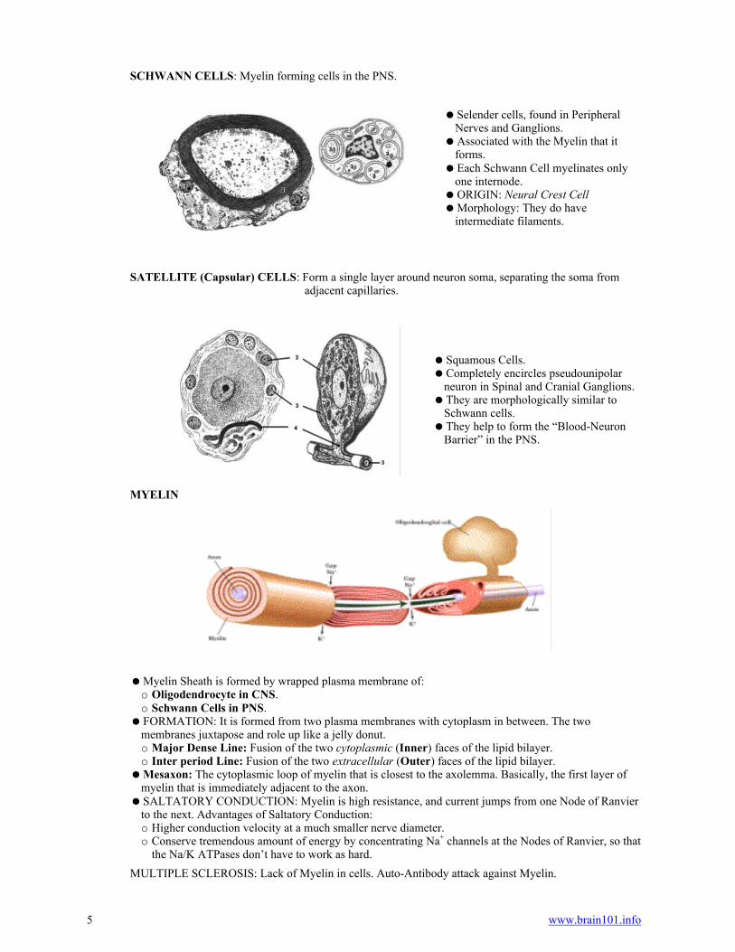
MYELIN
Myelin Sheath is formed by wrapped plasma membrane of:o Oligodendrocyte in CNS.o Schwann Cells in PNS.
FORMATION: It is formed from two plasma membranes with cytoplasm in between. The twomembranes juxtapose and role up like a jelly donut.o Major Dense Line: Fusion of the two cytoplasmic (Inner) faces of the lipid bilayer.o Inter period Line: Fusion of the two extracellular (Outer) faces of the lipid bilayer.
Mesaxon: The cytoplasmic loop of myelin that is closest to the axolemma. Basically, the first layer ofmyelin that is immediately adjacent to the axon.
SALTATORY CONDUCTION: Myelin is high resistance, and current jumps from one Node of Ranvierto the next. Advantages of Saltatory Conduction:o Higher conduction velocity at a much smaller nerve diameter.o Conserve tremendous amount of energy by concentrating Na+ channels at the Nodes of Ranvier, so that
the Na/K ATPases don’t have to work as hard.
MULTIPLE SCLEROSIS: Lack of Myelin in cells. Auto-Antibody attack against Myelin.

6 www.brain101.info
HISTOLOGY OF SELECTIVE CNS PARTS
LAYERS OF THE RETINA: From the first layer that the light contacts, to the last layer that it contacts.
Choroid: The material between the sclera and the beginning of the retina, through which the lighttravels.
Pigmented Epithelium: Pigments in this layer absorbs a lot of the light initially.o It also supplies Vitamin-A to (and exchanges it with) the photoreceptor cells.
Outer Segment: Contains the outer segment of rods and cones. Inner Segment: Containing the inner segment of rods and cones. Outer Nuclear Layer: Contains the nuclei of the rods and cones. Outer Plexiform Layer: Contains:o Horizontal Cellso Bipolar Cellso Processes and synapses from the rods and cones.
Inner Nuclear Layer: Contains the nuclei and soma of the Bipolar, Amacrine, and Horizontal cells. Inner Plexiform Layer: Contains processes and synapses from the Bipolar, Amacrine, and Horizontal
cells. Ganglion Cell Layer: Contains the Ganglion Cells, which ultimately converge on the Optic Nerve. Nerve Fiber Layer: Contains the axons of ganglion cells. Inner Limiting Membrane
CEREBELLUM LAYERS
MOLECULAR LAYER: Outermost layer of grey matter, containing relatively few, unmyelinatedfibers.o Basket Cells: They send out processes in the molecular layer that interconnect Purkinje cells.
PURKINJE CELLS: Middle border layer containing huge neurons that have fine dendrites and axonsextending beyond.o Purkinje Dendrite extends into the molecular layer (outer layer), where they receive efferent signals
from the cerebellar cell bodies.o Purkinje Axons extends into the granular layer (inner layer), where they relay efferent signals
ultimately to the white matter of the cerebellar core. GRANULAR LAYER: The innermost layer, containing numerous cells, and containing axons that
extend into the molecular layer to meet up with the Purkinje dendrites. WHITE MATTER: Beneath the granular layer is the white matter core of the cerebellum, which is the
interface between the cerebellum and the brainstem.o All incoming fibers have multiple connections in white matter and then make their way up to the
cerebellar cortex, via the Purkinje cells.o Outgoing fibers go back to the white matter and down the brainstem, once again via Purkinje cells.
DORSAL ROOT GANGLION: Consists of pseudounipolar cells on the dorsal root (intervertebralforamen) of the spinal column. Both processes of a dorsal root ganglion cells are considered to be axons.Dorsal root ganglia are sensory neurons.
Central Process: Transmits the sensory information into spinal column. Peripheral Process: Receives information from the periphery at the respective dermatomal level.
BLOOD-BRAIN BARRIER
Brain Arteries: They are covered by Astrocyte End-Feet and Pia Mater. Brain Capillaries: The primary barrier is the Capillary Endothelium, which has tight junctions.o Diffusion in and out of capillary is tightly regulated.o Basement Membrane surrounds the outside of the capillaries.o Astrocyte End-Feet are outside the basement membrane.
CEREBROSPINAL FLUID (CSF)
The average production is 500 ml/day. Total amount in average adult is 100 to 150 ml. (only 25 ml. of CSF is available in ventricles). Functions:o Transport of glucose and other products to CNS.o Removes waste products and drugso Supports and cushions the brain against trauma.o Carries hormones from the hypothalamus.

7 www.brain101.info
Contents: It is made primarily in choroid plexus (70%), with some capillary ultrafiltrate (18%), andglucose oxidation products (12%).
Properties:o Clear fluid, isotonic with Serum (290-295 mOsm/L).o PH = 7.33 (Less than blood PH = 7.36-7.40).o Concentration of Glucose and Protein is lower in CSF than in Serum. (Protein increases in CNS
tumors).o Concentration of K+, Ca2+ and HCO3
- ions is lower in CSF than in Serum.o Concentration of Na+, Cl- and Mg2+ ions is higher in CSF than in Serum.
Most of CSF returns to venous system (Superior Sagital Sinus) via Arachnoid Granulation. CHOROID PLEXUS: Contain numerous villi, made of ependymal cuboidal epithelial cells.o Located in each lateral ventricle, the third and forth ventricles.o The Ependymal cells have a basement membrane, and beneath that is the stromal core, in which the
blood vessels are found.o Each choroid plexus is supplied by an artery.
BRAIN MENINGES
Dura Mater: Attached to skull, made of collagen. Arachnoid Mater: Interdigitating fibers make it seem several layers of thick.o The arachnoid mater is avascular.
Subarachnoid Space: Contains the cerebrospinal fluid.o Arachnoid Trabecula forms a web-like structure in this space.
Pia Mater: Adherent to the brain.o The pia mater is highly vascular.o It follows vessels into the brain, forming reflections off of them as they enter brain.o Glial Limitans is just deep to the pia mater, separating brain from vessels. It is made of astrocyte
end-feet.
VENTRICULAR SYSTEM
General Flow of CSF: The two LATERAL VENTRICLES ------> FORAMEN OF MONROE ------>THIRD VENTRICLE ------> CEREBRAL AQUEDUCT ------> FOURTH VENTRICLE
Foramen of Monroe: Connects each Lateral Ventricle to the Third Ventricle. Cerebral Aqueduct: Connects the Third Ventricle to the Fourth Ventricle. Central Canal: The Central Canal of the Spinal Cord is formed as a continuation of the Fourth
Ventricle, as it narrows through the Foramen Magnum. Median Aperture (Foramen of Magendie): Connects the Third Ventricle to the Subarachnoid Space,
medially. Lateral Aperture (Foramen of Luschka): Connects the Third Ventricle to the Subarachnoid Space,
laterally. ARACHNOID GRANULATIONS: This is how CSF leaves the Ventricles, to enter the Superior
Sagital Sinus. HYDROCEPHALUS: Any blockage of the flow of cerebrospinal fluid will result in hydrocephalus.

8 www.brain101.info
LATERAL VENTRICLES: They are the primary makers of CSF, and they have four major parts,corresponding to the cerebral hemispheres.
Anterior Horn: That part in the Frontal Lobe. Body: That part in the Parietal Lobe. Inferior Horn: That part in the Temporal Lobe. Posterior Horn: That part in the Occipital Lobe.
BLOOD SUPPLY TO THE CNS
ANTERIOR CIRCULATION: Basically the Carotid System.
Supplies:o The Eye (via Ophthalmic Arteries) off of Internal Carotid.o The anterior (forebrain) deep structures (via Anterior Cerebral) of each cerebral hemisphere.o The lateral surface of each cerebral hemisphere (via Middle Cerebral)o The medial forebrain, as far back as the Parieto-Occipital Sulcus (via Anterior Cerebral)
FOUR SEGMENTS OF THE INTERNAL CAROTID:o Cervical Segment: From Bifurcation to The Carotid Canalo Petrous Segment: As it goes through the Carotid Canal, in Petrous Temporal bone.o CAROTID SIPHON (S-SHAPED): An S-Shape is made from two segments: BRANCHES = a few small branches to supply the dura mater in the cavernous sinus. BRANCHES = Middle Cerebral Artery and Anterior Cerebral Artery.
BRANCHES OF THE INTERNAL CAROTID
o Ophthalmic Artery: The eye, anastomosis with supraorbital face, Meninges and Falx Cerebri.o Posterior Communicating Artery joins the Posterior Cerebral Arteryo Anterior Choroidal Artery: Supplies the choroid plexus in the anterior horn of the lateral ventricle.o Anterior Cerebral Arteryo Middle Cerebral Artery

9 www.brain101.info
Anterior Cerebral Artery Middle Cerebral ArteryPosterior Cerebral Artery Basilar Artery Anterior Choroidal Artery .
ANTERIOR CEREBRAL ARTERY: Enters cranial cavity at the Longitudinal Fissure.o Anastomoses with other anterior cerebral artery via the Anterior Communicating.o TWO SEGMENTS: Gives off the anteromedial and medial striate arteries through the anterior perforating substance. It divides into branches to supply the medial, frontal cerebral cortex up to the parieto-occipital
fissure.o SUPPLIES the entire medial surface of each cerebral hemisphere, except for the Occipital Pole.
MIDDLE CEREBRAL ARTERY: Enters the Sylvian Fissure and then bifurcates into two mainbranches (Anterior and Posterior)o SUPPLIES
POSTERIOR CIRCULATION: Basically the vertebral system.
Supplies:o The Spinal Cord.o The Brain Steam: Medulla, Pons, most of Mesencephalono All of the Cerebellum
SEGMENTS OF THE VERTEBRAL ARTERIES:o Soft Tissue Segment: Subclavian Arteries ------> C6 Foramen Transversarium, i.e. until the point
when they enter the interior of the vertebral canal.o Intervertebral Segment: Inside the vertebral canal, from C6 ------> Atlas ------> Foramen Magnumo Intracranial Segment: That portion within the dura, distal to the foramen magnum. POSTERIOR INFERIOR CEREBELLAR ARTERY (PICA) supplies the lateral medulla and
part of the cerebellum. Paired arteries.o BASILAR ARTERY: The terminal segment of the Vertebral Arteries, where the two vertebras join
each other. ANTERIOR INFERIOR CEREBELLAR ARTERY (AICA): Sends numerous branches to
caudal Pons and Rostral Medulla. SUPERIOR CEREBELLAR ARTERY (SCA): Superior aspect of Cerebellum, given off before
the Basilar joins the Circle of Willis POSTERIOR CEREBRAL ARTERY (PCA): Given off in the Circle of Willis
Anterior Inferior Cerebellar Artery (AICA):o Supplies the Pontomedullary Junctiono Then ascend to cerebellum to supply its named part.
POSTERIOR CEREBRAL ARTERIES: The terminal branch of the Basilar Artery. These arteriesofficially begin the posterior limb of the Circle of Willis.

10 www.brain101.info
CIRCLE OF WILLIS: THE ANASTOMOTIC ARTERIAL CONNECTIONS SUPPLYING THECRANIAL CAVITY. THE TWO MAIN SUPPLIES TO THE BRAIN ARE THE INTERNALCAROTID AND VERTEBRAL ARTERIES, AND THEY COMMUNICATE THROUGH THE CIRCLEOF WILLIS.
All three of the Cerebral Arteries are given off in the Circle of Willis, while the Cerebellar Arteries aregiven off before the Circle of Willis.o POSTERIOR CEREBRAL ARTERIES: Terminal Branches of the Basilar.o MIDDLE CEREBRAL ARTERIES: Branches off at the point where the Internal Carotids join the
circle.o ANTERIOR CEREBRAL ARTERIES: The junction of the Anterior Communicating and Internal
Carotid Arteries. Posterior Communicating Artery connects the Posterior Cerebral (from Basilar) to the Internal Carotid
Artery. This is the major anastomosis between the Carotid and Vertebral arterial channels. Anterior Communicating Artery: Connects the Anterior Cerebellar Arteries to each other. This is the
major anastomosis of the Right and Left Internal Carotids with each other. UNEVEN DISTRIBUTION: It is not uncommon to find one side of the Circle-of-Willis arteries too
much larger than the other, carrying the majority of blood-flow.