Embed Size (px)
Citation preview

JOURNAL OF VIROLOGY,0022-538X/99/$04.0010
Mar. 1999, p. 2309–2320 Vol. 73, No. 3
Copyright © 1999, American Society for Microbiology. All Rights Reserved.
Identification of Retroviral Late Domains asDeterminants of Particle Size
LAURENCE GARNIER,1 LESLIE J. PARENT,2 BENJAMIN ROVINSKI,3
SHI-XIAN CAO,3 AND JOHN W. WILLS1*
Department of Microbiology and Immunology,1 and Department of Medicine,2 PennsylvaniaState University College of Medicine, Hershey, Pennsylvania 17033, and Department
of Molecular Virology, Pasteur Merieux-Connaught Canada,North York, Ontario M2R 3T4, Canada3
Received 6 July 1998/Accepted 13 November 1998
Retroviral Gag proteins, in the absence of any other viral products, induce budding and release of spherical,virus-like particles from the plasma membrane. Gag-produced particles, like those of authentic retrovirions,are not uniform in diameter but nevertheless fall within a fairly narrow distribution of sizes. For the humanimmunodeficiency virus type 1 (HIV-1) Gag protein, we recently reported that elements important for con-trolling particle size are contained within the C-terminal region of Gag, especially within the p6 sequence (L.Garnier, L. Ratner, B. Rovinski, S.-X. Cao, and J. W. Wills, J. Virol. 72:4667–4677, 1998). Deletions andsubstitutions throughout this sequence result in the release of very large particles. Because the size determi-nant could not be mapped to any one of the previously defined functions within p6, it seemed likely that itsactivity requires the overall proper folding of this region of Gag. This left open the possibility of the sizedeterminant residing in a subdomain of p6, and in this study, we examined whether the late domain (the regionof Gag that is critical for the virus-cell separation step) is involved in controlling particle size. We found thatparticles of normal size are produced when p6 is replaced with the totally unrelated late domain sequencesfrom Rous sarcoma virus (contained in its p2b sequence) or equine infectious anemia virus (contained in p9).In addition, we found that the large particles released in the absence of p6 require the entire CA and adjacentspacer peptide sequences, whereas these internal sequences of HIV-1 Gag are not needed for budding (orproper size) when a late domain is present. Thus, it appears the requirements for budding are very differentin the presence and absence of p6.
The major structural proteins of human immunodeficiencyvirus type 1 (HIV-1) are initially synthesized in the form of apolyprotein precursor, Pr55gag (Fig. 1). The Gag polyproteinsassemble at the plasma membrane in a process that leads to therelease of spherical, enveloped, and immature virions. A num-ber of studies have demonstrated that Pr55gag is the only viralprotein essential for assembly and release of viral particles (10,17, 18, 33).
Very late during budding or immediately after, cleavage ofPr55gag by the virally-encoded protease releases the matureproducts p17 (MA [matrix]), p24 (CA [capsid]), p7 (NC [nu-cleocapsid]), and the C-terminal peptide p6, as well as twosmall peptides, SP2 (spacer peptide 2) and SP1 (12, 16). How-ever, it is the uncleaved Gag protein that directs the assemblyand budding events (for a review, see reference 9). It is nowclear that the MA sequence contains the M (membrane bind-ing) domain, which specifically directs Gag proteins to themembrane. Within the NC sequence, two copies of the I (in-teraction) domain mediate tight interactions between Gagmolecules at the membrane to give particles their proper den-sity. Finally, the separation of the particle from the cell surfaceis mediated by the L (late) domain, which resides within the p6sequence. Although the M, I, and L domains have been shownto be sufficient for the release of virus particles of normaldensity (for a review, see reference 9), they are insufficient forthe production of normal-sized particles.
A recent study of Rous sarcoma virus (RSV [20]) has dem-onstrated that the size determinants map to the segment ofGag consisting of CA plus the spacer peptides located betweenCA and NC. Small deletions throughout CA-SP result in par-ticles that are large and heterogeneous (20). In the case ofHIV-1, the arrangement of size determinants is very different,and although the spacer peptide (SP1) following CA may be animportant size determinant, the CA sequence appears to be farless critical (8, 19). Thus, while some CA mutants have beenreported to have altered sizes (3, 6), many others have beenshown to have normal size, although infectivity is lost (31). Byemploying rate-zonal gradients to systematically study the ef-fect of deletions throughout the Gag protein, we recently re-ported that the HIV-1 CA sequence does not control particlesize during budding. Rather, the C-terminal sequences of Gag,and especially p6, are critical for determining HIV-1 particlediameter (8). Thus, Gag proteins with deletions within MA,CA, or the N-terminal part of NC produce particles of normalsize, while those with defects in the p6 sequence or the C-terminal part of NC release very large particles, even thoughproper buoyant density was retained. The importance of p6 isfurther emphasized by its ability to restore normal size to RSVCA mutants that otherwise produce large and heterogeneousparticles in its absence (8).
How the p6 sequence functions to constrain the size of anemerging particle is unknown. In our previous study, the sizedeterminant could not be mapped to motifs within p6 havingknown functions, including the minimal sequence that definesthe L domain (8); rather, the folded structure of the entire p6seemed to be important. Thus, it was not possible to rule out arole for the L domain in controlling particle size. To further
* Corresponding author. Mailing address: Department of Microbi-ology and Immunology, Pennsylvania State University College of Med-icine, 500 University Dr., P.O. Box 850, Hershey, PA 17033. Phone:(717) 531-3528. Fax: (717) 531-6522. E-mail: [email protected].
2309
on Decem
ber 1, 2018 by guesthttp://jvi.asm
.org/D
ownloaded from

examine this possibility, the RSV p2b and equine infectiousanemia virus (EIAV) p9 sequences and their associated Ldomains were moved to the C terminus of HIV-1 Gag in placeof p6. In both cases, the presence of the heterologous L do-main was sufficient to restore normal particle size as measuredby sedimentation experiments. These findings demonstratethat the L domains from three unrelated Gag proteins canfunction independently as determinants of HIV-1 particle size.Further evidence for this was obtained from experiments withEIAV, which showed that the C-terminal sequences of thislentiviral Gag protein, like those of HIV-1, are critical fordetermining particle size and can be functionally replaced withthe L domains of RSV and HIV. We also uncovered evidencethat the HIV-1 Gag protein utilizes a different mechanism forbudding when p6 is absent, a pathway that requires the pres-ence of an intact CA-SP1 sequence.
MATERIALS AND METHODS
All of the constructs used in this study lack viral protease activity (2, 5, 38). AllDNA manipulations were carried out by using standard methods (34). Recom-binant plasmids were propagated in Escherichia coli DH-5a with YT mediumcontaining ampicillin (25 mg/ml). Each construct was sequenced, and two inde-pendent clones of each mutant were analyzed in transfection experiments tominimize possible unwanted mutations.
Construction of p6 deletion mutants. All gag alleles were expressed by using asimian virus 40 (SV40)-based vector. Constructs pSV.M1.HIV Gag and pSV.M1.HIVDp6 have been previously reported (8).
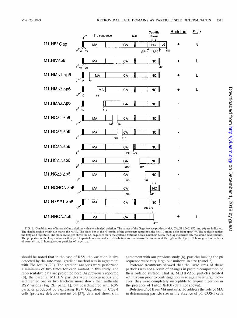
To combine previously described MA deletion mutations with a p6 deletion,pSV.M1.HMD1, pSV.M2.HMD2, and pSV.M1.HMD3 (8) were digested withMluI and SpeI, and each of the resulting small fragments was ligated into theMluI-SpeI sites of pSV.M1.HIVDp6. The recombinants were named pSV.M1.HMD1.Dp6, pSV.M2.HMD2.Dp6, and pSV.M1.HMD3.Dp6, respectively (Fig. 1).
To combine previously described CA, NC, and SP1 deletion mutations with ap6 deletion, pSV.M1.HCD4, pSV.M1.HCD5, pSV.M1.HCD6, pSV.M1.HCD7,pSV.M1.HCD8, pSV.M1.HCNCD, and pSV.M1.HDSP1 (8) were digested withBglII and BssHII and then ligated with an oligonucleotide pair containing a stopcodon inserted in place of the first p6 codon (8). The resulting constructs werenamed pSV.M1.HCD4.Dp6, pSV.M1.HCD5.Dp6, pSV.M1.HCD6.Dp6, pSV.M1.HCD7.Dp6, pSV.M1.HCD8.Dp6, pSV.M1.HCNCD.Dp6, and pSV.M1.HDSP1.Dp6, respectively (Fig. 1).
To combine a previously described NC deletion mutation with a p6 deletion,pSV.M1.HIVDp6 was digested with ApaI and BglII, treated with T4 DNA poly-merase, and religated. One foreign residue (Ser) was introduced at the site of thedeletion. The resulting construct was named pSV.M1.HNCD.Dp6 (Fig. 1).
Construction of HIV-p2 deletion mutants. To insert the RSV p2b sequence(and its associated L domain) in the place of the HIV-p6 sequence, pSV.M1.HIVGag and previously described CA, SP1, and NC deletion mutants (8) weredigested with BglII and SpeI and ligated with the fragment containing the p2bsequence, resulting from the digestion of pSV.RHB.T10C.tp2 (25) with BglII-SpeI. The resulting constructs were named pSV.M1.HG.tp2, pSV.M1.HCD4.tp2,pSV.M1.HCD5.tp2, pSV.M1.HCD6.tp2, pSV.M1.HCD7.tp2, pSV.M1.HCD8.tp2,pSV.M1.HCNCD.tp2, and pSV.M1.HDSP1.tp2 (Fig. 4).
To insert the RSV p2b sequence in the place of the HIV-1 p6 sequence in theNC deletion mutant M1.HNCD (8), pSV.M1.HG.tp2 was digested with ApaI andBglII, treated with T4 DNA polymerase, and religated. The resulting constructwas named pSV.M1.HNCD.tp2 (Fig. 4).
Chimeric HIV-EIAV gag alleles. To place the EIAV p9 sequence (and itsassociated L domain) in the place of HIV-1 p6, pSV.M1.HIV Gag,pSV.M1.HCD4, pSV.M1.HCD7, pSV.M1.HCNCD, and pSV.M1.HCDSP1were digested with ApaI-EcoRV and ligated with the fragment containing the p9sequence, resulting from the digestion of pSV.RHE.p9 (25) with ApaI andEcoRV. The recombinants were named pSV.M1.HCD4.tp9, pSV.M1.HCD7.tp9,pSV.M1.HCNCD.tp9, and pSV.M1.HDSP1.tp9 (Fig. 4).
Four other HIV-EIAV chimeras used in this study (Fig. 6, pSV.EG.p6 [28],pSV.RHE.T10C2 [25], pSV.REI.T10C [25], and pSV.RHE.p9.T10C [25]) aswell as two EIAV constructs (pSV.EG [28] and pSV.EG.p92 [28]) have beenpreviously reported.
Construction of M1.HG.CY. PCR amplification of the iso-1-cytochrome csequence of pSV.MYCY (36) was performed to create a BglII site immediatelyupstream of the fourth codon and an BssHII site immediately downstream of thestop codon by using 59-ATAGGAGGGGAGATCTGGAAGGCCGTTTCTGCTAAGA-39 as the upstream primer and 59-ATCCTACAGCGCGCTTACTCACAGGCTTTTT-39 as the downstream primer. The amplified fragment was di-gested with BglII and BssHII (restriction endonuclease recognition sites areunderlined in the primer sequences) and ligated into plasmid pSV.M1.HIV Gag(8), which had been digested with the same enzymes. The recombinant wasnamed pSV.M1.HG.CY.
Transfection of cells and metabolic labeling. COS-1 cells were grown in 35-mm- or 60-mm-diameter dishes in Dulbecco’s modified Eagle medium (DMEM;GIBCO BRL) supplemented with 3% fetal bovine serum and 7% bovine calfserum (HyClone, Inc.). These cells were transfected with XbaI-digested andligated plasmids by the DEAE-dextran-chloroquine method as described previ-ously (38). At 48 h after transfection, the COS-1 cells were labeled with [35S]me-thionine (10 mCi or 50 mCi; .1,000 Ci/mmol). After 2.5 h of labeling, the cellsand growth medium from each labeled culture were mixed with lysis buffercontaining protease inhibitors, and the Gag proteins were immunoprecipitatedfor 1 h at 4°C with a human HIV immunoglobulin (27), electrophoresed insodium dodecyl sulfate-12% polyacrylamide gels, and visualized by fluorography.The autoradiograms were then quantitated by laser densitometry.
Density gradient analysis. To ensure that the Gag deletion mutants used inthis study produced dense particles, several of the key constructs (M1.HG.tp2,M1.HG.tp9, M1.HCD6.tp2, M1.HNCD.tp2, and M1.HCD7.tp9) were tested bycentrifugation of the samples through density gradients as previously described(8). All of them released dense particles (data not shown). Many of the otherconstructs have been analyzed previously and found to be released into denseparticles, including M1.HIV Gag (8), M1.HIV.Dp6 (8), EG.p92 (28), EG (28),EG.p6 (28), REI.T10C (25), and RHE.p9.T10C (25).
Rate-zonal gradient analysis. Two days posttransfection, COS-1 cells werelabeled in methionine-free, serum-free Dulbecco’s medium supplemented with[35S]methionine (50 mCi; .1,000 Ci/mmol) for 5 h in 0.5 ml. After the labelingperiod, the medium was immediately centrifuged at a low speed to remove cel-lular debris. Radiolabeled infectious RSV was added to each sample to providean internal size marker. This virus, obtained from RSV-infected turkey embryofibroblasts (TEF) which were propagated in supplemented F10 medium as de-scribed previously (15), was labeled with [35S]methionine as described above.Similarly, EIAV-infected equine dermal cells (American Type Culture Collec-tion catalog no. CCL 57, kindly provided by Ron Montelaro and Bridget Puffer,University of Pittsburgh, Pittsburgh, Pa.) propagated in DMEM supplementedwith 10% fetal bovine serum, were labeled as described above. Each mixture waslayered onto 10 to 30% sucrose and centrifuged at 83,500 3 g for 0.5 h in aBeckman SW41Ti. From each gradient, 0.6-ml fractions were collected, and Gagproteins were immunoprecipitated with a mixture of anti-RSV and anti-HIVantisera (27) and subjected to electrophoresis. For EIAV constructs, we used amixture of anti-RSV and anti-EIAV antisera (a kind gift from Ron Montelaroand Bridget Puffer, University of Pittsburgh). All gradients were repeated at leastonce to confirm the results.
RESULTS
We recently reported that important size-controlling ele-ments within the HIV-1 Gag protein are contained within itsC-terminal sequences and especially within the entire p6 se-quence (8). Moreover, we found no evidence that the HIV-1CA sequence plays any role in determining particle size. Incontrast, previous studies of RSV showed that the CA-SPsequence of its Gag protein is critical for normal size, and smalldeletions throughout it result in the release of large, hetero-geneously-sized particles (20). These results suggested thateither lentiviruses (HIV-1) and oncoviruses (RSV) have dif-ferent size controlling elements or that the impact of HIV-1CA deletions on particle size cannot be detected when the p6sequence is present within Pr55gag. In other words, the CAsequence of HIV might be important for the mechanism oflarge particle production that occurs when p6 is absent.
To further examine the role of the CA sequence as well asMA, SP1, and NC domains in determining particle size in theabsence of p6, we removed the p6 sequence from a collectionof previously described HIV-1 Gag deletion mutants (8). Eachof the HIV-1 constructs has the first 10 residues of the Srcprotein in place of the 32-residue M domain of HIV-1 (Fig. 1).As previously reported (8), this modification has no effect onparticle size but leads to enhanced production of Gag proteinsand rapid release of virus-like particles into the growth me-dium after transfection (M1.HIV Gag, Fig. 2A, lanes 3 and18). To assess particle size, medium samples from transfectedCOS-1 cells were analyzed in 10 to 30% rate-zonal sucrosegradients. We chose this method of measuring particle sizebecause this technique, unlike electron microscopy (EM) anal-ysis, allows the entire population of particles to be detectedregardless of their morphological appearance. That is, verylarge particles might go unrecognized by EM methods. It
2310 GARNIER ET AL. J. VIROL.
on Decem
ber 1, 2018 by guesthttp://jvi.asm
.org/D
ownloaded from
should be noted that in the case of RSV, the variation in sizedetected by the rate-zonal gradient method was in agreementwith EM results (20). The gradient analyses were performeda minimum of two times for each mutant in this study, andrepresentative data are presented here. As previously reported(8), the parental M1.HIV particles were homogeneous andsedimented one or two fractions more slowly than authenticRSV virions (Fig. 2B, panel 1), but cosedimented with RSVparticles produced by expressing RSV Gag alone in COS-1cells (protease deletion mutant 3h [37]; data not shown). In
agreement with our previous study (8), particles lacking the p6sequence were very large but uniform in size (panel 2).
Protease treatments showed that the large sizes of theseparticles was not a result of changes in protein composition ortheir outside surface. That is, M1.HIVDp6 particles treatedwith trypsin prior to centrifugation were again very large; how-ever, they were completely susceptible to trypsin digestion inthe presence of Triton X-100 (data not shown).
Deletion of p6 from MA mutants. To address the role of MAin determining particle size in the absence of p6, COS-1 cells
FIG. 1. Combinations of internal Gag deletions with a terminal p6 deletion. The names of the Gag cleavage products (MA, CA, SP1, NC, SP2, and p6) are indicated.The shaded region within CA marks the MHR. The black box at the N termini of the constructs represents the first 10 amino acids from pp60v-src. The squiggle depictsthe fatty acid myristate. The black rectangles above the NC sequence mark the cysteine-histidine boxes. Numbers below the Gag molecules refer to amino acid residues.The properties of the Gag mutants with regard to particle release and size distribution are summarized in columns at the right of the figure: N, homogeneous particlesof normal size; L, homogeneous particles of large size.
VOL. 73, 1999 RETROVIRAL LATE DOMAINS AS PARTICLE SIZE DETERMINANTS 2311
on Decem
ber 1, 2018 by guesthttp://jvi.asm
.org/D
ownloaded from

were transfected with M1.HMD1.Dp6, M1.HMD2.Dp6, andM1.HMD3.Dp6 (Fig. 1). While intracellular synthesis of Gagproteins was similar to wild-type levels for M1.HMD1.Dp6and M1.HMD2.Dp6, Gag expression was severely reduced inM1.HMD3.Dp6 (Fig. 2A, compare lanes 3 and 5 to 7). Analysisof the media fractions revealed that M1.HMD1.Dp6 andM1.HMD2.Dp6 were present but greatly reduced for particlerelease (reduction about fivefold, Fig. 2A, compare lanes 18and 20 to 21), while M1.HMD3.Dp6 was not detectable,which is due either to a lower efficiency of budding or to alower level of expression (Fig. 2A, compare lanes 18 and22). For comparison, an assembly-incompetent deletion mu-tant (RHB.T10C [25]), which lacks L domains needed forbudding, is shown in lane 17. Gradient analyses revealed thatboth M1.HMD.Dp6 and M1.HMD2.Dp6 still produced largeparticles like the Dp6 parent, although those of M1.HMD1.Dp6were more heterogeneous in size (Fig. 2B, panels 3 and 4).Although the efficiency of budding is reduced for the p6 dele-tion mutants when combined with MA deletions, these dataindicate that MA does not influence particle size.
Deletion of p6 from CA, SP1 mutants. To address the role ofCA and SP1 in large-particle production, COS cells were trans-fected with a collection of HIV CA and SP1 deletion mutants,all of which lacked the p6 sequence (Fig. 1). While intra-cellular synthesis of Gag proteins was similar to wild-typelevels in M1.HCD4.Dp6, M1.HCD5.Dp6, M1.HCD6.Dp6, andM1.HDSP1.Dp6, the level of Gag expression was severely re-duced in M1.HCD7.Dp6 and M1.HCD8.Dp6 (Fig. 2A, comparelanes 3 and 8 to 12 and 14). Unexpectedly, all of the CA andSP1 deletion mutants were defective for particle release (Fig.2A, compare lanes 18 and 22 to 27 and 29). This was surprisingbecause the very same CA and SP1 deletions in the presence ofp6 do not greatly affect particle release or size (8). Thus, itappears that the HIV-1 CA and SP1 sequences are requiredfor and may drive the production of large particles in theabsence of p6 (see Discussion).
Deletion of p6 from NC mutants. To determine the contri-bution of NC to particle size, we removed p6 from two previ-ously described NC deletion mutants (8). M1.HCNCD.Dp6contains a deletion which removes a portion of the CA up-stream of the major homology region (MHR) along with SP1and the first six residues from NC, while M1.HNCD.Dp6 lacksthe second half of the NC, SP2, and p6 but retains I domainactivity (Fig. 1). Both proteins were produced at wild-typelevels (Fig. 2A, compare lanes 3 and 13 and 15).M1.HNCD.Dp6 was released into the medium with approx-imately 50% of wild-type efficiency (Fig. 2A, compare lanes18 and 30), whereas M1.HCNCD.Dp6 was completely defec-tive for particle release (Fig. 2A, compare lanes 18 and 28),again suggesting the importance of CA and SP1 in the pro-duction of large particles in the absence of p6. When ana-lyzed for size, M1.HNCD.Dp6 was found to be contained inrelatively homogeneous but very large particles (panel 5). Thissedimentation profile was identical to that for M1.HNCD (8),indicating once again that an important size determinant islocated within the C-terminal portion of NC.
Substitutions of p6 with RSV p2b and EIAV p9. Because wewere unable to analyze the impact that CA deletions mighthave on particle size in the absence of p6, we next attempted torestore budding to the HIV CA deletion mutants by including
heterologous L domains at their C termini. We decided to usethe RSV p2b and EIAV p9 sequences and their associated Ldomains because it has been previously shown that these se-quences can provide L domain activity in the context of het-erologous Gag proteins (25, 28). We hoped that they wouldrestore budding without themselves influencing particle size,since previous studies have not implicated L domains as sizedeterminants (8, 20). As a control, we first examined constructswith complete CA sequences. We found that the HIV-p9 chi-meric particles (produced by M1.HG.tp9, Fig. 3A) were re-leased into the medium with the same efficiency as M1.HIVGag (Fig. 3B, compare lanes 3 and 5), whereas the level of re-lease of HIV-p2 (produced by M1.HG.tp2, Fig. 3A) was re-duced about fourfold compared to wild-type level (Fig. 3B,compare lanes 3 and 4). This decrease is likely due to subop-timal positioning of the RSV L domain at the end of HIV Gag.Consistent with this, we previously noted a 50% decrease inparticle release for a RSV-HIV chimera containing the p2bsequence at this position (RHB.T10C.tp2 [25]).
As expected, subsequent sedimentation analyses showedthat both L domain chimeras produced particles of normaldensity when analyzed in 10 to 50% isopycnic sucrose gradients(data not shown). However, when particle size was analyzed in10 to 30% rate zonal gradients, particles for both chimeraswere not large but appeared essentially identical in size distri-bution to the control particles (Fig. 3C). This provided our firstindication that L domains may be important for size. To ad-dress the possibility that any random sequence placed at the Cterminus of HIV Gag would be sufficient to obtain normalparticle size, we analyzed chimera M1.HG.CY, which containsthe iso-1-cytochrome c sequence from Saccharomyces cerevi-siae in place of p6 (Fig. 3A). Although this sequence does notinterfere with budding when attached to the C terminus of theRSV Gag protein (36), we found that the HIV-cytochromechimera was severely defective for particle release, although itwas synthesized at levels similar to wild type (Fig. 3B, lanes 6).For comparison, an assembly-incompetent deletion mutant(T10C [37]), which lacks late domains needed for budding, isshown in lanes 2. The only known feature in common to p6, p9,and p2b is L domain activity, and thus, we conclude that the Ldomains play an active role in constraining particle size, atleast in the context of HIV-1 Gag.
Having found that heterologous L domains do influenceparticle size, we could not predict what would happen whensegments of CA or other regions of Gag were removed fromthe chimeras. For example, would a CA deletion block particlerelease for the p2b or the p9 chimera? If not, would particlesof normal or large size be released? To answer these questions,a variety of gag deletions were inserted into the HIV-p2 andHIV-p9 chimeras (Fig. 4). As before, budding efficiencies ofthe p2 chimeras were lower than those observed for the HIV-p9 constructs but sufficient for size analyses (data not shown).When sedimented through sucrose, each of the CA, CA-NC,and SP1 mutants produced relatively uniform populations ofparticles that were not large but were somewhat smaller thanthe internal RSV control (curves with triangular symbols inFig. 5A to F and H to L). For each mutant, the shift of the peakto a position four fractions or less above the control was quitereproducible and has been also seen in the case of M1.HG.tp9(Fig. 3C). The reason for this shift toward smaller particles
FIG. 2. Properties of p6-deleted Gag polyproteins. (A) COS-1 cells were transfected with the indicated DNAs and labeled as described in Materials and Methods.Molecular mass standards (in kilodaltons) are indicated to the left. (B) Distribution of particle size in rate-zonal gradients. COS-1 cells were transfected with theindicated DNAs, and after 48 h were labeled with [35S]methionine for 5 h. After the labeling period, particle sizes were analyzed as described in Materials and Methods.Radiolabeled RSV from infected TEF cells was added to the gradients to provide an internal control. Arrows indicate the direction of sedimentation.
VOL. 73, 1999 RETROVIRAL LATE DOMAINS AS PARTICLE SIZE DETERMINANTS 2313
on Decem
ber 1, 2018 by guesthttp://jvi.asm
.org/D
ownloaded from

FIG. 3. Replacement of p6 with heterologous L domains. (A) M1.HG.tp2 contains the L domain of RSV Gag, while M1.HG.tp9 contains the L domain of EIAV.M1.HG.CY contains the yeast iso-1-cytochrome c sequence, which does not have an L domain. The names of the Gag cleavage products (MA, CA, NC, p6, p2, andp9) are indicated. The black box at the N termini of the constructs represent the first 10 amino acids from pp60v-src. The squiggle represents the fatty acid myristate.(B) COS-1 cells were transfected with the indicated DNAs and labeled as described in Materials and Methods. Molecular mass standards (in kilodaltons) are indicatedto the left. (C) Distribution of particle size in rate-zonal gradients. COS-1 cells were transfected with the indicated DNAs and labeled with [35S]methionine for 5 h.After the labeling period, particle sizes were analyzed as described in Materials and Methods. HIV-p2 and HIV-p9 chimeras produced normal-sized particles. Arrowsindicate the direction of sedimentation.
2314
on Decem
ber 1, 2018 by guesthttp://jvi.asm
.org/D
ownloaded from

remains to be determined but may be due to the absence ofviral components other than Gag. Only M1.HNCD.tp2, whichlacks the second half of the NC sequence and the first fourresidues of SP2 (Fig. 4), produced large particles (panel G). Asimilar construct having p9 in the place of p2 was not tested,but when p6 is present on this mutant, large particles are alsoproduced (8). Thus, it appears that the CA sequence, theN-terminal part of NC, and SP1 are not important for HIV-1particle size when an L domain is present.
Analysis of size determinants of EIAV Gag. In view of thefundamental differences reported for the size determinants ofRSV (20) and HIV-1 (8), we decided to examine the impor-tance of the C terminus in another lentiviral Gag protein. Inour initial experiments, particles produced by expressing the
wild-type EIAV Gag protein in COS cells were subjected torate zonal analysis (Fig. 6, construct EG). Unexpectedly, theseparticles were relatively heterogeneous, although they over-lapped the normally-sized RSV particles (Fig. 7A). In contrast,infectious EIAV particles produced from equine dermal cellswere homogeneous and beautifully sedimented one to twofractions more slowly than authentic RSV virions (panel B).These results raise the possibility that there is a cell-specificfactor that contributes to the regulation of EIAV size or thatan EIAV component other than Gag influences particle size.Further experiments will be required to distinguish betweenthese two possibilities.
When the p9 sequence and its associated L domain weredeleted from EIAV Gag (EG.p92), large particles were re-
FIG. 4. Deletion of internal Gag sequences from the L domain chimeras. The names of the Gag cleavage products (MA, CA, NC, p6, p2, and p9) are indicated.The black box at the N termini of the constructs represents the first 10 amino acids from pp60v-src, and the squiggle represents the fatty acid myristate. The blackrectangles above the NC sequence mark the cysteine-histidine boxes. Numbers refer to amino acid residues. The column at the right of the figure summarizes the sizedistribution of the mutants: Sm, particles that are uniform in size but smaller in diameter; L, homogeneous particles of large size.
VOL. 73, 1999 RETROVIRAL LATE DOMAINS AS PARTICLE SIZE DETERMINANTS 2315
on Decem
ber 1, 2018 by guesthttp://jvi.asm
.org/D
ownloaded from

leased (panel C) in a manner similar to what we have observedwith HIV p6 deletion mutants. When the HIV p6 sequencewas inserted in place of the p9 sequence (EG.p6), the releasedparticles were once again rather uniform in size but slightlysmaller than RSV particles (panel D). This provides furtherevidence that the p6 sequence has an important role in con-straining particle size and can exert this function in a heterol-ogous system.
To examine whether p9 might be able to control the size ofan oncoviral Gag protein in a manner similar to that reportedfor p6 (8), RSV-EIAV chimeric Gag proteins were analyzed. ARSV capsid deletion mutant which produces heterogeneous-ly-sized particles (Myr1.R-3J [39]) is shown for comparisonin Fig. 7E. Placement of C-terminal sequences from EIAVGag onto the C terminus of such an RSV capsid mutant wassuffiient to restore the production of homogeneously-sizedparticles (REI.T10C, panel G), just as the C-terminal se-quences of HIV did (RHE.T10C, panel F). This result sug-gests that size determinants of EIAV Gag are indeed con-tained within its C-terminal sequences. The importance ofthe p9 sequence in determining particle size is emphasizedby chimera RHE.p9.T10C (Fig. 6), a RHE.T10C constructthat contains the p9 sequence in place of p6. As predicted,RHE.p9.T10C particles were found to be homogeneous (panelH). Based on our limited studies of the EIAV Gag protein, itssize determinants appear to be arranged similarly to those ofHIV.
DISCUSSION
In this report, we have demonstrated that three very differ-ent retroviral L domains act as major determinants of particlesize, at least in the context of HIV Gag. The substitution of theHIV p6 sequence with the RSV p2b or EIAV p9 sequencesand their associated L domains was sufficient to restore normal
sedimentation properties to HIV-1 particles. We chose thesucrose gradient approach rather than EM methods to analyzeparticle size. The advantages of gradient sedimentation includethe display of the entire population of particles and the stan-dardization provided by internal markers. However, the prin-cipal disadvantage of this technique is that variations in sedi-mentation rate do not allow easy calculation of the actual sizeof the particles. Therefore, it will be interesting to analyze theparticles produced by the different deletion mutants by EM inan attempt to determine their exact size and morphology.However, our transient expression systems make EM analysesdifficult because most of the cells in the transfected cultures donot express Gag.
Our data suggest that the HIV-1 Gag protein can participatein two very different mechanisms of budding. One requires theL domain but not the capsid and results in the production ofnormally-sized particles. The second mechanism is indepen-dent of the L domain and seems to be driven by CA-SP1 butcan produce only very large particles.
How do retroviral L domains influence particle size? Al-though we do not yet understand how L domains orchestratethe release of normal-sized particles, we favor the idea thatthey recruit host proteins to the site of budding for mediatingthe separation of the virus from the cell. The L domain mightbe directly or indirectly recognized near or within the neck ofthe stalk by cellular proteins, thereby creating the molecularmachinery needed for the release of a normal-sized particlefrom the plasma membrane. This model is in agreement withthe finding that the Y-X-X-L sequence in the L domain ofEIAV interacts in vitro and in vivo with the cellular AP-50medium chain subunit of the plasma membrane AP-2 complex(29), which in uninfected cells mediates endocytosis (22). Inthe case of RSV, the P-P-P-P-Y motif in its L domain has beenshown to bind to the WW domain of Yes-associated protein(Yap) in vitro (7). Although putative cellular proteins involved
FIG. 5. Heterologous L domains restore particle release and normal size to HIV Gag mutants. COS-1 cells were transfected with the indicated DNAs and labeled with[35S]methionine for 5 h. After the labeling period, particle sizes were analyzed as described in Materials and Methods. Arrows indicate the direction of sedimentation.
FIG. 6. Wild-type and chimeric forms of the EIAV Gag protein. The Gag protein of EIAV is illustrated at the top with the names of its cleavage products (MA,CA, SP, NC, and p9). Numbers below the Gag molecules refer to amino acid residues. Foreign sequences from RSV, HIV, and Src are indicated.
VOL. 73, 1999 RETROVIRAL LATE DOMAINS AS PARTICLE SIZE DETERMINANTS 2317
on Decem
ber 1, 2018 by guesthttp://jvi.asm
.org/D
ownloaded from

FIG. 7. Distribution of wild-type and recombinant EIAV Gag particles in rate-zonal gradients. COS-1 cells were transfected with the indicated deletion mutantsDNAs and labeled with [35S]methionine for 5 h. After the labeling period, particle sizes were analyzed as described in Materials and Methods. Arrows indicate thedirection of sedimentation.
2318
on Decem
ber 1, 2018 by guesthttp://jvi.asm
.org/D
ownloaded from

in budding have not yet been identified for RSV, several stud-ies suggest that cytoskeletal proteins (actin and myosin) mightparticipate in HIV-1 particle release (23, 26, 32, 35). More-over, a recent study has shown that HIV virions incorporateubiquitin and that a small amount of p6gag is covalently at-tached to single ubiquitin molecules inside HIV-1 virions (24).Free ubiquitin has also been detected in murine leukemiavirus, simian immunodeficiency virus, and RSV virions (24,30). These observations, coupled with the fact that severalcytoskeletal proteins as well as members of the microtubulenetwork are conjugated to single ubiquitin molecules (1, 4, 21),suggest that the presence of ubiquitin into released HIV-1 viri-ons might be the result of an interaction between viral struc-tural proteins and a monoubiquitinated cytoskeletal proteinduring assembly and/or budding. Interestingly, monoubiquiti-nation is believed to be a signal for plasma membrane receptorinternalization (13). Therefore, the identification of cellularproteins that interact specifically with different L domains maylead to a better understanding of the mechanism by whichthese domains define particle size.
What happens in the absence of L domains? In the case ofRSV and EIAV, when L is nonfunctional, virus particles ac-cumulate at the cell surface but fail to be released (25, 28, 40).With regard to HIV-1, multiple lines of evidence prove that p6contains a late budding function (11, 25), although this activityappears to be influenced by PR (14). More recently, it has beenfound that p6-deleted Gag particles are extremely large in size(8), suggesting that a mechanism of release independent of theL domain can take place. When p6 and its associated L domain(and any cellular proteins normally involved in virus-cell sep-aration) are absent, the particles grow very large, perhaps as aresult of many nascent particles coalescing. Unexpectedly, ourresults indicate that the CA-SP sequence is absolutely requiredfor this and may drive the release of these large particles. Thatis, all of the CA and SP1 deletion mutants were defective forparticle release in the absence of p6. Perhaps the rigid struc-ture created by the interactions among the intact CA-SP se-quences stabilizes the nascent particle long enough so that aless efficient or alternative mechanism of membrane separa-tion can occur (one not dependent on the L domain of p6).Although in our study we were unable to map an L domain-likesequence in the capsid, it may be that deletions throughout CAalter its overall conformation and hence prevent such a se-quence from being properly presented. Further experimentsare necessary to test these hypotheses.
Lentiviruses and oncoviruses have different size-controllingelements. Our results show that the size determinants of on-coviruses (RSV) and lentiviruses (HIV-1 and EIAV) are lo-cated at very different positions within Gag and raise the pos-sibility that they work through different mechanisms. Dataobtained with the EIAV chimeras indicate that the size-con-trolling elements, like those of HIV-1, are contained within theC-terminal sequences of Gag, and especially within the p9sequence. Unexpectedly, we also found that EIAV Gag parti-cles produced from COS cells were relatively heterogeneouscompared to infectious EIAV particles produced from equinecells. This suggests the possibility that EIAV Gag is betteradapted to host proteins present in equine cells than simiancells.
In conclusion, we have identified a novel function of theretroviral L domain. While further experiments are necessaryto understand the mechanisms used by these domains to con-strain the size of an emerging particle, these results indicatethat this function of L domain is conserved among retroviruses.
ACKNOWLEDGMENTS
We thank Ronald Montelaro and Bridget Puffer for providing EIAVantibodies and EIAV infected equine dermal cells. The HIV immu-noglobulin (from A. Prince) was obtained through the AIDS Researchand Reference Reagent Program, Division of AIDS, NIAID, NIH.
This work was supported by grants from the National Institutes ofHealth awarded to J.W.W. (CA47482) and L.J.P. (AI01148) and fromthe American Cancer Society awarded to J.W.W. (FRA-427). Supportfor L.G. was provided by Pasteur Merieux-Connaught Canada.
REFERENCES
1. Ball, E., C. C. Karlik, C. J. Beall, D. L. Saville, J. C. Sparrow, B. Bullard, andE. A. Fyrberg. 1987. Arthrin, a myofibrillar protein of insect flight muscle, isan actin-ubiquitin conjugate. Cell 51:221–228.
2. Bennett, R. P., T. D. Nelle, and J. W. Wills. 1993. Functional chimeras of theRous sarcoma virus and human immunodeficiency virus Gag proteins. J. Vi-rol. 67:6487–6498.
3. Chazal, N., C. Carriere, B. Gay, and P. Boulanger. 1994. Phenotypic char-acterization of insertion mutants of the human immunodeficiency virus type1 Gag precursor expressed in recombinant baculovirus-infected cells. J. Vi-rol. 68:111–122.
4. Corsi, D., L. Galluzzi, R. R. Crinelli, and M. Magnani. 1995. Ubiquitin isconjugated to the cytoskeletal protein a-spectrin in mature erythrocytes.J. Biol. Chem. 270:8928–8935.
5. Craven, R. C., R. P. Bennett, and J. W. Wills. 1991. Role of the avianretroviral protease in the activation of reverse transcriptase during virionassembly. J. Virol. 65:6205–6217.
6. Dorfman, T., F. Mammano, W. A. Haseltine, and H. G. Gottlinger. 1994.Role of the matrix protein in the virion association of the human immuno-deficiency virus type I envelope glycoprotein. J. Virol. 68:1689–1696.
7. Garnier, L., J. W. Wills, M. F. Verderame, and M. Sudol. 1996. WW domainsand retrovirus budding. Nature (London) 381:744–745.
8. Garnier, L., L. Ratner, B. Rovinski, S.-X. Cao, and J. W. Wills. 1998. Particlesize determinants in the human immunodeficiency virus type 1 Gag protein.J. Virol. 72:4667–4677.
9. Garnier, L., J. B. Bowzard, and J. W. Wills. 1998. Recent advances andremaining problems in HIV assembly. AIDS 12:S5–S16.
10. Gheysen, D., R. Yancey, E. Petrovskis, J. Timmins, and L. Post. 1989.Assembly and release of HIV-1 precursor Pr55gag virus-like particles fromrecombinant baculovirus-infected insect cells. Cell 59:103–112.
11. Gottlinger, H. G., T. Dorfman, J. G. Sodroski, and W. A. Haseltine. 1991.Effect of mutations affecting the p6 Gag protein on human immunodefi-ciency virus particle. Proc. Natl. Acad. Sci. USA 88:3195–3199.
12. Henderson, L. E., M. A. Bowers, R. C. Sowder II, S. A. Serabyn, D. G.Johnson, J. W. Bess, L. O. Arthur, D. K. Bryant, and C. Fenselau. 1992. Gagproteins of the highly replicative MN strain of human immunodeficiencyvirus type 1: posttranslational modifications, proteolytic processing, andcomplete amino acid sequences. J. Virol. 66:1856–1865.
13. Hinke, L., and H. Riezman. 1996. Ubiquitination of a yeast plasma mem-brane receptor signals its ligand-stimulated endocytosis. Cell 84:277–287.
14. Huang, M., J. M. Orenstein, M. A. Martin, and E. O. Freed. 1995. p6gag isrequired for particle production from full-length human immunodeficiencyvirus type 1 molecular clones expressing protease. J. Virol. 69:6810–6818.
15. Hunter, E. 1994. Biological techniques for avian sarcoma viruses. MethodsEnzymol. 58:379–392.
16. Kaplan, A. H., M. Manchester, and R. Swanstrom. 1994. The activity of theprotease of human immunodeficiency virus type 1 is initiated at the mem-brane of infected cells before the release of viral proteins and is required forrelease to occur with maximum efficiency. J. Virol. 68:6782–6786.
17. Karacostas, V., K. Nagashima, M. A. Gonda, and B. Moss. 1989. Humanimmunodeficiency virus-like particles produced by a vaccinia virus expres-sion vector. Proc. Natl. Acad. Sci. USA 86:8964–8967.
18. Krausslich, H. G., C. Ochsenbauer, A. M. Traenckner, K. Mergener, M.Facke, H. R. Gelderblom, and V. Bosch. 1993. Analysis of protein expressionand virus-like particle formation in mammalian cell lines stably expressingHIV-1 gag and env gene products with or without active HIV proteinase.Virology 192:605–617.
19. Krausslich, H. G., M. Facke, A.-M. Heuser, J. Konvalinka, and H. Zentgraf.1995. The spacer peptide between human immunodeficiency capsid andnucleocapsid proteins is essential for ordered assembly and viral infectivity.J. Virol. 69:3407–3419.
20. Krishna, N. K., S. Campbell, V. M. Vogt, and J. W. Wills. 1998. Geneticdeterminants of Rous Sarcoma virus particle size. J. Virol. 72:564–577.
21. Murti, K. G., H. T. Smith, and V. A. Fried. 1988. Ubiquitin is a componentof the microtubule network. Proc. Natl. Acad. Sci. USA 85:3019–3023.
22. Ohno, H., J. Stewart, M. C. Fournier, H. Bosshart, I. Rhee, S. Miyatake, T.Saito, A. Gallusser, T. Kirchhausen, and J. S. Bonifacino. 1995. Interactionof tyrosine-based sorting signals with clathrin-associated proteins. Science269:1872–1875.
23. Ott, D. E., L. V. Coren, B. P. Kane, L. K. Busch, D. G. Johnson, R. C. Sowder
VOL. 73, 1999 RETROVIRAL LATE DOMAINS AS PARTICLE SIZE DETERMINANTS 2319
on Decem
ber 1, 2018 by guesthttp://jvi.asm
.org/D
ownloaded from

II, E. N. Chertova, L. O. Arthur, and L. E. Henderson. 1996. Cytoskeletalproteins inside human immunodeficiency virus type 1 virions. J. Virol. 70:7734–7743.
24. Ott, D. E., L. V. Coren, T. D. Copeland, B. P. Kane, D. G. Johnson, R. C.Sowder II, Y. Yoshinaka, S. Oroszlan, L. O. Arthur, and L. E. Henderson.1998. Ubiquitin is covalently attached to the p6Gag proteins of human im-munodeficiency virus type 1 and simian immunodeficiency virus and to thep12Gag protein of moloney murine leukemia virus. J. Virol. 72:2962–2968.
25. Parent, L. J., R. P. Bennett, R. C. Craven, T. D. Nelle, N. K. Krishna, J. B.Bowzard, C. B. Wilson, B. A. Puffer, R. C. Montelaro, and J. W. Wills. 1995.Positionally independent and exchangeable late budding functions of theRous sarcoma virus and human immunodeficiency virus Gag proteins. J. Vi-rol. 69:5455–5460.
26. Pearce-Pratt, R., D. Malamud, and D. M. Phillips. 1994. Role of the cy-toskeleton in cell-to-cell transmission of human immunodeficiency virus.J. Virol. 68:2898–2905.
27. Prince, A. M., B. Horowitz, L. Baker, R. W. Shulman, H. Ralph, J. Valinsky,A. Cudell, B. Brotman, W. Boehle, F. Rey, L. Barbosa, M. Piet, H. Reesink,N. Lelie, M. Tersmette, F. Miedema, G. Nemo, C. L. Nastala, J. S. Allan,D. R. Lee, and J. W. Eichberg. 1988. Failure of a human immunodeficiencyvirus (HIV) immune globulin to protect chimpanzees against experimentalchallenge with HIV. Proc. Natl. Acad. Sci. USA 85:6944–6948.
28. Puffer, B. A., L. J. Parent, J. W. Wills, and R. C. Montelaro. 1997. Equineinfectious anemia virus utilizes a YLLL motif within the late assemblydomain of the Gag p9 protein. J. Virol. 71:6541–6546.
29. Puffer, B. A., S. C. Watkins, and R. C. Montelaro. 1998. Equine infectiousanemia virus gag polyprotein late domain specifically recruits cellular AP-2adapter protein complexes during virion assembly. J. Virol. 72:10218–10221.
30. Putterman, D., R. B. Pepinsky, and V. M. Vogt. 1990. Ubiquitin in avianleukosis virus particles. Virology 176:633–637.
31. Reicin, A. S., A. Ohagen, L. Yin, S. Hoglund, and S. P. Goff. 1996. The roleof Gag in human immunodeficiency virus type 1 virion morphogenesis andearly steps of the viral life cycle. J. Virol. 70:8645–8652.
32. Rey, O., J. Canon, and P. Krogstad. 1996. HIV-1 Gag protein associates withF-actin present in microfilaments. Virology 220:530–534.
33. Royer, M., M. Cerutti, B. Gay, S.-S. Hong, G. Devauchelle, and P. Boulanger.1991. Functional domains of HIV-1 gag-polyprotein expressed in baculovi-rus-infected cells. Virology 184:417–422.
34. Sambrook, J., E. F. Fritsch, and T. Maniatis. 1989. Molecular cloning: alaboratory manual. Cold Spring Harbor Laboratory Press, Cold Spring Har-bor, N.Y.
35. Sasaki, H., M. Namakura, T. Ohno, Y. Matsuda, Y. Yuda, and Y. Nonomura.1995. Myosin-actin interaction plays an important role in human immuno-deficiency virus type 1 release from host cells. Proc. Natl. Acad. Sci. USA 92:2026–2030.
36. Weldon, R. A., Jr., C. R. Erdie, M. G. Oliver, and J. W. Wills. 1990. Incor-poration of chimeric Gag protein into retroviral particles. J. Virol. 64:4169–4179.
37. Weldon, R. A., Jr., and J. W. Wills. 1993. Characterization of a small (25-kilodalton) derivative of the Rous sarcoma virus Gag protein competent forparticle release. J. Virol. 67:5550–5561.
38. Wills, J. W., R. C. Craven, and J. A. Achacoso. 1989. Creation and expressionof myristylated forms of Rous sarcoma virus gag protein in mammalian cells.J. Virol. 63:4331–4343.
39. Wills, J. W., R. C. Craven, R. A. Weldon, Jr., T. D. Nelle, and C. R. Erdie.1991. Suppression of retroviral MA deletions by the amino-terminal mem-brane-binding domain of p60src. J. Virol. 65:3804–3812.
40. Wills, J. W., C. E. Cameron, C. B. Wilson, Y. Xiang, R. P. Bennett, and J.Leis. 1994. An assembly domain of the Rous sarcoma virus Gag proteinrequired late in budding. J. Virol. 68:6605–6618.
2320 GARNIER ET AL. J. VIROL.
on Decem
ber 1, 2018 by guesthttp://jvi.asm
.org/D
ownloaded from
![SUBMITTED TO IEEE TRANSACTIONS ON PATTERN ANALYSIS … · for recognition in specialized domains (e.g., animal recognition [8] or face identification [6]), we demon-strate that by](https://img.dokumen.tips/doc/110x75/5f71ba8b5f4bab4fc232bf79/submitted-to-ieee-transactions-on-pattern-analysis-for-recognition-in-specialized.jpg)