Embed Size (px)
Citation preview

Retroviral oncogenesisTwo major mechanisms identified in animals…
Retroviral Transduction mediated by the “acutely-transforming retroviruses”
Rous Sarcoma Virus (RSV) Abelson Murine Leukemia Virus (A-MuLV)
these retroviruses carry a transduced oncogene
Proviral Integration mediated by the “slowly-transforming retroviruses”
Avian Leukosis Virus (ALV) Murine Leukemia Virus (MuLV) Mouse Mammary Tumor Virus (MMTV)
normal retroviral genome (e.g., gag, pol, env)

Human retroviruses
1977 - Human T-cell Leukemia Virus (HTLV) causes an acute T cell leukemia endemic in Japan and on some Caribbean islands mechanism of HTLV-mediated transformation?
not retroviral transduction not proviral insertion
the mechanism is poorly understood. May involve the HTLV regulatory proteins, tax and rex.
2006 - Xenotropic MuLV-related virus (XMRV) implicated in human prostate cancer, but remains
controversial.
Oncogene Hypothesis - alterations in a specific subset of cellular genes (the proto-oncogenes) can promote cancer formation.
although the mechanisms of retroviral-induced tumorigenesis are not directly relevant to human cancer, these studies served to identify a large number of proto-oncogenes.
Studies of retroviral-induced tumorigenesis in animals
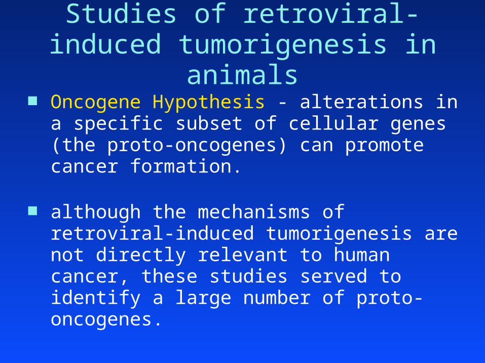
Many proto-oncogenes identified in studies of retroviral tumorigenesis in animals
retroviraltransduction H-ras
K-ras
SrcYesAblFosCbl
proviralintegration
EviWntLckPim
MycMyb
➔ Are these proto-oncogenes involved in human cancer?

Oncogenes II
Organization of today’s lecture in vitro gene transfer experiments in vivo gene transfer experiments chromosome abnormalities chromosome abnormalities in lymphoid tumors chromosome abnormalities in solid tumors gene amplification the proteins encoded by proto-oncogenes targeted cancer therapy (e.g., the Ph1 chromosome)

Retroviral infection of chick embryo fibroblasts (CEFs)
almost every RSV virion can transform a fibroblast to yield a visible focus. # of foci is proportional to # of RSV virions provides an ideal method to titer RSV
high efficiency reflects the activities of: viral surface protein (which facilitates infection) viral integrase (which facilitates proviral integration)
efficiency of transformation: ~ 1 focus/cell (at saturating quantities of virus: # virions >> # fibroblasts)
CEFs(106 cells/plate)
infect with ALVno foci
infect with RSVmany foci

Transfection of CEFs with proviral DNA
donor DNAs: the RSV provirusthe ALV provirus (control)
note: apply saturating quantities of DNA to cultured cells. (# of proviral DNA molecules >> # of fibroblasts)
Some cells are transformed by the RSV (but not the ALV) proviral DNA to yield visible foci
This is a less efficient process than viral infection: transfection vs. infection (perhaps 1/10 cells are transfected) proviral integration without integrase (perhaps occurs in only
1/102 of the transfected cells)
efficiency of transformation: ~ 1 focus/103 cells

CEF transfection with chicken genomic DNA
donor DNA: the genome of uninfected cell “ “ “ ALV-infected
cell “ “ “ RSV-transformed cell
a few cells are transformed by genomic DNA of an RSV-transformed cell
this is an inefficient process transfection vs. infection (perhaps 1/10 cells are transfected) proviral integration without integrase (perhaps in 1/102 of
transfected cells) the likelihood that the integrated DNA will include the RSV
provirus (perhaps 1/102)
efficiency of transformation: ~ 1 focus/105 cells

Though seemingly inefficient, the activity of a single oncogene (proviral v-src) can be readily detected within the background of an entire cellular genome.
Screening more than 106 cells in a focus formation assay is quite easy.
Can this assay be used to detect and isolate activated proto-oncogenes in human tumors ?

Transfection of rodent fibroblasts with human genomic DNA
Donor DNA - the genome of a human tumor from a tumor cell line or a primary tumor
Control DNA - genome of normal human cells from blood or other normal tissues

NIH3T3 cells
a permanent cell line derived from a “post-crisis” culture of normal mouse fibroblasts
properties of NIH3T3 cells… are immortal are contact-inhibited require anchorage for growth do not form tumors in nude mice
thus, grows as amonolayer in vitro

Transfection of NIH3T3 cells with human genomic DNA
Results of transfection experiment:
positive results with ~ 20% of human tumors seen in a broad spectrum of tumors, including
sarcomas, carcinomas, leukemias and lymphomas
normal cells 1 / 107 cells*
tumor A 1 / 107 cells
tumor B 1 / 105 cells !!
tumor C 1 / 107 cells
genomicDNA
transformedfoci

New properties of in vitro-transformed NIH3T3 cells
parental transformed immortality + + focus formation – + colony formation – + in soft agar tumor formation – + in nude mice
also, genomic DNA of transformed NIH3T3 cells is active in subsequent in vitro-transformation assays (unlike DNA of parental NIH3T3 cells).

Sequential transformation of NIH3T3 cells
EJ cells human bladder
carcinoma100 % human DNA
~ 1 % human DNA ~ 99 % mouse DNA
the genome of the primary transformants contains… mostly mouse genomic DNA (~99%) some newly integrated human genomic DNA (~1%)
including, the EJ cell transforming sequences!!
NIH-3T3 1’- transformant
DNA

Sequential transformation of NIH3T3 cells
EJ cells human bladder
carcinoma
NIH-3T3 1’- transformant
100 % human DNA
~ 1 % human DNA ~ 99 % mouse DNA
the tertiary transformants should contain the EJ cell transforming sequences, but little other human DNA.
DNA
NIH-3T3 2’- transformant
~ 0.01 % human DNA ~ 99.99 % mouse DNA
DNA
NIH-3T3 3’- transformant
DNA

What is the transforming agent in human tumor DNA?
isolate human DNA fragments from tertiary-transformants. test each fragment for the ability to transform NIH3T3 cells
in vitro. the transforming activity of EJ cells could be ascribed to a
single fragment of DNA. sequence analysis revealed that the transforming fragment is
the c-H-ras gene - a known proto-oncogene that had already been identified in animal studies!! the transduced oncogene of Harvey Sarcoma Virus (HSV), an
acutely-transforming mouse retrovirus. the targeted proto-oncogene of retroviral integration in ALV-
induced nephroblastomas.

The Ras gene family similar approaches were used to isolate transforming
genes from many human tumor cell lines:
c-H-ras •transduced oncogene of HSV
EJ cells bladder carcinoma
T24 cells bladder carcinoma
c-K-ras •transduced oncogene of KirstenSarcoma Virus, another acutely-transforming mouse retrovirus
Lx-1 cells lung carcinoma
SK-CO-1 cells colon carcinoma
N-ras •novel member of Ras family
SK-N-SH cells neuroblastoma
HL60 cells leukemia
transforming Ras oncogenes were also detected in DNA from primary human tumors

The three members of the Ras family represent most, but not all, of the human oncogenes isolated on the basis of NIH3T3 cell transformation.
~ 15 % of all human tumors harbor a Ras gene that has the ability to transform NIH3T3 cells.
However, Ras genes from normal human DNA do not transform NIH-3T3 cells.
the Ras gene of EJ cells is malignantly “activated”
The Ras gene family

Malignant activation of the Ras genes compare nucleotide sequences of…
transforming Ras genes from human tumors non-transforming Ras genes from normal cells
Each transforming Ras gene has a point mutation that yields an amino acid substitution at residue 12, 13, or 61.
Mutation of the same residues is also seen in the retrovirally-transduced Ras oncogenes!! Harvey Sarcoma Virus v-H-ras (G12R mutation) Kirsten Sarcoma Virus v-K-ras (G12D mutation)
malignant activation of Ras proteins by point mutation !!

Other human oncogenes identified on the basis of NIH3T3 transformation
Many encode protein tyrosine kinases: Neu (Her2) - isolated from a neuroblastoma
- activated by missense mutation Trk - isolated from a colon carcinoma
- activated by fusion (with tropomyosin) Ret - isolated from a thyroid carcinoma
- activated by fusion (with PTC1 and others) oncogenic fusion proteins involving tyrosine kinases (PTC1-Ret):
– the N-terminal sequences of the oncoprotein (Ret) are often replaced by the fusion partner (PCT1), resulting in deregulation of the kinase domain in the C-terminal half of the oncoprotein.

More proto-oncogenes identification of transforming DNA fragments in NIH3T3
transformation assays increased the number of known proto-oncogenes.
retroviraltransduction
NIH3T3transformation
N-rasNeuRetTrk
H-rasK-ras
YesAblFosCbl
proviralintegration
EviWntEviLck
Src
MycMyb

Oncogene complementation in vitro conduct focus formation assay using…
primary rat embryo fibroblasts (REFs) these have not undergone “crisis”
NIH3T3 REFs
immortality: + –
focus formation: – –
transfect REFs with expression plasmids encoding… ras* – c-H-ras with an oncogenic mutation at amino acid 12 myc – high levels of c-myc protein

Oncogene complementation in vitro results of focus formation assay…
NIH3T3 REFs
ras* + – (senescence)
myc – – (apoptosis) (apoptosis)
ras* + myc + +
ras* and myc cooperation is required for transformation of primary fibroblasts.

in vivo gene transfer experiments
fibroblasts transformed in vitro with activated proto-oncogenes will induce tumors in nude mice. can activated proto-oncogenes also induce in vivo tumors
directly ? can activated proto-oncogenes induce in vivo tumors from
cells of other (non-fibroblast) cell lineages ?
these questions can be addressed by developing transgenic animals that express activated proto-oncogenes

in vivo gene transfer experiments the MMTV-ras* transgene
encodes the activated c-H-ras gene from the EJ tumor line with oncogenic missense mutation G12V
transcriptional regulation of the transgene is mediated by the LTR element of the Mouse Mammary Tumor Virus (MMTV).
the LTR of MMTV is tissue-specific, inducing transcription primarily in mammary epithelial cells.
MMTVLTR c-H-ras (G12V)

MMTV-ras* transgenic mice
female (but not male) MMTV-ras* transgenic mice develop mammary carcinomas.
about half the female transgenic mice form palpable mammary carcinomas in six months (T50 ~ 6 months).
the carcinomas are monoclonal (even though the transgene is expressed in all developing mammary epithelial cells).
the latency and monoclonality indicate that additional somatic events are necessary for tumor formation from MMTV–ras* mammary cells.

MMTV-myc transgenic mice
the MMTV-myc transgene
female (but not male) MMTV-myc transgenic mice develop mammary carcinomas.
the T50 of female transgenic mice is ~ 11 months.
the carcinomas are monoclonal
additional somatic events are also necessary for tumor formation from MMTV–myc mammary cells.

mate MMTV-ras* and MMTV-myc mice.
female (but not male) double transgenic mice develop mammary carcinomas.
much shorter latency (T50 ~ 6 weeks). cooperation between activated ras and myc gene in vivo.
again, the tumors are monoclonal
additional somatic events are also necessary for tumor formation from double-transgenic mammary cells.
Oncogene complementation in vivo

Cytogenetics
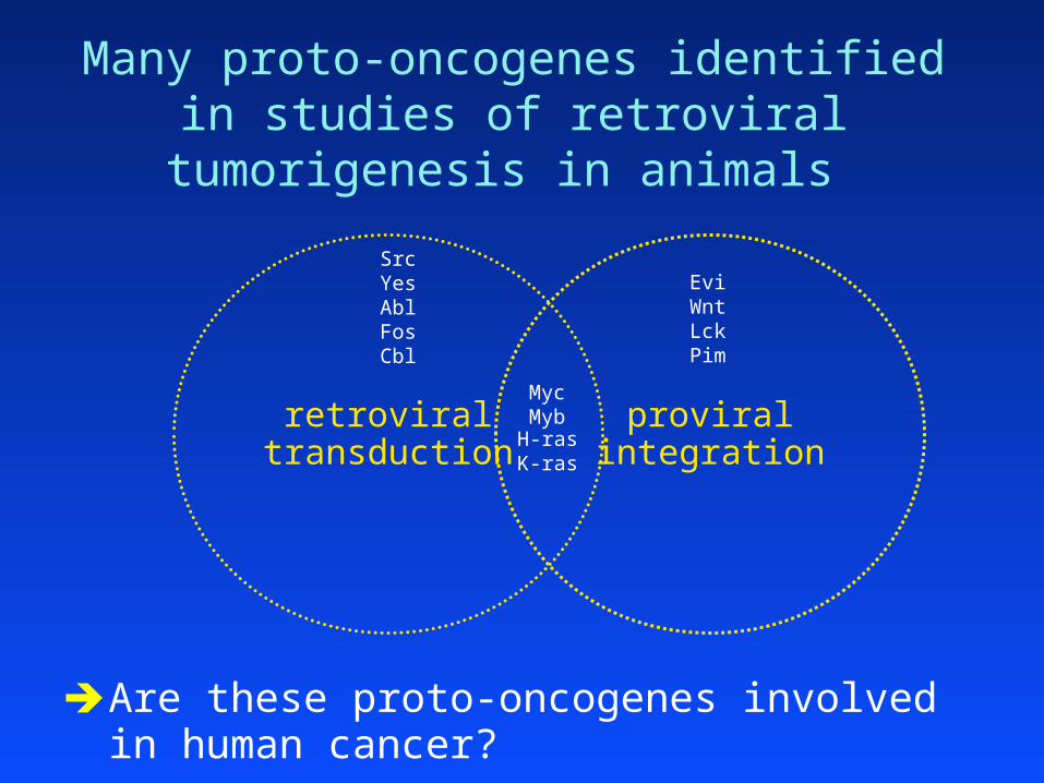
Cytogenetics of normal cells
traditional karyotype analysis chromosomes condense during mitosis. visible under the light microscope. mitotic chromosomes can be stained with certain dyes. for a given dye (e.g., Giemsa), each chromosome produces a
unique and reproducible banding pattern.
the karyotype of normal human diploid cells one pair of sex chromosomes and 22 pairs of autosomes. males: 46XY females: 46XX

Cytogenetics of tumor cells - I
karyotypes of tumor cells tumor cells often have unstable chromosomes
most tumor cells have chromosome abnormalities abnormalities in chromosome number (aneuploidy) abnormalities in chromosome structure
Translocations Inversions Deletions DNA amplification (HSRs, DMs)
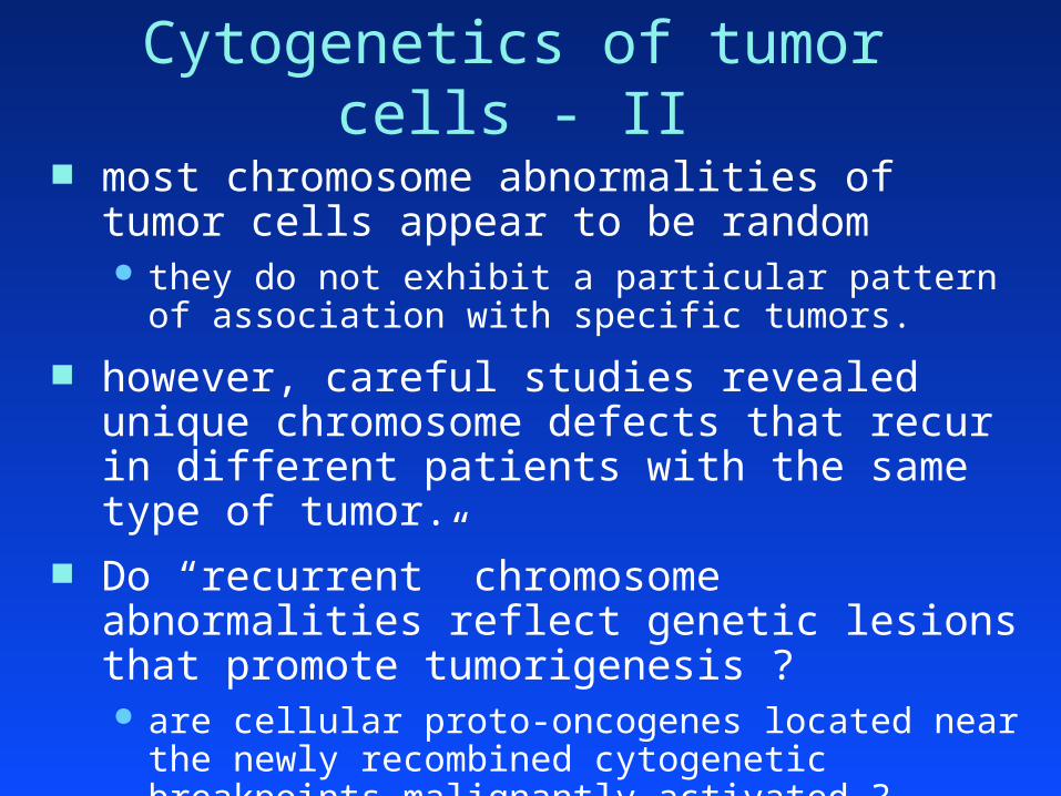
most chromosome abnormalities of tumor cells appear to be random they do not exhibit a particular pattern of association with
specific tumors.
however, careful studies revealed unique chromosome defects that recur in different patients with the same type of tumor.
Do “recurrent” chromosome abnormalities reflect genetic lesions that promote tumorigenesis ? are cellular proto-oncogenes located near the newly
recombined cytogenetic breakpoints malignantly activated ?
Cytogenetics of tumor cells - II

Carcinomas often have highly complex karyotypes with many, seemingly random, chromosome abnormalities.
Hematopoietic tumors and sarcomas have less complex karyotypes. These often feature recurrent chromosome abnormalities that are characteristic of a particular type of cancer.
Cytogenetics of tumor cells - III

In 1960, Nowell observed an altered (shorter) form of chromosome 22 in many patients with chronic myelogenous leukemia (CML)
Philadelphia chromosome (Ph1) only found in the leukemic cells of CML patients
Ph1 only associated with certain malignancies: 95% of CML 20% of adult acute lymphoblastic leukemias (ALL) 5% of childhood ALL
Hypothesis: Ph1 represents a genetic lesion that can promote the formation of CML or ALL
The Philadelphia (Ph1) chromosome

chromosome banding techniques revealed…
that Ph1 is the product of a reciprocal translocation between chromosomes 9 and 22:
t(9;22)(q34;q11)
t(9;22) has breakpoints at positions 9q34 and 22q11
Hypothesis: Ph1 promotes leukemia by malignantly activating a proto-oncogene located at 9q34 or 22q11
Ph1 produced by a chromosome translocation

the t(9;22)(q34;q11) chromosome translocation
9 and 22 are the chromosomes involved
translocation breakpoints occur at positions 9q34 and 22q11
chromosome short and long arms are “p” and “q”, respectively.
9q34 is giemsa band 34 on long arm of chromosome 9
22q11 is giemsa band 11 on long arm of chromosome 22
Cytogenetics terminology

the (9;22)(q34;q11) translocation
9
22
reciprocal translocation chromosomes 9 and 22
normalchromosomes
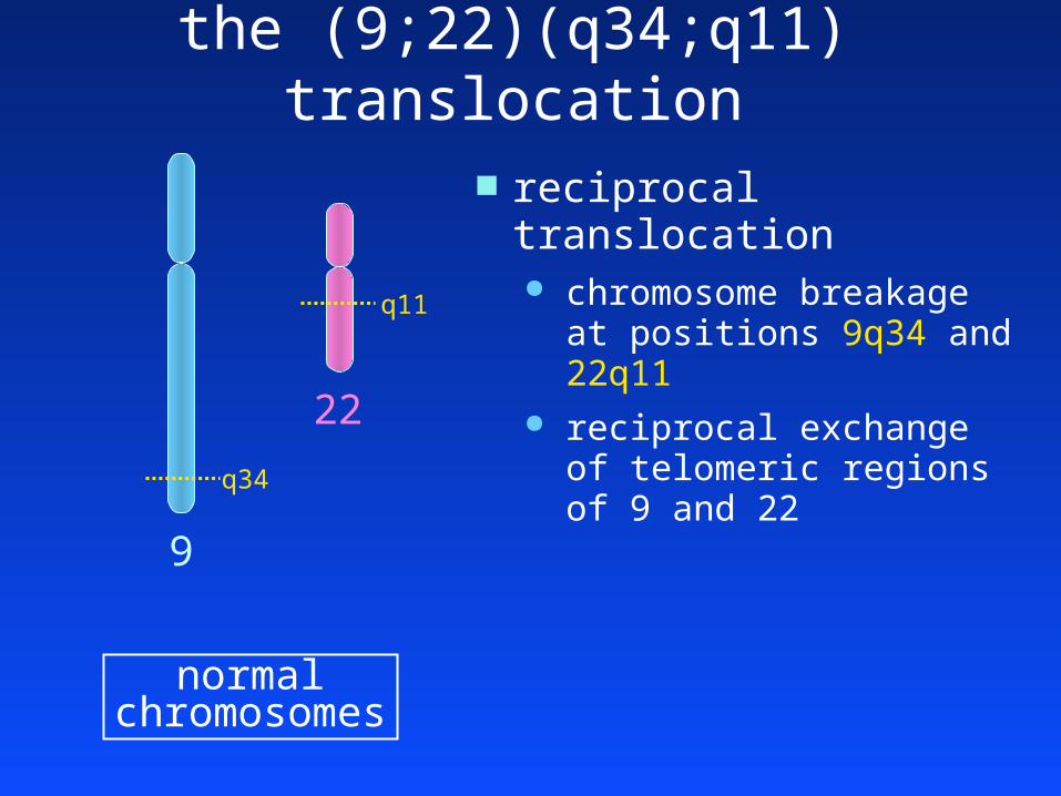
the (9;22)(q34;q11) translocation
reciprocal translocation chromosome breakage at
positions 9q34 and 22q11 reciprocal exchange of
telomeric regions of 9 and 22
9
q11
22
q34
normalchromosomes

the (9;22)(q34;q11) translocation
translocatedchromosomes
the reciprocal translocation yields two derivative chromosomes:
the der(9) chromosome has the centromere of chr. 9
the der(22) chromosome has the centromere of chr. 22 same as Ph1
der(9)
der(22)
Ph1

the (9;22)(q34;q11) translocation
normalchromosomes
chromosome breakage… disrupts the BCR gene at 22q11 disrupts the ABL gene at 9q34
ABL is the cellular form of v-abl, a retrovirally-tranduced oncogene!!
the der(22) junction has a … BCR-ABL fusion gene
9
22
ABL
BCR

the BCR-ABL fusion gene
der(22)
5’ 3’
lies at the junction of der(22) contains:
transcriptional promoter of BCR one or more 5’-coding exons of BCR coding exons 2-12 of ABL
BCR
genomicDNA
junction
22q11 9q34
ABL

the BCR-ABL fusion protein RNA transcription of BCR-ABL fusion gene spliced BCR-ABL messenger RNA the BCR-ABL fusion polypeptide
5’ 3’BCR ABL
genomicDNA
junction
BCR-ABL fusion protein
- transcription- translation

Malignant activation of the ABL protein the normal ABL protein
a non-receptor protein tyrosine kinase phosphorylation of ABL substrates promotes cell growth enzymatic activity of normal ABL is tightly controlled
the BCR-ABL fusion protein retains the tyrosine kinase activity of ABL, but… its enzymatic activity cannot be downregulated due to:
transcriptional deregulation by the BCR promoter replacement of N-terminal ABL sequences with BCR sequences
the fusion protein constitutively promotes cell growth.
Þ BCR-ABL is a malignantly activated form of the ABL proto-oncogene.

Animal models of BCR-ABL leukemia
BCR-ABL retrovirus infect murine hematopoietic stem cells in vitro use these stem cells to reconstitute lethally-irradiated
mice 50% of mice develop CML-like disease
BCR-ABL transgene produce mouse strain with a BCR-ABL transgene mice develop acute leukemia within two months of birth
The tyrosine kinase activity of BCR-ABL is essential for its transforming potential.

Clinical impact of Ph1 chromosome
diagnosis
prognosis
disease monitoring
treatment

Burkitt’s lymphoma
B cell malignancy endemic in the malarial belt common in immunosuppressed populations
cytogenetic abnormalities associated with BL ~ 90% of patients: t(8;14)(q24;q32) ~ 5% “ “ “ t(2;8)(p12;q24) ~ 5% “ “ “ t(8;22)(q24;q11)
a common breakpoint at 8q24

Burkitt’s lymphoma II in situ chromosome hybridization of known oncogenes:
In 1982, the MYC gene was localized to 8q24.
Southern analysis of genomic DNA from BL cells: MYC gene rearranged in most cases of BL.
isolation of rearranged MYC fragment from BL cells: rearranged fragment encompasses the t(8;14) junction.
5’ 3’
MYC
t(8:14)
junction
14q32 8q24
exon2
exon3
???

Burkitt’s lymphoma III 14q32 break: immunoglobulin heavy chain gene (IgH) !!
translocation break locust(8;14)(q24;q32) 14q32 IgHt(2;8)(p12;q24) 2p12 Igk (kappa light chain gene)t(8;22)(q24;q11) 22q11 Ig l (lambda light chain gene)
MYC is malignantly activated upon juxtaposition with any of the three immunoglobulin loci.
this makes sense… MYC activation by proviral integration causes B cell tumors in
chickens congenitally infected with ALV. The Ig genes undergo DNA recombination during normal
B cell development The Ig genes are transcriptionally active in normal B cells

Follicular lymphoma a distinct histopathologic type of B cell malignancy
> 85% of cases have the t(14;18)(q32;q21) 14q32 breaks in the IgH locus. 18q21 breaks in the BCL2 gene. a new proto-oncogene not encountered in animal studies ?
BCL2/IgH transgenic mice develop B cell tumors !
t(14;18)
junction
14q32 18q21IgH BCL2
5’ 3’

Mantle cell lymphoma another histopathologic type of B cell malignancy
~ 30% of cases have the t(11;14)(q13;q32) 14q32 breaks in the IgH locus. 11q13 breaks near the cyclin D1 gene (CCND1).
cyclin D1 is a positive regulator of cell cycle progression that drives the G1/S transition by phosphorylating Rb
5’ 3’
CCND1
t(11;14)
junction
14q32 11q13IgH

T cell acute lymphoblastic leukemia (T-ALL)
clinically homogenous disease
cytogenetically heterogeneous (in contrast with BL or FL) over 12 different recurrent translocations seen in T-ALL each found in a small proportion (< 5%) of T-ALL patients
T-ALL derived from immature T cells (thymocytes)
The Ig genes serve as activating loci in translocations of B cell tumors. Thus, do the TCR genes serve a similar role in translocations of T cell tumors ? T cell receptor (TCR) b chain gene maps to 7q34
“ “ “ “ /a d chain gene maps to 14q11

Recurrent translocations of T-ALL translocation patients TCR oncogene?
t(8;14)(q24;q11) ~ 2% /a d t(7;14)(q24;q11) ~ 2% /a d t(7;9)(q34;q34) ~ 1% b t(10;14)(q24;q11) ~ 3% /a d t(1;14)(p34;q11) ~ 3% /a d t(7;9)(q34;q32) ~ 2% b t(7;19)(q34;p13) < 1% b t(11;14)(p15;q11) ~ 1% /a d t(11;14)(p13;q11) ~ 5% /a d and others…..

Recurrent translocations of T-ALL translocation patients TCR oncogene
t(8;14)(q24;q11) ~ 2% /a d MYCt(7;14)(q24;q11) ~ 2% /a d LCKt(7;9)(q34;q34) ~ 1% b NOTCH1t(10;14)(q24;q11) ~ 3% /a d HOX11t(1;14)(p34;q11) ~ 3% /a d TAL1t(7;9)(q34;q32) ~ 2% b TAL2t(7;19)(q34;p13) < 1% b LYL1t(11;14)(p15;q11) ~ 1% /a d LMO1t(11;14)(p13;q11) ~ 5% /a d LMO2and others…..
all activate a proto-oncogene by recombination with the TCR locus at either 7q34 or 14q11

Recurrent translocations of T-ALL translocation patients TCR oncogene
t(8;14)(q24;q11) ~ 2% /a d MYC
MYC activation by IgH in Burkitt’s lymphoma MYC activation by TCR in T-ALL MYC activated by retroviral transduction in T cell
tumors of cats

Recurrent translocations of T-ALL translocation patients TCR oncogene
t(8;14)(q24;q11) ~ 2% /a d MYCt(7;14)(q24;q11) ~ 2% /a d LCK
LCK activated by retroviral integration in mouse thymomas

Recurrent translocations of T-ALL translocation patients TCR oncogene
t(8;14)(q24;q11) ~ 2% /a d MYCt(7;14)(q24;q11) ~ 2% /a d LCKt(7;9)(q34;q34) ~ 4% b NOTCH1
NOTCH1 was a novel proto-oncogene; its malignant potential was confirmed by tumor induction in transgenic mice
NOTCH1 is malignantly activated by point mutations in almost 50% of T-ALL patients!!

Recurrent translocations of T-ALL translocation patients TCR oncogene
t(8;14)(q24;q11) ~ 2% /a d MYCt(7;14)(q24;q11) ~ 2% /a d LCKt(7;9)(q34;q34) ~ 4% b NOTCH1t(10;14)(q24;q11) ~ 3% /a d HOX11
HOX11 was a novel proto-oncogene; its malignant potential was confirmed by tumor induction in transgenic mice
HOX11 is malignantly activated by ectopic expression in ~25% of T-ALL patients!!

Recurrent translocations of T-ALL translocation patients TCR oncogene
t(8;14)(q24;q11) ~ 2% /a d MYCt(7;14)(q24;q11) ~ 2% /a d LCKt(7;9)(q34;q34) ~ 4% b NOTCH1t(10;14)(q24;q11) ~ 3% /a d HOX11t(1;14)(p34;q11) ~ 3% /a d TAL1
TAL1 was a novel proto-oncogene; its malignant potential was confirmed by tumor induction in transgenic mice
TAL1 is malignantly activated by gene rearrangement and ectopic expression in over 50% of T-ALL patients!!

Recurrent translocations of T-ALL translocation patients TCR oncogene
t(8;14)(q24;q11) ~ 2% /a d MYCt(7;14)(q24;q11) ~ 2% /a d LCKt(7;9)(q34;q34) ~ 4% b NOTCH1t(10;14)(q24;q11) ~ 3% /a d HOX11t(1;14)(p34;q11) ~ 3% /a d TAL1t(7;9)(q34;q32) ~ 2% b TAL2t(7;19)(q34;p13) < 1% b LYL1
TAL1, TAL2, and LYL2 encode highly related proteins presumably, activation of these genes represents an
equivalent step in T cell leukemogenesis

Recurrent translocations of T-ALL translocation patients TCR oncogene
t(8;14)(q24;q11) ~ 2% /a d MYCt(7;14)(q24;q11) ~ 2% /a d LCKt(7;9)(q34;q34) ~ 4% b NOTCH1t(10;14)(q24;q11) ~ 3% /a d HOX11t(1;14)(p34;q11) ~ 3% /a d TAL1t(7;9)(q34;q32) ~ 2% b TAL2t(7;19)(q34;p13) < 1% b LYL1t(11;14)(p15;q11) ~ 1% /a d LMO1t(11;14)(p13;q11) ~ 5% /a d LMO2
LMO1 and LMO2 encode highly related proteins LMO- and TAL-related proteins form stable nuclear
complex; thus, a common pathway for T-ALL

Recurrent translocations of T-ALL Analysis of chromosome translocations identified three
major pathways of T cell leukemogenesis… NOTCH1 pathway HOX11, HOX11L2 pathway TAL1, TAL2, LYL1, LMO1, LMO2 pathway
Almost all T-ALL patients show malignant activation of at least one of these pathways
Many T-ALL patients show malignant activation of two or more of these pathways

Karyotypes of human sarcomas often display recurrent chromosome abnormalities
many of these engender fusions with the EWS gene
chromosome fusion DNA-binding translocation tumor type protein domain
t(11;22)(q24;q12) Ewings sarcoma EWS-FLI1 ETS family
t(21;22)(q22;q12) Ewings sarcoma EWS-ERG ETS family
t(7;22)(p22;q12) Ewings sarcoma EWS-ETV1 ETS family
t(7;22)(q12;q12) Ewings sarcoma EWS-ETV4 ETS family
t(7;22)(q12;q12) clear cell sarcomaEWS-ATF1 bZIP family
t(11;22)(p13;q12) desmo. sarcoma EWS-WT1 Zn finger family
each of the EWS partners harbors a DNA-binding domain
each fused to the transcriptional activation domain of EWS

Are recurrent chromosome abnormalities relevant in human epithelial tumors ?

Recurrent chromosome abnormalities in human carcinoma (2002) ?
t(12;15)(p13;q25): encodes ETV6-NTRK3 fusion gene ETV6 (TEL), a transcription factor of the “ETS” family Neurotrophin-3 receptor, a TM protein tyrosine kinase
t(12;15) in - congenital fibrosarcoma (1998) - cellular mesoblastic nephroma (1998) - acute myeloid leukemia (1999)
2002 – t(12;15) found in secretory breast cancer (SBC) ETV6-NTRK3 transcripts observed in 12 of 13 SBC cases! not observed in other types of breast carcinoma
The first recurrent translocation in human carcinoma!!

Recurrent chromosome abnormalities in human carcinoma (2005) ?
Tomlins et al., Science 310: 644-648, 2005
Recurrent gene fusions in prostate carcinomas the TMPRSS2-ERG and TMPRSS2-ETV1 fusions retain…
non-coding exon 1 of androgen-inducible TMPRSS2 gene coding sequences for the DNA binding domains of either
ERG or ETV1, transcription factors of the “ETS” family result in deregulated expression of truncated ETS proteins
ERG and ETV1 gene fusions seen in 23 of 29 cases of prostate cancer !!
more to come ?

In general, chromosome translocations activate proto-oncogenes by one of two mechanisms:
1. By generating an aberrant fusion gene comprised of exons from two genes located adjacent to the recombined cytogenetic breakpoints
2. By transcriptional deregulation of a proto-oncogene at one cytogenetic breakpoint upon juxtaposition with regulatory sequences from the other breakpoint

Gene amplification
methotrexate
DHF THFDHFR dNTP
synthesis
Gene amplification in response to metabolic stress a common mechanism by which mammalian cells acquire
resistance to certain metabolic inhibitors
DHFR and methotrexate DHFR (dihydrofolate reductase): an enzyme required for
dNTP biosynthesis (and, in turn, DNA synthesis) methotrexate: an analog of dihydrofolate that irreversibly
binds and inhibits DHFR

Methotrexate-resistant cell lines
methotrexate resistance treat cells with methotrexate.
select for cells that grow in the presence of methotrexate.
methotrexate-resistant cells show gross amplification of the DHFR gene (and increased levels of DHFR protein)!!
DHFR gene amplification is manifested cytogenetically by the presence of... DMs (double minutes) HSRs (homogeneously staining regions)

DMs double minute (DM) chromosomes
small, independent, chromosome-like structures comprised of multiple tandem “amplicons” of genomic DNA
that harbor the selected gene (e.g., DHFR) lack centromeres genetically unstable - the maintenance of DMs requires
continued selection.


HSRs homogenously-staining regions (HSRs)
multiple tandem amplicons that harbor the selected gene (e.g., DHFR) incorporated into a chromosome
stain homogenously (i.e., lack the banding pattern of normal chromosomal material)
derived by integration of DM sequences into a chromosome genetically stable


Gene amplification in human tumors
neuroblastomas DMs and HSRs are very common in human neuroblastomas correlate with a poor prognosis the amplicons of these structures often cross-hybridize with
sequences from the c-Myc gene
amplification of the MYC gene family in tumors c-Myc: neuroblastomas N-Myc: neuroblastomas L-Myc: lung carcinomas
amplification of the MDM2 gene in human sarcomas


Proteins encoded by proto-oncogenes Many proto-oncogenes encode components of
signal transduction pathways that promote cell proliferation in response to… internal cues extracellular stimuli
Growth factors Growth factor receptors Cytoplasmic transducers Nuclear factors

Growth factors and their receptors Growth factors
Simian Sarcoma Virus harbors a tranduced oncogene (v-sis) The c-sis gene encodes B chain of platelet-derived growth
factor (PDGF) Mechanism: autocrine stimulation of cell growth

Figure 5.11c The Biology of Cancer (© Garland Science 2014)
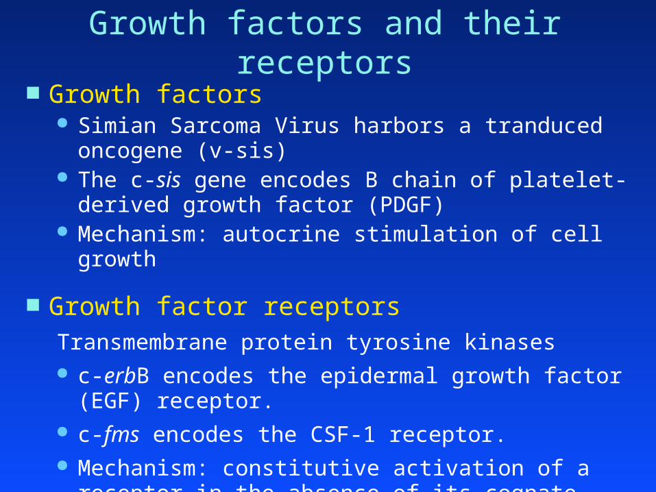
Growth factors and their receptors Growth factors
Simian Sarcoma Virus harbors a tranduced oncogene (v-sis) The c-sis gene encodes B chain of platelet-derived growth
factor (PDGF) Mechanism: autocrine stimulation of cell growth
Growth factor receptorsTransmembrane protein tyrosine kinases c-erbB encodes the epidermal growth factor (EGF) receptor. c-fms encodes the CSF-1 receptor. Mechanism: constitutive activation of a receptor in the absence
of its cognate ligand.

Figure 5.11a The Biology of Cancer (© Garland Science 2014)

Cytoplasmic transducers Protein tyrosine kinases: c-src, c-fes, c-abl
Protein serine/threonine kinases: c-raf and c-mos
the Ras proteins (small G proteins) have a covalently-attached farnesyl group that inserts into the
inner leaflet of the cell membrane bind the guanine nucleotides GDP and GTP GTP-bound Ras proteins stimulate downstream pathways
(including the Raf/MAP kinase cascade) have GTPase activity: convert GTP to GDP Oncogenic mutations ablate the GTPase activity. Thus, mutant
Ras proteins constitutively stimulate downstream pathways.

Figure 5.30 The Biology of Cancer (© Garland Science 2007)

Nuclear factors Transcription factors activated by signaling pathways
c-myc
c-jun
c-fos

Other categories of proto-oncogenes Negative regulators of the tumor suppressor pathways
MDM2 (amplified in human sarcomas) is a feedback inhibitor of the p53 tumor suppressor.
Cyclin D1 (translocated in centrocytic B cell lymphomas) associates with CDK4/6 to form kinase complexes that phosphorylate Rb and inhibit its tumor suppression activity.
Inhibitors of apoptosis BCL2 (translocated in follicular B cell lymphomas) encodes an
anti-apoptotic protein. E2A-HLF, a fusion gene associated with pre-B cell acute
lymphoblastic leukemia, encodes a transcription factor that suppresses apoptosis.

Clinical impact of Ph1 chromosome
diagnosis
prognosis
disease monitoring
treatment

Diagnosis & Prognosis
diagnosis The presence of Ph1 in granulocytic cells of PB or BM is
tantamount to a diagnosis of CML
prognosis Ph1-positive ALL has an especially poor outcome in both
children (~5%) and adults (~20%)
disease monitoring
treatment

CML progression chronic (pre-malignant) phase
leukocytosis and circulating immature granulocytic cells chronic phase lasts ~5 years Ph1 translocation occurs in a hematopoietic progenitor cell Ph1 cells differentiate normally
accelerated phase additional genetic lesions? rising numbers of Ph1 blasts and basophils in PB or BM lasts 6–18 months
blast crisis acute myeloid or lymphoid leukemia lasts 3–6 months

Disease Monitoring
to measure the levels of Ph1-positive cells during… the chronic phase and accelerating phase response to chemotherapy or transplantation
Ph1-positive cells detected by… cytogenetic analysis of metaphase spreads fluorescent in situ chromosome hybridization (FISH) of
interphase cells quantitative PCR (or RT-PCR) of patient DNA (or RNA)
using primers that flank the translocation junction.

conventional CML treatment chronic phase
hydroxyurea (RNR inhibitor) for cytoreductive therapy does not significantly alter disease progression
interferon a allogeneic stem cell transplantation
only proven curative therapy not an option for most CML patients
blast crisis acute myeloid leukemia
standard induction chemotherapy: 20% response complete remission: < 10%
acute lymphoid leukemia standard induction chemotherapy: 50% response complete remission: < 10%

Gleevac (imatinib mesylate or STI571)
the deregulated enzymatic (tyrosine kinase) activity of BCR-ABL is the essential transforming event in CML
Ciba-Geigy (Novartis) identified a synthetic tyrosine kinase inhibitor (2-phenolaminopyrimidine)
blocks the ATP-binding site of tyrosine kinases
screened compounds for increased potency and specificity
Gleevac: specific inhibitor of … ABL c-Kit PDGF-Rb

cellular effects of Gleevac
inhibits proliferation of cell lines containing BCR-ABL
inhibits clonal growth of Ph1 cells from CML patients
inhibits the in vivo growth of BCR-ABL-expressing cells in animal models of CML

Clinical study of Gleevac Phase 1, dose-escalating trial (NEJM 344:1031, 2001)
chronic phase CML patients for whom interferon treatment hadfailed
daily doses ranging from 25 to 1000 mg
all doses tolerated very well
low doses (25, 50, 85 mg): half of patients removed from study within 2 mos. due to increasing WBC counts
high doses (300-1000 mg): 53 of 54 patients showed a complete hematologic response within a month maintained for 310 days in 51 patients


![Retroviruses - 2013 (FN) [Compatibility Mode]](https://img.dokumen.tips/doc/110x75/577cdda21a28ab9e78ad6fbf/retroviruses-2013-fn-compatibility-mode.jpg)


![Oncogenesis Virica Y Microbiana J(1)[1]](https://img.dokumen.tips/doc/110x75/5563f160d8b42ae33c8b4a49/oncogenesis-virica-y-microbiana-j11.jpg)