Embed Size (px)
Citation preview

Lymphohematopoietic progenitors immortalized by a retroviral vector harboring a dominant-negative retinoic acid receptor can recapitulate lymphoid, myelold, and erythrold development Schickwann Tsai, 1 Stephen Bar te lmez , 2 Ewa Si tnicka, 2 and Steven Col l ins
Fred Hutchinson Cancer Research Center, Seattle, Washington 98104 USA; ~Pathology Department, University of Washington, Seattle, Washington 98195 USA
The lymphohematopoietic progenitors represent <0.01% of nucleated marrow cells. Here, we describe the immortalization of the murine lymphohematopoietic progenitors by a retroviral vector harboring a dominant-negative retinoic acid receptor. The immortalized progenitors proliferate as a stem-cell-factor-dependent clonal line EML that spontaneously generates pre-pro-B lymphocytes and erythroid and myeloid progenitors. Upon stimulation with intedeukin-7 and stromal cells, the pre-pro-B lymphocytes express RAG-1 and undergo D-[ rearrangements of the immunoglobulin heavy-chain genes. With erythropoietin the erythroid progenitors proliferate and differentiate into red cells. Generation of the common progenitors for neutrophils and macrophages is suppressed in EML but is inducible by high concentrations of retinoic acid. An additional block in neutrophil differentiation occurs at the promyelocyte stage but can also be overcome by high concentrations of retinoic acid. These studies demonstrate a reproducible way to immortalize lymphohematopoietic progenitors and implicate specific roles for retinoic acid receptors at two distinct stages of hematopoiesis.
[Key Words: Retinoic acid receptors; lymphohematopoietic progenitors; B-cell development; myelopoiesis; erythropoiesis]
Received August 30, 1994; revised version accepted October 13, 1994.
The existence of a common progenitor that gives rise to all lymphoid and hematopoietic (or "myeloerythroid") cells was initially demonstrated by transplantation ex- periments using bone marrow cells carrying X-ray-in- duced chromosomal markers (Wu et al. 1968). More re- cently, direct marking of bone marrow cells with retro- viral vectors has further comfirmed the existence of a common lymphohematopoietic progenitor cell (Lemis- chka et al. 1986). Through a combination of physical and immunological purification methods, the murine lym- phohematopoietic stem cells have been purified to near homogeneity (Spangrude et al. 1988). Their frequency is estimated to be 0.01-0.005% of all nucleated cells in the bone marrow. The scarcity of the lymphohematopoietic stem cells and the difficulty in their purification and maintenance have hampered the effort to dissect the mo- lecular mechanism controlling lymphoid and he- matopoietic development. Thus, the availability of a continuous cell line capable of both lymphoid and mye-
1Corresponding author.
loerythroid differentiation ih vitro would greatly facili- tate such research.
In this paper we demonstrate that the lymphohe- matopoietic progenitors can be immortalized by a retro- viral vector containing a dominant-negative retinoic acid receptor construct and that the resulting lymphohe- matopoietic progenitor cell line exhibits two develop- mental defects in the myeloid lineage that can be cor- rected with high concentrations of retinoic acid (RA). The RARs are members of the steroid/thyroid hormone receptors that function as ligand-inducible transcription factors (Evans 1988). Clues suggesting that RARs are in- volved in regulating hematopoiesis come from both lab- oratory and clinical observations; RA induces the HL-60 human leukemia cell line to differentiate into mature neutrophils, and this process is mediated through RARs (Breitman et al. 1980; Collins et al. 1990). In most cases of acute promyelocytic leukemia (APL), the gene of RARa on chromosome 17 is translocated and fused with the PML gene on chromosome 15 (Alcalay et al. 1990; Borrow et al. 1990; de The et al. 1990), and the leukemic cells from these patients can be induced by RA to differ-
GENES & DEVELOPMENT 8:2831-2841 © 1994 by Cold Spring Harbor Laboratory Press ISSN 0890-9369/94 $5.00 2831
Cold Spring Harbor Laboratory Press on April 2, 2018 - Published by genesdev.cshlp.orgDownloaded from

Tsai et al.
entiate into mature neutrophils both in vitro and in vivo (Huang et al. 1988). The precise role of the PML--RARa fusion gene in the pathogenesis of APL is yet to be re- solved. Attempts to demonstrate a dominant-negative function for P M L - R A R a fusion gene using transient ex- pression assay systems yielded inconsistent results (Kak- izuka et al. 1991; de The et al. 1991). On the other hand, we have shown that the expression of a dominant-nega- tive RARot (RARot403) in normal mouse bone marrow cells leads to a differentiation block in the neutrophil lineage at the promyelocyte stage (Tsai and Collins 1993). This finding provides direct evidence that RARs play important roles at the promyelocyte stage of neu- trophil differentiation and raises the possibility that a novel dominant-negative mechanism may underlie the action of the PML-RARa fusion protein (Dyck et al. 1994; Weis et al. 1994). We have also shown that when the dominant-negative RARot403 is expressed in a mul- tipotent, interleukin-3 (IL-3)-dependent myeloid cell line FDCP mix A4, it triggers a rapid switch from spontane- ous neutrophil and macrophage development to the pro- duction of mast cells (Tsai et al. 1992). This developmen- tal switch appears to occur near or at the lineage decision stage rather than at the neutrophilic promyelocyte stage. Taken together, these observations indicate that RARs not only play important roles in controlling the terminal differentiation of neutrophilic promyelocytes but also may influence hematopoietic lineage development at the multipotent progenitor stage.
To investigate the developmental effects of the domi- nant-negative RARot403 on normal, primitive lympho- hematopoietic progenitors, we used retroviral vector- mediated gene transfer to tranduce fresh mouse bone marrow cells with this dominant-negative RAR and cul- tured the cells in a medium containing hematopoietic growth factors that promote the survival and prolifera- tion of primitive lymphohematopoietic progenitors. Us- ing this approach we have reproducibly established con- tinuous cell lines that are dependent on stem cell factor (SCF; also known as c-kit ligand)(Zsebo et al. 1990) for survival and proliferation yet can undergo orderly lym- phoid, myeloid, and erythroid differentiation in vitro. In addition to demonstrating a reproducible way to immor- talize lymphohematopoietic progenitors, our present findings also provide the first evidence that RA and RARs may modulate myeloid lineage development prior to the stage of the so-called colony-forming unit-granu- locyte/macrophage (CFU-GM). Further observations suggest that there may be a causal link between the sup- pression of myeloid development and the immortaliza- tion of these lymphohematopoietic progenitors.
Results
Infection of mouse bone marrow wi th a retroviral vector LRARa403SN and the es tabl ishment of a s tem cell factor-dependent cell l ine
We have described previously the construction of the retroviral vector LRARot403SN harboring a truncated
RARot cDNA insert (Tsai et al. 1992). The truncated RARot, designated RARo~403, contains the first 403 amino acids of human RARot but is truncated in the ligand-binding domain and the activation function do- main-1 (AF-1; Nag, pal et al. 1992). It exhibits strong dom- inant-negative activity against endogenous RARs in transient expression assays in 3T3 fibroblasts, CV-1 cells, and hematopoietic cells (Tsai et al. 1992; Damm et al. 1993; Tsai and Collins 1993).
Bone marrow cells (10 s) of male BDFI mice who re- ceived 5-fluorouracil 5 days earlier were infected with the retroviral vector LRAR~403SN and subsequently cultured in a medium containing SCF, IL-3, and eryth- ropoietin IEpo) as detailed in Materials and methods. No G418 selection was applied. An SCF-dependent cell line, designated EML, emerged from this culture. It is nonad- herent and has been in continuous passage for > 1 year with a doubling time of 18-20 hr. In the absence of SCF, the majority (>95%) of the EML cells die within 24 hr, with the rest dying during the next 72 hr. It harbors the LRAR~403SN provirus genome (Fig. 1A) and expresses high levels of the retroviral message encoding the dom- inant-negative RAR (Fig. 1B). Many cells (20--30%) in the EML line have a characteristic hand-mirror shape (Fig. 6A, below).
To ensure the clonality of the cell line, it was first cloned by limiting dilution and recloned at a low cell density in a methylcellulose culture medium as detailed in Materials and methods. Southern analysis reveals that all clones have a single, identical integration site (Fig. 1A), indicating that they are all clonal derivatives of the same parental cell. Importantly, the clonal line and its subclones that we have examined possess similar lineage repertoires described below, although only the data ob- tained with EML C1 and its subclone EML C1.4 will be presented in this paper.
The dominant-negative RARa403 is essential for establishing the EML cell line
To determine whether the dominant-negative RARot403 plays an essential role in the establishment of the EML cell line, we repeated the infection of post-5-fluorouracil mouse bone marrow with the control vector LXSN as well as LRAR~403SN and processed the cells in parallel. An uninfected bone marrow culture was also included as an additional control. To increase the number of lym- phohematopoietic progenitors as potential targets for retroviral infection, 1,000,000 bone marrow mononu- clear cells (rather than l0 s as in the initial isolation of the EML cell line) were used. Using this approach we have reproducibly isolated EML-like cell lines from mouse bone marrow cells infected with the LRARot403SN vector. In contrast, no cell lines could be isolated from parallel cultures of uninfected or LXSN (control)-infected mouse bone marrow cells. Thus, the dominant-negative RARot403 is essential for the estab- lishment of the EML cell line. Southern analysis of the proviral integration sites revealed the presence of multi- ple clones on day 76 after retroviral infection, all of
2832 GENES & DEVELOPMENT
Cold Spring Harbor Laboratory Press on April 2, 2018 - Published by genesdev.cshlp.orgDownloaded from

Immortalized lymphohematopoietic progenitors
= ~ E r,r'. r,.~ r,.~
1 - _.1 --3 . - I
1 2 3 4
C s ~ I .U Z
1 2
S (,..)
_ J
LU
1
4 .7 - -
3 .6 - -
2,6--
b
Neo probe RARc~ probe Neo probe
Figure 1. Integration and expression of the LRARct403SN pro- virus in the EML line. (A) Southern analysis of the proviral integration sites. Genomic DNAs were digested with EcoRI (which cuts only once within the provirus upstream of the neo sequence), blotted, and hybridized with a neo probe. (Lane I) Uncloned EML line that has been passaged for 21/2 months since retroviral infection and is already dominated by one clone; (lane 2) a clonal line (EML C 1) obtained by limiting dilution cloning, followed by recloning in semisolid medium; (lane 3) EML C1.4 recloned again from EML C1 in semisolid medium. All clones share the same, single proviral integration site. Comparison of the signal intensity on Southern blot with that of a reference cell line indicates that there is one copy of the provirus per genome. (B) Northern analysis of the expression of the RARe,403 in EML. Five micrograms of total RNA of EML C1 was analyzed using a human RARa probe. The 4.7-kb retroviral message harboring the RARe~403 sequence and the 3.6- and 2.6- kb endogenous mouse RARa mRNAs are indicated. (C) Multi- clonal origin of EML-like cell lines. Additional EML-like cell lines were established as described for the original EML cell line except that 1,000,000 rather than 105 bone marrow mononu- clear cells were used in retroviral infection. Genomic DNA was obtained from a bulk culture 76 days after retroviral infection, digested with EcoRI, and subjected to Southern analysis. (Lane 1) EML C1; (lane 2) day 76 bulk culture of EML-like cell lines from an independent infection experiment. Four dominant clones are visible and show different integration sites from EML C1.
which integrated at different sites from EML C1 (Fig. 1C), indicat ing that a common retroviral integration site is probably not required for the immorta l iza t ion of EML- like cells. The lymphohematopoie t ic potential of these new clones was verified after subcloning (not shown). Despite the fact that no G418 selection was applied throughout the experiment, all independent ly isolated EML-like cell l ines we have examined harbored the LRARa403SN provirus, albeit at different integration sites.
The EML cell line contains cells expressing B-lymphocyte-specific antigen B220
The cell surface antigen profile of a representative clone
(C1) of the EML cell l ine is examined by flow cytometry using a panel of monoclonal antibodies: (1) anti-Sca-1, which is specific for s tem cel l s /pr imi t ive progenitors (Spangrude et al. 1988); (2) anti-B220 (clone RA3-6B2), which is specific for the B-lymphocyte lineage (Coffman and Weissman 1981); (3)anti-Mac-l, which is specific for macrophage/neutrophi l lineages; {4) monoclonal anti- body "7/4 ," which is specific for the neutrophi l l ineage (Hirsch and Gordon 1983); and (5) monoclonal ant ibody "ter 119," which recognizes erythroid precursors (Ikuta et al. 1990).
As shown in Figure 2, 50% of EML C1 cells are posi- tive for Sea-l, - 5 3 % express B220, - 5 1 % express ter 119, very few express 7/4 or Mac-1. A s imilar surface antigen profile is observed in subclones Cl.1, C1.3, and C1.4 of EML, although the actual percentages vary (not shown). The B220 antigen, a member of the " leukocyte common ant igen" (LCA) family, is specific for pr imi t ive as well as mature B cells (for review, see Thomas 1989). Thus, the results of flow cytometry indicate that cells of B-lymphoid and erythroid lineages are spontaneously generated in the EML C1 cell line.
The B-cell progenitors in the EML CI cell line are pre-pro-B ce i l s
To determine the developmental stage of the putat ive B-cell progenitors present in the EML C1 line, we exam- ined (1) the expression of immunog lobu l in heavy chain (IgH) and the pr imit ive lymphocyte-specif ic gene RAG-1 (recombination-activating gene-1; Schatz et al. 1989} by Northern analyses, and (2) the rearrangement of D-J seg- ments of the IgH gene by polymerase chain reaction (PCR) amplif icat ion of genomic DNAs using specific oli- gonucleotide primers.
When the EML C1 cell l ine was main ta ined in me-
A s all I t - - i i , , i
U
",~ : ~ '.!, -.
10 o 10 t 10 z 103 104 10 ° 10 ~ 10 ;= 10 ~ 104 100 10 ~ 10 z 10 3 104
Fluorescence Intensity
Figure 2. Flow cytometry analysis of the cell surface antigens of EML C1. The profile of the isotype antibody (negative con- trol; thin line) is shown in the first panel and again in all other panels for direct comparison. The antibodies used and the pos- itive fractions (shaded areas) are indicated in each panel. The specificity of the monoclonal antibodies are as follows: stem cells/primitive progenitors (Sca-1); B cells (B220); neutrophils (7/4); macrophages and neutrophils (Mac-i); erythroid precur- sors (ter 119).
GENES & DEVELOPMENT 2833
Cold Spring Harbor Laboratory Press on April 2, 2018 - Published by genesdev.cshlp.orgDownloaded from
Tsai et al.
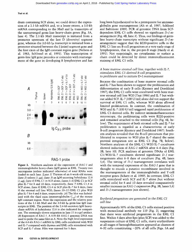
dium containing SCF alone, we could detect the expres- sion of a 2.1-kb mRNA and, to a lesser extent, a 3.0-kb mRNA corresponding to the Mu0 and I~ transcripts of the unrearranged germ-line heavy-chain genes (Fig. 3A, lane 4). The 2.1-kb Mu0 transcript is initiated from a promoter upstream of the last D (diversity) segment gene, whereas the 3.0-kb I~ transcript is initiated from a promoter situated between the J (joint) segment gene and the first exon of the IgH constant region gene (Nelson et al. 1983; Schlissel et al. 1991). This transcription of germ-line IgH gene precedes or coincides with rearrange- ment of the gene in developing B lymphocytes and has
long been hypothesized to be a prerequisite for immuno- globulin gene rearrangement (Alt et al. 1987; Schlissel and Baltimore 1989). PCR of genomic DNA from SCF- dependent EML C1 cells showed no significant D-J re- arrangement (Fig. 4B, lane 3). Thus, our findings of germ- line heavy-chain transcripts without significant D-J re- arrangement suggest that the B220-positive cells in the EML C1 line are B-cell progenitors at a very early stage of lymphopoiesis, that is, the pre-pro-B stage (Hardy et al. 1991). Not surprisingly, no cytoplasmic or surface chain could be detected by direct immunofluorescent staining of EML C1 cells.
A
kb
3.0--
2 .4-- 2 .1--
B
kb
6 . 6 -
I - - 0 3
1 2
"1o o ~ n o ned
,, ,, - - ,, ,, m +
-4- + I.I- IJ.- "4" -4- °R. u_ u_ u_ (.• G~ o o ,:'M rO r , j O rJ') 0 3 cM cM
0 3 0 3 0 3 + + ~ + '-:::t ,~. + + 4- .-I--
o • "~ r J ¢ j CJ (..) ( J r,~ rO -r-
- ¢ - I.k.I L U ILl LI.I l.l.I ~ 1.1.1 1.1.1
3 4 5 6 7 8 9 10 11
• ,,m
Ig C#3 probe
RAG-1 probe F i g u r e 3. Northern analyses of the expression of RAG-I and immunoglobulin heavy-chain (IgH) genes in EML. Twenty-two micrograms (unless indicated otherwise) of total RNAs were loaded in each lane. (Lane I) Thymus of an 8-week-old mouse; (lane 2) spleen (l ~g); (lane 3) an IgM-secreting hybridoma 12.8 (1 ~g); (lane 4) EML C1 in SCF alone; (lanes 5-6) EML C1 in SCF plus IL-7 for 8 and 10 days, respectively; (lane 7) EML C1.4 in SCF alone; (lane 8) EML C1.4 in SCF plus IL-7 for 8 days; (lane 9) the stromal cell line W20; (lanes 10--11) EML C1 plus W20 plus IL-7 for 6 and 8 days, respectively. (A) The blot was hybrid- ized with the third exon (immunoglobulin C~3) of the mouse IgH constant region. Note the expression and the relative posi- tions of the 2.1-kb Mu0 and the 3.0-kb Ill. germ-line IgH tran- scripts in EML. The position of the 2.4-kb secretory IgM heavy- chain mRNA of hybridoma 12.8 is also indicated for compari- son. The seemingly slower migration in lane 11 is a gel artifact. (B) Expression of RAG-1. A 0.95-kb RAG-1 genomic DNA was used to probe the same blot as A. Note the high-level expression of the 6.6-kb RAG-1 mRNA by EML cells cocultured with W20 and IL-7 compared with thymus and EML cells stimulated with SCF and IL-7 alone. Film was exposed for 5 days.
A bone marrow stromal cell line, together with IL-7, stimulates EML C1 -derived B-cell progenitors to proliferate and to initiate D-J rearrangement
Because the combination of bone marrow stromal cells and IL-7 has been shown to support the proliferation and differentiation of early B cells (Kierney and Dorshkind 1987), the EML C1 cells were cocultured with bone mar- row stromal cell line W20 (Tsai 1986) and IL-7 but with- out added SCF. IL-7 (100 U/ml) alone did not support the survival of EML C1 cells, whereas W20 alone allowed limited proliferation. In contrast, the combination of W20 and IL-7 (100 U/ml) triggered rapid proliferation of EML Cl-derived cells. By indirect immunofluorescence microscopy, the proliferating cells were B220-positive and remained attached to the stromal cells (Fig. 6E, be- low). The requirement of both stromal cells and IL-7 for proliferation is regarded as a characterisitic of early B-cell progenitors (Kierney and Dorshkind 1987). South- ern analysis revealed that the B-cell precursors that pro- liferated in response to W20 plus IL-7 shared the same proviral integration site as EML C1 (Fig. 7B, below). Northern analyses of the EML C1/W20/IL-7 cocultures showed induction of RAG-1 mRNA after 4--6 days (Fig. 3B, lane 10). PCR analyses of genomic DNAs of EML C1/W20/IL-7 cocultures showed significant D-J rear- rangements after 6-8 days of coculture (Fig. 4B, lanes 5,6). The timing of D-J rearrangement correlates well with the temporal expression of the RAG-1 gene that is thought to encode a putative recombinase involved in the rearrangements of the immunoglobulin and T-cell receptor genes (Schatz et al. 1989). In contrast, EML C1 cells stimulated with SCF and IL-7 alone (but without stromal cells) for 8 and 10 days revealed comparatively smaller increases in RAG-1 expression (Fig. 3B, lanes 5,6) and D-J rearrangements (not shown).
Erythroid progenitors are generated in the EML C1 cell line
Approximately 50% of the EML C1 cells stained positive with monoclonal antibody ter 119 (Fig. 2}, suggesting that there were erythroid progenitors in the EML C1 line. Within 4 days after Epo (plus SCF) was added to the suspension culture of EML C1 cells, many erythroblasts at all stages of hemoglobinization appeared as clusters of 4--32 cells consitituting - 2 0 % of all cells (Figs. 5A and
2834 GENES & D E V E L O P M E N T
Cold Spring Harbor Laboratory Press on April 2, 2018 - Published by genesdev.cshlp.orgDownloaded from

Immortalized lymphohematopoietic progenitors
A
- ,= . -m l l - .=~ , - y , " V H V H
DHL
D D D D D D
¢¢ #J
/," ~ ml V H V H D D DJ 1
J3
J2 J3 J4 a Pr0be
J 2 J 3 J 4
PCR Products: DJ 1 (1019 bp) DJ 2 (702 bp)
i Dj 3 (319 bp)
bp 1353 1,078
872
603
+ + + o o o
.~ ,.,-i + + +
1 2 3 4 5 6 7
-.~-.- DJ1
-~,- DJ 2
~.-DJ 3
CH
Germline
~" D& Rearrangement Figure 4. PCR detection of D-J rearrange- ments in EML Cl-derived B-cell progeni- tors. (A) The locations of the DHL and J3 primers and the 226-bp J3 Southern probe in IgH gene are depicted. The sizes (~319, ~702, and ~1019 bp)of various amplifica- tion products are approximate estimates. (B) D-I rearrangements detected by PCR of genomic DNAs. The DHL and J3 primers were used in PCR. The Southern blot of PCR products was hybridized with the 226- bp J3 probe. (Lane 1) d~X174/HaeIII digests as size markers; {lane 2) PCR buffer; (lane 3) EML C1 in SCF; (lanes 4-6) EML C1 cocul- tured with stromal cells W20 plus IL-7 for 4, 6, and 8 days, respectively; (lane 7) stro- real cell line W20.
6B, below). The level of globin mRNA in these SCF/Epo- treated EML C1 cells could approach that of hexameth- ylene bisacetamide (HMBA)-induced routine erythroleu- kemia cell line (MEL) (Fig. 5B). When EML C1 cells were cultured in methylcellulose medium containing SCF and Epo, typical burst-forming unit-erythroid (BFU-E)-de- rived colonies containing thousands of erythroblasts as well as mixed colonies composed of both undifferenti- ated EML C1 cells and BFU-E-derived subcolonies were observed. The frequency of BFU-Es is -1 -2% of the total population in EML C 1. These observations indicate that the SCF-dependent EML C1 cells spontaneously gener- ate erythroid progenitors that terminally differentiate in response to Epo.
A
30
o~" 25
N 2o
p, a . 15
m 5
0
0 0
0 0
I I I I
3 5 7 9 11 13 15 17 19 21 23
Days
B ~ = x x x
~ u.I u.I 4- 4- +
r ~ CO m m + + 4- 4- 4-
I 2 3 4 5
~-Globin Probe
Figure 5. Erythroid differentiation of EML C1. (A) Time course of the appearance of benzidine (hemoglobin)-positive erythro- blasts after addition of Epo. Numbers are means of duplicates. (B) Northern analysis of the expression of mouse [~major globin mRNA. (Lane 1 ) HMBA-induced MEL cell line; (lanes 2-5) EML C1 ceils stimulated with both SCF (200 ng/ml) and Epo (8 U/ml) for 0, 1, 3, and 5 days, respectively. The lower message level in lane 4 is the result of the suboptimal culture condition caused by a higher cell density.
Induction of myelodd progenitors (CFU-GMs) from EML C1 cells
Few EML C 1 cells express neutrophil or macrophage lin- eage-specific surface markers (Fig. 2), suggesting that very few myeloid cells or their progenitors are generated in EML C 1 cells maintained in SCF alone. Colony assays of CFU-GMs using murine granulocyte/macrophage col- ony-stimulating factor (GM-CSF) detected very few or no CFU-GMs in EML C1 cells (Table 1). Costimulation of EML C1 with both SCF and IL-3 resulted in some in- crease in CFU-GMs. More importantly, simultaneous treatment of EML C1 cells with SCF, IL-3, and high con- centrations of RA (0.5-1x10 -s M) dramatically in- creased the generation of CFU-GMs (Table 1), which formed colonies of neutrophils, macrophages, and pro- myelocytes in semisolid medium in the presence of GM- CSF. This induction of CFU-GMs by RA has never been described before. Interestingly, the combination of SCF, GM-CSF, and 10 -s M RA did not induce the formation of CFU-GMs (not shown). This is consistent with the ob- servation that multiple growth factors such as IL-3 and GM-CSF applied sequentially are needed for successful generation of CFU-GMs from stem cells (Metcalf 1993). The optimal concentration of RA for induction of CFU- GMs from EML C1 cells was 10 -s M with the half-opti- mal concentration being 10-6 M. The induction of CFU- GMs by RA (in the presence of SCF and IL-3) could be detected as early as 24 hr after adding RA. This dramatic increase in CFU-GMs was accompanied by a reduction in the numbers of BFU-Es and of self-renewing clono- genic cells in SCF/IL-3/RA-treated EML C1 cultures when compared with cultures treated with SCF/IL-3 alone (Table 1). These results suggest that the formation of CFU-GMs is severely suppressed in the EML C1 cells, probably by the dominant-negative RARa403, and that this suppression can be overcome with high concentra- tions of KA in presence of IL-3.
GENES & DEVELOPMENT 2835
Cold Spring Harbor Laboratory Press on April 2, 2018 - Published by genesdev.cshlp.orgDownloaded from

Tsai et al.
Tab le 1. Effects of RA on the production of CFU-GM, BFU-E, and the self-renewal in EML C1
SCF + + + + IL-3 - - + + RA (10 -5 M) - + - +
Experiment 1 (n = 2) total cells on day 3
(X10 6) 2.0 0.6 3.9 3.1 total CFU-GM a 2.5 0 1,115 32,040 total SCF-supported
clonogenic cells b 5,200 1,000 7,400 2,000 Experiment 2 (n = 3)
total cells on day 4 (X10 6) 7.0 N.D. 17.4 13.3
total CFU-GM 0 N.D. 150 12,850 total SCF-supported
clonogenic cells 58,600 N.D. 6 8 , 6 0 0 8,600 total BFU-E c 182,800 N.D. 276,000 42,800
EML C1 cells (10 5) were cultured in a liquid medium containing SCF +- IL-3 + RA for 3 days (experiment 1) or 4 days (experiment 2). Cells were then washed with PBS and counted, and aliquots were cultured in methylcellulose medium to assay for various types of progenitors. Higher-passage cells were used in experi- ment 2. aFor CFU-GM assay, the methylcellulose medium was supple- mented with murine GM-CSF (10 ng/ml). bFor SCF-supported clonogenic cells, i.e., the self-renewing cells, the methylcellulose medium was supplemented with rat SCF (200 ng/ml). ¢For BFU-E assay, the methylcellulose medium was supple- mented with rat SCF {200 ng/ml) and Epo (4 U/ml). Only pure BFU-E colonies were counted.
Other lineages detected in the EML C1 cell line
When EML C1 cells were stimulated with IL-3 (which is essential for mast cell differentiation and survival) in liquid culture with or without SCF, we observed the ap- pearance of mast cells {Fig. 6C}. With simultaneous stim- ulation with SCF, IL-3, Epo, IL-6, and IL-11, occasional megakaryocytes were detected (Fig. 6D). When multiple growth factors such as SCF, IL-3, GM-CSF, and Epo were added to colony assays of EML C 1 cells, we could readily detect multilineage colonies. Taken together, our data indicate that the SCF-dependent EML C1 cell line con- tains progenitors capable of differentiation along B-lym- phocyte, erythrocyte, neutrophil, macrophage, mast cell, and megakaryocyte lineages.
The terminal differentiation of EML Cl-derived neutrophil precursors is further blocked at the promyelocyte stage by the dominant-negative RARa403
When EML Cl-derived CFU-GMs were cultured in liq- uid or semisolid media containing GM-CSF, they prolif- erated and differentiated along the neutrophil and mac- rophage lineages (Fig. 6F). However, the differentiation of many neutrophils was blocked at the promyelocyte stage. These blocked promyelocytes proliferated contin-
uously as GM-CSF-dependent cell lines and were desig- nated as EPRO tfor _EML-derived promyelocytes; see Ma- terials and methods for derivation of these cell lines; Fig. 6G). Flow cytometry analyses of cell surface markers of a representative EPRO cell line {C1) show that 57% are positive for the neutrophil lineage-specific antigen 7/4, few are positive for Sea-l, and none expresses B220 or Mac-1 {Fig. 7A). Southern analysis showed that EPRO C1 had the same proviral integration site as EML C1 and EML Cl-derived B-cell precursors supported by stromal cell W20 and IL-7 (Fig. 7B), again verifying the common origin of these cells. The GM-CSF-dependent EPRO cell lines are virtually identical to the GM-CSF-dependent MPRO (mouse promyelocyte) cell lines that we isolated directly from LRARa403SN-infected, GM-CSF-stimu- lated mouse bone marrow as reported previously (Tsai and Collins 1993). Like MPRO cells, the EPRO cells dif- ferentiate synchronously and rapidly (in 96-120 hr) into mature neutrophils when treated with high concentra- tions of RA (Fig. 6H). The optimal concentration for this induction of terminal differentiation is 0 .5-1x10 -5 M for EPRO cell line, similar to that for MPKO cells {Tsai and Collins 1993).
D i s c u s s i o n
In this paper we describe a unique, SCF-dependent lym- phohematopoietic progenitor cell line (designated EML) with erythroid, myeloid, and lymphoid potentials. To our knowledge, the EML cell line is the only SCF-depen- dent cell line with both lymphoid and myeloerythroid potentials. It was established from a mouse bone marrow infected with a retroviral vector (LRARa403SN) harbor- ing a dominant-negative RAR construct. Remarkably, all independently isolated EML-like cell lines harbor the LRARa403SN provirus despite the fact that the lympho- hematopoietic progenitors represented only a small frac- tion of the input population and that no G418 selection was applied after retroviral infection, suggesting that lymphohematopoietic progenitors infected with the pro- virus possessed survival advantages over the uninfected cells. Furthermore, it appears that no common integra- tion site was required to confer such survival advantages on the infected cells (Fig. 1C). Although the dominant- negative RAR~ construct is essential for the establish- ment of EML-like cell lines, it is possible that additional mutations may have contributed to the emergence of the dominant clones.
The EML cell line is unique in its ability to generate large numbers of pre-pro-B cells. Furthermore, these pre- pro-B cells can respond to simultaneous stimulation with bone marrow stromal cells (W20) and IL-7 by ex- pressing RAG-l) (Fig. 3B, lanes 10,11) and by initiating the rearrangement of the D and J segments of the IgH genes (Fig. 4B). The D-J rearrangement occurs around day 8, following the peak expression of RAG-1 on day 6. We have not detected cytoplasmic or membrane ix-chain protein by immunofluorescence in EML C1 cells stimu- lated with the W20 stromal cells and IL-7 for 10 days (not shown). It is of interest to determine if these pro-B cells
2836 GENES & DEVELOPMENT
Cold Spring Harbor Laboratory Press on April 2, 2018 - Published by genesdev.cshlp.orgDownloaded from

Immortalized lymphohematopoietic progenitors
A ~~ i l i ¸̧ ̧:~
c
o °o
p
F
O
t4
Oq :
Q
Figure 6. Erythroid, myeloid, and lym- phoid cells developed from EML C1. (A) EML CI cells in growth medium (note the three hand-mirror-shaped cells that are frequently seen in the EML C1 cell line); {B) a cluster of polychromatophilic eryth- roblasts; (C) three coarsely granulated mast cells (arrows); (D) a megakaryocyte (center) with marked polyploidy; (E} devel- oping B cells from a 14-day coculture of EML C1/W20/IL-7 [the large cell hugging the smaller B-cell precursors is a W20 stro- mal cell (arrow)]; (F) vacuolated macro- phages (arrows) surrounded by smaller neutrophils from an EML Cl-derived CFU-GM colony; (G) the neutrophilic pro- myelocyte cell line EPRO C1 derived from EML C1 (the cytoplasm contains numer- ous azurophilic granules); (H) neutrophils with segmented nuclei that have devel- oped from EPRO C1 treated with 10 -s M RA for 5 days. Wright-Giemsa stain. All have the same magnifications except F. Bars, 50 ~.
can continue their differentiation to the plasma cell stage and what stimuli are required for such processes.
The EML cell line also spontaneously generates cells recognized by the monoclonal antibody ter 119, which recognizes mostly erythroid precursors (Ikuta et al. 1990). Three to five days after addition of Epo to the liquid culture of EML, we detected the appearance of hemoglobinized erythroblasts that formed aggregates of 4-32 cells (Fig. 6B). The rapidity of their appearance and their limited proliferative capacity suggest that these erythroblasts are the progenies of the so-called colony- forming units-erythroid ICFU-Es). In addition, large multicentric erythroid colonies containing several thou- sands of erythroblasts developing over 7-10 days could readily be demonstrated in clonal cultures of EML in methylcellulose medium supplemented with SCF and
Epo. These are derived from the so-called BFU-Es, which are developmentally more primitive than the CFU-Es and eventually differentiate into CFU-Es. Although flow cytometry indicated slightly more than 50% of EML C1 cells expressed the B-cell lineage-specific antigen B220 or the erythroid precursor-specific antigen ter 119 {Fig. 2), we believe this slight "overlapping" of the percentages of B220- and ter 119-positive cells resulted from minor im- precisions in setting fluorescence gates during data anal- ysis and did not always occur.
In contrast with its spontaneous generation of large numbers of B-lymphoid and -erythroid progenitors, the SCF-dependent EML C1 cells produce very few progeni- tors for the neutrophil and macrophage lineages (CFU- GMs; Table 1 ). This is in sharp contrast to the normal multipotent hematopoietic progenitors that readily gen-
GENES & DEVELOPMENT 2837
Cold Spring Harbor Laboratory Press on April 2, 2018 - Published by genesdev.cshlp.orgDownloaded from

Tsai et al.
A
ZE ~,.. ,.d,
Neg.
MAC-1
10 0 1'01 1~ 1'0 3
B220
A | i
7/4
J .
i i i
10 4 10 0 101 10 2 10 3 10 4
B { ~ + +
12345
Fluorescence Intensity Neo Probe
Figure 7. Phenotype and clonal origin of the EPRO cell line. (A) Flow cytometry analyses of the cell surface antigens of EPRO C1. EPRO C1 is a GM-CSF-dependent neutrophilic promyelo- cyte cell line derived from EML as described in Materials and methods. The isotype antibody (negative control; thin line) pro- file is shown in the first panel and again in all other panels for direct comparison. The positive fractions (shaded areas) are in- dicated in each panel. Most cells in EPRO C1 are stained by neutrophil lineage-specific monoclonal antibody 7/4. (B) Retro- viral integration site of EPRO C1. Genomic DNAs from EML C1 {lane 1), EML Cl-derived B-cell precursors expanded on stro- mal cell line W20 plus IL-7 for 8 days (lane 2) or 17 days (lane 3), EPRO C1 (lane 4), and W20 alone (lane 5) were digested with EcoRI and analyzed as described in Fig. 1A. Samples in lanes 2 and 3 also contained DNAs of the W20 stromal cell line.
erate CFU-GMs in vitro. Intriguingly, this deficiency of CFU-GMs in EML C1 cells can be overcome with a com- bination of IL-3 and high concentrations of RA (10-6_ 10 -s M), leading to a dramatic increase in the number of CFU-GMs (Table 1). (For comparison, the serum concen- tration of RA was estimated to be 10-9-10 -8 M; DeRuyter et al. 1979.) This phenomenon has never been described before but is in line with our previous obser- vations that the expression of the dominant-negative RARc~403 in an IL-3-dependent multipotent hematopoi- etic cell line (FDCP mix A4) severely suppressed spon- taneous neutrophil and macrophage development (Tsai et al. 1992) and that the dominant-negative effects of RARa403 in myeloid cells could be overcome or by- passed by high concentrations (10-6-10 -s M) of RA in both trans-act ivat ion and differentiation assays (Tsai and Collins 1993). Thus, it appears that the inhibition of CFU-GM formation in EML C1 is attributable to the activity of the dominant-negative RARc~403, and this in- hibition can be overcome or bypassed by high concen- trations of RA.
Further analyses revealed that the increase in CFU- GMs was accompanied by a decrease in SCF-responsive, self-renewing clonogenic cells and in the total number of EML C1 cells after 3 or 4 days of treatment with SGF/ IL-3/RA (Table 1). These opposite trends, that is, a de- crease in SCF-dependent self-renewal and an increase in
CFU-GMs, suggest that high concentrations of RA may have induced the multipotent progenitors in EML C1 cell line to commit to the neutrophil/macrophage differ- entiation pathway. As a result, the number of SCF-re- sponsive, self-renewing clonogenic cells decreases, which in turn leads to a decrease in BFU-Es (Table 1, experiment 2). Although an additional, direct inhibitory effect of RA on self-renewing cells (as well as BFU-Es) cannot be ruled out, these observations raise the possi- bility that the immortalization of EML cells may be an immediate consequence of the suppressive effect of the dominant-negative RARa403 on myeloid commitment. The primitive lymphohematopoietic progenitors are characterized by their ability to self-renew and to com- mit to several differentiation pathways. For primitive lymphohematopoietic progenitors to exist as a continu- ous cell line like EML, the probability of its self-renewal in vitro must exceed its probability of commitment to various differentiation pathways. By virtue of its ability to interfere with the progenitors' ability to commit to the myeloid differentiation pathway, the dominant-neg- ative RARa403 may have tipped the balance of self-re- newal versus differentiation in favor of self-renewal, leading to the establishment of the EML cell line.
A second block in the differentiation of EML-derived neutrophil precursors occurs downstream at the promy- elocyte stage, leading to the establishment of GM-CSF- dependent promyelocyte cell lines such as EPRO (Fig. 6G). High concentrations of RA (10-s-10 -6 M) induce these cells to terminally differentiate into mature neu- trophils (Fig. 6H). The biological properties (i.e., devel- opmental stage, growth factor requirements, surface an- tigens, and RA responsiveness) of EPRO cells are virtu- ally identical to those of GM-CSF-dependent MPRO cell lines that we previously established from LRAR~403SN- infected, GM-CSF-stimulated mouse bone marrow cells (Tsai and Collins 1993). Because we have shown that the RA-reversible differentiation block in MPRO cells is at- tributable to the dominant-negative effects of RARe403 (Tsai and Collins 1993), we believe that the RA-revers- ible differentiation block in EPRO cells is also caused by the dominant-negative RARe~403.
In summary, our data indicate that RA and RARs play important roles in the development of neutrophils at two distinct stages in the EML C1 cell line: The first one is mapped to the pre-CFU-GM stage, whereas the second one occurs at the neutrophilic promyelocyte stage. Our findings suggest that RARs may be part of the combina- torial mix of transcriptional regulators that converge be- fore the establishment of a "nodal decision point" (Las- sar and Weintraub 1992) in myeloid lineage develop- ment. They also raise the possibility of influencing the self-renewal of lymphohematopoietic progenitors in vitro by adding strong antagonists of RA to the culture medium. The effects of the dominant-negative RARc~403 on myeloid lineage development in EML are summa- rized in Figure 8.
Because of the altered physiology brought on by the dominant-negative RAR, the EML cell line can not be equated with normal lymphohematopoietic progenitors.
2 8 3 8 G E N E S & DEVELOPMENT
Cold Spring Harbor Laboratory Press on April 2, 2018 - Published by genesdev.cshlp.orgDownloaded from

Immortalized lymphohematopoietic progenitors
SCF Megakaryocyte
\ ~ EML st,orna Mast IL-3 [ :.~!~,: -~,. 1L-7
< 4 =
n,L3 / ~ ji~ ~-~
Erythrocyte V
~GM-CSF
Macrophage
~ Promyelocyte
RARo~403 BLOCK
GM-CSF
Neutrophil
Figure 8. A schematic summary of the effects of the dominant- negative RARa403 on myeloid development in the EML cell line. The SCF-dependent, self-renewing lymphohematopoietic progenitor in EML cell line is depicted at the top. The lineages that have been observed in the EML cell line are indicated. The developmental blocks imposed by the dominant-negative RAR (RARe403) are mapped to the pre-CFU-GM and the neutro- philic promyelocyte stages. The block to CFU-GM formation may have increased the probability of self-renewal of the lym- phohematopoietic progenitor. Both developmental blocks can be overcome by high concentrations (0.1 x 10 s to 1 x 10 -s M) of RA, leading to the production of mature neutrophils and a de- crease in the self-renewal of EML.
Nevertheless, it appears to be the closest in vitro approx- imation of the common lymphohematopoietic progeni- tor that has been established to date. As such, it provides a unique model system for studying the molecular con- trol of early erythroid, myeloid, and lymphoid lineage development.
Materials and methods Bone marrow
Six-week-old male BDFL (C57BL/6 x DBA/2) (the Jackson Labo- ratory) were injected intraperitoneally with 5-fluorouracil (100 mg/kg of body weight) 5 days prior to bone marrow harvest. The low-density marrow cell fraction containing hematopoietic pro- genitors was collected by density centrifugation over Nycodenz (specific gravity, 1.080; GIBCO).
Retroviral vectors
The structure of the dominant-negative RARa403 cDNA, the construction of the retroviral vectors LXSN and LRARc~403SN, and the establishment of helper virus-free amphotropic retrovi- ral producer cell lines PA317/LXSN and PA317/LRARa403SN have been reported (Miller and Rosman 1989; Tsai et al. 1992). The viral titers of the supernatants of the producer cells average 1 x 10⁶ to 5 x 10⁶/ml as assayed on NIH-3T3 cells.
Retroviral infection and establishment of lymphohematopoietic progenitor cell lines
Post-5-fluorouracil low-density BDF t mouse bone marrow cells were infected by cocultivation with unirradiated, subconfluent amphotropic retroviral producer cells for 2 days in IMDM (Is- cove's modified Dulbecco medium) supplemented with 20% (vol/vol) horse serum, 20% WEHI 3B conditioned medium [(vol/vol) as a source of murine IL-3], routine GM-CSF (2.5 ng/ ml; Immunex), human IL-I~ (10 ng/ml; Amgen), human IL-6 (20 ng/ml; Amgen), and polybrene (4 ~g/ml). In the initial iso- lation of the EML cell line, only l0 s bone marrow mononuclear cells were used in the cocuhure infection; in later experiments 106 cells were used. Nonadherent, infected cells were subse- quently cultured in IMDM supplemented with 20% horse se- rum, rat SCF (200 ng/ml; Amgen), 0.25% WEHI-3B-conditioned medium (with the final concentration of IL-3 estimated to be 2.5-5.0 ng/ml), and human Epo (8 U/ml; Amgen). Cells were subcultured every 2-3 days. EML-like cells became the domi- nant cell type in 11/2-2 months. Although low concentrations of IL-3 were necessary in the establishment of the cell lines, higher concentrations of IL-3 promoted the growth of mast cells that seemed to retard the emergence of EML-like cells. Cell lines established from LRARa403SN-infected cultures were main- tained in IMDM supplemented with 20% horse serum and rat SCF (200 ng/ml; Amgen) alone. Subcloning of the cell line was carried out first by limiting dilution at 0.4 clonogenic cells per well in 96-well plates. Lines obtained by limiting dilution clon- ing were recloned {300 cells per 35-ram dish) in semisolid cul- ture medium containing 0.8% methylcellulose plus SCF (200 ng/ml) once (EML C1) or twice (EML CI.I-C1.4). Individual colonies were picked with the aid of an inverted microscope and expanded in IMDM/20% horse serum/SCF (200 ng/ml).
Monoclonal antibodies
Affinity-purified anti-B220 (also known as anti-Ly-5 or CD45R; clone RA3-6B2), anti-Mac-l, fluorescein-conjugated goat anti- rat IgG (H + L) second antibody, anti-Sea-1, and rat IgG2a isotype (control) antibody were purchased from PharMingen. Monoclo- hal anitbody ter 119 was generously provided by Tatsuo Kina (Kyoto University, Japan). Monoclonal antibody 7/4 was a gift from Salmon Gordon (Oxford University, UK).
Induction of B-lymphoid, erythroid, and myeloid differentiation
The stromal cell line W20 was established from the bone mar- row of a W( + / + ) mouse and then subcloned four times (Tsai 1986). A clonal line W20 F1 was used in this experiment. It is adherent and has extensive, veil-like cytoplasm. Upon conflu- ency, the cells undergo adipogenesis. It is maintained in Dul- becco's modified Eagle medium (DMEM) supplemented with 10% FCS. To induce B-cell differentiation, one million EML cells that have been washed twice with phosphate-buffered sa- line (PBS) to remove SCF were added to a confluent layer of W20
GENES & DEVELOPMENT 2839
Cold Spring Harbor Laboratory Press on April 2, 2018 - Published by genesdev.cshlp.orgDownloaded from

Tsai et al.
grown in a 6-well plate and fed with RPMI 1640 supplemented with 5% FCS, 5x 10 -s M 2-mercaptoethanol, and human IL-7 ( 100 U/ml; Immunex) every 2-3 days. Total RNAs and genomic DNAs were prepared from the stromal cell/EML cell cocultures without separation of the two cell types.
For induction of erythroid differentiation in liquid culture, EML cells were cultured in IMDM supplemented with 20% horse serum, SCF (200 ng/ml), and Epo (8 U/ml). Benzidine (Sigma) staining was performed as described (Orkin et al. 1975). For BFU-E assay, EML cells were cultured in 0.8% methylcel- lulose culture medium supplemented with 20% horse serum, SCF (200 ng/ml), and Epo (8 U/ml).
For induction of CFU-GM, the EML cells were cultured in IMDM supplemented with 20% horse serum, rat SCF (200 ng/ ml) plus 5% WEHI conditioned medium. Cultures were treated with all-trans RA at various concentrations for 72-96 hr. Cells were then washed three times with PBS to remove RA and recultured in IMDM/methylcellulose (0.8%) supplemented with 20% horse serum and murine GM-CSF (10 ng/ml) for CFU-GM assay, SCF (200 ng/ml) for detecting self-renewing clonogenic cells, or SCF plus Epo (8 U/ml) for BFU-E assay.
Establishment of neutrophilic promyelocyte cell lines (EPRO) from EML
EML C1 cells induced with RA plus IL-3 (described above) were washed to remove exogenous RA and then cultured directly in liquid medium (IMDM/20% horse serum) plus GM-CSF (10 ng/ ml). Most cells died upon shifting to GM-CSF, whereas CFU- GMs proliferated and differentiated into neutrophils, promye- locytes, and macrophages. After 2-3 weeks, most of the growing cells were promyelocytes that depended on GM-CSF (10 ng/ml) for survival and proliferation. Alternatively, individual CFU- GM colonies (described above) were picked from the methylcel- lulose culture and expanded in liquid medium containing GM- CSF. Subcloning was done in methylcellulose supplemented with 20% horse serum and GM-CSF (10 ng/ml).
Southern and Northern analyses
For Southern analysis of the number and sites of proviral inte- grants, genomic DNA samples were isolated from various cell lines and digested with EcoRI, electrophoresed (25 ~g per lane), blotted onto nitrocellulose, and hybridized with a 32p nick- translated 0.9-kb neo probe. The correct size of the provirus within all cell lines was also verified by digestion with Sinai (which cut only within the long terminal repeats of the provi- rus) and analyzed by Southern analysis using neo as the probe (not shown). Northern blots of total RNAs from various cell lines and tissues were hybridized with the following nick-trans- lated probes: a 0.3-kb SalI-KpnI fragment of immunoglobulin C ~ genomic DNA clone encoding the third exon of the mouse IgH constant region gene (provided by Gregory Warr, Medical University of South Carolina, Charleston); a 0.95-kb BglII frag- ment containing part of the coding region of a mouse RAG-1 genomic clone (provided by Roger Perlmutter and Steve Ander- son, University of Washington, Seattle, WA); a mouse [~maior globin cDNA probe (obtained from Mark Groudine, Fred Hutch- inson Cancer Center, Seattle, WA), and a 226-bp fragment of the J3 segment of mouse IgH gene prepared as described below.
PCR detection of D-J rearrangement
The primers for PCR detection of D-J rearrangements were DHL, GMTTTTTGTSAAGGGATCTACTACTGTG and J3, TTCTCACAAGAGTCCGATAGACCCTGG; (5' --* 3'; M = A
or C; S = C or G). Each 100-~1 PCR reaction contained 0.2 ~g of spleen genomic DNA or 2 ~g of test template DNAs, 10 mM Tris (pH 8.3 at room temperature), 50 mM KC1, 1.5 mM MgC12, 0.01% gelatin, 100 ng of each primer, and 1 unit of Taq poly- merase (Amplitaq; Cetus). Thirty cycles of amplifications were performed. Each cycle consisted of 94°C for 1 min, 60°C for 1 min, and 72°C for 2 min, followed by a single 10 min at 72°C. Twelve microliters of PCR products were electrophoresed in 1% agarose, blotted onto nitrocellulose membranes, and probed with a nick-translated 226-bp J3 probe. The Jg probe was pre- pared by PCR amplification of the spleen DNA of a BDF1 mouse using DHL and J3 primers, followed by TA cloning of the -319- bp PCR product into the pT7Blue vector (Novagene). The se- quence of the -319-bp PCR product was verified as those of D-J3 by Taq DyeDeoxy Terminator Cycle Sequencing (Applied Biosystems) using the DHL and J3 primers. The pT7-J3 plasmid was digested with HindIII and StyI and the 226-bp fragment containing the J3 sequence internal to the DHL and J3 primers was gel-purified and nick-translated before hybridization.
A c k n o w l e d g m e n t s
We wish to thank Mark Groudine, Eric Milner, Irv Bernstein, and Jerry Radich for helpful discussions and Hal Weintraub for critical comments on this manuscript. Many thanks go to LeM- oyne Mueller and Paula Ladne for excellent technical assis- tance. This work was supported by National Cancer Institute grants CA01676 {S.T.) and CA58292 (S.J.C.).
The publication costs of this article were defrayed in part by payment of page charges. This article must therefore be hereby marked "advertisement" in accordance with 18 USC section 1734 solely to indicate this fact.
R e f e r e n c e s
Alcalay, M., D. Zangrilli, P. Pandolfi, L. Longo, A. Mencarelli, A. Giacomucci, M. Rocchi, A. Biondi, A. Rambaldi, F. Lo Coco, D. Diverio, D. Donti, E. Donti, F. Grignani, and P. Pelicci. 1991. Translocation breakpoint of acute promyelo- cytic leukemia lies within the retinoic acid receptor a locus. Proc. Natl. Acad. Sci. 88: 1977-1981.
Alt, F., T. Blackwell, and G. Yancopoulos. 1987. Development of the primary antibody repertoire. Science 238: 1079-1087.
Borrow, J., A.D. Goddard, D. Sheer, and E. Solomon. 1990. Mo- lecular analysis of acute promyelocytic leukemia break- points cluster region on chromosome 17. Science 249: 1577- 1580.
Breitman, T.R., S.E. Selonick, and S.J. Collins. 1980. Induction of differentiation of the human promyelocytic leukemia cell line (HL-60) by retinoic acid. Proc. Natl. Acad. Sci. 77: 2936-- 2940.
Coffman, R.L. and I.L. Weissman. 1981. B220: A B cell-specific member of the T200 glycoprotein family. Nature 289: 681- 683.
Collins, S.J., K. Robertson, and L. Mueller. 1990. Retinoic acid- induced granulocytic differentiation of HL-60 myeloid leu- kemia cells is mediated directly through the retinoic acid receptor (RARc~). Mol. Cell. Biol. 10: 2154-2161.
Damm, K., R.A. Heyman, K. Umesono, and R.M. Evans. 1993. Functional inhibition of retinoic acid response by dominant negative retinoic acid receptor mutants. Proc. Natl. Acad. Sci. 90: 1989-2993.
DeRuyter, M.G., W. Lambert, and P. DeLunheer. 1979. Retinoic acid: An endogenous compound of human blood. Unequiv- ocal demonstration of endogenous retinoic acid in normal
2840 GENES & DEVELOPMENT
Cold Spring Harbor Laboratory Press on April 2, 2018 - Published by genesdev.cshlp.orgDownloaded from

Immortalized lymphohematopoietic progenitors
physiological conditions. Anal. Biochem. 98: 402--409. De The, H., C. Chomienne, M. Lanotte, L. Degos, and A. De-
jearl. 1990. The t( 15; 17) translocation of acute promyelocytic leukemia fuses the retinoic acid receptor c, gene to a novel transcribed locus. Nature 347: 558-561.
de The, H., C. Lavau, A. Marchio, C. Chomienne, L. Degos, and A. Dejean. 1991. The PML-RARe~ fusion mRNA generated by the t( 15; 17) translocation in acute promyelocytic leuke- mia encodes a functionally altered RAR. Cell 66: 675-684.
Dyck, J.A., G.G. Maul, W.H. Miller Jr., J.D. Chen, A. Kakizuka, and R.M. Evans. 1994. A novel macromolecular structure is a target of the promyelocyte-retinoic acid receptor oncopro- tein. Cell 76: 333-343.
Evans, R.M. 1988. The steroid and thyroid hormone receptor superfamily. Science 240: 889-895.
Hardy, R.R., C.E. Carmack, S.A. Shinto, J.D. Kemp, and K. Hay- akawa. 1991. Resolution and characterization of pro-B and pre-pro-B cell stages in normal mouse bone marrow. J. Exp. Med. 173: 1213-1225.
Hirsch, S. and S. Gordon. 1983. Polymorphic expression of a neutrophil differentiation antigen revealed by monoclonal antibody 7/4. Immunogenetics 18: 229-239.
Huang, M.-E., Y.-C. Ye, S.-R. Chen, J.-R. Chai, J.-X. Lu, L. Zhoa, H.T. Gu, and Z.-Y Wang. 1988. Use of all-trans retinoic acid in the treatment of acute promyelocytic leukemia. Blood 72: 567-572.
Ikuta, K., T. Kina, I. MacNeil, N. Uchida, B. Peault, Y.-H. Chien, and I.L. Weissman. 1990. A developmental switch in thymic lymphocyte maturation potential occurs at the level of he- matopoietic stem cells. Cell 62: 863-874.
Kakizuka, R., W.H. Miller Jr., K. Umesono, R.P. Warrell, S.R. Frankel, V.V.V.S. Murty, E. Dmitrovsky, and R.M. Evans. 1991. Chromosomal translocation t(15; 17) in human acute promyelocytic leukemia fuses RARe~ with a novel putative transcription factor, PML. Cell 66: 663-674.
Kiemey, P.C. and K. Dorshkind. 1987. B lymphocyte precursors and myeloid progenitors survive in diffusion chamber cul- ture but B cell differentiation requires close association with stromal cells. Blood 70: 1418-1424.
Lassar, A.B. and H. Weintraub. 1992. The myogenic helix-loop- helix family: Regulators of skeletal muscle determination and differentiation. In Transcriptional regulation {ed. S.L. McKnight and K.R. Yamamoto), pp. 1037-1061. Cold Spring Harbor Laboratory Press, Cold Spring Harbor, New York.
Lemischka, I.R., D.H. Raulet, and R.C. Mulligan. 1986. Devel- opmental potential and behavior of hematopoietic stem cells. Cell 45: 917-927.
Metcalf, D. 1993. Hematopoietic regulators: Redundancy or subtlety? Blood 8 2 : 3 5 1 5 - 3 5 2 3 .
Miller, A.D. and G.J. Rosman. 1989. Improved retroviral vectors for gene transfer and expression. BioTechniques 7: 980-990.
Nagpal, 8., M. Saunders, P. Kastner, B. Durand, H. Nakshatri, and P. Chambon. 1992. Promoter context- and response el- ement-dependent specificity of the transcriptional activa- tion and modulating functions of retinoic acid receptors. Cell 70: 1007-1019.
Nelson, K.J., J. Haimovich, and R.P. Perry. 1983. Characteriza- tion of productive and sterile transcripts from the immuno- globulin heavy-chain locus: Processing of ~m and ~s mRNA. Mol. Cell. Biol. 3: 1317-1332.
Orkin, S.H., F. Harosi, and P. Leder. 1975. Differentiation in erythroleukemic cells and their somatic hybrids. Proc. Natl. Acad. Sci. 72: 98-102.
Schatz, D.G., M.A. Oettinger, and D. Baltimore. 1989. The V(D)J recombination activating gene, RAG-1. Cell 59: 1035-1048.
Schlissel, M.S. and D. Baltimore. 1989. Activation of i m m u n o -
globulin kappa gene rearrangement correlates with induc- tion of germline kappa gene transcription. Cell 8: 1001- 1007.
Schlissel, M.S., L.M. Corcoran, and D. Baltimore. 1991. Virus- transformed pre-B cells show ordered activation but not in- activation of immunoglobulin gene rearrangement and tran- scription. J. Exp. Med. 173: 711-720.
Spangrude, G.J., S. Heimfeld, and I.L. Weissman. 1988. Purifi- cation and characterization of mouse hematopoietic stem cells. Science 241: 58-62.
Thomas, M.L. 1989. The leukocyte common antigen family. Annu. Rev. Immunol. 7: 339-369.
Tsai, S. 1986. "The functional roles of stromal cells in erythro- poiesis." Ph.D. thesis, Harvard University, Cambridge, MA.
Tsai, S. and S.J. Collins. 1993. A dominant-negative retinoic acid receptor blocks neutrophil differentiation at the promy- elocyte stage. Proc. Natl. Acad. Sci. 90: 7153-7157.
Tsai, S., S. Bartelmez, R. Heyman, K. Damm, R. Evans, and S.J. Collins. 1992. A mutated retinoic acid receptor-e~ exhibiting dominant-negative activity alters the lineage development of a multipotent hematopoietic cell line. Genes & Dev. 6: 2258-2269.
Weis, K., S. Rambaud, C. Lavau, J. Jansen, T. Carvalho, M. Carmo-Fonseca, A. Lamond, and A. Dejean. 1994. Retinoic acid regulates aberrant nuclear localization of PML-RARe~ in acute promyelocytic leukemia cells. Cell 76: 345-356.
Wu, A.M., J.E. Till, L. Siminovitch, and E.A. McCulloch. 1968. Cytological evidence for a relationship between normal he- matopoietic colony-forming cells and cells of lymphoid sys- tem. J. Exp. Med. 127: 455--464.
Zsebo, K.M., J. Wypych, I.K. McNeice, H.S. Lu, K.A. Smith, S.B. Karkare, R.K. Sachdev, V.N. Yuschenkoff, N.C. Birkett, L.R. Williams, V.N. Satyagal, W. Tung, R.A. Bosselman, E.A. Mendiaz, and K.E. Langley. 1990. Identification, purifica- tion, and biological characterization of hematopoietic stem cell factor from Buffalo rat liver-conditioned medium. Cell 63: 195-201.
GENES & DEVELOPMENT 2841
Cold Spring Harbor Laboratory Press on April 2, 2018 - Published by genesdev.cshlp.orgDownloaded from

10.1101/gad.8.23.2831Access the most recent version at doi: 8:1994, Genes Dev.
S Tsai, S Bartelmez, E Sitnicka, et al. lymphoid, myeloid, and erythroid development.
recapitulateharboring a dominant-negative retinoic acid receptor can Lymphohematopoietic progenitors immortalized by a retroviral vector
References
http://genesdev.cshlp.org/content/8/23/2831.full.html#ref-list-1
This article cites 34 articles, 17 of which can be accessed free at:
License
ServiceEmail Alerting
click here.right corner of the article or
Receive free email alerts when new articles cite this article - sign up in the box at the top
Copyright © Cold Spring Harbor Laboratory Press
Cold Spring Harbor Laboratory Press on April 2, 2018 - Published by genesdev.cshlp.orgDownloaded from