Embed Size (px)
Citation preview
• HYMENOLEPIS DIMINUTA
• ECHINOCOCCUS GRANULOSUS
• ECHINOCOCCUS MULTILOCULARIS
By Sree keerthi
HYMENOLEPIS DIMINUTACOMMON NAME:rat tapeworm.• Parasite of rat,mouse & man.
HABITAT• Adult worm resides in distal
portion of ileum.
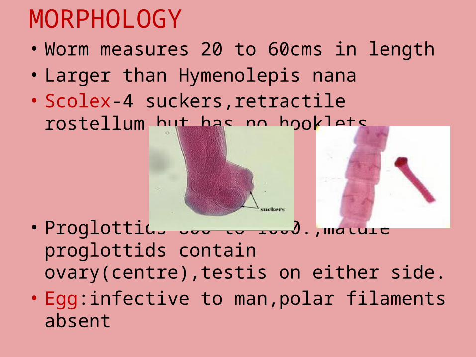
MORPHOLOGY • Worm measures 20 to 60cms in length• Larger than Hymenolepis nana• Scolex-4 suckers,retractile rostellum
but has no hooklets.
• Proglottids-800 to 1000.,mature proglottids contain ovary(centre),testis on either side.
• Egg:infective to man,polar filaments absent

LIFE CYCLE
PATHOGENESIS
• Rodents become infected by ingestion of arthropod hosts.
• Man acquires infection accidentally by ingestion of infected fleas containing cysticercoid larvae.
CLINICAL MANIFESTATIONS
• Most common in children.
• Usually asymptomatic but noted symptoms are mild diarrhoea,abdominal pain & vague GIT manifestations.
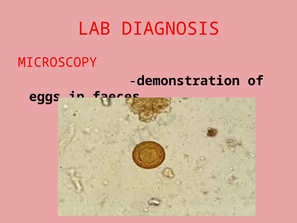
LAB DIAGNOSIS
MICROSCOPY -demonstration of eggs in
faeces
TREATMENT• Praziquantel• Niclosamide.
PROPHYLAXIS• Personal hygeine• Rodent control.
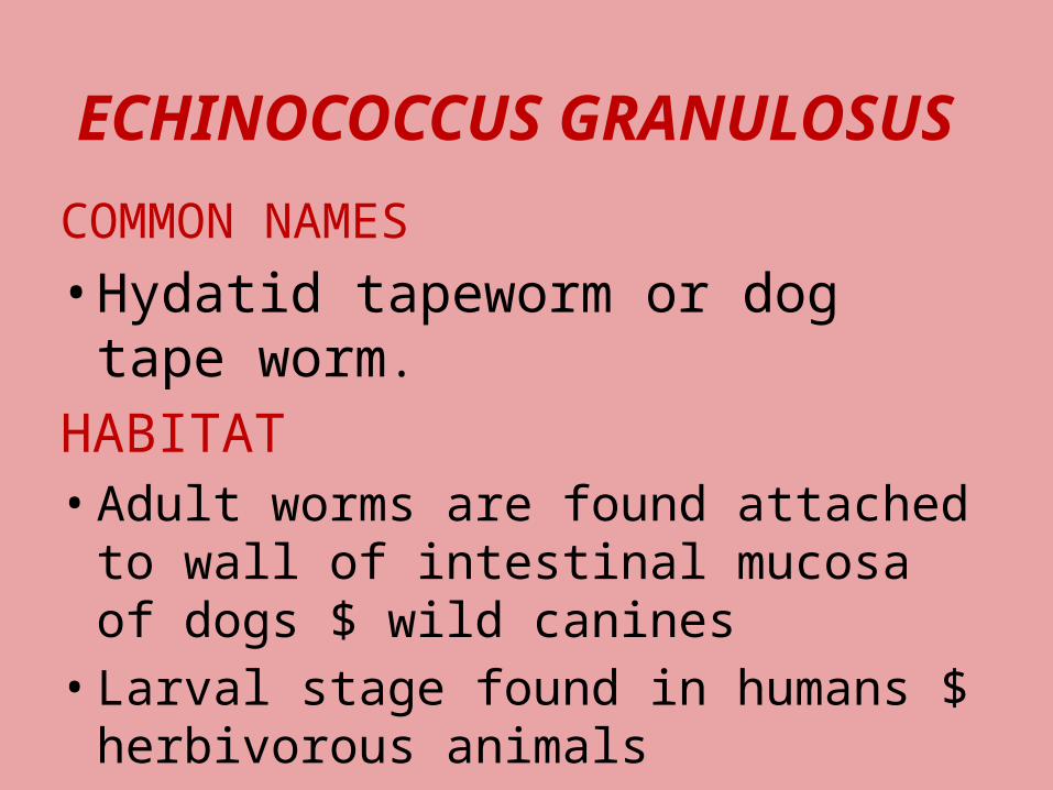
ECHINOCOCCUS GRANULOSUS
COMMON NAMES
• Hydatid tapeworm or dog tape worm.
HABITAT• Adult worms are found attached
to wall of intestinal mucosa of dogs $ wild canines
• Larval stage found in humans $ herbivorous animals
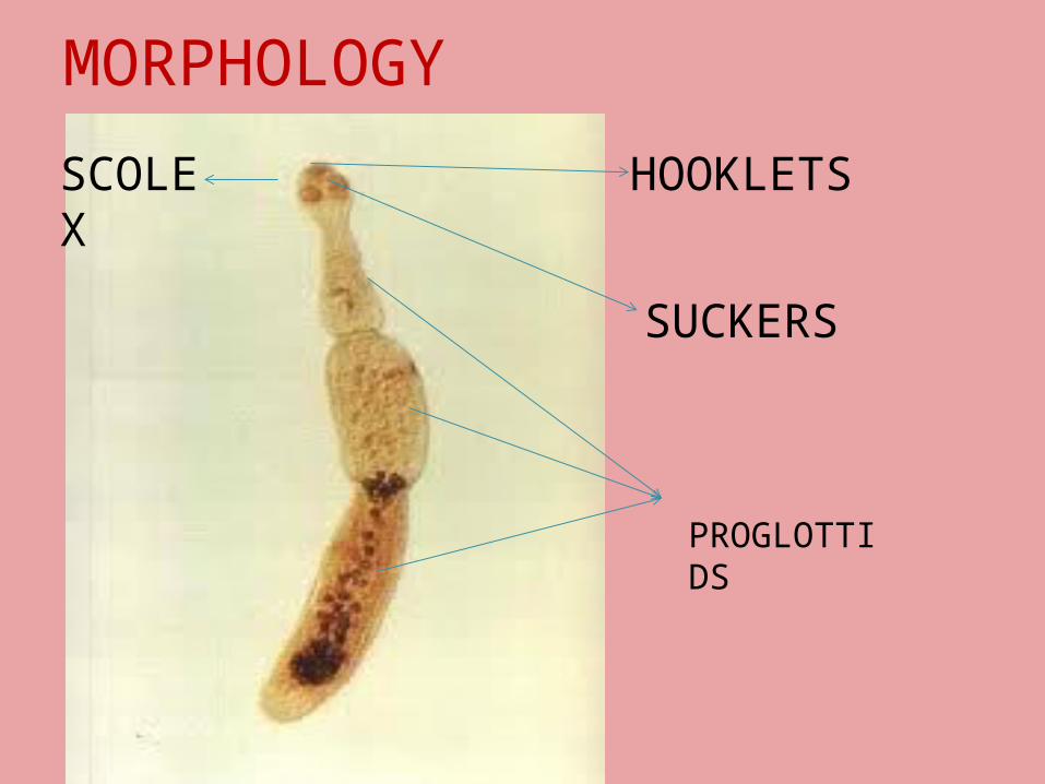
MORPHOLOGYM HOOKLETS
SUCKERS
SCOLEX
PROGLOTTIDS

EGG
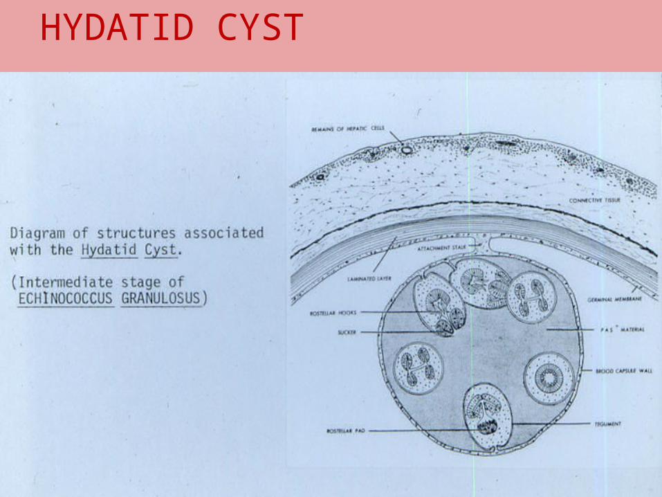
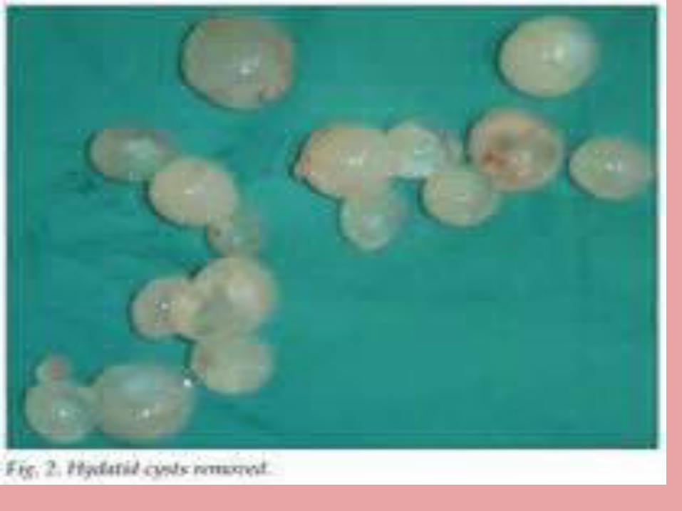
HYDATID CYST
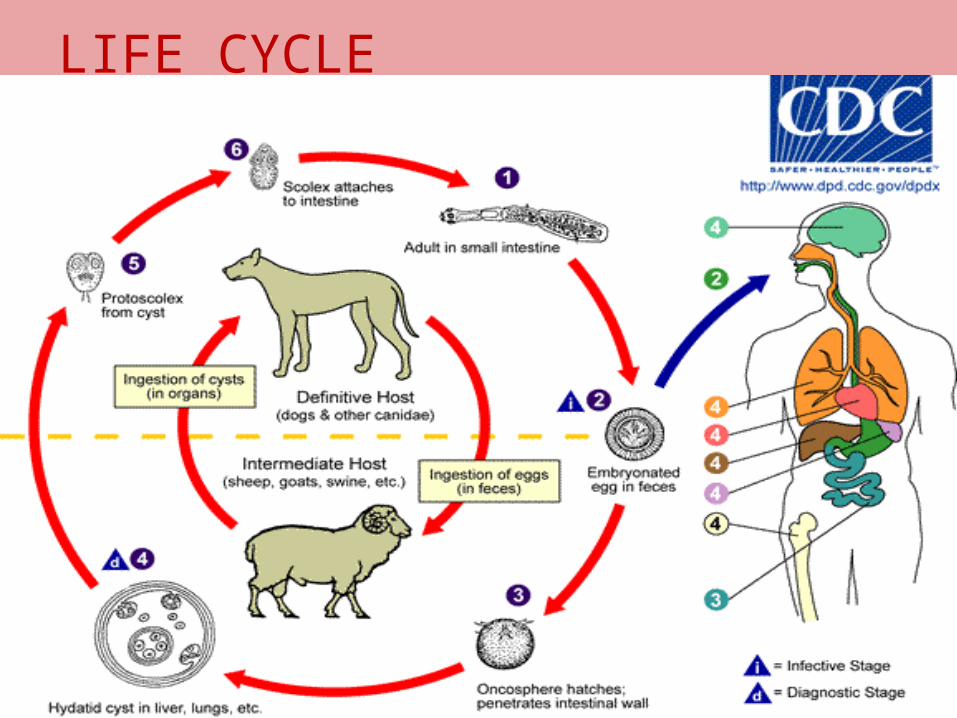
LIFE CYCLE
PATHOGENESIS• Depending on modes of development
cystic echinococcosis is of two types 1.primary cystic echinococcosis: -occurs after peroral infection
with E.granulosus eggs that gives rise to hydatid cyst in different parts of the body.
-most commonly these are found in liver and lung
2.secondary cystic echinococcosis: -occurs by rupture of primary
hydatid cyst by trauama
Cont…………… -in this condition protoscolices are
carried by blood circulation to different sites which develop into secondary hydatid cyst.
The growing cyst surrounded by three layers :
1.pericyst containing fibrous tissue.2.middle layer containing
fibroblasts,eosinophils& blood vessels.3.inner layer containing radially arranged
gaint cells &eosinophils.
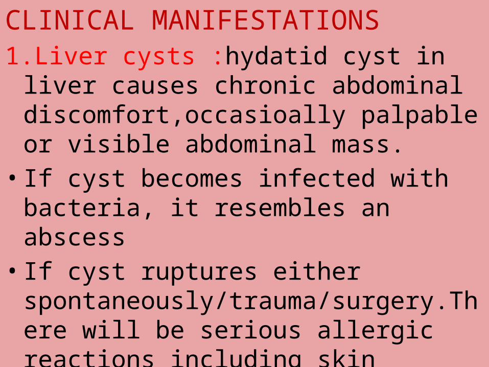
CLINICAL MANIFESTATIONS1.Liver cysts :hydatid cyst in liver
causes chronic abdominal discomfort,occasioally palpable or visible abdominal mass.
• If cyst becomes infected with bacteria, it resembles an abscess
• If cyst ruptures either spontaneously/trauma/surgery.There will be serious allergic reactions including skin rash,anaphylactic shock or death

2.lung cysts• Cysts are asymptomatic until they
become large enough to cause cough,shortness of breath,chest pain.
• Cyst rupture may lead to expectoration of hydatid fluid or membranes followed by the infection &lung abscess.
• If rupture occur into the lung,it cause pneumothorax,empyema,allergic reactions & even anaphylactic shock
3.other sites• Spleen• Kidney• CNS• Bones• Heart • Muscles • FGT• Eyes
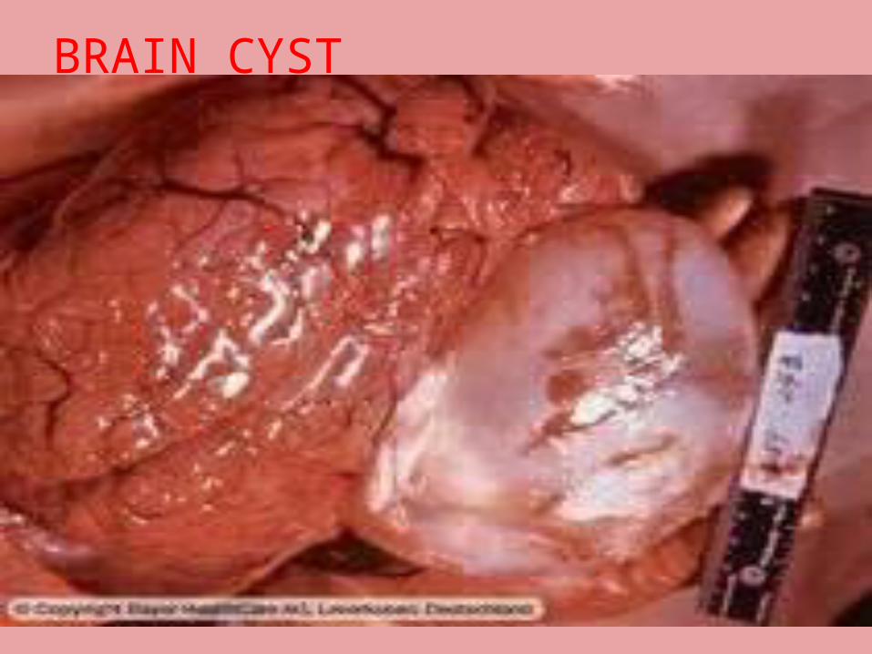
BRAIN CYST

LIVER CYSTS
LAB DIAGNOSIS
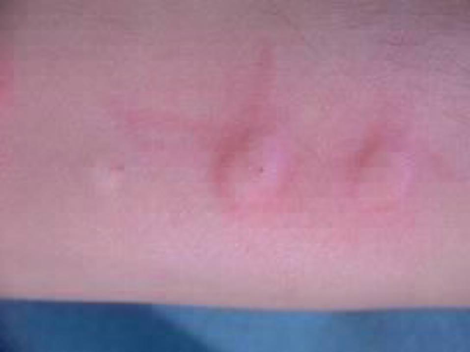
1. CASONI skin test
• It is immediate hypersensitivity skin test
• Ag used is sterile hydatid fluid drawn from unilocular hydatid cysts from sheep , pig , cattle , man
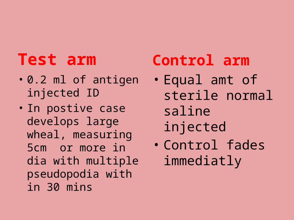
Test arm• 0.2 ml of antigen
injected ID• In postive case
develops large wheal, measuring 5cm or more in dia with multiple pseudopodia with in 30 mins
Control arm• Equal amt of
sterile normal saline injected
• Control fades immediatly
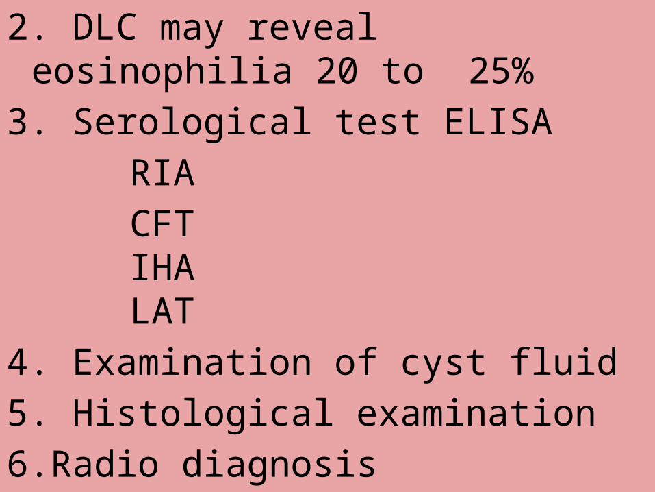
2. DLC may reveal eosinophilia 20 to 25%
3. Serological test ELISARIACFTIHALAT
4. Examination of cyst fluid5. Histological examination6.Radio diagnosis
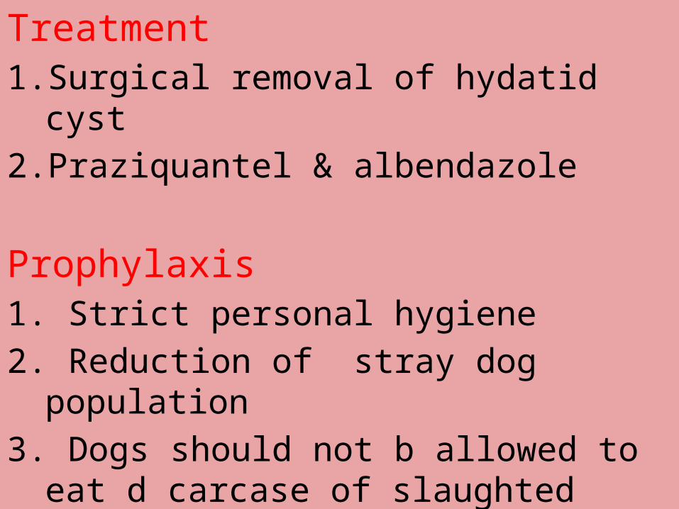
Treatment1. Surgical removal of hydatid cyst2. Praziquantel & albendazole
Prophylaxis1. Strict personal hygiene2. Reduction of stray dog population3. Dogs should not b allowed to eat d
carcase of slaughted animals
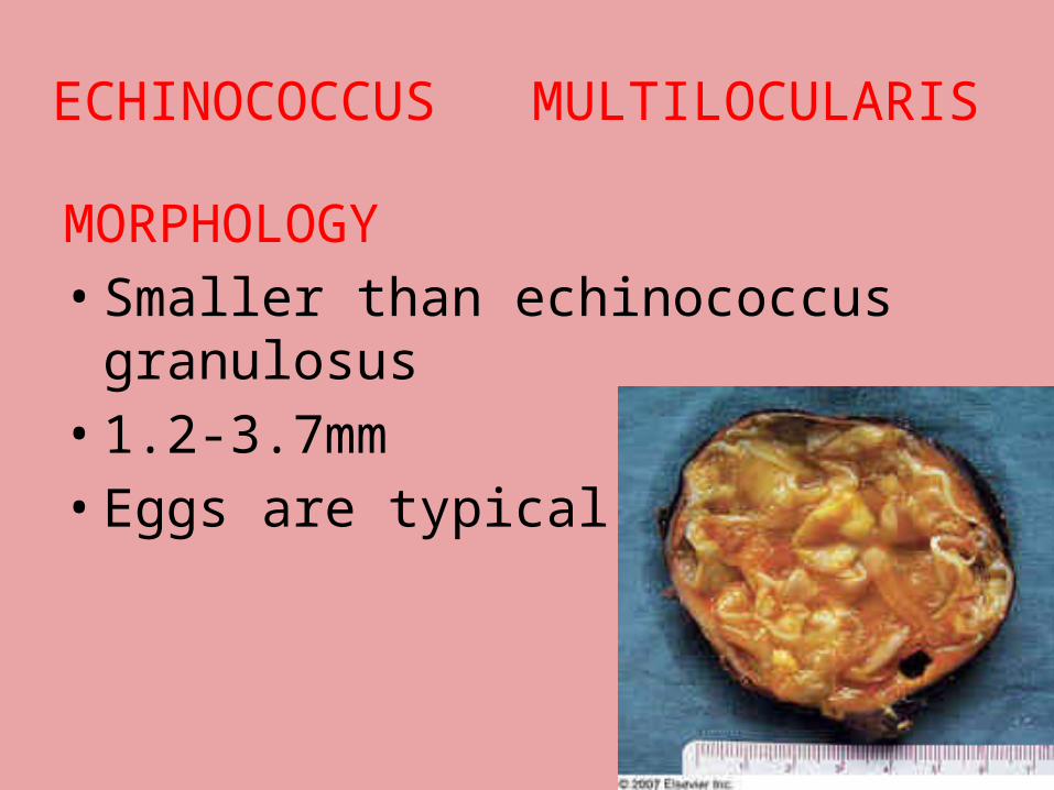
ECHINOCOCCUS MULTILOCULARIS
MORPHOLOGY• Smaller than echinococcus
granulosus• 1.2-3.7mm • Eggs are typical Taenia like
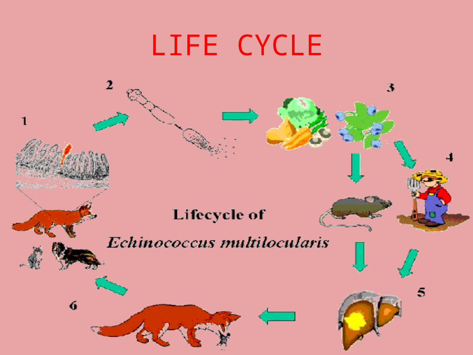
LIFE CYCLE
PATHOGENESIS• Alveolar echinococcosis/Alveolar or
multilocular Hydatid cyst.• ORGAN-Liver.• Destruction of liver parenchyma-
Hepatic failure.• Lesions spread to surrounding tissues
$ body sites via Lymphatic/Haematogenous routes.
• Central necrosis $ cavitation occurs $ cavity contains little or no fluid.
LAB DIAGNOSIS
• BIOPSY of affected organ,identification depends on recognition of germinal & laminated membranes.
• Radiodiagnosis:CT Scan,Ultra sound.
• Immunological Tests
TREATMENT
• Surgical removal of Hydatid cyst• Praziquantel & Albendazole.
PROPHYLAXIS• Strict personal hygiene.• Reduction of stray dog
population.

THANK YOU