Embed Size (px)
Citation preview
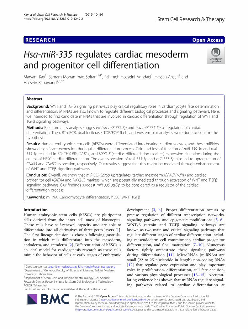
RESEARCH Open Access
Hsa-miR-335 regulates cardiac mesodermand progenitor cell differentiationMaryam Kay1, Bahram Mohammad Soltani1,4*, Fahimeh Hosseini Aghdaei1, Hassan Ansari2 andHossein Baharvand2,3,5*
Abstract
Background: WNT and TGFβ signaling pathways play critical regulatory roles in cardiomyocyte fate determinationand differentiation. MiRNAs are also known to regulate different biological processes and signaling pathways. Here,we intended to find candidate miRNAs that are involved in cardiac differentiation through regulation of WNT andTGFβ signaling pathways.
Methods: Bioinformatics analysis suggested hsa-miR-335-3p and hsa-miR-335-5p as regulators of cardiacdifferentiation. Then, RT-qPCR, dual luciferase, TOP/FOP flash, and western blot analyses were done to confirm thehypothesis.
Results: Human embryonic stem cells (hESCs) were differentiated into beating cardiomyocytes, and these miRNAsshowed significant expression during the differentiation process. Gain and loss of function of miR-335-3p and miR-335-5p resulted in BRACHYURY, GATA4, and NKX2-5 (cardiac differentiation markers) expression alteration during thecourse of hESC cardiac differentiation. The overexpression of miR-335-3p and miR-335-5p also led to upregulation ofCNX43 and TNNT2 expression, respectively. Our results suggest that this might be mediated through enhancementof WNT and TGFβ signaling pathways.
Conclusion: Overall, we show that miR-335-3p/5p upregulates cardiac mesoderm (BRACHYURY) and cardiacprogenitor cell (GATA4 and NKX2-5) markers, which are potentially mediated through activation of WNT and TGFβsignaling pathways. Our findings suggest miR-335-3p/5p to be considered as a regulator of the cardiacdifferentiation process.
Keywords: miRNA, Cardiomyocyte differentiation, hESC, WNT, TGFβ
IntroductionHuman embryonic stem cells (hESCs) are pluripotentcells derived from the inner cell mass of blastocysts.These cells have self-renewal capacity and are able todifferentiate into all derivatives of three germ layers [1].The first lineage decision is chosen following gastrula-tion in which cells differentiate into the mesoderm,endoderm, and ectoderm [2]. Differentiation of hESCs isan ideal model for cardiogenesis research as these cellsmimic the behavior of cells at early stages of embryonic
development [3, 4]. Proper differentiation occurs byprecise regulation of different transcription networks,signaling pathways, and epigenetic modifications [5, 6].WNT/β catenin and TGFβ signaling pathways areknown as two main and critical signaling pathways thatregulate different stages of cardiac differentiation includ-ing mesendoderm cell commitment, cardiac progenitordifferentiation, and final maturation [7–10]. Numerousfactors tightly orchestrate these signaling pathwaysduring differentiation [11]. MicroRNAs (miRNAs) aresmall (22 to 25 nucleotide in length) non-coding RNAs[12] that regulate gene expression and play importantroles in proliferation, differentiation, cell fate decision,and various physiological processes [13–15]. Accumu-lating evidence has shown that miRNAs regulate signal-ing pathways related to cardiac differentiation at
© The Author(s). 2019 Open Access This article is distributed under the terms of the Creative Commons Attribution 4.0International License (http://creativecommons.org/licenses/by/4.0/), which permits unrestricted use, distribution, andreproduction in any medium, provided you give appropriate credit to the original author(s) and the source, provide a link tothe Creative Commons license, and indicate if changes were made. The Creative Commons Public Domain Dedication waiver(http://creativecommons.org/publicdomain/zero/1.0/) applies to the data made available in this article, unless otherwise stated.
* Correspondence: [email protected]; [email protected] of Genetics, Faculty of Biological Sciences, Tarbiat ModaresUniversity, Tehran, Iran2Department of Stem Cells and Developmental Biology, Cell ScienceResearch Center, Royan Institute for Stem Cell Biology and Technology,ACECR, Tehran, IranFull list of author information is available at the end of the article
Kay et al. Stem Cell Research & Therapy (2019) 10:191 https://doi.org/10.1186/s13287-019-1249-2
posttranscriptional and posttranslational levels [16, 17].For instance, miR-15, miR-16 [18], and miR-430 [19]are identified as regulators of cell fate acquisitionthrough targeting the TGFβ signaling pathway. miR-1and miR-133 promote mesoderm formation [20] andmiR-499 promotes CPC differentiation into cardiomyo-cytes [21]. Here, we screen miRNAs that might be in-volved in cardiac differentiation through regulation ofWNT/β catenin and TGFβ signaling pathways. Bio-informatics analyses indicate that miR-335-3p andmiR-335-5p might regulate these two signaling path-ways through targeting core members of the pathways.Gain- and loss-of-function studies were performed toverify the exact role of these two miRNAs in cardiacdifferentiation. Our findings demonstrate that thesetwo miRNAs might regulate cardiac differentiation byactivating WNT and TGFβ signaling pathways. Thisactivation led to enhanced mesoderm cell commitmentand promoted cardiac progenitor cell differentiation.
Materials and methodsCell culture and differentiationHEK293 and SW480 cells were maintained in Dulbecco’smodified Eagle’s medium (DMEM) (Gibco), supple-mented with 10% heat-inactivated fetal bovine serumand 1% antibiotics (100 U/mL penicillin and 100 μg/mLstreptomycin) (Gibco). Cells were grown at 37 °C in ahumidified atmosphere with 5% CO2. The hESC lineRH5 [22] was expanded under feeder-free conditions onMatrigel-coated plates. Cardiomyocyte differentiationoccurred in a chemically defined medium, as previouslydescribed [23, 24] with minor modifications. Cells werestimulated with 20 ng/mL fibroblast growth factor 2(FGF2), 20 ng/mL activin A, and 10 ng/mL BMP4 in thefirst 36 h for mesoderm induction; then, cells weretreated with 20 ng/mL FGF2, 50 ng/mL BMP4, 0.5 mMretinoic acid, and 5 mM WNT inhibitor (IWP2) fromday 1.5 to day 5. Finally, cells were treated with 5 ng/mLFGF2 and 10 ng/mL BMP4 which resulted in cardiomyo-cyte differentiation. Samples were collected at differenttime points (0, 0.5, 1, 1.5, 2, 5, and 12 days) of differenti-ation for expression analysis.
Transfection of hESCsGain- and loss-of-function studies were done in (day 0) D0of differentiation. The miRCURY LNA™ microRNA mimic(Exiqon, Denmark) for miR-335-3p (MIMAT0004703),miR-335-5p (MIMAT0000765), and mimic control as wellas miRIDIAN microRNA miR-335-3p and miR-335-5phairpin inhibitors and miRIDIAN microRNA hairpininhibitor control (Dharmacon) were used for gain- andloss-of-function studies in which 8 × 10⁵ cells were platedin each 3.5-cm tissue culture dish, 24 h before transfection.When cells reached 80% confluence, they were transfected
by 50 nM siRNA or 5 nM mimic structures using Lipofec-tamine® 3000 reagent, based on the manufacturer’s instruc-tions. The efficiency of siRNAs and microRNA mimicstransfection was evaluated using BLOCK-iT Alexa FluorRed fluorescent oligo (Invitrogen).
RNA extraction and quantitative RT-PCRTotal RNA of harvested cells was extracted using TRI-zol™ reagent (Invitrogen, USA) according to the manu-facturer’s protocol. The total RNA was used for cDNAsynthesis after being treated with RNase-free DNase(Takara, Japan) in order to remove any DNA contamin-ation. cDNAs were synthesized using RevertAid™Reverse Transcriptase (Fermentase, Lithuania) accordingto the manufacturer’s instructions. For miRNA detec-tion, polyA tail was added to 3′ end of RNAs beforecDNA synthesis. RT-qPCR was performed using specificprimers (Table S1) by StepOne Real-Time PCR system(Applied Biosystems). GAPDH and small nucleolar RNA,C/D box 48 (SNORD48) were used as internal controls fornormalization of mRNAs and miRNA expression.
Immunocytochemistry (ICC)The seeded cells were washed once with phosphate buff-ered saline (PBS) and fixed with 4% (w/v) paraformalde-hyde (PFA) for 15 min at room temperature. The cellswere permeabilized using PBS containing 0.2% TritonX-100 for 10 min followed by blocking in a solution con-taining PBS and 10% Donkey serum for 1 h at roomtemperature. Next, cells were treated with primary anti-body overnight at 4 °C. After three washes in PBS, cellswere incubated with secondary antibody for 1 h at roomtemperature in the dark. Finally, cells were incubatedwith DAPI for nucleic acid staining and imaged with afluorescent microscope (IX71, Olympus, Japan). Imageoverlays and contrast enhancement were performedusing ImageJ software.
Dual luciferase assayDual luciferase assay was utilized to validate the directinteraction between miR-335-3p and miR-335-5p andtheir target genes 3′-UTRs. To this aim, HEK293 cellswere co-transfected with a psiCHECK-2 vectors includ-ing 3′-UTR of APC, AXIN-I, and SMAD7 andmiR-335-3p, miR-335-5p mimics, and siRNA structuresin 48-well plates. As negative controls, mimic and siRNAscramble were used in co-transfection. Luciferase activitywas measured 48 h after transfection using the Dual Lu-ciferase Reporter Assay System (Promega, USA) accord-ing to the manufacturer’s instructions.
TOP/FOP reporter assaysTOP/FOP reporter assays were carried out using theDual-Glo luciferase assay kit (Promega), based on the
Kay et al. Stem Cell Research & Therapy (2019) 10:191 Page 2 of 13

manufacturer’s instructions. SW480 cells were trans-fected with 1μg of constitutively active vector encodingRenilla luciferase, responsive firefly luciferase reporterplasmid Top Flash, each mimic and siRNA correspond-ing to the miR-335-3p, miR-335-5p and their relatedscrambled. Cells were harvested after 48 h, and both fire-fly and Renilla luciferase activity were measured in threebiological replicates according to the manufacturer’s in-structions. The firefly luciferase activity was normalizedagainst Renilla luciferase activity.
Western blottingThe protein was extracted from the samples. Samplescontaining 40 μg purified protein were separated by 12%SDS/PAGE, transferred to PVDF membranes (SantaCruz), and run at 100 V for 1.5 h at room temperature.The PVDF membrane was subsequently blocked by 5%BSA in phosphate-buffered saline (PBS) containing 0.1%Tween for 1 h at room temperature, followed by over-night incubation at 4 °C with phospho BRACHYURYprimary antibodies (1:500, Cell Signaling). The blotmembrane was washed and incubated with anti-rabbitsecondary antibody (1:1000, Santa Cruz) for 1 h at roomtemperature. The protein bands were visualized by anenhanced chemiluminescence (ECL) detection system(Amersham, Piscataway, NJ). The membranes werestripped and re-probed with β-actin for verification ofprotein loading. The bands were quantified using animage analyzer program (ImageJ).
ResultsBioinformatics analyses introduced miR-335 as a potentialregulator of cardiac differentiationWNT and TGFβ signaling pathways are known as mainplayers in cardiac differentiation. In this study, we aimedto find out miRNAs that are crucially important for driv-ing cardiac differentiation through regulation of WNTand TGFβ signaling pathways. To this aim, a four-stepfiltering approach was performed to nominate somemiRNAs (Fig. 2a). At the first step, 973 miRNAs werepredicted to target WNT and TGFβ signaling pathwaysusing miRWalk, a target prediction resource that usesseveral miRNA target prediction tools. Then, consider-ing the number of genes which are targeted by a miRNAin each pathway, the numbers of MREs (miRNA recog-nition elements) in 3′UTR sequences of each targetgene, and the annealing and conservation status of eachMRE, seven candidate miRNAs were chosen for furtheranalyses (Additional file 1: Table S2). Among candidatemiRNAs, miR-335-3p/5p hosted by mesoderm-specifictranscript (MEST) gene were chosen for further investi-gations concerning its role in cardiogenesis.
Differentiation of hESCs into cardiomyocytesIn vitro differentiation of hESC, RH5, to cardiomyocyte-like cells was successfully performed (Fig. 1a). Differenttime points representing different cell fates duringcardiac differentiation were considered (i.e., pluripotentstem cells (day 0), mesendoderm (day 1.5), cardiac pro-genitor (day 5), and cardiomyocyte cells (day 12)).NANOG stem cell marker expression was substantiallydecreased while the BRACHYURY (mesendodermalmarker) expression level reached its highest level inmesendoderm stage (day 1.5) (Fig. 1b). In addition,NKX2-5 and ISL1 (early cardiac differentiation markers)expression levels elevated from day 2 and substantiallyincreased during the process. As expected, cardiac-spe-cific markers (TNNT and MYH6) were significantly in-creased at the final stage (day 12) (Fig. 1b), followed byspontaneous beating of the cardiac cells (Additional file 2:Movie S1). The flow cytometry analysis indicated that ~51% of the cells were BRACHYURY positive on day 1.5,~ 87% of the cells were NKX2-5 positive on day 5(cardiac progenitor cells) and ~ 96% of the cells wereMYH6 positive on day 12 (the final step of cardiomyo-cyte differentiation) (Fig. 1c). Furthermore, immuno-staining indicated that hESCs were positive for OCT4,whereas hESC-CMs stained positive for cardiac-specificmarkers MYH6 (Fig. 1d), approving that these cells weresuccessfully differentiated into cardiomyocytes.
MiR-335 expression pattern during the human cardiacdifferentiation processMiR-335-3p and miR-335-5p expression status was mea-sured at seven time points of human cardiac differenti-ation process, using RT-qPCR. Both miR-335-3p andmiR-335-5p had relatively high levels of expressionduring this process (data not shown); however, their ex-pression pattern was different. While miR-335-3p wastransiently upregulated at day 1 and substantially down-regulated to the end of differentiation process,miR-335-5p showed no significant alteration during thedifferentiation (Fig. 2b).
The effect of MiR-335 expression alteration on cardiacdifferentiationIn order to examine the effect of miR-335-3p/5p expres-sion alteration on cardiac differentiation, gain- andloss-of-function study was performed according tomiR-335-3p and miR-335-5p expression profiles (Fig. 2c).In this part, miR-335-3p and miR-335-5p mimics or theircorresponding siRNAs were transfected into differentiat-ing hESCs on D0 to induce specific overexpression anddownregulation, respectively. Red fluorescent oligotransfection indicated ~ 45% transfection efficiency(Fig. 2d), and specific up- and downregulation of eachmiRNA was further confirmed using RT-qPCR, 24 and
Kay et al. Stem Cell Research & Therapy (2019) 10:191 Page 3 of 13

Fig. 1 (See legend on next page.)
Kay et al. Stem Cell Research & Therapy (2019) 10:191 Page 4 of 13

(See figure on previous page.)Fig. 1 Characterization of hESC-derived cardiomyocyte. a Schematic description of cardiomyocyte differentiation protocol. Activin A and BMP4compounds were used to induce differentiation of hESCs into mesoderm. Then, WNT inhibitor reagent (IWP2) was used to induce cardiacprogenitor cell development followed by cardiomyocyte formation. b Time-dependent expression of NANOG (pluripotency marker), BRACHYURY(mesoderm marker), NKX2-5, ISL1 (cardiac progenitor cell markers), TNNT2, and MYH6 (cardiomyocyte markers) during the cardiac differentiationprocess. RT-qPCR data are presented as mean ± SEM normalized against day 0 data, for n = 3 independent experiments. GAPDH was used as ahousekeeping gene. c Flow cytometry results confirmed the expression of BRACHYURY (day 1.5), NKX2-5 (day 5), and MYH6 (day 12) during thedifferentiation process. d Immunocytochemistry analysis of OCT4 (pluripotent hESCs) and MYH6 (cardiomyocytes) revealed the stemness potencyof hESCs and successful differentiation of cardiomyocyte. Differentiated cardiomyocytes are shown in two magnitudes
Fig. 2 MiR-335 expression alteration during cardiac differentiation. a Schematic presentation of different stages of miRNA screening. b miR-335-3p(top) and miR-335-5p (bottom) expression pattern during hESC cardiac differentiation. c Gain- and loss-of-function strategy in which miR-335-specific mimics and siRNAs were transfected on day 0 of differentiation process. d Flow cytometry results showed that hESCs were efficientlytransfected by miR-335 specific mimics or siRNAs along with 50 nM BLOCK-iT Alexa Fluor Red Fluorescent oligonucleotide. e The RT-qPCRconfirmed significant (P value < 0.05) overexpression of miR-335-3p (top-left) and miR-335-5p (bottom-left) but significant downregulation of miR-335-3p (top- right) and miR-335-5p (bottom-right), 24 and 48 h after transfection. RT-qPCR data are presented as mean ± SEM normalized againstday 0 data. GAPDH was used as a housekeeping gene
Kay et al. Stem Cell Research & Therapy (2019) 10:191 Page 5 of 13

48 h post transfection (Fig. 2e). Immunostaining of thecells transfected by mimic-scr, mimic-3p, or mimic-5pindicated successful cardiac differentiation (Fig. 3a). Todetermine the differentiation stage which has been mostaffected by miR-335 expression alteration, the expressionlevel of stage-specific markers was measured byRT-qPCR. Here, the expressions of BRACHUYRY andMESP1 as mesodermal markers, NKX2-5 and GATA4 ascardiac progenitor markers, HCN4 and ISL1 as progeni-tor markers of the first and second heart field, andTNNT2 and CNX43 as cardiomyocyte markers wereanalyzed. RT-qPCR data indicated that 48 h post-trans-fection of mimic-3p or mimic-5p, the expression level ofBRACHYURY was significantly increased (Fig. 3b). Con-sistently, downregulation of miR-335-3p was followed bysignificant reduction of BRACHUYRY expression(Fig. 3b). The results of western blotting also confirmedthe increased protein level of BRACHYURY followingmimic-3p and mimic-5p treatments compared to themimic-scr control (Fig. 3b, right). Increased or decreasedlevel of miR-335 did not show a significant effect onMESP1 expression level (Fig. 3c). Interestingly, GATA4and NKX2-5 expression levels were significantly in-creased following miR-335-3p overexpression while theydecreased after using their related siRNA (Fig. 3d, e).Similar significant results were obtained for miR-335-5pagainst GATA4, but results were non-significant againstNKX2-5 expression (Fig. 3d, e).Following transfection of mimic-3p and mimic-5p,
HCN4 (the first heart field marker) was significantly up-regulated as well; however, miR-335-3p and miR-335-5pdownregulation results were not consistent with it(Fig. 3f). MiR-335 expression alteration also has effecton ISL1 (the second heart field marker) expression.Transfection of both mimic-3p and mimic-5p resulted indownregulation of ISL1 expression while, miR-335downregulation resulted in increased ISL1 expression(Fig. 3g). Overexpression of miR-335-3p significantly up-regulated CNX43 expression but suppressed the expres-sion of TNNT2 at the final stage of the differentiationprocess (Fig. 3h, i). Interestingly, miR-335-5p overex-pression could significantly upregulate TNNT2 with nosignificant effects on CNX43 expression.
MiR-335 as an activator of WNT signaling pathwayTarget prediction tools predicted 5 and 1 potential targetsites for miR-335-3p within the 3′UTR sequences ofAPC and AXIN-I genes, respectively. For miR-335-5p, asingle recognition site was predicted within 3′UTRsequences of both APC and AXIN-I genes (Fig. 4a).Alterations in the expression of APC and AXIN-I targetgenes were investigated 48-h post-transfection ofmiR-335 mimics or siRNAs on D0 of hESC differenti-ation. Also, direct interaction of each miRNA mimic and
3′UTR sequences of APC and AXIN-I predicted targetgenes was investigated in HEK293 cells, using dual lucif-erase reporter assay.RT-qPCR indicated that up- or downregulation of
both miR-335-3p (Fig. 4b, top) and miR-335-5p (Fig. 4c,top) did not significantly affect APC gene expression indifferentiating cells. Co-transfection of reporter con-struct containing APC-3′UTR and miR-335-3p mimic inHEK293 cells resulted in a significant reduction of lucif-erase activity, and consistently, luciferase activity wasincreased after the application of siRNA againstmiR-335-3p, compared to scrambled controls (Fig. 4b,bottom). This effect was abrogated using off target UTR(the same length of UTR with no available target site formiRNA), thereby approving the direct interactionbetween miR-335-3p and APC 3′UTR. Similar dual lucif-erase assay results indicated that miR-335-5p was notinteracting with APC 3′UTR sequence (Fig. 4c, bottom).Transfection of differentiating hESCs with miR-335-3p
mimic resulted in significant downregulation of AXIN-Iexpression, compared to scrambled control transfection(Fig. 4d, top). Furthermore, dual luciferase assayconfirmed the direct interaction between miR-335-3pmimic and 3′UTR sequence of AXIN-I (Fig. 4d, bottom).Transfection with miR-335-5p mimic also caused a sig-nificant reduction in AXIN-I transcripts as examined byRT-qPCR (Fig. 4e, top). However, dual luciferase assaydid not show a direct interaction between miR-335-3pand 3′UTR sequence of AXIN-I gene (Fig. 4e, bottom).TOP/FOP flash assay indicated that overexpression of
miR-335-3p or miR-335-5p led to significant upregula-tion of WNT signaling pathway in SW480 cells, com-pared to mimic-scr controls (Fig. 4f). Similar results tothose of Top/Fop flash assay were observed whenmiR-335 was downregulated by specific siRNA (Fig. 4f).The RT-qPCR analysis against CYCLIND1 and c-MYC(downstream targets of WNT signaling pathway) alsoconfirmed the upregulation of WNT signaling, followingmiR-335-5p overexpression (Additional file 1: Figure S1).Alterations in miR-335-3p expression did notsignificantly affect CYCLIND1 and c-MYC expression(Additional file 1: Figure S1). Altogether, these datasuggested that miR-335-3p might enhance the WNTsignaling through targeting the inhibitory components ofWNT signaling pathway (i.e., APC and AXIN-I) whilemiR-335-5p could activate this pathway indirectly with-out any direct effect on these two target genes.
MiR-335 as an inducer of TGFβ signaling pathwayThe in silico analyses suggested that miR-335 could regu-late TGFβ signaling through targeting SMAD7 transcripts.It was predicted that both miR-335-3p and miR-335-5ptarget inhibitory SMAD7 gene (Additional file 1: FigureS2A). RT-qPCR indicated that SMAD7 expression was
Kay et al. Stem Cell Research & Therapy (2019) 10:191 Page 6 of 13

Fig. 3 MiR-335 regulates earlier stages of hESC cardiac differentiation. a Immunocytochemistry analysis showed that overexpression of hsa-miR335-3por hsa-miR335-5p on day 0 did not affect the final outcome of cardiac differentiation on day 12 (cardiomyocytes), as detected by MYH6 expression inall groups. b Overexpression of both hsa-miR335-3p (top) and hsa-miR335-5p (bottom) resulted in significant elevation of BRACHYURY (mesodermmarker) expression. Consistently, hsa-miR335-3p downregulation resulted in reduced expression of BRACHYURY. Western blot results indicate anincreased level of BRACHYURY following the mimic-3p and mimic-5p treatment, compared to mimic-scr control (right). c No significant expressionalteration was detected for MESP1 following alterations in hsa-miR335 expression. d, e Expressions of GATA4 and NKX2-5 (main cardiac progenitormarkers) were both increased following miR-335-3p (top) and miR-335-5p (bottom) overexpression. Downregulation of miR-335 had reverse effect onboth of these cardiac progenitor markers. f Expression of HCN4 (the first heart field marker) was significantly increased following miR-335-3p (top) andmiR-335-5p (bottom) overexpression. g Alterations in the expression of miR-335-3p (top) and miR-335-5p (bottom) had a significant reverse effect onISL1 (the second heart field marker) expression. h, i Has-miR-335-3p (top) overexpression increased CNX43 expression but decreased TNNT2 expression.However, has-miR-335-5p decreased CNX43 but enhanced TNNT2 expressions. RT-qPCR data are presented as mean ± SEM normalized against mimic-scr and siRNA-scr. GAPDH was used as a housekeeping gene
Kay et al. Stem Cell Research & Therapy (2019) 10:191 Page 7 of 13

significantly reduced following miR-335-3p and miR-335-5p overexpression, 48 h after transfection in differentiatinghESCs. Consistently, downregulation of these miRNAshad reverse effects (though non-significant) on SMAD7expression (Fig. 5a, top).Luciferase activity was decreased following miR-335-3p
overexpression, and this suppression was relieved bydownregulation of miR-335-3p as detected by dual
luciferase assay (Fig. 5b, top). Significant reductions in lu-ciferase activity were also detected after miR-335-5p over-expression (Fig. 5a, bottom), while miR-335-5pdownregulation could not rescue luciferase activity, sig-nificantly (Fig. 5b, bottom). Overall, these data suggestedthat both miR-335-3p and miR-335-5p were capable oftargeting SMAD7 transcript. The effect of MiR-335 over-expression on other TGFβ signaling pathway components
Fig. 4 MiR-335 activates WNT signaling through targeting APC and AXIN-I. a Pairing status of miR-335-3p and miR-335-5p with MREs within 3′UTRsequences of APC and AXIN-I. b Alterations in the expression of miR-335-3p did not significantly change the transcription level of APC gene (top);however, dual luciferase assay supported a direct interaction between them (bottom). c Neither RT-qPCR nor dual luciferase assay showed aninteraction between miR-335-5p and APC. d Alterations in the expression of miR-335-3p significantly changed the AXIN-I transcript level (top), anddual luciferase assay indicated a direct interaction between them (bottom). e Although RT-qPCR suggested downregulation of AXIN-I followingmiR-335-5p overexpression (top), dual luciferase assay did not show an interaction between them (bottom). f Overall inductive effect of both miR-335-3p (top) and miR-335-5p (bottom) on WNT signaling as detected by TOP/FOP flash assay. All experiments were done in three biologicalreplicates and results are presented as mean ± SEM
Kay et al. Stem Cell Research & Therapy (2019) 10:191 Page 8 of 13

was also investigated using RT-qPCR. Results indicatedthat both miR-335-3p and miR-335-5p overexpression ledto TGFβR-I upregulation (Fig. 5c, d). Alterations in the ex-pression of miR-335 had no significant effect on SMAD2and SMAD3 expression (Additional file 1: Figure S2B-C).Altogether, these data suggest that both miR-335-3p andmiR-335-5p might activate TGFβ signaling pathwaythrough targeting SMAD7 transcript.
DiscussionTGFβ and WNT signaling pathways have some pleio-tropic and multifunctional effects and regulate several
biological processes including embryonic development,as well as cell differentiation, proliferation, and survival[25–27]. These two pathways play critical roles inorchestration of cardiac development and differentiation[28–30]. It is known that regulation of TGFβ and WNTsignaling pathway is essential for mesoderm cell fatecommitment, as well as cardiomyocyte progenitor cellformation and cardiomyocyte maturation [31]. Thesecardiogenesis steps are closely regulated by differentmechanisms at posttranscriptional and posttranslationallevels. MiRNAs are known to regulate different biopro-cesses through targeting related signaling pathways
Fig. 5 MiR-335 activates TGFβ signaling pathway through targeting SMAD7. a Alterations in the expression of SMAD7 followed byoverexpression and downregulation of miR-335-3p (top) and miR-335-5p (bottom). b Dual luciferase assay study confirmed a directinteraction between miR-335-3p (top) and miR-335-5p (bottom), and SMAD7 3′UTR. Alteration in the expression of TGFβR-I was assessed byRT-qPCR followed by miR-335-3p and miR-335-5p overexpression (c) and downregulation (d) in hESC. RT-qPCR data are presented asmean ± SEM normalized against mimic-scr and siRNA-scr. GAPDH was used as a housekeeping gene
Kay et al. Stem Cell Research & Therapy (2019) 10:191 Page 9 of 13

[32–34]. For example, hsa-miR-590-5p was shown to beinvolved in cardiogenesis through regulating TGFβsignaling [35]. Also, hsa-miR-23b cluster was shown toinduce proliferation in hepatocytes through TGFβ path-way inhibition [36] and hsa-miR-126 is known to in-duce VEGF signaling and promote angiogenesis [37].Here, we intended to introduce miRNAs whichfine-tune TGFβ and WNT signaling pathways, duringthe cardiogenesis process. Bioinformatics analyses in-troduced 7 miRNAs which had multiple targets in bothWNT and TGFB signaling pathways (Additional file 1:Table S2). Finally, has-miR-335 that is well conservedin mammals and is under the control of mesoderm-spe-cific enhancers in the second intron of MEST [38](Additional file 1: Figure S3), was chosen for further in-vestigation. Also, miR-335 was reported to be involvedin the fate commitment of mesoderm during the mouseembryonic differentiation [38]. MiR-335-3p (miRBaseID: MIMAT0004703) and miR-335-5p (miRBase ID:MIMAT0000765) were both highly expressed in matureheart [38]. It is predicted that these two miRNAs targetdifferent genes in WNT and TGFβ signaling pathways(Additional file 1: Tables S3–S4, Figure S4).H5 human ESCs were successfully differentiated into
beating cardiomyocytes after 12 days (Fig. 1a). Then,the expression pattern of mesoderm, cardiac progeni-tor, and mature cardiomyocyte molecular markers de-fined the span of each stage (Fig. 1b) which was furtherconfirmed by flow cytometry (Fig. 1c) and ICC results(Fig. 1d). RT-qPCR results indicated a distinct expres-sion pattern for miR-335-3p and miR-335-5p during thecardiac differentiation process. While miR-335-3pexpression was significantly altered (up to six folds)during the mesoderm stage (before day 1.5), miR-335-5p was changed much less (1.5-fold) at this stage.miR-335-5p expression was strongly altered at the pro-genitor stage (after day 1.5) (Fig. 2b). Up- and downreg-ulation of miR-335 since D0 of the differentiationprocess lasted until the progenitor stage of the process,2 days post-transfection (Fig. 2e). ICC results indicatedthat miR-335 expression alterations did not change thefate of differentiating cells towards cardiomyocytes(Fig. 3a). RT-qPCR as well as western blot results indi-cated that both miR-335-3p and miR-335-5p gain offunctions enhanced mesodermal cell commitment, asmirrored by the increased BRACHYURY expressionlevel. Results of loss-of-function study also supportedgain-of-function results for miR-335-3p (Fig. 3b). It wasconsistent with a previous report showing that miR-335stabilizes the lineage decision in mouse ESCs formesendoderm formation [38]. Also, Schoeftner et al.consistently reported that miR-335 targets OCT4 andRb to control mESC proliferation and induced differen-tiation [39].
Following miR-335-3p or miR-335-5p gain- andloss-of-functions, cardiac progenitor markers (GATA4and NKX2-5) were also up- and down-regulated,respectively (Fig. 3d, e). Upregulation of GATA4 andNKX2-5 progenitor markers could enhance the firstheart field progenitor cell commitment through upregu-lating HCN4 (Fig. 3f) and downregulating ISL1 expres-sion (Fig. 3g). RT-qPCR results against late cardiacdifferentiation markers (TNNT2 and CNX43) suggestthat miR-335-3p and miR-335-5p might have comple-mentary effects on the process.While miR-335-3p downregulated TNNT2 expression,
miR-335-5p significantly upregulated it within the cells(Fig. 3i). Similar results were also obtained for CNX43expression after alterations in miR-335-3p and miR-335-5p expression (Fig. 3h). Altogether, these data suggestthat miR-335 enhances cardiac differentiation throughupregulation of the expression of mesoderm (BRACHY-URY) and cardiac progenitor marker (GATA4 andNKX2-5) genes. Nevertheless, molecular mechanism(s)via which these miRNAs regulate the expression of thesemarkers remained undiscovered.Bioinformatics analyses indicated that miR-335 regulates
WNT and TGFβ signaling pathways. Moreover, RT-qPCR(Fig. 4b–e, top) and dual luciferase assay (Fig. 4b–e, bot-tom) showed that miR-335-3p specifically target 3′UTRsequences of APC and AXIN-I, which are two main mem-bers of WNT inhibitor complex. These data suggestedthat miR-335-3p but not miR-335-5p could regulate APCexpression at the post-transcriptional level.Although miR-335-5p had no direct interaction with
3′UTR of these two target genes, but RT-qPCR re-sults indicated a significant downregulation of AXIN-Itranscript (Fig. 4e, top). Thus, it could be concludedthat miR-335-5p indirectly downregulated AXIN-Iexpression without interacting with its transcripts.Consistently, both of these miRNAs were capable ofactivating WNT signaling pathway as detected byTop/Fop flash assay (Fig. 4f). This is also consistentwith a previous report which showed that miR-335-5pactivates WNT signaling pathway through DKK1downregulation [40].Interaction between both miR-335-3p and miR-335-5p
and SMAD7 3′UTR was also confirmed by RT-qPCR(Fig. 5a) as well as dual luciferase assay (Fig. 5b). Resultsindicated that these two miRNAs could enhanceTGFβR-I expression (Fig. 5c, d). There are twoSMAD-binding elements within TGFβR-I promoter rec-ognized by SMAD7, which inhibit TGFβR-I expression[41]. Downregulation of SMAD7 by these two miRNAscould lessen the inhibitory effect of SMAD7 andenhance TGFβR-I expression. Altogether, the resultsproposed that miR-335-3p and miR-335-5p could acti-vate WNT and TGFβ signaling pathways.
Kay et al. Stem Cell Research & Therapy (2019) 10:191 Page 10 of 13

LEF-1/β-catenin complex is reported to bind to theTCF binding site at the BRACHYURY promoter se-quence and enhance its expression [42]. TGFβ1 also in-duces the expression of BRACHYURY in humancarcinoma cells [43]. In other words, activation of WNTand TGFβ signaling pathways could enhance BRACHY-URY expression, as also observed in the current experi-ment. Furthermore, WNT signaling pathway activationwas shown to enhance GATA4 [44] as well as NKX2-5expression through hindering HDAC1 inhibitory effect[45]. There is also evidence showing that TGFβ activa-tion enhances GATA4 and NKX2-5 expression [46–48].
ConclusionBased on the bioinformatics and experimental valida-tions, we propose a regulatory network for miR-335-3pand miR-335-5p during cardiomyocyte differentiation(Fig. 6). Accordingly, both miR-335-3p and miR-335-5pmight activate WNT and TGFβ signaling pathways that,
in turn, induce mesoderm (BRACHYURY) and progeni-tor (GATA4 and NKX2-5) marker expression andcardiac differentiation.
Additional files
Additional file 1: Table S1. Primer sequences used in this research.Table S2. Final list of candidate miRNAs. Table S3. List of potential miR-335-3p target genes related to WNT and TGFβ signaling pathways. TableS4. List of potential miR-335-5p target genes related to WNT and TGFβsignaling pathways. Figure S1. RT-qPCR results of C-MYC and CCND1 aftermimics and siRNA treatment for miR-335-3p (A) and miR-335-5p (B). Allexperiments were done in three biological replicates and presented asmean ± SEM. Figure S2. RT-qPCR results of SMAD2 and SMAD3 expres-sion. A) Pairing status of miR-335-3p (left) and miR-335-5p (right), with 3′UTR of SMAD7 gene. B) SMAD2 expression was not significantly changedfollowed by miR-335-3p (top) and miR-335-5p (bottom) overexpression. C)RT-qPCR data also showed no significant alterations in SMAD3 expressionfollowing miR-335-3p (top) and miR-335-5p (bottom) overexpression. Alldata are presented as mean ± SEM normalized against mimic-scr andsiRNA-scr. GAPDH was used as a housekeeping gene. Figure S3. Gen-omic location of mir-335 presented in UCSC genome browser. miR-335 is
Fig. 6 Schematic presentation of miR-335 involvement in cardiomyocyte differentiation procedure (details are discussed in the text)
Kay et al. Stem Cell Research & Therapy (2019) 10:191 Page 11 of 13

located within the second intron of MEST gene, containing twoconserved mature miRNAs (highlighted in red) including miR-335-3p andmiR-335-5p. Figure S4. The potential targets of miR-335 in TGFβ (A) andWNT (B) signaling pathways according to the KEGG pathway. The targetgenes are marked with red stars. (DOCX 474 kb)
Additional file 2: Movie S1. (AVI 2586 kb)
AcknowledgementsAuthors would like to thank all members of both departments for their kindcomments.
FundingThis work was supported by a grant from Royan Institute and the IranNational Science Foundation (INSF, grant no. 96001316) to H.B., and NIMAD(Grant No. 943387) and TMU to B.M.S.
Availability of data and materialsAll data generated or analyzed during this study are included in thispublished article [and its supplementary information files].
Authors’ contributionsMK contributed to do experiment design and carried out the experiment,and also contributed in the manuscript preparation. HA and FHA did theexperiment. BMS and HB did the experiment design, result interpretation,and manuscript preparation. All authors read and approved the finalmanuscript.
Ethics approval and consent to participateThis study was approved by the Ethical/Scientific Committee of RoyanInstitute.
Consent for publicationNot applicable.
Competing interestsThe authors declare that they have no competing interests.
Publisher’s NoteSpringer Nature remains neutral with regard to jurisdictional claims inpublished maps and institutional affiliations.
Author details1Department of Genetics, Faculty of Biological Sciences, Tarbiat ModaresUniversity, Tehran, Iran. 2Department of Stem Cells and DevelopmentalBiology, Cell Science Research Center, Royan Institute for Stem Cell Biologyand Technology, ACECR, Tehran, Iran. 3Department of DevelopmentalBiology, University of Science and Culture, Tehran, Iran. 4Department ofMolecular Genetics, Faculty of Biological Sciences, Tarbiat Modares University,14115-111, Tehran, Iran. 5Royan Institute, P.O. Box: 16635-148, BanihashemSq., Banihashem St., Ressalat Highway, Tehran 1665659911, Iran.
Received: 24 September 2018 Revised: 6 April 2019Accepted: 30 April 2019
References1. Evans MJ, Kaufman MH. Establishment in culture of pluripotential cells from
mouse embryos. nature. 1981;292(5819):154.2. Burdon T, Smith A, Savatier P. Signalling, cell cycle and pluripotency in
embryonic stem cells. Trends Cell Biol. 2002;12(9):432–8.3. Keller GM. In vitro differentiation of embryonic stem cells. Curr Opin Cell
Biol. 1995;7(6):862–9.4. Vidarsson H, Hyllner J, Sartipy P. Differentiation of human embryonic stem
cells to cardiomyocytes for in vitro and in vivo applications. Stem Cell RevRep. 2010;6(1):108–20.
5. Verma V, Purnamawati K, Shim W. Steering signal transduction pathwaytowards cardiac lineage from human pluripotent stem cells: a review. CellSignal. 2013;25(5):1096–107.
6. Olson EN. Gene regulatory networks in the evolution and development ofthe heart. Science. 2006;313(5795):1922–7.
7. Arnold SJ, Robertson EJ. Making a commitment: cell lineage allocation and axispatterning in the early mouse embryo. Nat Rev Mol Cell Biol. 2009;10(2):91.
8. Gadue P, Huber TL, Paddison PJ, Keller GM. Wnt and TGF-β signaling arerequired for the induction of an in vitro model of primitive streak formationusing embryonic stem cells. Proc Natl Acad Sci. 2006;103(45):16806–11.
9. Fujimori K, Matsumoto T, Kisa F, Hattori N, Okano H, Akamatsu W. Escapefrom pluripotency via inhibition of TGF-β/BMP and activation of Wntsignaling accelerates differentiation and aging in hPSC progeny cells. StemCell Reports. 2017;9(5):1675–91.
10. Sakaki-Yumoto M, Katsuno Y, Derynck R. TGF-β family signaling in stemcells. Biochimica et Biophysica Acta (BBA)-General Subjects. 2013;1830(2):2280–96.
11. Chew CL, Conos SA, Unal B, Tergaonkar V. Noncoding RNAs: masterregulators of inflammatory signaling. Trends Mol Med. 2017;24(1):66–84.
12. Denli AM, Tops BB, Plasterk RH, Ketting RF, Hannon GJ. Processing of primarymicroRNAs by the microprocessor complex. Nature. 2004;432(7014):231.
13. Guo H, Ingolia NT, Weissman JS, Bartel DP. Mammalian microRNAspredominantly act to decrease target mRNA levels. Nature. 2010;466(7308):835.
14. Yao S. MicroRNA biogenesis and their functions in regulating stem cellpotency and differentiation. Biological Procedures Online. 2016;18(1):8.
15. Shenoy A, Blelloch RH. Regulation of microRNA function in somatic stemcell proliferation and differentiation. Nat Rev Mol Cell Biol. 2014;15(9):565.
16. Espinoza-Lewis RA, Wang D-Z. MicroRNAs in heart development. Curr TopDev Biol 100: Elsevier; 2012. p. 279–317.
17. Gama-Carvalho M, Andrade J, Brás-Rosário L. Regulation of cardiac cell fateby microRNAs: implications for heart regeneration. Cells. 2014;3(4):996–1026.
18. Martello G, Zacchigna L, Inui M, Montagner M, Adorno M, Mamidi A, et al.MicroRNA control of nodal signalling. Nature. 2007;449(7159):183.
19. Choi W-Y, Giraldez AJ, Schier AF. Target protectors reveal dampening andbalancing of nodal agonist and antagonist by miR-430. Science. 2007;318(5848):271–4.
20. Ivey KN, Muth A, Arnold J, King FW, Yeh R-F, Fish JE, et al. MicroRNAregulation of cell lineages in mouse and human embryonic stem cells. CellStem Cell. 2008;2(3):219–29.
21. Sluijter JP, van Mil A, van Vliet P, Metz CH, Liu J, Doevendans PA, et al.MicroRNA-1 and-499 regulate differentiation and proliferation in human-derived cardiomyocyte progenitor cells. Arterioscler Thromb Vasc Biol. 2010;30(4):859–68.
22. Baharvand H, Ashtiani SK, Taee A, Massumi M, Valojerdi MR, Yazdi PE, et al.Generation of new human embryonic stem cell lines with diploid andtriploid karyotypes. Develop Growth Differ. 2006;48(2):117–28.
23. Bernardo AS, Faial T, Gardner L, Niakan KK, Ortmann D, Senner CE, et al.BRACHYURY and CDX2 mediate BMP-induced differentiation of human andmouse pluripotent stem cells into embryonic and extraembryonic lineages.Cell Stem Cell. 2011;9(2):144–55.
24. Mendjan S, Mascetti VL, Ortmann D, Ortiz M, Karjosukarso DW, Ng Y, et al.NANOG and CDX2 pattern distinct subtypes of human mesoderm duringexit from pluripotency. Cell Stem Cell. 2014;15(3):310–25.
25. Dobaczewski M, Chen W, Frangogiannis NG. Transforming growth factor(TGF)-β signaling in cardiac remodeling. J Mol Cell Cardiol. 2011;51(4):600–6.
26. Bujak M, Frangogiannis NG. The role of TGF-β signaling in myocardialinfarction and cardiac remodeling. Cardiovasc Res. 2007;74(2):184–95.
27. Marvin MJ, Di Rocco G, Gardiner A, Bush SM, Lassar AB. Inhibition of Wntactivity induces heart formation from posterior mesoderm. Genes Dev.2001;15(3):316–27.
28. Lian X, Hsiao C, Wilson G, Zhu K, Hazeltine LB, Azarin SM, et al. Robustcardiomyocyte differentiation from human pluripotent stem cells viatemporal modulation of canonical Wnt signaling. Proc Natl Acad Sci. 2012;109(27):E1848–E57.
29. Ozhan G, Weidinger G. Wnt/β-catenin signaling in heart regeneration. CellRegeneration. 2015;4(1):3.
30. Lim J-Y, Kim WH, Kim J, Park SI. Involvement of TGF-β1 signaling incardiomyocyte differentiation from P19CL6 cells. Molecules & Cells (SpringerScience & Business Media BV). 2007;24(3):431–6.
31. Paige SL, Plonowska K, Xu A, Wu SM. Molecular regulation of cardiomyocytedifferentiation. Circ Res. 2015;116(2):341–53.
32. Inui M, Martello G, Piccolo S. MicroRNA control of signal transduction. NatRev Mol Cell Biol. 2010;11(4):252.
33. Najafi H, Soltani BM, Dokanehiifard S, Nasiri S, Mowla SJ. Alternative splicing ofthe OCC-1 gene generates three splice variants and a novel exonic microRNA,which regulate the Wnt signaling pathway. RNA. 2017;23(1):70–85.
Kay et al. Stem Cell Research & Therapy (2019) 10:191 Page 12 of 13
34. Dokanehiifard S, Soltani BM. Hsa-miR-11181 regulates Wnt signalingpathway through targeting of APC2 transcripts in SW480 cell line. Gene.2018;641:297–302.
35. Ekhteraei-Tousi S, Mohammad-Soltani B, Sadeghizadeh M, Mowla SJ, Parsi S,Soleimani M. Inhibitory effect of hsa-miR-590-5p on cardiosphere-derivedstem cells differentiation through downregulation of TGFB signaling. J CellBiochem. 2015;116(1):179–91.
36. Rogler CE, LeVoci L, Ader T, Massimi A, Tchaikovskaya T, Norel R, et al.MicroRNA-23b cluster microRNAs regulate transforming growth factor-beta/bone morphogenetic protein signaling and liver stem cell differentiation bytargeting Smads. Hepatology. 2009;50(2):575–84.
37. Fish JE, Santoro MM, Morton SU, Yu S, Yeh R-F, Wythe JD, et al. miR-126regulates angiogenic signaling and vascular integrity. Dev Cell. 2008;15(2):272.
38. Yang D, Lutter D, Burtscher I, Uetzmann L, Theis FJ, Lickert H. miR-335promotes mesendodermal lineage segregation and shapes a transcriptionfactor gradient in the endoderm. Development. 2014;141(3):514–25.
39. Schoeftner S, Scarola M, Comisso E, Schneider C, Benetti R. An Oct4-pRbaxis, controlled by MiR-335, integrates stem cell self-renewal and cell cyclecontrol. Stem Cells. 2013;31(4):717–28.
40. Zhang J, Tu Q, Bonewald LF, He X, Stein G, Lian J, et al. Effects of miR-335-5p in modulating osteogenic differentiation by specifically downregulatingWnt antagonist DKK1. J Bone Miner Res. 2011;26(8):1953–63.
41. Yao W, Pan Z, Du X, Zhang J, Li Q. miR-181b-induced SMAD7 downregulationcontrols granulosa cell apoptosis through TGF-β signaling by interacting withthe TGFBR1 promoter. J Cell Physiol. 2018;233(9):6807–21.
42. Arnold SJ, Stappert J, Bauer A, Kispert A, Herrmann BG, Kemler R. Brachyuryis a target gene of the Wnt/β-catenin signaling pathway. Mech Dev. 2000;91(1–2):249–58.
43. Larocca C, Cohen JR, Fernando RI, Huang B, Hamilton DH, Palena C. Anautocrine loop between TGF-β1 and the transcription factor brachyurycontrols the transition of human carcinoma cells into a mesenchymalphenotype. Mol Cancer Ther. 2013;12(9):1805–15.
44. Huang J, Guo X, Li W, Zhang H. Activation of Wnt/β-catenin signalling viaGSK3 inhibitors direct differentiation of human adipose stem cells intofunctional hepatocytes. Sci Rep. 2017;7:40716.
45. Liu Z, Li T, Liu Y, Jia Z, Li Y, Zhang C, et al. WNT signaling promotes Nkx2. 5expression and early cardiomyogenesis via downregulation of Hdac1.Biochimica et Biophysica Acta (BBA)-Molecular Cell Research. 2009;1793(2):300–11.
46. Jamali M, Karamboulas C, Rogerson PJ, Skerjanc IS. BMP signaling regulatesNkx2-5 activity during cardiomyogenesis. FEBS Lett. 2001;509(1):126–30.
47. Haveri H, Ashorn M, Iltanen S, Wilson DB, Andersson LC, Heikinheimo M.Enhanced expression of transcription factor GATA-4 in inflammatory boweldisease and its possible regulation by TGF-β1. J Clin Immunol. 2009;29(4):444–53.
48. Li T-S, Komota T, Ohshima M, Qin S-L, Kubo M, Ueda K, et al. TGF-β inducesthe differentiation of bone marrow stem cells into immaturecardiomyocytes. Biochem Biophys Res Commun. 2008;366(4):1074–80.
Kay et al. Stem Cell Research & Therapy (2019) 10:191 Page 13 of 13