Embed Size (px)
Citation preview
Case Report
Copyright ©2012 Marshfield Clinic
Gastric Antral Vascular Ectasia:
Case Report and Review of the Literature
Papia Kar, MD; Subhashis Mitra, MD; Jeffrey M Resnick, MD;
and Camille F Torbey, MD, FACP, AGAF
Running header: Gastric antral vascular ectasia
Corresponding author:
Camille F. Torbey, MD, FACP, AGAF Received: July 18, 2011
Department of Gastroenterology 1st Revision: May 12, 2012
1000 N Oak Ave 2nd Revision: October 5, 2012
Marshfield, WI 54449 Accepted: October 10, 2012
Tel: 715-221-7833
Fax: 715-387-5663 doi:10.3121/cmr.2012.1036
Email: [email protected]
. Published online ahead of print December 21, 2012 as doi:10.3121/cmr.2012.1036Rapid ReleaseCM&R
Copyright 2012 by Marshfield Clinic.
Kar et al. doi:10.3121/cmr.2012.1036
Gastric antral vascular ectasia Page 2
Copyright ©2012 Marshfield Clinic
Abstract
Gastric antral vascular ectasia is the source of up to 4% of nonvariceal upper gastrointestinal
bleeding. It can present with occult bleeding requiring transfusions or with acute gastrointestinal
bleeding. It is associated with significant morbidity and mortality and has been associated with
such underlying chronic diseases as scleroderma, diabetes mellitus, and hypertension.
Approximately 30% of cases are associated with cirrhosis. We report two cases of gastric
antral vascular ectasia with two strikingly different endoscopic appearances. We further
describe the clinical, endoscopic, histologic, and therapeutic aspects of this entity.
Keywords: Endoscopy; Gastric antral vascular ectasia; Gastrointestinal bleeding; GAVE
Kar et al. doi:10.3121/cmr.2012.1036
Gastric antral vascular ectasia Page 3
Copyright ©2012 Marshfield Clinic
astric antral vascular ectasia (GAVE) is a rare cause of upper gastrointestinal (GI)
bleeding, accounting for 4% of nonvariceal upper GI bleeding and associated with
occult bleeding that manifests as iron deficiency anemia.1 In recent years, it has
emerged as a distinctive, well-defined entity within the spectrum of acquired vasculopathies of
the stomach.
We report two cases of GAVE presenting with two different endoscopy findings. We discuss the
etiology, endoscopic features, histology, pathogenesis, and management options of this rare
entity, and present a review of the pertinent literature.
Case Presentation 1
A male patient, aged 57 years, presented with a one week history of hematemesis. He described
three episodes of coffee ground emesis, about a half-cupful on each occasion. The most recent
episode occurred on the day of admission. His past medical history was significant for alcoholic
cirrhosis with portal hypertension. He had an episode of hematochezia in the past that was
diagnosed as a diverticular bleed on colonoscopy. His social history was significant for alcohol
abuse, but he had quit drinking about 10 years prior.
His vital signs showed a temperature of 98.9o F, pulse 102 beats/minute, respiratory rate 16
breaths/minute, and blood pressure 120/79 mm Hg. Physical examination revealed a pale,
jaundiced, malnourished male with ascites, pedal edema, and bilateral basal crackles. Initial
laboratory studies were significant for hemoglobin of 8.8 gm/dL (reference range 12.9–17.3
g/dL). Serum liver tests revealed total bilirubin of 1.9 mg/dL (reference range 0.4–1.4 mg/dL),
G
Kar et al. doi:10.3121/cmr.2012.1036
Gastric antral vascular ectasia Page 4
Copyright ©2012 Marshfield Clinic
aspartate aminotransferase 48 U/L (reference range 15–46 U/L), alanine aminotransferase 22
U/L (reference range 10–61 U/L), and alkaline phosphatase 409 U/L (reference range 98–317
U/L).
The patient received two units of packed red blood cell transfusion on admission. An
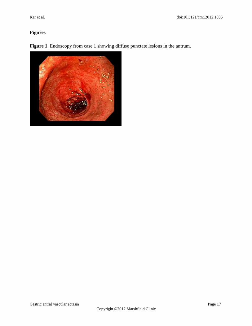
esophagogastroduodenoscopy (EGD) showed diffuse punctate hemorrhage in the antrum,
compatible with the punctate form of GAVE commonly encountered among cirrhotic patients,
with signs of active bleeding (figure 1). There was no evidence of esophageal or gastric varices.
The proximal portion of the stomach revealed changes consistent with portal hypertensive
gastropathy, characterized by the so-called “mosaic” pattern of the gastric wall along with
mucosal red spots, without active bleeding. The bleeding sites were successfully treated with
argon plasma coagulation (APC). He was monitored closely and had no further episodes of
bleeding. His hemoglobin remained stable before discharge. The patient was lost to follow-up at
our facility.
Case Presentation 2
A Caucasian female, aged 68 years, was admitted with complaints of exertional dyspnea and
orthopnea of 2 weeks duration. She denied any chest pain or palpitations. Past medical history
was significant for coronary artery disease, morbid obesity, hypertension, and poorly controlled
type 2 diabetes mellitus.
Her vital signs showed a temperature of 97.7o F, pulse 90 beats/minute, respiratory rate 18
breaths/minute, and blood pressure 143/61 mmHg. Physical examination revealed conjunctival
Kar et al. doi:10.3121/cmr.2012.1036
Gastric antral vascular ectasia Page 5
Copyright ©2012 Marshfield Clinic
pallor and bilateral basal crackles with leg edema. Rectal examination revealed hemoccult
positive stool. Laboratory studies revealed a hemoglobin of 6.6 gm/dL (reference range 11.7–
15.5 gm/dL) with a hematocrit of 21.7% (reference range 35–46%). Platelet count was normal at
254x103/µL. Iron studies revealed a serum iron level of 11 g/dL (reference range 40–160
g/dL) with a total iron binding capacity of 570 g/dL (reference range 260–420 g/dL).
The patient received four units of packed red cell transfusion, and her hematocrit was closely
monitored. She underwent an EGD, which showed areas of hyperemic streaks alternating with
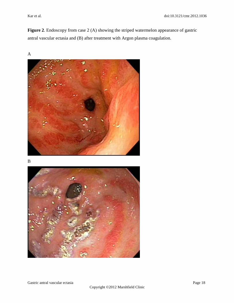
normal-appearing mucosa in the antrum, consistent with “watermelon stomach” (figure 2A). A
biopsy was obtained. The bleeding sites responded well to APC (figure 2B). The hemoglobin
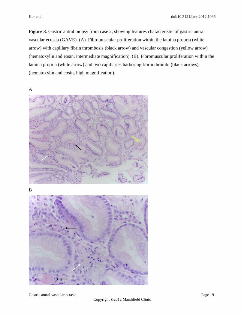
remained stable post-procedure. The gastric antral biopsy showed patchy fibromuscular
proliferation of the lamina propria, accompanied by dilated/congested capillaries and fibrin
thrombi; the histologic features were characteristic of GAVE (figure 3).
Over an 18-month follow-up period, the patient was treated with four sessions of APC. She
required a blood transfusion for severe anemia on two occasions. After being placed on iron
supplementation, her hemoglobin level increased from 8.6 gm/dL to 10.5 gm/dL, and there has
been a significant reduction in transfusion dependence.
Discussion
The first report of GAVE in the literature in 1953 was by Rider et al2, who described the
condition in an elderly female patient with chronic iron deficiency anemia. An antrectomy
specimen revealed “erosive atrophic gastritis with marked veno-capillary ectasia.”2 In 1984,
Kar et al. doi:10.3121/cmr.2012.1036
Gastric antral vascular ectasia Page 6
Copyright ©2012 Marshfield Clinic
Jabbari et al3
described the characteristic endoscopic findings of “longitudinal antral folds …
converging on the pylorus.” The endoscopic appearance resembled the stripes on a watermelon,
and they coined the term “watermelon stomach.”3
Generally, GAVE is associated with underlying chronic illness. Cirrhosis of the liver has been
found in 30% of cases.4 Among patients without cirrhosis, GAVE is usually associated with
underlying autoimmune conditions.5 Gostout et al
5 reported that 62% of patients had coexistent
autoimmune connective tissue disorder, particularly Raynaud’s phenomenon, which is noted in
31% of patients with GAVE. Other underlying conditions include systemic sclerosis and CREST
syndrome (calcinosis, Raynaud’s syndrome, esophageal dysmotility, sclerodactyly, and
telangiectasia).4-7
There have been published case reports of GAVE in patients with essential
hypertension,8 chronic renal failure,
8 acute myeloid leukemia,
9 and bone marrow transplant.
10
In a single-center study of 45 patients with GAVE, 71% were female, with an average age of 73
years.5 In that study, approximately 89% of patients presented with transfusion-dependent
anemia.5
More recently, two papers have reported two characteristic endoscopic appearances of
GAVE with different epidemiological aspects. Cirrhosis is commonly associated with a diffuse
appearance of GAVE, involving the whole antrum without interruption; whereas non-cirrhotic
patients are more likely to have a typical striped “watermelon” appearance.11,12
Our two patients
fit these respective endoscopic patterns. Epidemiological differences have been noted in these
two groups, and characteristics of the two patterns are listed in table 1.5,11
Kar et al. doi:10.3121/cmr.2012.1036
Gastric antral vascular ectasia Page 7
Copyright ©2012 Marshfield Clinic
The pathophysiology of GAVE remains unclear. Several theories have been proposed, including
achlorhydria, hypergastrinemia, and low pepsinogen levels. The pathogenesis of the histologic
changes, most notably the lamina propria fibromuscular proliferation and vascular dilatation with
thrombosis, is unclear. Increased levels of hormones with vasodilating properties, such as gastrin
and prostaglandin E2, have been observed in patients with GAVE, and it has been suggested that
failure of liver processing functions may lead to a build-up of these hormones, contributing to the
pathogenesis of GAVE.13
Several studies have suggested the mechanical stress theory in the pathogenesis of GAVE.
Charneua et al14
found that antral motility time was significantly increased in patients with
GAVE when compared to healthy controls. Quintero et al15
postulated that peristaltic waves of
loosely attached antral mucosa draw the mucosa in longitudinal folds and induce fibromuscular
proliferation and secondary formation of ectatic vessels. Fibromuscular proliferation and
capillary ectasia with microvascular thrombosis of the lamina propria form the histological
hallmark of GAVE; the theory of mechanical stress is therefore strengthened by the histological
findings in GAVE.10,16-18
Due to the increasing use of endoscopy and increased awareness of the condition, GAVE is now
more frequently diagnosed.18
It is important that GAVE be distinguished from portal
hypertensive gastropathy (PHG) and antral gastritis, especially in patients with portal
hypertension. Patients with GAVE have more advanced cirrhosis (by Child-Pugh scoring), lower
serum gastrin, and greater blood loss than patients with PHG.19,20
While PHG can be controlled
Kar et al. doi:10.3121/cmr.2012.1036
Gastric antral vascular ectasia Page 8
Copyright ©2012 Marshfield Clinic
through the use of medications to relieve portal hypertension, endoscopic intervention remains
the mainstay of therapy for GAVE.
Endoscopically, PHG is typically more predominant in the fundus and the body of the stomach,
whereas GAVE mainly involves the antrum.9,19
However, severe PHG can occur in the antrum
and resemble GAVE.21
It is unclear as to whether GAVE may involve the proximal stomach. In
two separate published case series, the patient numbers were small, and neither documented the
presence of thrombi at the proximal stomach; therefore, it is possible that some patients had
coexistent GAVE and PHG. 22,23
The role of biopsies becomes important, since microscopically,
GAVE demonstrates more prominent vascular ectasia, fibrin thrombosis (P = 0.006), and
fibromuscular proliferation/fibrohyalinosis than PHG.19
Treatment of GAVE includes initial resuscitation and symptomatic therapy with intravenous
fluids and blood products.17
Medical therapy aimed at reducing portal pressures has had limited
success in the treatment of GAVE.16
Corticosteroids have been used in patients to treat GAVE;
in a case series of eleven patients treated with oral steroids, six had complete resolution of
bleeding over a mean follow-up period of three years.24,25
There have been a few reports where control of bleeding was achieved with estrogen and
progesterone.26,27
The rationale for hormonal therapy was the observation that epistaxis due to
Osler-Weber-Rendu syndrome decreased during pregnancy. In a study involving six patients
with GAVE, Tran et al26
documented cessation of bleeding and reduction in transfusion
requirements in patients treated with ethinyl estradiol with no change in endoscopic appearance.
Kar et al. doi:10.3121/cmr.2012.1036
Gastric antral vascular ectasia Page 9
Copyright ©2012 Marshfield Clinic
There have also been reports of using tranexamic acid in the treatment of GAVE, with reduction
in bleeding and transfusion requirements.28
The mainstay of therapy remains endoscopic.13,17,18, 29,30
Neodymium: yttrium-aluminum-garnet
(Nd:YAG) laser has been widely used in the treatment of GAVE.17,29
In one study, 13
transfusion-dependent patients were treated with Nd:YAG laser, of whom 12 had remarkable
response with elimination of transfusion requirement over a median follow-up of two years.8,31
Argon plasma coagulation (APC) has been found to be equally effective in the treatment of
GAVE and superior in cost, convenience, and complication rates.29
APC is a non-contact electro-
coagulation technique using a jet of ionized argon gas to deliver monopolar current to the target
tissues.32
In a study of 26 patients treated with APC, transfusion requirement elimination was
seen in 77% over a mean follow-up of 16 months.33
The benefit of APC lies in the consideration
that the depth of penetration can be easily controlled, thus avoiding excessive blood loss. We
used this modality in both of our patients, with significant improvement in bleeding. Multiple
sessions may be required to reduce future episodes and/or decrease transfusion dependence, as
has been noted in our second patient.
Recently, Wells et al34
reported a study demonstrating the superiority of endoscopic band
ligation in the treatment of GAVE when compared to endoscopic thermal therapy. Endoscopic
band ligation is the current standard of care for variceal ligation. In patients with GAVE,
endoscopic band ligation resulted in increased hemoglobin levels and decreased blood
transfusions and hospital admissions compared to endoscopic thermal therapy. Additionally,
fewer treatment sessions were necessary, likely due to the ability of endoscopic band ligation to
Kar et al. doi:10.3121/cmr.2012.1036
Gastric antral vascular ectasia Page 10
Copyright ©2012 Marshfield Clinic
treat a larger area of the mucosa at once. No randomized, prospective studies have been
performed to conclusively demonstrate the superiority of endoscopic band ligation over
conventional therapies, but it offers a promising new therapeutic option.34
Before the advent of endoscopic therapy, GAVE was commonly treated by antrectomy.3,17,18
However, surgical resection is often associated with significant morbidity and mortality and
should be reserved for patients who do not respond to medical or endoscopic treatment.
Gross et al35
reported results of their pilot study that used radiofrequency ablation (the HALO90
system; BÂRRX Medical, Inc, Sunnyvale, CA) for the treatment of GAVE in six patients. All
six patients responded favorably with improved post-procedure hemoglobin levels and a
decrease in transfusion requirement with no significant complications. Endoscopic ablation has
been found to be effective in Barrett esophagus; however, it has not been well-studied in vascular
ectasia. It may be another tool in the treatment of GAVE.
Conclusion
GAVE is a rare cause of upper gastrointestinal bleeding, presenting with symptoms ranging from
occult bleeding to acute blood loss requiring resuscitation. The etiology of GAVE is unknown,
and the associated comorbidities of patients with GAVE are varied, including cirrhosis and
several autoimmune disorders. The cases presented here demonstrate the two different
appearances of GAVE in patients with distinct underlying conditions and underscore the
importance of correct diagnosis. Patients with cirrhosis may have a diffuse punctate appearance
of the antrum on endoscopy in contrast to the typical striped watermelon appearance seen in
Kar et al. doi:10.3121/cmr.2012.1036
Gastric antral vascular ectasia Page 11
Copyright ©2012 Marshfield Clinic
association with other conditions. It is important to distinguish this entity from PHG and antral
gastritis, as the management is different. Argon plasma coagulation has been employed
successfully in the treatment of GAVE, as in the cases presented here, though multiple sessions
may be required. Several other endoscopic interventions are available, and surgical antrectomy is
generally considered as a last resort. Juxtaposition of these two cases illustrates the two
strikingly different presentations, etiologies, and endoscopic appearances of GAVE.
Acknowledgements
The authors thank the Marshfield Clinic Research Foundation’s Office of Scientific Writing and
Publication for editorial assistance in the preparation of this manuscript.
Kar et al. doi:10.3121/cmr.2012.1036
Gastric antral vascular ectasia Page 12
Copyright ©2012 Marshfield Clinic
References
1. Dulai GS, Jensen DM, Kovacs TO, Grainek IM, Jutabha R. Endoscopic treatment outcomes in
watermelon stomach patients with and without portal hypertension. Endoscopy
2004;36:68-72.
2. Rider JA, Klotz AP, Kirsner JB. Gastritis with veno-capillary ectasia as a source of massive
gastric hemorrhage. Gastroenterology 1953;24:118-123.
3. Jabbari M, Cherry R, Lough JO, Daly DS, Kinnear DG, Goresky CA. Gastric antral vascular
ectasia: the watermelon stomach. Gastroenterology 1984;87:1165-1170.
4. Ward EM, Raimondo M, Rosser BG, Wallace MB, Dickson RD. Prevalence and natural
history of gastric antral vascular ectasia in patients undergoing orthotopic liver
transplantation. J Clin Gastroenterol 2004;38:898-900.
5. Gostout CJ, Viggiano TR, Ahlquist DA, Wang KK. Larson MV, Balm R. The clinical and
endoscopic spectrum of the watermelon stomach. J Clin Gastroenterol 1992;15:256-263.
6. Beales IL. Watermelon stomach in the CREST syndrome. Postgrad Med J 1994;70:766-767.
7. Watson M, Hally RJ, McCue PA, Varga J, Jiménez SA. Gastric antral vascular ectasia
(watermelon stomach) in patients with systemic sclerosis. Arthritis Rheum
1996;39:341-346.
8. Liberski SM, McGarrity TJ, Hartle RJ, Varano V, Reynolds D. The watermelon stomach:
long-term outcome in patients treated with Nd:YAG laser therapy. Gastrointest Endosc
1994;40:584-587.
9. Takahashi T, Miya T, Oki M, Sugawara N, Yoshimoto M, Tsujisaki M. Severe hemorrhage
from gastric vascular ectasia developed in a patient with AML. Int J Hematol
2006;83:467-468.
Kar et al. doi:10.3121/cmr.2012.1036
Gastric antral vascular ectasia Page 13
Copyright ©2012 Marshfield Clinic
10. Tobin RW, Hackman RC, Kimmey MB, Durtschi MB, Hayashi A, Malik R, McDonald MF,
McDonald GB. Bleeding from gastric antral vascular ectasia in marrow transplant
patients. Gastrointest Endosc 1996;44:223-229.
11. Ito M, Uchida Y, Kamano S, Kawabata H, Nishioka M. Clinical comparisons between two
subsets of gastric antral vascular ectasia. Gastrointest Endosc 2001;53:764-770.
12. Chaves DM, Sakai P, Oliveira CV, Cheng S, Ishioka S. Watermelon stomach: clinical
aspects and treatment with argon plasma coagulation. Arq Gastroenterol
2006;43:191-195.
13. Ripoll C, Garcia-Tsao G. The management of portal hypertensive gastropathy and gastric
antral vascular ectasia. Digest Liv Dis 2011;43:345-351.
14. Charneau J, Petit R, Calès P, Dauver A, Boyer J. Antral motility in patients with cirrhosis
with or without gastric antral vascular ectasia. Gut 1995;37:488-492.
15. Quintero E, Pique JM, Bombi JA, Bordas JM, Sentis J, Elena M, Bosch J, Rodes J. Gastric
mucosal vascular ectasias causing bleeding in cirrhosis. A distinct entity associated with
hypergastrinemia and low serum levels of pepsinogen I. Gastroenterology
1987;93:1054-1061.
16. Selinger CP, Ang YS. Gastric antral vascular ectasia (GAVE): an update on clinical
presentation, pathophysiology and treatment. Digestion 2008;77:131-137.
17. Novitsky YW, Kercher KW, Czerniach DR, Litwin DE. Watermelon stomach:
pathophysiology, diagnosis, and management. J Gastrointest Surg 2003;7:652-661.
18. Sebastian S, O'Morain CA, Buckley MJ. Review article: current therapeutic options for
gastric antral vascular ectasia. Aliment Pharmacol Ther 2003;18:157-165.
Kar et al. doi:10.3121/cmr.2012.1036
Gastric antral vascular ectasia Page 14
Copyright ©2012 Marshfield Clinic
19. Burak KW, Lee SS, Beck PL. Portal hypertensive gastropathy and gastric antral vascular
ectasia (GAVE) syndrome. Gut 2001;49:866-872.
20. Payen JL, Cales P, Voigt JJ, Barbe S, Pilette C, Dubuisson L, Desmorat H, Vinel JP, Kervran
A, Chayvialle JA, et al. Severe portal hypertensive gastropathy and antral vascular ectasia
are distinct entities in patients with cirrhosis. Gastroenterology 1995;108:138-144.
21. Thuluvath PJ, Yoo HY. Portal hypertensive gastropathy. Am J Gastroenterol
2002;97:2973-2978.
22. Stotzer PO, Willén R, Kilander AF. Watermelon stomach: not only an antral disease.
Gastrointest Endosc. 2002;55:897–900.
23. Al-Haddad M, Ward EM, DeVault KR, Bouras EP, Raimondo M. Vascular ectasia of the
proximal stomach. Dig Dis Sci. 2007;52:1367–1369.
24. Bhowmick BK. Watermelon stomach treated with oral corticosteroid. J R Soc Med
1993;86:52.
25. Calam J, Walker RJ. Antral vascular lesion, achlorhydria, and chronic gastrointestinal blood
loss: response to steroids. Dig Dis Sci 1980;25:236-239.
26. Tran A, Villeneuve JP, Bilodeau M, Willems B, Marleau D, Fenyves D, Parent R, Pomier-
Layrargues G. Treatment of chronic bleeding from gastric antral vascular ectasia (GAVE)
with estrogen-progesterone in cirrhotic patients: an open pilot study. Am J Gastroenterol
1999;94:2909-2911.
27. Manning RJ. Estrogen/progesterone treatment of diffuse antral vascular ectasia. Am J
Gastroenterol 1995;90:154-156.
28. Khan S, Vaishnavi A. Pharmacotherapy for gastric antral vascular ectasia: dramatic response
to tranexamic acid. Gastrointest Endosc 2009;70:191-192.
Kar et al. doi:10.3121/cmr.2012.1036
Gastric antral vascular ectasia Page 15
Copyright ©2012 Marshfield Clinic
29.Rosenfeld G, Enns R. Argon photocoagulation in the treatment of gastric antral vascular
ectasia and radiation proctitis. Curr Endoscop Prac 2009;23:801-804.
30.Ripoll C, Garcia-Tsao G. Management of gastropathy and gastric vascular ectasia in portal
hypertension. Clin Liver Dis 2010;14:281-295.
31. Gostout CJ, Ahlquist DA, Radford CM, Viggiano TR, Bowyer BA, Balm RK. Endoscopic
laser therapy for watermelon stomach. Gastroenterology 1989;96:1462-1465.
32. Dumot JA, Greenwald BD. Argon plasma coagulation, bipolar cautery, and cryotherapy:
ABC's of ablative techniques. Endoscopy 2008;40:1026-1032.
33. Kwan V, Bourke MJ, Williams SJ, Gillespie PE, Murray MA, Kaffes AJ, Henriquez MS,
Chan RO. Argon plasma coagulation in the management of symptomatic gastrointestinal
vascular lesions: experience in 100 consecutive patients with long-term follow-up. Am J
Gastroenterol 2006;101:58-63.
34. Wells CD, Harrison ME, Gurudu SR, Crowell MD, Byrne TJ, Depetris G, Sharma VK.
Treatment of gastric antral vascular ectasia (watermelon stomach) with endoscopic band
ligation. Gastrointest Endosc 2008;68:231-236.
35. Gross SA, Al-Haddad M, Gill KR, Schore AN, Wallace MB. Endoscopic mucosal ablation
for the treatment of gastric antral vascular ectasia with the HALO90 system: a pilot
study. Gastrointest Endosc 2008;67:324-327.
Author Affiliations
Papia Kar, MD*; Subhashis Mitra MD*; Jeffrey M Resnick, MD†; Camille F. Torbey MD,
FACP, AGAF‡
From the Departments of *Internal Medicine, †Lab-Pathology, and
‡Gastroenterology,
Marshfield Clinic, Marshfield, Wisconsin
Kar et al. doi:10.3121/cmr.2012.1036
Gastric antral vascular ectasia Page 16
Copyright ©2012 Marshfield Clinic
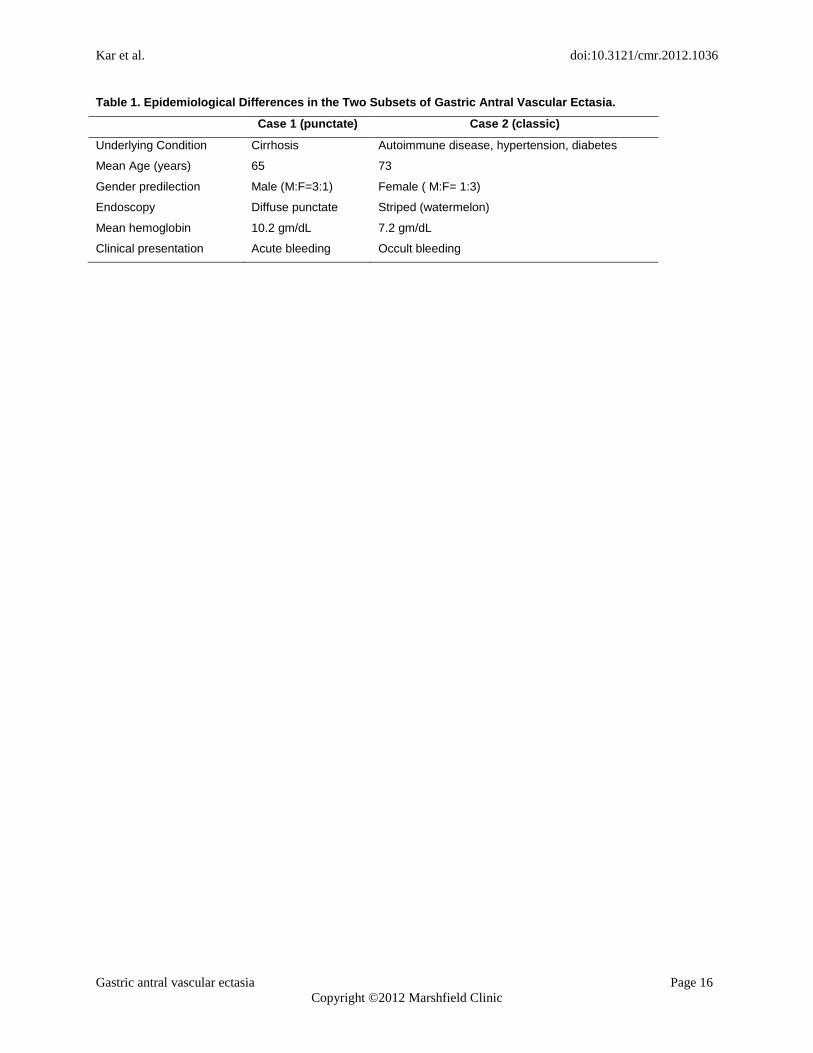
Table 1. Epidemiological Differences in the Two Subsets of Gastric Antral Vascular Ectasia.
Case 1 (punctate) Case 2 (classic)
Underlying Condition Cirrhosis Autoimmune disease, hypertension, diabetes
Mean Age (years) 65 73
Gender predilection Male (M:F=3:1) Female ( M:F= 1:3)
Endoscopy Diffuse punctate Striped (watermelon)
Mean hemoglobin 10.2 gm/dL 7.2 gm/dL
Clinical presentation Acute bleeding Occult bleeding
Kar et al. doi:10.3121/cmr.2012.1036
Gastric antral vascular ectasia Page 17
Copyright ©2012 Marshfield Clinic
Figures
Figure 1. Endoscopy from case 1 showing diffuse punctate lesions in the antrum.
Kar et al. doi:10.3121/cmr.2012.1036
Gastric antral vascular ectasia Page 18
Copyright ©2012 Marshfield Clinic
Figure 2. Endoscopy from case 2 (A) showing the striped watermelon appearance of gastric
antral vascular ectasia and (B) after treatment with Argon plasma coagulation.
A
B
Kar et al. doi:10.3121/cmr.2012.1036
Gastric antral vascular ectasia Page 19
Copyright ©2012 Marshfield Clinic
Figure 3. Gastric antral biopsy from case 2, showing features characteristic of gastric antral
vascular ectasia (GAVE). (A). Fibromuscular proliferation within the lamina propria (white
arrow) with capillary fibrin thrombosis (black arrow) and vascular congestion (yellow arrow)
(hematoxylin and eosin, intermediate magnification). (B). Fibromuscular proliferation within the
lamina propria (white arrow) and two capillaries harboring fibrin thrombi (black arrows)
(hematoxylin and eosin, high magnification).
A
B


![Gastric antral vascular ectasia in a patient with lupus undergoing … · 2020. 11. 10. · gastrin, CKD, and connective tissue disease [7]. CKD is one of the commonest health problems](https://img.dokumen.tips/doc/110x75/6109f020aaa0b405bd08aedf/gastric-antral-vascular-ectasia-in-a-patient-with-lupus-undergoing-2020-11-10.jpg)