Embed Size (px)
Citation preview

Gastric Antral Vascular Ectasia: The Invaluable Role of the
Gastric Biopsy in its Detection and Confirmation
Kevin Turner
Robert M. Genta
Miraca Life Sciences Research Institute Irving, Texas

Background
• Gastric vasculopathies consist predominantly of two entities: oGastric antral vascular ectasia (GAVE) o Portal hypertensive gastropathy (PHG)
• The classically described GAVE patient is an
elderly woman with iron-deficiency anemia and a “watermelon” appearance of the gastric mucosa on endoscopy.
Background • There are two published histologic scoring systems:
o Gilliam et al. (1989) – Devised a system requiring vascular
abnormality (thrombi/ectasia) and spindle cell proliferation of the lamina propria to distinguish GAVE from normal, acute gastritis, and atrophic gastritis.
o Payen et al. (1995) – Devised a similar system using
vascular abnormality, spindle cell proliferation, and fibrohyalinosis to distinguish GAVE from PHG in cirrhotic patients.

Histologic Findings
Background • There is wide variability in the workup of GAVE.
o Clinicians:
1) The frequency in which suspected GAVE is biopsied varies amongst clinicians.
o Pathologists:
1) Scoring systems are not generally used. 2) Crucial features (thrombi) are often patchy.
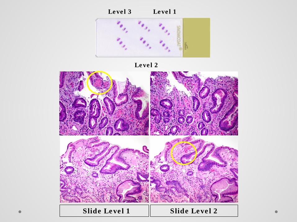
Slide Level 1 Slide Level 2
Level 3
Level 2
Level 1
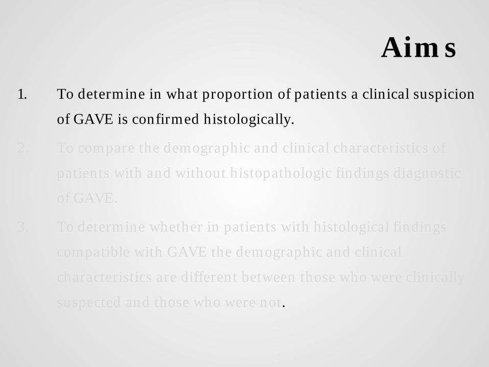
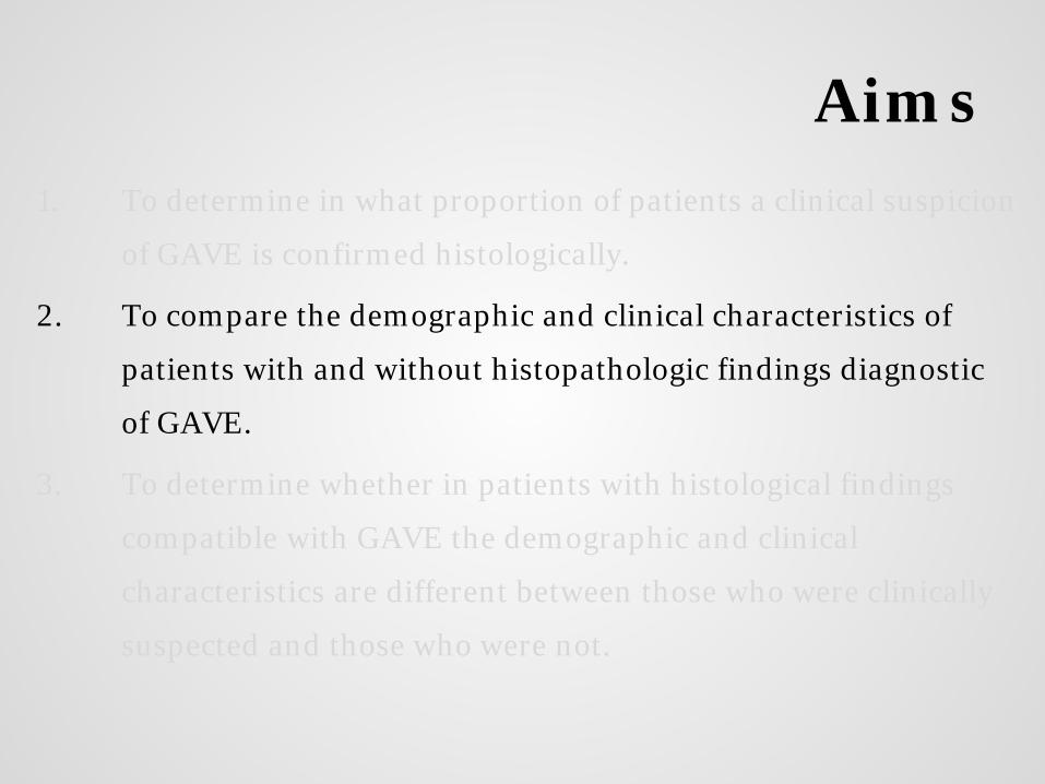
Aims 1. To determine in what proportion of patients a clinical suspicion
of GAVE is confirmed histologically.
2. To compare the demographic and clinical characteristics of
patients with and without histopathologic findings diagnostic
of GAVE.
3. To determine whether in patients with histological findings
compatible with GAVE the demographic and clinical
characteristics are different between those who were clinically
suspected and those who were not.
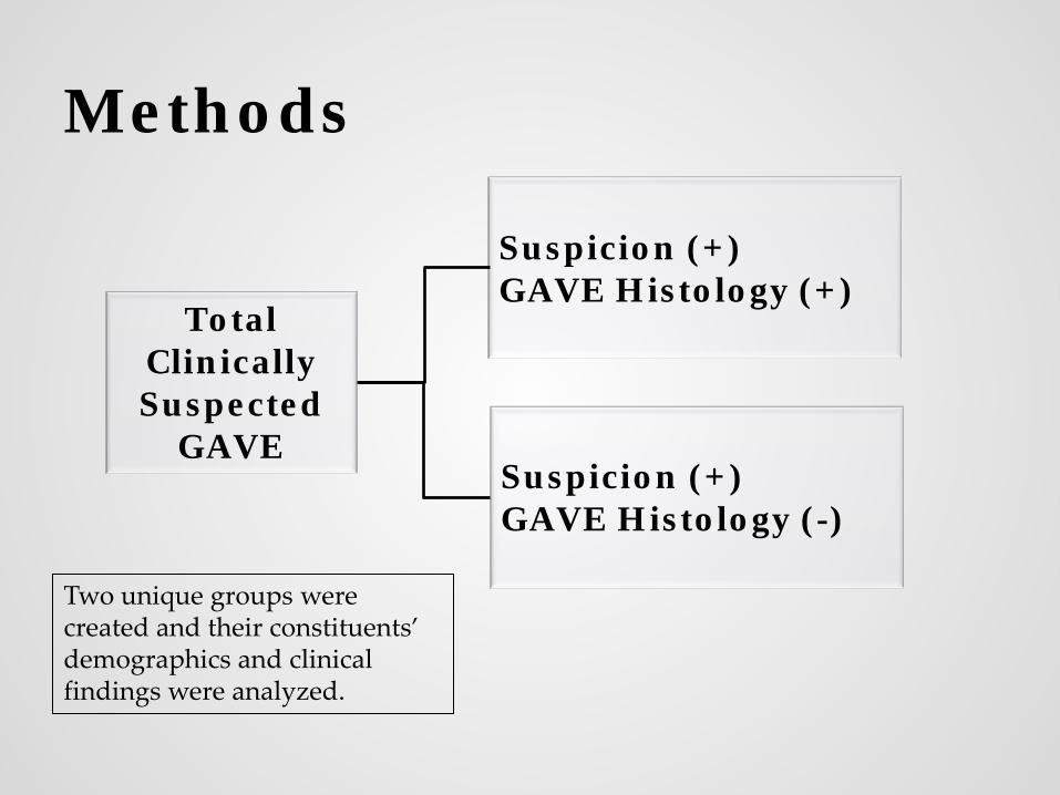
Methods
Two unique groups were created and their constituents’ demographics and clinical findings were analyzed.
Total Clinically Suspected
GAVE Suspicion (+) GAVE Histology (-)
Suspicion (+) GAVE Histology (+)
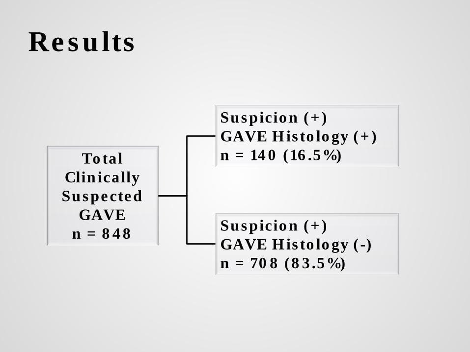
Results
Total Clinically Suspected
GAVE n = 848 Suspicion (+)
GAVE Histology (-) n = 708 (83.5%)
Suspicion (+) GAVE Histology (+) n = 140 (16.5%)
Aims 1. To determine in what proportion of patients a clinical suspicion
of GAVE is confirmed histologically.
2. To compare the demographic and clinical characteristics of
patients with and without histopathologic findings diagnostic
of GAVE.
3. To determine whether in patients with histological findings
compatible with GAVE the demographic and clinical
characteristics are different between those who were clinically
suspected and those who were not.

Results
Histo (+) (n=140) Histo (-) (n=708) Age (yrs, median) 67 64 Women (%) 98 (70) 430 (61) Men (%) 42 (30) 278 (39)
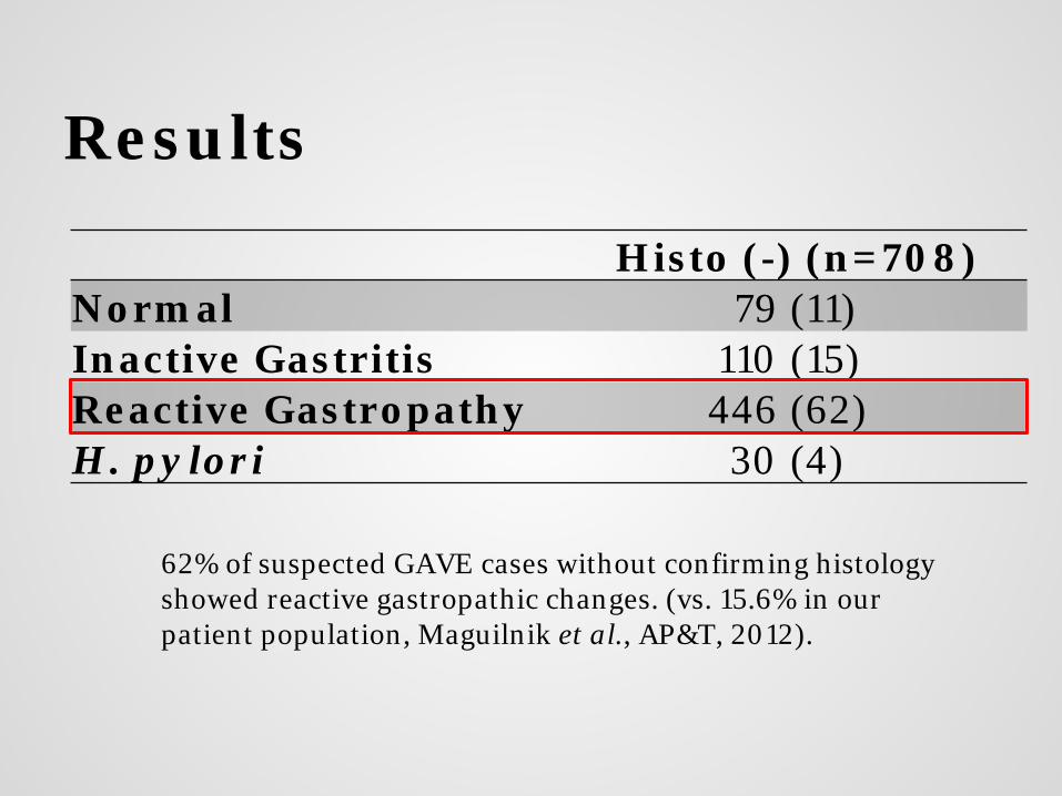
Results
Histo (-) (n=708) Normal 79 (11) Inactive Gastritis 110 (15) Reactive Gastropathy 446 (62) H. pylori 30 (4)
62% of suspected GAVE cases without confirming histology showed reactive gastropathic changes. (vs. 15.6% in our patient population, Maguilnik et al., AP&T, 2012).

Histo (+) (n=140) Histo (-) (n=708) OR (95% CI)
Clinical Cirrhosis (%) 1 (0.7) 35 (5) 0.14 (0.02 - 1.02)
Abdominal Pain (%) 23 (16) 122 (17) ns
Anemia (%) 79 (57) 221 (31) 2.85 (1.97 - 4.13)
Nausea (%) 1 (0.7) 51 (7) 0.11 (0.01 - 0.78)
Vomiting (%) 1 (0.7) 22 (3) ns Weight Loss (%) 4 (3) 31 (4) ns
Bleeding (%) 2 (1.4) 31 (4) ns
Results
Aims 1. To determine in what proportion of patients a clinical suspicion
of GAVE is confirmed histologically.
2. To compare the demographic and clinical characteristics of
patients with and without histopathologic findings diagnostic
of GAVE.
3. To determine whether in patients with histological findings
compatible with GAVE the demographic and clinical
characteristics are different between those who were clinically
suspected and those who were not.
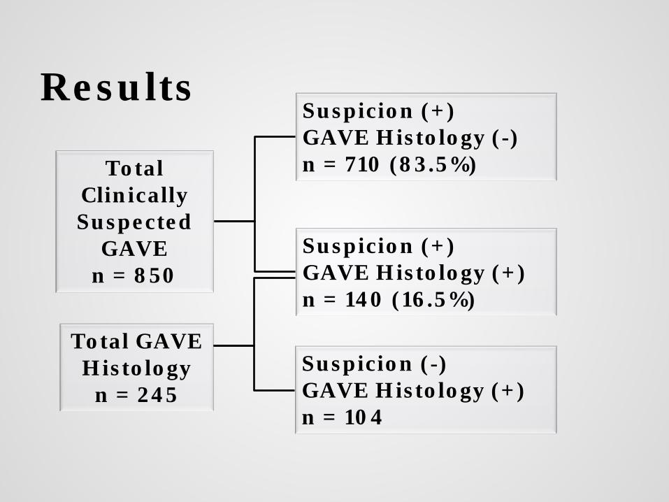
Results
Total Clinically Suspected
GAVE n = 850
Suspicion (+) GAVE Histology (+) n = 140 (16.5%)
Suspicion (+) GAVE Histology (-) n = 710 (83.5%)
Total GAVE Histology
n = 245 Suspicion (-) GAVE Histology (+) n = 104
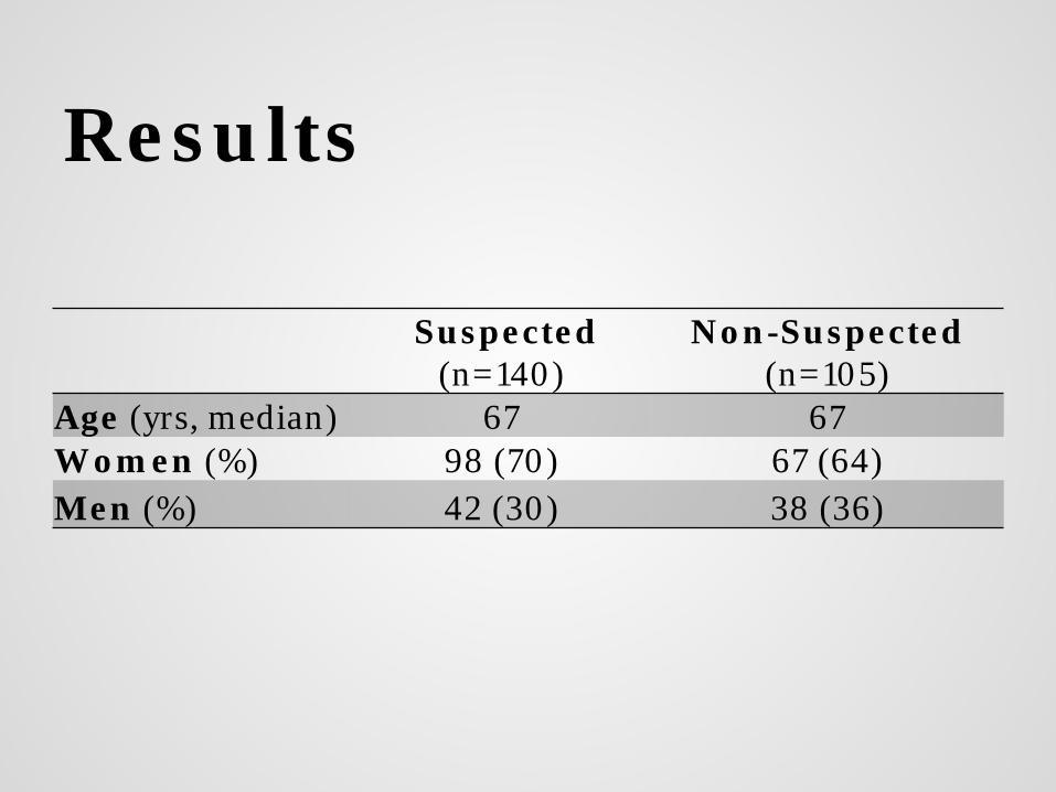
Results
Suspected (n=140)
Non-Suspected (n=105)
Age (yrs, median) 67 67 Women (%) 98 (70) 67 (64) Men (%) 42 (30) 38 (36)

Results
Suspected
(n=140) Non-Suspected
(n=105) OR
(95% CI) Clinical Cirrhosis (%) 1 (0.7) 11 (10.5) 0.06 (0.01 – 0.48)
Abdominal Pain (%) 23 (16.4) 25 (23.8) 0.45 (0.24 – 0.83)
Anemia (%) 79 (56.4) 32 (30.5) 2.95 (1.73 – 5.04)
Nausea (%) 1 (0.7) 8 (7.6) 0.09 (0.01 – 0.79)
Vomiting (%) 1 (0.7) 7 (6.7) 0.10 (0.01 – 0.83)
Weight Loss (%) 4 (2.9) 8 (7.6) ns
Bleeding (%) 2 (1.4) 1 (0.9) ns
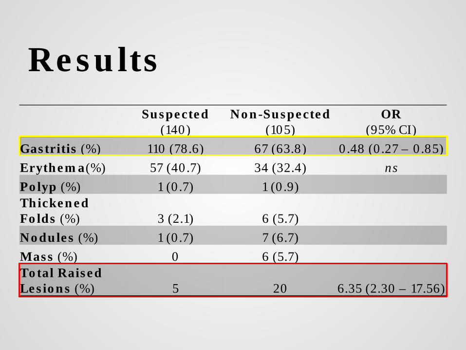
Results
Suspected
(140) Non-Suspected
(105) OR
(95% CI) Gastritis (%) 110 (78.6) 67 (63.8) 0.48 (0.27 – 0.85) Erythema(%) 57 (40.7) 34 (32.4) ns Polyp (%) 1 (0.7) 1 (0.9) Thickened Folds (%) 3 (2.1) 6 (5.7) Nodules (%) 1 (0.7) 7 (6.7) Mass (%) 0 6 (5.7) Total Raised Lesions (%) 5 20 6.35 (2.30 – 17.56)

“Consistent with GAVE”

Summary
• Only 16.5% of cases biopsied to rule out GAVE had confirmatory histopathologic features
• Of those without confirming histology, the most common histopathologic finding was reactive gastropathy.

Summary • In 42.5% of patients with histopathologic
features compatible with GAVE, a clinical suspicion was not conveyed to the pathologist
• Unsuspected cases were more frequently from males, those with a history of cirrhosis, and patients presenting with nausea, vomiting, and weight loss; but less frequently with anemia.

Summary
Endoscopically, unsuspected cases showed raised
lesions including nodules, thickened folds, polyps,
and masses more frequently than erythematous
streaks.

Conclusion Our study shows that the gastric biopsy is valuable in the diagnosis of GAVE in two ways.
1. Confirmation: GAVE is infrequently confirmed histologically, and endoscopically suspicious gastric mucosa is often associated with reactive gastropathy, gastritis, or normal findings
2. Detection: The gastric biopsy can detect a subset of patients without the classic presenting endoscopic appearance or symptoms.

Aims 1. To determine in what proportion of patients a clinical suspicion of
GAVE is confirmed histologically.
2. To compare the demographic and clinical characteristics of patients
with and without histopathologic findings diagnostic of GAVE.
3. To determine whether patients with the histological findings
compatible with GAVE are the demographic and clinical
characteristics different between those who were clinically suspected
and those who were not.
4. To determine whether patients with GAVE histology in the absence of
clinical suspicion have, or develop GAVE clinically (underway).
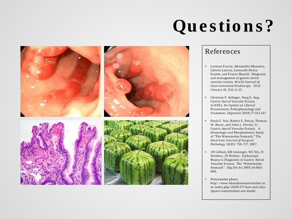
Questions? References • Lorenzo Fuccio, Alessandro Mussetto,
Liboria Laterza, Leonardo Henry Eusebi, and Franco Bazzoli. Diagnosis and management of gastric antral vascular ectasia. World Journal of Gastrointestinal Endoscopy. 2013 January 16; 5(1): 6-13.
• Christian P. Selinger, Yeng S. Ang. Gastric Antral Vascular Ectasia (GAVE): An Update on Clinical Presentation, Pathophysiology and Treatment. Digestion 2008;77:131-137.
• Paula F. Suit, Robert E. Petras, Thomas W. Bauer, and John L. Petrini, Jr. Gastric Antral Vascular Ectasia. A Hiostologic and Morphometric Study of “The Watermelon Stomach.” The American Journal of Surgical Pathology 11(10): 750-757, 1987.
• JH Gilliam, KR Geisinger, WC Wu, N Weidner, JE Richter. Endoscopic Biopsy is Diagnostic in Gastric Antral Vascular Ectasia. The “Watermelon Stomach”. Dig Dis Sci 1989;34:885-888.
• Watermelon photo: http://www.whataboutwatermelon.com/index.php/2009/07/how-and-why-square-watermelons-are-made/