Embed Size (px)
Citation preview
Osteomyelitis
David ShearerDave Lowenberg
Created June 2016
Definitions
• Osteomyelitis – Infection involving bone• Acute osteomyelitis
– Infection of short duration– Characterized by suppuration (i.e. abscess) but not
biofilm– Systemic symptoms common

Definitions
• Chronic osteomyelitis– Long standing infection (weeks to years)– Characterized by necrotic bone and bacterial
colonies in protein/polysaccharide matrix (biofilm)– Often no systemic symptoms
• Occurs along spectrum with no clear time cutoff to separate acute vs. chronic infection

Etiologies• Hematogenous
– Metaphysis of long bones • Most common in children
– Vertebral osteomyelitis• Contiguous spread
– Post-traumatic• Open fractures• Infections associated with deep
implants– Prosthetic Joint Infections
• Vascular Insufficiency and/or Diabetes– Secondary to ulceration– Commonly affects the forefoot bones

Epidemiology• Estimates vary widely, but overall
increasing incidence in US – Increasing
• Osteomyelitis from a contiguous focus of infection (e.g. post-trauma, post-surgery)
• Osteomyelitis of the foot and ankle related to diabetes
– Stable/Decreasing • Hematogenous osteomyelitis in children
Kremers, et al. JBJS 2015

Pathogens
• Staph aureus most common (45% in series by Kremers et al., JBJS, 2015)
• Staph epidermidis and steptococcal species next most common
• Diabetes more commonly polymicrobial

Pathophysiology: Implant-associated osteomyelitis
– Planktonic cells attach to metal substrate
– Initial cells undergo apoptosis
– “Sacrificial cells” become matrix for biofilm

Establishment of Infection Biofilm occurs
due to the organized cell death of the first waves of bacterial invasion on a host site (“death of the privates, corporals, and sergeants.”) Reprinted with permission from McPherson EJ, Peters CL: Musculoskeletal Infection, in Flynn
JM (ed): Orthopaedic Knowledge Update 10. Rosemont, IL, American Academy of OrthopaedicSurgeons, 2011. Figure 4, page 243, OKU 10

Presumed Timeline(Definitive time for biofilm unknown)

Importance of Bacterial “Phase”in the Host
Planktonic This represents the
initial innoculumphase.
The bacteria have a high metabolic rate.
They are “free floating”
Cause systemic symptoms
Biofilm (Sessile) This represents the semi-
dormant bacterial phase where the microbe is “trying” to live in a symbiotic state.
Low metabolic rate. Adherent to the biofilm. 103 times less sensitive to most
antibiotics. Represents 98% of biofilm
population
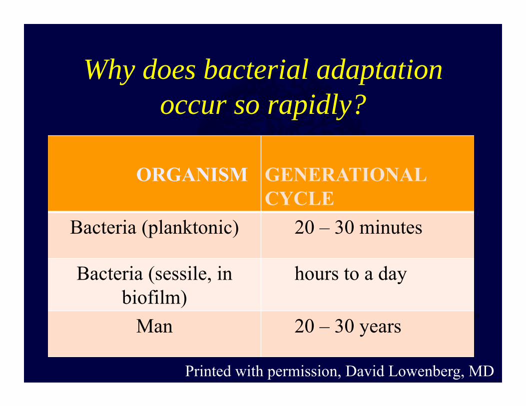
Why does bacterial adaptation occur so rapidly?
ORGANISM GENERATIONALCYCLE
Bacteria (planktonic) 20 – 30 minutes
Bacteria (sessile, inbiofilm)
hours to a day
Man 20 – 30 years
Printed with permission, David Lowenberg, MD

Biofilm antibiotic resistance
1. Cells hidden within hydrophobic matrix2. Low metabolic rate (sessile cells)
– Impossible to achieve effective dose safely with systemic antibiotics
3. Ability to mutate due to short generational cycle

Clinical evaluation: Pertinent history• Characterize infection
– Clinical history (e.g. onset, timeline)
– Prior treatment– Prior surgeries
• Characterize host– Age– Comorbidities – Habits (tobacco, alcohol, drugs) – Social support, housing– Baseline function (ambulatory
status, assistive devices) Vs.

Physical exam• Rule out sepsis (fever,
tachycardia, hypotension)• Signs of active infection
– Warmth– Redness– Drainage
• Soft-tissues– Open wounds– Sinus tracts– Scars
• Evaluate for limb deformity (limb length, alignment)
• Evaluate joints above and below affected area
• Neurovascular status of limb
Imaging studies
– Plain x-rays• First line exam
– CT • Less sensitive than MRI, but more
specific for bony changes that may require debridement
• Useful for assessing for union in cases of infection associated with fracture implant

Plain x-rays• Virtually always the first line
exam• Can be normal for 2-3 weeks
after onset• Sensitivity can be variable,
specificity is higher• Findings
– Periosteal thickening– Lytic lesions with surrounding
sclerosis– Osteopenia– Loss of trabecular architecture
• Sequestrum: Dead bone walled off in granulation tissue
• Involucrum: Reactive bone that surrounds the sequestrum
Sequestrum
Involucrum

MRI• Characterizes both bone and
soft-tissue infection• Quite sensitive but often not
specific, and tends to overcall the extent of the lesion due to edema
• Best read on the T2 sequence.
• May be obstructed by hardware
• Not necessary in every case

63 y/o M with chronic recurrent Stage 3 tibial osteomyelitis
Printed with permission, David Lowenberg, MD

Nuclear medicine– Technetium 99 Bone scan
• Detects new bone formation• High sensitivity (90-100%), but poor specificity
(~30%)
– Tagged WBC scan• Good sensitivity (~90%), moderate specificity
(~60%)• Particularly useful in chronic osteomyelitis when
hardware or other factors preclude MRI– In general nuclear medicine rarely adds to diagnosis and
treatment plan

Laboratory evaluation• WBC
– Low sensitivity (normal in many cases of chronic osteomyelitis)• Platelets
– 500-1000K can be indicative of acute phase infection• ESR/CRP
– Improved sensitivity– Lack specificity– CRP more responsive to change– Negative ESR and CRP cannot definitively rule out osteomyelitis
• Only ~50% of chronic musculoskeletal infections will have elevated inflammatory markers
• Labs for drug toxicity (e.g. creatinine, liver enzymes)• Labs to evaluate comorbidities (e.g. blood glucose, Hba1c for diabetes)

Classification: Cierny-Mader
Anatomic type + Host = Clinical Stage
George Cierny

Cierny-Mader Classification
Modified with permission from Ziran BH, Rao Nalini: Infections, in Baumgaertner MR, Tornetta P (eds): Orthopaedic Knowledge Update Trauma 3. Rosemont, IL, American Academy of Orthopaedic Surgeons, 2005, pp 131-139. Figure 2, page 132, OKU Trauma 3
I II III IV

Cierny-Mader Staging SystemSTAGE ANATOMIC TYPE TYPICAL
ETIOLOGYTREATMENT
1 Medullary Infected intramedullary nail
Removal of the infected implant andisolated intramedullarydébridement
2 Superficial; no full-thicknessinvolvement of cortex
Chronic wound, leading tocolonization and focalinvolvement of a superficial areaof bone under the wound
Remove layers of infected bone untilviable bone is identified
Printed with permission, David Lowenberg, MD

Cierny-Mader Staging SystemSTAGE ANATOMIC
TYPETYPICAL ETIOLOGY
TREATMENT
3 Full-thickness involvement of acortical segment of bone;endosteum is involved, implyingintramedullary spread
Direct trauma with resultantdevascularization and seeding ofthe bone
Noninvolved bone is present at sameaxial level, so the osteomyeliticportion can be excised withoutcompromising skeletal stability.
4 Infection is permeative, involving a segmental portion of the bone.
Major devascularization withcolonization of the bone
Resection leads to a segmental ornear-segmental defect, resulting inloss of limb stability.
Printed with permission, David Lowenberg, MD

Cierny-Mader Physiologic HostType Infection Status Perpetuating
FactorsTreatment
A Normal physiologicresponse
Little or no systemic or localcompromise
No contraindications to surgical treatment
B (local) Locally activeImpairment of response
Prior trauma, or surgery to area;chronic sinus; free flap; impaired local vascular supply
Consider healing potential of soft tissues and bone, consider adjunctive measures
B (systemic) Systemically activeImpairment of response
Diabetes, immunosuppression, vascular, ormetabolic disease
Treat correctable metabolic/nutritionalabnormalities first
C Severe infection Severe systemic compromise andstressors
Suppressive treatmentor amputation
Printed with permission, David Lowenberg, MD

Treatment approach1. Determine clinical stage2. Develop treatment plan
– A or B host• Limb salvage
– C host• Palliation (Limited I&D, antibiotic suppression)• Amputation
– When limb salvage or palliation not safe or feasible
3. Medical optimization Treat correctable systemic medical comorbidities
Example: Improved glycemic control for diabetic

Antibiotic suppression
• Reserved for type C host (treatment worse than disease)
• Affects planktonic cell state onlyprevent systemic symptoms• Cells may remain in sessile state unaffected
by systemic antibiotics

Limb salvage: Surgical Principles
1. Excise ALL devitalized/infected bone and soft-tissue
2. Manage the dead space3. Address soft-tissue envelope4. Reconstruct the bone defect
– Reconstruction always the last stage
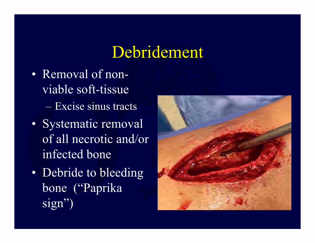
Debridement• Removal of non-
viable soft-tissue– Excise sinus tracts
• Systematic removal of all necrotic and/or infected bone
• Debride to bleeding bone (“Paprika sign”)
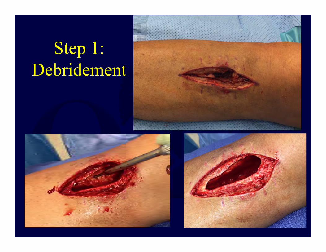
Step 1: Debridement
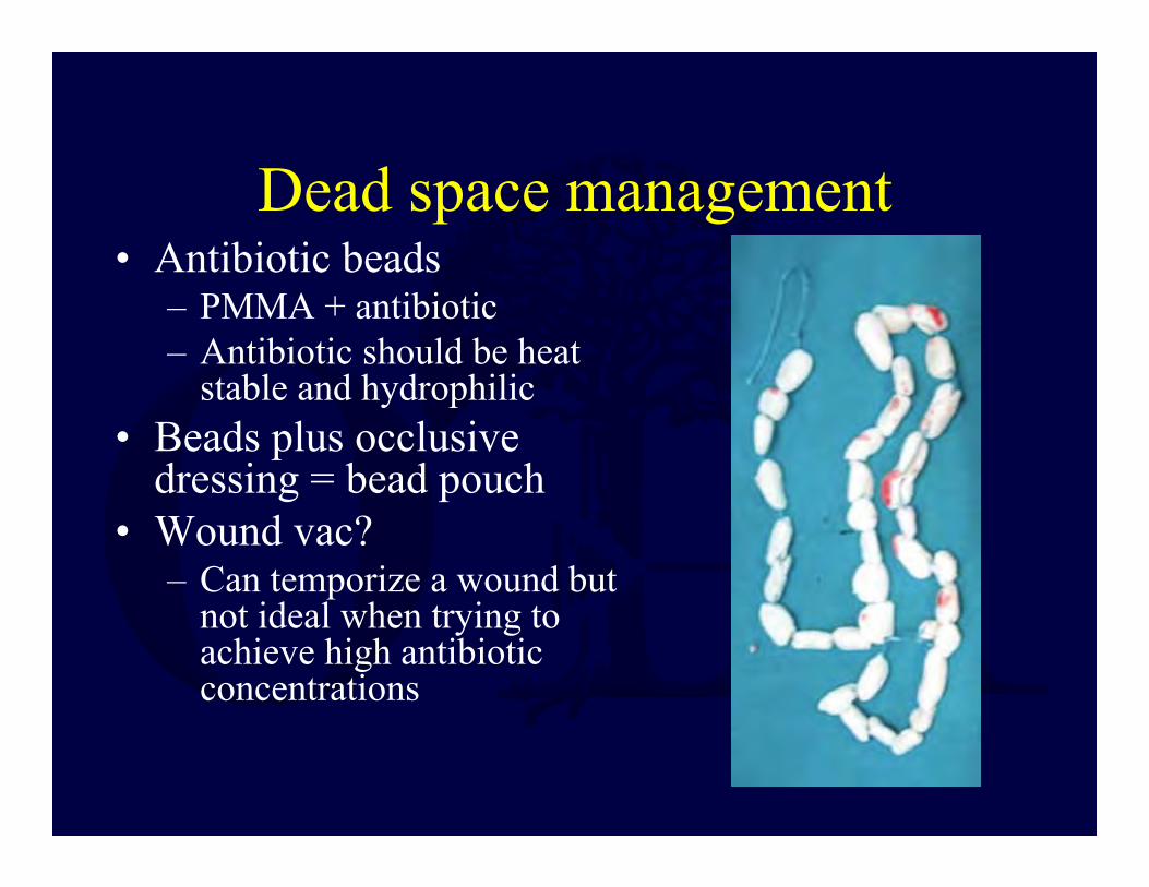
Dead space management• Antibiotic beads
– PMMA + antibiotic– Antibiotic should be heat
stable and hydrophilic• Beads plus occlusive
dressing = bead pouch• Wound vac?
– Can temporize a wound but not ideal when trying to achieve high antibiotic concentrations

Example: Dead space management antibiotic beads

Open Antibiotic Bead Pouch
• Highly useful for short periods to sterilize a wound as well as preserve bone and soft tissue health following diaphysectomy for osteomyelitis.
Printed with permission, David Lowenberg, MD

Open Antibiotic Bead Pouch
• 5 days following diaphysectomy at time of soft tissue reconstruction.
Printed with permission, David Lowenberg, MD
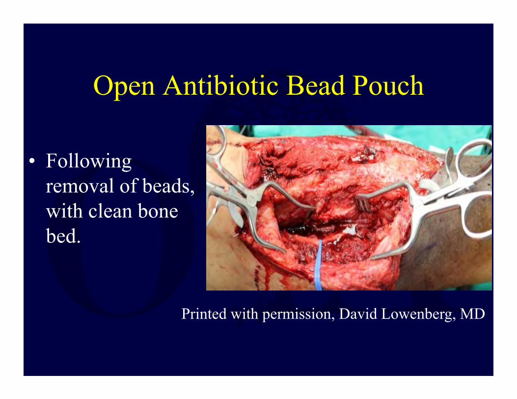
Open Antibiotic Bead Pouch
• Following removal of beads, with clean bone bed.
Printed with permission, David Lowenberg, MD

Soft-tissue reconstruction• Based on
reconstructive ladder• Often requires local
or free-tissue transfer– Must have skilled
microsurgeonavailable
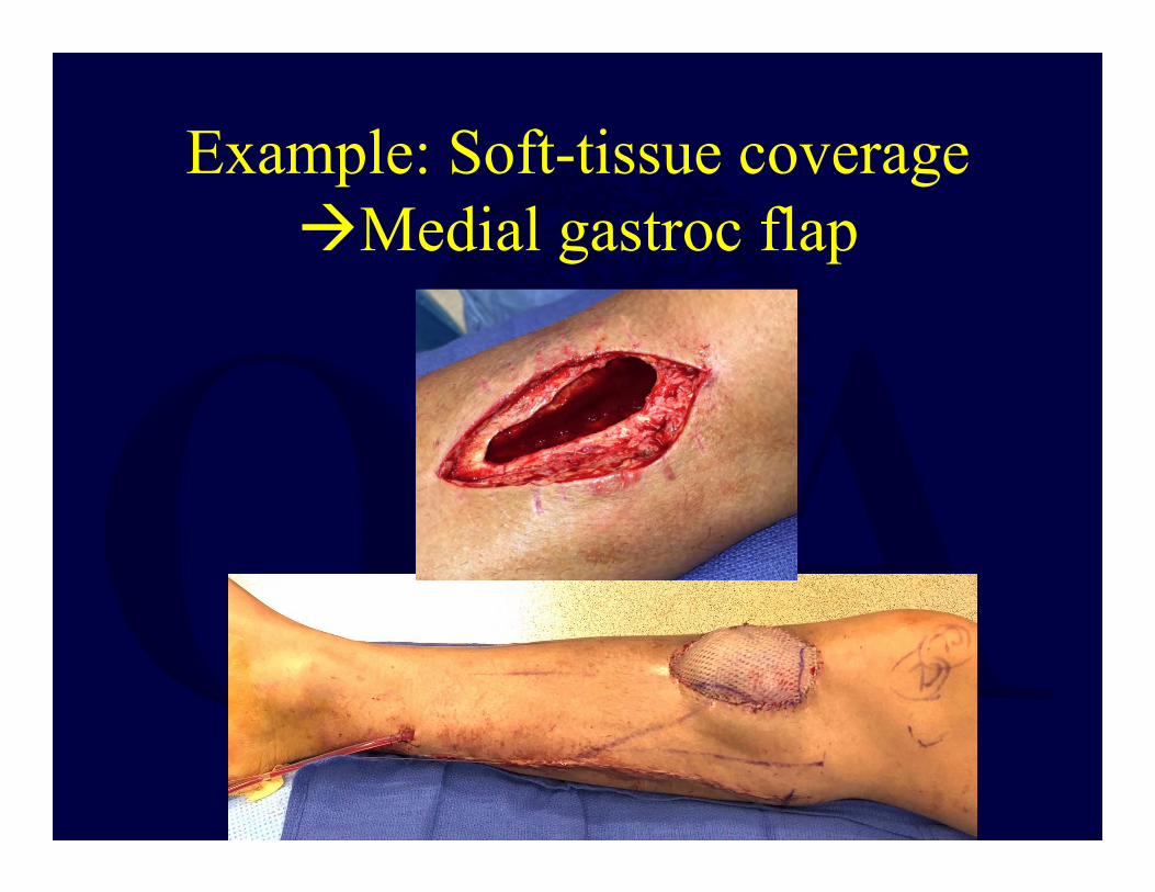
Example: Soft-tissue coverageMedial gastroc flap

Example: Anterolateral thigh free flap

Bone reconstruction• Non-segmental defects
– Additional stability may not be needed– Plan for bone grafting 6-8 weeks after
infection eradicated• Segmental defects
– Need provisional stability (most commonly external fixator)
– Plan for bone defect• Masquelet technique• Bone transport

Masquelet technique (Induced membrane)
• Antibiotic spacer placement + soft-tissue coverage
• Staged Bone grafting (6-8 weeks later)• Reported success ~80% for implant dependent
union• 10-12 months for union, weight bearing
Induced membrane properties
• Membrane secrets BMP-2, VEGF and other growth factors
• Peak at 4 weeks after membrane induction then decreases rapidly (Aho et al. JBJS 2013)
Bone transport• Corticotomy opposite
the defect• Segment transported
gradually, new bone formed by distraction osteogenesis
• Multiple techniques– External fixation
• Uniplanar• Ring fixator
– Transport over nail
From Giannikas et al. JBJS 2005
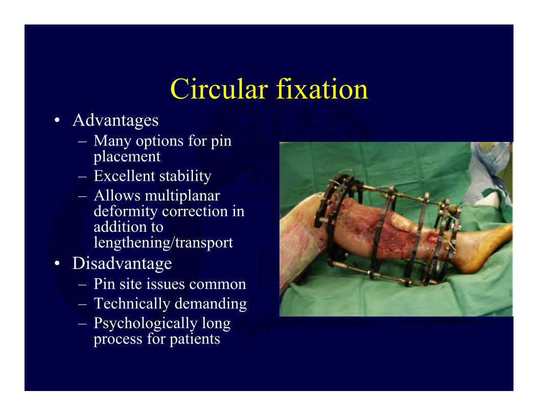
Circular fixation• Advantages
– Many options for pin placement
– Excellent stability– Allows multiplanar
deformity correction in addition to lengthening/transport
• Disadvantage– Pin site issues common– Technically demanding– Psychologically long
process for patients
Shortening
• Acute shortening >3cm may cause arterial flow impairment
• Results in limb length discrepancy and muscle shortening/dysfunction
• Reasonable option for small bone defects and/or resources limited
Infections associated with trauma implants
• Three scenarios:1. Stable hardware, fracture healed2. Stable hardware, fracture not healed3. Unstable hardware, fracture not healed

Stable hardware, fracture healed
• TreatmentI&D, remove hardware• Follows Stage 3 treatment principles• Typically no need for additional bony
stabilization assuming non-segmental defect

Stable hardware, fracture not healed
• If infection acute, can attempt I&D, retain hardware, suppress until fracture healing
• Goal to convert from Stage 4 to Stage 3 osteomyelitis
• 71% success in achieving fracture healing with antibiotic suppression (Berkes et al. JBJS 2010)
– Requires eventual hardware removal in ~30% cases– Hardware removal less likely in proximal (e.g. pelvis)
vs. distal locations (e.g. tibia)• If fracture healing achieved, principles follow
Stage 3 treatment

Unstable Hardware, Fracture Not Healed
• I&D, removal of hardware• Equivalent to Stage 4 Osteomyelitis (i.e.
Segmental Defect)• Requires strategy for bone stability (e.g.
ring fixator, antibiotic nail, etc.) and management of segmental bone defect

Results (of Comprehensive, Multidisciplinary Treatment Protocol)
• 2207 cases from 1981 through 2007– 1898 limb-salvage protocols– 230 amputations (as primary treatment)
• 85% overall success (infection-free, functional reconstruction at 2 years)– A-hosts 96%– B-hosts 74% – Limb-salvage 84%– Amputation 91%
Cierny G. Surgical Treatment of Osteomyelitis: Plastic and Reconstructive Surgery. 2011 Jan;127:190S–204S.

Results (cont)• Treatment failures (n=319)
– 43% aseptic nonunions– 28% wound sloughs– 15% unanticipated impairment – 12% recurrent sepsis – 2% deaths
• 82% success with retreatment• Overall 2 year success rate of 95%
– 99% A hosts– 90% B hosts
Cierny G. Surgical Treatment of Osteomyelitis: Plastic and Reconstructive Surgery. 2011 Jan;127:190S–204S.

Case Examples

Cierny-Mader Stage 1 …. A confined intramedullary
process
Printed with permission, David Lowenberg, MD
Cierny Mader Stage 1
• Currently the most common cause is secondary to infected intramedullary implants.

66 y/o F now 8 years s/p tibial rodding. With chronic leg pain and limited ability to ambulate.
Printed with permission, David Lowenberg, MD

Tc99 performed

CT of right leg
Printed with permission, David Lowenberg, MD

What do you do???A. Tell her that you can’t cure chronic pain.B. Take punch biopsies of bone.C. Start her on empiric Doxycycline.D. Stage her for neoplasm then perform open
biopsy with later plan for wide en bloc resection.
E. Call it for what it is, Type 1 C-M osteomyelitis, and treat appropriately.

Cierny-Mader Stage 2 Osteomyelitis
• In clinical practice, the rarest form of osteomyelitis seen.
• With the wider use of Negative Pressure Therapy, there has been a resurgence in cases.
Printed with permission, David Lowenberg, MD

Cierny Mader Stage 3 Osteomyelitis
• The most common form of osteomyelitis seen in clinical practice.
• Requires the basic tenants of osteomyelitis surgery to be followed:
1. Surgical resection2. Dead space management3. Soft tissue reconstruction4. Bone reconstruction

80 y/o F s/p hematogenous distal femoral osteomyelitis at age 15
• Initially treated with surgical debridement.• This remained completely quiescent for 65
years until she developed a mild case of the flu and presented draining with a distal lateral femoral sinus tract.
• Had remained completely active and asymptomatic until this event.

80 y/o F s/p hematogenous distal femoral osteomyelitis at age 15
Printed with permission, David Lowenberg, MD

Saucerization of the femur, removal of all infected necrotic bone, dead
space management
Printed with permission, David Lowenberg, MD

Saucerization of the femur, removal of all infected necrotic bone, dead
space management
Printed with permission, David Lowenberg, MD

Saucerization of the femur, removal of all infected necrotic bone, dead
space management
Printed with permission, David Lowenberg, MD
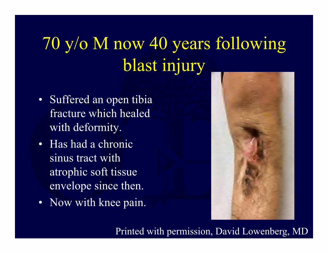
70 y/o M now 40 years following blast injury
• Suffered an open tibia fracture which healed with deformity.
• Has had a chronic sinus tract with atrophic soft tissue envelope since then.
• Now with knee pain.
Printed with permission, David Lowenberg, MD

70 y/o M with 40 year sinus tract
Printed with permission, David Lowenberg, MD
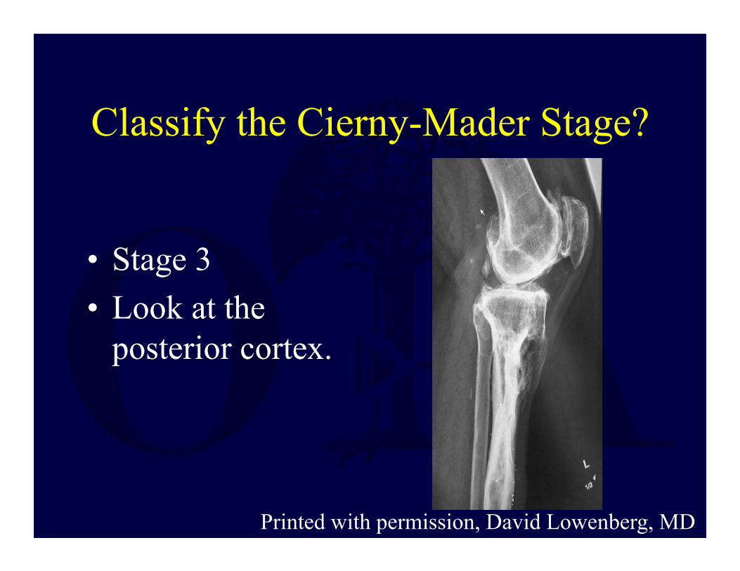
Classify the Cierny-Mader Stage?
• Stage 3• Look at the
posterior cortex.
Printed with permission, David Lowenberg, MD
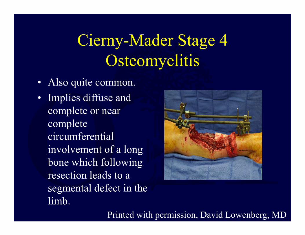
Cierny-Mader Stage 4 Osteomyelitis
• Also quite common. • Implies diffuse and
complete or near complete circumferential involvement of a long bone which following resection leads to a segmental defect in the limb.
Printed with permission, David Lowenberg, MD

What is an Infected Nonunion???
By definition it is a nonunion of a fracture
Printed with permission, David Lowenberg, MD
28 y/o M s/p Peds. Vs. MVA
• 28 y/o M (6’4” tall, 275 pounds) status post crush injury to leg when pinned by bumper of a car traveling at 35 MPH to rear of his tow truck.
• Initially rodded, then 3 week delay in flap coverage.

28 y/o M s/p Peds. Vs. MVA
• Persisitent drainage under free flap for 3 months.
• Treated with 3 months of IV antibiotics.
• Referred 4 months after injury with persistent drainage under flap.
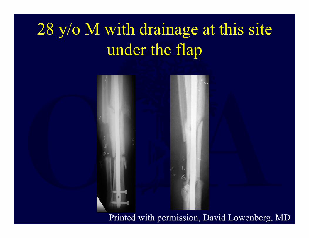
28 y/o M with drainage at this site under the flap
Printed with permission, David Lowenberg, MD

28 y/o M
• Note the cortical density that has developed at the intercalary segment.
Printed with permission, David Lowenberg, MD
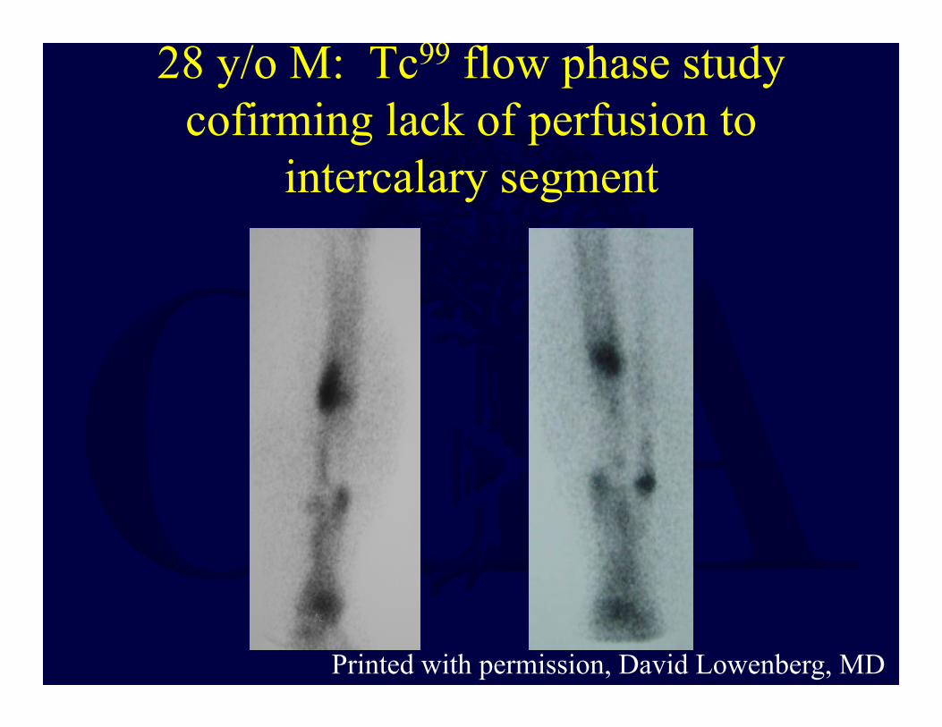
28 y/o M: Tc99 flow phase study cofirming lack of perfusion to
intercalary segment
Printed with permission, David Lowenberg, MD
28 y/o M: C-M Stage 4 osteomyelitis
• Complete devascularization of intercalary segment.
• Treated with en bloc resection and antibiotic nail, followed by bone transport.

28 y/o M: C-M Stage 4 osteomyelitis
Printed with permission, David Lowenberg, MD

47 y/o F s/p low energy distal tibia shaft fracture treated with
IM rodding• At the time of rodding a tourniquet was utilized.• The tibia was reamed up in size due to her small
intramedullary diameter.• Developed swelling and a new fracture at the isthmus
proximally which was not present previously.• Then developed drainage and soft tissue breakdown
necessitating free flap placement.• Underwent debridement and antibiotic nail and beads
but still concern for infection.• Referred then for care.

47 y/o female with infected nonunion of tibia:Note density developing of intercalary segment
Printed with permission, David Lowenberg, MD

Underwent flap elevation and exploration, intercalary segment avascular and infected, C-M
Stage 4 osteomyelitis
Printed with permission, David Lowenberg, MD

Conclusions
• Osteomyelitis after trauma is increasing• Biofilm is the hallmark of chronic infection that
makes osteomyelitis a surgical disease• Thorough workup and staging of the bone and the host
using the Cierny-Mader Classification is crucial to developing an effective treatment plan
• Systematic approach can lead to successful outcomes (1. Debridement, 2. Dead space management, 3. Soft-tissue coverage, 4. Address bone defect)
References1. Kremers HM, Nwojo ME, Ransom JE, et al. Trends in the Epidemiology of Osteomyelitis: A Population-Based Study, 1969 to 2009. The Journal of Bone & Joint Surgery. 2015 May 20;97(10):837–845.2. Taylor BC, Hancock J, Zitzke R, et al. Treatment of bone loss with the induced membrane technique: techniques and outcomes. Journal of orthopaedic trauma. 2015;29(12):554–557.3. Aho O-M, Lehenkari P, Ristiniemi J, et al. The Mechanism of Action of Induced Membranes in Bone Repair. The Journal of Bone and Joint Surgery (American). 2013 Apr 3;95(7):597.4. Masquelet AC, Begue T. The Concept of Induced Membrane for Reconstruction of Long Bone Defects. Orthopedic Clinics of North America. 2010 Jan;41(1):27–37.5. Cierny G, Mader JT, Penninck JJ. The Classic: A Clinical Staging System for Adult Osteomyelitis: Clinical Orthopaedics and Related Research. 2003 Sep;414:7–24.6. Blyth MJG, Kincaid R, Craigen MAC, et al. The changing epidemiology of acute and subacute haematogenousosteomyelitis in children. Bone & Joint Journal. 2001;83(1):99–102.7. Elliott IS, Groen RS, Kamara TB, et al. The Burden of Musculoskeletal Disease in Sierra Leone. Clinical Orthopaedics and Related Research®. 2015 Jan;473(1):380–389.8. Eralp L, Kocaoglu M, Rashid H. Reconstruction of Segmental Bone Defects Due to Chronic Osteomyelitis with Use of an External Fixator and an Intramedullary Nail: Surgical Technique. JBJS Essential Surgical Techniques. 2007 Sep 1;os-89(2_suppl_2):183–195.9. Mauffrey C, Hake ME, Chadayammuri V, et al. Reconstruction of Long Bone Infections Using the Induced Membrane Technique: Tips and Tricks. Journal of orthopaedic trauma. 2016;30(6):e188–e193.10. Waldvogel FA, Papageorgiou PS. Osteomyelitis: the past decade. N. Engl. J. Med. 1980 Aug 14;303(7):360–370.ics. 2015;30(3):156–160.

11. Lazzarini L, Mader JT, Calhoun JH. Osteomyelitis in long bones. J Bone Joint Surg Am. 2004;86(10):2305–2318.12. Waldvogel FA, Medoff G, Swartz MN. Osteomyelitis: a review of clinical features, therapeutic considerations and unusual aspects. N. Engl. J. Med. 1970 Jan 22;282(4):198–206.13. Archdeacon MT, Messerschmitt P. Modern papineau technique with vacuum-assisted closure. Journal of orthopaedic trauma. 2006;20(2):134–137.14. Azi M, Teixeira A, Cotias R, et al. Membrane Induced Osteogenesis in the Management of Post-traumatic Bone Defects: Journal of Orthopaedic Trauma. 2016 Apr;1.15. Berkes M. Maintenance of Hardware After Early Postoperative Infection Following Fracture Internal Fixation</article-title> The Journal of Bone and Joint Surgery (American). 2010 Apr 1;92(4):823.16. Lowenberg DW, Buntic RF, Buncke GM, et al. Long-term results and costs of muscle flap coverage with Ilizarov bone transport in lower limb salvage. Journal of orthopaedic trauma. 2013;27(10):576–581.17. Jauregui JJ, Bor N, Thakral R, et al. Life-and limb-threatening infections following the use of an external fixator. Bone Joint J. 2015;97(9):1296–1300.18. Paley D, Herzenberg JE. Intramedullary infections treated with antibiotic cement rods: preliminary results in nine cases. J Orthop Trauma. 2002 Dec;16(10):723–729.19. Chong K-W, Woon CY-L, Wong M-K. Induced Membranes--A Staged Technique of Bone-Grafting for Segmental Bone Loss: Surgical Technique. JBJS Essential Surgical Techniques. 2011 Mar 16;os-93(Supplement_1):85–91.20. Lowenberg DW, Githens M. Complex Limb Reconstruction With Simultaneous Muscle Transfer and Circular External Fixation. Techniques in Orthopaed

21. Baldan M, Gosselin RA, Osman Z, et al. Chronic osteomyelitis management in austere environments: the International Committee of the Red Cross experience. Tropical Medicine & International Health. 2014 Jul;19(7):832–837.22. Sachs BL, Shaffer JW. A staged Papineau protocol for chronic osteomyelitis. Clin. Orthop. Relat. Res. 1984 Apr;(184):256–263.23. Mader JT, Cripps MW, Calhoun JH. Adult posttraumatic osteomyelitis of the tibia. Clinical orthopaedics and related research. 1999;360:14–21.24. Ziran BH, Rao N, Hall RA. A Dedicated Team Approach Enhances Outcomes of Osteomyelitis Treatment: Clinical Orthopaedics and Related Research. 2003 Sep;414:31–36.25. Shirwaiker RA, Springer BD, Spangehl MJ, et al. A Clinical Perspective on Musculoskeletal Infection Treatment Strategies and Challenges. Journal of the American Academy of Orthopaedic Surgeons. 2015;23(suppl):S44–S54.