Embed Size (px)
Citation preview
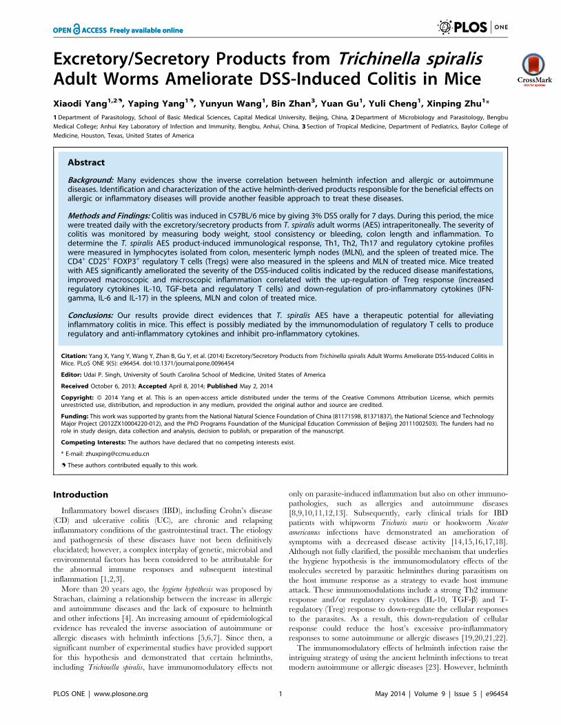
Excretory/Secretory Products from Trichinella spiralisAdult Worms Ameliorate DSS-Induced Colitis in MiceXiaodi Yang1,2., Yaping Yang1., Yunyun Wang1, Bin Zhan3, Yuan Gu1, Yuli Cheng1, Xinping Zhu1*
1 Department of Parasitology, School of Basic Medical Sciences, Capital Medical University, Beijing, China, 2 Department of Microbiology and Parasitology, Bengbu
Medical College; Anhui Key Laboratory of Infection and Immunity, Bengbu, Anhui, China, 3 Section of Tropical Medicine, Department of Pediatrics, Baylor College of
Medicine, Houston, Texas, United States of America
Abstract
Background: Many evidences show the inverse correlation between helminth infection and allergic or autoimmunediseases. Identification and characterization of the active helminth-derived products responsible for the beneficial effects onallergic or inflammatory diseases will provide another feasible approach to treat these diseases.
Methods and Findings: Colitis was induced in C57BL/6 mice by giving 3% DSS orally for 7 days. During this period, the micewere treated daily with the excretory/secretory products from T. spiralis adult worms (AES) intraperitoneally. The severity ofcolitis was monitored by measuring body weight, stool consistency or bleeding, colon length and inflammation. Todetermine the T. spiralis AES product-induced immunological response, Th1, Th2, Th17 and regulatory cytokine profileswere measured in lymphocytes isolated from colon, mesenteric lymph nodes (MLN), and the spleen of treated mice. TheCD4+ CD25+ FOXP3+ regulatory T cells (Tregs) were also measured in the spleens and MLN of treated mice. Mice treatedwith AES significantly ameliorated the severity of the DSS-induced colitis indicated by the reduced disease manifestations,improved macroscopic and microscopic inflammation correlated with the up-regulation of Treg response (increasedregulatory cytokines IL-10, TGF-beta and regulatory T cells) and down-regulation of pro-inflammatory cytokines (IFN-gamma, IL-6 and IL-17) in the spleens, MLN and colon of treated mice.
Conclusions: Our results provide direct evidences that T. spiralis AES have a therapeutic potential for alleviatinginflammatory colitis in mice. This effect is possibly mediated by the immunomodulation of regulatory T cells to produceregulatory and anti-inflammatory cytokines and inhibit pro-inflammatory cytokines.
Citation: Yang X, Yang Y, Wang Y, Zhan B, Gu Y, et al. (2014) Excretory/Secretory Products from Trichinella spiralis Adult Worms Ameliorate DSS-Induced Colitis inMice. PLoS ONE 9(5): e96454. doi:10.1371/journal.pone.0096454
Editor: Udai P. Singh, University of South Carolina School of Medicine, United States of America
Received October 6, 2013; Accepted April 8, 2014; Published May 2, 2014
Copyright: � 2014 Yang et al. This is an open-access article distributed under the terms of the Creative Commons Attribution License, which permitsunrestricted use, distribution, and reproduction in any medium, provided the original author and source are credited.
Funding: This work was supported by grants from the National Natural Science Foundation of China (81171598, 81371837), the National Science and TechnologyMajor Project (2012ZX10004220-012), and the PhD Programs Foundation of the Municipal Education Commission of Beijing 20111002503). The funders had norole in study design, data collection and analysis, decision to publish, or preparation of the manuscript.
Competing Interests: The authors have declared that no competing interests exist.
* E-mail: [email protected]
. These authors contributed equally to this work.
Introduction
Inflammatory bowel diseases (IBD), including Crohn’s disease
(CD) and ulcerative colitis (UC), are chronic and relapsing
inflammatory conditions of the gastrointestinal tract. The etiology
and pathogenesis of these diseases have not been definitively
elucidated; however, a complex interplay of genetic, microbial and
environmental factors has been considered to be attributable for
the abnormal immune responses and subsequent intestinal
inflammation [1,2,3].
More than 20 years ago, the hygiene hypothesis was proposed by
Strachan, claiming a relationship between the increase in allergic
and autoimmune diseases and the lack of exposure to helminth
and other infections [4]. An increasing amount of epidemiological
evidence has revealed the inverse association of autoimmune or
allergic diseases with helminth infections [5,6,7]. Since then, a
significant number of experimental studies have provided support
for this hypothesis and demonstrated that certain helminths,
including Trichinella spiralis, have immunomodulatory effects not
only on parasite-induced inflammation but also on other immuno-
pathologies, such as allergies and autoimmune diseases
[8,9,10,11,12,13]. Subsequently, early clinical trials for IBD
patients with whipworm Trichuris muris or hookworm Necator
americanus infections have demonstrated an amelioration of
symptoms with a decreased disease activity [14,15,16,17,18].
Although not fully clarified, the possible mechanism that underlies
the hygiene hypothesis is the immunomodulatory effects of the
molecules secreted by parasitic helminthes during parasitism on
the host immune response as a strategy to evade host immune
attack. These immunomodulations include a strong Th2 immune
response and/or regulatory cytokines (IL-10, TGF-b) and T-
regulatory (Treg) response to down-regulate the cellular responses
to the parasites. As a result, this down-regulation of cellular
response could reduce the host’s excessive pro-inflammatory
responses to some autoimmune or allergic diseases [19,20,21,22].
The immunomodulatory effects of helminth infection raise the
intriguing strategy of using the ancient helminth infections to treat
modern autoimmune or allergic diseases [23]. However, helminth
PLOS ONE | www.plosone.org 1 May 2014 | Volume 9 | Issue 5 | e96454
infections in humans could also lead to pathology and disease.
Direct treatment with the living worm is widely unacceptable
ethically and physically. Therefore, the proteins secreted by
parasitic helminthes involved in the immunomodulation have
become more attractive targets as a safe substitute for living
parasite infections for autoimmune therapies [24].
Ruyssers et al. [25] and Cancado et al. [26] recently
demonstrated the therapeutic potential of excretory/secretory
(ES) products from adult hookworms, Ancylostoma caninum and A.
ceylanicum, on experimental colitis in mice, predominantly through
the down-regulation of Th1 and Th17 cytokines. Although
helminthic ES products could directly modulate dendritic cells
(DC), suppress the expression of co-stimulatory MHCII and
produce anti-inflammatory cytokines [20], modulation of the
immune system by ES products derived from different helminth
species or developmental stages may act differently [27,28,29,30].
T. spiralis is one of the most widespread zoonotic parasitic
nematodes in the world. Its life cycle is completed in a single host
and includes three stages: muscle larvae inside skeletal striated
muscle cells, adult worms in the small intestine, and newborn
larvae in the lymphatic vessels and bloodstream. During different
phases or stages of parasite growth, T. spiralis interacts with the
host immune system to evade or inhibit the host immune response
through releasing a number of proteins into its surrounding
environment, which are considered to be crucial for its successful
invasion and survival within the host [31,32,33,34]. Several studies
have recently demonstrated that Trichinella secreted proteins or
infection itself are able to induce a strong Th2/Treg response and
the production of Th2/immunoregulation cytokines, e.g., IL-4,
IL-5, IL-10, IL-13, and TGF-b [29,32,35,36,37], which are
associated with the amelioration of autoimmune diseases such as
colitis [38,39], allergic airway inflammation [40,41], Type I
diabetes [42] and autoimmune encephalomyelitis [43].
In this study, we further explored the therapeutic effects of ES
products from T. spiralis adult worms (AES) on DSS-induced colitis
in C57BL/6 mice. The goal of this study is to better understand
the possible mechanisms behind their treatment efficacy as a
potential strategy to develop therapeutics for incurable immune
disorders using products derived from helminths, which have lived
within humans for millions of years.
Materials and Methods
Ethics statementAll experimental animals were purchased from Laboratory
Animal Services Center of Capital Medical University (Beijing,
China). All experimental procedures were reviewed and approved
by the Capital Medical University Animal Care and Use
Committee and were consistent with the NIH Guidelines for the
Care and Use of Laboratory Animals.
Animals and DSS-induced colitisFemale C57BL/6 mice, aged 6–8 weeks and free of specific
pathogens, were provided by the Laboratory Animal Center,
Academy of Military Medical Sciences.
The colitis was induced in C57BL/6 mice with dextran sodium
sulfate (DSS) as previously described [44]. Briefly, DSS (40 kDa,
Applichem, Germen) was dissolved in sterile filtered water at a
final concentration of 3% and presented to mice as drinking water
for 7 consecutive days. Freshly made DSS solution was provided
daily in drinking water. Negative control animals received filtered
water only. During the colitis induction, all mice were treated with
Trichinella AES as described below. On the 8th day, all animals
were sacrificed at the end of experiments by cervical dislocation.
Preparation of parasites and excretory/secretoryproducts from adults (AES)
T. spiralis (strain ISS 533) was maintained in female ICR mice.
Muscle larvae were recovered from the muscles of infected mice
using a standard pepsin/hydrochloric acid digestion method [45].
Adult T. spiralis worms were obtained from the intestine of a rat
orally infected with 12,000 muscle larvae after 84 hours [46]. T.
spiralis AES were prepared and collected as previously described
[29]. Briefly, T. spiralis adult worms recovered from intestine of
infected rat were washed three times with phosphate-buffered
saline (PBS) and then cultured in RPMI-1640 medium supple-
mented with 100 U/ml penicillin, 100 U/ml streptomycin and
0.25 mg/ml amphotericin B at 37uC and 5% CO2 for 48 hours.
The culture supernatant was collected by centrifugation, then
filtered through a 0.45 micron syringe filter and buffer exchanged
into PBS. The protein concentrations of the prepared ES products
were determined using BCA assay (Pierce, USA).
Experimental designIn the first experiment, we investigated the effect of T. spiralis
adult ES products on inhibiting the development of DSS-induced
colitis in mice. Briefly, the mice were divided into 4 groups (12–16
mice each group). The first two groups of mice were induced with
DSS to develop colitis as described above. Concurrently, each
group of mice was intraperitoneally injected daily with 25 mg of
AES (DSS-AES) or PBS only as a control (DSS-PBS) in a total
volume of 100 ml for 7 days of concomitant DSS-colitis induction.
The other two groups of mice were treated with the same amount
of AES or PBS for 7 days without colitis induction as a control
(AES-control and PBS-control, respectively).
After seven days of treatment, the mice were sacrificed, their
spleens, mesenteric lymph nodes (MLNs), and colon were
aseptically removed and the cells were isolated for cell culture
for cytokine profiling. Their entire large intestines were collected
for measuring the pathology and cytokine profile. The pathology
of each intestine was evaluated based on 5 criteria described in
detail as below: clinical disease activity index, the length of the
colon, macroscopic score, microscopic inflammation score and
myeloperoxidase (MPO) activity. Except for the principal inves-
tigator, all investigators were blind to all solution contents and
mice groups until the end of the experiments.
Clinical disease scoreThe mice were observed daily for morbidity and given a clinical
disease score (disease activity index, DAI) between 0 and 12 based
on the following characteristic criteria: weight loss, diarrhea, and
bleeding feces [44] (Table 1).
Macroscopic inflammation scoreAfter being sacrificed, the mouse colon was removed and
opened longitudinally, and the colonic damage was assessed
macroscopically. Briefly, four parameters were taken into account:
presence of adhesions, degree of colonic ulcerations, wall
thickness, and degree of mucosal edema. Each parameter was
given a score from 0 (normal) to 3 (severe) as previously described
[47]. The total score ranged from a minimum of 0 to a maximum
of 12. The length of colon as a way to evaluate the extent of
inflammation [51] was also measured.
Microscopic inflammation scoreSmall segments of the colon taken for histopathology examina-
tion were fixed in 4% neutral-buffered formalin, embedded into
paraffin, sectioned at 5 mm thickness and stained with hematoxylin
ES of T. spiralis Adult Worms Ameliorate Colitis
PLOS ONE | www.plosone.org 2 May 2014 | Volume 9 | Issue 5 | e96454
and eosin. To evaluate the severity of inflammation, we adopted
the histological damage score from Obermeier et al. [48] based on
the following parameters: (a) Epithelial damage (0 point = none, 1
point = minimal loss of goblet cells, 2 points = extensive loss of
goblet cells, 3 points = minimal loss of crypts and extensive loss of
goblet cells, and 4 points = extensive loss of crypts), (b) Infiltration
(0 point = none, 1 point = infiltration around crypt bases, 2
points = infiltration in the muscularis mucosa, 3 points = extensive
infiltration in the muscularis mucosa with edema, and 4
points = infiltration of the submucosa). The total score ranged
from a minimum of 0 to a maximum of 8.
MPO activity assayMyeloperoxidase (MPO) activity, an enzyme occurring nearly
exclusively in neutrophils, was determined using a MPO assay kit
(Nanjing Jiancheng Bio-engineering Institute, China). Briefly,
100 mg of colon tissue was cut and homogenized in 1.9 ml of
50 mM PBS, pH 6.0, containing 0.5% hexadecyltrimethyl
ammonium hydroxide and centrifuged at 12,000 rpm (4uC) for
20 min. The protein concentration of the colon extract superna-
tant was measured using a BCA protein assay kit (Thermo
Scientific, USA). One hundred microliters of the supernatant was
transferred into 2.9 ml of PBS (pH 6.0) containing 0.17 mg/ml
3,39-dimethoxybenzidine and 0.0005% H2O2. The MPO activity
of the supernatant was determined by measuring the H2O2-
dependent oxidation of 3,39-dimethoxybenzidine and is expressed
as units per gram of total protein (U/g).
Lymphocyte isolation and multiplex cytokine profilingSpleens and MLNs were removed aseptically from the
experimental mice, and the cells were isolated and suspended in
RPMI-1640 supplemented with 5% bovine fetal serum, 100 mM
L-glutamine, 100 U/ml penicillin, and 100 mg/ml streptomycin.
Colons were collected and thoroughly washed with PBS and then
cut into 0.5 cm pieces. The epithelia were removed by incubation
with 1 mM DTT (Sigma-Aldrich, USA) and 1 mM EDTA
(Sigma-Aldrich, USA) in RPMI 1640 medium supplemented with
5% FCS at 37uC for 20 min with gentle shaking. After repeating
this step twice, the tissue was cut into smaller pieces and then
incubated for 40 min at 37uC in 20 ml of RPMI 1640 containing
25 mM HEPES, 2 mM L-glutamine, 1 mM sodium pyruvate,
100 U/ml penicillin, 10 mg/ml gentamicin, 100 mg/ml strepto-
mycin, and 1 mg/ml Liberase Research Grade Purified Enzymes
(Roche, Mannheim, Germany). The cells suspension was filtered
through a 100-mm filter and washed, and the lamina propria
mononuclear cells (LPMC) were harvested by discontinuous 30/
70% percoll gradient centrifugation.
The cells were subjected to a cytokine assay using IFN-c, IL-4,
IL-6, IL-10 (BD Biosciences, USA) and IL-17 (R&D Systems,
USA) ELISPOT sets according to the manufacturer’s instructions.
Briefly, a total of 16106 cells were added to each well of 96-well
MultiScreen HTS Filter plates (Millipore, USA) pre-coated with
anti-mouse IL-4, IL-6, and IL-10, whereas 26105 cells were added
being stimulated with anti-CD3 and anti-CD28 (BD Biosciences,
USA) at a concentration of 1 mg/ml for 48 h (37uC), the cells were
incubated with biotin-conjugated secondary antibodies and
streptavidin-HRP (for the IFN-c, IL-4, IL-6 and IL-10 assay).
Single cytokine-positive cells were visualized by adding an AEC
substrate and counted using an ELISPOT reader (CTL, USA)
with the Immunospot image analyzer software (version 4.0). For
the IL-17 assay, streptavidin-AP and BCIP/NBT chromogen were
used [49,50].
In addition, the levels of IL-13 and TGF-b in the cell culture
medium were determined using ELISA kits following the
manufacturer’s instructions (R&D Systems, USA). Briefly, in
which TGF-b was measured, cells (16106) were switched to
serum-free media with 4 changes in media over a 24-hour period
to reduce the background level of TGF-b provided by the cell
culture serum. Samples were collected after being cultured for
24 hours past the last medium change, treated with 1 N HCl and
then neutralized with 1.2 N NaOH/0.5 M HEPES to activate
latent TGF-b. Hence, the presented data reflect the total TGF-bin the samples.
Fluorescence-activated cell sorting (FACS) analysis ofCD4 CD25 FOXP3 Tregs in the spleen and MLNlymphocytes
+ + +
To evaluate the Tregs induced by treatment with the T. spiralis
ES products, the isolated cells from the spleens and MLN of the
treated mice were sorted by FACS using reagents from BD
Pharmingen (USA). The cell surfaces were blocked with rat anti-
mouse CD16/CD32 mAb for 15 min (4uC) and then incubated
with anti-mouse CD3e, CD4, or CD25 mAbs or their isotype
controls for surface marker staining. Subsequently, the cells were
treated with FACS lysing solution for 10 min. After washing,
fixation, permeabilization and a second blocking, the intracellular
labeling of the Foxp3 protein was performed by treating the cells
with anti-mouse Foxp3 mAb for 30 min on ice in the dark. The
cells were washed twice and resuspended in 200 ml PBS
containing 1% FBS followed by immediate flow cytometric
analysis (BD Biosciences, USA).
Statistical analysesAll data are presented as the mean 6 standard error of the
mean (SEM) and evaluated using a one-way ANOVA analysis and
SPSS 11.5 software. P,0.05 was regarded as statistically
significant. All statistical analyses were performed using GraphPad
Prism software.
Results
Effect of T. spiralis AES on relieving the severity of theDSS-induced acute colitis
After 7 days of oral DSS administration, all mice developed
significant colitis manifestations such as weight loss, diarrhea,
rectal bleeding, and physical weakness compared to the control
mice treated with PBS only. The intraperitoneal treatment with
Table 1. The criteria for scoring the DSS-induced colitis (DAI).
Pathologic Score 0 1 2 3 4
Weight loss none 1–5% 5–10% 10–15% .15%
Stool shape normal between loose stool between watery diarrhea
Stool bleeding none between slight bleeding between gross bleeding
doi:10.1371/journal.pone.0096454.t001
ES of T. spiralis Adult Worms Ameliorate Colitis
PLOS ONE | www.plosone.org 3 May 2014 | Volume 9 | Issue 5 | e96454
to plates pre-coated with anti-IFN-c and IL-17 antibodies. After
25 mg of AES significantly reduced the clinical score for the DSS-
induced colitis in the mice (DSS-AES) compared to the PBS
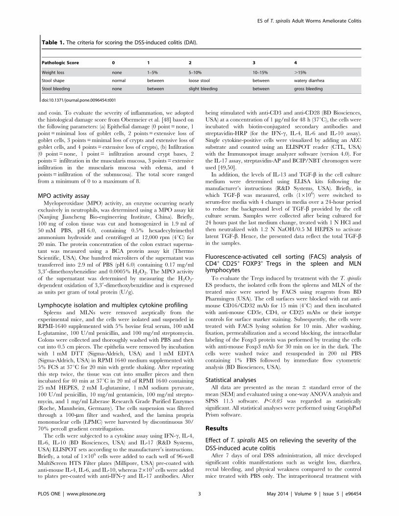
control (DSS-PBS) (Fig. 1, P,0.05). These improvements included
lower disease activity indices (DAI, Fig. 1A), less body weight loss
(Fig. 1B), less diarrhea or bleeding diarrhea and less colon
shrinking (Fig. 1C, D, E). The shrinking or shortening of the colon
and bleeding inside the colon were identified as the major signs of
colitis induced by DSS [51]. Apparently, there was no DSS-
induced bleeding or significant colon shrinking observed in mice
treated with AES compared to PBS control mice (Fig. 1D). Similar
to the PBS control, T. spiralis AES itself did not lead to obvious
pathology to the treated mice except for minor intestinal edema at
the intraperitoneal injection site (Fig. 1).
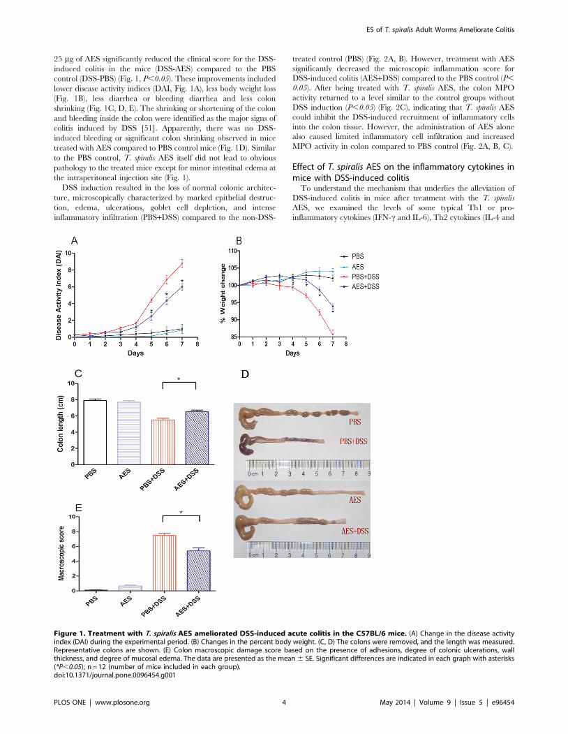
DSS induction resulted in the loss of normal colonic architec-
ture, microscopically characterized by marked epithelial destruc-
tion, edema, ulcerations, goblet cell depletion, and intense
inflammatory infiltration (PBS+DSS) compared to the non-DSS-
treated control (PBS) (Fig. 2A, B). However, treatment with AES
significantly decreased the microscopic inflammation score for
DSS-induced colitis (AES+DSS) compared to the PBS control (P,
0.05). After being treated with T. spiralis AES, the colon MPO
activity returned to a level similar to the control groups without
DSS induction (P,0.05) (Fig. 2C), indicating that T. spiralis AES
could inhibit the DSS-induced recruitment of inflammatory cells
into the colon tissue. However, the administration of AES alone
also caused limited inflammatory cell infiltration and increased
MPO activity in colon compared to PBS control (Fig. 2A, B, C).
Effect of T. spiralis AES on the inflammatory cytokines inmice with DSS-induced colitis
To understand the mechanism that underlies the alleviation of
DSS-induced colitis in mice after treatment with the T. spiralis
AES, we examined the levels of some typical Th1 or pro-
inflammatory cytokines (IFN-c and IL-6), Th2 cytokines (IL-4 and
Figure 1. Treatment with T. spiralis AES ameliorated DSS-induced acute colitis in the C57BL/6 mice. (A) Change in the disease activityindex (DAI) during the experimental period. (B) Changes in the percent body weight. (C, D) The colons were removed, and the length was measured.Representative colons are shown. (E) Colon macroscopic damage score based on the presence of adhesions, degree of colonic ulcerations, wallthickness, and degree of mucosal edema. The data are presented as the mean 6 SE. Significant differences are indicated in each graph with asterisks(*P,0.05); n = 12 (number of mice included in each group).doi:10.1371/journal.pone.0096454.g001
ES of T. spiralis Adult Worms Ameliorate Colitis
PLOS ONE | www.plosone.org 4 May 2014 | Volume 9 | Issue 5 | e96454
IL-13), a Th17 cytokine (IL-17) and regulatory cytokines (IL-10
and TGF-b) secreted by spleen, MLN and colon lymphocytes from
differently treated mice using ELISPOT or ELISA.
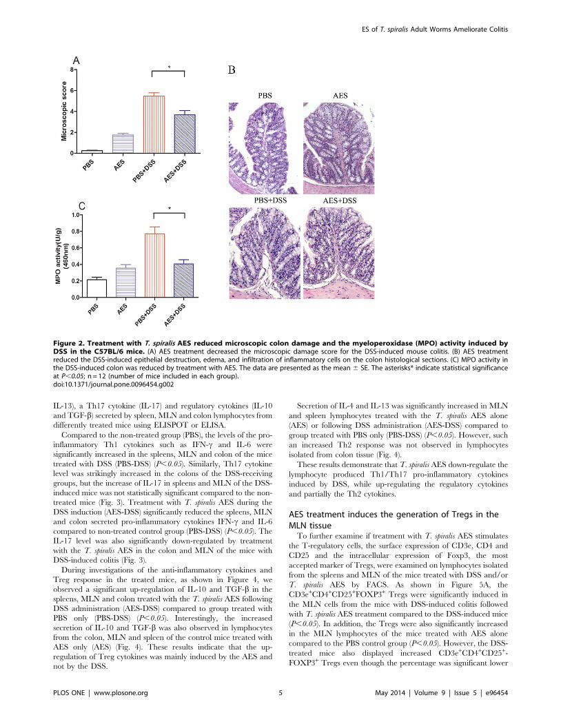
Compared to the non-treated group (PBS), the levels of the pro-
inflammatory Th1 cytokines such as IFN-c and IL-6 were
significantly increased in the spleens, MLN and colon of the mice
treated with DSS (PBS-DSS) (P,0.05). Similarly, Th17 cytokine
level was strikingly increased in the colons of the DSS-receiving
groups, but the increase of IL-17 in spleens and MLN of the DSS-
induced mice was not statistically significant compared to the non-
treated mice (Fig. 3). Treatment with T. spiralis AES during the
DSS induction (AES-DSS) significantly reduced the spleens, MLN
and colon secreted pro-inflammatory cytokines IFN-c and IL-6
compared to non-treated control group (PBS-DSS) (P,0.05). The
IL-17 level was also significantly down-regulated by treatment
with the T. spiralis AES in the colon and MLN of the mice with
DSS-induced colitis (Fig. 3).
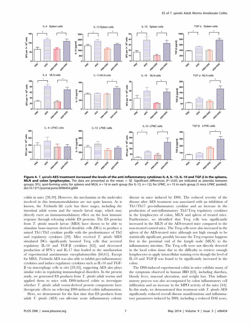
During investigations of the anti-inflammatory cytokines and
Treg response in the treated mice, as shown in Figure 4, we
observed a significant up-regulation of IL-10 and TGF-b in the
spleens, MLN and colon treated with the T. spiralis AES following
DSS administration (AES-DSS) compared to group treated with
PBS only (PBS-DSS) (P,0.05). Interestingly, the increased
secretion of IL-10 and TGF-b was also observed in lymphocytes
from the colon, MLN and spleen of the control mice treated with
AES only (AES) (Fig. 4). These results indicate that the up-
regulation of Treg cytokines was mainly induced by the AES and
not by the DSS.
Secretion of IL-4 and IL-13 was significantly increased in MLN
and spleen lymphocytes treated with the T. spiralis AES alone
(AES) or following DSS administration (AES-DSS) compared to
group treated with PBS only (PBS-DSS) (P,0.05). However, such
an increased Th2 response was not observed in lymphocytes
isolated from colon tissue (Fig. 4).
These results demonstrate that T. spiralis AES down-regulate the
lymphocyte produced Th1/Th17 pro-inflammatory cytokines
induced by DSS, while up-regulating the regulatory cytokines
and partially the Th2 cytokines.
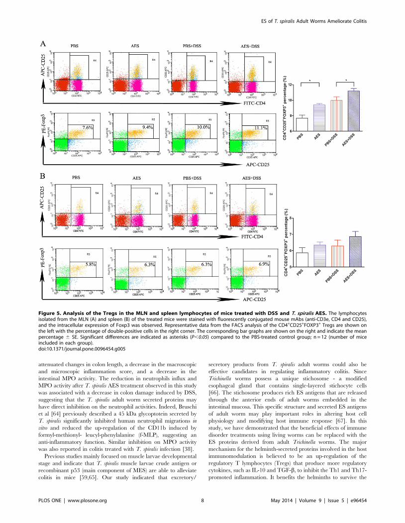
AES treatment induces the generation of Tregs in theMLN tissue
To further examine if treatment with T. spiralis AES stimulates
the T-regulatory cells, the surface expression of CD3e, CD4 and
CD25 and the intracellular expression of Foxp3, the most
accepted marker of Tregs, were examined on lymphocytes isolated
from the spleens and MLN of the mice treated with DSS and/or
T. spiralis AES by FACS. As shown in Figure 5A, the
CD3e+CD4+CD25+FOXP3+ Tregs were significantly induced in
the MLN cells from the mice with DSS-induced colitis followed
with T. spiralis AES treatment compared to the DSS-induced mice
(P,0.05). In addition, the Tregs were also significantly increased
in the MLN lymphocytes of the mice treated with AES alone
compared to the PBS control group (P,0.05). However, the DSS-
treated mice also displayed increased CD3e+CD4+CD25+-
FOXP3+ Tregs even though the percentage was significant lower
Figure 2. Treatment with T. spiralis AES reduced microscopic colon damage and the myeloperoxidase (MPO) activity induced byDSS in the C57BL/6 mice. (A) AES treatment decreased the microscopic damage score for the DSS-induced mouse colitis. (B) AES treatmentreduced the DSS-induced epithelial destruction, edema, and infiltration of inflammatory cells on the colon histological sections. (C) MPO activity inthe DSS-induced colon was reduced by treatment with AES. The data are presented as the mean 6 SE. The asterisks* indicate statistical significanceat P,0.05; n = 12 (number of mice included in each group).doi:10.1371/journal.pone.0096454.g002
ES of T. spiralis Adult Worms Ameliorate Colitis
PLOS ONE | www.plosone.org 5 May 2014 | Volume 9 | Issue 5 | e96454
compared to the group followed with AES treatment. It is possible
to observe increased Tregs for autoimmune diseases; however, the
functions of the Tregs in these diseases are typically impaired, and
the impaired Tregs are usually unable to regulate the excessive
immunopathology [52].
Although not significant, T. spiralis AES also tend to augment
the percentage of the Tregs cell subset in the spleens of mice with
DSS-induced colitis (P = 0.19) (Fig. 5B). Apparently, these results,
combined with the cytokine profile, suggest that the T. spiralis AES
treatment could induce the generation of Tregs in mice to suppress
the inflammatory response induced by the DSS.
Discussion
Abundant evidence has demonstrated that helminth infections
have the ability to ameliorate IBD [20,53]. Elliott et al. [54] were
the first to propose using helminthic parasites as a practical
treatment for Crohn’s disease. The preventive and therapeutic
effects of different helminth species and developmental stages on
experimental colitis were then investigated [55,56,57,58]. Treat-
ment with nematode, trematode or cestode helminths or their
products reduced the severity of colitis in different animal models
[38,59].
Trichinella infections have been found to down-regulate the
inflammatory immunopathology caused by autoimmune diseases
in various animal models such as experimental autoimmune
encephalomyelitis (EAE) in rats [11,43,60,61] and experimental
Figure 3. Treatment with the T. spiralis AES reduced the levels of the DSS-induced pro-inflammatory cytokines IFN-c, IL-6 and IL-17in the spleens, MLN and colon lymphocytes. The data are presented as the mean 6 SE. The asterisks* indicate statistical significance at P,0.05;SFU, spot-forming units; for spleens and MLN, n = 16 in each group (for IL-17, n = 12); for LPMC, n = 15 in each group (3 mice LPMC pooled).doi:10.1371/journal.pone.0096454.g003
ES of T. spiralis Adult Worms Ameliorate Colitis
PLOS ONE | www.plosone.org 6 May 2014 | Volume 9 | Issue 5 | e96454
colitis in mice [38,39]. However, the mechanism or the molecules
involved in this immunomodulation are not quite known. As is
known, the Trichinella life cycle has three stages, including the
intestinal adult worm and the muscle larval stage, which may
directly exert an immunomodulatory effect on the host immune
response through releasing soluble ES proteins. The ES proteins
from T. spiralis muscle larvae (MES) have shown to be able to
stimulate bone-marrow derived dendritic cells (DCs) to produce a
mixed Th1/Th2 cytokine profile with the predominance of Th2
and regulatory cytokines [29]. Mice received T. spiralis MES
simulated DCs significantly boosted Treg cells that secreted
regulatory IL-10 and TGF-b cytokines [62], and decreased
production of IFN-c and IL-17 that leaded to the amelioration
of experimental autoimmune encephalomyelitis [60,61]. Except
for MES, Trichinella AES was also able to inhibit pro-inflammatory
cytokines and induce regulatory cytokines such as IL-10 and TGF-
b via macrophage cells in vitro [29,32], suggesting AES also plays
similar roles in regulating immunological disorders. In the present
study, we generated ES products from T. spiralis adult worms and
applied them to mice with DSS-induced colitis to investigate
whether T. spiralis adult worm-derived protein components have
therapeutic effects on relieving DSS-induced colitis inflammation.
Here, we demonstrate for the first time that ES products from
adult T. spiralis (AES) can alleviate acute inflammatory colonic
disease in mice induced by DSS. The reduced severity of the
disease after AES treatment was associated with an inhibition of
Th1/Th17 pro-inflammatory cytokine and an increase in the
production of anti-inflammatory Th2/Treg regulatory cytokines
in the lymphocytes of colon, MLN and spleen of treated mice.
Furthermore, we identified that Treg cells was significantly
increased in the MLN of the AES-treated mice compared to the
non-treated control mice. The Treg cells were also increased in the
spleen of the AES-treated mice although not high enough to be
statistically significant, possibly because the Treg response happens
first in the proximal end of the lymph node (MLN) to the
inflammatory intestine. The Treg cells were not directly detected
in the local colon tissue due to the difficulty to retrieve enough
lymphocytes to apply intracellular staining even though the level of
IL-10 and TGF-b was found to be significantly increased in the
colon.
The DSS-induced experimental colitis in mice exhibits many of
the symptoms observed in human IBD [63], including diarrhea,
bloody feces, mucosal ulceration, and weight loss. This inflam-
matory process was also accompanied by colon inflammatory cell
infiltration and an increase in the MPO activity of the mice [44].
In this study, we demonstrated that treatment with T. spiralis AES
significantly reduced overall disease manifestations and inflamma-
tory parameters induced by DSS, including a reduced DAI score,
Figure 4. T. spiralis AES treatment increased the levels of the anti-inflammatory cytokines IL-4, IL-13, IL-10 and TGF-b in the spleens,MLN and colon lymphocytes. The data are presented as the mean 6 SE. Significant differences (P,0.05) are indicated as asterisks betweengroups; SFU, spot-forming units; for spleens and MLN, n = 16 in each group (for IL-13, n = 12); for LPMC, n = 15 in each group (3 mice LPMC pooled).doi:10.1371/journal.pone.0096454.g004
ES of T. spiralis Adult Worms Ameliorate Colitis
PLOS ONE | www.plosone.org 7 May 2014 | Volume 9 | Issue 5 | e96454
attenuated changes in colon length, a decrease in the macroscopic
and microscopic inflammation score, and a decrease in the
intestinal MPO activity. The reduction in neutrophils influx and
MPO activity after T. spiralis AES treatment observed in this study
was associated with a decrease in colon damage induced by DSS,
suggesting that the T. spiralis adult worm secreted proteins may
have direct inhibition on the neutrophil activities. Indeed, Bruschi
et al [64] previously described a 45 kDa glycoptotein secreted by
T. spiralis significantly inhibited human neutrophil migrations in
vitro and reduced the up-regulation of the CD11b induced by
formyl-methionyl- leucyl-phenylalanine (f-MLP), suggesting an
anti-inflammatory function. Similar inhibition on MPO activity
was also reported in colitis treated with T. spiralis infection [38].
Previous studies mainly focused on muscle larvae developmental
stage and indicate that T. spiralis muscle larvae crude antigen or
recombinant p53 (main component of MES) are able to alleviate
colitis in mice [59,65]. Our study indicated that excretory/
secretory products from T. spiralis adult worms could also be
effective candidates in regulating inflammatory colitis. Since
Trichinella worms possess a unique stichosome - a modified
esophageal gland that contains single-layered stichocyte cells
[66]. The stichosome produces rich ES antigens that are released
through the anterior ends of adult worms embedded in the
intestinal mucosa. This specific structure and secreted ES antigens
of adult worm may play important roles in altering host cell
physiology and modifying host immune response [67]. In this
study, we have demonstrated that the beneficial effects of immune
disorder treatments using living worms can be replaced with the
ES proteins derived from adult Trichinella worms. The major
mechanism for the helminth-secreted proteins involved in the host
immunomodulation is believed to be an up-regulation of the
regulatory T lymphocytes (Tregs) that produce more regulatory
cytokines, such as IL-10 and TGF-b, to inhibit the Th1 and Th17-
promoted inflammation. It benefits the helminths to survive the
Figure 5. Analysis of the Tregs in the MLN and spleen lymphocytes of mice treated with DSS and T. spiralis AES. The lymphocytesisolated from the MLN (A) and spleen (B) of the treated mice were stained with fluorescently conjugated mouse mAbs (anti-CD3e, CD4 and CD25),and the intracellular expression of Foxp3 was observed. Representative data from the FACS analysis of the CD4+CD25+FOXP3+ Tregs are shown onthe left with the percentage of double-positive cells in the right corner. The corresponding bar graphs are shown on the right and indicate the meanpercentage 6 SE. Significant differences are indicated as asterisks (P,0.05) compared to the PBS-treated control group; n = 12 (number of miceincluded in each group).doi:10.1371/journal.pone.0096454.g005
ES of T. spiralis Adult Worms Ameliorate Colitis
PLOS ONE | www.plosone.org 8 May 2014 | Volume 9 | Issue 5 | e96454

host Th1-mediated cellular attack, simultaneously preventing the
host’s immune system from overreacting to innate or external
immunogens or allergens [7,23]. Th17 is another subset of CD4 T
cells that produce IL-17, a pro-inflammatory cytokine involved in
the autoimmune diseases and other immunopathology [61,68]. In
this study, we have observed the high level of pro-inflammatory
cytokines (IFN-c, IL-6 and IL-17) produced by lymphocytes from
spleen, MLN or colon of mice with DSS-induced colitis, further
suggesting these pro-inflammatory cytokines are involved in the
inflammation of colitis. The IL-6 also contributes to the
differentiation of Th17 cells, aggravating immunopathogenesis of
IBD [69]. After being treated with T. spiralis AES, the Th1 and
Th17 cytokines were significantly reduced, simultaneously corre-
lating with the boost of Treg cells and their produced regulatory
cytokines (IL-10 and TGF-b). An increased IL-10 level was also
detected in patients infected with Schistosome, who displayed a less
severe skin-prick test to allergen [70]. Mice with IL-10 knocked
out developed more severe lupus, an autoimmune disease
mediated by pathogenic Th1 cytokine responses [71]. These
results have led to the suggestion that the IL-10 and/or TGF-bsecreted by Tregs in response to a chronic helminth infection or
helminth-secreted molecules directly moderate Th1-mediated
immunopathology. In this study, increased Treg cells as well as
the higher level of their secreted IL-10/TGF-b, and decreased
pro-inflammatory cytokines including Th1 and Th17, were
observed in DSS-induced colitis mice treated with T. spiralis
AES, further suggesting the immunomodulatory effects of T.
spiralis AES and its pharmaceutical potential for the treatment of
autoimmune or allergic diseases.
Helminthic infections or worm-derived products typically
induce strong Th2 immune response which contributes to the
protective immunity [27,29]. The Th2 cytokines IL-4 and IL-13
were apparently boosted in MLN and spleen lymphocytes of mice
treated with T. spiralis AES in this study, but not significantly in
treated mouse colons that represents a local response to DSS-
induced colitis. The Th2 response in colon upon T. spiralis AES
treatment may be offset by a strong local inflammatory reaction.
Ruyssers et al. [25] and Cancado et al. [26] also observed that
treatment with helminth proteins did not significantly alter the
level of IL-4 in the colon with colitis. In fact, Bodammer et al. [57]
have shown soluble egg antigen of Schistosome failed to improve
colitis even though it induced a robust Th2 response, suggesting
that Th2 response may not be essential for the control of colonic
inflammation. Therefore, our results suggest that the beneficial
effect of T. spiralis AES to inflammatory colitis may not be Th2-
mediated, but rather act through an overall effect of boosting Treg
and restraining Th1/Th17.
In this study we have showed the T. spiralis adult secreted
proteins (AES) apparently inhibited DSS-induced inflammation
mainly through inhibiting Th1 cytokines and stimulating regula-
tory T-cells, however, AES themselves tend to cause minor
inflammation in administrated mice, observed by limited inflam-
matory cell infiltration and increased MPO activity compared to
PBS control (Fig. 2A, B, C). The increases of relevant Th1/Th2
cytokines were also observed in the AES-alone treated mice
compared to PBS control except for stimulating Treg cytokines
(IL-10 and TGF-b) (Fig. 3, 4). It correlates with the findings that T.
spiralis derived antigens induced a mixed Th1/Th2 responses
through stimulating dendritic cells [29]. It was assumed that T.
spiralis worms secrete various proteins that play different biological
and immunological functions except for the immunomodulatory
effects [29,30,31]. Therefore, it is important to identify the active
components in the complex of the nematode-secreted proteins that
contribute only to the immunomodulation of inflammatory and
auto-immune diseases. Using the identified active protein(s), rather
than the AES complex, for the therapy or prevention of
autoimmune diseases is a more feasible approach.
In conclusion, T. spiralis adult ES products ameliorate DSS-
induced colitis in mice, however, these worm-derived products are
difficult to be manufactured on a large-scale, and their compli-
cated components may lead to undesired side-effects or other
safety issues. Studies are currently underway to screen for and
ultimately to identify the effector molecule(s) from T. spiralis AES
that play roles in immunomodulatory effects for potential large-
scale pharmaceutical applications for allergic or autoimmune
diseases.
Acknowledgments
We thank Xiaoqin Chen, Jing Yang, Xi Zhao, Zhifei Zhang, Jingjing
Huang, Fengyun Wang, Shijuan Cui, Jin Pan and Zhiyong Tao for their
technical assistance.
Author Contributions
Conceived and designed the experiments: XPZ BZ. Performed the
experiments: XDY. Analyzed the data: YPY YYW YLC YG. Contributed
reagents/materials/analysis tools: YPY. Wrote the paper: XDY XPZ.
References
1. Neuman MG, Nanau RM (2012) Inflammatory bowel disease: role of diet,
microbiota, life style. Transl Res 160: 29–44.
2. Xavier RJ, Podolsky DK (2007) Unravelling the pathogenesis of inflammatory
bowel disease. Nature 448: 427–434.
3. Bene L, Falus A, Baffy N, Fulop AK (2011) Cellular and Molecular Mechanisms
in the Two Major Forms of Inflammatory Bowel Disease. Pathol Oncol Res
17:463–472.
4. Strachan DP (1989) Hay fever, hygiene, and household size. BMJ 299: 1259–
1260.
5. Flohr C, Quinnell RJ, Britton J (2009) Do helminth parasites protect against
atopy and allergic disease? Clin Exp Allergy 39: 20–32.
6. Van Riet E, Hartgers FC, Yazdanbakhsh M (2007) Chronic helminth infections
induce immunomodulation: consequences and mechanisms. Immunobiology
212: 475–490.
7. Erb KJ (2009) Can helminths or helminth-derived products be used in humans
to prevent or treat allergic diseases? Trends in Immunology 30: 75–82.
8. Reardon C, Sanchez A, Hogaboam CM, McKay DM (2001) Tapeworm
infection reduces epithelial ion transport abnormalities in murine dextran sulfate
sodium-induced colitis. Infect Immun 69: 4417–4423.
9. Leung J, Hang L, Blum A, Setiawan T, Stoyanoff K, et al. (2012) Heligmosomoides
polygyrus abrogates antigen-specific gut injury in a murine model of inflammatory
bowel disease. Inflamm Bowel Dis 18:1447–1455.
10. Smith P, Mangan NE, Walsh CM, Fallon RE, McKenzie AN, et al. (2007)
Infection with a helminth parasite prevents experimental colitis via a
macrophage-mediated mechanism. J Immunol 178: 4557–4566.
11. Gruden-Movsesijan A, Ilic N, Mostarica-Stojkovic M, Stosic-Grujicic S, Milic
M, et al. (2008) Trichinella spiralis: Modulation of experimental autoimmune
encephalomyelitis in DA rats. Experimental Parasitology 118: 641–647.
12. Kim SE, Kim JH, Min BH, Bae YM, Hong ST, et al. (2012) Crude extracts of
Caenorhabditis elegans suppress airway inflammation in a murine model of allergic
asthma. PLoS One 7: e35447.
13. Song X, Shen J, Wen H, Zhong Z, Luo Q, et al. (2011) Impact of Schistosoma
japonicum infection on collagen-induced arthritis in DBA/1 mice: a murine model
of human rheumatoid arthritis. PLoS One 6: e23453.
14. Croese J, O’Neil J, Masson J, Cooke S, Melrose W, et al. (2006) A proof of
concept study establishing Necator americanus in Crohn’s patients and reservoir
donors. Gut 55: 136–137.
15. Summers RW, Elliott DE, Urban JJ, Thompson RA, Weinstock JV (2005)
Trichuris suis therapy for active ulcerative colitis: a randomized controlled trial.
Gastroenterology 128: 825–832.
16. Summers RW, Elliott DE, Qadir K, Urban JJ, Thompson R, et al. (2003)
Trichuris suis seems to be safe and possibly effective in the treatment of
inflammatory bowel disease. Am J Gastroenterol 98: 2034–2041.
ES of T. spiralis Adult Worms Ameliorate Colitis
PLOS ONE | www.plosone.org 9 May 2014 | Volume 9 | Issue 5 | e96454

17. Broadhurst MJ, Leung JM, Kashyap V, McCune JM, Mahadevan U, et al.
(2010) IL-22+ CD4+ T cells are associated with therapeutic Trichuris trichiura
infection in an ulcerative colitis patient. Sci Transl Med 2: 60r–88r.
18. Bager P, Kapel C, Roepstorff A, Thamsborg S, Arnved J, et al. (2011) Symptoms
after ingestion of pig whipworm Trichuris suis eggs in a randomized placebo-controlled double-blind clinical trial. PLoS One 6: e22346.
19. Elliott DE, Weinstock JV (2012) Helminth-host immunological interactions:prevention and control of immune-mediated diseases. Ann N Y Acad Sci 1247:
83–96.
20. Whelan RA, Hartmann S, Rausch S (2012) Nematode modulation ofinflammatory bowel disease. Protoplasma 249:871–886.
21. Elliott DE, Weinstock JV (2009) Helminthic therapy: using worms to treatimmune-mediated disease. Adv Exp Med Biol 666: 157–166.
22. Weinstock JV (2012) Autoimmunity: The worm returns. Nature 491: 183–185.23. Navarro S, Ferreira I, Loukas A (2013) The hookworm pharmacopoeia for
inflammatory diseases. Int J Parasitol 43: 225–231.
24. Osada Y, Kanazawa T (2010) Parasitic helminths: new weapons againstimmunological disorders. J Biomed Biotechnol 2010: 743758.
25. Ruyssers NE, De Winter BY, De Man JG, Loukas A, Pearson MS, et al. (2009)Therapeutic potential of helminth soluble proteins in TNBS-induced colitis in
mice. Inflamm Bowel Dis 15: 491–500.
26. Cancado GG, Fiuza JA, de Paiva NC, Lemos LD, Ricci ND, et al. (2011)Hookworm products ameliorate dextran sodium sulfate-induced colitis in
BALB/c mice. Inflamm Bowel Dis 17:2275–86.27. Balic A, Harcus Y, Holland MJ, Maizels RM (2004) Selective maturation of
dendritic cells by Nippostrongylus brasiliensis-secreted proteins drives Th2 immuneresponses. Eur J Immunol 34: 3047–3059.
28. Grainger JR, Smith KA, Hewitson JP, McSorley HJ, Harcus Y, et al. (2010)
Helminth secretions induce de novo T cell Foxp3 expression and regulatoryfunction through the TGF-beta pathway. J Exp Med 207: 2331–2341.
29. Ilic N, Worthington JJ, Gruden-Movsesijan A, Travis MA, Sofronic-Milosavl-jevic L, et al. (2011) Trichinella spiralis antigens prime mixed Th1/Th2 response
but do not induce de novo generation of Foxp3+ T cells in vitro. Parasite Immunol
33:572–582.30. Ilic N, Colic M, Gruden-movsesijan A, Majstorovic I, Vasilev S, et al. (2008)
Characterization of rat bone marrow dendritic cells initially primed by Trichinella
spiralis antigens. Parasite Immunol 30: 491–495.
31. Nagano I, Wu Z, Takahashi Y (2009) Functional genes and proteins of Trichinella
spp. Parasitol Res 104: 197–207.
32. Bai X, Wu X, Wang X, Guan Z, Gao F, et al. (2012) Regulation of cytokine
expression in murine macrophages stimulated by excretory/secretory productsfrom Trichinella spiralis in vitro. Mol Cell Biochem 360: 79–88.
33. Sun S, Wang X, Wu X, Zhao Y, Wang F, et al. (2011) Toll-like receptoractivation by helminths or helminth products to alleviate inflammatory bowel
disease. Parasit Vectors 4: 186.
34. Bruschi F, Chiumiento L (2011) Trichinella inflammatory myopathy: host orparasite strategy? Parasit Vectors 4: 42.
35. Fabre MV, Beiting DP, Bliss SK, Appleton JA (2009) Immunity to Trichinella
spiralis muscle infection. Vet Parasitol 159: 245–248.
36. Geiger SM, Fujiwara RT, Freitas PA, Massara CL, Dos SCO, et al. (2011)Excretory-secretory products from hookworm l(3) and adult worms suppress
proinflammatory cytokines in infected individuals. J Parasitol Res 2011: 512154.
37. Del PG, Chiumiento L, Amedei A, Piazza M, D’Elios MM, et al. (2008)Immunosuppression of TH2 responses in Trichinella spiralis infection by
Helicobacter pylori neutrophil-activating protein. J Allergy Clin Immunol 122:908–913.
38. Khan WI, Blennerhasset PA, Varghese AK, Chowdhury SK, Omsted P, et al.
(2002) Intestinal nematode infection ameliorates experimental colitis in mice.Infect Immun 70: 5931–5937.
39. Cho MK, Park MK, Kang SA, Choi SH, Ahn SC, et al. (2012) Trichinella spiralis
Infection Suppressed Gut Inflammation with CD4(+)CD25(+)Foxp3(+) T Cell
Recruitment. Korean J Parasitol 50: 385–390.
40. Aranzamendi C, de Bruin A, Kuiper R, Boog CJ, van Eden W, et al. (2013)Protection against allergic airway inflammation during the chronic and acute
phases of Trichinella spiralis infection. Clin Exp Allergy 43: 103–115.41. Park HK, Cho MK, Choi SH, Kim YS, Yu HS (2010) Trichinella spiralis:
Infection reduces airway allergic inflammation in mice. Exp Parasitol 127:539–544.
42. Saunders KA, Raine T, Cooke A, Lawrence CE (2007) Inhibition of
autoimmune type 1 diabetes by gastrointestinal helminth infection. InfectImmun 75: 397–407.
43. Gruden-Movsesijan A, Ilic N, Mostarica-Stojkovic M, Stosic-Grujicic S, MilicM, et al. (2010) Mechanisms of modulation of experimental autoimmune
encephalomyelitis by chronic Trichinella spiralis infection in Dark Agouti rats.
Parasite Immunol 32: 450–459.44. Alex P, Zachos NC, Nguyen T, Gonzales L, Chen TE, et al. (2009) Distinct
cytokine patterns identified from multiplex profiles of murine DSS and TNBS-induced colitis. Inflamm Bowel Dis 15: 341–352.
45. Chen X, Yang Y, Yang J, Zhang Z, Zhu X (2012) RNAi-Mediated Silencing ofParamyosin Expression in Trichinella spiralis Results in Impaired Viability of the
Parasite. PLoS One 7: e49913.
46. Martinez-Gomez F, Santiago-Rosales R, Ramon BC (2009) Effect of Lactobacillus
casei Shirota strain intraperitoneal administration in CD1 mice on the
establishment of Trichinella spiralis adult worms and on IgA anti-T. spiralis
production. Vet Parasitol 162: 171–175.
47. Menachem Y, Trop S, Kolker O, Shibolet O, Alper R, et al. (2005) Adoptive
transfer of NK 1.1+ lymphocytes in immune-mediated colitis: a pro-
inflammatory or a tolerizing subgroup of cells? Microbes Infect 7: 825–835.
48. Obermeier F, Kojouharoff G, Hans W, Scholmerich J, Gross V, et al. (1999)
Interferon-gamma (IFN-gamma)- and tumour necrosis factor (TNF)-induced
nitric oxide as toxic effector molecule in chronic dextran sulphate sodium (DSS)-
induced colitis in mice. Clin Exp Immunol 116: 238–245.
49. Yang Y, Zhang Z, Yang J, Chen X, Cui S, et al. (2010) Oral vaccination with
Ts87 DNA vaccine delivered by attenuated Salmonella typhimurium elicits a
protective immune response against Trichinella spiralis larval challenge. Vaccine
28:2735–2742.
50. Yang Y, Yang X, Gu Y, Wang Y, Zhao X, et al. (2013) Protective immune
response induced by co-immunization with the Trichinella spiralis recombinant
Ts87 protein and a Ts87 DNA vaccine. Vet Parasitol 194:207–210.
51. Axelsson LG, Landstrom E, Bylund-Fellenius AC (1998) Experimental colitis
induced by dextran sulphate sodium in mice: beneficial effects of sulphasalazine
and olsalazine. Aliment Pharmacol Ther 12: 925–934.
52. Buckner JH (2010) Mechanisms of impaired regulation by CD4(+)CD25(+)
FOXP3(+) regulatory T cells in human autoimmune diseases. Nat Rev Immunol
10: 849–859.
53. Weinstock JV, Elliott DE (2012) Translatability of helminth therapy in
inflammatory bowel diseases. Int J Parasitol 43:245–251.
54. Elliott DE, Urban JJ, Argo CK, Weinstock JV (2000) Does the failure to acquire
helminthic parasites predispose to Crohn’s disease? FASEB J 14: 1848–1855.
55. Moreels TG, Nieuwendijk RJ, De Man JG, De Winter BY, Herman AG, et al.
(2004) Concurrent infection with Schistosoma mansoni attenuates inflammation
induced changes in colonic morphology, cytokine levels, and smooth muscle
contractility of trinitrobenzene sulphonic acid induced colitis in rats. Gut 53: 99–
107.
56. Elliott DE, Li J, Blum A, Metwali A, Qadir K, et al. (2003) Exposure to
Schistosome eggs protects mice from TNBS-induced colitis. Am J Physiol
Gastrointest Liver Physiol 284: G385–G391.
57. Bodammer P, Waitz G, Loebermann M, Holtfreter MC, Maletzki C, et al.
(2011) Schistosoma mansoni infection but not egg antigen promotes recovery from
colitis in outbred NMRI mice. Dig Dis Sci 56: 70–78.
58. Elliott DE, Metwali A, Leung J, Setiawan T, Blum AM, et al. (2008)
Colonization with Heligmosomoides polygyrus suppresses mucosal IL-17 production.
J Immunol 181: 2414–2419.
59. Motomura Y, Wang H, Deng Y, El-Sharkawy RT, Verdu EF, et al. (2009)
Helminth antigen-based strategy to ameliorate inflammation in an experimental
model of colitis. Clin Exp Immunol 155: 88–95.
60. Sofronic-Milosavljevic L, Radovic I, Ilic N, Majstorovic I, Cvetkovic J, et al.
(2013) Application of dendritic cells stimulated with Trichinella spiralis excretory-
secretory antigens alleviates experimental autoimmune encephalomyelitis. Med
Microbiol Immunol 202:239–249.
61. Wu Z, Nagano I, Asano K, Takahashi Y (2010) Infection of non-encapsulated
species of Trichinella ameliorates experimental autoimmune encephalomyelitis
involving suppression of Th17 and Th1 response. Parasitol Res 107: 1173–1188.
62. Gruden-Movsesijan A, Ilic N, Colic M, Majstorovic I, Vasilev S, et al. (2011)
The impact of Trichinella spiralis excretory-secretory products on dendritic cells.
Comp Immunol Microbiol Infect Dis 34:429–439.
63. Elson CO, Sartor RB, Tennyson GS, Riddell RH (1995) Experimental models
of inflammatory bowel disease. Gastroenterology 109: 1344–1367.
64. Bruschi F, Carulli G, Azzara A, Homan W, Minnucci S, et al. (2000) Inhibitory
effects of human neutrophil functions by the 45-kD glycoprotein derived from
the parasitic nematode Trichinella spiralis. Int Arch Allergy Immunol 122: 58–65.
65. Du L, Tang H, Ma Z, Xu J, Gao W, et al. (2011) The Protective Effect of the
Recombinant 53-kDa Protein of Trichinella spiralis on Experimental Colitis in
Mice. Dig Dis Sci 56:2810–7.
66. Xu D, Nagano I, Takahashi Y (1997) Electron microscopic observations of the
stichosome during the normal development of Trichinella spiralis from muscle
larvae to adult worms in BALB/c mice. J Electron Microsc (Tokyo) 46:439–442.
67. Ilic N, Gruden-Movsesijan A, Sofronic-Milosavljevic L (2012) Trichinella spiralis:
shaping the immune response. Immunol Res 52:111–119.
68. Steinman L (2010) Mixed results with modulation of TH-17 cells in human
autoimmune diseases. Nat Immunol 11:41–44.
69. Mudter J, Neurath MF (2007) Il-6 signaling in inflammatory bowel disease:
pathophysiological role and clinical relevance. Inflamm Bowel Dis; 13: 1016-
1023.
70. Van den Biggelaar AH, van Ree R, Rodrigues LC, Lell B, Deelder AM, et al.
(2000) Decreased atopy in children infected with Schistosoma haematobium: a role
for parasite-induced interleukin-10. Lancet 356: 1723–1727.
71. Yin Z, Bahtiyar G, Zhang N, Liu L, Zhu P, et al. (2002) IL-10 regulates murine
lupus. J Immunol 169: 2148–2155.
ES of T. spiralis Adult Worms Ameliorate Colitis
PLOS ONE | www.plosone.org 10 May 2014 | Volume 9 | Issue 5 | e96454