Embed Size (px)
Citation preview
POSTER PRESENTATION Open Access
Evaluation of functional arterial spin labeling datausing a perfusion templateJan Petr2,3*, Elise Bannier4,5, Hélène Raoult1,2, Jean-Christophe Ferré1,2, Jean-Yves Gauvrit1,2, Christian Barillot2,3
From Twentieth Annual Computational Neuroscience Meeting: CNS*2011Stockholm, Sweden. 23-28 July 2011
ASL allows non-invasive imaging and quantification ofbrain perfusion by magnetically labeling blood in thebrain-feeding arteries. In this study, a template createdfrom perfusion images of 25 resting healthy subjects wasused to automatically detect hyper perfusion patterns of8 other subjects. DARTEL registration was used toimprove the precision of the template and partialvolume correction to prevent interpolation artifacts.MR imaging was performed on a 3T MR scanner with a
32-channel head coil and consisted in a 1x1x1mm3 3D T1and a PICORE Q2TIPS ASL with 3x3x7mm3 pixel size,TR/TE=3000/25ms, TI=1700ms and Q2TIPS saturation[3] at 700ms. ASL images were acquired in 25 healthy sub-jects (mean age 31.6). Additionally, 8 healthy right-handedsubjects underwent fASL study following a bloc-designexperiment with seven interleaved 30s-phases of rest andmotor task of the dominant hand. The following proces-sing was applied: a) Perfusion was quantified [2]; b) ASLimages were co-registered with their T1 images; c) T1images were segmented to GM/WM regions; d) Partialvolume effects in ASL images were corrected for usinghigh-resolution segmentation [4]; e) T1 images werealigned to the ICBM-152 template [5]; f) T1 images werealigned using DARTEL registration [1]; g) ASL imageswere spatially normalized using the transformations frome,f); h) The perfusion template was created as the meanand variance of the spatially normalized ASL images of the25 resting subjects. For each of the 8 fASL subjects, theactivated image was created by averaging the imagesacquired during activity phases. Hyperperfused areas wereidentified by comparison with the template (p<0.001). Toexamine false positive hyperperfusion detection, hyper-per-fusion areas were assessed on each of the 25 patients usinga template created from the 24 remaining patients.
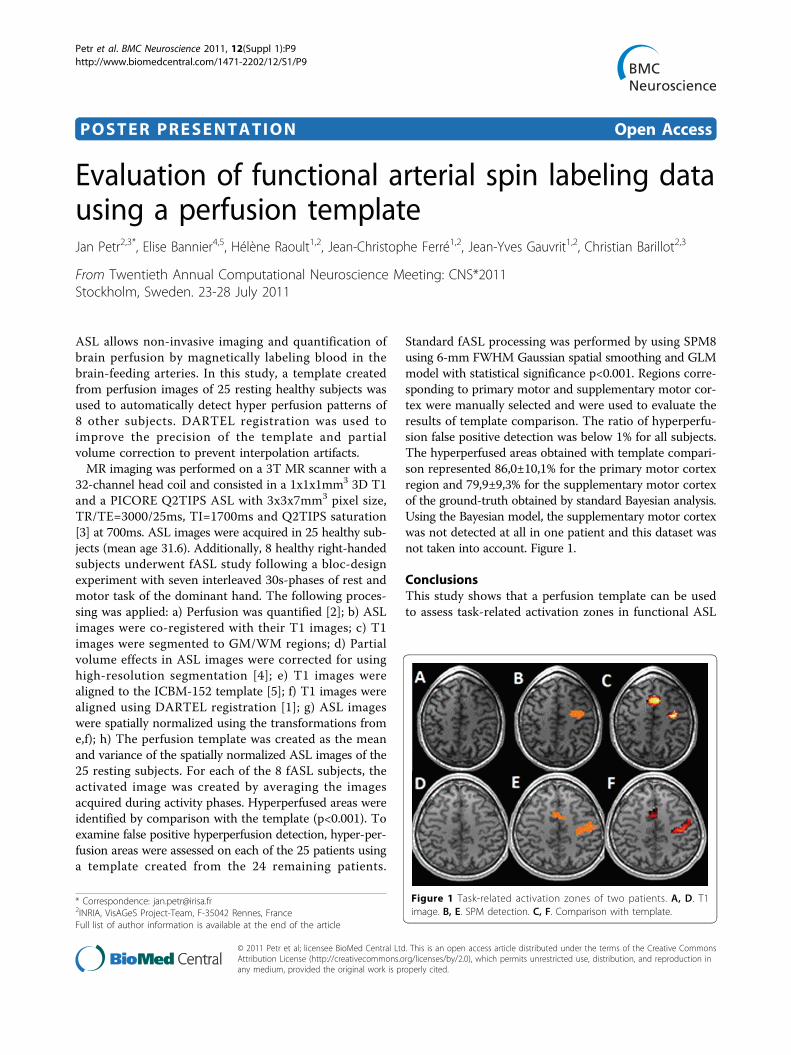
Standard fASL processing was performed by using SPM8using 6-mm FWHM Gaussian spatial smoothing and GLMmodel with statistical significance p<0.001. Regions corre-sponding to primary motor and supplementary motor cor-tex were manually selected and were used to evaluate theresults of template comparison. The ratio of hyperperfu-sion false positive detection was below 1% for all subjects.The hyperperfused areas obtained with template compari-son represented 86,0±10,1% for the primary motor cortexregion and 79,9±9,3% for the supplementary motor cortexof the ground-truth obtained by standard Bayesian analysis.Using the Bayesian model, the supplementary motor cortexwas not detected at all in one patient and this dataset wasnot taken into account. Figure 1.
ConclusionsThis study shows that a perfusion template can be usedto assess task-related activation zones in functional ASL
* Correspondence: [email protected], VisAGeS Project-Team, F-35042 Rennes, FranceFull list of author information is available at the end of the article
Figure 1 Task-related activation zones of two patients. A, D. T1image. B, E. SPM detection. C, F. Comparison with template.
Petr et al. BMC Neuroscience 2011, 12(Suppl 1):P9http://www.biomedcentral.com/1471-2202/12/S1/P9
© 2011 Petr et al; licensee BioMed Central Ltd. This is an open access article distributed under the terms of the Creative CommonsAttribution License (http://creativecommons.org/licenses/by/2.0), which permits unrestricted use, distribution, and reproduction inany medium, provided the original work is properly cited.

data while using only activated phase. Two assumptionscan be made to explain why standard functional analysisyields slightly larger activation regions. First, the use ofFWHM 6mm Gaussian kernel possibly enlarges thedetected zones. Second, the data analyzed using SPMcontains both resting and activated phases whereas onlythe activated phase was compared to the template.Future work will focus on detection of hyperperfusionin different neurodegenerative diseases taking intoaccount registration issues of pathological T1 images.
Author details1Neuroradiology Dept., University Hospital of Rennes, F-35043 Rennes,France. 2INRIA, VisAGeS Project-Team, F-35042 Rennes, France. 3INSERM,U746, F-35042 Rennes, France. 4University of Rennes I, CNRS, UMR 6074,IRISA, F-35042 Rennes, France. 5Neurinfo Platform, University Hospital ofRennes, F-35043 Rennes, France.
Published: 18 July 2011
References1. Ashburner J: A fast diffeomorphic image registration algorithm.
NeuroImage 2007, 38:95-113.2. Buxton RB, Frank LR, Wong LC, Siewert B, Warach S, Edelman RR: A general
kinetic model for quantitative perfusion imaging with arterial spinlabeling. Magn Reson Med 1998, 40:383-396.
3. Luh WM, Wong EC, Bandettini PA, Hyde JS: QUIPSS II with thin-slice TI1periodic saturation: a method for improving accuracy of quantitativeperfusion imaging using pulsed ASL. Magn Reson Med 41:1246-1254.
4. Petr J, Ferre JC, Gauvrit JY, Barillot C: Denoising ASL MRI using tissuepartial volume. SPIE 2010.
5. Mazziotta JC, Toga AW, Evans A, Fox P, Lancaster J: A Probablistic Atlas ofthe Human Brain: Theory and Rationale for Its Development. NeuroImage1995, 2:89-101.
doi:10.1186/1471-2202-12-S1-P9Cite this article as: Petr et al.: Evaluation of functional arterial spinlabeling data using a perfusion template. BMC Neuroscience 2011 12(Suppl 1):P9.
Submit your next manuscript to BioMed Centraland take full advantage of:
• Convenient online submission
• Thorough peer review
• No space constraints or color figure charges
• Immediate publication on acceptance
• Inclusion in PubMed, CAS, Scopus and Google Scholar
• Research which is freely available for redistribution
Submit your manuscript at www.biomedcentral.com/submit
Petr et al. BMC Neuroscience 2011, 12(Suppl 1):P9http://www.biomedcentral.com/1471-2202/12/S1/P9
Page 2 of 2