Embed Size (px)
Citation preview

European Journal of Radiology 30 (1999) 115–124
Perfusion magnetic resonance imaging with continuous arterial spinlabeling: methods and clinical applications in the central nervous
system
John A. Detre *, David C. Alsop
Departments of Neurology and Radiology, Uni6ersity of Pennsyl6ania, 3400 Spruce Street, Philadelphia, PA 19104, USA
Received 8 March 1999; accepted 25 March 1999
Abstract
Several methods are now available for measuring cerebral perfusion and related hemodynamic parameters using magneticresonance imaging (MRI). One class of techniques utilizes electromagnetically labeled arterial blood water as a noninvasivediffusible tracer for blood flow measurements. The electromagnetically labeled tracer has a decay rate of T1, which is sufficientlylong to allow perfusion of the tissue and microvasculature to be detected. Alternatively, electromagnetic arterial spin labeling(ASL) may be used to obtain qualitative perfusion contrast for detecting changes in blood flow, similar to the use of susceptibilitycontrast in blood oxygenation level dependent functional MRI (BOLD fMRI) to detect functional activation in the brain. Theability to obtain blood flow maps using a non-invasive and widely available modality such as MRI should greatly enhance theutility of blood flow measurement as a means of gaining further insight into the broad range of hemodynamically relatedphysiology and pathophysiology. This article describes the biophysical considerations pertaining to the generation of quantitativeblood flow maps using a particular form of ASL in which arterial blood water is continuously labeled, termed continuous arterialspin labeling (CASL). Technical advances permit multislice perfusion imaging using CASL with reduced sensitivity to motion andtransit time effects. Interpretable cerebral perfusion images can now be reliably obtained in a variety of clinical settings includingacute stroke, chronic cerebrovascular disease, degenerative diseases and epilepsy. Over the past several years, the technical andtheoretical foundations of CASL perfusion MRI techniques have evolved from feasibility studies into practical usage. Currentlyexisting methodologies are sufficient to make reliable and clinically relevant observations which complement structural assessmentusing MRI. Future technical improvements should further reduce the acquisition times for CASL perfusion MRI, while increasingthe slice coverage, resolution and stability of the images. These techniques have a broad range of potential applications in clinicaland basic research of brain physiology, as well as in other organs. © 1999 Published by Elsevier Science Ireland Ltd. All rightsreserved.
Keywords: Continuous arterial spin labeling; Neuroimaging; Perfusion MRI; Quantitative blood flow maps
1. Introduction
Several methods are now available for measuringcerebral perfusion and related hemodynamic parame-ters using MRI. One class of techniques utilizes electro-magnetically labeled arterial blood water as a
non-invasive diffusible tracer for blood flow measure-ments, in a manner analogous to that used for 15Opositron emission tomography (PET) scanning [1]. Thismethod is schematically illustrated in Fig. 1. The elec-tromagnetically labeled tracer has a decay rate of T1,which is sufficiently long to allow perfusion of thetissue and microvasculature to be detected. Becausewater is a diffusible tracer, blood flow can be quantifiedin well characterized physiological units of ml/100 g/min using this approach. Alternatively, electromagnetic
* Corresponding author. Tel.: +1-215-349-8465; fax: +1-215-349-5579.
E-mail address: [email protected] (J.A. Detre)
0720-048X/99/$ - see front matter © 1999 Published by Elsevier Science Ireland Ltd. All rights reserved.PII: S 0720 - 048X(99 )00050 -9
J.A. Detre, D.C. Alsop / European Journal of Radiology 30 (1999) 115–124116
arterial spin labeling (ASL) may be used to obtainqualitative perfusion contrast for detecting changes inblood flow, similar to the use of susceptibility contrastin blood oxygenation level dependent functional MRI(BOLD fMRI) to detect functional activation in thebrain [2–5].
A variety of studies in both animal models andhuman subjects have demonstrated that blood flow canbe accurately quantified using ASL [1,6–11]. Suchquantitative measurements of regional perfusion werepreviously obtainable only with exogenous tracer meth-ods and ionizing radiation using positron emission to-mography (PET), single photon emission computedtomography (SPECT) or xenon enhanced X-ray com-puted tomography (XeCT). The ability to obtain bloodflow maps using a non-invasive and widely availablemodality such as MRI should greatly enhance theutility of blood flow measurement as a means of gain-ing further insight into the broad range of hemodynam-ically related physiology and pathophysiology.
Using electromagnetically labeled arterial water as atracer for measuring perfusion can be thought of as astandard diffusible tracer method as originally de-scribed by Kety [12]. Tissue water is in constant ex-change with blood water flowing through capillariesand venules. ASL applied proximal to the tissue ofinterest is used to modulate the magnetization of theprotons in the arterial water. Due to the constantexchange of water between blood and tissue compart-ments, proximal ASL alters the total magnetization inthe distal tissue. The extent of this alteration is deter-mined by comparison with images acquired with con-trol labeling that does not modulate the magnetizationof arterial water. With an additional knowledge of the
regional T1 of tissue, quantitative blood flow maps canbe generated [6].
This article will describe the biophysical consider-ations pertaining to the generation of quantitativeblood flow maps using a particular form of ASL inwhich arterial blood water is continuously labeled,termed continuous arterial spin labeling (CASL). Thisapproach has been chosen because it maximizes thesensitivity of the method to blood flow [13] and is mostreadily quantified. In addition, a variety of applicationsof CASL perfusion MRI to central nervous systemdisorders will be described. Studies of cerebral bloodflow (CBF) are likely to have clinical utility in diagnos-ing and managing many central nervous system disor-ders. Because flow can be quantified directly, thisapproach is also ideal for comparison of resting cere-bral blood flow across patient groups or before andafter pharmacological interventions. However, the gen-eral principles of ASL and CASL are not limited tostudies of the brain, and are likely to have a broadrange of applications in other tissues as well.
2. Methodological considerations for CASL perfusionMRI
Over the past few years, theoretical and methodolog-ical developments have done much to improve theaccuracy and reliability of perfusion quantification withASL. Recently a quantitative validation of CASL per-fusion MRI with arterial spin labeling by comparisonwith 15O PET measurements of CBF was reported byYe et al. [14]. This study was carried out in 12 volun-teers and demonstrated an excellent correlation both inperfusion contrast and in quantitative CBF valuesacross modalities.
2.1. Continuous arterial in6ersion
CASL was the first method used for ASL imaging ofperfusion [6]. CASL can be implemented either withpseudo-continuous saturation or by flow driven adia-batic inversion [15,16]. Since inversion produces twicethe signal of saturation, it is generally preferred. Adia-batic inversion is achieved by applying a constant mag-netic field gradient and a constant radiofrequency (RF)irradiation at a frequency determined by the desiredinversion plane, as illustrated in Fig. 2. Simulations andexperimental data indicate that flow driven inversion isquite efficient over a physiologically relevant range ofvelocities [17]. Adiabatic inversion is relatively simple toimplement, but it has certain disadvantages. Many MRscanners use pulsed radiofrequency (RF) amplifierswhich cannot be operated continuously, though contin-uous RF can be closely approximated by using multipleshorter pulses spaced closely together. The continuous
Fig. 1. Schematic diagram illustrating CASL in comparison to steady-state nuclear medicine approaches. In the nuclear medicine approach,a radioactive tracer is administered at a constant rate, resulting in aconstant arterial concentration. Regional tissue blood flow can thenbe calculated based on the regional tracer concentration in the tissuedetermined by imaging, the arterial concentration of the tracer and aknowledge of the decay rate of the tracer. In CASL perfusion MRI,the arterial supply is continuously inverted, producing a knowndegree of arterial labeling. The regional accumulation of the label ismeasured in the tissues by comparison with a control image acquiredwithout labeling. The decay rate for the tracer is the measurablequantity, T1.
J.A. Detre, D.C. Alsop / European Journal of Radiology 30 (1999) 115–124 117
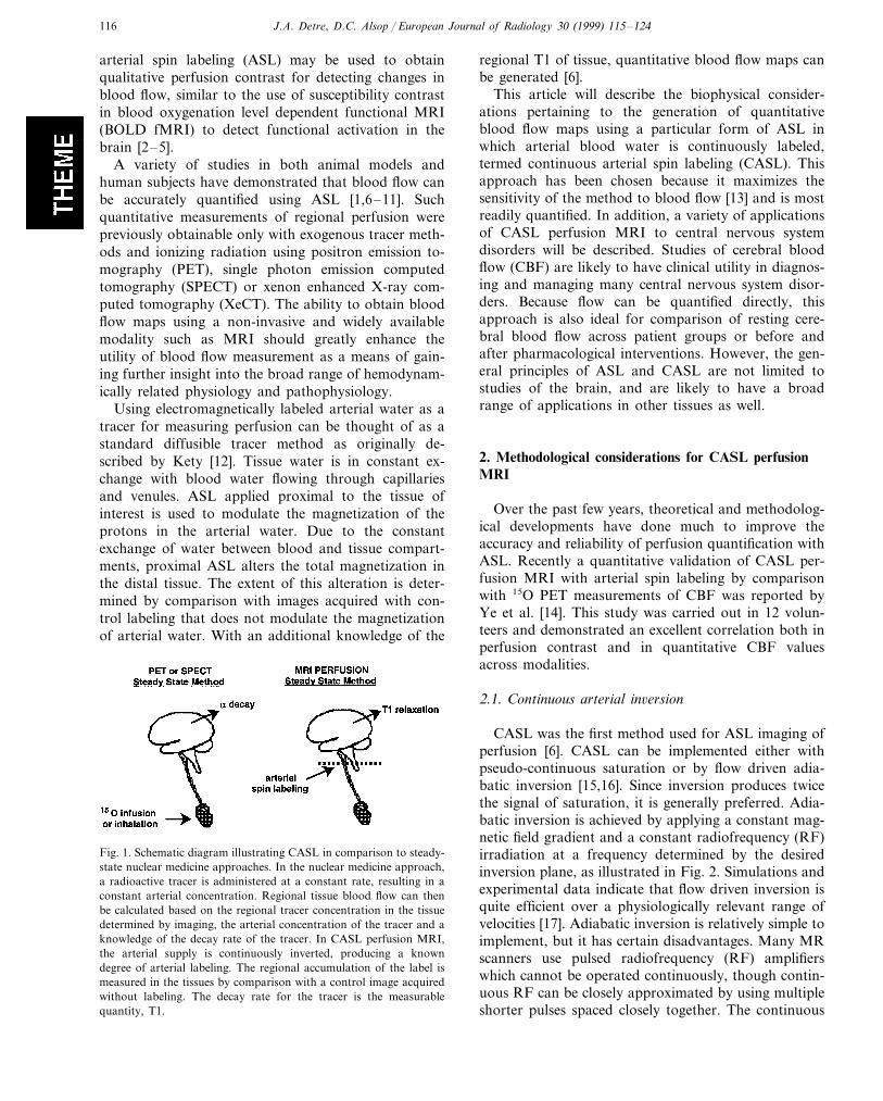
Fig. 2. Schematic diagram illustrating velocity driven adiabatic inver-sion used for CASL. Blood flowing along the direction of a magneticfield gradient in the presence of constant or pseudo-constant radiofre-quency (B1, gray arrow) experiences a frequency sweep. The diagramshows changes in magnetization in the rotating frame of the appliedradiofrequency. As long as the conditions for adiabatic fast passageare met, the magnetization (M, black arrow) follows the Beffective
(dotted arrow) until it is inverted.
plane and at an intermediate location between ASL andcontrol labeling. An additional complication of off-res-onance saturation is that both the T1 and restingmagnetization of the brain tissue (Mb
0) are reduced,which decreases the observed signal, affecting quantifi-cation and reducing the signal-to-noise ratio. For cor-rect quantification, T1 should be measured while theRF irradiation is on, but Mb
0 should be measured in itsabsence [13]. Usually the reverse is done, but since theratio of Mb
0 to 1 in tissue is usually independent ofoff-resonance saturation [13,21], quantification isunaffected.
To allow multislice imaging, several approaches tocircumventing the off-resonance limitations have beenproposed. A hardware approach using a separate coilplaced proximal to the image slice to label arterial spinswhile a second volume coil is used for imaging wasproposed by Silva et al. [8], and implemented for imag-ing of the rat. With active decoupling between the twocoils, the labeling coil produces negligible RF power inthe imaged tissue so off-resonance saturation is virtu-ally eliminated. While this approach is highly successfulin the rat, the extra hardware requirements can becumbersome for human applications and it can restrictthe physical location of ASL. The second coil must belarge enough to produce sufficient RF at the vessels forinversion while remaining small enough and distantenough to produce negligible RF in the imaged area. Atwo coil implementation for human perfusion imaginghas been reported [22] but the labeling efficiency hasnot yet been optimized.
A single coil approach to controlling for off-reso-nance effects utilizes an alternative control irradiationwhich mimics the frequency dependent off-resonanceeffects of the labeling pulse. The control is an ampli-tude modulated version of the labeling [23]. When aconstant RF irradiation at a fixed frequency, f0, ismultiplied by a sine wave at frequency f1, the signalproduced is mathematically identical to continuous ir-radiation at two different frequencies, f0+ f1 and f0−f1. The combined effect is that spins will be invertedtwice, resulting in no net effect. This is illustrated inFig. 3. Because the average power and center frequencyof the amplitude modulated control are identical to thelabeling RF irradiation, the off-resonance effects of thecontrol are nearly identical to those of the labeling.This frequency independent matching of the off-reso-nance of labeling and control pulse makes multisliceCASL readily possible. In addition, as with the two coilapproach, the orientation of perfusion images is nolonger restricted to a plane parallel to the ASL plane.For clinical applications, a multislice technique ishighly desirable both to increase coverage through thebrain and because it is not always possible to knowwhich slices are of interest prior to scanning. Theextension of ASL MRI to a multislice modality has
irradiation can also deposit significant RF power intothe subject which may be limiting as CASL is imple-mented at higher magnetic fields, though human perfu-sion scans at 1.5 T can be performed within standardsafety guidelines for RF deposition. The greatest com-plication of adiabatic inversion labeling is the off-reso-nance saturation of the imaged tissue by labeling RF[18]. Since the effects of CASL on distal image intensitycan only be measured in comparison to a controllabeling condition, off-resonance effects of the controllabeling condition must be identical.
2.2. Off-resonance effects and multislice CASL
Due to magnetization transfer, off-resonance RF inbiological tissues causes a decrease in both signal andT1 [19]. In the original implementations of CASL per-fusion MRI, control images were acquired with labelingapplied an equal distance distal to the perfusion imag-ing slice by reversing the frequency offset of the RF [9].Because off-resonance saturation is highly symmetricwith frequency, this largely controls for differences inoff-resonance saturation. Cycling of the gradient ampli-tude was additionally proposed as a method to removeresidual errors due to small asymmetries in the off-reso-nance response [20]. Unfortunately, the distal controlmethod works for only one slice parallel to the labeling
J.A. Detre, D.C. Alsop / European Journal of Radiology 30 (1999) 115–124118
been a major technical advance which has greatly in-creased the feasibility of clinical use.
2.3. Motion effects
ASL perfusion imaging involves the subtraction oftwo images with signal intensities differing by only afew percent or less. Motion between scans can thereforepotentially lead to large errors in the measured perfu-sion. Single excitation imaging techniques such as echo-planar imaging [24] have helped to dramatically reducemotion related errors. Using echoplanar imaging, mo-tion is rarely a problem except with the most uncooper-ative subjects. Single excitation imaging reduces motionerrors in two ways. First, individual images are freefrom the nonlocal phase artifacts that motion canproduce in multi-excitation images. Second, label andcontrol images can be interleaved on the time scale ofseconds. This rapid interleaving acts to attenuate thelow frequency components of motion and other spuri-ous signals which tend to be much larger than thehigher frequencies [25]. Echoplanar imaging itself doessuffer from geometric distortion and chemical shiftartifacts. Recently single shot rapid aquisition withrelaxation enhancement (RARE) sequences have beenevaluated for ASL perfusion imaging [26] and showpromise.
2.4. Vascular transit time errors
The ASL label decays with T1, approximately 1 s at1.5 T, permitting far greater time resolution than withany other perfusion imaging technique. While thisproperty is advantageous for brain activation studies, itcan be limiting in efforts to measure extremely low
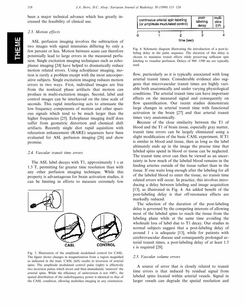
Fig. 4. Schematic diagram illustrating the introduction of a post-la-beling delay in the pulse sequence. The duration of this delay ischosen to minimize transit effects while preserving sufficient spinlabeling to visualize perfusion. Delays of 900–1500 ms are typicallyused.
flow, particularly as it is typically associated with longarterial transit times. Considerable evidence also sug-gests that microvascular transit times are highly vari-able both anatomically and under varying physiologicalconditions. The arterial transit time can have importanteffects on the measured signal and consequently onflow quantification. Our recent studies demonstratelarge changes in arterial transit time with functionalactivation in the brain [27] and that arterial transittimes vary anatomically.
Because of the close similarity between the T1 ofblood and the T1 of brain tissue, especially gray matter,transit time errors can be largely eliminated using aslight modification of the basic CASL experiment. If T1is similar in blood and tissue, then as long as the labelultimately ends up in the image the precise time thatlabeled spins spend in blood or tissue can be neglected.The transit time error can then be viewed as an uncer-tainty in how much of the labeled blood remains in thefeeding arteries outside of the tissue rather than in thetissue. If one waits long enough after the labeling for allof the labeled blood to enter the tissue, no transit timerelated errors will occur. In practice, this involves intro-ducing a delay between labeling and image acquisition[13], as illustrated in Fig. 4. An added benefit of thepost-labeling delay is that off-resonance effects aremarkedly reduced.
The selection of the duration of the post-labelingdelay is governed by the competing interests of allowingmost of the labeled spins to reach the tissue from thelabeling plane while at the same time avoiding thewholesale loss of label due to T1 decay. Our studies innormal subjects suggest that a post-labeling delay ofaround 1 s is adequate [13], while for patients withcerebrovascular disease and consequently prolonged ar-terial transit times, a post-labeling delay of at least 1.5s is required [28].
2.5. Vascular 6olume errors
A source of error that is closely related to transittime errors is that induced by residual signal fromlabeled spins located within arterial vessels. Signal inlarger vessels can degrade the spatial resolution and
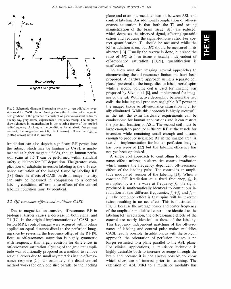
Fig. 3. Illustration of the amplitude modulated control for CASL.The figure shows changes in magnetization from a region magnifiedas indicated in the inset. CASL (left) results in inversion of arterialspins. The amplitude modulated control pulse (right) is effectivelytwo inversion pulses which invert and then immediately ‘uninvert’ thearterial spins. While the efficiency of uninversion is not 100%, thespatial distribution of the radiofrequency power is identical to that ofthe CASL condition, allowing multislice imaging in any orientation.

J.A. Detre, D.C. Alsop / European Journal of Radiology 30 (1999) 115–124 119
image quality by introducing bright linear or point-likeartifacts in the perfusion images. The methods dis-cussed above for eliminating transit time sensitivity alsoeliminate vascular volume signal because they allow thelabeled blood reach the tissue. However, it is difficult toverify that there is no residual label in smaller vesselswhich are too small to produce gross artifacts or de-grade resolution. Indeed, it may be too stringent torequire that the all of the signal be in the tissue sincethis would mandate an extremely long post-labelingdelay that severely reduces the sensitivity of the mea-surement. Theoretical analyses using two transit times,one from the labeling plane to vessels within the imagewhich are too small to degrade image resolution and alonger one to the tissue itself, suggest that labeled signalin small vessels need not degrade quantification [13]. Itis nonetheless important to minimize any aspects of theimaging sequence which might differentially affect arte-rial blood and tissue spins. These aspects include T2weighting, since arterial blood T2 is approximatelytwice that of brain, and flow attenuation as might beintroduced by strong bipolar gradients. Strong bipolargradients have previously been used to eliminate thelarge vessel vascular volume artifacts in perfusion sensi-tive images without post-labeling delays [9,29].
Fig. 4 shows an example of multislice CASL perfu-sion MRI from a normal volunteer obtained using theamplitude modulated control. In this case, a fractionalecho imaging scheme was used to increase the numberof slices which could be acquired before decay of theASL, though much of our clinical data has been ac-quired from a lesser number of slices using full echoimaging. Images shown in Fig. 4 were acquired with arepitition time (TR) of 4 s, an echo time (TE) of 12 ms,an acquisition time per image of 40 ms and a post-la-beling delay of 1000 ms for the inferior-most slice. The
matrix is 64×40 in a 24×15 cm field of view with aslice thickness of 5 mm. Image acquisition took 6 min(45 averages).
3. Results of clinical applications of CASL in thecentral nervous system
3.1. Cerebral perfusion in acute stroke
Since hypoperfusion is the proximate cause of allstroke, it is an obvious application for perfusion imag-ing methods. The measurement of cerebral perfusionusing MRI, in combination with diffusion MRI, isbecoming an important part of the evaluation of acutestroke. These data are used to confirm the diagnosis ofstroke, to establish a baseline against which stroketherapies can be assessed, and to contribute to progno-sis. Most perfusion studies in acute stroke have utilizeda bolus-tracking approach in which the first passage ofan MRI contrast agent is imaged dynamically. Suchdata can be analyzed to yield maps of mean transit timeand blood volume which are related to perfusion. Al-though the bolus tracking method provides relativelyhigh signal-to-noise for perfusion abnormalities, its suc-cessful implementation requires administration of con-trast through a large bore intravenous catheter, oftenwith a power injector. The cost of the contrast agentcan also represent a considerable expense, particularlyif multiple repeated measurements are desired. CASLperfusion MRI provides a means of noninvasivelyquantifying cerebral perfusion in the acute setting andis ideal in instances where intravenous access is difficultto obtain or where serial perfusion measurements arerequired, Fig. 5.
Fig. 5. Example of a multislice perfusion map acquired from a normal volunteer using a post-labeling delay of 1 s and a fractional echo echoplanarimaging scheme which allows up to 24 slices to be acquired within 1 s. The grayscale shows CBF ranging from 0–150 ml/100 g/min.
J.A. Detre, D.C. Alsop / European Journal of Radiology 30 (1999) 115–124120
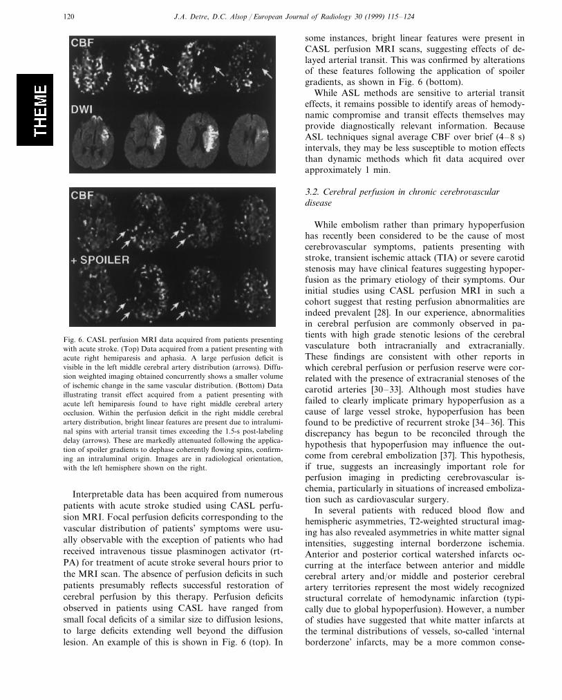
Fig. 6. CASL perfusion MRI data acquired from patients presentingwith acute stroke. (Top) Data acquired from a patient presenting withacute right hemiparesis and aphasia. A large perfusion deficit isvisible in the left middle cerebral artery distribution (arrows). Diffu-sion weighted imaging obtained concurrently shows a smaller volumeof ischemic change in the same vascular distribution. (Bottom) Dataillustrating transit effect acquired from a patient presenting withacute left hemiparesis found to have right middle cerebral arteryocclusion. Within the perfusion deficit in the right middle cerebralartery distribution, bright linear features are present due to intralumi-nal spins with arterial transit times exceeding the 1.5-s post-labelingdelay (arrows). These are markedly attenuated following the applica-tion of spoiler gradients to dephase coherently flowing spins, confirm-ing an intraluminal origin. Images are in radiological orientation,with the left hemisphere shown on the right.
some instances, bright linear features were present inCASL perfusion MRI scans, suggesting effects of de-layed arterial transit. This was confirmed by alterationsof these features following the application of spoilergradients, as shown in Fig. 6 (bottom).
While ASL methods are sensitive to arterial transiteffects, it remains possible to identify areas of hemody-namic compromise and transit effects themselves mayprovide diagnostically relevant information. BecauseASL techniques signal average CBF over brief (4–8 s)intervals, they may be less susceptible to motion effectsthan dynamic methods which fit data acquired overapproximately 1 min.
3.2. Cerebral perfusion in chronic cerebro6asculardisease
While embolism rather than primary hypoperfusionhas recently been considered to be the cause of mostcerebrovascular symptoms, patients presenting withstroke, transient ischemic attack (TIA) or severe carotidstenosis may have clinical features suggesting hypoper-fusion as the primary etiology of their symptoms. Ourinitial studies using CASL perfusion MRI in such acohort suggest that resting perfusion abnormalities areindeed prevalent [28]. In our experience, abnormalitiesin cerebral perfusion are commonly observed in pa-tients with high grade stenotic lesions of the cerebralvasculature both intracranially and extracranially.These findings are consistent with other reports inwhich cerebral perfusion or perfusion reserve were cor-related with the presence of extracranial stenoses of thecarotid arteries [30–33]. Although most studies havefailed to clearly implicate primary hypoperfusion as acause of large vessel stroke, hypoperfusion has beenfound to be predictive of recurrent stroke [34–36]. Thisdiscrepancy has begun to be reconciled through thehypothesis that hypoperfusion may influence the out-come from cerebral embolization [37]. This hypothesis,if true, suggests an increasingly important role forperfusion imaging in predicting cerebrovascular is-chemia, particularly in situations of increased emboliza-tion such as cardiovascular surgery.
In several patients with reduced blood flow andhemispheric asymmetries, T2-weighted structural imag-ing has also revealed asymmetries in white matter signalintensities, suggesting internal borderzone ischemia.Anterior and posterior cortical watershed infarcts oc-curring at the interface between anterior and middlecerebral artery and/or middle and posterior cerebralartery territories represent the most widely recognizedstructural correlate of hemodynamic infarction (typi-cally due to global hypoperfusion). However, a numberof studies have suggested that white matter infarcts atthe terminal distributions of vessels, so-called ‘internalborderzone’ infarcts, may be a more common conse-
Interpretable data has been acquired from numerouspatients with acute stroke studied using CASL perfu-sion MRI. Focal perfusion deficits corresponding to thevascular distribution of patients’ symptoms were usu-ally observable with the exception of patients who hadreceived intravenous tissue plasminogen activator (rt-PA) for treatment of acute stroke several hours prior tothe MRI scan. The absence of perfusion deficits in suchpatients presumably reflects successful restoration ofcerebral perfusion by this therapy. Perfusion deficitsobserved in patients using CASL have ranged fromsmall focal deficits of a similar size to diffusion lesions,to large deficits extending well beyond the diffusionlesion. An example of this is shown in Fig. 6 (top). In

J.A. Detre, D.C. Alsop / European Journal of Radiology 30 (1999) 115–124 121
quence of hypoperfusion [38,39]. While borderzone in-farctions have clearly been associated with reducedsystemic perfusion, the contribution of this mechanismto overall stroke incidence was thought to be low.However, the presence of borderzone ischemic changesin patients presenting with TIA carries an extremelypoor prognosis [40].
Under non-pathological conditions, CBF is main-tained over a broad range of perfusion pressures [41].This property of the cerebrovascular system is termed‘autoregulation’. Because autoregulatory mechanisms inthe cerebral vasculature can maintain CBF throughvasodilatation, it has been suggested that CBF alone isan inadequate measure of hemodynamic compromise[42]. While resting reductions in perfusion are clearlyabnormal, alterations in hemodynamic reserve are alsosignificant because they suggest that the autoregulatorycapacity of the cerebral vasculature may be exhausted.In the absence of intact autoregulation, cerebral perfu-sion becomes dependent on arterial blood pressure.Cerebrovascular reserve is tested by measuring the in-crease in CBF induced by carbon dioxide inhalation oracetazolamide administration (‘cerebrovascular reactiv-ity’). A number of studies have demonstrated thatcerebrovascular reserve impairment is particularly sig-nificant in patients with borderzone ischemia [43,44]and that abnormalities in augmentation are predictiveof stroke [35,36,45]. Perfusion MRI provides a conve-nient method for quantitatively and non-invasivelymeasuring the effects of pharmacological augmentationthroughout the brain. An example of CASL perfusionMRI obtained before and after intravenous acetazo-lamide is shown in Fig. 7 and demonstrates impaired
cerebrovascular reserve in the vascular distribution of aright middle cerebral artery stenosis.
3.3. Cerebral perfusion in degenerati6e disease
Functional imaging studies of Alzheimer’s disease(AD) and frontotemporal dementia (FD) with PET andSPECT have demonstrated specific deficits inmetabolism and flow in cortical association areas thatare characteristic for the type of dementia. StructuralMRI has also been used to quantify hippocampal andcortical atrophy. CASL perfusion MRI in conjunctionwith structural MRI offers the possibility of obtainingboth functional and structural information during asingle scanning session. Sandson et al. [46] have re-ported an initial evaluation of single-slice qualitativepulsed ASL imaging for functional studies of AD. Theywere able to detect temporoparietal flow deficits relativeto controls.
We have evaluated multislice CASL perfusion MRIin a cohort of 14 patients with AD and 11 patients withFD as determined by clinical criteria. Statistical com-parison of CBF and gray matter density across subjectswas possible because of brain registration. We used amodification of the methods of statistical parametricmapping (SPM; Wellcome Institute of Cognitive Neu-rology) to perform statistical analysis of the three-di-mensional dataset to assess both the significance andthe magnitude of the group differences. Our betweengroup analysis consisted of comparing CBF in the twodementias with nine normal elderly controls. An Fstatistic for group differences was calculated on a pixelby pixel basis and then converted to an approximate z
Fig. 7. Effect of acetazolamide challenge in a patient with right middle cerebral artery stenosis. Resting CBF (top row) appears symmetrical,though some transit effect is observed in the right middle cerebral artery distribution (arrow). Following 1 g intravenous acetazolamide, flow isincreased everywhere except in the right middle cerebral artery distribution (middle row). This is best observed in the percent change map (bottomrow) in which a reduction of flow in the right middle cerebral artery distribution is observed, indicating an impaired hemodynamic reserve. Imagesare in radiological orientation.

J.A. Detre, D.C. Alsop / European Journal of Radiology 30 (1999) 115–124122
Fig. 8. Statistical maps showing regional reductions in CBF in patients with dementia as compared to age-matched controls. Regions withsignificant cortical flow reductions are superimposed upon a surface rendered brain in a standard space. (Left) Patients with Alzheimer’s disease(AD) show prominent temporoparietal flow changes with lesser changes frontally. (Right) Patients with frontal dementia (FD) show morewidespread flow reductions frontally, though parietal flow changes are also observed. Orientation of the maps is indicated as left (L), right (R),front (F) and back (B).
statistic. To permit intuitive display of the three-dimen-sional data, surface projections on a rendered three-di-mensional brain was used. The z scores along theprojection line were integrated over the 3 cm closest tothe surface. The integrated values were corrected forthe correlations induced by the 10 mm smoothing andremapped to z scores. Projection was performed for allvoxels above a threshold of PB0.05 after correctionfor multiple voxel comparisons.
CBF in Alzheimer’s patients demonstrated very sig-nificant deficits bilaterally in parietal temporal, frontaland posterior cingulate cortex (Fig. 8, left). In contrast,CBF deficits in frontotemporal dementia occurred inthe anticipated frontal and anterior temporal regions aswell as superior parietal cortex (Fig. 8, right). Sincelittle pathology has been reported in superior parietalcortex in FD, low flow in this region may represent adiaschisis phenomenon brought on by destruction ofclosely connected frontal association cortex. Initialanalysis of a very mildly demented cohort indicate asimilar pattern of flow deficits but with reducedamplitude.
By correlating changes in regional cerebral perfusionwith cognitive task performance, it is possible to iden-tify brain regions in which significant perfusion abnor-malities are associated with specific cognitive deficits.Statistical analysis was also performed to compare theflow decreases in the AD patients with the nature andseverity of their cognitive dysfunction. Correlationswith several measures were very significant. The mini-mental state exam score (MMSE) correlated moststrongly with left temporoparietal junction and poste-rior cingulate CBF, as illustrated in Fig. 9. Dorsolateralprefrontal cortex was also significant. The strong later-ality of this measure, given the bilateral average flowdecrease in the patients is notable. This approach repre-sents a method of functional imaging that yields task-
specific patterns of functional abnormality. However,since only resting perfusion is measured, the interpreta-tion of the imaging data is not confounded by taskperformance.
3.4. Cerebral perfusion in temporal lobe epilepsy
Epilepsy is among the most common neurologicaldisorders, affecting approximately 1% of the popula-tion. The most common seizure type in adults withmedically intractable epilepsy is complex partialseizures, typically arising from the temporal lobe. Tem-poral lobectomy has been validated as a highly effectivetreatment for temporal lobe epilepsy (TLE). The suc-cess of resection of mesial temporal structures corre-
Fig. 9. Correlation of CBF changes with task performance on themini-mental status examination (MMSE) in patients with Alzheimer’sdisease. Left hemispheric changes correlate prominently with poorperformance on the MMSE, which is primarily a verbally based test.

J.A. Detre, D.C. Alsop / European Journal of Radiology 30 (1999) 115–124 123
Fig. 10. CASL perfusion MRI in a patient with left temporal lobeepilepsy, showing subtle hypoperfusion in left mesial temporal struc-tures (arrows). Images are in radiological orientation.
while increasing the slice coverage, resolution, and sta-bility of the images. However, currently existingmethodologies are sufficient to make reliable and clini-cally relevant observations which complement struc-tural assessment using MRI. Because tissue perfusion isa fundamental physiological process and is affected in abroad range of pathophysiological disorders, thesetechniques have a broad range of potential applicationsin clinical and basic research of brain physiology, aswell as in other organs.
Acknowledgements
The authors acknowledge support from the NationalInstitutes of Health, the American Heart Association,and the Whitaker Foundation.
References
[1] Detre JA, Zhang W, Roberts DA, Silva AC, Williams DS,Grandis DJ, Koretsky AP, Leigh JS. Tissue specific perfusionimaging using arterial spin labeling. NMR Biomed 1994;7:75–82.
[2] Edelman RR, Siewert B, Darby DG, Thangaraj V, Nobre AC,Mesulam MM, Warach S. Qualitative mapping of cerebral bloodflow and functional localization with echo-planar MR imagingand signal targeting with alternating radio frequency. Radiology1994;192:513–20.
[3] Kim SG. Quantification of relative cerebral blood flow changeby flow-sensitive alternating inversion recovery (FAIR) tech-nique: application to functional mapping. Magn Reson Med1995;34:293–301.
[4] Kwong KK, Chesler DA, Weisskoff RM, Donahue KM, D’AvisTL, Ostergaard L, Campbell TA, Rosen BR. MR perfusionstudies with T1-weighted echo planar imaging. Magn ResonMed 1995;34:878–87.
[5] Talagala SL, Noll DC. Functional MRI using strady statearterial water labeling. Magn Reson Med 1998;39:179–83.
[6] Detre JA, Leigh JS, Williams DS, Koretsky AP. Perfusionimaging. Magn Reson Med 1992;23:37–45.
[7] Roberts DA, Detre JA, Bolinger L, Insko EK, Leigh JS Jr.Quantitative magnetic resonance imaging of human brain perfu-sion at 1.5 T using steady-state inversion of arterial water. ProcNatl Acad Sci USA 1994;91:33–7.
[8] Silva AC, Zhang W, Williams DS, Koretsky AP. Multislice MRIof rat brain perfusion during amphetamine stimulation usingarterial spin labeling. Magn Reson Med 1995;33:209–14.
[9] Williams DS, Detre JA, Leigh JS, Koretsky AP. Magneticresonance imaging of perfusion using spin inversion of arterialwater. Proc Natl Acad Sci USA 1992;89:212–6.
[10] Zhang W, Williams DS, Detre JA, Koretsky AP. Measurementof brain perfusion by volume-localized NMR spectroscopy usinginversion of arterial water spins: accounting for transit time andcross relaxation. Magn Reson Med 1992;25:362–71.
[11] Zhang W, Silva AC, Williams DS, Koretsky AP. NMR measure-ment of perfusion using arterial spin labeling without saturationof macromolecular spins. Magn Reson Med 1995;33:370–6.
[12] Kety SS, Schmidt CF. The detemination of cerebral blood flowin man by the use of nitrous oxide in low concentrations. Am JPhysiol 1945;143:53–66.
lates with the extent of resection as well as abnormali-ties in functional activity in this tissue.
Lateralization of TLE is predicted by interictal hy-pometabolism using fluorodeoxyglucose PET (FDG-PET) [47]. The presence of interictal abnormalities onPET or SPECT scanning [48,49] has also been associ-ated with an improved outcome from epilepsy surgery.In some studies, uncoupling of CBF and metabolismhas been demonstrated in the resting state [50,51], withFDG-PET showing better lateralization than 15O PETmeasurements of CBF.
CASL perfusion MRI can successfully detect interic-tal asymmetries in mesial temporal lobe (mTL) perfu-sion in patients with TLE. It is readily combined withroutine structural assessment and offers an inexpensiveand completely non-invasive means of screening forasymmetries in interictal mesial temporal lobe (mTL)function, providing information using magnetic reso-nance which is complementary to structural imagingand metabolite levels measured by 31P and 1H NMRspectroscopy. In our preliminary experience in patientswith TLE, there was reasonably good agreement be-tween lateralized temporal lobe hypoperfusion observedusing CASL perfusion MRI and both clinical lateralityand PET hypometabolism. Lateralization by CASL andFDG-PET showed complete agreement in nine of tenpatients studied in our initial series, based on a regionof interest analysis of mTL structures. An example ofleft mTL hypoperfusion demonstrated by CASL perfu-sion MRI is shown in Fig. 10.
4. Conclusions
Over the past several years, the technical and theoret-ical foundations of CASL perfusion MRI techniqueshave evolved from feasibility studies into practicalusage. Future technical improvements should furtherreduce the acquisition times for CASL perfusion MRI,
J.A. Detre, D.C. Alsop / European Journal of Radiology 30 (1999) 115–124124
[13] Alsop DC, Detre JA. Reduced transit-time sensitivity in nonin-vasive magnetic resonance imaging of human cerebral bloodflow. J Cereb Blood Flow Metab 1996;16:1236–49.
[14] Ye FQ, Berman KF, Ellmore T, Esposito G, Van Horn JD,Yang Y, Duyn J, Salustri C, Smith A, Frank JA, WeinbergerDR, McLaughlin AC. H2
15O PET validation of arterial spintagging measurements of cerebral blood flow in humans.ISMRM 5th Annual Meeting Book of Abstracts, 1997, p. 87.
[15] Dixon WT, Du LN, Faul DD, Gado M, Rossnick S. Projectionangiograms of blood labeled by adiabatic fast passage. MagnReson Med 1986;3:454–62.
[16] Sardashti M, Schwartzberg DG, Stomp GP, Dixon WT. Spinlabeling angiography of the carotids by presaturation and sim-plified adiabatic inversion. Magn Reson Med 1990;15:192–200.
[17] Maccotta L, Detre JA, Alsop DC. The efficiency of adiabaticinversion for perfusion imaging by arterial spin labeling. NMRBiomed 1997;10:216–21.
[18] Wolff SD, Balaban RS. Magnetization transfer contrast (MTC)and tissue water proton relaxation in vivo. Magn Reson Med1989;10:135–44.
[19] Yeung HN, Swanson SD. Transient decay of longitudinal mag-netization in heterogeneous spin systems under selective satura-tion. J Magn Reson 1992;99:466–79.
[20] Pekar J, Jezzard P, Roberts DA, Leigh JSJ, Frank JA,McLaughlin AC. Perfusion imaging with compensation forasymmetric magnetization transfer effects. Magn Reson Med1996;35:70–9.
[21] Zhang W, Silva AC, Williams DS, Koretsky AP. The effect ofcross-relaxation on the measurement of perfusion by arterial spinlabeling using two isolated RF coils. SMRM 12th Annual Meet-ing Book of Abstracts 1993, p. 617.
[22] Zaharchuk G, Ledden PJ, Kwong KK, Rosen BR. Multislicearterial spin labeling of perfusion territory and functional activa-tion in humans using two coils. ISMRM 5th Annual MeetingBook of Abstracts 1997, p. 79.
[23] Alsop DC, Detre JA. Multisection cerebral blood flow MRimaging with continuous arterial spin labeling. Radiology1998;208:410–6.
[24] Stehling MK, Turner R, Mansfield P. Echo planar imaging:magnetic resonance imaging in a fraction of a second. Science1991;254:43–50.
[25] Siewert B, Bly BM, Schlaug G, Darby DG, Thangaraj V,Warach S, Edelman RR. Comparison of the BOLD- and EPIS-TAR-technique for functional brain imaging by using signaldetection theory. Magn Reson Med 1996;36:249–55.
[26] Chen Q, Siewert B, Bly B, Warach S, Edelman R. STAR-HASTE: Perfusion imaging without magnetic susceptibility arti-fact. Magn Reson Med 1997;38:404–8.
[27] Gonzalez-At JB, Alsop DC, Detre JA. Cerebral perfusion andarterial transit time changes during task activation determinedwith continuous arterial spin labeling. Magn Reson Med (sub-mitted for publication).
[28] Detre JA, Alsop DC, Vives LR, Maccotta L, Teener JW, RapsEC. Noninvasive MRI evaluation of cerebral blood flow incerebrovascular disease. Neurology 1998;50:633–41.
[29] Ye FQ, Mattay VS, Jezzard P, Frank JA, Weinberger DR,McLaughlin AC. Correction for vascular artifacts in cerebralblood flow values measured using arterial spin tagging tech-niques. Magn Reson Med 1997;37:226–35.
[30] Carpenter DA, Grubb RL Jr., Powers WJ. Borderzone hemody-namics in cerebrovascular disease. Neurology 1990;40:1587–92.
[31] Leblanc R, Yamamoto YL, Tyler JL, Diksic M, Hakim A.Borderzone ischemia. Ann Neurol 1987;22:707–13.
[32] Nighoghossian N, Trouillas P, Philippon B, Itti R, Adeleine P.Cerebral blood flow reserve assessment in symptomatic versusasymptomatic high-grade internal carotid artery stenosis. Stroke1994;25:1010–3.
[33] Powers WJ, Tempel LW, Grubb RL Jr. Influence of cerebralhemodynamics on stroke risk: one-year follow-up of 30 medi-cally treated patients. Ann Neurol 1989;25:325–30.
[34] Bogousslavsky J, Delaloye-Bischof A, Regli F, Delaloye B.Prolonged hypoperfusion and early stroke after transient is-chemic attack. Stroke 1990;21:40–6.
[35] Gur AY, Bova I, Bornstein NM. Is impaired cerebral vasomotorreactivity a predictive factor of stroke in asymptomatic patients?Stroke 1996;27:2188–90.
[36] Webster MW, Makaroun MS, Steed DL, Smith HA, JohnsonDW, Yonas H. Compromised cerebral blood flow reactivity is apredictor of stroke in patients with symptomatic carotid arteryocclusive disease. J Vasc Surg 1995;21:338–44.
[37] Caplan LR, Hennerici M. Impaired clearance of emboli(washout) is an important link between hypoperfusion, em-bolism, and ischemic stroke. Arch Neurol 1998;55:1475–82.
[38] Mull M, Schwarz M, Thron A. Cerebral hemispheric low-flowinfarcts in arterial occlusive disease: lesion patterns and an-giomorphological conditions. Stroke 1997;28:118–23.
[39] Weiller C, Ringelstein E, Reiche W, Buell U. Clinical andhemodynamic aspects of low-flow infarcts. Stroke 1991;22:1117–23.
[40] Dutch TIA Trial Study Group. Predictors of major vascularevents in patients with a transient ischemic attack or nondis-abling stroke. Stroke 1993;24:527–31.
[41] Lassen NA. Control of cerebral circulation in health and disease.Circ Res 1974;34:749–60.
[42] Powers WJ. Cerebral hemodynamics in ischemic cerebrovasculardisease. Ann Neurol 1991;29:231–40.
[43] Baumgartner RW, Regard M. Role of impaired CO2 reactivityin the diagnosis of cerebral low flow infarcts. J Neurol Neuro-surg Psychiatry 1994;57:814–7.
[44] Ringelstein EB, Weiller C, Weckesser M, Weckesser S. Cerebralvasomotor reactivity is significantly reduced in low-flow as com-pared to thromboembolic infarctions: the key role of the circle ofWillis. J Neurol Sci 1994;121:103–9.
[45] Widder B, Kleiser B, Krapf H. Course of cerebrovascular reac-tivity in patients with carotid artery occlusions. Stroke1994;25:1963–7.
[46] Sandson TA, O’Connor M, Sperling RA, Edelman RR, WarachS. Noninvasive perfusion MRI in Alzheimer’s disease: a prelimi-nary report. Neurology 1996;47:1339–42.
[47] Henry TR, Engel JJ, Mazziotta JC. Clinical evaluation of inter-ictal fluorine-18-fluorodeoxyglucose PET in partial epilepsy. JNucl Med 1993;34:1892–8.
[48] Manno EM, Sperling MR, Ding X, Jaggi J, Alavi A, O’ConnorMJ, Reivich M. Predictors of outcome after anterior temporallobectomy: positron emission tomography. Neurology1994;44:2331–6.
[49] Weinand ME, Carter LP. Surface cortical cerebral blood flowmonitoring and single photon emission computed tomography:prognostic factors for selecting temporal lobectomy candidates.Seizure 1994;3:55–9.
[50] Gaillard WD, Fazilat S, White S, Malow B, Sato S, Reeves P,Herscovitch P, Theodore WH. Interictal metabolism and blood-flow are uncoupled in temporal-lobe cortex of patients withcomplex partial epilepsy. Neurology 1995;45:1841–7.
[51] Leiderman DB, Balish M, Sato S, Kufta C, Reeves P, GaillardWD, Theodore WH. Comparison of PET measurements ofcerebral blood flow and glucose metabolism for the localizationof human epileptic foci. Epilepsy Res 1992;13:153–7.
.