Embed Size (px)
Citation preview
ERP Peaks Review 1
LINKING BRAINWAVES TO THE BRAIN: AN ERP PRIMER
Alexandra P. Fonaryova Key, Guy O. Dove, and Mandy J. Maguire
Psychological and Brain Sciences
University of Louisville
Louisville, Kentucky
Short title: ERPs Peak Review.
Key Words: ERP, peak, latency, brain activity source, electrophysiology.
Please address all correspondence to:
Alexandra P. Fonaryova Key, Ph.D. Department of Psychological and Brain Sciences 317 Life Sciences, University of Louisville Louisville, KY 40292-0001. [email protected]
ERP Peaks Review 2
Linking Brainwaves To The Brain: An ERP Primer
Alexandra Fonaryova Key, Guy O. Dove, and Mandy J. Maguire
Abstract This paper reviews literature on the characteristics and possible interpretations of the event-
related potential (ERP) peaks commonly identified in research. The description of each peak
includes typical latencies, cortical distributions, and possible brain sources of observed activity
as well as the evoking paradigms and underlying psychological processes. The review is
intended to serve as a tutorial for general readers interested in neuropsychological research and a
references source for researchers using ERP techniques.
ERP Peaks Review 3
Linking Brainwaves To The Brain: An ERP Primer
Alexandra P. Fonaryova Key, Guy O. Dove, and Mandy J. Maguire
Over the latter portion of the past century recordings of brain electrical activity such as
the continuous electroencephalogram (EEG) and the stimulus-relevant event-related potentials
(ERPs) became frequent tools of choice for investigating the brain’s role in the cognitive
processing in different populations. These electrophysiological recording techniques are
generally non-invasive, relatively inexpensive, and do not require participants to provide a motor
or verbal response. Furthermore, virtually identical procedures can be used across the entire life
span (e.g., Molfese & Molfese, 1979; Molfese & Hess, 1978; Molfese & Schmidt, 1983; Nelson,
et al., 1998). However, while the ongoing EEG reflects a wide-range of neural activity related to
the various sensory and cognitive functions, it also reflects the myriad of self-regulation
processes ongoing in the brain at the same time (e.g., maintaining body temperature, heart rate,
breathing). This intermixing of signals makes it difficult to separate cognitive and physiological
contributors to the observed EEG. In contrast, the ERP approach permits investigators to link
recorded signals with stimulus events more directly by focusing on the change in
electrophysiological signal that occurs immediately following the stimulus event (Callaway, et
al., 1975; Rockstroh, et al., 1982). The smaller size of the ERPs relative to other physiological
events can make it difficult to discern the relevant signal. To accommodate these factors,
researchers employ repeated presentations of the evoking stimulus to average out potentially
unrelated events1.
ERPs have been successfully used to study both general and specific aspects of an
individual’s response to events in the external as well as internal environment (e.g., Molfese,
1978a,b). Neuropsychological research of cognitive functioning in various populations also
demonstrated that ERP components could serve as informative markers of neurodevelopmental
status in general as well as the reflect development of more specific abilities (Courchesne, 1978).
Additional advantages of the ERP technique over other procedures include (1) very fine temporal
1 Recently, Makeig et al. (2002) demonstrated that some ERP features are not independent of the background EEG and therefore proposed a single-trial rather than average analysis approach for ERP data that would provide more detailed information about cortical dynamics.
ERP Peaks Review 4
resolution (on the order of milliseconds or even fractions of a millisecond) that reveals even
momentary changes in patterns of brain activation that otherwise could go unnoticed, and (2)
relatively gross level spatial resolution capabilities that allow for theorizing about the distribution
of brain mechanisms that subserve these cognitive functions.
ERP waveforms are typically described in terms of positive and negative peaks (i.e., the
most positive and negative deflections in the wave). At a general level, the labeling can refer to
the sequence in which the peak occurs while at the same time indicating its polarity. For
example, “N1” would refer to the first negative going peak in the waveform while “P2” would
label the second positive peak. The naming scheme for ERP components can also identify the
positive and negative peaks by their latency (usually defined as the time from stimulus onset).
“N100” in this example refers to the negative peak that occurs 100 ms following stimulus onset.
“P300” would identify the positive peak that occurred 300 ms post stimulus onset.
In contrast to this objective peak naming convention, functional descriptions of ERP
peaks refer to their psychological interpretation. In the past, Donchin (1978) proposed a
distinction between exogenous and endogenous components, suggesting that the former were
sensitive mainly to physical properties of external stimuli while the latter were affected by
information processing and could be elicited even by the event absence. However, further
research indicated that some components appeared to share characteristics of both groups (e.g.,
N1, P2; Shibasaki & Miyazaki, 1992) depending on the stimulus properties. While a variety of
terms were proposed for this subgroup, such as transient (Hugdahl, 1995) or mesogenous
components (Fabiani, et al., 2000), in general, functional descriptions of the ERPs have shifted
away from such classification toward identifying more specific cognitive processes reflected by
each peak.
In addition to the latency measures and functional interpretations, ERP descriptors often
include topographical scalp distributions or identify electrodes where maximum amplitudes are
typically observed. Such information can be useful for interpreting ERP peaks that may occur at
the same time but over different scalp areas reflecting different cognitive processes. However,
the scalp distribution does not necessarily correspond to the actual brain areas generating the
signal. The ERPs are generally believed to reflect post-synaptic (dendritic) potentials (Allison, et
al., 1986) of a fairly extensive set of neurons activated in close temporal proximity. The
orientation of the cortical columns generating the signal may affect whether the electrodes detect
ERP Peaks Review 5
a signal and where on the scalp it is maximal. If the columns are perpendicular to the scalp, the
likelihood of recording a strong signal is good. At the same time, columns from different brain
areas may project to the same scalp area resulting in a larger signal (if the polarities are the
same). Further, if the cell columns are oriented parallel to the scalp or at some other angle to it,
the signal may project to an area away from the nearest electrode above it and thus fail to be
recorded or be noted by electrodes over other scalp locations (e.g., a signal originating in the left
hemisphere may be maximal over the right hemisphere). Because of this imperfect relationship
between the observed scalp topography and the actual brain structures involved in generating it,
recently, the scientific community has moved another step forward to extending ERP
descriptions to include the potential brain sources of observed activity rather than focusing on
scalp distributions alone.
Given the great variety of ERP paradigms, analyses, and proposed implications, a reader
may find it challenging to make sense of the reported findings or integrate them into the more
general frame of psychology. Currently, there are several reviews of ERP components available
(e.g., Fabiani, et al., 2000; Hugdahl, 1995, etc.), however, we are not aware of any papers that
describe a wide range of ERP components and include all four characteristics: peak latency,
cognitive functional significance, scalp distributions, and component brain sources. This review
is intended to fill that void. The following sections describe most commonly identified
components of adult ERPs: P1, N1, P2, N2, Mismatch negativity (MMN), P3a, P3b, N400, and
P600 in the order they appear in the brainwave2. For the purpose of consistency and clarity, the
peaks are identified by their polarity and place in the sequence of components rather than by
exact latency due to possible variations in the latter due to developmental, environmental, or
clinical effects (unless the latency is the predominant descriptor of the peak). Because peak
characteristics can vary as a function of stimulus modality and reference location, our review
separates data for auditory and visual paradigms and notes the references used to identify
2 This list is not assumed to be exhaustive. Other ERP components such as the Contingent Negative Variation (CNV; Hillyard & Picton, 1987), Left Anterior Negativity (LAN; Friederici & Mecklinger, 1996), Late Positive Potential (LPP; Cuthbert, et al., 1995), and Positive Slow Wave (PSW; N. Squires, et al. 1975) are not included in the current review due to a lack of information regarding their sources and/or the limited space available to cover a large amount of research.
ERP Peaks Review 6
topographic maxima. Finally, different techniques used for source localization of the observed
ERPs rely on different principles and therefore can produce conflicting results. Thus, findings
from intracranial recordings, functional magnetic resonance imaging (fMRI),
magnetoencephalography (MEG), brain electromagnetic source analysis (BESA), positron
emission tomography (PET), or low-resolution brain electromagnetic tomography (LORETA)
may not always agree. Consequently, the specific method for source localization is noted for
each brain source listed in this paper.
P1. This peak is not always easily identified, but when present, occurs approximately 50 ms after
an auditory stimulus onset (also known as P50) or about 100 ms after the onset of a visual
stimulus. Functionally, this component is usually interpreted as a neurophysiological indicator of
preferential attention to sensory inputs (suppression of unattended information) and is thought to
reflect the general level of arousal.
Auditory: The auditory P1 appears earlier in time (shorter latency) over posterior scalp
electrode sites but with larger amplitudes over frontal and/or central regions. Nagamoto et al.
(1991) reported that the peak was largest over the Cz electrode (nose reference). The distribution
is symmetrical over the two hemispheres except for the anterior temporal regions where larger
amplitudes are noted over the left hemisphere. Overall, peak amplitude and latency appears to
decrease with age to the point where the peak disappears (Coch, et al., 2002).
Auditory P1 has been frequently associated with auditory inhibition (Waldo, et al., 1992)
and typically tested in a sensory gating paradigm where paired clicks are presented at relatively
short inter-stimulus intervals. The amplitude of the averaged ERP to the second of the paired
clicks is reduced compared to the averaged response to the first click. The magnitude of this
suppression is commonly interpreted as a neurophysiological index of sensory gating. Reduced
suppression is frequently reported for certain neuropsychiatric disorders, including mania and
schizophrenia, where peak amplitude to paired stimuli is reported to be approximately equal
(Siegel, et al., 1984; Waldo, et al., 1991; Jin, et al., 1997; Patterson, et al., 2000). P1 latency is
often used clinically to diagnose neurodegenerative diseases, such as multiple sclerosis and
Parkinson’s disease (Squires & Ollo, 1986).
Buchwald et al. (1992) proposed that the P1 response is associated with the ascending
reticular activating system (RAS) and its post-synaptic thalamic targets. Using a MEG approach,
ERP Peaks Review 7
Thoma et al. (2003) and Huotilainen (1998) independently localized the sources of the auditory
P1 in the superior temporal gyrus. Weisser et al (2001) coregistered auditory evoked potentials
(AEPs) and magnetic fields (AEFs). The resulting equivalent dipole model consisted of one
source in the auditory cortex of each hemisphere as well as a radially oriented medial frontal
source. Similar findings identifying frontal and temporal generators were reported by Potts et al.,
(1998) using current source density approach.
Visual: The visual P1 response is different from the auditory component in terms of the
evoking stimulus, neurocognitive and neurophysiological mechanism, peak latency, scalp
distribution, and neural sources. The visual P1 is typically recorded in a checkerboard-reversal
task or similar light-flashes paradigms but can also be present for other visual stimuli (e.g., faces)
and is largest over the occipital regions (Hugdahl, 1995). A negative component may be present
at the same latency over frontal and central regions (Rossion, Campanella, et al., 1999; left
earlobe reference). The amplitude of P1 generally varies with the amount of attention (Mangun,
et al., 1993 - Posner’s cuing paradigm; Clark & Hillyard, 1996 - spatial selective attention). Luck
(1995) proposed that P1 reflects suppression of noise because the amplitude decreased for
unattended locations and did not increase for attended stimuli. Mangun et al. (1993) interpreted it
to reflect encoding of form and color (ventral “what” pathway). Further, the amplitude of P1
increased when speed of response was emphasized, suggesting that this peak may also reflect the
level of arousal (Vogel & Luck, 2000).
Probable sources for the visual P1 were identified using PET, BESA, and LORETA
methods in ventral and lateral occipital regions (Clark, et al., 1996; Gomez, et al., 1994),
suggesting a striate (Strik, et al., 1998) or extrastriate (posterior fusiform gyrus) origin (Heinze,
et al., 1994). Rossion et al. (1999) submitted data from a face identification paradigm to BESA
and reported similar sources as well as sources in posterior-parietal regions indicating additional
involvement of dorsal and ventral neural components.
N1. This component was originally investigated by Hillyard et al. (1973) in a dichotic listening
paradigm and is one of the most easily identified components regardless of the specific analysis
approach employed. There is good convergence in findings based on analyses of principal
components analysis (PCA) factor scores (Beauducel, et al., 2000), baseline-to-peak amplitude
ERP Peaks Review 8
(Pekkonen, et al., 1995; Sandman & Patterson, 2000), and latency measures (Segalowitz &
Barnes, 1993).
Generally, N1 is assumed to reflect selective attention to basic stimulus characteristics,
initial selection for later pattern recognition, and intentional discrimination processing (e.g.,
Vogel & Luck, 2000). Latency and amplitude of the peak depend on the stimulus modality.
Auditory stimuli elicit a larger N1 with shorter latency than visual stimuli (Hugdahl, 1995).
Auditory: For auditory stimuli, N1 typically occurs approximately 100 ms after stimulus
onset and has maximum amplitude over frontocentral areas (Vaughn & Ritter, 1970; nose
reference) or the vertex (Picton, et al., 1974). More recent studies differentiated it into three
different components with maximum amplitudes over temporal areas (latency 75 ms and 130 ms)
and over vertex (latency 100 ms; McCallum & Curry, 1980; Giard, et al., 1994; nose reference).
Naatanen and Picton (1987) reviewed the three components of N1 and proposed that the early
temporal and vertex components reflect sensory and physical properties of the stimuli (e.g.,
intensity, location, timing in regards to other stimuli) while the later temporal component appears
to be less specific in its response and reflects transient arousal.
The amplitude of the auditory N1 is enhanced by increased attention to the stimuli
(Hillyard et al, 1973; Knight, et al., 1981; Ritter, et al., 1988; Mangun, 1995) and by increasing
the inter-stimulus interval (Hari, et al., 1982). The latter has been attributed to contributions of
additional sources from frontal cortical areas (Hari, et al., 1982).
N1 appears to be most likely generated by sources in primary auditory cortex in the
temporal lobe (Vaughn & Ritter, 1970). MEG, BESA, and lesions studies consistently localize
auditory N1 in superior temporal plane (e.g., Papanicolaou, et al., 1990; Scherg, et al., 1989;
Knight, et al., 1988). However, several studies proposed additional sources in the frontal lobe
that could be activated from the temporal lobe (e.g., Giard, et al., 1994; current source density
and equivalent current dipoles analysis).
Visual: The visual N1 component is usually largest over the occipital region (Hopf, et al.,
2002; reference not mentioned) or the inferior temporal regions (Bokura, et al., 2001; average
reference). N1 amplitude is typically larger in stimulus discrimination tasks (Mangun & Hillyard,
1990; Vogel & Luck, 2000), but is reduced when the stimuli are presented at short intervals. The
increased amplitude is attributed to enhanced processing of the attended location (Luck, 1995;
Coull, 1998), including spatial properties of the stimuli (Mangun et al., 1993), and is not due to
ERP Peaks Review 9
arousal because the amplitudes were larger in a task that placed no emphasis on the speed of
response (Vogel & Luck, 2000). It is also not affected by inhibition as indicated by the lack of
Go/No-Go response differences (Bokura, et al., 2001). Additionally, similar to the auditory N1, a
visual N1 was noted to include at least two distinct subcomponents, one occurring at 100 ms
over the central midline sites and another present at 165 ms over the posterior sites (Vogel &
Luck, 2000; average mastoids). The researchers attributed the more anterior visual N1 solely to
response preparation processes because it could be eliminated by not requiring a motor response
and decreased SOA.
Using a combination of techniques (MEG, ERP, and MRI), Hopf et al. (2002) located
visual N1 sources in the inferior occipital lobe and the occipito-temporal junction. However,
Bokura et al., (2001) using the LORETA approach, identified additional sources of the visual N1
in the inferior temporal lobe.
P2. The P2, like the N1 and P1, has long been considered to be an “obligatory cortical
potential” since it has low interindividual variability and high replicability (Roth, et al., 1975;
Sandman & Patterson, 2000; Shelley, et al., 1991). The P2 component has been identified in
many different cognitive tasks including selective attention (Johnson, 1989; Hackley, et al. 1990;
Hillyard, et al., 1973), stimulus change (Naatanen, 1990), feature detection processes (Luck &
Hillyard, 1994), and short-term memory (Golob & Starr, 2000; Starr & Barrett, 1987). Similar to
N1, P2 has been consistently identified by PCA factor scores (Beauducel, et al., 2000), baseline-
to-peak amplitude (Beauducel, et al., 2000; Sandman, & Patterson, 2000), and latency measures
(Segalowitz & Barnes, 1993). Functional interpretations of the P2 include attention modulation
of non-target stimuli (Novak et al., 1992) and stimulus classification (Garcia-Larrea et al., 1992).
Auditory: In the auditory modality, P2 often occurs together with N1 (referred to as
N1/P2 complex) and shares many characteristics of the preceding component, yet the two peaks
can be dissociated experimentally and developmentally (Hugdahl, 1995; Oades, et al., 1997; see
Crowley & Colrain, 2004 for a review). The maximum amplitude of the P2 can span a broader
latency range (150-275 ms) compared to the N1 (Dunn, et al., 1998), and can be double-peaked
(Hyde, 1997; Ponton, et al., 1996). The scalp distribution of the P2 is less localized than that of
the N1 (Naatanen, 1992) but typically the highest amplitude is noted over the central region
ERP Peaks Review 10
using either the left mastoid or the linked earlobes references (Holcomb, et al., 1986; Iragui, et
al., 1993), therefore this peak is often referred to as a “vertex potential” or “vertex positivity”.
P2 is sensitive to physical parameters of the stimuli, such as pitch (Novak, et al., 1992)
and loudness (Hegerl & Juckel, 1993; Hillyard & Picton, 1987). Similar to the N1, the amplitude
of the P2 peak gets larger as the stimulus intensity increases, however, opposite to the N1, it
continues to increase for stimuli with intensity above 70 dB (Adler & Adler, 1989). Participant
differences, such as reading ability, can also affect the P2 amplitude to auditory stimuli (Bernal,
et al., 2000).
Generators for the auditory P2 are thought to be located mainly in the primary and
secondary auditory cortices (Zouridakis, et al., 1998; MEG). ). Combined analyses using MEG
and intracranial recordings identified possible P2 sources in planum temporale while MEG alone
also located an additional source in auditory association complex (Area 22; Godey et al., 2001).
When using dipole source analysis, both the N1 and P2 elicited by auditory stimuli are often
represented by two dipoles: one for the primary auditory cortex and one for the secondary
auditory cortex (Hegerl, et al., 1994; Scherg & Berg, 1991). Using BESA and LORETA to
identify dipole locations for the N1/P2 component, Mulert et al. (2002) identified one in the
superior temporal region with a tangential orientation while the second was located in the
temporal lobe with a radial orientation, but sources specific to P2 have not been reliably
separated from the N1 generators. Some evidence toward independent generators of the P2
comes from lesion studies reporting that damage to the temporo-parietal areas did not affect
properties of the P2 but resulted in reduction of the N1 (Knight et al., 1988
Visual: In the visual domain, topographic distribution of the P2 is characterized by a
positive shift at the frontal sites around 150-200 ms after stimulus onset (right mastoid reference;
Heslendfeld, et al., 1997; Kenemans et al., 1993; Van der Stelt et al., 1998) and a large
negativity, approximately 200 ms following stimulus onset at the occipital sites (Talsma & Kok,
2001; right earlobe reference). The amplitude of a visual P2 increases with the complexity of the
stimuli (Pernet, et al., 2003). Using BESA dipole analysis, Talsma and Kok (2001) reported a
symmetrical dipole pair localized in the inferior occipital (extrastriate) areas. However, the
researchers noted that both topographic distribution and the exact dipole positions varied slightly
for the attended and not attended visual stimuli.
ERP Peaks Review 11
N2. The N2 component is characterized by higher interindividual variation (Michalewski, et al.,
1986; Pekkonen, et al. 1995) and has multiple psychological interpretations including orienting
response (Loveless, 1983), stimulus discrimination (Ritter et al., 1983; Satterfield, et al., 1990),
and target selection (Donchin, et al., 1978), possibly reflecting task demands (Johnson, 1989;
Duncan, et al., 1994). Findings also show that the N2 is smaller in amplitude and shorter in
latency for shorter interstimulus intervals (Polich & Bondurant, 1997).
Very few studies have investigated the “basic” N2 peak first reported by K. Squires and
colleagues (1975). In their study, participants viewed two stimuli; the first was expected to give
information about the upcoming second image. When that image did not match what was
expected, they observed a larger N2 with frontal distribution, compared to when these
expectations were met. At present, N2 it is considered to be a family of responses that differ
based on the features of the eliciting stimuli, such as modality (Donchin, et al., 1978) and trial
presentation parameters (Ceponiene, et al., 2002). These components share some of their
functional interpretation with the mismatch negativity (MMN; see below) because both appear to
indicate a detection of a deviation between a particular stimulus and the subject’s expectation.
However, unlike the MMN studies, in order for the N2 to be present the subject must pay
attention to the stimuli.
Auditory: Auditory stimuli elicit the highest N2 amplitudes over the central parietal
region (Simson, et al., 1977; nose reference). Based on scalp current density analysis, Bruneau
and Gomot (1998) suggest that the auditory N2 has bilateral sources in the supratemporal
auditory cortex.
Visual: Visual stimuli were reported to elicit the highest N2 amplitudes over the
preoccipital region (Simson, et al., 1977; nose reference). The N2 to visual stimuli has been
shown to vary based on the task type (semantic vs. physical discrimination; Ritter et al., 1983)
and stimulus type, such as written words, pictures of objects, or human faces. Using intracranial
electrodes placed directly on the cortex, Allison, et al (1999) observed that letter-strings of
recognizable nouns produced a N2 component at the fourth occipital gyrus near the
occipitotemporal sulci. Pictures of complex objects, such as cars and butterflies, elicited an N2
response over the inferior lingual gyrus medially and the middle occipital gyrus laterally. This
effect was not present for scrambled pictures. Face recognition tasks elicit an N2 at the fusiform
gyrus and inferior temporal or occipital gyri just lateral to the occipito-temporal or inferior
ERP Peaks Review 12
occipital sulci. The differential processing of human faces has led many researchers to
investigate the visual processing of human faces (see N170 below). These differing distributions
indicate that the N2 peak may reflect category-specific processing (Allison, et al., 1999).
N2 and Inhibition. One of the variants of N2 is associated with the Go/No-Go paradigm,
in which the participant is asked to respond to some stimuli (Go trials), and inhibit the response
to another class of stimuli (No-Go trials). The ERPs on No-Go trials are characterized by a large
negative peak relative to the Go trials between 100 and 300 ms after stimulus onset (Eimer,
1993; Jodo & Kayama, 1992; Kok, 1986; Kopp, et al., 1996; Pfefferbaum, et al., 1985). Given
the nature of this task, it is often thought to be associated with response inhibition (Jodo &
Kayama, 1992; Gemba & Sasaki, 1989; Sasaki & Gemba, 1993). Pfefferbaum, et al. (1985)
showed that this response occurred both in relation to overt and covert responses, indicating that
the N2 Go/ No-Go effect cannot be completely attributed to motor responses. Instead, it appears
to be present whenever responses must be interrupted (Kopp, et al., 1996).
The amplitude and polarity of the N2 inhibition response can change depending on the
complexity of the task. The amplitude of N2 was noted to increase when subjects had less time to
respond (Jodo & Kayama, 1992). In some instances, the Go/No-Go response has also been
reported as a positive peak (Schiller, et al., 2003; left mastoid reference), possibly due to large
amplitude of the P300 in difficult tasks (Keifer et al., 1998).
The N2 for both visual and auditory tasks is especially pronounced over the fronto-
central electrodes when the Go response is withheld (Gemba & Sasaki, 1989; Jodo & Kayama,
1992; Mathalon, et al., 2003; Miltner, et al, 2003; Pfefferbaum, et al., 1985; Thorpe, et al., 1996),
regardless of the reference point, such as the ear lobes (Jodo & Kayama, 1992), left ear, and
(Miltner, et al., 20003), the linked mastoids (Mathalon, et al, 2003).
Mathalon et al. (2003) using both ERP and fMRI identified the involvement of the caudal
and motor anterior cingulate cortices during both correctly and incorrectly inhibited responses
suggesting that the N2 reflects general inhibitory responses.
N170. The N170 peak is another member of the N2 family and ranges in latency
between 156 and 189 ms (Bentin, et al., 1996; George, et al., 1996; Jemel, et al., 2003; Rossion,
et al., 1999; Taylor, et al., 1999). It is associated primarily with visual processing of human
faces. The topographic distribution of the N170 component for both familiar and unfamiliar
ERP Peaks Review 13
faces is largest over the occipito-temporal regions (Allison, et al., 1999; Bentin, et al., 1996;
George, et al., 1996; Jemel, et al., 2003). These results are consistent across studies and
reference points, such as the mastoids (Allison, et al., 1999) and the nose (Jemel, et al. 2003).
N170 amplitude is significantly larger in response to faces than other natural or human-made
objects (Bentin & Deouell, 2000; Eimer, 2000) and patients suffering from prosopagnosia do not
show an N170 response to faces (Bentin & Deouell, 2000). However, recently, Tanaka and
Curran (2001) proposed that the N170 is not specific to human faces but reflects expert object
recognition. In their study, that dog experts showed an increased N170 to pictures of dogs but not
birds, while bird experts showed the opposite effect.
Intracranial recordings of evoked potentials (Allison, et al., 1999; Bentin et al, 1996) and
fMRI studies (Kanwisher, et al., 1997; McCarthy, et al., 1997) all point to the fusiform gyrus as
the possible neuroanatomical substrate of N170. However, source localization of the N170 using
BESA identified the potential source in lateral occipitotemporal region outside the fusiform
gyrus (Schweinberger, et al., 2002).
Mismatch Negativity (MMN). First described by Naatanen et al. (1978), the MMN is a negative
deflection that has a typical latency of 100-250 ms. The amplitude is largest at frontal and central
electrode sites (Fabiani, et al., 2000; Liebenthal, et al., 2003) and has been replicable with
different reference points including the tip of the nose (e.g., Pekkonen, Rinne, & Naatanen, 1995;
Liebenthal, et al., 2003), the earlobe and noncephalic locations (Aarts, Kraus, McGee, & Nicol,
1991). MMN is elicited using an oddball paradigm where an occasional deviant stimulus is
presented in a stream of more frequent standard stimuli (but see Naatanen et al., 2004 for a 5-
deviants paradigm). Because MMN paradigms typically do not require attention to the stimuli,
they have been widely used in developmental research (Csepe, 1995; Csepe, et al., 1992; Kraus,
et al., 1999) and sleep studies (Alho, et al., 1990; Campbell, et al., 1991). Though MMN is
associated with considerable test-retest reliability (Pekkonen, et al., 1995), it may be affected by
many paradigm characteristics. Some reports indicate a substantially reduced MMN response
for trials with short SOA (Schröger, 1996) and in subjects not attending to the stimuli
(Paavilainen, et al., 1991). MMN characteristics may also depend on the number of trials because
too many deviant trials may allow a subject to habituate to the particular stimulus, thus
diminishing the MMN amplitude. McGee et al. (2001) mapped the habituation of adults,
ERP Peaks Review 14
children, and guinea pigs for complex and simple stimuli and found that as the number of
exposures increased, the size of the MMN response decreased in a nonlinear fashion. Further, the
exact time for habituation varied as a function of the complexity of the stimuli.
Auditory: In the auditory modality, the MMN can be evoked by any perceivable physical
deviance from the standard stimulus, such as changes in tone duration, frequency, intensity, and
interstimulus interval (Rosburg, 2003). It is thought to be an index of the early, preattentive
sensory memory, most likely only echoic memory (Naatanen, 1992). Most often MMN is used as
a measure of subject’s ability to discriminate linguistic stimuli (e.g., speech sounds with different
voice onset time or place of articulation; Naatanen, 1992). ERPs elicited by the standard stimuli
are subtracted from the ERPs for the deviants. The resulting difference wave is typically used in
the statistical analyses. The subtracted component generally displays onset latency as short as 50
ms and a peak latency of 100-200 ms (Naatanen, 1992).
Rosburg (2003), using MEG, reported that dipole locations for the MMN were located
within the temporal lobe but exact placement varied based in the stimulus properties. Dipoles for
frequency and duration deviants differed significantly from each other in the anterior-posterior
direction and were located significantly inferior in comparison to the intensity deviants.
Leibenthal et al. (2003) recorded fMRI and ERP data simultaneously and noted increased BOLD
signal in the right superior temporal gyrus and the right superior temporal plane.
Visual: The MMN for visual stimuli has been difficult to obtain (Fabiani, et al., 2000),
although there is some evidence that it can be captured with optical imagining techniques.
Source localization techniques suggest the involvement of the primary visual cortex and/or
adjacent areas (Gratton, 1997; Gratton, et al. 1998).
P3: At this time, the P3 is the most extensively researched ERP component. It was first identified
by Sutton and colleagues (1965) in a cuing paradigm as a pronounced positivity over parietal
areas (one third of a distance from Cz to external auditiory meatus; bilateral earlobe reference)
that occurred in response to an unexpected stimulus type approximately 300 ms after stimulus
onset. This effect was present for auditory (clicks) and visual (light flashes) stimuli. Currently,
the most typical paradigm for eliciting the P3 component, also known as P3b, is the oddball
paradigm where a target stimulus is presented infrequently among more common distracter
stimuli. However, Polich et al. (1994) noted that P3 could also be elicited in a single stimulus
ERP Peaks Review 15
paradigm where a rare stimulus is presented randomly in time. Unlike the MMN paradigms, for a
P3 to be elicited, the subject must pay attention and respond (overtly or covertly) to the stimuli.
Additionally, the ratio of target to distracter stimuli must be low (the fewer targets the larger the
peak). P3 amplitude is affected by attention (Strandburg, et al., 1996; Overtoom, et al., 1998),
stimulus probability, and stimulus relevance as well as by the amount of processing resources
available, such as in single vs. dual tasks (Donchin, et al., 1986), the quality of selection
(Johnstone, et al., 1996), and attention allocation (Jonkman, et al., 2000). Polich (1990) indicated
that length of the interstimulus interval could also affect the amplitude independently of stimulus
probability with shorter intervals resulting in a larger P3. P3 latency was reported to vary with
stimulus complexity (McCarthy & Donchin, 1981), effectiveness of selection (Robaey, et al.,
1992; Taylor, et al., 1997) and sustained attention (Strandburg, et al., 1996).
The issue of modality effects on the P3 is not very clear. Some findings suggest that P3
characteristics are not identical across various modalities (e.g., Johnson, 1989, 1993). Katayama
and Polich (1999) used a 3-stimulus oddball paradigm and reported larger P3 amplitudes for
visual stimuli while the auditory stimuli were associated with shorter latencies. Similar findings
have been noted by others using the traditional oddball design (e.g., Simson et al., 1977; Picton
et al., 1984). Nevertheless, the general consensus in the field is that stimulus modality has no
significant effect on the P3 amplitude and latency (Simson et al., 1977; Picton et al., 1984) or
scalp topography (Polich et al., 1996).
The functional interpretation of the classic P3 is diverse – some view it as an indicator of
memory updating (Donchin & Coles, 1988) while others believe that it reflects a combination of
processes that vary by task and situation, including more elaborate active stimulus discrimination
and response preparation (Verleger, 1988). P3 latency is assumed to reflect the duration of
stimulus evaluation (Donchin & Coles, 1988). The P3 component has also attracted attention in
clinical studies. Because P3 amplitude varies with the amount of attention paid to the stimuli,
this component is widely studied in populations with attention deficits (e.g., ADHD) where it is
interpreted to reflect information regarding various attentional functions. Further, P3 latency was
reported to be related to cognitive abilities with shorter latencies associated with better
performance (Emmerson, et al., 1990; Polich & Martin, 1992).
Sources of the P3 are not clearly identified but intracranial recordings indicate that at
least some are expected to be in the medial temporal lobe (Neshige & Luders, 1992; O’Donnel,
ERP Peaks Review 16
et al., 1993), including the hippocampal region (Paller, McCarthy, et al, 1992), parahippocampal
gyrus, amygdala, or thalamus (Katayama, et al., 1985). Tarkka et al. (1995) investigated the
possible sources and reported that selecting only one region (e.g., hippocampus or thalamus)
resulted in poor BESA model fit, but combining the different locations produced a better model.
Their findings are consistent with earlier observations using MEG analyses that located sources
in the floor of Sylvian fissure (superior temporal gyrus) as well as deeper sources in the thalamus
and/or hippocampus (Papanicolaou, et al., 1992; Rogers, et al., 1991). Lesion and BESA data
suggest that at least some of the P3 generators are located deep within the temporo-parietal area
or in the temporo-parietal junction (Knight et al, 1989; Hegerl & Frodl-Bauch, 1997).
P3a. A variant of P3, known as P3a, appears to have a different scalp distribution with
frontal maximum and slightly shorter latency for stimuli in visual (Courchesne, et al., 1975; right
mastoid reference) vs. auditory (Knight, 1984) and somatosensory (Yamaguchi & Knight, 1991)
modalities. This frontal P3a occurs when a subject is not required to actively respond to the
targets (N. Squires, et al., 1975) or when a novel stimulus is added to the standard 2-stimulus
oddball paradigm (Coull, 1998).
Frontal P3a is assumed to reflect involuntary attention as well as inhibition. In Go/No-Go
paradigms, P3a was larger in amplitude in No-Go than Go conditions (maximal at parietal sites
for Go) (Kopp, et al., 1996; Fallgatter & Strick, 1999; Bokura, et al., 2001). Regarding its neural
substrate, Bokura and colleagues used LORETA approach and identified sources of P3a in the
medial parietal lobe (317 ms) and in the left superior prefrontal cortex (651 ms) for Go trials; for
the No-Go trials the sources originated in the left lateral orbitofrontal cortex (365 ms; similar to
Weisbrod, et al, 2000; Casey, et al., 1997). Underscoring the prefrontal cortex connection, P3a
can be reduced by lesions to frontal cortex (Knight, 1991). Using BESA, Hegerl & Bauch (1997)
located auditory P3a near the superior temporal plane in both hemispheres. Similar to the P3b
results, these findings were highly reliable as evidenced by almost identical replication across
two separate data sets from 54 adults collected three weeks apart.
N400. This negative component occurs approximately 400 ms after stimulus onset and is usually
associated with visual and auditory sentence comprehension tasks. This phenomenon was first
identified by Kutas and Hillyard (1980a, 1980b) in a paradigm where words of a sentence were
visually presented one after another at fixed intervals in a serial manner. The last word of the
ERP Peaks Review 17
sentence was syntactically appropriate and either congruous (“He took a sip from the water
fountain”) or incongruous (“He took a sip from the transmitter”) with the rest of the sentence.
The incongruous words elicited a larger amplitude N400 response than the congruous words.
Further, the amplitude of the N400 was correlated with the degree of incongruency of the
sentence and the final word. Kutas and Hillyard (1983) reported that the N400 effect was elicited
for semantic, but not syntactic deviations from expected endings. The N400 is also elicited in
semantic word pairs (Rugg, 1985), semantic priming tasks (Bentin, et al., 1985; Ruz, et al., 2003)
and matching semantic material to visual displays (Huddy, et al., 2003).
The amount of attention necessary to produce the N400 and the precise cognitive
processes involved remain unclear (Osterhout & Holcomb, 1995). Holcomb (1988) reported that
the N400 is more robust with when attention is required but can occur even when participants are
not attending to the stimuli. However, Bentin et al. (1995) observed that in a dichotic listening
task, the N400 was absent for material presented in the unattended ear. The amount of effortful
semantic processing required is also unclear. Kutas and Hillyard (1993) identified an N400
effect in tasks that did not require any semantic processing while Chwilla et al. (1995) found no
N400 when the attention was not directed to the meaning of the stimuli (see also Ruz, et al.,
2003). One consistent finding is that N400 can be elicited by anomalies in language presented in
various modalities, including auditory presentation (Connolly, et al., 1992; Connolly & Phillips,
1994; McCallum, et al., 1984; Holcomb, et al., 1992) and American Sign Language (Neville,
1985). However, N400 did not occur when participants were presented with anomalies in music,
which is believed to involve a structure similar to language (Besson, et al., 1994; Besson &
Macar, 1986). More recently N400 response was also noted in response to incongruent solutions
for mathematical multiplication problems (Niedeggen, et al., 1999).
For both visual and auditory stimuli, the N400 is larger for over the parietal and temporal
regions in the right hemisphere (Atchley & Kwasny, 2003 – linked mastoids; Holcomb, et al.,
1992 – left mastoid reference). N400 latency varies with the modality of the task with visual
stimuli resulting in an earlier peak relative to the auditory presentation (475 ms vs 525 ms) but
only over the temporal, anterior temporal and frontal sites (Holcomb, et al., 1992). Further, the
shortest latency in the visual modality was noted over the parietal and temporal sites, while in the
auditory modality it was recorded over parietal and occipital areas (Holcomb, et al., 1992).
ERP Peaks Review 18
Hemisphere asymmetry for latency measures was noted only in the visual modality where N400
occurred earlier over the left hemisphere (Holcomb, et al., 1992).
The N400 is likely to arise from multiple generators that are segregated both functionally
(Nobre & McCarthy, 1994) and spatially (Halgren, et al., 1994; McCarthy, et al., 1995). Results
of intracortical recordings point to the parahippocampal anterior fusiform gyrus (McCarthy et al,
1995; Nobre, et al., 1994) or medial temporal structures near the hippocampus and amygdala
(Smith, et al., 1986; Halgren, et al., 1994; Nobre & McCarthy, 1995), while others suggest
locations in the lateral temporal region (Simos, et al., 1997; MEG).
P600: This component has two functionally different interpretations, one associated with
memory processes and another related to language. Although both peaks have roughly similar
topographies, they appear to have different brain sources.
Some researchers proposed that the P600 component, especially the one associated with
language, is a delayed variant of the P3 because both peaks have relatively similar scalp
distributions and are both sensitive to probability manipulations (e.g., Gunter, et al. 1997;
Coulson, et al. 1998). However, Osterhout et al. (1996) reported evidence that the P3 and P600
have sufficiently different scalp topography, are differentially sensitive to manipulations of
stimuli and tasks, and have additive effects when they are co-elicited (see also Osterhout &
Hagoort, 1999).
P600 and memory: This version of P600 is typically observed in recognition/recall
memory paradigms and is often referred to as an old/new effect. Typically, the peak onsets at
400 ms and continues for approximately 400-600ms (Allan, et al. 1998). Maximum amplitudes
are noted over left temporo-parietal regions in studies using linked mastoid references (Rugg et
al. 1995; Donaldson & Rugg, 1999) or average reference (Curran, 1999; 2000; Curran & Cleary
2003).
The P600 old/new effect often co-occurs in time with a frontal N400 effect present over
left fronto-central areas starting at 300-500 ms post-stimulus and continuing to 1200 ms and
beyond (Allan, et al., 1998; Curran, 1999; 2000; Curran & Cleary, 2003; Wilding & Rugg,
1996). Jordan et al. (1995) noted that during the learning phase of a free recall task larger N400
and P600 amplitudes were elicited by items that were later forgotten. However, the two
components have different functional interpretations. P600 is assumed to reflect recognition of
ERP Peaks Review 19
the stimuli (Rugg & Doyle 1992; Rugg, 1995b; Allan, et al., 1998) while frontal N400 is
associated with stimulus familiarity (Curran & Cleary, 2003; Allan, et al., 1998; Duzel, et al.,
2001; Friedman & Johnson, 2000; Guillem, et al., 2001; Mecklinger, 2000; Nessler, et al, 2001;
Rugg, et al., 1995; Rugg, et al., 1998; Wilding & Rugg, 1996; 1997a,b).
Numerous studies of recognition memory reported a larger P600 in response to ‘old’
stimuli previously presented to the subject compared to ‘new’ stimuli that were not experienced
before (e.g., Smith, 1993; Rugg & Doyle, 1992) while the opposite is true for frontal N400
(Johnson, et al., 1998). The P600 old/new effect also occurs for items that are incorrectly judged
as ‘new’ (Duzel, et al., 1997). In addition, it is often larger for correctly recognized words than
falsely recognized lures (Curran, 2000) and can be affected by depth of processing (Paller &
Kutas, 1992; Paller, et al., 1995; Rugg, et al., 1998; Rugg, et al., 2000), and the amount of
retrieved episodic information (see Friedman & Johnson, 2000 for a review). Further, the
amplitude of the P600 peak increases with better memory performance (Curran & Cleary, 2003;
Olichney, et al., 2000). A number of experiments have demonstrated that P600 old/new effects
could also occur in the absence of intentional retrieval (Paller & Kutas, 1992; Paller, et al. 1995;
Curran, 1999). However, some have reported that intentional retrieval resulted in enhanced P600
old/new effects (Paller & Gross, 1998; Badgaiyan & Posner, 1997).
Although most of the P600 studies involve visually presented stimuli, some work has
employed auditory stimuli. For example, Curran (unpublished manuscript, cited in Curran &
Cleary, 2003) noted no difference in the size of the P600 when the words were studied in one
modality but tested in another. Similarly, Wilding and Rugg (1996; 1997b) reported the old/new
P600 effect after training subjects on auditory stimuli and testing them when the same stimuli
were presented visually. These findings suggest that the component is not modality specific.
Various techniques consistently identified several brain sources for the P600 old/new
effect. Using intracranial ERP recordings during continuous recognition tasks, Guillem et al.
(1995) noted P600 responses in prefrontal regions and anterior temporal lobe structures. Further,
Guillem et al. (1999) reported a large amplitude P600 response in the anterior cingulate gyrus.
Similar findings were obtained in studies employing PET and ERP methods. PET data indicated
that rCBF in the left posterior hippocampus, left frontal and temporal cortex, and left anterior
cingulate were greater during the recognition of deeply processed (sentence generation vs.
alphabetic judgment) words (Rugg, et al., 1998). Henson et al. (1999) utilized event-related
ERP Peaks Review 20
fMRI imaging and found that during the study period, words subsequently given recalled versus
familiar judgments were associated with increased activity in a posterior left prefrontal region.
However, during the memory task, recalled words were associated with enhanced responses in
anterior left prefrontal, left parietal, and posterior cingulate regions relative to familiar
judgments.
P600 - Syntactic Positive Shift (SPS): Kutas and Hillyard (1983) first reported that
syntactic anomalies elicited a small early negativity and a small later positivity rather than a
standard N400 response. A decade later, two independent research teams identified a specific
component, variously referred to as P600 (Hagoort, et al. 1993) or the Syntactic Positive Shift
(Osterhout & Holcomb, 1992). This component typically consists of a slow positive shift, lasting
up to 300 ms, that begins approximately 500 ms after word onset and is widely distributed across
the scalp with posterior maxima (Brown, Hagoort, et al. 2000). Most researchers working on the
P600/SPS reference scalp electrodes to either a single or linked mastoids.
The P600/SPS is typically elicited by various syntactic or morphosyntactic violations (for
a review see Osterhout, et al., 1997), including violations of agreement (Hagoort, et al., 1993;
Coulson, et al., 1998), phrase structure (Neville, et al., 1991; Hagoort, et al., 1993), subjacency
(Neville, et al., 1991), and subcategorization frame (Osterhout & Holcomb, 1992; Hagoort &
Brown, 2000). It has also been elicited by syntactically ambiguous sentences (Frisch et al,.
2002). The P600/SPS was reported in studies using English (Neville, et al., 1991), Dutch
(Hagoort, et al., 1993), German (Rosler, et al., 1993), and Italian languages (Angrilli, et al.,
2002). Although it is usually elicited by visually presented written stimuli, it can also be elicited
using naturally produced speech (Friederici, et al., 1993; Hagoort & Brown, 2000).
The P600/SPS is commonly thought to reflect additional syntactic processing in response
to a parsing failure (Hagoort et al. 1993; Osterhout 1994; Friederici and Mecklinger 1996;
Hagoort et al. 2003). It is found in correlation with not only syntactically incorrect sentences that
require repair (Neville et al., 1991; Hagoort et al., 1993;, Osterhout & Mobley, 1995; Coulson et
al., 1998) but also with syntactic anomalies such as garden-path sentences that require reanalysis
(Osterhout & Holcomb, 1992; Mecklinger et al. 1995). The P600/SPS has also been recently
shown to occur in response to syntactic ambiguity (Frisch, et al. (2002). Münte, et al. (1998) has
challenged the syntactic specificity of the P600/SPS. Examining ERP responses to
ERP Peaks Review 21
morphosyntactic, semantic and orthographic violations, they found that each elicited similar late
positivities.
Investigation of the neuroanatomical sources of the P600/SPS using rapid-presentation
event-related fMRI methods has identified greater activation in the superior parietal cortex and
the precuneus and posterior cingulate on the medial surface in response to morphosyntactic
violations compared to normal sentences (Kuperberg, et al., 2003). Aphasic patients with lesions
in basal ganglia failed to display a P600 effect in response auditory stimuli containing syntactic
violations but had a clear P3b in response to an oddball paradigm (Frisch, et al. 2003). Another
study involving patients with left subcortical lesions restricted to the basal ganglia found a
modulated P600/SPS response with a reduced amplitude compared to that of normal individuals
(Friederici et al., 1999). These results suggest that the basal ganglia play a crucial role in the
modulation of the syntactic P600 but not in the modulation of the P3b.
Conclusions
The purpose of this review was to provide a comprehensive summary of the peak
characteristics, paradigms, and typical interpretations of the results for the commonly identified
ERP components.
From the review, it is evident that a notion of individual peaks reflecting single cognitive
processes is a long outmoded view. In the early years of electrophysiological research,
equipment limitations made it very difficult or impossible to record and/or analyze more than a
single peak or to record from more than a few electrode sites. This may have led investigators to
conclude that the measured component was the sole indicator of the cognitive process in
question. In the interim, decades of research and advances in technology have increasingly
demonstrated that each of the ERP components can be elicited by multiple stimuli and paradigms
that tap different cognitive processes. This view is consistent with the common understanding of
brain organization – the same structures may participate in different processes to varying degrees
at different times.
Further, it clear that peak characteristics can be affected by the procedures used to record
ERPs. Differences in number of trials or length of the intertrial intervals, variations in stimulus
ERP Peaks Review 22
intensity or modality can contribute to inconsistent outcomes. Therefore, to increase the chance
of successful replication, investigators must routinely report (and review) such details.
Additionally, it is our intention to caution researchers about potential problems of
interpretation, directly linking the scalp distribution of an ERP component with brain structures
located below the specific electrodes. As noted in this review and elsewhere (e.g., Coles & Rugg,
1995), brain sources of the components are often located not immediately below the electrode
that recorded the maximum amplitude. In some cases, the sources are not even in the same
hemisphere. Development of carbon electrodes as well as brain source analysis software now
allows researchers to co-register ERPs with fMRI data to map ERP components onto brain
structures and to model potential sources of the observed activity across procedures. Therefore, a
change in the language used to report electrophysiological results is needed. We propose that
investigators guard against using brain structure terminology, such as “frontal regions produced a
larger peak” and instead indicate the electrode locations, e.g., “electrodes over frontal regions
recorded larger amplitudes”. For a researcher to make a claim regarding the source of activity,
the method used to determine the source (e.g., MEG, BESA, etc.) must be described.
Finally, combining ERP measures with other behavioral indicators (e.g., response time,
number of correct responses, scores on standardized assessments) invariably provides more
detailed information concerning the cognitive processes under study. This also provides a means
to map the ERP findings onto the extensive behavioral literature that already exists. Such an
approach may lead to increased understanding of brain-behavior relationships and to the
development of innovative neurocognitive assessment techniques that may be increasingly
sensitive to otherwise less noticeable changes.
ERP Peaks Review 23
References Aarts, N., Kraus, N., McGee, T., & Nicol, T. (1991). MMN and P3a: Characteristics of non-task
related late auditory evoked potentials. American Academy of Audiology Abstracts, 3, 32. Adler, G., & Adler, J. (1989). Influence of stimulus intensity on AEP components in the 80- to
200-millisecond latency range. Audiology, 28, 316-324. Alho, K, Saino, N., Sajaniemi, N., Reinikainen, K., & Naatanen, R. (1990). Event-related brain
potentials of human newborns to pitch change of an acoustic stimulus. Electroencephalography and Clinical Neurophysiology, 77, 151-155.
Allan, K., Wilding, E.I., & Rugg, M.D. (1998). Electrophysiological evidence for dissociable processes contributing to recollection. Acta Psychologica, 998, 231-252.
Allison, T., Wood, C.C., & McCarthy, G.M. (1986). The central nervous system. In M.G.H. Coles, E. Donchin, & S.W. Porges (Eds.), Psychophysiology: Systems, processes, and applications (pp. 5-25). New York: Guilford.
Allison, T., Puce, A., Spencer, D., & McCarthy, G. (1999). Electrophysiological studies of human face perception: I. Potentials generated in occipitotemporal cortex by face and non-face stimuli. Cerebral Cortex, 9, 415-430.
Angrilli, A., Penolazzi, B., Vespignani, F., De Vincenzi, M., Job, R., Ciccarelli, L., & Palomba, D. (2002). Cortical brain responses to semantic incongruity and syntactic violation in Italian language: an event-related potential study. Neuroscience Letters 322, 5-8.
Atchley, R.A. & Kwasny, K.M. (2003). Using event-related potentials to examine hemisphere differences in semantic processing. Brain and Cognition, 53, 133-138.
Badgaiyan, R.D. & Posner, M.I. (1997). Time course of cortical activation in implicit and explicit recall. The Journal of Neuroscience, 17, 4904-4913.
Beauducel, A., Debener, S., Brocke, B., & Kayser, J. (2000). On the reliability of augmenting/reducing: Peak amplitudes and principal component analysis of auditory evoked potentials. Journal of Psychophysiology, 14, 226-240.
Bentin, S., McCarthy, G., & Wood, C.C. (1985). Event-related potentials, lexical decision and semantic priming. Electroencephalography and Clinical Neurophysiology, 60, 343-355.
Bentin, S., Kutas, M., & Hillyard, S.A. (1995). Semantic processing and memory for attended and unattended words in dichotic listening: Behavior and electrophysiological evidence. Journal of Experimental Psychology: Human Perception and Performance, 21, 54-67.
Bentin, S., Allison, T., Puce, A., Perez, E., & McCarthy, G. (1996). Electrophysiological studies of face perception in humans. Journal of Cognitive Neuroscience, 8, 551-565.
Bentin, S. & Deouell, L.Y. (2000). Structural encoding and identification in face processing: ERP evidence for separate mechanisms, Cognitive Neuropsychology, 17, 35-54.
Bernal, J., Harmony, R., Rodríguez, M. Reyes, A., Yáñez, G., Fernández, T., Galán, L., Silva, J., Fernández-Bouzas, A., Rodríguez, H., Guerrero, V., & Marosi, E. (2000). Auditory event-related potentials in poor readers. International Journal of Psychophysiology, 36, 11-23.
ERP Peaks Review 24
Besson, M., & Macar, F. (1986). Visual and auditory event-related potentials elicited by linguistic and non-linguistic incongruities. Neuroscience Letters, 63, 109-114.
Besson, M., Faita, F., & Requin, J. (1994). Brain waves associated with musical incongruities differ for musicians and non-musicians. Neuroscience Letters, 168, 101-105.
Bokura, H., Yamaguchi, S., & Kobayashi, S. (2001). Electrophysiological correlates for response inhibition in a Go/NoGo task. Clinical Neurophysiology, 112, 2224-2232.
Brown, C., Hagoort, P., & Kutas, M. (2000). Postlexical integration processes in language comprehension: Evidence from brain-imaging research. In M. S. Gazzaniga (Ed.), The New Cognitive Neurosciences (pp. 881-895). Cambridge, MA: MIT.
Bruneau, N. & Gomot, M. (1998). Auditory evoked potentials (N1 wave) as indices of cortical development. In B. Garreau (Ed.) Neuroimaging in child neuropsychiatric disorders (pp. 113-124). Berlin: Springer.
Buchwald, J.S., Erwin, R., Van Lancker, D., Guthrie, D., Schwafel, J., & Tanguay, P. (1992). Midlatency auditory evoked responses: P1 abnormalities in adult autistic subjects. Electroencephalography & Clinical Neurophysiology, 84, 164-171.
Calloway, E., Gruae, S., & Shatton, M. (1975). Brain electrical potentials and individuals psychological differences. New York: Grune & Stratton.
Campbell, K., Bell, I., & Bastien, C. (1991). Evoked potential measures of information processing during natural sleep. In R. Broughton & R. Ogilvie (Eds.), Sleep arousal and performance (pp. 88-116). Cambridge, MA: Birkhauser Boston.
Casey, B. J., Trainor, R., Orendi, J., Schubert, A., Nystrom, L., Giedd, J., Castellanos, F.X., Haxby, J., Noll, D., Cohen, J., Forman, S., Dahl, R., & Rapoport, J. (1997). A developmental functional MRI study of prefrontal activation during performance of a Go-No-Go task. Journal of Cognitive Neuroscience, 9, 835-847.
Ceponiene, R., Rinne, T., & Naatanen, R. (2002). Maturation of cortical sound processing as indexed by event related potentials. Clinical Neurophysiology, 113, 870-882.
Chwilla, D.J., Brown, C.M., & Hagoort, P. (1995). The N400 as a function of the level of processing. Psychophysiology, 32, 274-285.
Clark, V., Fan, S, & Hillyard, S. (1996). Identification of early visual evoked potential generators by retinotopic and topographic analyses. Human Brain Mapping, 2, 170-187.
Clark, V., & Hillyard, S. (1996). Spatial selective attention affects early extrastriate components of the visual evoked potential. Journal of Cognitive Neuroscience, 8, 387-402.
Coch, D., Groissi, G., Coffey-Corina, S., Holcomb, P.J., & Neville, H.J. (2002). A developmental investigation of ERP auditory rhyming effects. Developmental Science, 5, 467-489.
Coles, M., & Rugg, M. (1995). Event-related brain potentials: An introduction. In M. Rugg & M. Coles (Eds.), Electrophysiology of Mind: Event-Related Brain Potentials and Cognition. (pp.1-26). New York: Oxford University Press.
ERP Peaks Review 25
Connolly, J. F., Phillips, N. A., Steward, S. H., & Brake, W. G. (1992). Event-related potential sensitivity to acoustic and semantic properties of terminal words in sentences. Brain and Language, 43, 1-18.
Connolly, J. F., & Phillips, N.A. (1994). Event-related potential components reflect phonological and semantic processing of the terminal word of spoken sentences. Journal of Cognitive Neuroscience, 6, 256-266.
Coull, J. (1998). Neural correlates of attention and arousal; Insights from electrophysiology, functional neuroimaging and psychopharmacology. Progress in Neurology, 55, 343-361.
Coulson, S., King, J., & Kutas, M. (1998). Expect the unexpected: event-related brain response to morphosyntactic violations. Language and Cognitive Processes, 13, 21-58.
Courchesne, E. (1978). Neurophysiological correlates of cognitive development: Changes in long-latency event-related potentials from childhood to adulthood. Electroencephalography & Clinical Neurophysiology, 45, 468-482.
Courchesne, E., Hillyard, S.A., & Galambos, R. (1975). Stimulus novelty, task relevance and the visual evoked potential in man. Electroencephalography & Clinical Neurophysiology. 39, 131-143.
Crowley, K., & Colrain, I. (2004). A review of the evidence for P2 being an independent component process: Age, sleep, and modality. Clinical Neurophysiology, 115, 732-744.
Csepe, V. (1995). On the origin and development of the mismatch negativity. Ear and Hearing, 16, 90-103.
Csepe, V., Dieckmann, B., Hoke, M., & Ross, B. (1992). Mismatch negativity to pitch change of acoustic stimuli in pre-school and school-aged children. Proceedings of the Evoked Potential International Congress: EPIC-X, 10, 32.
Curran, T. (1999). The electrophysiology of incidental and intentional retrieval: ERP old/new effects in lexical decision and recognition memory. Neuropsychologia, 37, 771-785.
Curran, T. (2000). Brain potentials of recollection and familiarity. Memory and Cognition, 20, 923-938.
Curran, T. & Cleary, A.M. (2003). Using ERPs to dissociated recollection from familiarity in picture recognition. Cognitive Brain Research, 15, 191-205.
Cuthbert, B.N., Schupp, H., McManis, M., Hillman, C., Bradley, M.M., Lang, P.J. (1995). Cortical slow waves: Emotional perception and processing. Psychophysiology, 32, S26.
Donchin, E. (1978). Use of scalp distribution as a dependent variable in event-related potential studies: Excerpts of preconference correspondence. In D. Otto (Ed.), Multidisciplinary perspectives in event-related brain potentials research (pp.501-510), Washington, DC: US Government Printing Office.
Donchin, E. (1981). Surprise! . . . Surprise? Psychophysiology, 18, 493-513. Donchin, E., Ritter, W., & McCallum, W.C. (1978). Cognitive psychophysiology: the
endogenous components of the ERP. In E. Calaway, P. Tueting, & S.H. Koslow, (Eds.), Event-related potentials in man (pp. 349-441). Academic Press: New York.
ERP Peaks Review 26
Donchin, E., Miller, G.A., & Farwell, L.A. (1986). The endogenous components of the event-related potential--a diagnostic tool? Progress in Brain Research, 70, 87-102.
Donchin, E., & Coles, M. (1988). Is the P300 component a manifestation of context updating? Behavioral & Brain Sciences, 11, 357-427.
Duncan, C.C., Rumsey, J.M., Wilkniss, S.M., Denckla, M.B., et al. (1994). Developmental dyslexia and attention dysfunction in adults: Brain potential indices of information processing. Psychophysiology, 31, 386-401.
Dunn, B. R., Dunn, D. A., Languis, M. & Andrews, D. (1998). The relation of ERP components to complex memory processing, Brain and Cognition, 36, 355-376.
Duzel, E., Yonelinas, A.P., Mangun, G.R., Heinze, H.-J., & Tulving, E. (1997). Event-related potential correlates of two states of conscious awareness in memory. Proceedings of the National Academy of the Sciences, USA, 94, 5973-5978.
Duzel, E., Vargha-Khadem, F., Heinze, H.J., & Mishkin, M. (2001). Brain activity evidence for recognition without recollection after early hippocampal damage. Proceedings of the National Academy of Science, USA, 98, 8101-8106.
Eimer, M. (1993). Effects of attention and stimulus probability on ERPs in a Go/Nogo task. Biological Psychology, 35, 123-138.
Emmerson, R., Dustman, R., Shearer, D., & Turner, C. (1990). P3 latency and symbol digit performance correlations in aging. Experimental Aging Research, 15, 151-159.
Fabiani, M., Gratton, G., & Coles, M. G. H. (2000). Event-related brain potentials: Methods, theory, and applications. In J. T. Cacioppo, L. G., Tassinary, & G. G., Berntson (Eds.), Handbook of psychophysiology, 2nd edition (pp. 53-84). Cambridge, UK: Cambridge University Press.
Fallgatter, A., & Strick, W. (1999). The NoGo-anteriorization as a neurophysiological standard-index for cognitive response control. International Journal of Psychophysiology, 32, 233-238.
Falkenstein, M., Hoormann, J., & Hohnsbein, J. (1999). ERP components in Go/Nogo tasks and their relation to inhibition. Acta Psychologica, 101, 267-291.
Friederici, A., Pfeifer, E., & Hahne, A. (1993). Event-related brain potentials during natural speech processing: Effects of semantic, morphological and syntactic violations. Cognitive Brain Research 1, 183-192.
Friederici, A. D., & Mecklinger, A. (1996). Syntactic parsing and revealed by brain processes: first-pass and second-pass parsing processes. Journal of Psycholinguistic Research, 25, 157-176.
Friederici, A. D., von Cramon, D. Y., & Kotz, S. A. (1999). Language related brain potentials in patients with cortical and subcortical left hemisphere lesions. Brain, 122, 1033-1047.
Friedman, D., & Johnson, R. (2000). Event–related potential (ERP) studies of memory encoding and retrieval: A selective review. Microscopy Research and Technique, 51, 6-28.
Frisch, S., M. Schlesewsky, Saddy, D., & Alpermann, A. (2002). The P600 as an indicator of syntactic ambiguity. Cognition 85, B83-B92.
ERP Peaks Review 27
Frisch, S., Kotz, S. A., & von Cramon, D. Y. (2003). Why the P600 is not just a P300: the role of the basal ganglia. Clinical Neurophysiology, 114, 336-340.
Garcia-Larrea, L., Lukaszewicz, A., & Mauguiere, F. (1992). Revisiting the oddball paradigm. Non-target vs. neutral stimuli and the evaluation of ERP attentional effects. Neuropsychologia, 30, 723-741.
Gemba, H. & Sasaki, K. (1989). Potential related to no-go reaction of go/no-go had movement task with color discrimination in human. Neuroscience Letters, 101, 262-268.
George, N., Evans, J., Fiori, N, Davidoff, J., & Renault, B. (1996). Brain events related to normal and moderately scrambled faces. Cognitive Brain Research, 4, 65-76.
Giard, M.H., Perrin, F., Echallier, J.F., Thevenet, M., Fromenet, J.C., & Pernier, J. (1994). Dissociation of temporal and frontal components in the human auditory N1 wave: A scalp current density and dipole model analysis. Electroencephalography and Clinical Neurophysiology, 92, 238-252.
Golob, E. J. & Starr, A. (2000). Age-related qualitative differences in auditory cortical responses during short-term memory. Clinical Neurophysiology, 111, 2234-2244.
Gomez, C., Clark, V., Luck, S., Fan, S., & Hillyard, S. (1994). Sources of attention-sensitive visual event-related potentials. Brain Topography, 7, 41-51.
Gratton, G. (1997). Attention and probability effects in the human occipital cortex: An optical imaging study. Neuroreport, 8, 1749-1753.
Gratton, G., Gabiani, M., Goodman-Wood, M.R., & DeSoto, M.C., (1998). Pre- and post-stimulus activation of response channels: A psychophysiological analysis. Journal of Experimental Psychology: Human Perception and Performance, 14, 331-341.
Guillem, F., N’Kaoua, B., Rougier, A., & Claverie, B. (1995). Intracranial topography of event-related potentials (N400/P600) elicited during a continuous recognition memory task. Psychophysiology, 32, 382-392.
Guillem, F., Rougier, A., & Claverie, B. (1999). Short- and long-delay intracranial ERP effects dissociate memory systems in the human brain. Journal of Cognitive Neuroscience, 11, 437-458.
Guillem, F., Bieu, M., & Debruille, J.B., (2001). Dissociating memory processes involved in direct and indirect tests with ERPS to unfamiliar faces, Cognitive Brain Research, 11, 113-125.
Gunter, T. C., Stowe, L. A., & Mulder, G. M. (1997). When syntax meets semantics. Psychophysiology, 34, 660-676.
Hackley, S.A., Woldorff, M., & Hillyard, S.A. (1990). Cross-modal selective attention effects on retinal, myogenic, brainstem, and cerebral evoked potentials. Psychophysiology, 27, 195-208.
Hagoort, P., Brown, C. M., & Groothusn, J. (1993). The syntactic positive shift as an ERP measure of syntactic processing. Language and Cognitive Processes, 8, 439-483.
ERP Peaks Review 28
Hagoort, P., & C. M. Brown (2000). ERP effects of listening to speech compared to reading: the p600/sps to syntactic violations in spoken sentences and rapid serial visual presentation. Neuropsychologia 38, 11531-1459.
Hagoort, P., Wassenaar, M., & Brown C. M. (2003). Syntax-related ERP-effects in Dutch, Cognitive Brain Research, 16, 38-50.
Halgren, E., Baudena, P., Heit, G., Clarke, J. M., Marinkovic, K., & Clarke, M. (1994). Spatio-temporal stages in face and word processing. 1. Depth-recorded potentials in the human occipital, temporal and parietal lobes [corrected]. [Published erratum appears in Journal of Physiology Paris, 88, following ISI]. Journal of Physiology Paris, 88, 1-50.
Hari, R., Kaila, K., Katila, T., Tuomisto, T., & Varpula, T. (1982). Interstimulus interval dependence of the auditory vertex response and its magnetic counterpart: implications for their neural generation. Electroencephalography and Clinical Neurophysiology, 54, 561-569.
Hegerl, U. & Juckel, G. (1993). Intensity dependence of auditory evoked potentials as an indicator of central serotonergic neurotransmission: A new hypothesis. Biological Psychiatry, 33, 173-187.
Hegerl, U., Gallinat J., & Mrowinski, D. (1994). Intensity dependence of auditory evoked dipole source activity. International Journal of Psychophysiology, 17, 1-13.
Hegerl, U., & Frodl-Bauch, T. (1997). Dipole source analysis of P300 component of the auditory evoked potential: a methodological advance? Psychiatry Research: Neuroimaging Section, 74, 109-118.
Heinze, H., Mangun, G., Burchert, W., Hinrichs, H., Scholtz, M., Münte, T., Gös, A., Scherg, M., Johaness, S., Hundeshagen, H., Gazzaniga, M., & Hillyard, S. (1994). Combined spatial and temporal imaging of brain activity during selective attention in humans. Nature, 372, 543-546.
Henson, R.N.A., Rugg, M.D., Shallice, T., Josephs, O., & Dolan, R.J. (1999). Recollection and familiarity in recognition memory: An event-related fMRI study. Journal of Neuroscience, 19, 3962-3972.
Heslenfeld, D., Kenemans, J.L., Kok, A.,& Molenaar, P. C. M. (1997). Feature processing and attention in the human visual system: An overview. Biological Psychology, 45, 183-215.
Hillyard, S.A., Hink, R.F., Schwent, V.L., & Picton, T.W. (1973). Electrical signs of selective attention in the human brain. Science, 182, 177-180.
Hillyard, S.A., & Picton, T.W. (1987). Electrophysiology of cognition. In V. B. Mountcastle, F. Plum and S. R. Geiger. (Eds.), Handbook of Physiology. Section 1: The Nervous System. Vol. 5, (pp. 519-584). Washington, D.C.: American Physiology Society.
Holcomb, P.J. (1988). Automatic and attentional processing: An event-related brain potential analysis of semantic priming, Brain and Language, 35, 66-85.
Holcomb, P. J., Ackerman, P. T., & Dykman, R. A. (1986). Auditory event-related potentials in attention and reading disabled boys . International Journal of Psychophysiology, 3, 263-273.
ERP Peaks Review 29
Holcomb, P. J., Coffey, S. A., & Neville, H. J. (1992). Visual and auditory sentence processing: A developmental analysis using event-related potentials, Developmental Neuropsychology, 8, 203-241.
Hopf, J.-M., Vogel, E., Woodman, G., Heinze, H.-J., & Luck, S. (2002). Localizing visual discrimination processes in time and space. Journal of Neurophysiology, 88, 2088-2095.
Huddy, V., Schweinberger, S. r., Jentzsch, I., & Burton,A. M. (2003). Matching faces for semantic information and names: an event related brain potentials study. Cognitive brain Research, 17, 314-326.
Hugdahl, K. (1995). Psychophysiology: The mind-body perspective. Harvard University Press: Cambridge.
Huotilainen, M., Winkler, I., Alho, K., Escera, C., Virtanen, J., Ilmoniemi, R.J., Jaaskelainen, I.P., Pekkonen, E., & Naatanen, R. (1998). Combined mapping of human auditory EEG and MEG responses. Electroencephalography & Clinical Neurophysiology, 108, 370-379.
Hyde, M. (1997). The N1 response and its applications. Audiology and Neurootology, 26, 281-307.
Iragui, V.J., Kutas, M., Mitchiner, M. R., & Hillyard, S. A. (1993). Effects of aging on event related potentials and reaction times in an auditory oddball task. Psychophysiology, 30, 10-22.
Jemel, B., Pisani, M., Calabria, M., Crommelinck, M., & Bruyer, R. (2003). Is the N170 for faces cognitively penetrable? Evidence from repetition priming of Mooney faces of familiar and unfamiliar persons. Cognitive Brain Research, 17, 431-446.
Jin, Y., Potkin, S.G., Patterson, J.V., Sandman, C.A., Hetrick, W.P., & Bunney Jr., W.E. (1997). Effects of P50 temporal variability on sensory gating in schizophrenia. Psychiatry Research, 70, 71-81.
Jodo, E. & Kayama, Y. (1992). Relation of a negative ERP component to response inhibition in a Go/NoGo task. Electroencephalography and Clinical Neurophysiology, 82, 477-482.
Johnson, R. (1989). Developmental evidence for modality dependent P300 generators: A normative study, Psychophysiology, 26, 651-667.
Johnson, R. (1993). On the neural generators of the P300 component of the event-related potential. Psychophysiology, 30, 90-97.
Johnson, R., Kreiter, K., Russo, B., & Zhu, J. (1998). A spatio-temporal analysis of recognition-related event-related brain potentials. Journal of Psychophysiology, 29, 83-104.
Johnstone, S.J., Barry, R.J., Anderson, J.W., & Coyle, S.F. (1996). Age-related changes in child and adolescent event-related potential component morphology, amplitude and latency to standard and target stimuli in an auditory oddball task. International Journal of Psychophysiology, 24: 223-238.
Jonkman, L. M., Kemner, C., Verbaten, M., van Engeland, H., Camfferman, G., Buitelaar, J., & Koelega, H.. (2000). Attentional capacity, a probe ERP study: Differences between
ERP Peaks Review 30
children with attention-deficit hyperactivity disorder and normal control children and effects of methylphenidate. Psychophysiology, 37, 334-346.
Jordan, J. S., Kotchoubey, B., Groezinger, B., & Westphal, K. P. (1995). Evoked brain potentials and memory: More positivity in response to forgotten items. Neuroreport, 6, 1913-1916.
Kanwisher, N., McDermott, J, & Chun, M.M. (1997). The fusiform face area: A module in human extrastriate cortex specialized for face perception. Journal of Neuroscience, 17, 4302-4311.
Katayama, Y., Tsukiyama, T., & Tsubokawa, T. (1985). Thalamic negativity associated with the endogenous late positivity component of cerebral evoked potential (P300): recordings using discriminative aversive conditioning in humans and cats. Brain Research Bulletin, 14, 223-226.
Katayama, Y.& Polich, J. (1999). Auditory and visual P300 topography from a 3-stimulus paradigm. Clinical Neurophysiology, 110, 463-468.
Kenemans, J.L., Kok, A., & Smulders, F.T. (1993). Event-related potentials to conjunctions of spatial frequency and orientation as a function of stimulus parameters and response requirements. Electroencephalography and Clinical Neurophysiology, 88, 51-63.
Kiefer, M., Marzinsik, F., Weisbrod, M., Scherg, M., & Spitzer, M. (1998). The time course of brain activation during response inhibition. Neuroreport: an International Journal for the Rapid Communication of Research in Neuroscience, 9, 765-770.
Knight, R. (1984). Decreased response to novel stimuli after prefrontal lesions in man. Electroencephalography & Clinical Neurophysiology. 59, 9-20.
Knight, R. (1991). Evoked potential studies of attention capacity in human frontal lobe lesions. In H.S. Levin, H.M. Eisenberg, and A.L. Benton (eds.) Frontal lobe function and dysfunction (pp. 139-153). New York: Oxford University Press.
Knight, R.T., Hillyard, S.A., Woods, D.L., & Neville, H.J. (1981). The effects of frontal cortex lesions on event-related potentials during auditory selective attention. Electroencephalography and Clinical Neurophysiology, 52, 571-582.
Knight, R.T., Scabini, D., Woods, D.L., & Clayworth, C.C. (1988). The effects of lesions of superior temporal gyrus and inferior parietal lobe on temporal and vertex components of the human AEP. Electroencephalography & Clinical Neurophysiology, 70, 499-508.
Knight, R.T., Scabini, D., Woods, D.L., & Clayworth, C.C. (1989). Contributions of temporal-parietal junction to the human auditory P3. Brain Research, 502, 109-116.
Kok, A. (1986). Effects of degradation of visual stimuli on components of the event-related potential (ERP) in go/no-go reaction tasks. Biological Psychology, 23, 21-38.
Kopp, B., Mattler., U., Goertz, R., & Rist, F. (1996). N2, P3, and the lateralized readiness potential in a nogo task involving selective response priming. Electroencephalography & Clinical Neurophysiology. 99, 19-27.
Kraus, N., McGee, T., Carrell, T. D., King, C., Tremblay, K., & Nicol, T. (1993). Speech-evoked cortical potentials in children. Journal of the American Academy of Audiology, 4, 238-248.
ERP Peaks Review 31
Kraus, N., Koch, D.B., McGee, T.J., Nicol, T.G., & Cunningham, J. (1999). Speech-sound discrimination in school-age children: psychophysical and neurophysiologic measures. Journal of Speech, Language, & Hearing Research, 42, 1042-1060.
Kuperberg, G. R., Holcomb, P. J., Sitnikova, T., Greve, D., Dale, A. M., & Caplan, D. Distinct patterns of neural modulation during the processing of conceptual and syntactic anomalies. Journal of Cognitive Neuroscience, 15, 272-293.
Kutas, M. & Hillyard, S. A. (1980a). Reading between the lines: Event-related brain potentials during natural speech processing. Brain and Language ,11, 354 - 373.
Kutas, M. & Hillyard, S. A. (1980b). Reading senseless sentences: Brain potentials reflect semantic incongruity. Science, 207, 203-205.
Kutas, M., & Hillyard, S.A. (1983). Event-related brain potentials to grammatical errors and semantic anomalies. Memory and Cognition, 11, 539-550.
Kutas, M., Hillyard, S.A., & Gazzaniga, M.S. (1988). Processing of semantic anomaly by right and left hemispheres of commissurotomy patients. Evidence from event-related brain potentials. Brain, 111, 553-576.
Kutas, M. & Hillyard, S.A. (1993). An electrophysiological probe of incidental semantic association. Journal of Cognitive Neuroscience, 1, 38-49.
Leppänen, P.H.T., Richardson, U., Pihko, E., Eklund, K.M., Guttorm, T.K., Aro, M., & Lyytinen, H. (2002). Brain responses to changes in speech sound durations differ between infants with and without familial risk for dyslexia. Developmental Neuropsychology, 22, 407-422.
Liebenthal, E., Ellingson, M.L., Spanaki, M. V., Prieto, T. E., Ropella, K. M., & Binder, J. R. (2003). Simultaneous ERP and fMRI of the auditory cortex in a passive oddball paradigm. Neuroimage, 19, 1395-1404.
Loveless, N.E. (1983). Event–related brain potentials and human performance. In: A. Gale & J. A. Edwards (Eds.), Physiological Correlates of Human Behavior, vol. 2. Academic Press Inc.: London.
Luck, S. (1995). Multiple mechanisms of visual-spatial attention: recent evidence from human electrophysiology. Behavioural Brain Research, 71, 113-123.
Luck, S.J., & Hillyard, S.A. (1994). Spatial filtering during visual search: evidence from human electrophysiology. Journal of Experimental Psychology: Human Perception & Performance, 20, 1000-1014.
Makeig, S., Westerfield, M., Jung, T., Enghoff, S., Townsend, J., Courchesne, E., & Sejnowski, T. (2002). Dynamic brain sources of visual evoked responses, Science, 295(5555), 690-694.
Mangun, G. (1995). Neural mechanisms of visual selective attention. Psychophysiology, 32, 4-18.
Mangun, G.R. & Hillyard, S.A. (1990). Allocation of visual attention to spatial location; Event-related brain potentials and detection performance. Perception and Psychophysiology, 47, 532-550.
ERP Peaks Review 32
Mangun, G.R., Hillyard, S.A., & Luck, S.J. (1993). Electrocortical substrates of visual selective attention. In D. Meyer & S. Kornblum (Eds). Attention and performance 14: Synergies in experimental psychology, artificial intelligence, and cognitive neuroscience (pp. 219-243). MIT Press.
Mathalon, D.H., Whitfield, S.L., & Ford, J.M. (2003). Anatomy of an error: ERP and fMRI. Biological Psychology, 64, 119-141.
McCallum, W.C. & Curry, S.H. (1980). The form and distribution of auditory evoked potentials and CNVs when stimuli and responses are lateralized. Progress in Brain Research, 54, 767-775.
McCallum, W.C., Farmer, S.F., & Pocock, P.V. (1984). The effects of physical and semantic incongruities on auditory event-related potentials. Electroencephalography and Clinical Neurophysiology, 59, 477-488.
McCarthy, G., & Donchin, E. (1981). A metric for thought: a comparison of P300 latency and reaction time. Science. 211, 77-80.
McCarthy, G., Nobre, A. C., Bentin, S., & Spencer, D. D., (1995). Language-related field potentials in the anterior-medial temporal lobe: I. Intracranial distribution and neural generators. Journal of Neuroscience, 15, 1080-1089.
McCarthy, G., Puce, A., Gore, J., & Allison, T. (1997). Face-specific processing in the human fusiform gyrus. Journal of Cognitive Neuroscience, 16, 605-610.
McGee, T.J., King, C., Tremblay, K., Nicol, T.G., Cunningham, J., & Kraus, N. (2001). Long-term habituation of the speech-elicited mismatch negativity. Psychophysiology, 38, 653-658.
Mecklinger, A. (2000). Interfacing mind and brain: A neurocognitive model of recognition memory. Psychophysiology, 37, 565-582.
Mecklinger, A., Schriefers, H. Steinhauer, K., & Friederici, A. D. (1995). Processing relative clauses varying on syntactic and semantic dimensions: An analysis with event-related potentials. Memory and Cognition, 23, 477-523.
Michalewski, H. H., Prasher, D.K., & Starr, A. (1986). Latency variability and temporal interrelationship of the auditory event-related potentials (N1, P2, N2, and P3) in normal subjects. Electroencephalography and Clinical Neurophysiology, 65, 59-71.
Miltner, W.H.R., Lemke, U., Weiss, T., Holroyd, C., Scheffers, M. K., & Coles, M.G.H. (2003). Implementation of error-processing in the human anterior cingulated cortex: A source analysis of the magnetic equivalent of the error-related negativity, Biological Psychology, 64, 157-166.
Molfese, D.L. (1978a). Neuroelectrical correlates of categorical speech perception in adults. Brain and Language, 5, 25-35.
Molfese, D.L. (1978b). Left and right hemisphere involvement in speech perception: Electro-physiological correlates. Perception and Psychophysics, 23, 237-243
Molfese, D. L., & Hess, T. M. (1978). Speech perception in nursery school age children: Sex and hemispheric differences. Journal of Experimental Child Psychology, 26, 71-84.
ERP Peaks Review 33
Molfese, D. L., & Molfese, V. J. (1979). Hemisphere and stimulus differences as reflected in the cortical responses of newborn infants to speech stimuli. Developmental Psychology, 15, 505- 511.
Molfese, D. L., & Schmidt, A. L. (1983). An auditory evoked potential study of consonant perception. Brain and Language, 18, 57-70.
Mulert, C., Juckel, G., Augustin, H., & Hegerl, U. (2002). Comparison between the analysis of the loudness dependency of the auditory N1/P2 component with LORETA and dipole source analysis in the prediction of treatment response to the selective serotonin reuptake inhibitory citalopram in major depression, Clinical Neurophysiology, 113, 1566-1572.
Naatanen, R. (1990). The role of attention in auditory information processing as revealed by event-related potentials and other brain measures of cognitive function. Behavioral Brain Sciences, 13, 201-233.
Naatanen, R. (1992). Attention and brain function. Hillsdale, NJ: Erlbaum. Naatanen, R., Gaillard, A. W., & Mantysalo, S., (1978). Early selective-attention effect on
evoked potential reinterpreted. Acta Psychologica, 42, 313-329. Naatanen, R., & Picton, T. W. (1987). The N1 wave of the human electric and magnetic response
to sound: A review and analysis of the component structure. Psychophysiology, 24, 375-425.
Naatanen, R., Pakarinen, S., Rinne, T., & Takkegata, R. (2004). The mismatch negativity (MMN): Towards the optimal paradigm. Clinical Neurophysiology, 115, 140-144.
Nagamoto, H.T., Adler, L.E., Waldo, M.C., Griffith, J., & Freedman, R. (1991). Gating of auditory response in schizophrenics and normal controls. Effects of recording site and stimulation interval on the P50 wave. Schizophrenia Research, 4, 31-40.
Nelson, C., Thomas, K., de Haan, M., & Wewerka, S. (1998). Delayed recognition memory in infants and adults as revealed by event-related potentials. International Journal of Psychophysiology, 29, 145-165.
Neshige, R., & Luders, H. (1992). Recording of event-related potentials (P300) from human cortex. Journal of Clinical Neurophysiology, 9, 294-298.
Nessler, D., Mecklinger, A., & Penney, T.B. (2001). Event related brain potentials and illusory memories: the effects of differential encoding. Cognitive Brain Research, 10, 283-301.
Neville, H. J. (1985). Biological constraints on semantic processing: A comparison of spoken and signed languages, Psychophysiology, 22, 576.
Neville, H., Nicol, J.L., Barss, A., Foster, K.I., & Garrett, M.F. (1991). Syntactically based sentence processing classes: Evidence from event-related brain potentials. Journal of Cognitive Neuroscience, 3, 151 - 165.
Niedeggen, M., Rosler, F., & Jost, K. (1999). Processing of incongruous mental calculation problems: Evidence for an arithmetic N400 effect. Psychophysiology, 36, 307-324.
Nigam, A., Hoffman, J.E., & Simons, R.F. (1992). N400 to semantically anomalous pictures and words. Journal of Cognitive Neuroscience, 4, 15-22.
ERP Peaks Review 34
Nobre, A. C., Allison, T., & McCarthy, G. (1994). Word recognition in the human inferior temporal lobe. Nature, 372, 260-263.
Nobre, A. C., & McCarthy, G. (1994). Language-related ERPs: scalp distributions and modulation by word type and semantic priming. Journal of Cognitive Neuroscience, 6, 233-255.
Nobre, A.C., McCarthy, G. (1995). Language-related field potentials in the anterior-medial temporal lobe: II. Effects of word type and semantic priming. Journal of Neuroscience. 15, 1090-1098.
Novak, G., Ritter, W., & Vaughan Jr., H. (1992). Mismatch detection and the latency of temporal judgments. Psychophysiology, 29, 398-411.
Oades, R., Dittmann-Balcar, A., & Zerbin, D. (1997). Development and topography of auditory event-related potentials (ERPs): Mismatch and processing negativity in individuals 8-22 years of age. Psychophysiology, 34, 677-693.
O’Donnel, B., Cohen, R., Hokama, H., Cuffin, B., Lippa, C, Shelton, M., & Drachman, D. (1993). Electrical source analysis of auditory ERPs in medial temporal lobe amnestic syndrome. Electroencephalography and clinical Neurophysiology, 87, 394-402.
Olichney, J.M., Petten, C., Paller, K.A.V., Salmon, D.P., Iragui, V.J., & Kutas, M. (2000). Word repetition in amnesia: Electrophysiological measures of impaired and spared memory, Brain, 123, 1948-1963.
Osterhout, L. (1994). Event-related brain potentials as tools for comprehending sentence comprehension. In C. Clifton & L. Frazier & K. Rayner (Eds.), Perspectives on sentence processing (pp. 15-44). Hillsdale: Erlbaum.
Osterhout, L., & Holcomb, P. J. (1992). Event-related brain potentials elicited by syntactic anomaly. Journal of Memory and Language, 3, 785-806.
Osterhout, L., & Holcomb, P.J. (1995). Event-related potentials and language comprehension. In M. D. Rugg, & M. G. H. Coles (Eds.), Electrophysiology of mind. Oxford University Press: Oxford, pp. 171-215.
Osterhout, L., McKinnon, R., Bersick, M., & Corey, V. (1996). On the language specificity of the brain response to syntactic anomalies: Is the syntactic positive shift a member of the P300 family? Journal of Cognitive Neuroscience, 8, 507-526.
Osterhout, L., McLaughlin, J., & Bersick, M. (1997). Event-related brain potentials and human language. Trends in Cognitive Science, 1, 203-209.
Osterhout, L., & Hagoort, P. (1999). A superficial resemblance does not necessarily mean you are part of the family: Counterarguments to Coulson, King, and Kutas (1998) in the P600/SPS-P300 debate. Language and Cognitive Processes, 14, 1-14.
Overtoom, C. C., Verbaten, M. N., Kemner, C., Kenemans, L., van Engeland, H., Buitelaar, J., Camfferman, G., & Koelega, H. (1998). Associations between event-related potentials and measures of attention and inhibition in the Continuous Performance Task in children with ADHD and normal controls. Journal of the American Academy of Child & Adolescent Psychiatry, 37, 977-985.
ERP Peaks Review 35
Paavilainen, P., Alho, K., Reinikainen, K., Sams, M., & Naatanen, R. (1991). Right-hemisphere dominance of different mismatch negativities. Electroencephalography and Clinical Neurophysiology, 78, 466-479.
Paller, K.A., & Kutas, M. (1992). Brain potentials during memory retrieval provide neurophysiological support of the distinction between conscious recollection and priming. Journal of Cognitive Neuroscience, 4, 375-391.
Paller, K.A., McCarthy, G., Roessler, E., Alllison, T., & Wood, C. (1992). Potentials evoked in human and monkey medial temporal lobe during auditory and visual oddball paradigms. Electroencephalography and clinical Neurophysiology, 84, 269-279.
Paller, K.A., Kutas, M., & McIsaac, H.K. (1995). Monitoring conscious recollection via the electrical activity of the brain. Psychological Science, 6, 107-111.
Paller, K.A., & Gross, M. (1998). Brain potentials associated with perceptual priming vs. explicit remembering during the repetition of visual word-form. Neuropsychologia, 36, 559-571.
Patterson, J.V., Jin, Y., Gierczak, M., Hetrick, W.P., Potkin, S., Bunney, W.E., & Sandman, C.A. (2000). Effects of temporal variability on p50 and the gating ratio in schizophrenia: a frequency domain adaptive filter single-trial analysis. Archives of General Psychiatry, 57, 57-64.
Papanicolaou, A., Baumann, S., Rogers, R., Saydjari, C., Amparo, E., & Eisenberg, H. (1990). Localization of auditory response sources using magnetoencephalography and magnetic resonance imaging. Archives of Neurology, 47, 33-37.
Papanicolaou, A., Baumann, S., & Rogers, R. (1992). Source estimation of late components of emitted tone evoked magnetic fields. In M. Hoke, S. Erne, et al. (Eds.), Biomagnetism: Clinical Aspects. (pp.177-180). Elsevier, Amsterdam.
Pekkonen, E., Rinne, T., & Naatanen, R. (1995). Variability and replicability of the mismatch negativity. Electroencephalography and Clinical Neurophysiology, 96, 546-554.
Pernet, C., Basan, S., Doyon, B., Cardebat, D., Demonent, F., & Celsis, P. (2003). Neural timing of visual implicit categorization. Cognitive Brain Research, 17, 327-328.
Pfefferbaum, A., Ford, J. M., Weller, B. J., & Kopell, B. S. (1985). ERPs to response production and inhibition. Electroencephalography and Clinical Neurophysiology, 60, 423-434.
Picton, T.W., Hillyard, S.A. Krausz, H.I., & Galambos, R. (1974). Human auditory evoked potentials. Audiology and Neurootology, 36, 179-190.
Picton, T., Stuss, D., Champagne, S., & Nelson, R. (1984). The effects of age on human event-related potentials. Psychophysiology, 21, 312-325.
Polich, J. (1990). Probability and inter-stimulus interval effects on the P300 from auditory stimuli. International Journal of Psychophysiology, 10, 163-170.
Polich, J. & Martin, S. (1992). P300, cognitive capability, and personality: A correlational study of university undergraduates. Personality and Individual Differences, 13, 533-543.
Polich, J., Eischen, S., & Collins, G. (1994). P300 from a single auditory stimulus. Electroencephalography & Clinical Neurophysiology, 92, 253-261.
ERP Peaks Review 36
Polich, J., Eischen, S., & Collins, G. (1996). P300, stimulus intensity, modality and probability. International Journal of Psychophysiology, 23, 55-62.
Polich, J. & Bondurant, T. (1997). P300 sequence effects, probability, and interstimulus interval. Physiology and Behavior, 61, 843-849.
Ponton, C.W., Don, M, Eggermont, J.J., Waring, M.D., & Masuda, A. (1996). Maturation of human cortical auditory function: Differences between normal-hearing children and children with cochlear implants. Ear and Hearing, 17, 430-437.
Potts, G., Dien, J., Hartry-Speiser, A., McDougal, L., & Tucker, D. (1998). Dens sensor array topography of th event-related potential to task-relevant auditory stimuli. Electroencephalography and clinical Neurophysiology, 106, 444-456.
Ritter, W., Simson, R., & Vaughan, H. (1983). Event-related potential correlates of two stages of information processing in physical and semantic discrimination tasks. Psychophysiology, 20, 168-179.
Ritter, W., Simson, R., & Vaughan, H. (1988). Effects of the amount of stimulus information processed on negative event-related potentials. Electroencephalography and clinical Neurophysiology, 69, 244-258.
Robaey, P., Breton, F., Dugas, M., & Renault, B. (1992). An event-related potential study of controlled and automatic processes in 6-8-year-old boys with attention deficit hyperactivity disorder. Electroencephalography & Clinical Neurophysiology, 82, 330-340.
Rockstroh, B., Elbert, T., Birbaumer, N., & Lutzenberger, W. (1982). Slow Brain Potentials and Behavior. Baltimore: Urban-Schwarzenberg.
Rogers, R., Papanicolaou, A., Baumann, S., Bourbon, W., Alagarsamy, S., & Eisenberg, H. (1991). Localization of P3 sources using magnetoencephalography and magnetic resonance imaging. Electroencephalography and clinical Neurophysiology, 79, 308-321.
Rosburg, T. (2003). Left hemispheric dipole locations of the neuromagnetic mismatch negativity to frequency, intensity and duration deviants. Cognitive Brain Research, 16, 83-990.
Rosler, F., A. Friederici, Puts, P., & Hahne, A. (1993). Event-related brain potentials while encountering semantic and syntactic constraint violations. Journal of Cognitive Neuroscience 5, 345-362.
Rossion, B., Belvenne, J. F., Dabatisse, D., Goffaux, V., Bruyer, R., Crommelinch, M, & Guerit, J.M. (1999). Spatio-temporal localization of the face inversion effect: An event related potentials study. Biological Psychology, 50, 173-189.
Rossion, B., Campanella, S., Gomez, C., Delinte, A,. Debatissem, D., Liard, L., Dubois, S., Bruyerm R., Grommelinck, M., & Guerit, J.-M. (1999). Task modulation of brain activity related to familiar and unfamiliar face processing: an ERP study. Clinical Neurophysiology, 110, 449-462.
Roth, W.T., Kopell, B.S., Tinklenberg, J.R., Huntsberger, G.E., & Kraemer, H.C. (1975). Reliability of the contingent negative variation and the auditory evoked potential. Electroencephalography and Clinical Neurophysiology, 65, 45-50.
ERP Peaks Review 37
Rugg, M. D. (1985). The effect of semantic priming and word repetition on event-related potentials, Psychophysiology, 22, 642-647.
Rugg, M. D. (1995a). Cognitive event-related potentials: Intracranial and lesion studies. In Baron, J. C. & Grafman, J. (Eds.) Handbook of Neuropsychology, vol. 10 (pp.165-186). Amsterdam: Elsevier.
Rugg, M. D. (1995b). ERP studies of memory, in M.D. In Rugg & M.G.H. Coles (Eds.), Electrophysiology of Mind (pp. 132-170). New York: Oxford University Press.
Rugg, M.D., & Doyle, M.C. (1992). Event-related potentials and recognition memory for low-frequency and high-frequency words. Journal of Cognitive Neuroscience, 4, 69-79.
Rugg, M.D., C. Cox, Doyle, M., & Wells, T. (1995). Event-related potentials and the recollection low and high frequency words. Neuropsychologia 33, 471-484.
Rugg, M.D., Schloerscheidt, A.M., & Mark, R.E. (1998). An electrophysiological comparison of two indices of recollection. Journal of Memory and Language, 39, 47-69.
Rugg, M.D., Allan, C.S., & Birch, C.S. (2000). Electrophysiological evidence for the modulation of retrieval orientation by depth of study processing. Journal of Cognitive Neuroscience, 12, 664-678.
Ruz, M., Madrid, E., Lupiáñez, J., & Tudela, P. (2003). High density ERP indices of conscious and unconscious semantic priming, Cognitive Brain Research, 17, 719-731.
Sandman, C., & Patterson, J. (2000). The auditory event-related potential is a stable and reliable measure in elderly subjects over a 3 year period. Clinical Neurophysiology, 111, 1427-1437.
Sasaki, K., & Gemba, H. (1993). Prefrontal cortex in the organization and control of voluntary movement, in: T. Ono, L.R. Squire, M.E. Raichle, E.I. Perret & M. Fukuda (Eds.), Brain Mechanisms of Perception and Memory: From Neuron to Behavior (pp. 473-496). New York: Oxford University Press.
Satterfield, J. H., Schell, A. M., Nicholas, T. W., Satterfield, B. T., & Freese, T.W., (1990). Ontogeny of selective attention effects on event-related potentials in attention-deficit hyperactivity disorder and normal boys. Biological Psychiatry, 28, 879-903.
Scherg, M., Vajsar, J., & Picton, T. (1989). A source analysis of the late human auditory evoked potentials. Journal of Cognitive Neuroscience, 1., 336-355.
Scherg, M., & Berg, P. (1991). Use of prior knowledge in brain electromagnetic source analysis. Brain Topography, 4, 143-150.
Segalowitz, S., & Barnes, K. (1993). The reliability of ERP components in the auditory oddball paradigm. Psychophysiology, 30, 451-459.
Schiller, N. O., Bles, M., & Jansma, B. M. (2003). Tracking the time course of phonological encoding in speech production: An event-related brain potential study. Cognitive Brain Research, 17, 819-831.
Schröger, E. (1996). The influence of stimulus intensity and inter-stimulus interval on th detection of pitch and loudness changes. Electroencephalography & Clinical Neurophysiology, 100, 517-526.
ERP Peaks Review 38
Schweinberger, S., Pickering, E., Jentzsch, I., Burton, A., & Kaufmann, J. (2002). Event-related potential evidence for a response of inferior temporal cortex to familiar face repetitions. Cognitive Brain Research, 14, 398-409.
Shelley, A.M., Ward, P.B., Michie, P. T., Andrews, S., Michell, P.G., Catts, S.V., & McConaghy, N. (1991). The effect of repeated testing on ERP components during auditory selective attention. Psychophysiology, 28, 496-510.
Shibasaki, H. & Miyazaki, M. (1992). Event-related potentials studies in infants and children, Journal of Clinical Neurophysiology, 9, 408-418.
Siegel, C., Waldo, M., Mizner, G., Adler, L.E., & Freedman, R. (1984). Deficits in sensory gating in schizophrenic patients and their relatives. Evidence obtained with auditory evoked responses. Archives of General Psychiatry, 41, 607-612.
Simos, P.G., Basile, L.F.H., & Papanicolaou, A.C. (1997). Source localization of the N400 response in a sentence-reading paradigm using evoked magnetic fields and magnetic resonance imaging. Brain Research, 762, 29-39.
Simson, R., Vaughan Jr., H. G., & Ritter, W. (1977). The scalp topography of potentials in auditory and visual discrimination tasks. Electroencephalography & Clinical Neurophysiology, 42, 528-604.
Smith, M.E., Stapleton, J.M., & Halgren, E. (1986). Human medial temporal lobe potentials evoked in memory and language tasks. Electroencephalography & Clinical Neurophysiology, 63, 145-159.
Smith, M.E. (1993). Neurophysiological manifestations of recollective experience during recognition memory judgments, Journal of Cognitive Neuroscience, 5, 1-13.
Squires, K. C., Squires, N. K., & Hillyard, S. A. (1975). Decision-related cortical potentials during an auditory signal detection task with cued intervals. Journal of Experimental Psychology: Human Perception and Performance, 1, 168-279.
Squires, N. K., & Ollo, C. (1986). Human evoked potentials techniques: Possible applications to neuropsychology. In J.H. Hannay (Ed.), Experimental techniques in human neuropsychology (pp.386-418). New York: Oxford University Press.
Squires, N.K., Squires, K.C., & Hillyard S.A. (1975). Two varieties of long-latency positive waves evoked by unpredictable auditory stimuli in man. Electroencephalography & Clinical Neurophysiology, 38, 387-401.
Strandburg, R. J., Marsh, J. T., Brown, W., Asarnow, R., Higa, J., Harper, R., & Guthrie, D. (1996). Continuous-processing-related event-related potentials in children with attention deficit hyperactivity disorder. Biological Psychiatry, 40, 964-980.
Starr, A., & Barrett, G. (1987). Disordered auditory short-term memory in man and event-related potentials. Brain, 110, 935-959.
Strik, W., Fallgatter, A., Brandeis, D., & Pascual-Marqui, R. (1998). Three-dimensional tomography of event-related potentials during response inhibition: Evidence for phasic frontal lobe activation. Electroencephalography & Clinical Neurophysiology, 108, 406-413.
ERP Peaks Review 39
Sutton, S., Tueting, P., Zubin, J., & John, E.R. (1965). Evoked potential correlates of stimulus uncertainty. Science, 150, 1187-1188.
Talsma, D. & Kok, A. (2001). Nonspatial intermodal selective attention is mediated by sensory brain areas: Evidence from event related potentials. Psychophysiology, 38, 736-751.
Tanaka, J. W. & Curran, T. (2001). A neural basis for expert object recognition. Psychological Science, 12, 43-43.
Tarkka, I., Stokic, D., Basil, L., & Papanicolaou, A. (1995). Electric source localization of the auditory P300 agrees with magnetic source localization. Electroencephalography & Clinical Neurophysiology. 96, 538-545.
Taylor, M. J., Sunohara, G. A., Khan, S., & Malone, M. (1997). Parallel and serial attentional processes in ADHD: ERP evidence. Developmental Neuropsychology, 13, 531-540.
Taylor, M.J., McCarthy, G., Saliba, E., & Degiovanni, E. (1999). ERP evidence of developmental changes in the processing of faces. Clinical Neurophysiology, 110, 910-915.
Thorpe, S. Fize, C. & Marlot, C. (1996). Speed of processing in the human visual system. Nature, 381, 520-522.
Thoma, R.J., Hanlon, F.M., Moses, S.N., Edgar, J.C., Huang, M., Weisend, M.P., Irwin, J., Sherwood, A., Paulson, K., Bustillo, J., Adler, L.E., Miller, G.A., & Canive, J.M. (2003). Lateralization of auditory sensory gating and neuropsychological dysfunction in schizophrenia. American Journal of Psychiatry, 160, 1595-1605.
van Berkum, J. J. A., Zwitserlood, P., Hagoort, P. & Brown, C. M. (2003). When and how do listeners relate a sentence to the wider discourse? Evidence from the N400 effect. Cognitive Brain Research, 17, 701-718.
van der Stelt, O., Kok, A., Smulders, F.T.Y., Snel, J., & Gunning, B. (1998). Cerebral event-related potentials associated with selective attention to color: Developmental changes from childhood to adulthood. Psychophysiology, 35, 227-239.
van Veen, V. & Carter, C. S. (2002). The anterior cingulated as a conflict monitor: fMRI and ERP studies. Physiology and Behavior, 77, 477-482.
Vaughan, H.G. Jr. & Ritter, W. (1970). The sources of auditory evoked responses recorded from the human scalp. Electroencephalography and Clinical Neurophysiology, 28, 360-367.
Verleger, R. (1988). Event-related potentials and memory: A critique of the context updating hypothesis and an alternative interpretation of P3. Behavioral and Brain Sciences, 11, 343-356.
Vogel, E.K.& Luck, S.J. (2000). The visual N1 component as an index of a discrimination process. Psychophysiology, 37, 190-203.
Waldo, M.C., Carey, G., Myles-Worsley, M., Cawthra, E., Adler, L.E., Nagamoto, H.T., Wender, P., Byerley, W., Plaetke, R., & Freedman, R. (1991). Codistribution of a sensory gating deficit and schizophrenia in multi-affected families. Psychiatry Research. 39, 257-268.
ERP Peaks Review 40
Waldo, M., Gerhardt, G., Baker, N., Drebing, C., Adler, L., & Freedman, R. (1992). Auditory sensory gating and catecholamine metabolism in schizophrenic and normal subjects. Psychiatry Research, 44, 21-32.
Weisbrod, M., Kiefer, M., Marzinzik, F., & Spitzer, M. (2000). Executive control is disturbed in schizophrenia: Evidence from event-relate potentials in a Go/NoGo task. Biological Psychiatry, 47, 51-60.
Weisser, R., Weisbrod, M., Roehrig, M., Rupp, A., Schroeder, J., & Scherg, M. (2001). Is frontal lobe involved in the generation of auditory evoked P50? Neuroreport, 12, 3303-3307.
Wilding, E.I., & Rugg, M.D. (1996). An event-related potential study of recognition memory with and without retrieval of source. Brain, 119, 889-905.
Wilding, E.I., & Rugg, M.D. (1997a). Event-related potential and the recognition memory exclusion task. Neuropsychologia, 35, 119-128.
Wilding, E.I., & Rugg, M.D. (1997b). An event-related potential study of recognition memory for words spoken aloud or heard. Neuropsychologia, 35, 1185-1195.
Yamaguchi, S., & Knight, R. (1991). P300 generation by novel somatosensory stimuli. Electroencephalography & Clinical Neurophysiology. 78, 50-55.
Zouridakis, G., Simos, P.G., & Papanicolaou, A. (1998). Multiple bilaterally asymmetric cortical sources for the auditory N1m component. Brain Topography , 10, 183-189.

ERP Peaks Review 41
Footnotes
1. Thiis work was supported in part from grant support from NIH, R01HD17860, and the U.S. Department of Education, R215K000023.
2. The authors wish to thank Dr. Dennis L. Molfese for comments on earlier drafts of this
manuscript.
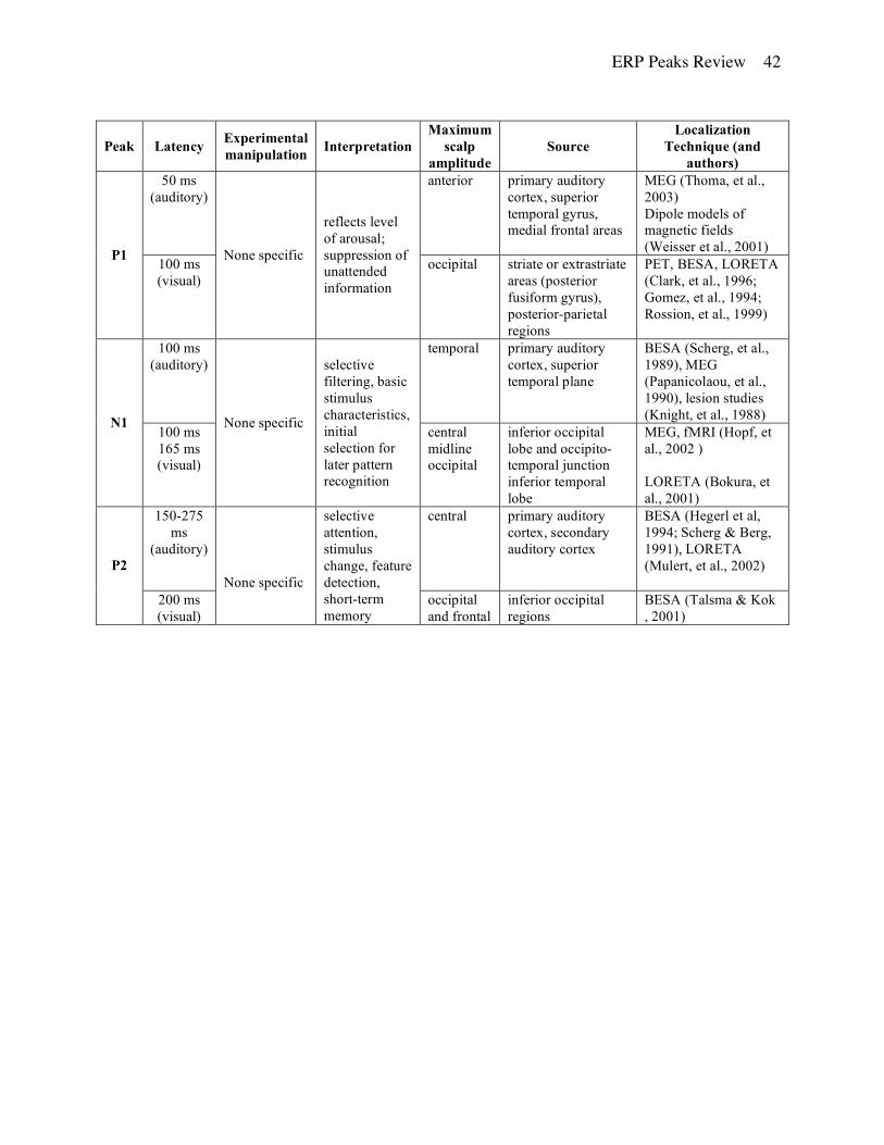
ERP Peaks Review 42
Peak Latency Experimental manipulation Interpretation
Maximum scalp
amplitude Source
Localization Technique (and
authors) 50 ms
(auditory) anterior
primary auditory cortex, superior temporal gyrus, medial frontal areas
MEG (Thoma, et al., 2003) Dipole models of magnetic fields (Weisser et al., 2001) P1 100 ms
(visual)
None specific
reflects level of arousal; suppression of unattended information
occipital striate or extrastriate areas (posterior fusiform gyrus), posterior-parietal regions
PET, BESA, LORETA (Clark, et al., 1996; Gomez, et al., 1994; Rossion, et al., 1999)
100 ms (auditory)
temporal
primary auditory cortex, superior temporal plane
BESA (Scherg, et al., 1989), MEG (Papanicolaou, et al., 1990), lesion studies (Knight, et al., 1988) N1
100 ms 165 ms (visual)
None specific
selective filtering, basic stimulus characteristics, initial selection for later pattern recognition
central midline occipital
inferior occipital lobe and occipito-temporal junction inferior temporal lobe
MEG, fMRI (Hopf, et al., 2002 ) LORETA (Bokura, et al., 2001)
150-275 ms
(auditory)
central
primary auditory cortex, secondary auditory cortex
BESA (Hegerl et al, 1994; Scherg & Berg, 1991), LORETA (Mulert, et al., 2002) P2
200 ms (visual)
None specific
selective attention, stimulus change, feature detection, short-term memory
occipital and frontal
inferior occipital regions
BESA (Talsma & Kok , 2001)

ERP Peaks Review 43
200 ms
(auditory) None specific
Detects changes in stimuli that are attended to
vertex, preoccipital, and frontal
supratemporal auditory cortex
scalp current density (Bruneau & Gomot, 1998)
156–189 ms
aka N170 (visual)
human faces, complex objects, words
Facial and/or expert object recognition
occipito-temporal
Fusiform gyrus Lateral occipitotemporal areas
Intracranial recordings (Allison, et al., 1999; Bentin, et al., 1996) fMRI (Kanwisher, et al., 1997; McCarthy, et al., 1997) BESA (Schweinberger, et al., 2002)
N2
100-300 ms Auditory &
Visual
Go/NoGo
inhibition
fronto-central Caudal and rostral anterior cingulated cortex
fMRI (Mathalon, et al., 2003)
MMN
100-250 ms (auditory)
physically different infrequent stimuli among other more frequent stimuli
early preattentive sensory memory
frontal, central Multiple stimulus-specific dipoles in the temporal lobe Right superior temporal gyrus and plane
MEG (Rosburg, 2003) fMRI-ERP (Leibenthal. et al., 2003)
300 ms Attention to stimuli, low probability of targets
memory updating, stimulus discrimination and responses preparation
centro-parietal
medial temporal lobe (thalamus, hippocampus), superior temporal gyrus, temporo-parietal junction
BESA (Tarkka, et al., 1995) MEG (Papanicolaou, et al., 1992) Lesion studies (Knight, et al., 1989) P3
300 ms Novel stimuli, not requiring attention
involuntary attention, inhibition
frontal medial parietal lobe, left superior prefrontal cortex, left lateral orbitofrontal cortex
LORETA (Bokura, et al., 2001) lesions (Knight, 1991)
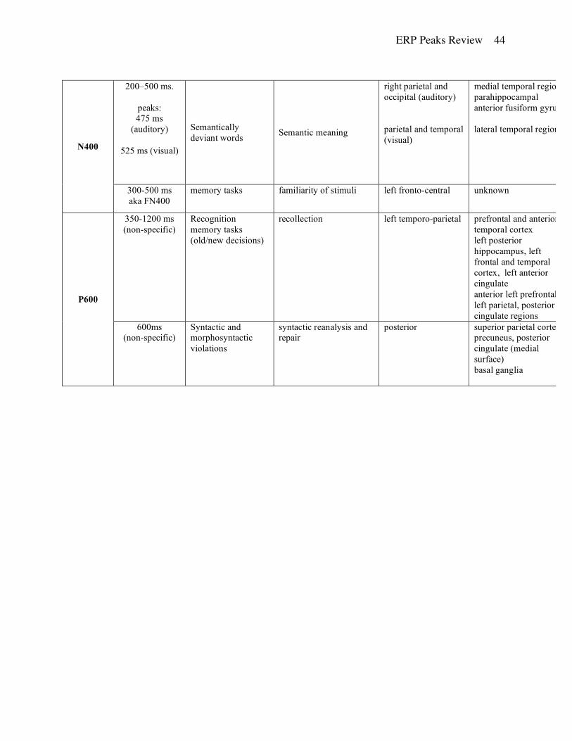
ERP Peaks Review 44
200–500 ms.
peaks: 475 ms
(auditory)
525 ms (visual)
Semantically deviant words Semantic meaning
right parietal and occipital (auditory) parietal and temporal (visual)
medial temporal regions parahippocampal anterior fusiform gyrus lateral temporal regions
Intracortical depth recordings (Smith, et al., 1986; Halgren et al., 1994; McCarthy et al., Nobre & McCarthy, 1995) MEG (Simos et al., 1997)
N400
300-500 ms aka FN400
memory tasks familiarity of stimuli left fronto-central unknown
350-1200 ms (non-specific)
Recognition memory tasks (old/new decisions)
recollection left temporo-parietal prefrontal and anterior temporal cortex left posterior hippocampus, left frontal and temporal cortex, left anterior cingulate anterior left prefrontal, left parietal, posterior cingulate regions
intracranial recordings (Guillem et al., 1996; 1999) PET (Rugg et al., 1996; 1998) fMRI (Henson et al., 1999)
P600
600ms (non-specific)
Syntactic and morphosyntactic violations
syntactic reanalysis and repair
posterior
superior parietal cortex, precuneus, posterior cingulate (medial surface) basal ganglia
fMRI (Kuperberg et al., 2003) lesions (Frisch et al., 2003)