Embed Size (px)
Citation preview

Ko et al. Appl Biol Chem (2021) 64:39 https://doi.org/10.1186/s13765-021-00609-2
ARTICLE
Emodin and rhein in Cassia tora ameliorates activity of mitochondrial enzymes involved in oxidative phosphorylation in the retina of diabetic miceEun Ko1†, Min Young Um2†, Taewon Han2, Sooim Shin1,3* and Moonsung Choi4,5*
Abstract
Cassia tora is an annual herb, which has pharmacological effects such as antioxidant, hypolipidemic, and antidiabetic effects. Accordingly, its effect on diabetes has been well-studied. However, it is unclear whether it has an effect on mitochondrial dysfunction associated with diabetes. In this study, the effects of emodin and rhein in C. tora seed ethanolic extract (ER/CSEE) on retinal mitochondrial function were examined in high-fat diet (HFD)-fed mice. HFD-fed mice exhibited decreased mitochondrial function followed by compensatory increase in the expression levels of mitochondrial enzymes. However, ER/CSEE treatment for 12 weeks ameliorated the activity of retinal mitochondrial complexes and reduced the expression level of enzymes involved in oxidative phosphorylation, except that of com-plex II and citrate synthase in citric acid cycle. This suggests that repairing capacity of enzymes in electron transport chain and citric acid cycle of mitochondria are different in response to the metabolic state. Therefore, it concluded that emodin and rhein play a pharmacological role in fat metabolism by influencing activities of enzymes in citric acid cycle linked with beta-oxidation in retina.
Keywords: Cassia tora, Emodin, Rhein, Mitochondria, Oxidative phosphorylation, Type 2 diabetes
© The Author(s) 2021. This article is licensed under a Creative Commons Attribution 4.0 International License, which permits use, sharing, adaptation, distribution and reproduction in any medium or format, as long as you give appropriate credit to the original author(s) and the source, provide a link to the Creative Commons licence, and indicate if changes were made. The images or other third party material in this article are included in the article’s Creative Commons licence, unless indicated otherwise in a credit line to the material. If material is not included in the article’s Creative Commons licence and your intended use is not permitted by statutory regulation or exceeds the permitted use, you will need to obtain permission directly from the copyright holder. To view a copy of this licence, visit http:// creat iveco mmons. org/ licen ses/ by/4. 0/.
IntroductionType 2 diabetes (T2D) is the most threatening metabolic disease, affecting more than 10% population worldwide [1]. A typical symptom of T2D is abnormal glycemic reg-ulation, and the long-term progression of T2D results in severe complications in critical organs in the human body [2]. Mitochondrion is the central organelle for glucose catabolism, in which adenosine triphosphate molecules
are produced from glucose-delivered metabolites in the process of oxidative phosphorylation (OXPHOS) [3]. Increased concentration of glucose in blood results in increased glucose uptake by the organs, thus leading to an overload of metabolites in the mitochondria. This can further lead to an imbalance in metabolism and elevate oxidative stress [4]. Several organs malfunction due to diabetes; particularly, metabolic imbalance and unregu-lated angiogenesis in the eye can cause diabetic retinopa-thy [5]. Diabetic retinopathy is the most frequent cause of blindness, with its prevalence reported in 60% of patients with T2D [6]. The pathogenesis of retinopathy involves swelling of the macula and proliferation of retinal blood vessels that eventually disrupt clear vision [7, 8].
Cassia tora is well known in East Asia as an alterna-tive medicine to treat diabetes and retinal dysfunction. Other than this, the whole plant, leaves, roots, and seeds
Open Access
*Correspondence: [email protected]; [email protected]†Eun Ko and Min Yong Um contributed equally to this work1 Department of Biotechnology and Bioengineering, College of Engineering, Chonnam National University, 77 Yongbong-ro, Buk-gu, Gwangju 61186, Republic of Korea4 Department of Optometry, College of Energy and Biotechnology, Seoul National University of Science and Technology, Seoul 01811, Republic of KoreaFull list of author information is available at the end of the article
Page 2 of 6Ko et al. Appl Biol Chem (2021) 64:39
of C. tora have been used as a traditional medicine to treat various diseases such as psoriasis, skin diseases, and cardiovascular diseases [9]. The extract of C. tora contains pharmacologically useful phenolic compounds such as anthraquinones, naphthopyrones, naphthalenes, and hydroanthracenes and their glucosides [10, 11]. In a previous study, when the butanol-extracted fraction of C. tora seeds was administrated to streptozotocin-induced diabetic rats, the diabetes was ameliorated by the reduc-tion of postprandial blood glucose and insulin secretion [12]. In addition, C. tora seed extract was shown to have hypolipidemic activity [13]. However, the effect of C. tora seeds on mitochondrial dysfunction associated with T2D are poorly examined. Therefore, we assessed the efficacy of emodin and rhein in C. tora seeds ethanolic extract (ER/CSEE) on the retinal health of mice with diabetes induced by a high-fat diet (HFD). Specifically, we exam-ined the effect of ER/CSEE treatment on the mitochon-drial function of the retina by measuring the activity of enzymes involved in OXPHOS thereby identify the link-age between the C. tora and functional improvement of mitochondria.
Materials and methodsPreparation of C. tora seed extractCassia tora seeds were bought from a commercial sup-plier Gyeongdong market (Seoul, Republic of Korea). The seeds of C. tora were ground into a powder with particle size of 40 mesh, and their extract was prepared in 70% ethanol. The extract was filtered and concentrated by rotary evaporation (Heidoph, LABORATA 4000, Schwa-bach, Germany). The concentrate was freeze dried and stored at − 20 °C till further analysis.
High‑performance liquid chromatography for the quantification of Emodin and RheinCSEE was subjected to high performance liquid chroma-tography (HPLC; 1290 Infinity II, Agilent Technologies, USA) to quantify the active compounds. Dried CSEE was dissolved in ethanol and heated at 60 °C for 20 min. This was followed by sonication, and further, the extract was filtered using a membrane filter of pore size 0.45 μm. A total of 10 μL sample was loaded to C18 (4.60 × 250 mm) column in CH3CN:10 mM/phosphate buffer (50:50 ratio; pH 2.6) with flow rate of 0.7 mL/min.
Animals and dietsAll procedures were performed according to Guide for Institutional Animal Care and Use Committee of the Korea Food Research Institute (KFRI-IACUC, KFRI-M-16037). In this study, 5-week-old male C57BL/6 J mice (Central Lab. Animal Inc., Seoul, Republic of Korea) were maintained in a cage at 21–25 °C, 50–60% humidity, 12 h
each of light and dark cycles, and ad libitum access to food and water. After acclimatization period of 1 week, the mice were divided into three groups depending on the diet type. They were as follows: normal diet (ND; 10% calories corresponding to fats, Research diets Inc., New Brunswick, USA), HFD (45% calories corresponding to fats, Research diets Inc., New Brunswick, USA), and HFD with 0.1% ER/CSEE (w/w). The diet was maintained for 12 weeks. After the experimental period, blood was obtained from the retro-orbital sinus of mice in the fast-ing state. The eyes were removed, weighed, immediately snap-frozen in liquid N2, and stored at − 80 °C till further analysis.
Assessment of mitochondrial oxidative phosphorylationUsing homogenized eyes, individual activity of com-plexes I, II, III, and IV was measured by spectrophotom-etry using S-3100 UV–Vis Spectrophotometer (Scinco, Seoul, Republic of Korea) [14]. The activity of complex I [nicotinamide adenine dinucleotide (NADH) dehydro-genase] was assessed using NADH and ubiquinone as substrates in 50 mM potassium phosphate at pH 7.5 con-taining 3 mg/mL bovine serum albumin (BSA), 0.1 mM NADH, 0.3 mM KCN, and 0.06 mM ubiquinone. The reaction for the complex I was monitored by checking absorbance at 340 nm; decrease in absorbance indicated oxidation of NADH. The activity of complex II (succinate dehydrogenase) was assessed using succinate and ubiqui-none as substrates in 50 mM potassium phosphate at pH 7.5 containing 1 mg/mL BSA, 0.05 mM decylubiqui-none, 20 mM succinate, 0.3 mM KCN, and 0.015% (w/v) 2,6-dichlorophenolindolphenol (DCPIP). The reaction for the complex II was monitored by measuring absorb-ance at 600 nm; decrease in absorbance indicated reduc-tion of DCPIP. The activity of complex III (cytochrome bc1 complex) was assessed using ubiquinol and ferric form of cytochrome c as substrates in 50 mM potassium phosphate at pH 7.5 containing 2.5% (v/v) Tween®20, 0.3 mM KCN, 0.1 mM decylubiquinol, 0.1 mM ethyl-enediaminetetraacetic acid, and 0.075 mM cytochrome c. The reaction for the complex III was monitored by measuring absorbance at 550 nm; increase in absorb-ance indicated reduction of cytochrome c. The activ-ity of complex IV (cytochrome c oxidase) was assessed using 0.06 mM ferrous form of cytochrome c as a sub-strate in 10 mM potassium phosphate at pH 7.0. The reaction for the complex IV was monitored by measur-ing absorbance at 550 nm; decrease in absorbance indi-cated oxidation of cytochrome c. Citrate synthase was quantified using acetyl CoA and oxaloacetate as sub-strates in 20 mM Tris buffer at pH 8.0 with 0.2% (v/v) triton X-100, 0.3 mM acetyl CoA, 0.5 mM oxaloacetate, and 0.1 mM 5,5ʹ-dithiobis-(2-nitrobenzoic acid) (DTNB).
Page 3 of 6Ko et al. Appl Biol Chem (2021) 64:39
The reaction was monitored by measuring absorbance at 412 nm; increase in absorbance indicated reduction of DTNB to 2-nitro-5-thiobenzoic acid (TNB). All chemi-cals used for the assays were purchased from Sigma Aldrich (St. Louis, USA).
Western blottingMitochondria was isolated from homogenized eyes using Mitochondria Isolation Kit for Tissue (Thermo Fisher Scientific, Waltham, USA). Isolated mitochondria was loaded on 12% gel prepared using TGX™ FastCast™ Acrylamide Kit (Bio-Rad, Hercules, USA), and isolated proteins were transferred to polyvinylidene fluoride (PVDF) membrane (Bio-Rad) using Trans-Blot Turbo Transfer system (25 V and 1A for 30 min: Turbo–Stand-ard). The membrane was blocked with 2.5% Blotting-Grade Blocker (Non-fat dry milk, Bio-Rad) in TBST buffer (20 mM Tris, 150 mM NaCl, and 0.1% Tween®20; pH 7.5) for 1 h at 26 °C followed by overnight incubation at 4 °C with two primary antibodies: anti-OxPhos (1:500 dilution; Anti-OxPhos complex kit; Invitrogen, Carlsbad, USA) and prohibitin (1:500 dilution; Santacruz, Dallas, USA). Further, the membrane was washed several times with TBST buffer and incubated with goat anti-mouse IgG (H + L) secondary antibody (1:3000 dilution) with horseradish peroxidase–conjugate (Invitrogen) for 1.5 h at 26 °C. The protein complexes were visualized using Clarity™ Western ECL Substrate (Bio-rad) on ChemiDoc System from Bio-rad. The samples were analyzed using Image Lab 6.0. (Bio-rad).
Statistical analysisResults are expressed as the means or as the mean ± SD. One-way analysis of variance (ANOVA) with post hoc Tukey test was used for statistical analysis using Graph-Pad Prism 8.0.1 software (GraphPad, Inc., La Jolla, USA). A p value of < 0.01 was considered statistically significant.
ResultsIdentification of emodin and rhein in CSEEEmodin and rhein are the anthraquinone metabolites known to have pharmacological effects [15–18]. For the quantification of them in CSEE, HPLC was performed. Emodin and rhein were found in CSEE, accounting for a total concentration of 8.51% (data not shown).
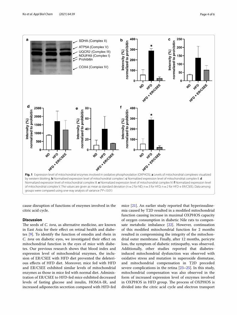
ER/CSEE alleviated a compensatory increase in mitochondrial enzymes induced by HFDTo assess the changes in the expression level of mitochon-drial enzymes, western blotting was performed. The ten-dency of expression level change depending on the diet group was similar for all the mitochondrial complexes. Compared with the normal diet group, the HFD group
exhibited increased expression levels of mitochondrial complexes I–V. However, administration of ER/CSEE to HFD mice resulted in a decrease in the expression levels of mitochondrial complexes, bringing the levels similar to those of normal diet group (Fig. 1). In a previous study, beta-cell mass and expression level of a specific protein exhibited compensatory increase in response to early diabetes [19]. This compensatory increase is the result of cellular response to ameliorate functions that might be disrupted by HFD [20]. Therefore, it can be assumed that the administration of ER/CSEE successfully repaired HFD-induced distorted metabolism in retinal cells or blocked the process of disruption.
ER/CSEE recovered the HFD‑induced disrupted function of mitochondrial enzymes involved in OXPHOSThe activity of mitochondrial enzymes involved in OXPHOS was investigated in the three mice groups. The activity of all mitochondrial enzymes reduced after HFD in comparison with normal diet. However, when ER/CSEE was administrated to HFD-fed mice, activity of some of the mitochondrial enzymes was drastically recovered (Fig. 2). In case of complexes I and IV, mito-chondrial activity was recovered and brought to the level similar to that of normal diet group. For complex III, although its activity in HFD with ER/CSEE group exhib-ited a slight decrease compared with ND, it was recov-ered to a considerable extent in comparison with that in HFD group.
HFD‑induced disrupted function of enzymes in the citric acid cycle was not recovered by ER/CSEEThough the activity of mitochondrial complexes I, III, and IV was recovered by ER/CSEE after HFD, that of complex II and citrate synthase did not exhibit any spe-cific change compared with that in HFD group (Fig. 2b and e). Mitochondrial complex II and citrate synthase are the enzymes involved in the citric acid cycle. NADH and FADH2, which act as crucial coenzymes for mitochon-drial complexes I and II, are produced in citric acid cycle. In normal glucose metabolism, glucose is converted into pyruvate through glycolysis. Before entering the citric acid cycle, acetyl CoA is produced by pyruvate dehydro-genase from pyruvate. In lipid metabolism, triglyceride breaks down into glycerol and fatty acids. Fatty acids are metabolized through beta-oxidation to produce NADH, FADH2, and acetyl CoA. Further, acetyl CoA enters the citric acid cycle. When diet contains high portion of fats, extra energy is stored in the form of triglycerides result-ing in a high influx of fatty acids for beta-oxidation. It leads to increased production of acetyl CoA as a sub-strate of the citric acid cycle, resulting in overload of metabolites in the citric acid cycle. Consequently, it may
Page 4 of 6Ko et al. Appl Biol Chem (2021) 64:39
cause disruption of functions of enzymes involved in the citric acid cycle.
DiscussionThe seeds of C. tora, as alternative medicine, are known in East Asia for their effect on retinal health and diabe-tes [9]. To identify the function of emodin and rhein in C. tora on diabetic eyes, we investigated their effect on mitochondrial function in the eyes of mice with diabe-tes. Our previous research shows that blood index and expression level of mitochondrial enzymes, the inclu-sion of ER/CSEE with HFD diet prevented the deleteri-ous effects of HFD diet. Moreover, mice fed with HFD and ER/CSEE exhibited similar levels of mitochondrial enzymes as those in mice fed with normal diet. Adminis-tration of ER/CSEE to HFD-fed mice exhibited decreased levels of fasting glucose and insulin, HOMA-IR, and increased adiponectin secretion compared with HFD-fed
mice [21]. An earlier study reported that hyperinsuline-mia caused by T2D resulted in a modified mitochondrial function causing increase in maximal OXPHOS capacity of oxygen consumption in diabetic Nile rats to compen-sate metabolic imbalance [22]. However, continuation of this modified mitochondrial function for 2 months resulted in compromising the integrity of the mitochon-drial outer membrane. Finally, after 12 months, pericyte loss, the symptom of diabetic retinopathy, was observed. Additionally, other studies reported that diabetes-induced mitochondrial dysfunction was observed with oxidative stress and mutation in superoxide dismutase, and mitochondrial compensation in T2D preceded severe complications in the retina [23–25]. In this study, mitochondrial compensation was also observed in the form of increased expression level of enzymes involved in OXPHOS in HFD group. The process of OXPHOS is divided into the citric acid cycle and electron transport
Fig. 1 Expression level of mitochondrial enzymes involved in oxidative phosphorylation (OXPHOS). a Levels of mitochondrial complexes visualized by western blotting. b Normalized expression level of mitochondrial complex I. c Normalized expression level of mitochondrial complex II. d Normalized expression level of mitochondrial complex III. e Normalized expression level of mitochondrial complex IV. f Normalized expression level of mitochondrial complex V. The values are given as mean ± standard deviation (n = 2 for ND; n = 3 for HFD; n = 2 for HFD + ER/CSEE). Data among groups were compared using one-way analysis of variance (*P < 0.01)

Page 5 of 6Ko et al. Appl Biol Chem (2021) 64:39
chain. In HFD with ER/CSEE group, the expression lev-els of mitochondrial enzymes of the electron transport chain were similar to those in normal diet group (Fig. 1). This indicated that ER/CSEE successfully prevented the metabolic imbalance in the early stage of diabetes, also averting mitochondrial compensation and diabetic retin-opathy. In contrast, the activity of the enzymes involved in OXPHOS in mitochondria exhibited different patterns according to the category of the enzymes (Fig. 2). In HFD with ER/CSEE group, enzyme activity of the complexes I, III and IV in electron transport chain was found to be recovered compared with that in HFD group. However, the activity of enzymes of the citric acid cycle, complex II, and citrate synthase did not recover. This suggests that the function of enzymes involved in the citric acid cycle and electron transport chain are differently repaired by the administration of emodin and rhein. Given that the beta-oxidation of fatty acid decomposition is directly linked to the citric acid cycle, it can be speculated that emodin and rhein in C. tora are involved in the ameliora-tion of overall fat metabolism with reduction of circulat-ing triglyceride and fatty acid by fat oxidation. It might result in burdening enzymes in the citric acid cycle before the direct action on the mitochondrial function improve-ment. In addition, it should be noted that the methodo-logical approach of the current study as assessing the mitochondrial enzyme activity provided a profound anal-ysis of the changes in enzyme function in response to the
metabolic state, in comparison to detecting the enzyme expression level by western blotting which exhibits superficial and similar changes in all complexes. In con-clusion, administration of the emodin and rhein to HFD mouse successfully enhanced mitochondrial enzymes’ activity in the electron transport chain and prevented the compensatory increase of mitochondrial enzyme expres-sion in retina in vivo that should lead to mitochondrial dysfunction and diabetic retinopathy induced by metabo-lites overload.
AbbreviationsCSEE: C. tora seed ethanolic extract; ER/CSEE: Emodin and rhein in C. tora seed ethanolic extract; HFD: High-fat diet; T2D: Type 2 diabetes; OXPHOS: Oxidative phosphorylation; NADH: Nicotinamide adenine dinucleotide; BSA: Bovine serum albumin; DCPIP: 2,6-dichlorophenolindolphenol; DTNB: 5,5ʹ-Dithiobis-(2-nitrobenzoic acid); TNB: 2-Nitro-5-thiobenzoic acid; PVDF: Polyvinylidene fluoride.
AcknowledgementsNot applicable.
Authors’ contributionsSS, MC conceived the project and designed the experiments. MU, EK and TH performed the experiments. SS, MC, MU and EK analyzed the data, and EK, and SS wrote the manuscript. All authors read and approved the final manuscript.
FundingThis work was supported by the National Research Foundation of Korea (Grant Number 2020R1A2C1101110) and by the Korea Institute of Planning and Evaluation for Technology in Food, Agriculture, Forestry (IPET) through High Value-added Food Technology Development Program, funded by the Ministry of Agriculture, Food and Rural Affairs (MAFRA) (Grant Number 313028-3).
Fig. 2 Activity of mitochondrial complexes and citrate synthase involved in OXPHOS. a Activity of mitochondrial complex I. b Activity of mitochondrial complex II. c Activity of mitochondrial complex III. d Activity of mitochondrial complex IV. e Activity of mitochondrial citrate synthase. Activities in ND are shown as white dots, HFD as black dots, and HFD + ER/CSEE as gray dots. Values are given as means (n = 7 for all groups)
Page 6 of 6Ko et al. Appl Biol Chem (2021) 64:39
Availability of data and materialsThe datasets used and/or analysed during the current study are available from the corresponding author on reasonable request.
Declarations
Competing interestsThe authors declare that they have no competing interests.
Author details1 Department of Biotechnology and Bioengineering, College of Engineering, Chonnam National University, 77 Yongbong-ro, Buk-gu, Gwangju 61186, Republic of Korea. 2 Research Division of Food Functionality, Korea Food Research Institute, Wanju 55365, Republic of Korea. 3 Interdisciplinary Program of Bioenergy and Biomaterials Graduate School, College of Engineering, Chon-nam National University, Gwangju 61186, Republic of Korea. 4 Department of Optometry, College of Energy and Biotechnology, Seoul National University of Science and Technology, Seoul 01811, Republic of Korea. 5 Convergence Institute of Biomaterials and Bioengineering, Seoul National University of Sci-ence and Technology, Seoul 01811, Republic of Korea.
Received: 24 February 2021 Accepted: 17 April 2021
References 1. Wang C-Y, Neil DL, Home P (2018) 2020 vision–an overview of prospects
for diabetes management and prevention in the next decade. Diabetes Res Clin Pr 143:101–112
2. Stolar M (2010) Glycemic control and complications in type 2 diabetes mellitus. Am J Med 123(3):S3–S11
3. Hatefi Y (1985) The mitochondrial electron transport and oxidative phos-phorylation system. Annu Rev Biochem 54(1):1015–1069
4. Rabøl R, Højberg PM, Almdal T, Boushel R, Haugaard SB, Madsbad S, Dela F (2009) Effect of hyperglycemia on mitochondrial respiration in type 2 diabetes. J Clin Endocr Metab 94(4):1372–1378
5. Martin A, Komada MR, Sane DC (2003) Abnormal angiogenesis in diabe-tes mellitus. Med Res Rev 23(2):117–145
6. Pugliese G, Tilton RG, Williamson JR (1991) Glucose-induced metabolic imbalances in the pathogenesis of diabetic vascular disease. Diabetes Metab Rev 7(1):35–59
7. Fong DS, Aiello L, Gardner TW, King GL, Blankenship G, Cavallerano JD, Ferris FL, Klein R (2004) Retinopathy in diabetes. Diabetes Care 27(suppl 1):s84–s87
8. Yau JW, Rogers SL, Kawasaki R, Lamoureux EL, Kowalski JW, Bek T, Chen S-J, Dekker JM, Fletcher A, Grauslund J (2012) Global prevalence and major risk factors of diabetic retinopathy. Diabetes Care 35(3):556–564
9. Jain S, Patil U (2010) Phytochemical and pharmacological profile of Cassia tora Linn.-An overview. Indian J of Nat Prod Resour 1:430–437
10. Hatano T, Uebayashi H, Ito H, Shiota S, Tsuchiya T, Yoshida T (1999) Phenolic constituents of Cassia seeds and antibacterial effect of some
naphthalenes and anthraquinones on methicillin-resistant Staphylococ-cus aureus. Chem Pharm Bull 47(8):1121–1127
11. El-Halawany AM, Chung MH, Nakamura N, Ma C-M, Nishihara T, Hattori M (2007) Estrogenic and anti-estrogenic activities of Cassia tora phenolic constituents. Chem Pharm Bull 55(10):1476–1482
12. Nam J, Choi H (2008) Effect of butanol fraction from Cassia tora L. seeds on glycemic control and insulin secretion in diabetic rats. Nutr Res Pract 2(4):240–246
13. Patil UK, Saraf S, Dixit V (2004) Hypolipidemic activity of seeds of Cassia tora Linn. J Ethnopharmacol 90(2–3):249–252
14. Spinazzi M, Casarin A, Pertegato V, Salviati L, Angelini C (2012) Assess-ment of mitochondrial respiratory chain enzymatic activities on tissues and cultured cells. Nat Protoc 7(6):1235
15. Chang C-H, Lin C-C, Yang J-J, Namba T, Hattori M (1996) Anti-inflam-matory effects of emodin from ventilago leiocarpa. Am J Chin Med 24(02):139–142
16. Li H-L, Chen H-L, Li H, Zhang K-L, Chen X-Y, Wang X-W, Kong Q-Y, Liu J (2005) Regulatory effects of emodin on NF-κB activation and inflam-matory cytokine expression in RAW 264.7 macrophages. Int J Mol Med 16(1):41–47
17. Zhong X-F, Huang G-D, Luo T, Deng Z-Y, Hu J-N (2012) Protective effect of rhein against oxidative stress-related endothelial cell injury. Mol Med Rep 5(5):1261–1266
18. Aviello G, Rowland I, Gill CI, Acquaviva AM, Capasso F, McCann M, Capasso R, Izzo AA, Borrelli F (2010) Anti-proliferative effect of rhein, an anthraquinone isolated from Cassia species, on Caco-2 human adenocar-cinoma cells. J Cell Mol Med 14(7):2006–2014
19. Araujo TG, de Oliveira AG, Saad MJA (2013) Insulin-resistance-associated compensatory mechanisms of pancreatic beta cells: a current opinion. Front Endocrinol 4:146
20. Montanya E (2014) Insulin resistance compensation: not just a matter of β-Cells? Diabetes 63(3):832–834
21. Ko E, Um MY, Choi M, Han T, Kim IH, Shin S (2020) Cassia tora seed improves pancreatic mitochondrial function leading to recovery of glucose metabolism. Am J Chin Med 48(03):615–629
22. Han WH, Gotzmann J, Kuny S, Huang H, Chan CB, Lemieux H, Sauve Y (2017) Modifications in retinal mitochondrial respiration precede type 2 diabetes and protracted microvascular retinopathy. Invest Ophth Vis Sci 58(10):3826–3839
23. Kowluru RA, Abbas SN (2003) Diabetes-induced mitochondrial dysfunc-tion in the retina. Invest Ophth Vis Sci 44(12):5327–5334
24. Kowluru RA, Mishra M (2015) Oxidative stress, mitochondrial damage and diabetic retinopathy. BBA-Mol Basis Dis 1852(11):2474–2483
25. Kowluru RA, Atasi L, Ho Y-S (2006) Role of mitochondrial superoxide dismutase in the development of diabetic retinopathy. Invest Ophth Vis Sci 4:1594–1599
Publisher’s NoteSpringer Nature remains neutral with regard to jurisdictional claims in pub-lished maps and institutional affiliations.