Embed Size (px)
Citation preview

8/20/2019 dry eyes ii
http://slidepdf.com/reader/full/dry-eyes-ii 1/142
THE OCULAR SURFACE / APRIL 2007, VOL. 5, NO. 2 / www.theocularsurface.com 59
2007 Report of the
International Dry EyeWorkShop (DEWS)
Sponsored by the
Tear Film & Ocular Surface Society
INTRODUCTION TO THE 2007 REPORT OF THE
INTERNATIONAL DRY EYE WORKSHOP DEWS
THE DEFINITION AND CLASSIFICATION OF DRY EYE DISEASE
THE EPIDEMIOLOGY OF DRY EYE DISEASE
METHODOLOGIES TO DIAGNOSE AND MONITOR DRY EYE DISEASE
DESIGN AND CONDUCT OF CLINICAL TRIALS
MANAGEMENT AND THERAPY OF DRY EYE DISEASE
RESEARCH IN DRY EYE
www.theocularsurface.com
A peer-reviewed journal, indexed in MEDLINE/PubMed and EMBASE
SPECIAL ISSUE
April 2007Volume 5, Number 2
OcularSurfaceTHE
A JOURNAL OF REVIEW LINKING LABORATORY SCIENCE, CLINICAL SCIENCE, AND CLINICAL PRACTICE

8/20/2019 dry eyes ii
http://slidepdf.com/reader/full/dry-eyes-ii 2/142
THE OCULAR SURFACE / APRIL 2007, VOL. 5, NO. 2 / www.theocularsurface.com60
Article submission: Questions regarding manuscript prepa-
ration not addressed in the Information for Authors may beaddressed to the Managing Editor: Susan Erickson, 130
Winchester St., Brookline, MA 02446. Tel: 617-731-3415;
Email: [email protected]. Information for Authors is
available at www.theocularsurface.com.
Subscription rates Individual: $154 per year in the US and
Canada; $199 per year rest of world (includes air mail deliv-
ery). Institution:$249 per year in the US and Canada; $299
per year rest of world (includes air mail delivery). Internet-
only subscription: $139 (US and rest of world). Print-only
subscription: $139 (US and Canada); $139 (rest of world).
Back issues are $45 per issue. Subscriptions may be ordered
online at www.theocularsurface.com or at The Ocular Surface, 75 Maiden Lane, Suite 408, New York, NY 10038, USA. TEL:
212-791-1440 FAX : 212-791-4980. Claims for missing issues
must be filed within 120 days of the issue date.
Indexing The Ocular Surface is indexed in MEDLINE/
PubMed and EMBASE. The Ocular Surface is printed on
acid-free paper that meets the requirements of ANSI/NISO
Z39.48-1992 (Permanence of Paper).
Library reprints Contact the Copyright Clearance Center,
Inc., 222 Rosewood Drive, Danvers, MA 01923 USA. TEL:
978-646-2600; FAX : 978-646-8600 E-MAIL: info@copyright.
com ONLINE: www.copyright.com.
Advertising and bulk reprint inquiries should be addressed
to LaVon Kellner, Ethis Communications, Inc., 75 Maiden
Lane, Suite 408, New York, NY 10038. TEL: 212-791-1440;
FAX : 212-791-4980; E-MAIL: [email protected]. Con-
tact authors for single reprint copies.
The Ocular Surface (ISSN 1542-0124) is published quarterly
by Ethis Communications, Inc., 75 Maiden Lane, Suite 408,
New York, NY 10038, USA. Copyright 2006 Ethis Commu-
nications, Inc. All rights reserved. Neither The Ocular Surface
nor the publisher assume any responsibility for any injury
and/or damage to persons or property as a matter of product
liability, negligence or otherwise, or from any use or operationof any methods, products, instructions or ideas contained in
this publication. No part of these contents may be reproduced
without permission.
PUBLISHER , DIRECTOR OF GLOBAL SALES: LaVon Marie Kellner
EDITORIAL DIRECTOR : David Kellner
ART DIRECTOR : DeborahAnne Chingas Sandke
PRODUCTION DIRECTOR : Charlotte L. Latham
CIRCULATION MANAGER : Simon Wang
Ocular SurfaceTHE
A JOURNAL OF REVIEW LINKING LABORATORY SCIENCE, CLINICAL SCIENCE, AND CLINICAL PRACTICE
A peer-reviewed journal

8/20/2019 dry eyes ii
http://slidepdf.com/reader/full/dry-eyes-ii 3/142
THE OCULAR SURFACE / APRIL 2007, VOL. 5, NO. 2 / www.theocularsurface.com 61 61
EDITOR -IN-CHIEF
Gary N. Foulks, MD, FACSProfessor, Ophthalmology,University of Louisville,
Louisville, KY
FOUNDING EDITOR
Michael A. Lemp, MD Washington, DC
SECTION EDITORS
LABORATORY SCIENCE
James V. Jester, PhDProfessor, Ophthalmology,
University of California, Irvine, CA
CLINICAL SCIENCE,
INNOVATIVE TECHNIQUES AND TECHNOLOGY
Gary N. Foulks, MD
CLINICAL PRACTICE
John Sutphin, Jr, MDProfessor and Chair, Ophthalmology,
University of Kansas Medical Center, Kansas City, KS
FEATURES EDITORS
Juan Murube, MD, PhDProfessor, Ophthalmology,
University of Alcala, Madrid, Spain
Gary D. Novack, PhDPresident, PharmaLogic
Development Inc., San Rafael, CA
MANAGING EDITOR
Susan EricksonBrookline, MA
Mark B. Abelson, MD, Director, Ophthalmic Research Associates, North Andover, MA
Penny A. Asbell, MD, Professor, Ophthalmology, Mount Sinai MedicalCenter, New York, NY
Christophe Baudouin, MD, PhD, Professor and Director, Ophthalmology,Quinze-Vingts Hôpital, University of Paris, Paris, France
Roger W. Beuerman, PhD, Professor, Ophthalmology, Cell Biology, and Anatomy, Louisiana State University Eye Center, New Orleans, LA,and Singapore Eye Research Institute, Singapore
Stefano Bonini, MD,
Professor and Chairman, Ophthalmology,University of Rome, Rome, Italy
Anthony Bron, FRCS, Professor Emeritus, Nuffield Laboratory ofOphthalmology, Oxford, UK
M. Reza Dana, MD, MPH, Senior Scientist, Schepens Eye ResearchInstitute, Boston, MA
Darlene A. Dartt, PhD, Senior Scientist, Schepens Eye ResearchInstitute, Boston, MA
Harminder S. Dua, MD, PhD, Professor, Ophthalmology and VisionScience, University of Nottingham, UK
Suzanne M. J. Fleiszig, OD, PhD, Associate Professor, Vision Scienceand Optometry, University of California Berkeley, Berkeley, CA
Desmond Fonn, Dip Optom, M Optom (NSW), FAAO, Professor,School of Optometry, University of Waterloo, Waterloo, Ontario, Canada
Ilene K. Gipson, PhD,
Senior Scientist, Schepens Eye Research Institute,Boston, MA
W. Bruce Jackson, MD, Professor and Chairman, Ophthalmology,University of Ottawa, Ottawa, Ontario, Canada
Winston W.-Y. Kao, PhD, Director, Ophthalmic Research, University ofCincinnati, Cincinnati, OH
Shigeru Kinoshita, MD, Professor and Chairman, Ophthalmology,Kyoto Prefectural University of Medicine, Kyoto, Japan
Friedrich E. Kruse, MD, Professor and Chairman, University EyeClinic, University of Erlangen-Nuremberg, Erlangen, Germany
Peter Laibson, MD, Co-director, Cornea Service, Wills Eye Hospital,Philadelphia, PA
Mark J. Mannis, MD, Professor, Ophthalmology, University ofCalifornia, Davis, Davis, CA
William D. Mathers, MD, Professor, Casey Eye Institute, Oregon HealthSciences University, Portland, OR
James P. McCulley, MD, Professor and Chairman, Ophthalmology,University of Texas Southwestern Medical Center, Dallas, TX
Austin K. Mircheff, PhD, Professor, Physiology and Biophysics,and Ophthalmology, Doheny Eye Institute, University of SouthernCalifornia, Los Angeles, CA
Teruo Nishida, MD, Professor and Chairman, Biomolecular Recognitionand Ophthalmology, Yamaguchi University School of Medicine,
Yamaguchi, JapanStephen C. Pflugfelder, MD, Professor, Ophthalmology, Baylor Collegeof Medicine, Houston, TX
Kenneth A. Polse, OD, MS, Professor, Vision Science and Optometry,University of California, Berkeley, Berkeley, CA
Gullapalli N. Rao, MD, Director, LV Prasad Eye Institute, Hyderabad,India
Maurizio Rolando, MD, Professor, Neuroscience and Ophthalmology,University of Genoa, Genoa, Italy
Janine Smith, MD, Deputy Clinical Director, National Eye Institute,National Institutes of Health, Bethesda, MD
Michael E. Stern, PhD, Research Investigator, Allergan Inc., Irvine, CA
David A. Sullivan, PhD, Senior Scientist, Schepens Eye ResearchInstitute, Boston, MA
Deborah F. Sweeney, OD, PhD,
Associate Professor, University of NewSouth Wales, Sydney, NSW, Australia
Donald T. Tan, FRCS, Director, Singapore Eye Research Institute,National University of Singapore, Singapore
Timo Tervo, MD, PhD, Professor, Applied Clinical Ophthalmology,Helsinki University Eye Hospital, Helsinki, Finland
Alan Tomlinson, PhD, DSc, FCOptom, Professor and Head, VisionSciences, Glasgow Caledonian University, Glasgow, Scotland, UK
Scheffer Tseng, MD, PhD, President, Ocular Surface Center, Miami, FL
Kazuo Tsubota, MD, Professor and Head, Department ofOphthalmology, Keio University School of Medicine, Tokyo, Japan
Graeme Wilson, PhD, Professor, Optometry, Indiana University,Bloomington, IN
EDITORS
EDITORIAL BOARD
Ocular SurfaceTHE
A JOURNAL OF REVIEW LINKING LABORATORY SCIENCE, CLINICAL SCIENCE, AND CLINICAL PRACTICE
A peer-reviewed journal
Additional biographical information is available at www.theocularsurface.com

8/20/2019 dry eyes ii
http://slidepdf.com/reader/full/dry-eyes-ii 4/142
THE OCULAR SURFACE / APRIL 2007, VOL. 5, NO. 2 / www.theocularsurface.com62
8/20/2019 dry eyes ii
http://slidepdf.com/reader/full/dry-eyes-ii 5/142
THE OCULAR SURFACE / APRIL 2007, VOL. 5, NO. 2 / www.theocularsurface.com 63
Editorial
DEWS Report: A Mission Completed
his issue of The Ocular Surface is very unusual. As the official report of the Dry Eye Work-Shop (DEWS), it is an encyclopedic review of dry eye disease and, additionally, a guide toresources archived on the internet. It is the product of a team of international experts who
have labored over 3 years to compile an evidence-based review of the present state of knowledge fordry eye disease and the methods used to evaluate, diagnose, and manage the disorder. It summarizesthe findings of current research and identifies future needs for a better understanding of the etiology,pathogenesis, and potential therapy of the disease.
The process of deliberation and discussion that underpins this arduous endeavor is described in the“Introduction” and in various chapters of the volume. Suffice it to say that an international communityof clinicians and scientists with expertise in all aspects of dry eye disease collaborated to search theliterature, collect and validate data, and incorporate it into reports. The process of commentary andadjudication of differing opinions was open, yet subject to several levels of validation. The productis a written document that serves as a guide to a vast amount of information that is archived both inthis special issue and on a supporting website (www.tearfilm.org) that is accessible to all.
The chapter on Definition and Classification expands the characterization of dry eye disease andplaces it within the perspective of ocular surface disease. The chapter on Epidemiology providescommentary on the implications of the disease, as well as comparison of the methods available toevaluate symptoms and factors contributory to the disease. The Diagnostic Methodologies chapter notonly provides valuable discussion of the parameters of dry eye disease, but also catalogs and validatesa vast collection of clinical and research methods, including questionnaires, to monitor the disease.The Research chapter summarizes past and present findings, and identifies areas whose further studywill contribute to the understanding of the etiopathogenesis and consequences of dry eye disease.The chapter on Clinical Trials provides recommendations with regard to both general and specific
guidelines for clinical trials in dry eye disease and identifies the idiosyncrasies and confoundingoutcome variables for such trials. The chapter on Management and Therapy catalogs the options fortherapy and recommends a contemporary strategy for management of dry eye disease.
As would be expected for a multifactorial disease that has many nuances in clinical and patho-logical expression, opinions differ even amongst the experts as to the most appropriate way to char-acterize and label some aspects of the disease. This proved true for the definition and classificationof the disease. Some key concepts in the appreciation of dry eye were identified from the literature.One such concept was the characterization of the Lacrimal Functional Unit,1 which has highlightedthe interdependence of components of the lacrimal system in maintaining the integrity of the ocularsurface. Some new concepts were constructed in the deliberation process of the Subcommittee work,including a concept suggested by Dr. Christophe Baudoin—a Vicious Circle of dry eye disease, bywhich various risk factors may interact to precipitate and perpetuate the condition.2 The concept of
the Ocular Surface System, developed by the Research Subcommittee, extends the scope of the ocularsurface to a collection of contiguous tissues that share embryonal, innervational, histological, andhormonal background.
The time and effort necessary to compile and collate this project and the summary documentwas extraordinary. The endeavor could never have been completed without the sponsorship andcommitment of The Tear Film & Ocular Surface Society and the officers and staff of that organization.The planning and execution of the organizational meetings, the coordination of the conferences forpresentation of the collected information, the facilitation of the discussions of the DEWS participants,and the administrative direction of the publication process were achieved through the tireless effortsof Dr. David A., Rose M. and Amy G. Sullivan. The deliberations of the Steering Committee wereessential to the completion of the task. Likewise, the leaders of the various Subcommittees were in-
T
THE OCULAR SURFACE / APRIL 2007, VOL. 5, NO. 2 / www.theocularsurface.com 65
8/20/2019 dry eyes ii
http://slidepdf.com/reader/full/dry-eyes-ii 6/142
THE OCULAR SURFACE / APRIL 2007, VOL. 5, NO. 2 / www.theocularsurface.com64
strumental in providing the buildingblocks for construction of the finalproduct. A special congratulations and
thank you is due Professor Anthony J. Bron, who devoted endless hoursand energy to leading the writing teamthrough multiple iterations of thetext and the references to provide aharmonization of the various reports.The ultimate coordination and editingof the document was in the capablehands of Susan Erickson, for whomwe are most appreciative. Particularappreciation is extended to EthisCommunications, Inc. for embracing
the publication of this work, whichshould serve as a valuable referencefor all those who investigate and man-age patients with dry eye disease. Lastbut far from least is a heartfelt thankyou to the Corporate Sponsors of theDry Eye WorkShop, who providedthe financial resources and encourage-ment to complete this project.
I wish you good reading and greatreferencing.
Gary N. Foulks, MD, FACSEditor-in-Chief
REFERENCES 1. Stern ME, Gao J, Siemasko KF, et al. The
role of the lacrimal functional unit in thepathophysiology of dry eye. Exp Eye Res 2004;78(3):409-16
2. Baudoin C. [The vicious circle in dry eyesyndrome: a mechanistic approach] J FrOphtalmol 2007;30:239-46
EDITORIAL continued
66

8/20/2019 dry eyes ii
http://slidepdf.com/reader/full/dry-eyes-ii 7/142
THE OCULAR SURFACE / APRIL 2007, VOL. 5, NO. 2 / www.theocularsurface.com 67
SPECIAL ISSUE
2007 Report of theInternational Dry Eye
WorkShop (DEWS)
Sponsored by the Tear Film & Ocular Surface Society
69 INTRODUCTION TO THE 2007 REPORT OF THE INTERNATIONAL DRY EYEWORKSHOP (DEWS)
71 MEMBERSHIP OF THE INTERNATIONAL DRY EYE WORKSHOP (DEWS)
73 GLOSSARY
75 THE DEFINITION AND CLASSIFICATION OF DRY EYE DISEASE
93 THE EPIDEMIOLOGY OF DRY EYE DISEASE
108 METHODOLOGIES TO DIAGNOSE AND MONITOR DRY EYE DISEASE
153 DESIGN AND CONDUCT OF CLINICAL TRIALS
163 MANAGEMENT AND THERAPY OF DRY EYE DISEASE
179 RESEARCH IN DRY EYE
195 INDEX
202 DISCLOSURE OF FINANCIAL/PROPRIETARY INTERESTS OF DEWSMEMBERSHIP
68 PROCEDURES FOR SUBMITTING REVIEWS TO THE OCULAR SURFACE
The 2007 International Dry Eye WorkShop was sponsored by The Tear Film & Ocular SurfaceSociety, which received support for DEWS from SOOFT Italia; Alcon Laboratories; Allergan;
McNeil Consumer Healthcare; Pfizer; Santen Pharmaceutical Co.; Bausch & Lomb;Novartis Pharmaceuticals; Advanced Vision Research; Inspire Pharmaceuticals; Vistakon;
Senju Pharmaceutical Co.; Kowa; Otsuka Pharmaceutical Co.; Alimera Sciences; Tomei; Nidek
TABLE OF CONTENTS APRIL 2007, VOLUME 5, NUMBER 2

8/20/2019 dry eyes ii
http://slidepdf.com/reader/full/dry-eyes-ii 8/142
THE OCULAR SURFACE / APRIL 2007, VOL. 5, NO. 2 / www.theocularsurface.com68
8/20/2019 dry eyes ii
http://slidepdf.com/reader/full/dry-eyes-ii 9/142
THE OCULAR SURFACE / APRIL 2007, VOL. 5, NO. 2 / www.theocularsurface.com 69
DEWS Introduction
Introduction to the Report of the
International Dry Eye WorkShop (2007)ry eye disease is a common yet frequently under-recognized clinical condition whose etiologyand management challenge clinicians and researchers alike. Advances in the understandingof the disease have been made over the past 10 years in areas of epidemiology, pathogenesis,
clinical manifestation, and possible therapy. This volume represents the work of many contribu-tors over a long period of deliberation and through an iterative process that included collection ofdata, presentation of summary reports in a conference format, and harmonization of reports by awriting team with interactive commentary by the entire group of participants in an internationalworkshop.
HistoryIn 1994, a workshop sponsored by the National Eye Institute and supported by industry con-
vened a group of scientists, clinicians, and researchers interested in dry eye to clarify the definitionand characteristics of dry eye disease and to recommend reliable parameters for conduct of clinicalresearch and conduct of clinical trials for dry eye disease.1 The report of that workshop has servedas a solid resource in the field for over 10 years, but the explosion of information in both basic andclinical research in the interim warranted repetition of the process. An initiative was suggested byKazuo Tsubota, MD, and endorsed by Michael A. Lemp, MD, to recruit an international panel ofexperts in dry eye disease to accomplish such a task, and preliminary meetings were held in 2001.2 Selection of the participants was based upon their prior history of peer-reviewed publication, level ofparticipation in previous dry eye meetings (including the NEI/Industry Workshop), and collaborationwith acknowledged experts in the field. The immensity of the task became immediately apparent and
the coordinating support of The Tear Film & Ocular Surface Society (TFOS) was solicited. David A.Sullivan, PhD, President of TFOS, committed the organizational and administrative support of TFOSand secured broad financial support from international corporations to facilitate the internationalDry Eye WorkShop (DEWS).
Process
The DEWS effort was chaired by Anthony J. Bron, FRCS, and directed by a Steering Commit-tee that proposed guidelines for the determination of acceptable levels of evidence and methods ofdocumentation to support such evidence. The first step involved the formation of subcommittees:Definition and Classification; Epidemiology; Diagnosis; Research; Clinical Trials, and Managementand Therapy, in addition to a Communications and Industrial Liaison committee. The scientific sub-committees were charged with identifying contemporary, evidence-based information about various
aspects of dry eye disease and summarizing the data in a conceptual format that was well documentedand well referenced. Chairpersons of the subcommittees developed goals for each of the workingcommittees and were responsible for coordinating the work. The second step was to hold a 3-daymeeting, during which committee reports were presented to the entire group and discussed in anopen forum, with all participants invited to comment or suggest additions to the reports. Finally, awriting team was established to review the reports and attempt to harmonize the presentation andcross-reference the information and concepts presented. The process of review and considerationwas ongoing over a period of several years. Reports were posted on an internet website for reviewand commentary by all participants and comments received were submitted to the subcommitteechairpersons for evaluation and response. The draft product was submitted to the Steering Commit-tee for final review and approval. All participants were required to provide disclosures of financial
D
8/20/2019 dry eyes ii
http://slidepdf.com/reader/full/dry-eyes-ii 10/142
THE OCULAR SURFACE / APRIL 2007, VOL. 5, NO. 2 / www.theocularsurface.com70
arrangements or conflicts of interest, and this information is posted on the website (www.tearfilm.org) and published at the end of this issue.
ProductIn addition to the report published in this special issue of The Ocular Surface, the DEWS findings
are available in an expanded electronic form on the TFOS website (www.tearfilm.org). This latterprovision has allowed the presentation of material excluded from the journal for reasons of space,such as appendices, extended bibliographies, and standardized templates describing diagnostic tests.Each chapter addresses a topic relevant to the understanding of dry eye disease and the combinedpublication represents a resource that will be valuable to clinicians, epidemiologists, basic and clini-cal scientists, and members of the pharmaceutical industry. The reader is encouraged to use theseresources extensively to support and enhance discussions in the text.
Acknowledgements
Because the DEWS report represents the integrated work of many participants, individual author-
ship is not assigned to the overall report or its chapters. Complete listing of the DEWS membershipis shown on the following pages, and Subcommittee members are designated in a footnote on thetitle page of each chapter. Special recognition of the efforts of several participants in the productionof this report is appropriate. The officers and administrative staff of The Tear Film & Ocular SurfaceSociety (TFOS), including David A. Sullivan, PhD, Rose M. Sullivan, and Amy G. Sullivan, wereessential to the compilation and circulation of schedules and documents. Christopher Paterson,PhD, facilitated the open meeting and discussion of the preliminary reports. Elizabeth Fini, PhD,recorded and transcribed the proceedings of the open discussion at the meeting. Anthony J. Bron,FRCS, served with dedication and energy as both Chairman of the entire DEWS workshop andChairman of the writing team. In his role as Chairman of the Communication Subcommittee andmember of the writing team, Gary N. Foulks, MD, provided valuable contributions both scientifi-cally and organizationally.
REFERENCES 1. Lemp MA. Report of the National Eye Institute/Industry Workshop on Clinical Trials in Dry Eye. CLAO J 1995;21:221-32 2. Dogru M, Stern ME, Smith JA, Foulks GN, Lemp MA, Tsubota K. Changing trends in the definition and diagnosis of dry eyes.
Am J Ophthalmol 2005;140:507-8
DEWS INTRODUCTION continued
8/20/2019 dry eyes ii
http://slidepdf.com/reader/full/dry-eyes-ii 11/142
THE OCULAR SURFACE / APRIL 2007, VOL. 5, NO. 2 / www.theocularsurface.com 71
STEERING COMMITTEE
Christophe Baudouin, MD, PhD, Quninze-VingtsHospital AP-HP, University of Paris, Ophthalmol-ogy,28 Rue de Charenton, Paris 75102, [email protected]
Anthony J. Bron, FRCS, DEWS Organizer,University of Oxford, Nuffield Laboratory ofOphthalmology, Walton Street, Oxford OX26HZ, UK. [email protected]
Murat Dogru, MD, Keio University School ofMedicine, Dept. of Ophthalmology, Shinano-machi 35, Shinjuku-ku, Tokyo 160-8582, Japan.
Gary N. Foulks, MD, University of Louisville,Dept. of Ophthalmology & Visual Science,Kentucky Lions Eye Center, 301 E MuhammadAli Blvd. Louisville, KY 40202, USA. [email protected]
Ilene K. Gipson, PhD, Schepens Eye ResearchInstitute, 20 Staniford Street, Boston, MA 02114,USA. [email protected]
Michael A. Lemp, MD, DEWS Organizer, George-town University, 4000 Cathedral Avenue NW,#828B, Washington DC, 20016 USA. [email protected]
J. Daniel Nelson, MD, Health Partners MedicalGroup, 8100 34th Avenue South - MS#21110R,Minneapolis, MN 55440-1309, USA. [email protected]
Kelly K. Nichols, OD, PhD, Ohio State University,College of Optometry, 338 W. 10th Avenue, Co-lumbus, OH 43210-1280, USA. [email protected]
Stephen C. Pflugfelder, MD, Baylor College ofMedicine, Cullen Eye Institute, 6565 FanninStreet, NC 205, Houston, TX 77030, USA. ste- [email protected]
Debra A. Schaumberg, ScD, OD, MPH, HarvardMedical School, Brigham and Womens Hospital,900 Commonwealth Avenue East, 3rd Floor,
Boston, MA 02215, USA. [email protected]
Janine A. Smith, MD, NEI, Office of ClinicalDirector, 10 Center Drive, MSC 1863, Bldg 10,Rm 10S227, Bethesda, MD 20892-1863, [email protected]
David A. Sullivan, PhD, DEWS Organizer,Schepens Eye Research Institute, 20 StanifordStreet, Boston, MA 02114, USA. [email protected]
Alan Tomlinson, PhD, Glasgow CaledonianUniversity, Vision Sciences, City Campus,Cowcaddans Road, Glasgow, Scotland G4 [email protected]
Kazuo Tsubota, MD, DEWS Organizer, KeioUniversity School of Medicine, Dept of Ophthal-mology, 35 Shinanomachi, Shinjuku-ku, Tokyo160-8582, Japan. [email protected]
COMMITTEE MEMBERS
Mark B. Abelson, MD, Ophthalmic ResearchAssociates, 863 Turnpike Street, N. Andover, MA01845, USA. [email protected]
Julie Albietz, PhD, The Eye Centre, River City,P.O. Box 2003, Milton 4064, Australia. [email protected]
Pablo Argüeso, PhD, Schepens Eye ResearchInstitute, 20 Staniford Street, Boston, MA 02114,USA. [email protected]
Penny Asbell, MD, Mount Sinai Medical Center,Ophthalmology, One Gustave L. Levy Place,#1183, New York, NY 10029, USA. [email protected]
Jules Baum, MD, Tufts University School ofMedicine, 81 Maugus Avenue, Wellesley Hills,MA 02481. [email protected]
Carolyn G. Begley, OD, MS, Indiana UniversitySchool of Optometry, 800 East Atwater Avenue,Bloomington, IN 47405, USA. [email protected]
Roger W. Beuerman, PhD, Singapore EyeResearch Institute, 11 Third Hospital Ave.,#06-00, Singapore 168751, Singapore. [email protected]
Stefano Bonini, MD, University of Rome, CampusBioMedico, Ophthalmology, Via Emilio Longoni83, Rome 00155, Italy. [email protected]
Igor Butovich, MS, PhD, University of TexasSouthwestern Medical Center, 5323 Harry HinesBlvd., Room E7.141, Dallas, TX 75390-7557,USA. [email protected]
Barbara Caffery, OD, MS, Caffery, Tepperman &Assoc., 77 Bloor Street W, Suite 1409, Toronto,
Ontario M5S 1M2, Canada. [email protected]
Margarita Calonge, MD, IOBA, Facultad de Me-dicina, University of Valladolid, Avenida Ramony Cajal 7, Valladolid 47005, Spain. [email protected]
Reza Dana, MD, MSc, MPH, Schepens EyeResearch Institute, Massachusetts Eye & EarInfirmary, 20 Staniford Street, Boston MA 02114,USA. [email protected]
Darlene A. Dartt, PhD, Schepens Eye ResearchInstitute, 20 Staniford Street, Boston, MA 02114,USA. [email protected]
Desmond Fonn, MOptom, University of Water-loo, CCLR School of Optometry, 200 UniversityAvenue W, Waterloo Ontario N2L 3G1, [email protected]
Daniel Gamache, PhD, Alcon Research Ltd,6201 South Freeway, MS R2-51, Fort Worth, TX76134, USA. [email protected]
Gerd Geerling, MD, PhD, University of Wuerz-burg, Ophthalmology, Josef-Schneider-Str.11, Wuerzburg, Bavaria 97080, [email protected] e
Eiko Goto, MD, Tsurumi University, Dept of Oph-thalmology, School of Dental Medicine, 2-1-3Tsurumi Tsurumi-ku, Yokohama City Kanagawa230-8501, Japan. [email protected]
Franz Grus, MD, PhD, University of Mainz, Exper-imental Ophthalmology, Langenbeckstr 1, Mainz55101, Germany. [email protected]
Bryan Ham, PhD, Pacific Northwest NationalLaboratory, PO Box 999 – Mail Stop K8-98, Rich-land, WA 99352, USA. [email protected]
Marcia Jumblatt, PhD, University of Louisville,Department of Ophthalmology, KentuckyLions Eye Center, 301 E Muhammad Ali Blvd.Louisville, KY 40202, USA. [email protected]
Shigeru Kinoshita, MD, PhD, Kyoto Prefec-tural Univ of Medicine, Ophthalmology, HirokojiKawaramachi Kamigyo-ku, Kyoto 602-0841,
Donald Korb, OD, Donald Korb & Assoc., 100Boylston Street, #550, Boston, MA 02116, [email protected]
Friedrich E. Kruse, MD, University Erlangen-Nürnberg, Department of Ophthalmology,Schwabachanlage 6, Erlangen 91054, [email protected]
Peter R. Laibson, MD, Wills Eye Hospital, Cor-nea Department, 840 Walnut Street, Ste 920,Philadelphia, PA 19107-5109, USA. plaibson@
willseye.org
James P. McCulley, MD, UT SouthwesternMedical School, Ophthalmology, 5323 HarryHines Blvd., Dallas TX 75390-9057, USA. [email protected]
Juan Murube, MD, PhD, University of Alcala,Moralzarzal St. 43, Madrid 28034, Spain. [email protected]
Gary Novack, PhD, PharmaLogic Development,Inc., 17 Bridgegate Drive, San Rafael, CA 94903,USA. [email protected]
DEWS Membership
Membership of the International Dry Eye WorkShop (DEWS)

8/20/2019 dry eyes ii
http://slidepdf.com/reader/full/dry-eyes-ii 12/142
THE OCULAR SURFACE / APRIL 2007, VOL. 5, NO. 2 / www.theocularsurface.com72
Yoko Ogawa, MD, Keio University School ofMedicine, Ophthalmology, 35 ShinanomachiShinjuku-ku, Tokyo 160-8582, Japan. [email protected]
George Ousler, III, Ophthalmic Research As-sociates, 863 Turnpike Street, N. Andover, MA
01845. [email protected]
Jerry R. Paugh, OD, PhD, Southern CaliforniaCollege of Optometry, 2575 Yorba Linda Blvd.,Fullerton, CA 92831, USA. [email protected]
Friedrich P. Paulsen, MD, PhD, Martin LutherUniversity of Halle-Wittenberg, Große Stein-straße 52 Halle (Saale) 06097, Germany. [email protected]
Ian E. Pearce, PhD, Glasgow Caledonian Uni-versity, Vision Sciences, Cowcaddens Road,Glasgow G4 OBA, Scotland, UK. [email protected]
Maurizio Rolando, MD, University of Genoa,
Dept Neuroscience Ophthalmology, Via Gor-gona 12 int 9, Genoa 16146, Italy. [email protected]
Oliver Schein, MD, Wilmer Eye Institute, 116 Wilmer Building, 600 North Wolfe Street ,Baltimore, MD 21287-9019, USA. oschein@ jhmi.edu
Jun Shimazaki, MD, Tokyo Dental College, 5-11-13 Sugano Ichikawa-shi, Chiba 272-8513, Japan. [email protected]
Michael E. Stern, PhD, Allergan, Inc., 2525Dupont Drive, RD3-2D, Irvine, CA 92612, [email protected]
Deborah F. Sweeney, PhD, Vision CooperatvieResearch Centre, Institute for Eye Research, POBox 6327 UNSW, Sydney NSW 1466, [email protected]
John M. Tiffany, PhD, University of Oxford,Nuffield Laboratory of Ophthalmology, WaltonStreet, Oxford OX2 6AW, UK. [email protected]
Ikuko Toda, MD, Minamiaoyama Eye Clinic,2-27-25 Minamiaoyama Minato-Ku, Tokyo 107-0062, Japan. [email protected]
John Ubels, PhD, Calvin College, Biology Depart-ment, 3201 Burton Street SE, Grand Rapids, MI49546, USA. [email protected]
Hitoshi Watanabe, MD, PhD, Kansai Rosai Hos-pital, Eye Division, 3-1-69 Inabasou, Amagasaki660-8511, Japan. [email protected]
Mark Willcox, PhD, The University of New South Wales, Institute for Eye Research, ExecutiveDirector of Science, Vision CRC, Gate 14 BarkerSreet, Sydney 2052, Australia. [email protected]
Clive G. Wilson, PhD, University of Strathclyde,41 Briarcroft Place, Glasgow G33 1RF, Scotland,UK. [email protected] ; [email protected]
Norihiko Yokoi, MD, PhD, Kyoto Prefectural Univ
of Medicine, Dept. of Ophthalmology, 465 Kaji-icho, Kawaramachi-Hirokoji, Kyoto 602-0841,
INDUSTRY LIAISON COMMITTEE
*Fouad Amer, MD, MPH, Global Head of ProjectManagement (Ophthalmics), Novartis Ophthal-mics, One Health Plaza, 104/2A11, East Hanover,NJ 07936, USA. [email protected]
Michael J. Brubaker, PhD, Director, R&D Dry
Eye, Alcon Research Ltd., 6201 South FreewayM/S TC-40, Fort Worth, TX 76132, USA. [email protected]
*Timothy Comstock, OD, MS, Director, Pharma-ceutical Clinical Science, Bausch & Lomb, Inc.,1400 N. Goodman Street, Rochester, NY 14609,USA. [email protected]
*David Eveleth, PhD, Executive Director, Medicaland Development Sciences, Pfizer, Inc., 10646Science Center Drive (CB10), San Diego, CA92121, USA. [email protected]
**William Florida, Global Director, Core Brands,Novartis Ophthalmic, PO Box WSJ-780.5.20,Basel CH-4002, Switzerland.
Fulvio Foschini, Vice President, SOOFT Italia,Contrada Molino 17, Montegiorgio AP 63025,Italy. [email protected]
Sherryl Frisch, MBA, MS, Director, MedicalAffairs/Clinical Development Eye Care, McNeilConsumer Healthcare Group, 201 Tabor Road,G3, Morris Plains, NJ 07950, USA. [email protected]
Jeffrey Gilbard, MD, President & CEO, AdvancedVision Research, 660 Main Street, Suite 1, Wo-burn MA 01801, USA. [email protected]
Kate Kline, Manager of Strategic Communica-tions for Dry Eye Marketing, Allergan, Inc.,2525 Dupont Drive, Irvine CA 92612, [email protected]
Masatsugu Nakamura, PhD, General Managerof Cornea & External Disease Group, SantenPharmaceutical, 8916-16 Takayama-cho Ikoma-shi, Nara 630-0101, Japan. [email protected]
**Ami Anand Shah, MD, Manager, Scientific andClinical Affairs, Global Pharmaceutical, Bausch& Lomb, 1400 N. Goodman Street, Rochester,NY 14609, USA.
Ian Vessey, Novartis Pharma AG, Ophthalmics,Strategic Marketing and Portfolio Management,Peter Merian Strasse 80, Basel CH 4052, Swit-zerland. [email protected]
* have replaced individuals no longer withrespective departments or companies
** no longer with company
DEWS MEMBERSHIP continued
8/20/2019 dry eyes ii
http://slidepdf.com/reader/full/dry-eyes-ii 13/142
THE OCULAR SURFACE / APRIL 2007, VOL. 5, NO. 2 / www.theocularsurface.com 73
DEWS Glossary
ACR50, ACR70 indices of physical and jointfunction developed by the American Collegeof Rheumatology to assess functional perfor-mance and limitation due to rheumatic disease.
ADDE Aqueous Deficient Dry eye, dry eye that isdue to decreased secretion of tear fluid fromthe lacrimal glands.
AKC atopic keratoconjunctivitis, an allergic con-dition associated with atopic disease produc-tive of inflammation of the ocular surface.
ARDE Age-Related Dry Eye, dry eye disease thatis concurrent with aging.
ATD Aqueous Tear Deficiency.
ATS Artificial Tear Substitute
BUT Fluorescein Break-Up Time or Test.
CAE Controlled Adverse Environment, an envi-ronment designed and constructed to providean environmental challenge to aggravate aclinical condition under study.
CCLR Centre for Contact Lens Research, Uni-versity of Waterloo, Ontario.
Challenge clinical trial a clinical trial that ob-serves the effect of a treatment or interventionunder environmental or activity conditionsthat stress or challenge a particular physicalor mental condition.
CIC Conjunctival Impression Cytology.
CLEK Collaborative Longitudinal Study ofKeratoconus.
CPT Conjunctival Provocation Test.CPT code current procedure terminology that
assigns a unique numerical code to proce-dures performed for conditions listed in theICD-9 codified disease list.
CVS Computer Vision Syndrome, the symptomsand signs produced by prolonged use of avideodisplay terminal and computer thatresults in decreased blink, increased tearinstability and symptoms of discomfort andfluctuation in vision.
DEQ The Dry Eye Questionnaire.
DES Dry Eye Syndrome, that collection of clinicalconditions that produce abnormalities of thetears and ocular surface, usually by decreased
tear production or increased tear evaporation.Dysfunctional tear syndrome the term recom-
mended by the International Delphi Panel todescribe abnormalities of the tear film and theconsequences to the ocular surface.
ECP Eosinophil Cationic Protein.
EDE Evaporative Dry Eye, dry eye that is due toincreased evaporation of the tear fluid fromthe surface of the eye.
Environmental clinical trial a clinical trial thatobserves the effect of a treatment or inter-vention under the ambient environmentalconditions present.
EQ-5D a standardized questionnaire for use as ameasure of health outcomes.
Equipoise (clinical research) a state ofuncertainty regarding whether alternativehealth care interventions will confer morefavorable outcomes, including balance ofbenefits and harms. Under the principleof equipoise, a patient should be enrolledin a randomized controlled trial only ifthere is substantial uncertainty (an expec-tation for equal likelihood) about whichintervention will benefit the patient most.
FBUT Fluorescein Break-Up Time or Test.
FCT Fluorescein Clearance Test. A test of tearturnover; see TCR.
FVA Functional Visual Acuity, a measure of visualacuity during a tightly controlled period of
time or environmental circumstance thatassesses visual acuity with the subject beingunable to compensate by blinking or adjust-ment to a visual challenge.
GCP Good Clinical Practices, those features ofconducting a clinical trial that are acceptedas proper methods for conducting a clini-cal trial.
Goblet cells specialized cells in the ocularsurface epithelium that secrete soluble andgel-forming mucins onto the ocular surfaceand into the tear film.
GVHD Graft Vs Host Disease, inflammation causedby engrafted immunocompetent cells that rec-ognize as foreign and attack cells of the host.
HADS Hospital Anxiety and Depression Scale,a scale developed to evaluate anxiety anddepression.
HLA Human Leukocyte Antigen.
ICAM-1 Intercellular Adhesion Molecule thatenables cell-to-cell adhesion. It is often amarker of inflammation.
ICD-9 International Classification of Disease thatassigns a unique numerical code to each disease.
IDEEL Impact of Dry Eye on Everyday Life, a setof questions framed to determine the levelof interference with activities of daily livingproduced by dry eye disease.
IL Interleukin.
Incidence the frequency of occurrence of a con-dition per total unit of population per periodof time (eg, x/100,000/yr).
International Conference on Harmonization conference that defined guidelines for ethicalconduct of human clinical trials.
International Dry Eye Workshop (DEWS) theinternational group conference that collatedevidence-based information describing theclinical condition of dry eye disease, includ-ing clinical, basic and clinical research, epide-miology and management of the condition.
IRB Institutional Review Board, institutionalcommittee of a defined composition thatis responsible for the review of the ethicalconstruction and conduct of a clinical trial in
compliance with accepted ethical guidelines.
ITT Intention To Treat population, all subjectsrandomized in a clinical trial based on theoriginal treatment to which they were as-signed, regardless of the treatment theyactually received or their adherence to thestudy protocol.
KCS Keratoconjunctivitis sicca, the conditionof dry eye and inflammation of the ocularsurface described by Henrik Sjögren, MD.Now commonly used interchangeably withdry eye syndrome.
La (SSB) a specific antigen expressed on cellsthat is a target for antibodies developed by theimmune response in Sjogren syndrome
LASIK Laser Assisted in-Situ Keratomileusis: theremoval of corneal tissue by laser beneath ananterior flap of cornea performed to correctrefractive error.
LFU Lacrimal Functional Unit, the integratedfunctional unit comprising the lacrimalsystem, the ocular surface and its accessoryglands and their neural interconnections thatis responsible for the maintenance of the tearfilm and protection of the transparency of thecornea and health of the ocular surface.
Likert score a method of grading a subjectivesymptom or objective sign of disease by useof a categorical scale.
LINE LASIK-Induced Neuro Epitheliopathy, aterm used to describe the symptom complex
of ocular irritation and ocular surface abnor-malities following LASIK surgery.
LIPCOF Lid Parallel Conjunctival Folds, anindicator of conjunctivochalasis.
LOCF Last Observation Carried Forward, astatistical technique to correct for missinginformation at a data collection point bycarrying forward the last clinical observationmade prior to the missing data.
M3 Muscarinic receptor, type 3.
MAP kinase Mitogen-Activated Protein kinase
MBI Maximum Blink Interval.
MFI Multi-dimensional Fatigue Inventory, a ques-tionnaire that catalogs multiple aspects of symp-toms contributing to or associated with fatigue.
MGD Meibomian Gland Dysfunction
MHC Major Histocompatibility Antigens ex-pressed on cells and determining immune rec-ognition in transplantation allograft reaction
MHT Menopausal Hormone Therapy, systemicreplacement of female sex hormones as atreatment for post-menopausal lack of estro-gen and/or other hormones.
MMP Matrix Metalloproteinase Proteolyticenzymes formed by tissues and inflamma-tory cells.
Mod ITT Modified Intent to Treat population,all subjects randomized to a clinical trial whoreceived at least one dose of medication orassigned intervention.
Report of the 2007 International Dry Eye WorkShop (DEWS) Glossary
8/20/2019 dry eyes ii
http://slidepdf.com/reader/full/dry-eyes-ii 14/142
THE OCULAR SURFACE / APRIL 2007, VOL. 5, NO. 2 / www.theocularsurface.com74
Mucins glycoproteins expressed on the ocularsurface or secreted into the tear film.
MUC-4 Mucins –soluble:
MUC1, MUC11, MUC-16 Mucins-membranespanning
MUC5AC the gel-forming mucin secreted by thegoblet cells of the ocular surface.
NEI-VFQ NEI Visual Function Questionnaire,a questionnaire developed by the NationalEye Institute to evaluate vision function inactivities of daily life.
NIBUT Non-Invasive Break-Up Time or Test
Nocebo a treatment or intervention that has nonegative direct effect on a condition undertreatment.
NSATD Non-Sjogren Aqueous Tear Deficiency.
NSSDE Non-Sjogren Syndrome-associated DryEye, ADDE that occurs in the absence ofSjogren Syndrome.
OPI Ocular Protection Index.
OR odds ratioOSDI Ocular Surface Disease Index, a set of
questions assessing the level of discomfortand interference with activities of daily livingproduced by ocular surface disease. (Devel-oped by Allergan, Inc for evaluation of dryeye disease).
OSS Ocular Surface System, the contiguous epi-thelia of the ocular surface which are derivedembryologically from the same surface epithe-lia and which are continuous, through ductalepithelia, with the acinar epithelia of the mainand accessory lacrimal glands, the meibomianglands and the nasolacrimal system.
Phenol red thread test measurement of tear
volume or change in tear volume with timeby observation of the amount of wetting ofa phenol red dye impregnated cotton threadplaced over the inferior eyelid.
PHS Physicians’ Health Study, a large, prospec-tive, long-term epidemiologic study of a co-hort of male physicians in the United States
Placebo a treatment or intervention that has nopositive direct effect on a condition undertreatment.
PP Per Protocol population, all subjects random-ized to an assigned treatment or interventionwho completed the treatment according toprotocol
Predictive value the likelihood that a test willreliably predict the presence of a given abnor-
mality in a population.Prevalence the frequency of occurrence of a con-
dition or disease in a cross-sectional populationsample (eg, x% of an evaluated population)
PRK photorefractive keratectomy: the removalof anterior corneal tissue by laser performedto correct refractive error.
QoL Quality of Life, the features of patientcomfort and activity that can be influencedby illness or injury.
RCT Randomized Clinical Trial, a clinical studyof two or more treatments or interventionsthat assigns subjects at random to each of thetreatment options.
Regression to the mean a statistical finding thatwith sequential observations, subject scorestend towards the mean of the original sample.
RK radial keratotomy, incisions made in a radialpattern about the mid-peripheral cornea tocorrect myopic refractive error.
Ro (SSA) a specific antigen expressed on cells that
is a target for antibodies developed by the im-mune response present in Sjogren Syndrome.
SBUT Symptomatic Tear Film Break-Up Time.
Schirmer test a test to measure change in tearvolume (production) by the observed wettingof a standardized paper strip placed over theinferior eyelid over a given period of time.
Schirmer test without anesthetic the testis performed without prior instillation oftopical anesthesia to the ocular surface.
Schirmer test with anesthetic the test is per-formed after prior instillation of atopical anesthetic to the ocular surface.
Secretagogue an agent that stimulates glandu-lar secretion.
Sensitivity the likelihood that a clinical test willdetect the presence of a given abnormality ina population.
SF-36 The 36 item Medical Outcome StudyShort-Form, a set of 36 questions that evalu-ate the level of interference with activities ofdaily living by a disease.
SLE Systemic Lupus Erythematosis.
Specificity the likelihood that a clinical testwill identify only the given abnormality ina population.
SSATD Sjogren Syndrome Aqueous Tear De-ficiency
SSDE Sjogren Syndrome-associated Dry Eye,ADDE that is associated with and caused bySjogren Syndrome.
S-TBUD Staring Tear Breakup Dynamics.
Surrogate marker a marker or parameter ofmeasurement that reflects or correlates witha different parameter of disease or tissuealteration. Surrogate markers may be director correlative. Direct surrogate markers arethose that derive from the same physical orchemical properties as the primary marker.Correlative surrogate markers are those thatcorrelate with the primary marker but can beproduced by other mechanisms as well.
TCR Tear Clearance Rate, the rate at which thepreocular tear film or an instilled marker ofthe tear is removed from the tear film by dilu-tion or drainage from the tear volume.
Tear Breakup Time (TBUT also: BUT, FBUTand TFBUT) The time to initial breakup ofthe tear film following a blink.
TFFL Tear Film Lipid Layer, the most anterior layerof the tear film, composed of meibomian lipidsthat limit evaporation and stabilize the tear film.
TFI a test of tear dynamics whose value is ob-tained by dividing the value of the Schirmertest with anesthesia by the tear clearance rate.
TFT Tear Ferning Test, a test that detects dry eyeon the basis of tear ferning patterns.
TSAS Tear Stability Analyses System
VAS Visual Analog Scale, a method of gradinga subjective symptom or objective sign of
disease by use of a measured linear scale.
VFQ-25 NEI-devised Visual Functioning Ques-tionnaire.
VKC Vernal Keratoconjunctivitis, an allergiccondition manifested by chronic and episodicinflammation of the ocular surface and papil-lary reaction of the conjunctiva.
VT-HRQ Vision-Targeted Health-Related Quality
of Life, a questionnaire that evaluates QOL ac-tivities related to or dependent upon vision.
WHS Women’s Health Study, a large, prospective,long-term epidemiologic study of a cohort ofwomen in the United States.
Xerophthalmia A bilateral ocular disease causedby Vitamin A deficiency, characterized bynight blindness, xerosis of the ocular surfaceand keratomalacia.
DEWS GLOSSARY continued
ABBREVIATIONS USED
↑ = Increase in/increased
↓ = Decrease in/decreased
∆ = Change in/changes to
–/– = Homozygous null mouse
ACAT-1 = Acyl-CoA:cholesterolacyltransferase-1
Auto-AG = Autoantigen
BUT = Breakup time
CALT = Conjunctiva-associated lymphoidtissue
Chr Bleph = Chronic blepharitis
CIC = Cicatrizing disease
Conj = Conjunctiva/conjunctival
Cont lens = Contact lens
DE = Dry eye
DES = Dry eye syndromeEDA = Ectodermal dysplasia
ENV STR = Environmental stress
epi = Epithelia/epithelial
Epi. Diff/sq metaplasia = Epithelialdifferentiation/squamous metaplasia
GVHD = Graft-versus-host disease
KCS = Keratoconjunctivitis sicca
Lac = Lacrimal
Meibom = Meibomian
↓MG = Loss of meibomian glands
MGD = Meibomian gland dysfunction
NSS = Non Sjögren’s syndrome
NSS/ACQ = Aqueous deficient non Sjögren’s
SyndromeNasolac = Nasolacrimal
NLD = Nasolacrimal duct
RA-MGD = Retinoic acid induced MGD
SCOP = Scopolamine
siRNA = Small interfering RNA
Spont DE = Spontaneous dry eye
SS = Sjogren Syndrome
TALT = Tear duct-associated lymphoidtissue
TBUT = Tear breakup time
Undif KCS = undifferentiatedkeratoconjunctivitis sicca
↓Vit A = Vitamin A-deficient
–Vit A = Vitamin A totally depleted

8/20/2019 dry eyes ii
http://slidepdf.com/reader/full/dry-eyes-ii 15/142
THE OCULAR SURFACE / APRIL 2007, VOL. 5, NO. 2 / www.theocularsurface.com 75
The Definition and Classification of Dry Eye Disease:
Report of the Definition and Classification Subcommittee ofthe International Dry Eye WorkShop (2007)
DEWS Definition and Classification
©2007 Ethis Communications, Inc. The Ocular Surface ISSN: 1542-
0124. (No authors listed). The definition and classification of dry eye
disease: report of the Definition and Classification Subcommittee of
the International Dry Eye WorkShop (2007). 2007;5(2):75-92.
ABSTRACT The aim of the DEWS Definition and Classifica-
tion Subcommittee was to provide a contemporary definition
of dry eye disease, supported within a comprehensive clas-
sification framework. A new definition of dry eye was devel-
oped to reflect current understanding of the disease, and the
committee recommended a three-part classification system.
The first part is etiopathogenic and illustrates the multiple
causes of dry eye. The second is mechanistic and shows how
each cause of dry eye may act through a common pathway.
It is stressed that any form of dry eye can interact with and
exacerbate other forms of dry eye, as part of a vicious circle.
Finally, a scheme is presented, based on the severity of the
dry eye disease, which is expected to provide a rational basis
for therapy. These guidelines are not intended to override the
clinical assessment and judgment of an expert clinician in
individual cases, but they should prove helpful in the conduct
of clinical practice and research.
KEYWORDS definition, DEWS, dry eye disease, Dry EyeWorkShop, etiopathogenesis, mechanism, severity grading
I. INTRODUCTION
he Definition and Classification Subcommitteereviewed previous definitions and classificationschemes for dry eye, as well as the current clinical
and basic science literature that has increased and clarifiedknowledge of the factors that characterize and contribute todry eye. Based on its findings, the Subcommittee presentsherein an updated definition of dry eye and classificationsbased on etiology, mechanisms, and severity of disease.
II. GOALS OF THE DEFINITION AND
CLASSIFICATION SUBCOMMITTEE
The goals of the DEWS Definition and ClassificationSubcommittee were to develop a contemporary definition ofdry eye disease and to develop a three-part classification ofdry eye, based on etiology, mechanisms, and disease stage.
The manner of working of the committee is outlined inthe introduction to this issue of The Ocular Surface. Further
details are published on the TFOS-DEWS web-site (www.tearfilm.org).
III. DEFINITION OF DRY EYE DISEASE
The committee reviewed the definition and classifica-tion presented at the 1995 National Eye Institute (NEI)/In-dustry Dry Eye Workshop, which was: Dry eye is a disorderof the tear film due to tear deficiency or excessive evaporation,which causes damage to the interpalpebral ocular surface andis associated with symptoms of ocular discomfort.1
The committee agreed that the definition could beimproved in the light of new knowledge about the roles of
tear hyperosmolarity and ocular surface inflammation indry eye and the effects of dry eye on visual function. Initiallytwo definitions were developed and presented to membersof the workshop. These “general” and “operational” defini-tions overlapped to some extent, and, therefore, in this finalreport, these versions have been combined to produce thefollowing definition:
Dry eye is a multifactorial disease of the tears and ocu-lar surface that results in symptoms of discomfort,2-4 visual disturbance,5-7 and tear film instability8-10 with
potential damage to the ocular surface. It is accompa-nied by increased osmolarity of the tear film11-14 and
inflammation of the ocular surface.15,16
T
Accepted for publication January 2007.
Definition and Classfication Subcommittee members: Michael A. Lemp, MD(Chair); Christophe Baudouin, MD, PhD; Jules Baum, MD; Murat Dogru,MD; Gary N. Foulks, MD; Shigeru Kinoshita, MD; Peter Laibson, MD; JamesMcCulley, MD; Juan Murube, MD, PhD; Stephen C. Pflugfelder, MD; MaurizioRolando, MD; Ikuko Toda, MD.
The Subcommittee is indebted to Professors A.J. Bron and G.N. Foulks fortheir invaluable contributions to the writing of this report.
Proprietary interests of Subcommittee members are disclosed on pages 202and 204.
Reprints are not available. Articles can be accessed at: www.tearfilm.org
Correspondence in regard to the this chapter should be addressed to Michael A. Lemp, MD, 4000 Cathedral Avenue NW, Apt 828B, Washington, DC 20016(Email: [email protected]. Tel: 202-338-6424)
8/20/2019 dry eyes ii
http://slidepdf.com/reader/full/dry-eyes-ii 16/142
THE OCULAR SURFACE / APRIL 2007, VOL. 5, NO. 2 / www.theocularsurface.com76
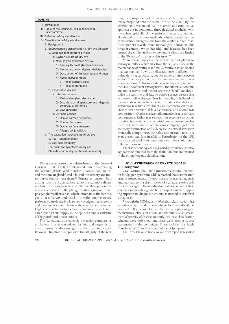
Dry eye is recognized as a disturbance of the LacrimalFunctional Unit (LFU), an integrated system comprisingthe lacrimal glands, ocular surface (cornea, conjunctivaand meibomian glands) and lids, and the sensory and mo-tor nerves that connect them.17 Trigeminal sensory fibersarising from the ocular surface run to the superior salivary
nucleus in the pons, from whence efferent fibers pass, in thenervus intermedius, to the pterygopalatine ganglion. Here,postganglionic fibers arise, which terminate in the lacrimalgland, nasopharynx, and vessels of the orbit. Another neuralpathway controls the blink reflex, via trigeminal afferentsand the somatic efferent fibers of the seventh cranial nerve.Higher centers feed into the brainstem nuclei, and there isa rich sympathetic supply to the epithelia and vasculatureof the glands and ocular surface.
This functional unit controls the major componentsof the tear film in a regulated fashion and responds toenvironmental, endocrinological, and cortical influences.
Its overall function is to preserve the integrity of the tear
film, the transparency of the cornea, and the quality of theimage projected onto the retina.17-20 At the 2007 Dry Eye WorkShop, it was noted that the corneal and conjunctivalepithelia are in continuity, through ductal epithelia, withthe acinar epithelia of the main and accessory lacrimalglands and the meibomian glands, which themselves ariseas specialized invaginations from the ocular surface. Also,
these epithelia have the same embryological derivation. Thisbroader concept, which has additional features, has beentermed the Ocular Surface System and is discussed furtherin the “Research” chapter of this issue.21
An important aspect of the unit is the part played bysensory impulses, which arise from the ocular surface, in themaintenance of resting tear flow. Currently, it is consideredthat waking tear flow is a reflex response to afferent im-pulses deriving particularly, but not entirely, from the ocularsurface.22 Sensory input from the nasal mucosa also makesa contribution.23 Disease or damage to any component ofthe LFU (the afferent sensory nerves, the efferent autonomicand motor nerves, and the tear-secreting glands) can desta-bilize the tear film and lead to ocular surface disease thatexpresses itself as dry eye. Tear film stability, a hallmark ofthe normal eye, is threatened when the interactions betweenstabilizing tear film constituents are compromised by de-creased tear secretion, delayed clearance, and altered tearcomposition. Ocular surface inflammation is a secondaryconsequence. Reflex tear secretion in response to ocularirritation is envisioned as the initial compensatory mecha-nism, but, with time, inflammation accompanying chronicsecretory dysfunction and a decrease in corneal sensationeventually compromises the reflex response and results ineven greater tear film instability. Perturbation of the LFU
is considered to play an important role in the evolution ofdifferent forms of dry eye.
The distinctions aqueous-deficient dry eye and evaporativedry eye were removed from the definition, but are retainedin the etiopathogenic classification.
IV. CLASSIFICATION OF DRY EYE DISEASE
A. Background
Vitali, writing about the harmonized classification crite-ria for Sjogren syndrome (SS) remarked that classificationcriteria are not necessarily appropriate for use in diagnosisand may lead to misclassification of a disease, particularly
in its early stages.24 In an individual patient, a classificationscheme can provide a guide, but an expert clinician, apply-ing appropriate diagnostic criteria, is needed to establisha diagnosis.
Although the NEI/Industry Workshop classification1 hasserved as a useful and durable scheme for over a decade, itdoes not reflect newer knowledge on pathophysiologicalmechanisms, effects on vision, and the utility of an assess-ment of severity of disease. Recently, two new classificationschemes were published, and these were used as sourcedocuments by the committee. These include: the TripleClassification25,26 and the report of the Delphi panel.27
The Triple Classification evolved from reports presented
OUTLINE
I. Introduction
II. Goals of the Definition and ClassificationSubcommittee
III. Definition of dry eye disease
IV. Classification of dry eye disease
A. Background
B. Etiopathogenic classification of dry eye disease
1. Aqueous tear-deficient dry eye
a. Sjogren syndrome dry eye
b. Non-Sjogren syndrome dry eye
1) Primary lacrimal gland deficiencies
2) Secondary lacrimal gland deficiencies
3) Obstruction of the lacrimal gland ducts
4) Reflex hyposecretion
a) Reflex sensory block
b) Reflex motor block
2. Evaporative dry eye
a. Intrinsic causes
1) Meibomian gland dysfunction
2) Disorders of lid aperature and lid/globecongruity or dynamics
3) Low blink rate
b. Extrinsic causes
1) Ocular surface disorders
2) Contact lens wear
3) Ocular surface disease
4) Allergic conjunctivitis
C. The causative mechanisms of dry eye
1. Tear hyperosmolarity 2. Tear film instability
D. The basis for symptoms in dry eye
E. Classification of dry eye based on severity
DEWS DEFINITION AND CLASSIFICATION
8/20/2019 dry eyes ii
http://slidepdf.com/reader/full/dry-eyes-ii 17/142
THE OCULAR SURFACE / APRIL 2007, VOL. 5, NO. 2 / www.theocularsurface.com 77
DRY EYE
Effect of theEnvironment
Milieu Interieur
Low blink rate behavior, VTU,
microscopy
Wide lid aperture
gazeposition
Aging
Low androgenpool
Systemic Drugs:
antihistamines,
beta-blockers,
antispasmodics,
diuretics, and
some psychotropic
drugs
Milieu Exterieur
Low relative humidityHigh wind velocity
Occupational
environment
Aqueous-deficient
SjogrenSyndrome
Dry Eye
Primary
Secondary
Non-SjogrenDry Eye
LacrimalDeficiency
Reflex Block
LacrimalGland Duct
Obstruction
SystemicDrugs
Evaporative
Extrinsic
Meibomian OilDeficiency
Intrinsic
Vitamin A-Deficiency
Topical DrugsPreservatives
Low Blink Rate
Drug ActionAccutane
Contact LensWear
Disordersof Lid
Aperture
Ocular SurfaceDisease
eg, Allergy
at the 14th Congress of the European Society of Ophthal-mology.25 After further clinical experience, an updated ver-sion was published in 2005, which presented three separateschemes: one based on etiopathogenesis; one based on theglands and tissues targeted in dry eye; and one based on
disease severity.26
The committee felt that the concept of three differentschemes serving different purposes was attractive, but itwas noted that evidence-based referencing was limited.For this reason, the scheme as a whole was not adopted,but many conceptual aspects were incorporated into thecommittee’s final schemes.
The Delphi Panel was a consensus group that met toreview the classification of dry eye.27 The panel proposedchanging the name of dry eye disease to dysfunctional tear syn-drome, suggesting that the name more accurately reflectedpathophysiological events in dry eye. However, although
the committee felt that the term embraced the essential
features of the disease, they concluded that retention of thename dry eye had much to recommend it and that its usewas embedded in the literature. The committee also rejecteda subdivision based on the presence or absence of lid dis-ease, because it is frequently difficult to identify the relative
contribution of lid disease to a particular case of dry eye.The majority of the Definition and Classification Sub-
committee was in favor of adopting a severity grading basedon the report of the Delphi Panel, recognizing it as a com-prehensive approach that could form the basis of therapyaccording to severity of the disease. As noted above, theTriple Classification also presented a severity grading.
B. Etiopathogenic Classification of Dry Eye Disease
The etiopathogenic classification developed by theSubcommittee is an updated version of that presented inthe NEI/Industry Workshop Report and reflects a more
contemporary understanding of dry eye disease (Figure 1).
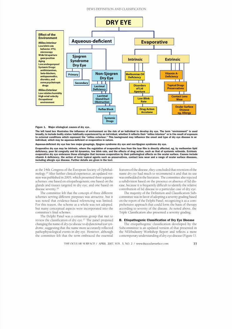
Figure 1. Major etiological causes of dry eye.
The left hand box illustrates the influence of environment on the risk of an individual to develop dry eye. The term “environment” is usedbroadly, to include bodily states habitually experienced by an individual, whether it reflects their “milieu interieur” or is the result of exposure
to external conditions which represent the “milieu exterieur.” This background may influence the onset and type of dry eye disease in anindividual, which may be aqueous-deficient or evaporative in nature.
Aqueous-deficient dry eye has two major groupings, Sjogren syndrome dry eye and non-Sjogren syndrome dry eye.
Evaporative dry eye may be intrinsic, where the regulation of evaporative loss from the tear film is directly affected, eg, by meibomian lipid
deficiency, poor lid congruity and lid dynamics, low blink rate, and the effects of drug action, such as that of systemic retinoids. Extrinsicevaporative dry eye embraces those etiologies that increase evaporation by their pathological effects on the ocular surface. Causes include
vitamin A deficiency, the action of toxic topical agents such as preservatives, contact lens wear and a range of ocular surface diseases,including allergic eye disease. Further details are given in the text.
DEWS DEFINITION AND CLASSIFICATION

8/20/2019 dry eyes ii
http://slidepdf.com/reader/full/dry-eyes-ii 18/142
THE OCULAR SURFACE / APRIL 2007, VOL. 5, NO. 2 / www.theocularsurface.com78
As in the 1995 report, the term dry eye is regarded as syn-onymous with the term keratoconjunctivitis sicca (KCS).
The classification has the following features:The left hand box in Figure 1 illustrates the influence of
environment on an individual’s risk of developing dry eye.The term environment is used broadly to include physiologi-cal variation between individuals (their milieu interieur ), as
well as the ambient conditions that they encounter (theirmilieu exterieur ).The milieu interieur implies physiological conditions
particular to an individual that could influence their riskof dry eye. For instance, a normal subject may have a lownatural blink rate, or the blink rate may be slowed for be-havioral or psychological reasons.28 Slowing of the blinkrate increases the blink interval and increases the periodof evaporative loss between each blink.29
Similarly, the natural height of the palpebral aperture inthe primary position varies between individuals and betweenethnic groups.30 The aperture is also wider in upgaze thandowngaze.31 Evaporative loss per eye increases with increas-ing palpebral width and is, therefore, increased in upgaze.32
Extensive evidence supports a role for the sex hormonesin the etiology of dry eye33 with the generalization that lowlevels of androgens and high estrogen levels are risk factorsfor dry eye. Biologically active, androgens promote lacrimaland meibomian gland function.33 Androgen deficiency isassociated with dry eye34 and may be prevented by topicalor systemic androgen therapy.35-38 Dry eye occurs in patientsexposed to anti-androgens in the treatment of prostaticcancer,39,40 and women with complete androgen insensitiv-ity syndrome show an increase in the signs and symptomsof dry eye, associated with evidence of meibomian gland
and goblet cell dysfunction.41-43 A significantly depletedandrogen pool in “non-autoimmune” dry eye associatedwith meibomian gland dysfunction (MGD) has been re-ported.44 Also, as noted elsewhere in this issue,45 femalesex and postmenopausal estrogen therapy are importantrisk factors for dry eye,46,47 and women with prematureovarian failure suffer from the symptoms and signs of dryeye, although their tear production is not affected.48
Lacrimal tear secretion is reduced by a number ofsystemic drugs, and these effects may be looked upon asdisturbances of the milieu interieur . Their details are dis-cussed later in this report. Aging is associated with physi-
ological changes that may predispose to dry eye, includingdecreased tear volume and flow, increased osmolarity,49 decreased tear film stability,50 and alterations in the com-position of the meibomian lipids.51
The milieu exterieur involves the occupational andexternal environments, which may represent risk factorsfor the development of dry eye. Evaporative water lossfrom the eye is increased in conditions of low relativehumidity, occurring either as part of natural variation atdifferent geographic locations or in special circumstancescreated by air-conditioning, air travel, or other artificialenvironments.52 Similarly, tear evaporation is increased by
exposure to high wind velocity, and this mechanism has
been incorporated into some of the newer experimentalmodels of dry eye.
Occupational factors may cause a slow blink rate, repre-senting a risk for dry eye in those working with video dis-play terminals.53 Other activities associated with decreasedblinking and an increase in palpebral width, including thatassociated with upgaze, have been reported to carry a risk
for the development of dry eye symptoms.The major classes of dry eye, as in the 1995 workshop,1 are still held to be aqueous tear-deficient dry eye ( ADDE)and evaporative dry eye (EDE). The category ADDE referschiefly to a failure of lacrimal secretion, and this approach isretained. However, it should be recognized that a failure ofwater secretion by the conjunctiva could also contribute toaqueous tear deficiency. The class EDE has been subdividedto distinguish those causes that are dependent on intrinsicconditions of the lids and ocular surface and those that arisefrom extrinsic influences.
Dry eye can be initiated in any of these classes, but theyare not mutually exclusive. It is recognized that disease initi-ated in one major subgroup may coexist with or even leadto events that cause dry eye by another major mechanism.This is part of a vicious circle of interactions that can amplifythe severity of dry eye. An example might be that all formsof dry eye cause goblet cell loss and that this, in turn, willcontribute to loss of tear film stability, to surface damageand evaporative water loss, and to symptoms resulting froma loss of lubrication and surface inflammatory events.
The major classes and subclasses of dry eye are de-scribed below.
1. Aqueous Tear-Deficient Dry Eye (Tear Deficient
Dry Eye; Lacrimal Tear Deficiency) Aqueous tear-deficient dry eye implies that dry eye is
due to a failure of lacrimal tear secretion. In any form ofdry eye due to lacrimal acinar destruction or dysfunction,dryness results from reduced lacrimal tear secretion andvolume.54,55 This causes tear hyperosmolarity, because,although the water evaporates from the ocular surface atnormal rates, it is from a reduced aqueous tear pool. Tearfilm hyperosmolarity causes hyperosmolarity of the ocularsurface epithelial cells and stimulates a cascade of inflam-matory events involving MAP kinases and NFkB signallingpathways56,57 and the generation of inflammatory cytokines
(interleukin (IL)-1α; -1β; tumor necrosis factor (TNF)-α)and matrix metalloproteinases (MMP-9).58 When lacrimaldysfunction is due to lacrimal gland infiltration and inflam-mation, inflammatory mediators generated in the gland areassumed to find their way into the tears and be deliveredto the ocular surface. However, when such mediators aredetected in the tears, it is not usually possible to knowwhether they derive from the lacrimal gland itself or fromthe ocular surface (conjunctiva and cornea).
It is uncertain whether evaporation is reduced59 or in-creased59-64 in ADDE. It is possible that this is determinedby the stage of the disease. Some studies suggest that the
reservoir of lid oil is larger in non-Sjogren syndrome dry
DEWS DEFINITION AND CLASSIFICATION
8/20/2019 dry eyes ii
http://slidepdf.com/reader/full/dry-eyes-ii 19/142
THE OCULAR SURFACE / APRIL 2007, VOL. 5, NO. 2 / www.theocularsurface.com 79
eye (NSSDE)65 and that the tear film lipid layer is thicker,66 but dynamic studies of the tear film lipid layer in ADDEhave shown that spreading of the lipid layer is delayed inthe interblink.67,68 Additionally, in severe ADDE, spread-ing may be undetectable by interferometry, suggesting amajor defect in the tear film lipid layer. Delayed or absentspreading of the tear film could lead to an increase in waterloss from the eye.
ADDE has two major subclasses, SS dry eye (SSDE)and non-SS dry eye.
a. Sjogren Syndrome Dry Eye Sjogren syndrome is an exocrinopathy in which the
lacrimal and salivary glands are targeted by an autoimmuneprocess; other organs are also affected. The lacrimal andsalivary glands are infiltrated by activated T-cells, whichcause acinar and ductular cell death and hyposecretionof the tears or saliva. Inflammatory activation within theglands leads to the expression of autoantigens at the surfaceof epithelial cells (eg, fodrin, Ro and La)69 and the retentionof tissue-specific CD4 and CD8 T-cells.70 Hyposecretion isamplified by a potentially reversible neurosecretory block,due to the effects of locally released inflammatory cytokines
or to the presence of circulating antibodies (eg, anti-M3
antibody) directed againstmuscarinic receptors with-in the glands.71-73
There are two formsof SS, and classificationcriteria have recently beenharmonized in a European-
American collaboration.74
Primary SS consists of theoccurrence of ADDE incombination with symp-toms of dry mouth, in thepresence of autoantibod-ies, evidence of reducedsalivary secretion and witha positive focus score onminor salivary gland bi-opsy.75,76 Details of the cri-teria are presented in Table1. Secondary SS consists ofthe features of primary SStogether with the featuresof an overt autoimmuneconnective disease, such asrheumatoid arthritis, whichis the most common, orsystemic lupus erythema-tosis, polyarteritis nodosa, Wegener’s granulomatosis,systemic sclerosis, primarybiliary sclerosis, or mixedconnective tissue disease.
Diagnostic criteria for eachof these connective tissue disorders have been published.77
The precise triggers leading to autoimmune acinardamage are not known in full, but risk factors includegenetic profile,78 androgen status79 (a low androgen poolfavoring an inflammatory environment within the targettissues), and exposure to environmental agents, rangingfrom viral infections affecting the lacrimal gland to pollutedenvironments. A nutritional deficiency in omega-3- andother unsaturated fatty acids and unsupplemented intakeof vitamin C has also been reported in patients with SS.80 It is generally accepted that environmental factors leading
to increased evaporative water loss from the eye (eg, lowhumidity, high wind velocity, and increased exposure of theocular surface) may act as a trigger by invoking inflamma-tory events at the ocular surface through a hyperosmolarmechanism (see Section V).
The ocular dryness in SSDE is due to lacrimal hypose-cretion and the accompanying characteristic inflammatorychanges in the lacrimal gland, together with the presenceof inflammatory mediators in the tears and within theconjunctiva.81 It is not known whether the conjunctivalchanges are due to an autoimmune targeting of this tissueor whether they are due to the effect of inflammatory media-
tors released from the lacrimal glands into the tears.
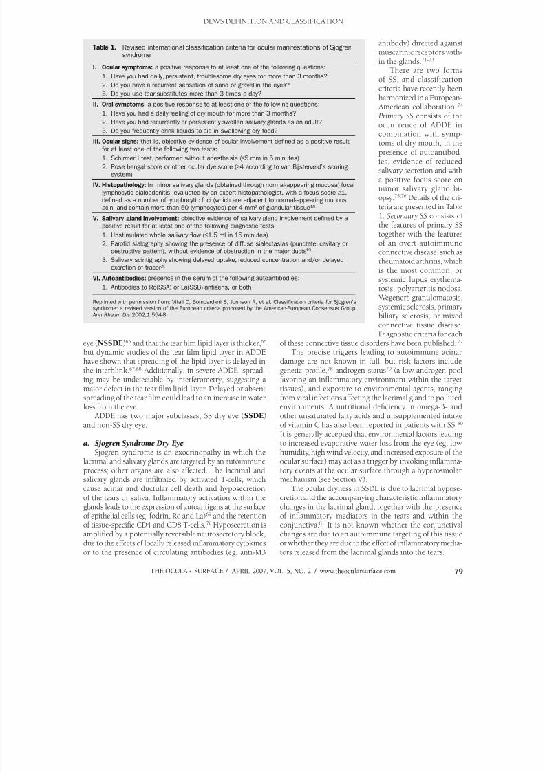
Table 1. Revised international classification criteria for ocular manifestations of Sjogrensyndrome
I. Ocular symptoms: a positive response to at least one of the following questions:
1. Have you had daily, persistent, troublesome dry eyes for more than 3 months?
2. Do you have a recurrent sensation of sand or gravel in the eyes?
3. Do you use tear substitutes more than 3 times a day?
II. Oral symptoms: a positive response to at least one of the following questions:
1. Have you had a daily feeling of dry mouth for more than 3 months?
2. Have you had recurrently or persistently swollen salivary glands as an adult?
3. Do you frequently drink liquids to aid in swallowing dry food?
III. Ocular signs: that is, objective evidence of ocular involvement defined as a positive result
for at least one of the following two tests:
1. Schirmer I test, performed without anesthesia (≤5 mm in 5 minutes)
2. Rose bengal score or other ocular dye score (≥4 according to van Bijsterveld’s scoring
system)
IV. Histopathology: In minor salivary glands (obtained through normal-appearing mucosa) focal
lymphocytic sialoadenitis, evaluated by an expert histopathologist, with a focus score ≥1,
defined as a number of lymphocytic foci (which are adjacent to normal-appearing mucous
acini and contain more than 50 lymphocytes) per 4 mm2 of glandular tissue18
V. Salivary gland involvement: objective evidence of salivary gland involvement defined by apositive result for at least one of the following diagnostic tests:
1. Unstimulated whole salivary flow (≤1.5 ml in 15 minutes)
2. Parotid sialography showing the presence of diffuse sialectasias (punctate, cavitary or
destructive pattern), without evidence of obstruction in the major ducts19
3. Salivary scintigraphy showing delayed uptake, reduced concentration and/or delayed
excretion of tracer20
VI. Autoantibodies: presence in the serum of the following autoantibodies:
1. Antibodies to Ro(SSA) or La(SSB) antigens, or both
Reprinted with permission from: Vitali C, Bombardieri S, Jonnson R, et al. Classification criteria for Sjogren’ssyndrome: a revised version of the European criteria proposed by the American-European Consensus Group.
Ann Rheum Dis 2002;1:554-8.
DEWS DEFINITION AND CLASSIFICATION

8/20/2019 dry eyes ii
http://slidepdf.com/reader/full/dry-eyes-ii 20/142
THE OCULAR SURFACE / APRIL 2007, VOL. 5, NO. 2 / www.theocularsurface.com80
The frequency of MGD is higher in patients with SSthan in the normal population; thus, a defective tear filmlipid layer may contribute to dry eye by leading to excessevaporation.82
b. Non-Sjogren Syndrome Dry EyeNon-Sjogren syndrome dry eye is a form of ADDE due
to lacrimal dysfunction, where the systemic autoimmunefeatures characteristic of SSDE have been excluded. Themost common form is age-related dry eye, to which theterm KCS has sometimes been applied in the past. However,as noted earlier, the term KCS is now used to describe anyform of dry eye. In the 1995 Dry Eye Workshop report, itwas referred to as primary lacrimal disease,1 but this term hasnot been generally adopted. The different forms of NSSDEare briefly discussed below (Table 2).
1) Primary Lacrimal Gland Deficiencies Age-Related Dry Eye (ARDE): There is some uncertainty
as to whether tear dynamics are affected by age in thenormal population.83 Mathers et al showed significant age-related correlations for tear evaporation, volume, flow, andosmolarity,49 but no such relationship was noted by Craigand Tomlinson84 or in other reports of tear turnover,85 tear evaporation86,87 and lipid layer.88 ARDE is a primarydisease.
With increasing age in the normal human population,there is an increase in ductal pathology that could promotelacrimal gland dysfunction by its obstructive effect.89,89a
These alterations include periductal fibrosis, interacinarfibrosis, paraductal blood vessel loss and acinar cellatrophy.89,89a Damato et al found lymphocytic glandular
infiltrates in 70% of lacrimal glands studied and consid-ered this to be the basis of the fibrosis. Appearances werelikened to the less severe grades of Sjogren syndrome. Theypostulated a sequence of periductal fibrosis, interacinarfibrosis and, finally, acinar atrophy. It has been suggestedthat the low-grade dacryoadenitis could be caused bysystemic infection or conjunctivitis89 or, alternatively, thatsubclinical conjunctivitis might be responsible for stenosisof the excretory ducts.89a
Congenital Alacrima: Congenital alacrima is a rare causeof dry eye in youth.90 It is also part of certain syndromes,91 including the autosomal recessive, triple A syndrome (All-
grove syndrome), in which congenital alacrima is associatedwith achalasia of the cardia, Addison’s disease, central neu-rodegeneration, and autonomic dysfunction. It is caused bymutations in the gene encoding the protein ALADIN, whichplays a role in RNA and/or protein trafficking between thenucleus and cytoplasm.92,93
Familial Dysautonomia: Lacrimal dysfunction is a majorfeature of the autosomal recessive disorder, familial dys-autonomia (Riley Day syndrome), in which a generalizedinsensitivity to pain is accompanied by a marked lack ofboth emotional and reflex tearing, within a multisystemdisorder. There is a developmental and progressive neuronal
abnormality of the cervical sympathetic and parasympa-
thetic innervations of the lacrimal gland and a defectivesensory innervation of the ocular surface, which affects bothsmall myelinated (Aδ) and unmyelinated (C) trigeminalneurons.94,95 The chief mutation affects the gene encodingan Iκ B kinase-associated protein.
2) Secondary Lacrimal Gland DeficienciesLacrimal gland infiltration: Lacrimal secretion may fail
because of inflammatory infiltration of the gland, as in:Sarcoidosis: Infiltration of the lacrimal gland by sarcoid
granulomata may cause dry eye.96 Lymphoma: Infiltration of the lacrimal gland by lym-
phomatous cells causes dry eye.97 AIDS: Dry eye may be caused by lacrimal gland infiltra-
tion by T-cells. However, in AIDS-related dry eye, unlikethe situation in SSDE, there is a predominance of CD8suppressor cells, rather than CD4, helper cells.98
Graft vs host disease (GVHD): Dry eye is a commoncomplication of GVHD disease, occurring typically around6 months after hematopoietic stem cell transplantation. It
is caused in part by lacrimal gland fibrosis due to colocali-
Table 2. Conditions associated with non-Sjogrensyndrome dry eye
Primary lacrimal gland deficiencies
Age-related dry eye
Congenital alacrima
Familial dysautonomia
Secondary lacrimal gland deficiencies
Lacrimal gland infiltration
Sarcoidosis
Lymphoma
AIDS
Graft vs host disease
Lacrimal gland ablation
Lacrimal gland denervation
Obstruction of the lacrimal gland ducts
Trachoma
Cicatricial pemphigoid and mucous membrane pemphigoid
Erythema multiforme
Chemical and thermal burns
Reflex hyposecretion
Reflex sensory block
Contact lens wear
Diabetes
Neurotrophic keratitis
Reflex motor block
VII cranial nerve damage
Multiple neuromatosis
Exposure to systemic drugs
DEWS DEFINITION AND CLASSIFICATION
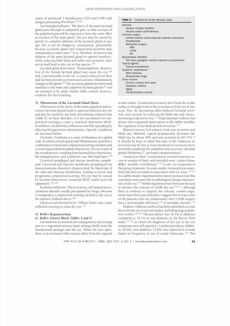
8/20/2019 dry eyes ii
http://slidepdf.com/reader/full/dry-eyes-ii 21/142
THE OCULAR SURFACE / APRIL 2007, VOL. 5, NO. 2 / www.theocularsurface.com 81
zation of periductal T-lymphocytes (CD4 and CD8) withantigen-presenting fibroblasts.99,100
Lacrimal gland ablation: The ducts of the main lacrimalgland pass through its palpebral part, so that excision ofthe palpebral part will be expected to have the same effectas excision of the main gland. Dry eye may be caused bypartial or complete ablation of the lacrimal gland at any
age, but is not an obligatory consequence, presumablybecause accessory gland and conjunctival secretion maycompensate in some cases.55 It is, therefore, of interest thatablation of the main lacrimal gland in squirrel monkeys,while reducing both basal and reflex tear secretion, doesnot in itself lead to dry eye in that species.101
Lacrimal gland denervation: Parasympathetic denerva-tion of the human lacrimal gland may cause dry eye,102 and, experimentally in the rat, it causes reduced tear flowand lacrimal protein secretion and activates inflammatorychanges in the gland.103 The accessory glands are innervatedsimilarly to the main and palpebral lacrimal glands104 andare assumed to be under similar reflex control; however,evidence for this is lacking.
3) Obstruction of the Lacrimal Gland DuctsObstruction of the ducts of the main palpebral and ac-
cessory lacrimal glands leads to aqueous-deficient dry eyeand may be caused by any form of cicatrising conjunctivitis(Table 2). In these disorders, it is not uncommon for con- junctival scarring to cause a cicatricial obstructive MGD.In addition, lid deformity influences tear film spreading byaffecting lid apposition and dynamics. Specific conditionsare discussed below.
Trachoma: Trachoma is a cause of blindness on a global
scale, in which corneal opacity and blindness are caused by acombination of tarsal and conjunctival scarring, trichiasis anda cicatrizing meibomian gland obstruction. Dry eye is part ofthe overall picture, resulting from lacrimal duct obstruction,lid malapposition, and a deficient tear film lipid layer.105
Cicatricial pemphigoid and mucous membrane pemphi- goid: Cicatricial and mucous membrane pemphigoid aremucocutaneous disorders characterized by blistering ofthe skin and mucous membranes, leading to severe andprogressive conjunctival scarring. Dry eye may be causedby lacrimal obstruction, cicatricial MGD, and/or poor lidapposition.106-108
Erythema multiforme: This is an acute, self-limited muco-cutaneous disorder usually precipitated by drugs, infectionor malignancy. Conjunctival scarring can lead to dry eye inthe manner outlined above.109
Chemical and thermal burns: Diffuse burns may causesufficient scarring to cause dry eye.110
4) Reflex Hyposecretiona) Reflex Sensory Block (Tables 2 and 3)
Lacrimal tear secretion in the waking state is due in largepart to a trigeminal sensory input arising chiefly from thenasolacrimal passages and the eye. When the eyes open,
there is an increased reflex sensory drive from the exposed
ocular surface. A reduction in sensory drive from the ocularsurface is thought to favor the occurrence of dry eye in twoways, first, by decreasing reflex-induced lacrimal secre-tion, and, second, by reducing the blink rate and, hence,increasing evaporative loss.111 Experimental evidence hasshown that trigeminal denervation in the rabbit modifiesthe regulation of lacrimal protein secretion.112
Bilateral sensory loss reduces both tear secretion andblink rate. Bilateral, topical proparacaine decreases theblink rate by about 30% and tear secretion by 60-75%.22
It should be kept in mind that part of the reduction insecretion may be due to local anesthesia of secretory nerveterminals supplying the palpebral and accessory lacrimalglands (Belmonte C: personal communication).
Contact Lens Wear: A reduction in corneal sensitivity oc-curs in wearers of hard- and extended wear- contact lenses(CLs), possibly contributing11,113 to dry eye symptoms inthis group of patients. In some studies, increased tear osmo-larity has been recorded in association with CL wear.113,114 In a rabbit model, trigeminal denervation increases tear filmosmolarity and causes the morphological changes character-istic of dry eye.115 Similar arguments have been put forward
to advance the concept of LASIK dry eye116,117; althoughthere is evidence to support the concept, counter argu-ments have been put forward to suggest that at least someof the patients who are symptomatic after LASIK surgeryhave a neurotrophic deficiency118 or neuralgic disorder.119
Diabetes: Diabetes mellitus has been identified as a riskfactor for dry eye in several studies, including large popula-tion studies.120-123 The prevalence was 18.1% in diabeticscompared to 14.1% in non-diabetics in the Beaver Damstudy,121,122 in which the diagnosis of dry eye or dry eyesymptoms were self-reported. A similar prevalence (diabet-ics 20.6%, non-diabetics 13.8%) was reported in a study
based on frequency of use of ocular lubricants.123 This
Table 3. Causes of ocular sensory loss
Infective
Herpes simplex keratitis
Herpes zoster ophthalmicus
Corneal surgery
Limbal incision (extra-capsular cataract extraction)
Keratoplasty
Refractive surgery
PRK LASIK
RK
Neurotrophic Keratitis
Vth nerve/ganglion section/injection/compression
Topical agents
Topical anaesthesia
Systemic medications
Beta blockers
Atropine-like drugs
Other causes
Chronic contact lens wear
Diabetes mellitus
Aging
Trichlorethylene toxicity
DEWS DEFINITION AND CLASSIFICATION
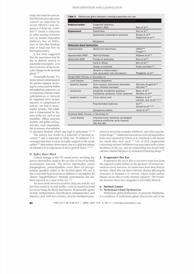
8/20/2019 dry eyes ii
http://slidepdf.com/reader/full/dry-eyes-ii 22/142
THE OCULAR SURFACE / APRIL 2007, VOL. 5, NO. 2 / www.theocularsurface.com82 82
study also noted an associa-tion between poor glycemiccontrol (as indicated byserum HbA1C) and fre-quency of drop use. Goeb-bels124 found a reductionin reflex tearing (Schirmer
test) in insulin-dependentdiabetics, but no differ-ence in tear film breakuptime or basal tear flow byfluorophotometry.
It has been suggestedthat the association may bedue to diabetic sensory orautonomic neuropathy, or tothe occurrence of microvas-cular changes in the lacrimalgland.123
Neurotrophic keratitis: Ex-tensive sensory denervation ofthe anterior segment, involv-ing the cornea and the bulbarand palpebral conjunctiva, asa component of herpes zosterophthalmicus or inducedby trigeminal nerve section,injection, or compression ortoxicity, can lead to neuro-trophic keratitis. This condi-tion is characterized by fea-tures of dry eye, such as tear
instability, diffuse punctatekeratitis, and goblet cell loss,and also, most importantly,the occurrence of an indolentor ulcerative keratitis, which may lead to perforation.115,125
The sensory loss results in a reduction of lacrimal se-cretion126 and a reduction in blink rate. In addition, it isenvisaged that there is a loss of trophic support to the ocularsurface125 after sensory denervation, due to a deficient releaseof substance-P or expression of nerve growth factor.127-131
b) Reflex Motor Block
Central damage to the VII cranial nerve, involving thenervus intermedius, leads to dry eye due to loss of lacrimalsecretomotor function. The nervus intermedius carriespostganglionic, parasympathetic nerve fibers (of pterygo-palatine ganglion origin) to the lacrimal gland. Dry eye isdue to lacrimal hyposecretion in addition to incomplete lidclosure (lagophthalmos). Multiple neuromatosis has alsobeen reported as a cause of dry eye.132
An association between systemic drug use and dry eyehas been noted in several studies, with decreased lacrimalsecretion being the likely mechanism. Responsible agentsinclude: antihistamines, beta blockers, antispasmodics, and
diuretics, and, with less certainty, tricyclic antidepressants,
selective serotonin reuptake inhibitors, and other psycho-tropic drugs.122 Additional associations with drying medica-tions were reported by Schein et al, unrelated to the diseasefor which they were used.133 Use of ACE (angiotensinconverting enzyme) inhibitors was associated with a lowerincidence of dry eye, and no relationship was found withcalcium channel blockers or cholesterol-lowering drugs.122
2. Evaporative Dry EyeEvaporative dry eye is due to excessive water loss from
the exposed ocular surface in the presence of normal lac-rimal secretory function. Its causes have been described asintrinsic, where they are due to intrinsic disease affecting lidstructures or dynamics, or extrinsic, where ocular surfacedisease occurs due to some extrinsic exposure. The bound-ary between these two categories is inevitably blurred.
a. Intrinsic Causes1) Meibomian Gland Dysfunction
Meibomian gland dysfunction, or posterior blepharitis,
is a condition of meibomian gland obstruction and is the
Table 4. Meibomian gland diseases causing evaporative dry eye
Category Disease References
Reduced number Congenital deficiency
Acquired—MGD Bron et al137
Replacement Dystichiasis Bron et al137
Dystichiasis lymphedema syndrome Brooks et al138
Kiederman et al139
Metaplasia
Meibomian Gland Dysfunction
Hypersecretory Meibomian seborrhoea Gifford140
Cowper141
Hyposecretory MGD Retinoid therapy Mathers et al142
Obstructive MGD Primary or secondary Bron et al143
Focal or diffuse Bron et al143
Simple or cicatricial Foulks and Bron134
Atrophic or inflammatory—
note association with dermatoses Pflugfelder et al144
Simple MGD: Primary, or Secondary to:
Local disease Anterior blepharitis
Systemic disease Acne rosacea; seborrhoeic dermatitis; McCulley Dougherty 145
atopy; icthyosis; psoriasis; McCulley 146
Syndromes Anhydrotic ectodermal dysplasia; Baum et al147
ectrodactyly syndrome; Turner syndrome Mondino et al148
Systemic toxicity 13-cis retinoic acid Mathers et al142
Lambert and Smith149,150
Polychlorinated biphenyls Ikui151
Ohnishi et al152,153
Epinephrine (rabbit) Jester et al154
Cicatricial MGD: Primary, or Secondary to:
Local disease Chemical burns; trachoma; pemphigoid; erythema multiforme; acne rosacea;
VKC and AKC
DEWS DEFINITION AND CLASSIFICATION
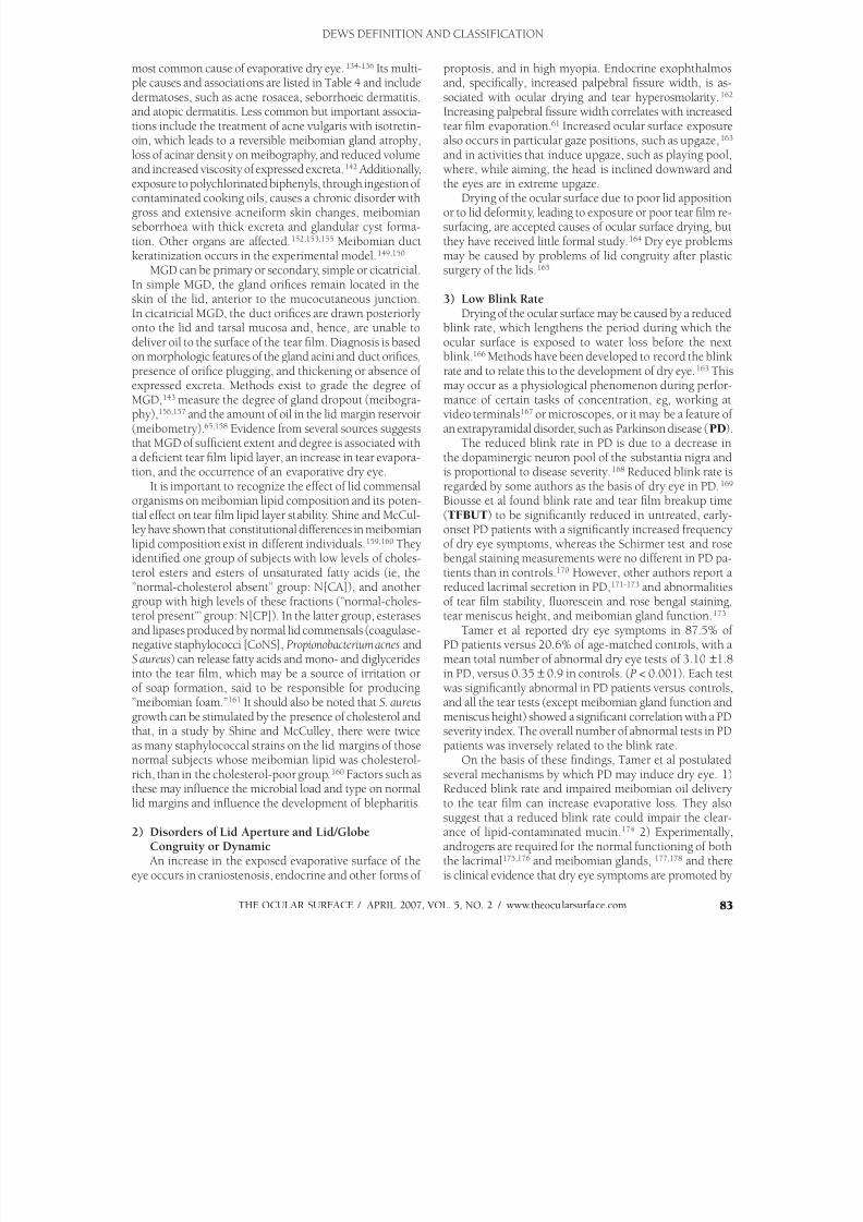
8/20/2019 dry eyes ii
http://slidepdf.com/reader/full/dry-eyes-ii 23/142
THE OCULAR SURFACE / APRIL 2007, VOL. 5, NO. 2 / www.theocularsurface.com 83 83
most common cause of evaporative dry eye.134-136 Its multi-ple causes and associations are listed in Table 4 and includedermatoses, such as acne rosacea, seborrhoeic dermatitis,and atopic dermatitis. Less common but important associa-tions include the treatment of acne vulgaris with isotretin-oin, which leads to a reversible meibomian gland atrophy,loss of acinar density on meibography, and reduced volume
and increased viscosity of expressed excreta.142
Additionally,exposure to polychlorinated biphenyls, through ingestion ofcontaminated cooking oils, causes a chronic disorder withgross and extensive acneiform skin changes, meibomianseborrhoea with thick excreta and glandular cyst forma-tion. Other organs are affected.152,153,155 Meibomian ductkeratinization occurs in the experimental model.149,150
MGD can be primary or secondary, simple or cicatricial.In simple MGD, the gland orifices remain located in theskin of the lid, anterior to the mucocutaneous junction.In cicatricial MGD, the duct orifices are drawn posteriorlyonto the lid and tarsal mucosa and, hence, are unable todeliver oil to the surface of the tear film. Diagnosis is basedon morphologic features of the gland acini and duct orifices,presence of orifice plugging, and thickening or absence ofexpressed excreta. Methods exist to grade the degree ofMGD,143 measure the degree of gland dropout (meibogra-phy),156,157 and the amount of oil in the lid margin reservoir(meibometry).65,158 Evidence from several sources suggeststhat MGD of sufficient extent and degree is associated witha deficient tear film lipid layer, an increase in tear evapora-tion, and the occurrence of an evaporative dry eye.
It is important to recognize the effect of lid commensalorganisms on meibomian lipid composition and its poten-tial effect on tear film lipid layer stability. Shine and McCul-
ley have shown that constitutional differences in meibomianlipid composition exist in different individuals.159,160 Theyidentified one group of subjects with low levels of choles-terol esters and esters of unsaturated fatty acids (ie, the”normal-cholesterol absent” group: N[CA]), and anothergroup with high levels of these fractions (”normal-choles-terol present”’ group: N[CP]). In the latter group, esterasesand lipases produced by normal lid commensals (coagulase-negative staphylococci [CoNS], Propionobacterium acnes andS aureus) can release fatty acids and mono- and diglyceridesinto the tear film, which may be a source of irritation orof soap formation, said to be responsible for producing
”meibomian foam.”161 It should also be noted that S. aureus growth can be stimulated by the presence of cholesterol andthat, in a study by Shine and McCulley, there were twiceas many staphylococcal strains on the lid margins of thosenormal subjects whose meibomian lipid was cholesterol-rich, than in the cholesterol-poor group.160 Factors such asthese may influence the microbial load and type on normallid margins and influence the development of blepharitis.
2) Disorders of Lid Aperture and Lid/GlobeCongruity or Dynamic An increase in the exposed evaporative surface of the
eye occurs in craniostenosis, endocrine and other forms of
proptosis, and in high myopia. Endocrine exophthalmosand, specifically, increased palpebral fissure width, is as-sociated with ocular drying and tear hyperosmolarity.162 Increasing palpebral fissure width correlates with increasedtear film evaporation.61 Increased ocular surface exposurealso occurs in particular gaze positions, such as upgaze,163 and in activities that induce upgaze, such as playing pool,
where, while aiming, the head is inclined downward andthe eyes are in extreme upgaze.Drying of the ocular surface due to poor lid apposition
or to lid deformity, leading to exposure or poor tear film re-surfacing, are accepted causes of ocular surface drying, butthey have received little formal study.164 Dry eye problemsmay be caused by problems of lid congruity after plasticsurgery of the lids.165
3) Low Blink RateDrying of the ocular surface may be caused by a reduced
blink rate, which lengthens the period during which theocular surface is exposed to water loss before the nextblink.166 Methods have been developed to record the blinkrate and to relate this to the development of dry eye.163 Thismay occur as a physiological phenomenon during perfor-mance of certain tasks of concentration, eg, working atvideo terminals167 or microscopes, or it may be a feature ofan extrapyramidal disorder, such as Parkinson disease (PD).
The reduced blink rate in PD is due to a decrease inthe dopaminergic neuron pool of the substantia nigra andis proportional to disease severity.168 Reduced blink rate isregarded by some authors as the basis of dry eye in PD.169 Biousse et al found blink rate and tear film breakup time(TFBUT) to be significantly reduced in untreated, early-
onset PD patients with a significantly increased frequencyof dry eye symptoms, whereas the Schirmer test and rosebengal staining measurements were no different in PD pa-tients than in controls.170 However, other authors report areduced lacrimal secretion in PD,171-173 and abnormalitiesof tear film stability, fluorescein and rose bengal staining,tear meniscus height, and meibomian gland function.173
Tamer et al reported dry eye symptoms in 87.5% ofPD patients versus 20.6% of age-matched controls, with amean total number of abnormal dry eye tests of 3.10 ±1.8in PD, versus 0.35 ± 0.9 in controls. (P < 0.001). Each testwas significantly abnormal in PD patients versus controls,
and all the tear tests (except meibomian gland function andmeniscus height) showed a significant correlation with a PDseverity index. The overall number of abnormal tests in PDpatients was inversely related to the blink rate.
On the basis of these findings, Tamer et al postulatedseveral mechanisms by which PD may induce dry eye. 1)Reduced blink rate and impaired meibomian oil deliveryto the tear film can increase evaporative loss. They alsosuggest that a reduced blink rate could impair the clear-ance of lipid-contaminated mucin.174 2) Experimentally,androgens are required for the normal functioning of boththe lacrimal175,176 and meibomian glands, 177,178 and there
is clinical evidence that dry eye symptoms are promoted by
DEWS DEFINITION AND CLASSIFICATION
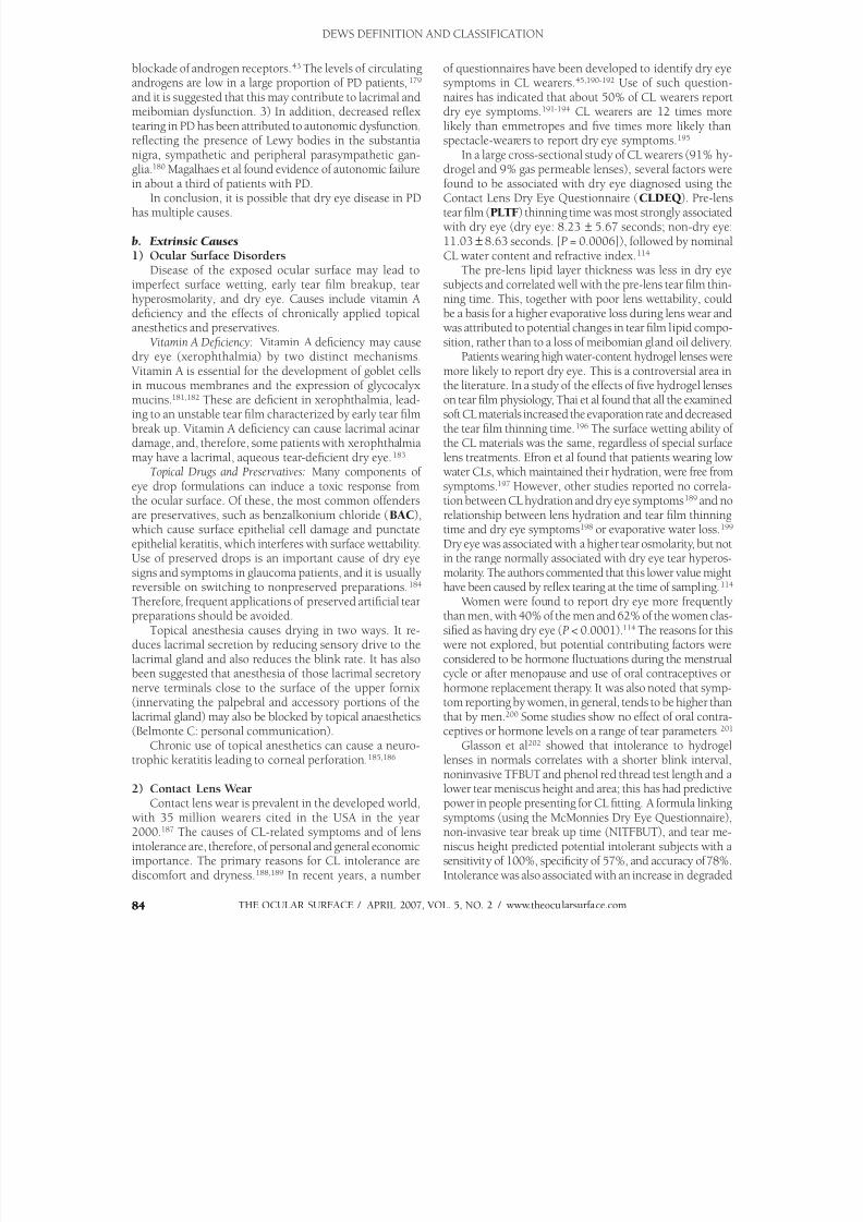
8/20/2019 dry eyes ii
http://slidepdf.com/reader/full/dry-eyes-ii 24/142
THE OCULAR SURFACE / APRIL 2007, VOL. 5, NO. 2 / www.theocularsurface.com84 84
blockade of androgen receptors.43 The levels of circulatingandrogens are low in a large proportion of PD patients,179 and it is suggested that this may contribute to lacrimal andmeibomian dysfunction. 3) In addition, decreased reflextearing in PD has been attributed to autonomic dysfunction,reflecting the presence of Lewy bodies in the substantianigra, sympathetic and peripheral parasympathetic gan-
glia.180
Magalhaes et al found evidence of autonomic failurein about a third of patients with PD.In conclusion, it is possible that dry eye disease in PD
has multiple causes.
b. Extrinsic Causes1) Ocular Surface Disorders
Disease of the exposed ocular surface may lead toimperfect surface wetting, early tear film breakup, tearhyperosmolarity, and dry eye. Causes include vitamin Adeficiency and the effects of chronically applied topicalanesthetics and preservatives.
Vitamin A Deficiency: Vitamin A deficiency may causedry eye (xerophthalmia) by two distinct mechanisms. Vitamin A is essential for the development of goblet cellsin mucous membranes and the expression of glycocalyxmucins.181,182 These are deficient in xerophthalmia, lead-ing to an unstable tear film characterized by early tear filmbreak up. Vitamin A deficiency can cause lacrimal acinardamage, and, therefore, some patients with xerophthalmiamay have a lacrimal, aqueous tear-deficient dry eye.183
Topical Drugs and Preservatives: Many components ofeye drop formulations can induce a toxic response fromthe ocular surface. Of these, the most common offendersare preservatives, such as benzalkonium chloride (BAC),
which cause surface epithelial cell damage and punctateepithelial keratitis, which interferes with surface wettability.Use of preserved drops is an important cause of dry eyesigns and symptoms in glaucoma patients, and it is usuallyreversible on switching to nonpreserved preparations.184 Therefore, frequent applications of preserved artificial tearpreparations should be avoided.
Topical anesthesia causes drying in two ways. It re-duces lacrimal secretion by reducing sensory drive to thelacrimal gland and also reduces the blink rate. It has alsobeen suggested that anesthesia of those lacrimal secretorynerve terminals close to the surface of the upper fornix
(innervating the palpebral and accessory portions of thelacrimal gland) may also be blocked by topical anaesthetics(Belmonte C: personal communication).
Chronic use of topical anesthetics can cause a neuro-trophic keratitis leading to corneal perforation.185,186
2) Contact Lens WearContact lens wear is prevalent in the developed world,
with 35 million wearers cited in the USA in the year2000.187 The causes of CL-related symptoms and of lensintolerance are, therefore, of personal and general economicimportance. The primary reasons for CL intolerance are
discomfort and dryness.188,189 In recent years, a number
of questionnaires have been developed to identify dry eyesymptoms in CL wearers.45,190-192 Use of such question-naires has indicated that about 50% of CL wearers reportdry eye symptoms.191-194 CL wearers are 12 times morelikely than emmetropes and five times more likely thanspectacle-wearers to report dry eye symptoms.195
In a large cross-sectional study of CL wearers (91% hy-
drogel and 9% gas permeable lenses), several factors werefound to be associated with dry eye diagnosed using theContact Lens Dry Eye Questionnaire (CLDEQ ). Pre-lenstear film (PLTF) thinning time was most strongly associatedwith dry eye (dry eye: 8.23 ± 5.67 seconds; non-dry eye:11.03 ± 8.63 seconds. [P = 0.0006]), followed by nominalCL water content and refractive index.114
The pre-lens lipid layer thickness was less in dry eyesubjects and correlated well with the pre-lens tear film thin-ning time. This, together with poor lens wettability, couldbe a basis for a higher evaporative loss during lens wear andwas attributed to potential changes in tear film lipid compo-sition, rather than to a loss of meibomian gland oil delivery.
Patients wearing high water-content hydrogel lenses weremore likely to report dry eye. This is a controversial area inthe literature. In a study of the effects of five hydrogel lenseson tear film physiology, Thai et al found that all the examinedsoft CL materials increased the evaporation rate and decreasedthe tear film thinning time.196 The surface wetting ability ofthe CL materials was the same, regardless of special surfacelens treatments. Efron et al found that patients wearing lowwater CLs, which maintained their hydration, were free fromsymptoms.197 However, other studies reported no correla-tion between CL hydration and dry eye symptoms189 and norelationship between lens hydration and tear film thinning
time and dry eye symptoms198 or evaporative water loss.199 Dry eye was associated with a higher tear osmolarity, but notin the range normally associated with dry eye tear hyperos-molarity. The authors commented that this lower value mighthave been caused by reflex tearing at the time of sampling.114
Women were found to report dry eye more frequentlythan men, with 40% of the men and 62% of the women clas-sified as having dry eye (P < 0.0001).114 The reasons for thiswere not explored, but potential contributing factors wereconsidered to be hormone fluctuations during the menstrualcycle or after menopause and use of oral contraceptives orhormone replacement therapy. It was also noted that symp-
tom reporting by women, in general, tends to be higher thanthat by men.200 Some studies show no effect of oral contra-ceptives or hormone levels on a range of tear parameters.201
Glasson et al202 showed that intolerance to hydrogellenses in normals correlates with a shorter blink interval,noninvasive TFBUT and phenol red thread test length and alower tear meniscus height and area; this has had predictivepower in people presenting for CL fitting. A formula linkingsymptoms (using the McMonnies Dry Eye Questionnaire),non-invasive tear break up time (NITFBUT), and tear me-niscus height predicted potential intolerant subjects with asensitivity of 100%, specificity of 57%, and accuracy of 78%.
Intolerance was also associated with an increase in degraded
DEWS DEFINITION AND CLASSIFICATION
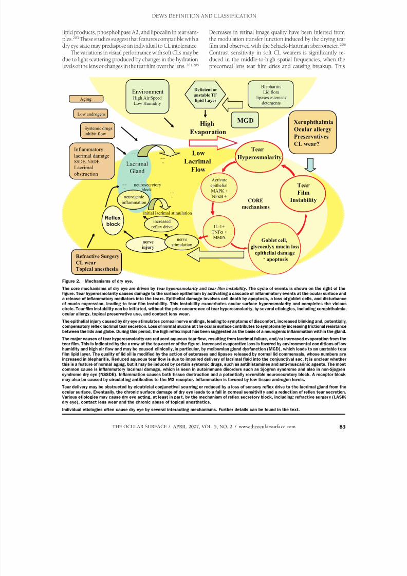
8/20/2019 dry eyes ii
http://slidepdf.com/reader/full/dry-eyes-ii 25/142
THE OCULAR SURFACE / APRIL 2007, VOL. 5, NO. 2 / www.theocularsurface.com 85 85
lipid products, phospholipase A2, and lipocalin in tear sam-ples.203 These studies suggest that features compatible with adry eye state may predispose an individual to CL intolerance.
The variations in visual performance with soft CLs may bedue to light scattering produced by changes in the hydrationlevels of the lens or changes in the tear film over the lens.204,205
Decreases in retinal image quality have been inferred fromthe modulation transfer function induced by the drying tearfilm and observed with the Schack-Hartman aberrometer.206 Contrast sensitivity in soft CL wearers is significantly re-duced in the middle-to-high spatial frequencies, when theprecorneal lens tear film dries and causing breakup. This
DEWS DEFINITION AND CLASSIFICATION
Table 4
CORE
mechanisms
Systemic drugs
inhibit flow
Refractive Surgery
CL wear
Topical anesthesia
Activate
epithelial
MAPK +
NF B +
Tear
Film
Instability
High
Evaporation
Low
Lacrimal
Flow
Goblet cell,
glycocalyx mucin lossepithelial damage
- apoptosis
Inflammatory
lacrimal damageSSDE; NSDE;
Lacrimal
obstruction
EnvironmentHigh Air Speed
Low Humidity
Blepharitis
Lid flora
lipases esterases
detergents
nerve
injury
Hyperosmolarity
Tear
Deficient or
unstable TF
lipid Layer
MGD Xerophthalmia
Ocular allergy
Preservatives
CL wear?
Low androgens
Aging
IL-1+
TNF +
MMPs
– –
–
LacrimalGland
– –
–
– –
–
++
+
initial lacrimal stimulation
increased
reflex drive
nerve
stimulation
neurogenic
inflammation
neurosecretory block
Reflex
block
Figure 2. Mechanisms of dry eye.
The core mechanisms of dry eye are driven by tear hyperosmolarity and tear film instability . The cycle of events is shown on the right of thefigure. Tear hyperosmolarity causes damage to the surface epithelium by activating a cascade of inflammatory events at the ocular surface and
a release of inflammatory mediators into the tears. Epithelial damage involves cell death by apoptosis, a loss of goblet cells, and disturbanceof mucin expression, leading to tear film instability. This instability exacerbates ocular surface hyperosmolarity and completes the viciouscircle. Tear film instability can be initiated, without the prior occurrence of tear hyperosmolarity, by several etiologies, including xerophthalmia,
ocular allergy, topical preservative use, and contact lens wear.
The epithelial injury caused by dry eye stimulates corneal nerve endings, leading to symptoms of discomfort, increased blinking and, potentially,compensatory reflex lacrimal tear secretion. Loss of normal mucins at the ocular surface contributes to symptoms by increasing frictional resistance
between the lids and globe. During this period, the high reflex input has been suggested as the basis of a neurogenic inflammation within the gland.
The major causes of tear hyperosmolarity are reduced aqueous tear flow, resulting from lacrimal failure, and/or increased evaporation from thetear film. This is indicated by the arrow at the top-center of the figure. Increased evaporative loss is favored by environmental conditions of low
humidity and high air flow and may be caused clinically, in particular, by meibomian gland dysfunction (MGD), which leads to an unstable tearfilm lipid layer. The quality of lid oil is modified by the action of esterases and lipases released by normal lid commensals, whose numbers areincreased in blepharitis. Reduced aqueous tear flow is due to impaired delivery of lacrimal fluid into the conjunctival sac. It is unclear whether
this is a feature of normal aging, but it may be induced by certain systemic drugs, such as antihistamines and anti-muscarinic agents. The mostcommon cause is inflammatory lacrimal damage, which is seen in autoimmune disorders such as Sjogren syndrome and also in non-Sjogren
syndrome dry eye (NSSDE). Inflammation causes both tissue destruction and a potentially reversible neurosecretory block. A receptor blockmay also be caused by circulating antibodies to the M3 receptor. Inflammation is favored by low tissue androgen levels.
Tear delivery may be obstructed by cicatricial conjunctival scarring or reduced by a loss of sensory reflex drive to the lacrimal gland from the
ocular surface. Eventually, the chronic surface damage of dry eye leads to a fall in corneal sensitivity and a reduction of reflex tear secretion.Various etiologies may cause dry eye acting, at least in part, by the mechanism of reflex secretory block, including: refractive surgery (LASIKdry eye), contact lens wear and the chronic abuse of topical anesthetics.
Individual etiologies often cause dry eye by several interacting mechanisms. Further details can be found in the text.

8/20/2019 dry eyes ii
http://slidepdf.com/reader/full/dry-eyes-ii 26/142
THE OCULAR SURFACE / APRIL 2007, VOL. 5, NO. 2 / www.theocularsurface.com86
could account for complaints of intermittent blurred visionin some CL wearers and may provide a stimulus to blink.207
3) Ocular Surface DiseaseThere is evidence that various forms of chronic ocular sur-
face disease result in destabilization of the tear film and add adry eye component to the ocular surface disease. Allergic eye
disease offers a well-studied example.208
Also, any form of dryeye, whatever its origins, may cause at least a loss of gobletcell numbers, so that an ocular surface element is added.209
4) Allergic Conjunctivitis Allergic conjunctivitis takes several forms, which in-
clude seasonal allergic conjunctivitis, vernal keratoconjunc-tivitis, and atopic keratoconjunctivitis. The general mecha-nism leading to disease is that exposure to antigen leads todegranulation of IgE-primed mast cells, with the release ofinflammatory cytokines. A Th2 response is activated at theocular surface, initially in the conjunctival and, later, in thecorneal epithelium, subsequently leading to submucosalchanges. There is stimulation of goblet cell secretion andloss of surface membrane mucins.210 Surface epithelial celldeath occurs, affecting conjunctival and corneal epithelium(punctate keratoconjunctivitis). Surface damage and therelease of inflammatory mediators leads to allergic symp-toms and to reflex stimulation of the normal lacrimal gland.
Surface irregularities on the cornea (punctate epithelialkeratitis and shield ulcer) and conjunctiva can lead to tearfilm instability and, hence, to a local drying componentto the allergic eye disease. In chronic disease, there maybe meibomian gland dysfunction, which could exacerbatesurface drying by interfering with the tear film lipid layer.
Lid swelling, eg, in vernal catarrh and atopic keratocon- junctivitis, can interfere with lid apposition and tear filmspreading, thus exacerbating the dry eye.
Ocular allergy was noted to be a risk factor for dry eye inthe Beaver Dam study, although the concomitant use of sys-temic medications, such as antihistamines, was recognizedas a potential contributor.122 Factors leading to a dry eye statein allergic eye disease are discussed by Fujishima et al.211
C. The Causative Mechanisms of Dry Eye
From the above discussion, it can be seen that certaincore mechanisms are envisaged at the center of the dry eye
process that can initiate, amplify, and potentially change thecharacter of dry eye over time. These are tear hyperosmolar-ity and tear film instability. This section is intended to showhow the several subclasses of dry eye activate these coremechanisms and explain the features of various forms ofdry eye. The interactions of various etiologies with thesecore mechanisms are summarized in Figure 2.
It should be noted that an attractive mechanistic schemafor dry eye has been presented in detail by Baudouin.212 Inthis concept, two levels of involvement are identified. Thefirst level includes the known risk factors or causes of dryeye that ultimately lead to a series of secondary biological
cascades, resulting in breakdown of the tear film and ocular
surface. This pathbreaking conceptual approach describesthe relationship of early disparate events to biological re-sponses common to all forms of dry eye, many of whichare mutually reinforcing. This leads to a vicious circle orloop. It is thought that early therapeutic intervention maydisrupt this loop. The schema in Figure 2, developed fromthe discussion of our Subcommittee, emphasizes the core
biological mechanisms described in this text.
1. Tear HyperosmolarityTear hyperosmolarity is regarded as the central mecha-
nism causing ocular surface inflammation, damage, andsymptoms, and the initiation of compensatory events indry eye. Tear hyperosmolarity arises as a result of waterevaporation from the exposed ocular surface, in situationsof a low aqueous tear flow, or as a result of excessive evapo-ration, or a combination of these events. Nichols et al havedemonstrated the wide variation of tear film thinning ratesin normal subjects, and it is reasonable to conclude that, fora given initial film thickness, subjects with the fastest thin-ning rates would experience a greater tear film osmolaritythan those with the slowest rates.114 Rapid thinning may behypothesized as a risk factor for tear hyperosmolarity.
Since the lacrimal fluid is secreted as a slightly hypoton-ic fluid, it will always be expected that tear osmolarity willbe higher in the tear film than in other tear compartments.There are also reasons to believe that osmolarity is higherin the tear film itself than in the neighboring menisci. Onereason for this is that the ratio of area to volume (whichdetermines the relative concentrating effect of evaporation)is higher in the film than the menisci.213
Hyperosmolarity stimulates a cascade of inflammatory
events in the epithelial surface cells, involving MAP kinasesand NFkB signalling pathways56 and the generation ofinflammatory cytokines (IL-1α; -1β; TNF-α) and MMPs(MMP9),58 which arise from or activate inflammatory cellsat the ocular surface.214 These concepts are supported bystudies of desiccating stress in the experimental model,215 which have demonstrated the evolution of inflammatorycytokine release and MMP activation.57 There is evidencethat these inflammatory events lead to apoptotic death ofsurface epithelial cells, including goblet cells216; thus, gobletcell loss may be seen to be directly related to the effects ofchronic inflammation.217,218 Goblet cell loss is a feature of
every form of dry eye, and consistent with this is the dem-onstration of reduced levels of the gel mucin MUC5AC indry eye.219,220 With the evolution of dry eye, other factorsare likely to amplify these initiating inflammatory events,and the contribution of direct autoimmune targeting of theocular surface cannot be excluded.
In the initial stages of dry eye, it is considered that ocularsurface damage caused by osmotic, inflammatory or me-chanical stresses (loss of surface lubrication) results in reflexstimulation of the lacrimal gland. Reflex trigeminal activity isthought to be responsible for an increased blink rate and acompensatory response, increased lacrimal secretion. In the
case of lacrimal gland insufficiency (SSDE or NSSDE), the
DEWS DEFINITION AND CLASSIFICATION
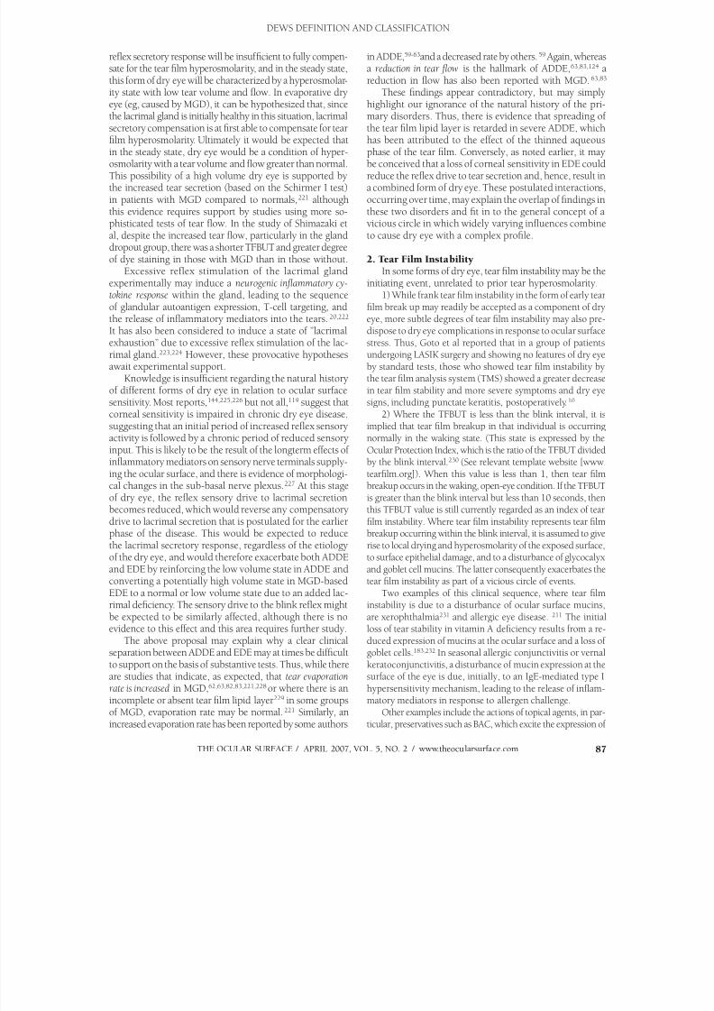
8/20/2019 dry eyes ii
http://slidepdf.com/reader/full/dry-eyes-ii 27/142
THE OCULAR SURFACE / APRIL 2007, VOL. 5, NO. 2 / www.theocularsurface.com 87
reflex secretory response will be insufficient to fully compen-sate for the tear film hyperosmolarity, and in the steady state,this form of dry eye will be characterized by a hyperosmolar-ity state with low tear volume and flow. In evaporative dryeye (eg, caused by MGD), it can be hypothesized that, sincethe lacrimal gland is initially healthy in this situation, lacrimalsecretory compensation is at first able to compensate for tear
film hyperosmolarity. Ultimately it would be expected thatin the steady state, dry eye would be a condition of hyper-osmolarity with a tear volume and flow greater than normal.This possibility of a high volume dry eye is supported bythe increased tear secretion (based on the Schirmer I test)in patients with MGD compared to normals,221 althoughthis evidence requires support by studies using more so-phisticated tests of tear flow. In the study of Shimazaki etal, despite the increased tear flow, particularly in the glanddropout group, there was a shorter TFBUT and greater degreeof dye staining in those with MGD than in those without.
Excessive reflex stimulation of the lacrimal glandexperimentally may induce a neurogenic inflammatory cy-tokine response within the gland, leading to the sequenceof glandular autoantigen expression, T-cell targeting, andthe release of inflammatory mediators into the tears.20,222 It has also been considered to induce a state of ”lacrimalexhaustion” due to excessive reflex stimulation of the lac-rimal gland.223,224 However, these provocative hypothesesawait experimental support.
Knowledge is insufficient regarding the natural historyof different forms of dry eye in relation to ocular surfacesensitivity. Most reports,144,225,226 but not all,119 suggest thatcorneal sensitivity is impaired in chronic dry eye disease,suggesting that an initial period of increased reflex sensory
activity is followed by a chronic period of reduced sensoryinput. This is likely to be the result of the longterm effects ofinflammatory mediators on sensory nerve terminals supply-ing the ocular surface, and there is evidence of morphologi-cal changes in the sub-basal nerve plexus.227 At this stageof dry eye, the reflex sensory drive to lacrimal secretionbecomes reduced, which would reverse any compensatorydrive to lacrimal secretion that is postulated for the earlierphase of the disease. This would be expected to reducethe lacrimal secretory response, regardless of the etiologyof the dry eye, and would therefore exacerbate both ADDEand EDE by reinforcing the low volume state in ADDE and
converting a potentially high volume state in MGD-basedEDE to a normal or low volume state due to an added lac-rimal deficiency. The sensory drive to the blink reflex mightbe expected to be similarly affected, although there is noevidence to this effect and this area requires further study.
The above proposal may explain why a clear clinicalseparation between ADDE and EDE may at times be difficultto support on the basis of substantive tests. Thus, while thereare studies that indicate, as expected, that tear evaporationrate is increased in MGD,62,63,82,83,221,228 or where there is anincomplete or absent tear film lipid layer229 in some groupsof MGD, evaporation rate may be normal.221 Similarly, an
increased evaporation rate has been reported by some authors
in ADDE,59-63and a decreased rate by others.59 Again, whereasa reduction in tear flow is the hallmark of ADDE,63,83,124 areduction in flow has also been reported with MGD.63,83
These findings appear contradictory, but may simplyhighlight our ignorance of the natural history of the pri-mary disorders. Thus, there is evidence that spreading ofthe tear film lipid layer is retarded in severe ADDE, which
has been attributed to the effect of the thinned aqueousphase of the tear film. Conversely, as noted earlier, it maybe conceived that a loss of corneal sensitivity in EDE couldreduce the reflex drive to tear secretion and, hence, result ina combined form of dry eye. These postulated interactions,occurring over time, may explain the overlap of findings inthese two disorders and fit in to the general concept of avicious circle in which widely varying influences combineto cause dry eye with a complex profile.
2. Tear Film InstabilityIn some forms of dry eye, tear film instability may be the
initiating event, unrelated to prior tear hyperosmolarity.1) While frank tear film instability in the form of early tear
film break up may readily be accepted as a component of dryeye, more subtle degrees of tear film instability may also pre-dispose to dry eye complications in response to ocular surfacestress. Thus, Goto et al reported that in a group of patientsundergoing LASIK surgery and showing no features of dry eyeby standard tests, those who showed tear film instability bythe tear film analysis system (TMS) showed a greater decreasein tear film stability and more severe symptoms and dry eyesigns, including punctate keratitis, postoperatively.10
2) Where the TFBUT is less than the blink interval, it isimplied that tear film breakup in that individual is occurring
normally in the waking state. (This state is expressed by theOcular Protection Index, which is the ratio of the TFBUT dividedby the blink interval.230 (See relevant template website [www.tearfilm.org]). When this value is less than 1, then tear filmbreakup occurs in the waking, open-eye condition. If the TFBUTis greater than the blink interval but less than 10 seconds, thenthis TFBUT value is still currently regarded as an index of tearfilm instability. Where tear film instability represents tear filmbreakup occurring within the blink interval, it is assumed to giverise to local drying and hyperosmolarity of the exposed surface,to surface epithelial damage, and to a disturbance of glycocalyxand goblet cell mucins. The latter consequently exacerbates the
tear film instability as part of a vicious circle of events.Two examples of this clinical sequence, where tear film
instability is due to a disturbance of ocular surface mucins,are xerophthalmia231 and allergic eye disease. 211 The initialloss of tear stability in vitamin A deficiency results from a re-duced expression of mucins at the ocular surface and a loss ofgoblet cells.183,232 In seasonal allergic conjunctivitis or vernalkeratoconjunctivitis, a disturbance of mucin expression at thesurface of the eye is due, initially, to an IgE-mediated type Ihypersensitivity mechanism, leading to the release of inflam-matory mediators in response to allergen challenge.
Other examples include the actions of topical agents, in par-
ticular, preservatives such as BAC, which excite the expression of
DEWS DEFINITION AND CLASSIFICATION

8/20/2019 dry eyes ii
http://slidepdf.com/reader/full/dry-eyes-ii 28/142
THE OCULAR SURFACE / APRIL 2007, VOL. 5, NO. 2 / www.theocularsurface.com88
inflammatory cell markers at the ocular surface, causing epithelialcell damage, cell death by apoptosis, and a decrease in goblet celldensity.233 There is both clinical and experimental evidence tosupport such events.234-238 In a study of patients treated for glau-
coma for at least one year, flow cytometry demonstrated a greaterexpression of inflammatory markers (HLA-DR and ICAM-1) inthose receiving preserved drops (BAC) than in normals or thosereceiving unpreserved drops. Use of preservative was associatedwith a lower expression of MUC5AC and the lowest MUC5AClevels were associated with the highest ICAM-1 and HLA-DRlevels.239 This negative correlation suggested inflammation as apossible basis for the decreased mucin expression, in addition toany direct effect of BAC on goblet cells themselves.
Considering the possible relationship between these find-ings and dry eye, Pisella et al, in an unmasked study of 4107glaucoma patients, found that the frequency of ocular surface
changes was twice as high in those receiving preserved dropsthan in those receiving unpreserved drops, and the frequencyof signs and symptoms was dose-related.184
CL wear may also provide a route of entry into the dry eyemechanism, a route in addition to reduced corneal sensitivity.For a considerable time, CL wear has been recognized to causechanges to the ocular surface epithelia. Knop and Brewitt dem-onstrated surface epithelial metaplasia and a reduced gobletcell density with hydrogel lens wear.240,241 Other studies haveshown an increase in goblet cell density evolving over a periodof 6 months in subjects wearing polymacon, galyfilcon, andsilicone hydrogel lenses.242,243 In another study, no change in
goblet cell density was found after 6 months wear of a daily
disposable lens with a 2-week wearing schedule, and furtherstudies suggest that the goblet cell responses may differ be-tween hard and soft CLs.244
A recent study combining impression cytology with flow
cytometry demonstrated an increase in inflammatory markers(HLA-DR and ICAM-1) at the ocular surface and a nonsig-nificant trend toward a decrease in the expression of mucinmarkers (MUC5AC) in patients with a history of chronic CLwear.245 A later study has shown no difference between CLwearers and non-CL wearers in mucin expression (MUC5ACand the carbohydrate epitope H185, a marker for MUC 16)in tears or impression cytology samples.182 In summary, it ap-pears that CL wear may activate proinflammatory markers andstimulate the ocular surface epithelia to a variable degree. It isnot yet possible to say whether these changes alone predisposeindividuals to the occurrence of dry eye with CL wear.
D. The Basis for Symptoms in Dry Eye
The basis for symptoms in dry eye is not truly knownbut may be surmised from a consideration of the etiologies,mechanisms, and responses of dry eye to therapy.246 The oc-currence of symptoms implies the activation of sensory nervessubserving nociception at the ocular surface.247,248 Candidatesinclude tear and ocular surface hyperosmolarity – includingtear film break-up in the interblink, shear-stress between thelids and globe in response to reduced tear volume, and/or thereduced expression of mucins at the ocular surface, the pres-ence of inflammatory mediators at the surface of the eye, and,
finally, hypersensitivity of the nociceptive sensory nerves.
Table 5. Dry eye severity grading scheme
Dry Eye SeverityLevel 1 2 3 4*
Discomfort, severity
& frequency
Mild and/or episodic;
occurs under
environmental stress
Moderate episodic or
chronic, stress or no
stress
Severe frequent or
constant without
stress
Severe and/or
disabling and constant
Visual symptomsNone or episodic mild
fatigue
Annoying and/or
activity-limiting
episodic
Annoying, chronic
and/or constant,
limiting activity
Constant and/or
possibly disabling
Conjunctival injection None to mild None to mild +/– +/++
Conjunctival staining None to mild Variable Moderate to marked Marked
Corneal staining
(severity/location)None to mild Variable Marked central
Severe punctate
erosions
Corneal/tear signs None to mild Mild debris, ↓ meniscus
Filamentary keratitis,
mucus clumping,
↑ tear debris
Filamentary keratitis,
mucus clumping,
↑ tear debris, ulceration
Lid/meibomian glands MGD variably present MGD variably present FrequentTrichiasis, keratinization,
symblepharon
TFBUT (sec) Variable ≤ 10 ≤ 5 Immediate
Schirmer score
(mm/5 min)Variable ≤ 10 ≤ 5 ≤ 2
*Must have signs AND symptoms. TBUT: fluorescein tear break-up time. MGD: meibomian gland disease
Reprinted with permission from Behrens A, Doyle JJ, Stern L, et al. Dysfunctional tear syndrome. A Delphi approach to treatment recommendations.Cornea 2006;25:90-7
DEWS DEFINITION AND CLASSIFICATION
8/20/2019 dry eyes ii
http://slidepdf.com/reader/full/dry-eyes-ii 29/142
THE OCULAR SURFACE / APRIL 2007, VOL. 5, NO. 2 / www.theocularsurface.com 89
E. Classification of Dry Eye on the Basis of Severity
The Subcommittee considered that there was consider-able clinical utility to adopting a classification of diseasebased on severity. The basic scheme of the Delphi PanelReport was adopted and modified to produce the thirdcomponent of the recommendation (Table 5).
REFERENCES 1. Lemp MA. Report of the National Eye Institute/Industry Workshop on
Clinical Trials in Dry Eye. CLAO J 1995;21:221-32 2. Begley CG, Chalmers RL, Abetz L, et al. The relationship between habitual
patient-reported symptoms and clinical signs among patients with dry eyeof varying severity. Invest Ophthalmol Vis Sci 2003;44:4753-61
3. Adatia FA, Michaeli-Cohen A, Naor J, et al. Correlation between cornealsensitivity, subjective dry eye symptoms and corneal staining in Sjogren’ssyndrome. Can J Ophthalmol 2004;39:767-71
4. Vitale S, Goodman LA, Reed GF, Smith JA. Comparison of the NEI-VFQand OSDI questionnaires in patients with Sjogren’s syndrome-related dryeye. Health Qual Outcomes 20041;2:44
5. Rieger G. The importance of the precorneal tear film for the quality ofoptical imaging. Br J Ophthalmol 1992;76:157-8
6. Liu Z, Pflugfelder SC. Corneal surface irregularity and the effect of artificialtears in aqueous tear deficiency. Ophthalmology 1999;106:936-43
7. Goto E, Yagi Y, Matsumoto Y, Tsubota K. Impaired functional visual acuityof dry eye patients. Am J Ophthalmol 2002;133:181-6
8. Holly F, Lemp MA. Formation and rupture of the tear film. Exp Eye Res 1973;15:515-25
9. Bron AJ. Diagnosis of dry eye. Surv Ophthalmol 2001;45 Suppl 2:S221-6 10. Goto T, Zheng X, Klyce SD, et al. A new method for tear film stability
using videokeratography. Am J Ophthalmol 2003;135:607-12 11. Farris RL, Stuchell RN, Mandel ID Tear osmolarity variation in the dry
eye. Trans Am Ophthalmol Soc 1986;84:250-68 12. Gilbard JP. Human tear film electrolyte concentrations in health and dry-
eye disease. Int Ophthalmol Clin 1994 Winter;34:27-36 13. Murube J. Tear osmolarity. Ocul Surf 2006;1;62-73 14. Tomlinson A, Khanal S, Ramaesh K, et al. Tear film osmolarity: determination
of a referent for dry eye diagnosis. Invest Ophthalmol Vis Sci 2006;47:4309-15 15. Pflugfelder SC, Jones D, Ji Z, et al. Altered cytokine balance in the tear fluid
and conjunctiva of patients with Sjogren’s syndrome keratoconjunctivitis
sicca. Curr Eye Res 1999;19:201-11 16. Tsubota K, Fujihara T, Saito K, Takeuchi T. Conjunctival epithelium ex-
pression of HLA-DR in dry eye patients. Ophthalmologica 1999;213:16-9 17. Stern ME, Beuerman RW, Fox RI, et al. The pathology of dry eye; the interac-
tion between the ocular surface and lacrimal glands. Cornea 1998;17:584-9 18. Pflugfelder SC, Solomon A, Stern ME. The diagnosis and management of
dry eye:a twenty-five year review. Cornea 2000;19:644-9 19. Beuerman RW, Mircheff A, Pflugfelder SC, Stern ME. The lacrimal func-
tional unit, in Pflugfelder SC, Beuerman RW, Stern ME (eds). Dry eye andocular surface disorders. New York, Marcel Dekker, 2004
20. Stern ME, Gao J, Siemarko KF, et al. The role of the lacrimal functionalunit in the pathophysiology of dry eye. Exp Eye Res 2004;78:409-6
21. (No authors listed). Research in dry eye: Report of the Research Subcom-mittee of the 2007 Dry Eye WorkShop (DEWS).Ocul Surf 2007;5:179-193
22. Jordan A, Baum J. Basic tear flow. Does it exist?Ophthalmology 1980;87:920. 23. Gupta A, Heigle T, Pflugfelder SC. Nasolacrimal stimulation of aqueous
tear production.Cornea 1997;16:645-8 24. Vitali C. Classification criteria for Sjogren’s syndrome. Ann Rheum Dis
2003;62:94-5 25. Murube J, Benitez del Castillo JM, Chenghou L, et al. The Madrid triple
classification system. Arch Soc Esp Oftalmol 2003;78;587-93 26. Murube J, Nemeth J, Hoh H, et al. The triple classification of dry eye for
practical clinical use. Eur J Ophthalmol 2005;15:660-7 27. Behrens A, Doyle JJ, Stern L, et al. Dysfunctional tear syndrome: A Delphi
approach to treatment recommendations. Cornea 2006;25:900-7 28. Doughty MJ, Naase T. Further analysis of the human spontaneous eye blink
rate by a cluster analysis-based approach to categorize individuals with‘normal’ versus ‘frequent’ eye blink activity.Eye Contact Lens 2006;32:294-9
29. Nakamori K, Odawara M, Nakajima T, et al. Blinking is controlled primar-ily by ocular surface conditions. Am J Ophthalmol 1997;124:24-30
30. Mangubat L, Luague S. Normal measurements of the palpebral fissure and theinterpalpebral distance among Filipinos. Philipp J Surg Surg Spec 1966;21:304-6
31. Stoller SH, Meyer DR. Quantitating the change in upper lid position during
downgaze. Ophthalmology 1994;101:1604-7 32. Cho P, Sheng C, Chan C, et al. Baseline blink rates and the effect of visual
task difficulty and position of gaze. Curr Eye Res 2000;20: 64-70 33. Sullivan DA. Sex and sex steroid influences on the dry eye syndrome, in
Pflugfelder SC, Beuerman RW, Stern ME (eds). Dry eye and ocular surfacedisorders. New York, Marcel Dekker, 2004
34. Sullivan DA. Androgen deficiency and dry eye syndromes. Arch SocEspanola Oftalmologia 2004;79:49-50
35. Sullivan DA. Tearful relationships? Sex, hormones and aqueous-deficientdry eye. Ocul Surf 2004;2:92-123
36. Worda C, Nepp J, Huber JC, Sator MO. Treatment of keratoconjunctivitissicca with topical androgen. Maturitas 2001;37, 209-12
37. Zeligs MA. Dehydroepiandrosterone therapy for the treatment of dry eyedisorders. Int Patent Application. WO 94/04155, 1994
38. Schiffman RM, Bradford R, Bunnell B, et al. A multi-center, double-masked,randomized, vehicle-controlled, parallel group study to evaluate the safetyand efficacy of testosterone ophthalmic solution in patients with meibo-mian gland dysfunction. ARVO e-abstract #5608, 2006 (www.arvo.org)
39. Krenzer KL, Dana MR, Ullman MD, et al. Effect of androgen deficiencyon the human meibomian gland and ocular surface. J Clin Endocr Metab 2000;85:4874-82
40. Sullivan BD, Evans JE, Krenzer KL, et al. Impact of antiandrogen treat-ment on the fatty acid profile of neutral lipids in human meibomian glandsecretions. J Clin Endocr Metab 2000;85:4866-73
41. Sullivan BD, Evans JE, Cermak JM, et al. Complete androgen insensitivitysyndrome: Effect on human meibomian gland secretions. Arch Ophthalmol
2002;120:1689-99 42. Cermak JM, Krenzer KL, Sullivan RM, et al. Is complete androgen insen-
sitivity syndrome associated with alterations in the meibomian gland andocular surface? Cornea 2003;22:516-21
43. Mantelli F, Moretti C, Micera A, Bonini S. Conjunctival mucin deficiencyin complete androgen insensitivity syndrome (CAIS). Graefes Arch Clin Exp Ophthalmol 2006 Nov 2; [Epub ahead of print]
44. Tamer C, Oksuz H, Sogut S. Androgen status of the nonautoimmune dryeye subtypes. Ophthalmic Res 2006;38:280-6
45. (No authors listed). Epidemiology of dry eye: Report of the Epidemiol-ogy Subcommittee of the 2007 Dry Eye WorkShop (DEWS). Ocul Surf 2007;5:93-107
46. Schaumberg DA, Buring JE, Sullivan DA, Dana MR. Hormone replacementtherapy and dry eye syndrome. JAMA 2001;286:2114-9
47. Schaumberg DA, Sullivan DA, Buring JE, Dana MR. Prevalence of dry eyesyndrome among US women. Am J Ophthalmol 2003;136:318-26
48. Smith JA, Vitale S, Reed GF, et al. Dry eye signs and symptoms in womenwith premature ovarian failure. Arch Ophthalmol 2004;122:151-6
49. Mathers WD, Lane JA, Zimmerman MB. Tear film changes associated withnormal aging. Cornea 1996;15: 229-34
50. Patel S, Farrell JC. Age-related changes in precorneal tear film stability.Optom Vis Sci 1989;66:175-8
51. Sullivan BD, Evans JE, Dana MR, Sullivan DA. Influence of aging on thepolar and neutral lipid profiles in human meibomian gland secretions.
Arch Ophthalmol 2006;124: 286-92 52. Paschides CA, Stefaniotou M, Papageorgiou J, et al. Ocular surface and
environmental changes. Acta Ophthalmol Scand 1998;876:74-7 53. Tsubota K, Nakamori K. Dry eyes and video display terminals N Engl J
Med 1993;328:584 54. Mishima S, Gasset A, Klyce S, Baum J. Determination of tear volume and
tear flow. Investigative Ophthalmology 1966; 5: 264-76 55. Scherz W, Dohlman CH. Is the lacrimal gland dispensable? Keratoconjunc-
tivitis sicca after lacrimal gland removal. Arch Ophthalmol 1975;93: 81-3 56. Li DQ, Chen Z, Song XJ, et al. Stimulation of matrix metalloproteinases
by hyperosmolarity via a JNK pathway in human corneal epithelial cells.Invest Ophthalmol Vis Sci 2004;45:4302-11
57. Luo L, Li DQ, Corrales RM, Pflugfelder SC. Hyperosmolar saline is a proinflam-matory stress on the mouse ocular surface. Eye Contact Lens 2005;31:186-93
58. De Paiva CS, Corrales RM, Villarreal AL, et al. Corticosteroid and doxycyclinesuppress MMP-9 and inflammatory cytokine expression, MAPK activation inthe corneal epithelium in experimental dry eye.Exp Eye Res2006;83:526-35
59. Tsubota K, Yamada M. Tear evaporation from the ocular surface. Invest Ophthalmol Vis Sci 1992;33:2942-50
60. Rolando M, Refojo MF, Kenyon KR. Increased tear evaporation in eyeswith keratoconjunctivitis sicca. Arch Ophthalmol 1983;101:557-8
61. Rolando M, Refojo MF, Kenyon KR. Tear water evaporation and eye surfacediseases. Ophthalmologica 1985;190:147-9
62. Mathers WD. Ocular evaporation in meibomian gland dysfunction and
dry eye. Ophthalmology 1993;100:347-51
DEWS DEFINITION AND CLASSIFICATION

8/20/2019 dry eyes ii
http://slidepdf.com/reader/full/dry-eyes-ii 30/142
THE OCULAR SURFACE / JULY 2006, VOL. 4, NO. 3 / www.theocularsurface.com90
63. Mathers WD, Lane JA, Sutphin JE, Zimmerman MB. Model for oculartear film function. Cornea 1996;15:110-9
64. Mathers WD, Daley TE. Tear flow and evaporation in patients with andwithout dry eye. Ophthalmology 1996;103:664-9
65. Yokoi N, Mossa F, Tiffany JM, Bron A J. Assessment of meibomian glandfunction in dry eye using meibometry. Arch Ophthalmol 1999;117:723-9
66. Yokoi N, Takehisa Y, Kinoshita S. Correlation of tear lipid layer interfer-ence patterns with the diagnosis and severity of dry eye. Am J Ophthalmol 1996;122:818-24
67. Owens H, Phillips JR. Tear spreading rates: post-blink. Adv Exp Med Biol
2002;506(Pt B):1201-4 68. Goto E, Tseng SC. Differentiation of lipid tear deficiency dry eye by kinetic
analysis of tear interference images. Arch Ophthalmol 2003;121:173-80 69. Nakamura H, Kawakamu A, Eguchi K. Mechanisms of autoantibody
production and the relationship between autoantibodies and the clinicalmanifestations in Sjogren’s syndrome. Trans Res 2006;148(6):281-8
70. Hayashi Y, Arakaki R, Ishimaru N. The role of caspase cascade on thedevelopment of primary Sjogren’s syndrome. J Med Invest 2003;50:32-8
71. Zoukhri D. Effect of inflammation on lacrimal gland function. Exp EyeRes 2006;82:885-98. [Epub 2005 Nov 23]
72. Dawson L, Tobin A, Smith P, Gordon T. Antimuscarinic antibodies inSjogren’s syndrome: where are we, and where are we going? ArthritisRheum 2005;52:2984-95
73. Dawson LJ, Stanbury J, Venn N, et al. Antimuscarinic antibodies in primarySjogren’s syndrome reversibly inhibit the mechanism of fluid secretion by hu-man submandibular salivary acinar cells. Arthritis Rheum 2006;54:1165-73
74. Vitali C, Bombardieri S, Jonsson R, et al. Classification criteria for Sjogren’ssyndrome: a revised version of the European criteria proposed by theAmerican-European Consensus Group. Ann Rheum Dis 2002;61:554-8
75. Vitali C, Bombardieri S, Moutsopoulos HM, et al. Preliminary criteria for theclassification of Sjogren’s syndrome. Results of a prospective concerted ac-tion supported by the European Community. Arthritis Rheum 1993;36:340-7
76. Fox RI, Robinson CA, Curd JG, et al. Sjogren’s syndrome. Proposed criteriafor classification. Arthritis Rheum 1986;29:477-585
77. Wiik A, Cervera R, Haass M, et al. European attempts to set guidelines for improv-ing diagnostics of autoimmune rheumatic disorders. Lupus 2006;15:391-6
78. Moutsopoulos HM, Manoussakis MN. Immunopathogenesis of Sjogrensyndrome, “facts and fancy.” Autoimmunity 1989;5:17-24
79. Sullivan DA, Belanger A, Cermak JM, et al. Are women with Sjogren’ssyndrome androgen deficient? J Rheumatology 2003;30:2413-9
80. Cermak JM, Papas AS, Sullivan RM, et al: Nutrient intake in women withprimary and secondary Sjogren’s syndrome.Eur J Clin Nutr 2003; 57:328-34
81. Jones DT, Monroy D, Ji Z, et al. Sjogren’s syndrome: cytokine and Epstein-Barr viral gene expression within the conjunctival epithelium. InvestOphthalmol Vis Sci 1994;35:3493-504
82. Shimazaki J, Goto E, Ono M, et al. Meibomian gland dysfunction inpatients with Sjogren syndrome. Ophthalmology 1998;105:1485-8
83. Tomlinson A, Khanal S. Assessment of tear film dynamics: quantificationapproach. Ocul Surf 2005;3:81-95
84. Craig JP, Tomlinson A. Age and gender effects on the normal tear film. Adv Exp Med Biol 1998; 438:411-5.
85. Sahlin S, Chen E. Evaluation of the lacrimal drainage function by the droptest. Am J Ophthalmol 1996;122:701-8
86. Rolando M, Refojo MF. Tear evaporimeter for measuring water evaporationrate from the tear film under controlleded conditions in humans. Exp Eye
Res 1983;36:25-33 87. Tomlinson A, Geisbrecht J. The aging tear film. Br J Contact Lens Assoc
1993;16;67-9
88. Norn MS. Semiquantitative interference study of the fatty layer of thepre-corneal film. Acta Ophthalmol (Copenh) 1979;57:766-74
89. Damato BE, Allan D, Murray SB, Lee WR. Senile atrophy of the humanlacrimal gland: the contribution of chronic inflammatory disease. Br JOphthalmol 1984;68:674-80
89a. Obata H, Yamamoto S, Horiuchi H, Machinami R. Histopathologic studyof human lacrimal gland. Statistical analysis with special reference to ag-ing. Ophthalmology 1995;102:678-86
90. Davidoff E, Friedman AH. Congenital alacrima. Surv Ophthalmol 1977;22:113-9
91. Arya SK, Chaudhuri Z, Jain R, et al. Congenital alacrima in Pierre Robinsequence. Cornea. 2004; 23:632-4
92. Brooks BP, Kleta R, Stuart C, et al. Genotypic heterogeneity and clinicalphenotype in triple A syndrome: a review of the NIH experience 2000-2005. Clin Genet 2005;68:215-21
93. Krumbholz M, Koehler K, Huebner A. Cellular localization of 17 natural
mutant variants of ALADIN protein in triple A syndrome—shedding light
on an unexpected splice mutation. Biochem Cell Biol 2006;84:243-9 94. Axelrod FB, Chelimsky GG, Weese-Mayer DE. Pediatric autonomic
disorders. Pediatrics 2006;118: 309-21 95. Gold-von Simson G, Axelrod FB. Familial dysautonomia: update and
recent advances. Curr Probl Pediatr Adolesc Health Care 2006;36:218-3796. James DG, Anderson R, Langley D, Ainslie D. Ocular sarcoidosis. Br J
Ophthalmol 1964;48:461-70 97. Heath P. Ocular lymphomas. Trans Am Ophthalmol Soc 1948;46:385-98 98. Itescu S, Brancato LJ, Buxbaum J, et al. A diffuse infiltrative CD8 lymphocy-
tosis syndrome in human immunodeficiency virus (HIV) infection: a host
immune response associated with HLA-DR5. Ann Intern Med 1990;112:3-10 99. Ogawa Y, Okamoto S, Wakui M, et al. Dry eye after haematopoietic stem
cell transplantation. Br J Ophthalmol 1999;83:1125-30100. Ogawa Y, Kuwana M, Yamazaki K, et al. Periductal area as the primary site
for T-cell activation in lacrimal gland chronic graft- versus-host disease.Invest Ophthalmol Vis Sci 2003;44:1888-96
101. Maitchouk DY, Beuerman RW, Ohta T, et al. Tear production after unilateralremoval of the main lacrimal gland in squirrel monkeys. Arch Ophthalmol 2000;118:246-52
102. Whitwell J. Denervation of the lacrimal gland.Br J Ophthalmol 1958;42:518-25 103. Maitchouk DY, Beuerman RW, Ohta T, et al. Tear production after unilateral
removal of the main lacrimal gland in squirrel monkeys. Arch Ophthalmol 2000;118:246-52
103. Nguyen DH, Vadlamudi V, Toshida H, Beuerman RW. Loss of parasym-pathetic innervation leads to sustained expression of pro-inflammatorygenes in the rat lacrimal gland. Auton Neurosci 2006;124:81-9
104. Seifert P, Stuppi S, Spitznas M. Distribution pattern of nervous tissue and pepti-dergic nerve fibers in accessory lacrimal glands.Curr Eye Res 1997;16: 298-302
105. Guzey M, Ozardali I, Basar E, et al. A survey of trachoma: the histopa-thology and the mechanism of progressive cicatrization of eyelid tissues.Ophthalmologica 2000;214: 277-84
106. Dart J. Cicatricial pemphigoid and dry eye.Semin Ophthalmol 2005;20: 95-100 107. Eschle-Meniconi ME, Ahmad SR, Foster CS. Mucous membrane pem-
phigoid: an update. Curr Opin Ophthalmol 2005;16:303-7 108. Hingorani M, Lightman S. Ocular cicatricial pemphigoid.Curr Opin Allergy
Clin Immunol 2006;6: 373-8 109. Power WJ, Ghoraishi M, Merayo-Lloves J, et al. Analysis of the acute ophthalmic
manifestations of the erythema multiforme/Stevens-Johnson syndrome/tox-ic epidermal necrolysis disease spectrum.Ophthalmology 1995;102:1669-76
110. Lemp MA. Basic principles and classification of dry eye disorders, in LempMA, Marquandt R (eds). The dry eye: a comprehensive guide. New York,Springer,1992,pp 101-31
111. Battat L, Macri A, Dursun D, Pflugfelder SC. Effects of laser in situkeratomileusis on tear production, clearance, and the ocular surface.Ophthalmology 2001;108:1230-5.
112. Nguyen DH, Beuerman RW, Toshida H. The effects of sensory and parasym-pathetic denervation on the kinases and initiation factors controlling pro-tein synthesis in the lacrimal gland. Adv Exp Med Biol 2002;506(PtA):65-70
113. Gilbard JP, Gray KL, Rossi SR. A proposed mechanism for increased tear-filmosmolarity in contact lens wearers. Am J Ophthalmology 1986;102:505-7
114. Nichols JJ, Sinnott LT. Tear film, contact lens, and patient-related factorsassociated with contact lens-related dry eye. Invest Ophthalmol Vis Sci 2006;47:1319-28
115. Gilbard JP, Rossi SR. Tear film and ocular surface changes in a rabbit modelof neurotrophic keratitis. Ophthalmology 1990;97:308-12
116. Albietz JM, Lenton LM, McLennan SG. Chronic dry eye and regression afterlaser in situ keratomileusis for myopia. J Cataract Refract Surg 2004;30:675-84
117. Albietz JM, Lenton LM, McLennan SG. Dry eye after LASIK: comparison of
outcomes for Asian and Caucasian eyes. Clin Exp Optom 2005;88:89-96 118. Wilson SE, Ambrosio R Jr. Laser in-situ keratomileusis-induced neuro-
tophic epitheliopathy. Am J Ophthalmol 2001;132:405 119. De Paiva CS, Pflugfelder SC. Corneal epitheliopathy of dry eye induces hyper-
esthesia to mechanical air jet stimulation. Am J Ophthalmol 2004;137: 109-15 120. Seifart U, Strempel I. The dry eye and diabetes mellitus. Ophthalmologe
1994;91:235-9 121. Moss SE, Klein R, Klein BE. Prevalence of and risk factors for dry eye syn-
drome. Arch Ophthalmol 2000;118:1264-8 122. Moss SE, Klein R, Klein BE. Incidence of dry eye in an older population.
Arch Ophthalmol 2004; 122: 369-73 123. Kaiserman I, Kaiserman N, Nakar S, Vinker S. Dry eye in diabetic patients.
Am J Ophthalmol 2005;139: 498-503 124. Goebbels M. Tear secretion and tear film function in insulin dependent
diabetics. Br J Ophthalmol 2000;84:19-21 125. Cavanagh HD, Colley AM. The molecular basis of neurotrophic keratitis.
Acta Ophthalmol Suppl 1989;192:115-34
DEWS DEFINITION AND CLASSIFICATION
8/20/2019 dry eyes ii
http://slidepdf.com/reader/full/dry-eyes-ii 31/142
THE OCULAR SURFACE / JULY 2006, VOL. 4, NO. 3 / www.theocularsurface.com 91
126. Heigle TJ, Pflugfelder SC. Aqueous tear production in patients withneurotrophic keratitis. Cornea 1996;15:135-8
127. Nishida T, Nakamura M, Konma T, et al. [Neurotrophic keratopathy--studies on substance P and the clinical significance of corneal sensation].Nippon Ganka Gakkai Zasshi 1997;101: 948-74
128. Lambiase A, Rama P, Bonini S, et al. Topical treatment with nerve growthfactor for corneal neurotrophic ulcers. N Engl J Med 1998;338:1174-80
129. Bonini S, Rama P, Olzi D, Lambiase A. Neurotrophic keratitis. Eye 2003;17:989-95
130. Yamada N, Yanai R, Inui M, Nishida T. Sensitizing effect of substance P on
corneal epithelial migration induced by IGF-1, fibronectin, or interleukin-6. Invest Ophthalmol Vis Sci 2005;46:833-9
131. Yamada N, Yanai R, Kawamoto K, et al. Promotion of corneal epithelialwound healing by a tetrapeptide (SSSR) derived from IGF-1. Invest Oph-thalmol Vis Sci 2006;47:3286-92
132. Baum JL, Adler ME. Pheochromocytoma, medullary thyroid carcinoma,multiple mucosal neuroma. A variant of the syndrome. Arch Ophthalmol 1972;87: 74-84
133. Schein OD, Hochberg MC, Munoz B, et al. Dry eye and dry mouth in theelderly: a population-based assessment. Arch Int Med 1999;159: 1359-63
134. Foulks G, Bron AJ. A clinical description of meibomian gland dysfunction.Ocul Surf 2003:107-26
135. Bron AJ, Tiffany JM. The contribution of Meibomian disease to dry eye .Cornea 2004;2:149-64
136. Bron AJ, Tiffany JM, Gouveia SM, et al. Functional aspects of the tear filmlipid layer. Exp Eye Res 2004;78 347-60
137. Bron AJ, Mengher LS. Congenital deficiency of meibomian glands. Br JOphthalmol 1987;71:312-4
138. Kriederman BM, Myloyde TL, Witte MH, et al. FOXC2 haploinsufficientmice are a model for human autosomal dominant lymphedema-distichiasissyndrome. Hum Mol Genet 2003;12:1179-85
139. Gifford SR. The etiology of chronic meibomitis. Am J Ophthalmol 1921;4: 566-70 140. Brooks BP, Dagenais SI, Nelson CC, et al. Mutation of the FOXC2 gene
in familial distichiasis. J AAPOS 2003;7:354-7 141. Cowper HW. Meibomian seborrhoea. Am J Ophthalmol 1922;5:25-30 142. Mathers WD, Shields WJ, Sachdev MS, et al. Meibomian gland morphology
and tear osmolarity changes with Acutane therapy.Cornea 1991;10:286-90 143. Bron AJ, Benjamin L, Snibson GR. Meibomian gland disease. Classification
and grading of lid changes. Eye 1991;5:395-411 144. Pflugfelder SC,Tseng, SC, Sanabria O, et al. Evaluation of subjective as-
sessments and objective diagnostic tests for diagnosing tear-film disordersknown to cause ocular irritation. Cornea 1998;17:38-56
145. McCulley JP, Dougherty JM. Blepharitis associated with acne rosacea andseborrheic dermatitis. Int Ophthalmol Clin 1985;25:159-72
146. McCulley JP: Meibomitis, in Kaufman HE, Barron BA, McDonald MB,et al (eds). The cornea. New York, London and Edinburgh, ChurchillLivingstone, 1988, pp125-37
147. Baum JL, Bull MJ. Ocular manifestations of the ectrodactyly, ectodermaldysplasia, cleft lip-palate syndrome. Am J Ophthalmol 1974;78:211-6
148. Mondino BJ, Bath PE, Foos RY, et al. Ocular manifestations of the ectro-dactyly, ectodermal dysplasia, cleft lip-palate syndrome. Am J Ophthalmol 1984;97:496-500
149. Lambert R, Smith RE. Hyperkeratinization in a rabbit model of meibomiangland dysfunction. Am J Ophthalmol 1988;105:703-5
150. Lambert RW, Smith RE. Effects of 13-cis-retinoic acid on the hamstermeibomian gland. J Invest Dermatol 1989;92:321-5
151. Ikui H, Sugi K, Uga S. Ocular signs of chronic chlorobiphenyl poisoning(Yusho). Fukuoka Igaku Zasshi. Fukuoka Acta Medica 1969;60:432
152. Ohnishi Y, Ikui S, Kurimoto S, Kawashima K. Further ophthalmic studiesof patients with chronic chlorobiphenyls poisoning. Fukuoka Igaku Zasshi.Fukuoka Acta Medica 1975;66:640
153. Ohnishi Y, Kohno T. Polychlorinated biphenyls poisoning in monkey eye.Invest Ophthalmol Vis Sci 1979;18:981-4
154. Jester JV, Nicolaides N, Kiss-Palvolgyi I, Smith RE. Meibomian glanddysfunction. II. The role of keratinization in a rabbit model of MGD.Invest Ophthalmol Vis Sci 1989b;30:936-45
155. Fu YA. Ocular manifestation of polychlorinated biphenyls intoxication. Am J Ind Med 1984;5:127-32
156. Robin JB, Jester JV, Nobe J, et al. In vivo transillumination biomicroscopyand photography of meibomian gland dysfunction. A clinical study. Oph-thalmology 1985;92:1423-6
157. Mathers W, Shields W, Sachdev M, et al. Meibomian gland dysfunctionin chronic blepharitis. Cornea 1991;10: 277-85
158. Chew CKS, Hykin PG, Jansweijer C, et al. The casual level of meibomian
lipids in humans. Curr Eye Res 1993;12:255-9
159. Shine WE, McCulley JP. The role of cholesterol in chronic blepharitis.Invest Ophthalmol Vis Sci 1991;32: 2272-80
160. Shine WE, Silvany R, McCulley JP. Relation of cholesterol-stimulatedStaphylococcus aureus growth to chronic blepharitis. Invest OphthalmolVis Sci 1993;34:2291-6
161. Dougherty JM, McCulley JP. Bacterial lipases and chronic blepharitis.Invest Ophthalmol Vis Sci 1986;27:486-91
162. Gilbard JP, Farris RL. Ocular surface drying and tear film osmolarity inthyroid eye disease. Acta Ophthalmol 1983;61:108-16
163. Tsubota K, Nakamori K. Effects of ocular surface area and blink rate on
tear dynamics. Arch Ophthalmol 1995;113:155-8 164. Lemp MA. Surfacing abnormalities in the preocular tear film and dry eye
syndromes. Int Ophthalmol Clin 1973;13:191-9 165. Rees TD, Jelks GW. Blepharoplasty and the dry eye syndrome: guidelines
for surgery? Plast Reconstr. Surg 1981;68:249-52 166. Abelson MB, Ousler GW III, Nally LA, et al. Alternative reference values
for tear film break up time in normal and dry eye populations. Adv ExpMed Biol 2002;506(Pt B):121-5
167. Nakamori K, Odawara M, Nakajima T, et al. Blinking is controlled primar-ily by ocular surface conditions. Am J Ophthalmol 1997;124:24-30
168. Lawrence MS, Redmond DE Jr, Elsworth JD, et al. The D1 receptorantagonist, SCH23390, induces signs of Parkinsonism in African greenmonkeys. Life Sci 1991;49:PL229-34
169. Karson CN, Burns RS, LeWitt PA, et al. Blink rates and disorders of move-ment. Neurology 1984;34:677-8
170. Biousse V, Skibell BC, Watts RL, et al. Ophthalmologic features of
Parkinson’s disease. Neurology 2004;62:177-80 171. Kwon OY, Kim SH, Kim JH, et al. Schirmer test in Parkinson’s disease. J
Korean Med Sci 1994;9:239-42 172. Bagheri H, Berlan M, Senard JM, et al. Lacrimation in Parkinson’s disease.
Clin Neuropharmacol 1994;17:89-91. 173. Tamer C, Melek IM, Duman T, Oksuz H. Tear film tests in Parkinson’s
disease patients. Ophthalmology 2005;112:1795 174. Driver PJ, Lemp MA. Meibomian gland dysfunction. Surv Ophthalmol
1996;40: 43-67 175. Sullivan DA, Krenzer KL, Sullivan BD, et al. Does androgen insufficiency
cause lacrimal gland inflammation and aqueous tear deficiency? InvestOphthalmol Vis Sci 1999;40:1261-5
176. Sullivan DA, Wickham LA, Rocha EM, et al. Androgens and dry eye inSjogren’s syndrome. Ann N Y Acad Sci 1999;876:312-24
177. Sullivan DA, Sullivan BD, Evans JE, et al. Androgen deficiency, Meibomiangland dysfunction, and evaporative dry eye. Ann N Y Acad Sci 2002;966:211-22
178. Sullivan DA, Schaumberg DA, Suzuki T, et al. Sex steroids, meibomian glanddysfunction and evaporative dry eye in Sjogren’s syndrome.Lupus 2002;11:667
179. Okun MS, Walter BL, McDonald WM, et al. Beneficial effects of testoster-one replacement for the nonmotor symptoms of Parkinson disease. ArchNeurol 2002;59:1750-3
180. Magalhaes M, Wenning GK, Daniel SE, Quinn NP. Autonomic dysfunc-tion in pathologically confirmed multiple system atrophy and idiopathicParkinson’s disease—a retrospective comparison. Acta Neurol Scand 1995;91:98-102
181. Tei M, Spurr-Michaud SJ, Tisdale AS, Gipson IK. Vitamin A deficiencyalters the expression of mucin genes by the rat ocular surface epithelium.Invest Ophthalmol Vis Sci 2000;41:82-8
182. Hori Y, Spurr-Michaud S, Russo CL, et al. Differential regulation of mem-brane-associated mucins in the human ocular surface epithelium. InvestOphthalmol Vis Sci 2004;45:114-22
183. Sommer A, Emran N. Tear production in a vitamin A responsive xeroph-
thalmia. Am J Ophthalmol 1982;93:84-7 184. Pisella PJ, Pouliquen P, Baudouin C. Prevalence of ocular symptoms and
signs with preserved and preservative free glaucoma medication. Br JOphthalmol 2002;86:418-23
185. Pharmakakis NM, Katsimpris JM, Melachrinou MP, Koliopoulos JX. Cor-neal complications following abuse of topical anesthetics. Eur J Ophthalmol 2002;12:373-8
186. Chen HT, Chen KH, Hsu WM. Toxic keratopathy associated with abuseof low-dose anesthetic: a case report. Cornea 2004;23:527-9
187. McMahon TT, Zadnik K. Twenty-five years of contact lenses: the impacton the cornea and ophthalmic practice. Cornea 2000;19:730-40
188. Schlanger JL. A study of contact lens failures. J Am Optom Assoc 1993;64:220–4 189. Pritchard N, Fonn D. Dehydration, lens movement and dryness ratings
of hydrogel contact lenses. Ophthalmic Physiol Opt 1995;15:281-6 190. (No authors cited). Methodologies to diagnose and monitor dry eye.
Report of the Diagnostic Subcommittee of the International Dry Eye
WorkShop (2007). Ocul Surf 2007;5:108-152
DEWS DEFINITION AND CLASSIFICATION
8/20/2019 dry eyes ii
http://slidepdf.com/reader/full/dry-eyes-ii 32/142
THE OCULAR SURFACE / APRIL 2007, VOL. 5, NO. 2 / www.theocularsurface.com92
191. Doughty MJ, Fonn D, Richter D, et al. A patient questionnaire approachto estimating the prevalence of dry eye symptoms in patients presentingto optometric practices across Canada. Optom Vis Sci 1997;74:624–31
192. Nichols JJ, Mitchell GL, Nichols KK, et al. The performance of the ContactLens Dry Eye Questionnaire as a screening survey for contact lens-relateddry eye. Cornea 2002;21:469–75
193. Begley CG, Caffery B, Nichols KK, Chalmers R. Responses of contact lenswearers to a dry eye survey. Optom Vis Sci 2000;77:40-6
194. Begley CG, Chalmers RL, Mitchell GL, et al. Characterization of ocular surfacesymptoms from optometric practices in North America.Cornea 2001;20:610-8
195. Nichols JJ, Ziegler C, Mitchell GL, Nichols KK. Self-reported dry eye dis-ease across refractive modalities. Invest Ophthalmol Vis Sci 2005;46:1911–4
196. Thai LC, Tomlinson A, Doane MG. Effect of refractive contact lens materi-als on tear physiology. Optom Vis Sci 2004;81:194-204
197. Efron N, Brennan NA. A survey of wearers of low water content hydrogelcontact lenses. Clin Exp Optom 1988;71:86–90
198. Fonn D, Situ P, Simpson T. Hydrogel lens dehydration and subjectivecomfort and dryness ratings in symptomatic and asymptomatic contactlens wearers. Optom Vis Sci 1999;76:700–4
199. Cedarstaff TH, Tomlinson A. A comparative study of tear evaporation rates andwater content on soft contact lenses. Am J Optom Physiol Opt 1983;60:167-74
200. Ladwig KH, Marten-Mittag B, Formanek B, Dammann G. Gender dif-ferences of symptom reporting and medical health care utilization in theGerman population. Eur J Epidemiol 2000;16:511–8
201. Tomlinson A, Pearce EI, Simmons PA, Blades K. Effect of oral contracep-tives on tear physiology. Ophthal Physiol Opt 2001;21:9-6
202. Glasson MJ, Keay L, Sweeney DF, et al. Tolerant and intolerant contactlens wearers show differences in clinical parameters and tear film volume.Invest Ophthalmol Vis Sci 2003;44:5116-24
203. Glasson MJ, Stapleton F, Willcox MD. Lipid, lipase and lipocalin differ-ences between tolerant and intolerant contact lens wearers. Curr Eye Res 2002;25:227-35
204. Timberlake GT, Doane MG, Bertera GH. Short term, low contrast visualacuity reduction associated with in vivo contact lens dry eye. Optom VisSci 1992;69:755-60
205. Lohmann CT, Fitzke F, O’Brart D, et al. Corneal light scattering and visualperformance in myopic individuals with spectacles, contact lens or excel-sior laser filter refractive keratectomy. Am J Ophthalmol 1993;115:444-53
206. Thibos LN, Cheng X, Bradley A. Design principles and limitations of wave-front guided contact lenses. Eye Contact Lens 2003;29(1 Suppl):S167-70
207. Thai LC, Tomlinson A, Ridder WH. Contact lens drying and visual perfor-mance: the vision cycle for contact lenses. Optom Vis Sci 2002;79: 81-8
208. Abelson MB, Smith L, Chapin M. Ocular allergic disease: Mechanisms,disease subtypes, treatment. Ocul Surf 2003;1:127-49
209. Ralph RA. Conjunctival goblet cell density in normal subjects and in dryeye syndromes. Invest Ophthalmol 1975;14:299-302
210. Kunert KS, Keane-Myers AM, Spurr-Michaud S, et al. Alteration in gobletcell numbers and mucin gene expression in a mouse model of allergicconjunctivitis. Invest Ophthalmol Vis Sci 2001;42:2483-9
211. Fujishima H, Toda I, Shimazaki J, Tsubota K. Allergic conjunctivitis anddry eye. Br J Ophthalmol 1996;80:994-7
212. Baudouin C. [The vicious circle in dry eye syndrome: a mechanistic ap-proach.] J Fr Ophtalmol 2007;30:239-46
213. Bron AJ, Tiffany JM, Yokoi N, Gouveia SM. Using osmolarity to diagnosedry eye: a compartmental hypothesis and review of our assumptions. AdvExp Med Biol 2002;506(PtB):1087-95
214. Baudouin C. The pathology of dry eye. Surv Ophthalmol 2001;45 Suppl2:S211-20
215. Niederkorn JY, Stern ME, Pflugfelder SC, et al. Dessicating stress inducesT-cell mediated Sjogren’s syndrome-like lacrimal keratoconjunctivitissicca. J Immunol 2006;176:3950-7
216. Yeh S, Song XJ, Farley W, et al. Apoptosis of ocular surface cells in experi-mentally induced dry eye. Invest Ophthalmol Vis Sci 2003;44:124-9
217. Kunert KS, Tisdale AS, Gipson IK. Goblet cell numbers and epithelialproliferation in the conjunctiva of patients with dry eye syndrome treatedwith cyclosporine. Arch Ophthalmol 2002;120:330-7
218. Brignole F, Pisella PJ, Goldchild M, et al. Flow cytometric analysis ofinflammatory markers in conjunctival epithelial cells of patients with dryeyes. Invest Ophthalmol Vis Sci 2000;41:1356-63
219. Zhao H, Jumblatt JE, Wood TO, Jumblatt MM. Quantification of MUC-S5AC protein in human tears. Cornea 2001;20:873-7
220. Argueso P, Balaram M, Spurr-Michaud S, et al. Decreased levels of thegoblet cell mucin MUC5AC in tears of Sjogren’s syndrome patients. InvestOphthalmol Vis Sci 2002;43:1004-1011
221. Shimazaki J, Sakata M, Tsubota K. Ocular surface changes and discom-fort in patients with meibomian gland dysfunction. Arch Ophthalmol1995;113:1266-70
222. Tsubota K. Tear dynamics and dry eye. Prog Retin Eye Res 1998;17:565-96 223. Qian L, Xie J, Rose CM, et al. Altered traffic to the lysozyme in an ex vivo
lacrimal acinar cell model for chronic muscarinic receptor stimulation.Exp Ey Res 2004;79:665-75
224. Tang NE, Zuure PL, Pardo RD, et al. Reflex lacrimation in patients withglaucoma and healthy control subjects by fluorophotometry. Invest Oph-thalmol Vis Sci 2000;41:709-14
225. Xu KP, Yagi Y, Tsubota K. Decrease in corneal sensitivity and change intear function in dry eye. Cornea 1995;15:235-9
226. Bourcier. Decreased corneal sensitivity in patients with dry eye. InvestOphthalmol Vis Sci 2005;46:2341-5
227. Benitez-Del-Castillo JM, Acosta MC, Wassfi MA, et al. Relation betweencorneal innervation with confocal microscopy and corneal sensitivity withnoncontact esthesiometry in patients with dry eye. Invest Ophthalmol VisSci 2007;48:173-81
228. Goto E, Endo K, Suzuki A, et al. Tear evaporation dynamics in normalsubjects and subjects with obstructive meibomian gland dysfunction.Invest Ophthalmol Vis Sci 2003;44:533-9
229. Craig JP, Tomlinson A. Importance of the lipid layer in human tear filmstability and evaporation. Optom Vis Sci 1997;74:8-13
230. Ousler GW, Emory TB, Welch D, Abelson MB. Factors that influencethe inter-blink interval (IBI) as measured by the ocular protection index(OPI). Poster presentation, ARVO, 2002
231. Sommer A: Nutritional blindness. Xerophthalmia and keratomalacia.Oxford, Oxford University Press, 1982
232. Sommer A, Green WR. Goblet cell response to vitamin A treatment forcorneal xerophthalmia. Am J Ophthalmol 1982;94:213-5
233. Rolando M, Brezzo G, Giordano P, et al, in Van Bijsterweld OP, Lemp MA, Spi-nelli D (eds). The effect of different benzalkonium chloride concentrationson human normal ocular surface. A controlled prospective impression cytol-ogy study.The lacrimal system. Amsterdam, Kugler & Ghedini, 1991:89-91
234. Becquet F, Goldschild M, Moldovan MS, et al. Histopathological effects oftopical ophthalmic preservatives on rat corneoconjunctival surface. CurrEye Res 1998;17:419-25
235. De Saint Jean M, Brignole F, Bringuier AF, et al. Effects of benzalkoniumchloride on growth and survival of Chang conjunctival cells. Invest Oph-thalmol Vis Sci 1999;40:619-30
236. Ishibashi T, Yokoi N, Kinoshita S. Comparison of the short-term effectson the human corneal surface of topical timolol maleate with and without
benzalkonium chloride. J Glaucoma 2003;12:486-90 237. Pisella PJ, Debbasch C, Hamard P, et al. Conjunctival proinflammatory and
proapoptotic effects of latanoprost and preserved and unpreserved timolol:an ex vivo and in vitro study. Invest Ophthalmol Vis Sci 2004;45:1360-8
238. De Saint Jean M, Debbasch C, Brignole F, et al. Toxicity of preserved andunpreserved antiglaucoma topical drugs in an in vitro model of conjunc-tival cells. Curr Eye Res 2000;20:85-94
239. Baudouin C, Pisella PJ, Fillacier K, et al. Ocular surface inflammatorychanges induced by topical antiglaucoma drugs: human and animalstudies. Ophthalmology 1999;106:556-3
240. Knop E, Brewitt H. Conjunctival cytology in asymptomatic wearers ofsoft contact lenses. Graefes Arch Clin Exp Ophthalmol 1992;230:340-7
241. Knop E, Brewitt H. Induction of conjunctival epithelial alterations bycontact lens wearing. A prospective study.Ger J Ophthalmol 1992;1:125-34
242. Connor CG, Campbell JB, Steel SA, Burke JH. The effects of daily wearcontact lenses on goblet cell density. J Am Optom Assoc 1994;65:792-4
243. Lievens CW, Connor CG, Murphy H. Comparing goblet cell densities in pa-tients wearing disposable hydrogel contact lenses versus silicone hydrogelcontact lenses in an extended-wear modality.Eye Contact Lens 2003;29:241-4
244. Aragona P, Ferreri G, Micali A, Puzzolo D . Morphological changes of theconjunctival epithelium in contact lens wearers evaluated by impressioncytometry. Eye 1998;12:461-6
245. Pisella PJ, Malet F, Lejeune S, et al. Ocular surface changes induced bycontact lens wear. Cornea 2001;20:820-5
246. Afonso A, Monroy D, Stern M, et al. Correlation of tear fluorescein clear-ance and Schirmer test scores with ocular irritation symptoms. Ophthal-mology 1999;106:803-10
247. Belmonte C, Tervo T. Pain in and around the eye, in McMahon S, Koltzen-burg M (eds). Wall and Melzack’s Textbook of Pain, 5th Edition. London,Elsevier Science, 2005
248. Belmonte C, Acosta MC, Gallar J. Neural basis of sensation in intact andinjured corneas. Exp Eye Res 2004;78:513-25
DEWS DEFINITION AND CLASSIFICATION
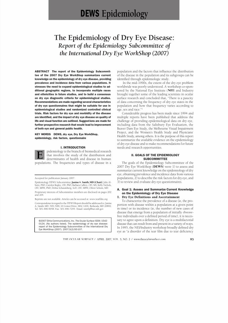
8/20/2019 dry eyes ii
http://slidepdf.com/reader/full/dry-eyes-ii 33/142
THE OCULAR SURFACE / APRIL 2007, VOL. 5, NO. 2 / www.theocularsurface.com 93
DEWS Epidemiology
©2007 Ethis Communications, Inc. The Ocular Surface ISSN: 1542-
0124. (No authors listed). The epidemiology of dry eye disease:
report of the Epidemiology Subcommittee of the International Dry
Eye WorkShop (2007). 2007;5(2):93-107.
ABSTRACT The report of the Epidemiology Subcommit-
tee of the 2007 Dry Eye WorkShop summarizes current
knowledge on the epidemiology of dry eye disease, providing
prevalence and incidence data from various populations. It
stresses the need to expand epidemiological studies to ad-
ditional geographic regions, to incorporate multiple races
and ethnicities in future studies, and to build a consensus
on dry eye diagnostic criteria for epidemiological studies.
Recommendations are made regarding several characteristics
of dry eye questionnaires that might be suitable for use in
epidemiological studies and randomized controlled clinical
trials. Risk factors for dry eye and morbidity of the disease
are identified, and the impact of dry eye disease on quality of
life and visual function are outlined. Suggestions are made for
further prospective research that would lead to improvement
of both eye and general public health.
KEY WORDS DEWS, dry eye, Dry Eye WorkShop,
epidemiology, risk factors, questionnaire
I. INTRODUCTION
pidemiology is the branch of biomedical researchthat involves the study of the distribution anddeterminants of health and disease in human
populations. The frequencies and types of disease in a
population and the factors that influence the distributionof the disease in the population and its subgroups can beidentified through epidemiologic study.
In the mid-1990s, the extent of the dry eye problemworldwide was poorly understood. A workshop co-spon-sored by the National Eye Institute (NEI) and Industrybrought together some of the leading scientists in ocularsurface research and concluded that, “There is a paucityof data concerning the frequency of dry eye states in thepopulation and how that frequency varies according toage, sex and race.”1
Considerable progress has been made since 1994 andmultiple reports have been published that address thechallenge of providing epidemiological data on dry eye,including data from the Salisbury Eye Evaluation, theBeaver Dam Eye Study, the Melbourne Visual ImpairmentProject, and the Women’s Health Study and Physicians’Health Study, among others. It is the purpose of this report
to summarize the available evidence on the epidemiologyof dry eye disease and to make recommendations for futureneeds and research opportunities.
II. GOALS OF THE EPIDEMIOLOGY
SUBCOMMITTEE
The goals of the Epidemiology Subcommittee of the2007 Dry Eye WorkShop (DEWS) were 1) to assess andsummarize current knowledge on the epidemiology of dryeye, obtaining prevalence and incidence data from variouspopulations, 2) to describe the risk factors for dry eye, and3) to review and evaluate dry eye questionnaires.
A. Goal 1: Assess and Summarize Current Knowledge
on the Epidemiology of Dry Eye Disease
1. Dry Eye Definitions and AscertainmentTo characterize the prevalence of a disease (ie, the pro-
portion with disease within a population at a given pointin time) or its incidence (ie, the number of new cases ofdisease that emerge from a population of initially disease-free individuals over a defined period of time), it is neces-sary to agree upon a definition. Dry eye is a multifactorialdisease that can result from and present in a variety of ways.In 1995, the NEI/Industry workshop broadly defined dry
eye as “a disorder of the tear film due to tear deficiency
The Epidemiology of Dry Eye Disease:
Report of the Epidemiology Subcommittee ofthe International Dry Eye WorkShop (2007)
Accepted for publication January 2007.
Epidemiology DEWS Subcommittee: Janine A. Smith, MD (Chair); Julie Al-
beitz, PhD; Carolyn Begley, OD, PhD; Barbara Caffery, OD, MS; Kelly Nichols,OD, MPH, PhD; Debra Schaumberg, ScD, OD, MPH; Oliver Schein, MD.
Proprietary interests of Subcommittee members are disclosed on pages 202and 204.
Reprints are not available. Articles can be accessed at: www.tearfilm.org
Correspondence in regard to the DEWS Report should be addressed to: Janine A. Smith, MD, NEI, NIH, 10 Center Drive, MSC 1204, Bethesda, MD 20892.Tel: 301-496-9058. Fax: 301-496-7295. Email: [email protected]
E
8/20/2019 dry eyes ii
http://slidepdf.com/reader/full/dry-eyes-ii 34/142
THE OCULAR SURFACE / APRIL 2007, VOL. 5, NO. 2 / www.theocularsurface.com94
or excessive tear evaporation which causes damage to theinterpalpebral ocular surface associated with symptoms ofocular discomfort.”1 In this definition, the term tear defi-ciency implied a deficiency of aqueous tears secreted by thelacrimal gland. The requirement of symptoms in the defini-tion is noteworthy, as it was not included in the definitionsestablished in all nations; for instance, it was absent from
the Japanese definition of dry eye until recently.2
2. Challenges in Dry Eye EpidemiologyNo single diagnostic test can be performed in the field
or in the clinic to reliably distinguish individuals withand without dry eye. Furthermore, although a variety ofdiagnostic tests are in common clinical usage, there is noconsensus on which combination of tests should be usedto define the disease, either in the clinic or for the purposesof a research protocol. A major stumbling block has beenthe reported lack of correlation between patients’ irritativeocular symptoms and the results of selected clinical testsfor dry eye. Much of this discrepancy can be explainedby the lack of repeatability of many of the clinical tests incommon use, with the implication that repeated measuresof the same test on the same subjects at different times arenot strongly correlated. Thus, it is not unexpected that suchtests will fail to correlate with each other.
Another plausible reason for a lack of correlation be-tween clinical tests and irritative symptoms may be the nat-ural variability of the disease process, the “subjective” natureof symptoms, and variability in pain thresholds and cogni-tive responses to questions about the physical sensations inthe eyes. Other factors could include the development ofrelative corneal anesthesia with aging and with worsening
disease, and the possibility that symptoms are related toparameters not measured by the tests currently employed.
Dry eye is a symptomatic disease, and, at the presenttime, symptom questionnaires are among the most repeat-able of the commonly used diagnostic tests. They mayprovide a more integrated view of the clinical conditionover time. Irritative symptoms are largely responsible for thepublic health burden and for the care-seeking behavior ofdry eye patients and their desire for therapy. Dry eye symp-toms also affect activities of daily living, adversely impact-ing important tasks such as driving. With these importantissues in mind, it should be noted that individual research
groups in various reports have used different operationaldefinitions of dry eye that are appropriate for their par-ticular purpose. It is of great importance to consider thesedifferences when interpreting and comparing such studies.
The Subcommittee examined data from a number oflarge cohort studies and paid particular attention to defini-tions employed and criteria used, including the require-ment for a certain number, frequency, and intensity ofsymptoms. It was also noted whether a clinical examina-tion was performed, or whether the study diagnosis wasbased on the history of dry eye diagnosed by a clinician.In some cases, measurements from objective tests were
recorded, such as tear production, staining of the ocular
OUTLINE
I. Introduction
II. Goals of the Epidemiology Subcommittee
A. Goal 1: Assess and summarize currentknowledge on the epidemiology of dry eyedisease
1. Dry eye definitions and ascertainment
2. Challenges in dry eye epidemiology
3. Summary of dry eye epidemiology data
a. Prevalence of dry eye
1) Combined prevalence data
2) Discussion/comments
b. Incidence of dry eye
c. Natural history
d. Effects of magnitude of prevalence ofdisease in population on positive andnegative predictive value
4. Morbidity of dry eye
a. Financial costs of dry eye
b. Impact of dry eye on quality of life
c. Burden of dry eye
d. Quality of Life in Sjogren syndrome
e. Impact on visual function
f. Ocular morbidity associated with dry eye
g. Future research directions
B. Goal 2: Describe the risk factors for dry eye
1. Bone marrow transplantation and cancer
2. Menopausal hormone therapy (MHT)
3. Sex hormones
4. Essential fatty acids
5. Low humidity environments6. Computer use
7. Contact lens wear
8. Refractive surgery
C. Goal 3: Review of Dry Eye Questionnaires
1. Features of dry eye questionnaires
a. McMonnies Dry Eye History Questionnaire
b. Canadian Dry Eye Epidemiology Study(CANDEES)
c. Ocular Surface Disease Index (OSDI)
d. Impact of Dry Eye on Everyday Life (IDEEL)
e. Salisbury Eye Evaluation Questionnaire
f. Dry Eye Epidemiology ProjectQuestionnaire
g. Women’s Health Study Questionnaire
h. National Eye Institute-Visual FunctionQuestionnaire (NEI-VFQ)
i. Dry Eye Questionnaire (DEQ) and ContactLens DEQ
j. Melbourne, Australia, Visual ImpairmentProject Questionnaire
2. Summary
3. Future Research
III. Conclusions
DEWS EPIDEMIOLOGY

8/20/2019 dry eyes ii
http://slidepdf.com/reader/full/dry-eyes-ii 35/142
THE OCULAR SURFACE / APRIL 2007, VOL. 5, NO. 2 / www.theocularsurface.com 95
surface, and tear film breakup time. The prevalence of dryeye, using these varying definitions, was tabulated for eachepidemiologic study and is listed in Table 1, along withthe corresponding estimates of population prevalence.
3. Summary of Dry Eye Epidemiology Data
a. Prevalence of Dry Eye 1) Combined Prevalence Data
Based on data from the largest studies of dry eye todate, the Women’s Health Study ( WHS), and the Physi-cians’ Health Study (PHS), and other studies,3-14 it hasbeen estimated that about 3.23 million women and 1.68million men, for a total of 4.91 million Americans 50 yearsand older have dry eye.7,14 Tens of millions more have lesssevere symptoms and probably a more episodic manifesta-tion of the disease that is notable only during contact withsome adverse contributing factor(s), such as low humidityor contact lens wear.
Comparison of age-specific data on the prevalence of
dry eye from large epidemiological studies reveals a rangeof about 5%11 to over 35%12 at various ages. However, itmust be noted that different definitions of dry eye wereemployed in these studies, and, therefore, caution is ad-vised in interpreting direct comparisons of these studies. Although very limited data exist on the potential effect of
race or ethnicity on dry eye prevalence, data from the WHSsuggest that the prevalence of severe symptoms and/orclinical diagnosis of dry eye may be greater in Hispanic and Asian, as compared to Caucasian, women. The combineddata from large population-based epidemiological studiesindicates that the number of women affected with dry eyeappears to exceed that of men.
2) Discussion/CommentsEach of the population-based studies evaluated used a
different definition of dry eye. Some studies included objec-tive examination, but many did not. Nevertheless, in view
of the poor performance (inconsistency, lack of repeatability,
Table 1. Summary of population-based epidemiologic studies of dry eye
Study N Age range Dry eye assessment Prevalence
US Studies
Salisbury Eye Study 3-5 2420 ≥ 65 y At least 1 of 6 symptoms (dryness, 14.6%
gritty/sandiness, burning, redness,
crusting on lashes, eyes stuck shut in
morning), occurring at least often.
Beaver Dam6 3722 ≥ 48 y “For the past 3 months or longer have 14.4%
you had dry eyes?” (If needed, described
as foreign body sensation with itching,
burning, sandy feeling, not related to allergy.)
Women’s Health Study 7 36995 ≥ 49 y Severe symptoms of dryness and irritation, 7.8%
either constantly or often, and/or the physician’s
diagnosis of dry eye as volunteered by the patient.
Physician’s Health 25655 ≥ 50, 55 y Severe symptoms of both dryness and irritation
Studies I and II8,9,14 either constantly or often and/or the physician’s
diagnosis of dry eye as volunteered by the patient.
Australian Studies
Blue Mountains10
1075 ≥ 50 y At least 1 of 4 symptoms regardless of 16.6% (at least severity, or at least 1 symptom with a 1 symptom)
moderate to severe ranking (dryness, 15.3% (3 or more
grittiness, itchiness, discomfort). symptoms)
Melbourne Visual 926 ≥ 40 y At least 1 of 6 “severe” symptoms, not 5.5%
Impairment Project11 attributed by the subject to hay fever
(discomfort, foreign body, itching, tearing,
dryness, photophobia).
Asian Studies
Shihpai12 2038 ≥ 65 y At least 1 of 6 symptoms, often or all 33.7%
of the time (dryness, gritty/sandiness,
burning, sticky, tearing, redness, discharge,
eyes stuck shut in morning).
Sumatra13 1058 ≥ 21 y At least 1 of 6 symptoms, often or all of 27.5%
the time (dryness, gritty/sandiness,
burning, redness, crusting on lashes,
eyes stuck shut in morning).
DEWS EPIDEMIOLOGY
8/20/2019 dry eyes ii
http://slidepdf.com/reader/full/dry-eyes-ii 36/142
THE OCULAR SURFACE / APRIL 2007, VOL. 5, NO. 2 / www.theocularsurface.com96
etc) of commonly used clinical tests and the importance ofsymptoms as an indicator of both the clinical and publicimpact of dry eye, these data from large epidemiologicalstudies have provided much needed information on theprevalence of dry eye.
The studies were performed in different populationsacross the world and, therefore, provide some valuable
information regarding potential differences in dry eye ac-cording to geographic region. In particular, data from thetwo studies performed in Asia suggest the possibility of ahigher prevalence of dry eye in those populations.12,13
The weight of the evidence from large epidemiologicalstudies indicates that female sex and older age increase therisk for dry eye; the Salisbury Eye Evaluation study is themost notable exception.3-5
An overall summary of data suggests that the preva-lence of dry eye lies somewhere in the range of 5-30%in the population aged 50 years and older. It is thoughtthat a proportion of the variation in observed prevalencebetween studies relates to differences in the definition ofdisease used; it is observed that the higher estimates arederived from studies in which a less restrictive definitionwas used, and the lower estimates are derived from thosestudies in which a more restrictive definition was used.Thus, one might surmise that the true prevalence of mod-erate-to-severe dry eye lies somewhere close to the lowerbound of the range, whereas inclusion of mild or episodiccases would bring the estimate in closer proximity to thehigher estimates observed.
Data from the largest US studies, the WHS7 and thePHS,8,9 yield estimates that 3.2 million women and 1.6million men aged 50 years or older suffer from moderate-
to-severe dry eye.
b. Incidence of Dry Eye Epidemiologic data on dry eye can be extracted from
data repositories and federal or public databases, eg, theMedicare/Medicaid databases or other data sources, such ashealth maintenance organizations. Ellwein and colleaguesfound that the dry eye case incidence per 100 fee-for-serviceMedicare beneficiaries increased by 57.4% from 1.22 in1991 to 1.92 in 1998.15 For comparison, cataract case in-cidence increased from 23.44 to 27.29 (16.4%), while thatof diabetic retinopathy increased from 1.36 to 2.55 (87.5%)
in the same time period. Case incidence may be particularlyuseful in evaluating the prevalence for chronic conditionsfor which yearly or more frequent visits are common.15
c. Natural History There is a paucity of data on the natural history of
untreated and treated dry eye. Data regarding the clinicalcourse of dry eye of varying severity and rates of progres-sion from mild to severe disease are also lacking. Suchinformation could be obtained from clinic-based popula-tions with use of standardized tests, and, similarly, baselinedata from clinical trials and other clinical studies could be
employed to obtain useful data. However, such informa-
tion is not yet available. Data from randomized controlledtrials (RCTs) include a wealth of information, which couldbe garnered from the placebo or vehicle-treated groups,both at baseline and at end of study; this would providesome crude natural history data, albeit from a selectedpopulation. At the DEWS meeting in Miami, Florida, inMay 2006, industry representatives to the DEWS group
and attendees were invited to work collaboratively toestablish procedures for sharing this valuable clinicaldata without compromise to proprietary information.The natural history of dry eye remains to be determined,including prognostic factors, the likelihood of disease pro-gression, and the rates of treatment adherence and discon-tinuation and the long-term effect of the use of lubricants.
Epidemiologic data can also be garnered from medicalclaims data. This should be interpreted with the caveat thatprevalence estimates based on claims provide different datathan population-based studies, because claims are made forsymptomatic disease for which diagnosis or treatment issought from the medical care system. Yazdani et al reviewedthe PharMetrics’ Integrated Outcomes database of medicalclaims for 10 million patients from 22 managed care plansand reported a prevalence of dry eye of 0.39% (27,289cases) in 1989.16 International Classification of Disease,Ninth Revision, Clinical Modification (ICD-9 CM) codeswere used to identify cases based on a diagnosis of dry eye(tear film insufficiency 375.15, keratoconjunctivitis sicca(KCS) 370.33, and sicca syndrome 710.2), and CurrentProcedural Terminology (CPT-4) procedure codes for clo-sure of the lacrimal punctum by thermocauterization, liga-tion, laser surgery, or plug were used to identify surgicallytreated cases of dry eye. In this managed care population,
dry eye was diagnosed or treated in 0.65% of women vs0.26% of men (P < 0.001), and dry eye rates increasedwith age, reaching the highest among women 75-79 yearsof age and men 80-84 years of age. This is one of a fewpapers that report a regional variation in the prevalence ofdry eye, with a high rate of 0.8% in the midwestern US,not explained by a higher proportion of women or elderly.16 There are several ICD-9-CM codes that can be applied todry eye cases, including: 370.33 keratoconjunctivitis sicca,non-Sjogren syndrome (SS); 370.34 keratoconjunctivitis,exposure; 372.52 xerosis, conjunctival; 375.15 tear filminsufficiency, unspecified (dry eye syndrome); and 710.20
keratoconjunctivitis sicca, SS.
d. Effects of Magnitude of Prevalence of Disease in Population on Positive and Negative Predictive Value
Community level surveys may overestimate rates of dryeye, due to higher response rates from ill, as opposed tohealthy, individuals. Medical insurance or pharmacy claimscollect data related to diagnoses made by a health care pro-vider, procedures performed, and medications dispensedwithin a specific population, such as a managed care popu-lation. Minority and low-income populations may be differ-entially affected by under-reporting associated with reduced
access to health care or decreased participation in research
DEWS EPIDEMIOLOGY

8/20/2019 dry eyes ii
http://slidepdf.com/reader/full/dry-eyes-ii 37/142
THE OCULAR SURFACE / APRIL 2007, VOL. 5, NO. 2 / www.theocularsurface.com 97
studies. Epidemiologic studies report varying prevalence ofdry eye because of all of these factors and, also, differencesin study populations (community-, clinic-, managed care-based), differences in disease definition, and the lack of astandardized diagnostic test or clinical algorithm of tests.
4. Morbidity of Dry Eye
The public health significance of dry eye is raised by thehigh prevalence of dry eye among the older age groups inmultiple population-based studies combined with the ag-ing of the population. US Census Bureau estimates suggestthat in the period between 2000 and 2050, the number ofpeople in the US aged 65-84 years will increase by 100%,and the number of people aged 85 years and older will in-crease by 333% (Source: U.S. Census Bureau, 2004, “U.S.Interim Projections by Age, Sex, Race, and Hispanic Ori-gin,” http://www.census.gov/ipc/www/usinterimproj/ InternetRelease Date: March 18, 2004). Similar trends are expectedin many other parts of the world.
a. Financial Costs of Dry Eye Few data exist on the direct and indirect costs of dry
eye. The economic impact of dry eye includes costs dueto health care system utilization, including office visits,surgical interventions, prescription medications, over-the-counter and complementary and alternative therapeutics,and purchase of specialized eye wear and other nonphar-macologic therapeutics, such as humidifiers. Indirect costsinclude lost work time and productivity, alteration in worktype or environment, decreased work time and days of workwith dry eye symptoms. In addition to the pain of dry eye,intangible costs include decreased leisure time, impaired
physical functioning and quality of life, impact on socialinteractions, and mental and general health.17
b. Impact of Dry Eye on Quality of LifeThe impact of dry eye on quality of life (QoL) is medi-
ated through 1) pain and irritative symptoms, 2) effect onocular and general health and well-being (general QoL),3) effect on perception of visual function (vision-relatedQoL), and 4) impact on visual performance. For example,the irritative symptoms of dry eye can be debilitating andresult in both psychological and physical effects that impactQoL.18 Dry eye also limits and degrades performance of
common vision-related daily activities, such as driving.19 The need for frequent instillation of lubricant eye drops canaffect social and workplace interactions. The cost of treat-ment and the lack of a cure for dry eye add to the impactof this important public health problem.
Various methods are available to assess the effect of dryeye on visual function and QoL. Non-disease-specific, “ge-neric” instruments like the Medical Outcome Study ShortForm-36 (SF-36) have been applied to dry eye. Utilityassessment, a tool used widely in medicine that permitsthe comparison of the effect of different diseases on QoLbased on strategies such as standard gamble, or trading
years of life for disease-free years, and other techniques,
has also been applied to dry eye.20 Interestingly, the util-ity scores for dry eye were similar to those for moderateangina.21 General vision-related questionnaires, such asthe NEI-Visual Function Questionnaire (NEI-VFQ ), havebeen used. Disease-specific instruments, like the OcularSurface Disease Index (OSDI) and the the Impact of DryEye on Everyday Life (IDEEL) questionnaire have also
been developed and validated specifically for research onthe impact of dry eye.22 These are discussed in detail andreferenced in Section C.
c. Burden of Dry Eye In a recent study among subgroups of 450 participants
in the WHS and 240 participants in the PHS,22a investi-gators used a supplementary dry eye syndrome (DES)questionnaire to ascertain how much a patient’s everydayactivities were limited by symptoms of dry eye and towhat degree problems with their eyes limited them in anumber of common activities of modern living, includ-ing reading, driving, working at the computer, profes-sional activity, and watching TV. By design, the study groupconsisted of one-third with clinically diagnosed DES orsevere symptoms and two-thirds without these charac-teristics. In pooled analyses controlled for age, diabetes,hypertension, and other factors, patients with DES weresignificantly more likely to report problems with reading,carrying out professional work, using a computer, watch-ing television, driving during the day, and driving at night.Overall, patients with DES were about three times morelikely to report problems with common activities than werethose without DES (P < 0.001). These data add furtherweight to the consideration of DES as a significant public
health problem that deserves attention in the clinic.22a Mertzanis et al described the relative burden of dry eye
by comparing a measure of general health-related QoL,the SF-36 responses from persons with and without dryeye against the US norm.18 The IDEEL questionnaire wasadministered to dry eye patients with non-SS KCS (deter-mined by ICD-9CM codes) or SS-related KCS (determinedby San Diego diagnostic criteria) and to control subjectsnot meeting dry eye diagnostic codes. The Survey Manualand Interpretation Guide provided the US normative data.These authors found that while non-SS KCS consistentlylimited daily roles, caused bodily pain or discomfort, and
decreased vitality or energy, this impact became clinicallysignificant when symptoms became moderate in severity. With increased severity of symptoms, other domains wereadversely affected, such as perceptions of health, physicalfunctioning, social functioning, and role-emotional limita-tion. Non-SS KCS had lower role-physical (effect size [ES]= –0.07), bodily pain (ES = –0.08), and vitality (ES = –0.11)scores than norms, but higher scores for general health,physical functioning, role-emotional and mental health,and social functioning. All SF-36 domains were lower (ESranged from –0.14 to 0.91) for the SS patients than adjustednorms except mental health (ES = 0.12) and role-emotional
(ES = –0.13). Regardless of severity of dry eye, patients
DEWS EPIDEMIOLOGY
8/20/2019 dry eyes ii
http://slidepdf.com/reader/full/dry-eyes-ii 38/142
THE OCULAR SURFACE / APRIL 2007, VOL. 5, NO. 2 / www.theocularsurface.com98
reported more limitations in roles due to physical prob-lems and bodily pain likely to affect daily activities. Withincreased severity, patients also reported deficits in generalhealth perception and vitality, and the most severely affectedpatients reported worse health-related QoL over all scales.The IDEEL showed greater discriminative validity for sever-ity levels of dry eye than the SF-36 or EuroQoL (EQ )-5D.23
d. Quality of Life in Sjogren Syndrome Sjogren syndrome is an autoimmune exocrinopathy that
may be associated with immunologic abnormalities and asevere form of dry eye. Vitale et al used a disease-specificinstrument, the OSDI, and a generic instrument developedfor ocular disease, the NEI-VFQ, to evaluate the effect of dryeye in patients with SS on vision-targeted QoL. Despite theless heterogeneous study population of a single disease withsevere dry eye, they found correlations of ocular surfaceparameters with vision-targeted health-related QoL to beweak or nonexistent, consistent with other studies dem-onstrating poor correlations between signs and symptomsof dry eye. Interestingly, the NEI-VFQ correlations withobjective ocular surface parameters were higher than thoseof the OSDI, which may have been due to the capture ofsymptom intensity in addition to frequency in the genericinstrument. Furthermore, the OSDI is targeted to howsymptoms affect current status with a 1-week recall period,whereas the NEI-VFQ may be more suited to capturingoverall impact of chronic ocular disease. It is importantto include assessments of Vision-Targeted Health-RelatedQuality of Life ( VT-HRQ ) and visual function to fully char-acterize the impact of dry eye on health status. The poorcorrelations with conventionally measured signs indicate
that an additional component of disease not captured byclinical examination is being captured.24
Sjogren syndrome can affect many organ systems, andafflicted patients have a reduced quality of life. Severalstudies have measured various aspects of this reduced QoL.Fatigue, anxiety, and depression are major aspects of SS.Thomas et al25 studied the impact of SS in terms of disabilityand QoL in a community-based sample. The majority ofwomen with SS reported interference in leisure activitiesand lifestyle.26 Higher levels of depression/anxiety andfatigue were evident in SS patients compared with non-SSpatients. SS patients had significantly lower scores on the
SF-36, indicating a greater impact on health status. TheSF-36 has been used by Sutcliffe et al,27 Strombeck et al,28 and others29 to show that disabling fatigue is an importantsymptom for many of these patients.
Godaert et al used the multi-dimensional fatigue inven-tory (MFI) to confirm that SS patients had substantiallyhigher levels of daily fatigue and that their fatigue increasedin the evening.30 Giles and Isenberg also noted increasedfatigue in SS patients, even compared to a population oflupus patients.31 Depression is also a prominent featureof SS. Stevenson et al used the Hospital Anxiety and De-pression Scale (HADS) to evaluate 40 SS patients and 40
controls. SS patients showed significantly higher scores.32
Valtysdottir et al also observed more psychiatric symptomsand worse well-being in patients with primary SS.33
e. Impact on Visual FunctionKnowledge is increasing about how dry eye limits and
degrades visual performance, including the conduct ofcommon vision-related daily activities. New methods of
measuring functional visual acuity have demonstrated theeffect of dry eye on visual performance. Distinct from high-contrast visual acuity, measured in a standardized way ata practitioner’s office, visual function is a measure of one’sability to perform vision-intensive tasks, such as reading,using a computer, professional work, driving at night, orwatching television. Visual complaints are highly prevalentamong dry eye patients.22,34,35 These are usually describedas disturbed vision or blurry, foggy vision that clears tem-porarily with the blink.34 These transient changes can beprofound, resulting in marked drops in contrast sensitivityand visual acuity,36 thus affecting workplace productivityand vision-related QoL.19,37
Corneal surface irregularity due to epithelial desicca-tion, tear film instability, and evaporation can be visualizedand quantified with use of tools ranging from corneal to-pography (surface regularity index) to complex instrumentslike wavefront analysis that quantify optical aberrations thatcan degrade the quality of vision and affect non-acuity vi-sual function. An uneven, disrupted tear film in the centralcornea can result in transient vision changes in the dry eyepatient.37,38 Optical aberrations created by tear film breakupbetween blinks contribute to a decline in retinal image qual-ity that can be measured by both objective and subjectivemethods. The Shack-Hartmann aberrometer measures real-
time changes in whole eye, higher order aberrations thatcan be attributed to the tear film,38,39 whereas aberrationsmodeled by changes in corneal topography are based on thefront surface of the eye only.40 Subjective methods can alsobe used to track changes in contrast sensitivity and visualacuity due to tear film disruption.41 Both topical applicationof artificial tears and punctal occlusion in dry eye patientshave been demonstrated to improve visual acuity, contrastsensitivity, and corneal epithelial regularity.36,42,43
f. Ocular Morbidity Associated With Dry Eye Disease Dry eye is associated with contact lens intolerance
and discontinuation of contact lens wear,44,45 can ad-versely affect refractive surgery outcomes,46,47 and maybe associated with increased risk of infection and com-plications with ocular surgery. Few data exist on the riskof infection due to dry eye. Cataract surgery in patientswith dry eye can be associated with ocular morbidity,especially in patients with connective tissue disorders.48
The large incision required for extracapsular cataractextraction was associated with decreased corneal sensa-tion, which can impair wound healing, interrupt normaltrophic factors, and render the cornea more vulnerable toepithelial breakdown in predisposed cases.49 In contrast,
small incision cataract surgery with phacoemulsification
DEWS EPIDEMIOLOGY

8/20/2019 dry eyes ii
http://slidepdf.com/reader/full/dry-eyes-ii 39/142
THE OCULAR SURFACE / APRIL 2007, VOL. 5, NO. 2 / www.theocularsurface.com 99
in patients with dry eye has not been associated with ahigher risk of complications in dry eye patients; Ram etal reported postoperative punctate epitheliopathy in 8/25eyes, epithelial defect in 8/25 eyes of 23 patients, and no
cases of infection or keratolysis.50
g. Future Research Directions A number of questions should be addressed in future
research on the epidemiology of dry eye. What is the natural history of dry eye syndrome? Is the
tissue damage to the ocular surface progressive? Do irritativesymptoms progress, or might they wane over time with thedevelopment of relative corneal anesthesia?
Can we quantify the risk of ocular surface infectionamong patients with dry eye? Is the amount of corneal stain-ing correlated with visual function/functional visual acuity?
What is the incidence of dry eye syndrome in thepopulation, and are there any identifiable demographiccorrelates (eg, age, sex, race/ethnicity)?
Suggested risk factors for dry eye need to be verifiedand quantified (diabetes mellitus, HIV/HTLV1, medications,menopause, alcohol, smoking, pollution, low humidity,various medical conditions, refractive surgery, androgendeficiency, and others). It needs to be determined whetherpredisposing genetic factors contribute to dry eye.
The effects of dry eye should be further defined in termsof QoL, impact on vision, impact on driving, psychologicalissues, cost of care, impact on the health care system, and
overall economic impact.
New diagnostic tests and disease biomarkers should bedeveloped to facilitate epidemiological and clinical research.
B. Goal 2. Describe the Risk Factors for Dry Eye Disease
In 1995, the NEI/Industry Workshop found “virtuallyno data in reference to risk factors for the development ofdry eye.”1 Since that time, epidemiological studies have onlybegun to address the evidence for potential lifestyle, dietary,behavioral, and other risk factors for dry eye, and furtherstudy is clearly needed. The Epidemiology Subcommitteenoted that risk factors might differ among certain subtypesof dry eye, which could dilute associations in population-based studies, in which all forms of dry eye are consideredtogether. Findings from studies in which a purely statistical,non-hypothesis-driven approach was used to study riskfactors must be viewed cautiously, as spurious results are
likely, and, at the same time, important associations couldhave easily been overlooked.
The Subcommittee recommends that future studies ofrisk factors for dry eye should concentrate on the exami-nation of biologically compelling hypotheses in a detailedfashion, with appropriate attention to all aspects of goodepidemiological study design (including sufficient studypower), analysis, and data presentation.
Substantiated risk factors for dry eye include female sex,older age, postmenopausal estrogen therapy,51 a diet that islow in omega 3 essential fatty acids or has a high ratio ofomega 6 to omega 3 fatty acids,52 refractive surgery,53 vitamin
A deficiency, radiation therapy, bone marrow transplanta-
Table 2. Risk factors for dry eye
Level of Evidence
Mostly consistent* Suggestive† Unclear‡
Older age Asian race Cigarette smoking
Female sex Medications Hispanic ethnicity
Postmenopausal estrogen therapy Tricyclic antidepressants
Omega-3 and Omega-6 fatty acids Selective serotonin reuptake inhibitors Anti-cholinergics
Medications Diuretics Anxiolytics
Antihistamines Beta-blockers Antipsychotics
Connective tissue disease Diabetes mellitus Alcohol
LASIK and refractive excimer laser surgery HIV/HTLV1 infection Menopause
Radiation therapy Systemic chemotherapy Botulinum toxin injection
Hematopoietic stem cell transplantation Large incision ECCE and penetrating keratoplasty
Isotretinoin Acne
Vitamin A deficiency Low humidity environments Gout
Hepatitis C infection Sarcoidosis Oral contraceptives
Androgen deficiency Ovarian dysfunction Pregnancy
* Mostly consistent evidence implies the existence of at least one adequately powered and otherwise well-conducted study published in a peer-reviewed journal, along with the existence of a plausible biological rationale and corroborating basic research or clinical data.
† Suggestive evidence implies the existence of either: 1) inconclusive information from peer-reviewed publications or 2) inconclusive or limited informa-tion to support the association, but either not published or published somewhere other than in a peer-reviewed journal
‡ Unclear evidence implies either directly conflicting information in peer-reviewed publications, or inconclusive information but with some basis for abiological rationale
DEWS EPIDEMIOLOGY
8/20/2019 dry eyes ii
http://slidepdf.com/reader/full/dry-eyes-ii 40/142
THE OCULAR SURFACE / APRIL 2007, VOL. 5, NO. 2 / www.theocularsurface.com100
tion, hepatitis C,54 and certain classes of systemic and ocularmedications, including anti-histamines (Table 2). Vitamin A deficiency is a well-recognized risk factor for dry eye,55 and the etiology of the nutritional deficiency now extendsfrom inadequate intake due to unavailability of food to al-coholism-related nutritional deficiency, bariatric surgery,56 malabsorption, eating disorders,57 and vegan diet.58
Other risk factors may include diabetes mellitus,59
human immunodeficiency virus, HIV60 and human T celllymphotropic virus-1 infection,61 connective tissue diseases,systemic cancer chemotherapy, and other medications, suchas isotretinoin,62 antidepressants, anxiolytics, beta-blockers,and diuretics. However, systematic, comprehensive studyof many of these factors is lacking. Conflicting results havebeen reported on the associations between dry eye andsome factors, including alcohol, cigarette smoking, caf-feine, acne,63 and menopausal status. Very few reports existon the risk of dry eye with use of oral contraceptives andpregnancy and the role of ethnicity in dry eye.64
1. Bone Marrow Transplantation and Cancer Allogeneic bone marrow transplantation has increased in
frequency, the indications for the procedure have expanded,and the survival rate is higher than ever before. Conditioningregimens and the use and amount of radiation therapy havealso changed, which has altered the clinical spectrum of ocu-lar graft vs host disease. Dry eye due to radiation therapy,65 systemic chemotherapy, or ocular graft vs host disease as acomplication of bone marrow transplantation can be seen incancer survivors.66,67 A significant pediatric population hasundergone bone marrow transplantation and is surviving todevelop chronic graft vs host disease and dry eye.68
2. Menopausal Hormone Therapy (MHT)In a study of over 25,000 women, postmenopausal es-
trogen therapy was found to be associated with an increasedprevalence of dry eye; the prevalence of dry eye was 5.93%in women not receiving therapy, 6.67% in those receivingestrogen combined with progesterone, and 9.05% in thosetaking estrogen alone.51 In post-menopausal women, foreach additional 3 years of MHT, the odds ratio (OR ) for riskof dry eye was 1.16 (1.09-1.24) after adjusting for age andother possible confounding factors. A prospective analysisof data from this study showed that the initiation of estro-
gen therapy preceded the diagnosis of dry eye syndrome.Corroborating evidence was subsequently found in theShihpai study,12 in which menopausal hormone therapywas associated with an increased risk of dry eye, OR=1.28,and in the Blue Mountains Eye Study, OR=1.7.10
3. Sex HormonesThe role of sex hormones in ocular surface homeostasis
has been recognized and the pathologic mechanism(s) bywhich disturbances may result in dry eye are being inves-tigated. Androgen levels decrease with aging in both menand women.69 Sex steroid deficiency, specifically involving
androgens, has been associated with dry eye in several
distinct clinical entities, such as congenital androgen insuf-ficiency syndrome,70,71 SS,72 premature ovarian failure,73
and anti-androgen medication treatment.74-76 The complexrole of sex hormones in ocular surface health and diseasewarrants further study. There are conflicting reports ofsmall studies of the risk of dry eye with oral contraceptiveuse, and minimal data are available regarding the effect
of pregnancy, hysterectomy, oophorectomy and ovariandysfunction on the ocular surface.77-79
4. Essential Fatty Acids A role for essential fatty acids in dry eye is supported by
largely consistent evidence. In a study of over 32,000 women,Miljanovic et al demonstrated about a 30% reduction inrisk for dry eye with each additional gram of omega-3 fattyacids consumed per day.52 Those who consumed 5 or more4-ounce servings of tuna per week had a > 60% reductionin risk of dry eye. A higher ratio of omega-6 to omega-3fatty acid consumption in the diet was associated with a sig-nificantly increased risk of DES (OR: 2.51; 95% confidenceinterval [CI]: 1.13, 5.58) for > 15:1 versus < 4:1 (P for trend= 0.01). Thus, the higher the level of intake of omega-3 fattyacids in relation to the most commonly consumed types ofomega-6 fatty acids, the lower the risk of dry eye. In supportof a role for essential fatty acids, another study showed thatwomen with SS had a significantly lower intake of omega-3fatty acids (with or without adjustment for energy intake), ascompared to age-matched controls.80 Furthermore, intake ofomega-3 fatty acids has been correlated with the polar lipidpattern of meibomian gland secretions in women with SS.81
5. Low Humidity Environments
Ocular irritative complaints, such as burning, dryness,stinging, and grittiness, are often reported in epidemiologicstudies of indoor environment, especially in offices wherehighly demanding visual and cognitive tasks are performed.82 While the exact cause of these symptoms remains unclear,ocular dryness due to increased tear evaporation may be dueto low humidity, high room temperature and air velocity, de-creased blink rate, or indoor pollution or poor air quality.83,84 Other ultra-low humidity environments, such as aircraftcabins, have also been associated with dry eye symptoms.85,86
6. Computer Use
Computer users often complain of eye strain, eyefatigue, burning, irritation, redness, blurred vision, anddry eyes, among other repetitive strain symptoms.87 Thisconstellation of ocular complaints resulting from videodisplay terminal operation and sustained visual attentionto a computer monitor, with an associated decreased blinkrate, can be regarded as a repetitive strain disorder, computervision syndrome ( CVS ). While asthenopia, glare, and accom-modative difficulty are all aspects of CVS, dry eye appears tocontribute to a major component of symptoms reported.88
7. Contact Lens Wear
Contact lens (CL) wear has often been reported to
DEWS EPIDEMIOLOGY
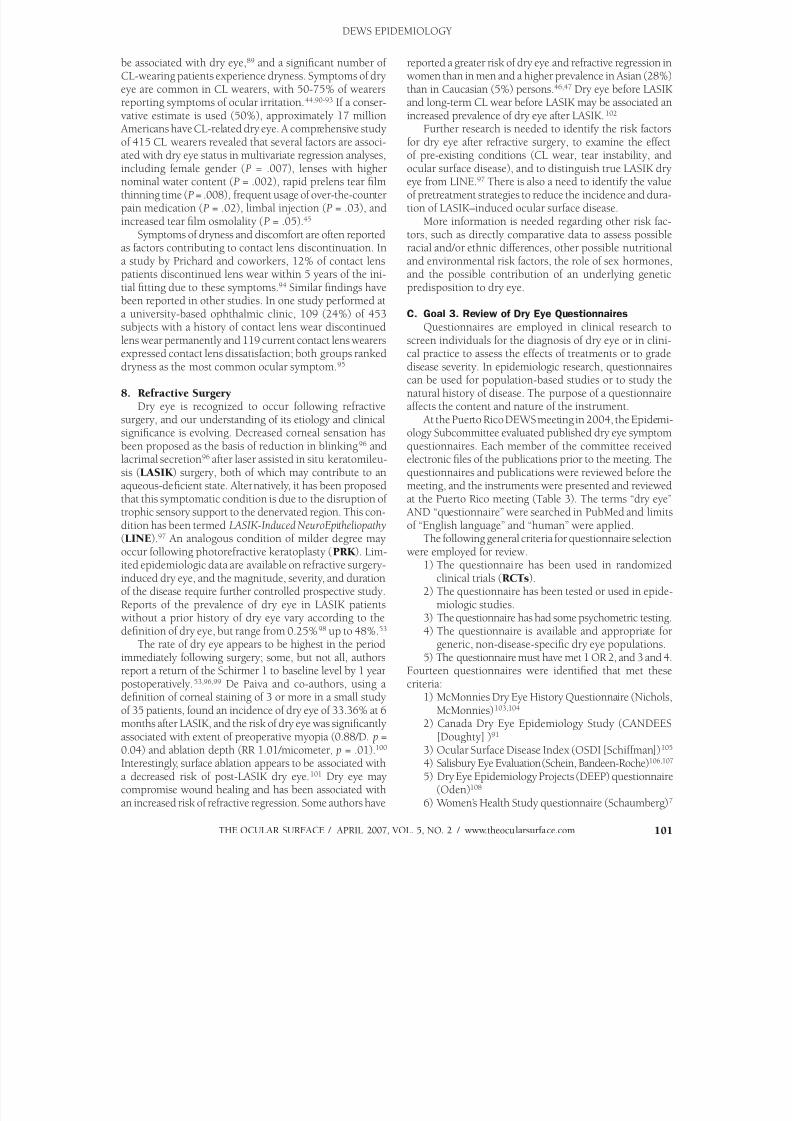
8/20/2019 dry eyes ii
http://slidepdf.com/reader/full/dry-eyes-ii 41/142
THE OCULAR SURFACE / APRIL 2007, VOL. 5, NO. 2 / www.theocularsurface.com 101
be associated with dry eye,89 and a significant number ofCL-wearing patients experience dryness. Symptoms of dryeye are common in CL wearers, with 50-75% of wearersreporting symptoms of ocular irritation.44,90-93 If a conser-vative estimate is used (50%), approximately 17 million Americans have CL-related dry eye. A comprehensive studyof 415 CL wearers revealed that several factors are associ-
ated with dry eye status in multivariate regression analyses,including female gender (P = .007), lenses with highernominal water content (P = .002), rapid prelens tear filmthinning time (P = .008), frequent usage of over-the-counterpain medication (P = .02), limbal injection (P = .03), andincreased tear film osmolality (P = .05).45
Symptoms of dryness and discomfort are often reportedas factors contributing to contact lens discontinuation. Ina study by Prichard and coworkers, 12% of contact lenspatients discontinued lens wear within 5 years of the ini-tial fitting due to these symptoms.94 Similar findings havebeen reported in other studies. In one study performed ata university-based ophthalmic clinic, 109 (24%) of 453subjects with a history of contact lens wear discontinuedlens wear permanently and 119 current contact lens wearersexpressed contact lens dissatisfaction; both groups rankeddryness as the most common ocular symptom.95
8. Refractive SurgeryDry eye is recognized to occur following refractive
surgery, and our understanding of its etiology and clinicalsignificance is evolving. Decreased corneal sensation hasbeen proposed as the basis of reduction in blinking96 andlacrimal secretion96 after laser assisted in situ keratomileu-sis (LASIK ) surgery, both of which may contribute to an
aqueous-deficient state. Alternatively, it has been proposedthat this symptomatic condition is due to the disruption oftrophic sensory support to the denervated region. This con-dition has been termed LASIK-Induced NeuroEpitheliopathy (LINE).97 An analogous condition of milder degree mayoccur following photorefractive keratoplasty (PRK ). Lim-ited epidemiologic data are available on refractive surgery-induced dry eye, and the magnitude, severity, and durationof the disease require further controlled prospective study.Reports of the prevalence of dry eye in LASIK patientswithout a prior history of dry eye vary according to thedefinition of dry eye, but range from 0.25%98 up to 48%.53
The rate of dry eye appears to be highest in the periodimmediately following surgery; some, but not all, authorsreport a return of the Schirmer 1 to baseline level by 1 yearpostoperatively.53,96,99 De Paiva and co-authors, using adefinition of corneal staining of 3 or more in a small studyof 35 patients, found an incidence of dry eye of 33.36% at 6months after LASIK, and the risk of dry eye was significantlyassociated with extent of preoperative myopia (0.88/D. p =0.04) and ablation depth (RR 1.01/micometer, p = .01).100 Interestingly, surface ablation appears to be associated witha decreased risk of post-LASIK dry eye.101 Dry eye maycompromise wound healing and has been associated with
an increased risk of refractive regression. Some authors have
reported a greater risk of dry eye and refractive regression inwomen than in men and a higher prevalence in Asian (28%)than in Caucasian (5%) persons.46,47 Dry eye before LASIKand long-term CL wear before LASIK may be associated anincreased prevalence of dry eye after LASIK.102
Further research is needed to identify the risk factorsfor dry eye after refractive surgery, to examine the effect
of pre-existing conditions (CL wear, tear instability, andocular surface disease), and to distinguish true LASIK dryeye from LINE.97 There is also a need to identify the valueof pretreatment strategies to reduce the incidence and dura-tion of LASIK–induced ocular surface disease.
More information is needed regarding other risk fac-tors, such as directly comparative data to assess possibleracial and/or ethnic differences, other possible nutritionaland environmental risk factors, the role of sex hormones,and the possible contribution of an underlying geneticpredisposition to dry eye.
C. Goal 3. Review of Dry Eye Questionnaires
Questionnaires are employed in clinical research toscreen individuals for the diagnosis of dry eye or in clini-cal practice to assess the effects of treatments or to gradedisease severity. In epidemiologic research, questionnairescan be used for population-based studies or to study thenatural history of disease. The purpose of a questionnaireaffects the content and nature of the instrument.
At the Puerto Rico DEWS meeting in 2004, the Epidemi-ology Subcommittee evaluated published dry eye symptomquestionnaires. Each member of the committee receivedelectronic files of the publications prior to the meeting. Thequestionnaires and publications were reviewed before the
meeting, and the instruments were presented and reviewedat the Puerto Rico meeting (Table 3). The terms “dry eye” AND “questionnaire” were searched in PubMed and limitsof “English language” and “human” were applied.
The following general criteria for questionnaire selectionwere employed for review. 1) The questionnaire has been used in randomized
clinical trials (RCTs).2) The questionnaire has been tested or used in epide-
miologic studies. 3) The questionnaire has had some psychometric testing. 4) The questionnaire is available and appropriate for
generic, non-disease-specific dry eye populations. 5) The questionnaire must have met 1 OR 2, and 3 and 4.Fourteen questionnaires were identified that met thesecriteria:
1) McMonnies Dry Eye History Questionnaire (Nichols,McMonnies)103,104
2) Canada Dry Eye Epidemiology Study (CANDEES[Doughty] )91
3) Ocular Surface Disease Index (OSDI [Schiffman])105 4) Salisbury Eye Evaluation (Schein, Bandeen-Roche)106,107
5) Dry Eye Epidemiology Projects (DEEP) questionnaire(Oden)108
6) Women’s Health Study questionnaire (Schaumberg)7
DEWS EPIDEMIOLOGY

8/20/2019 dry eyes ii
http://slidepdf.com/reader/full/dry-eyes-ii 42/142
THE OCULAR SURFACE / APRIL 2007, VOL. 5, NO. 2 / www.theocularsurface.com102
Table 3. Symptoms and quality of life instruments
Instrument Title/Description/Reference Authors/Report Questionnaire Summary Description/Use
McMonnies
Key questions in a dry eye history
(McMonnies)103
McMonnies. J Am Optomet-
ric Assoc 1986; 57(7):512-7
15 questions Screening questionnaire—
used in a clinic population
McMonnies
Reliability and validity of McMonnies
Dry Eye Index. (Nichols et al)104
Nichols, Nichols, Mitchell.
Cornea 2004;23(4):365-
71
Previously described Screening questionnaire
Dry eye clinic population
*CANDEES
A patient questionnaire approach to
estimating the prevalence of dry eye
symptoms in patients presenting to
optometric practices across Canada
(CANDEES)91
Doughty, Fonn, Richter,
et al. Optom Vis Sci
1997;74(8):624-31
13 questions Epidemiology of dry eye
symptoms in a large random
sample
OSDI
The Ocular Surface Disease Index105
Schiffman, Christianson,
Jacobsen, et al. Arch Oph-
thalmol 2000;118:615-21
12-item questionnaire Measures the severity of dry
eye disease; end points in
clinical trials, symptoms, func-
tional problems and environ-
mental triggers queried for the
past week
OSDI and NEI-VFQ comparison24
Vitale, Goodman, Reed,Smith. Health Quality Life
Outcomes 2004,2:44
Comparison of existingquestionnaires
Tested in Sjogren Syndromepopulation
IDEEL Comparing the discriminative
validity of two generic and one
disease-specific health-related
quality of life measures in a sample
of patients with dry eye23
Rajagopalan, Abetz, Mertz-
anis, et al. Value Health
2005 Mar-Apr;8(2):168-74
3 modules (57 questions):
1. Daily Activities
2. Treatment Satisfaction
3. Symptom Bother
Epidemiologic and clinical
studies
Salisbury Eye Evaluation
Relation between signs and symptoms
of dry eye in the elderly 106
Schein, Tielsch, Munoz
B, et al. Ophthalmology
1997;104:1395-1401
Standardized 6-question
questionnaire*
Population-based prevalence
survey for clinical and subjec-
tive evidence of dry eye
Salisbury Eye Evaluation
Self-reported assessment of dry eye
in a population-based setting107
Bandeen-Roche, Munoz,
Tielsch, et al. Ophthalmol
Vis Sci 1997;38(12):
2469-75
Standardized 6-question
questionnaire*
Population-based prevalence
survey for clinical and subjec-
tive evidence of dry eye
Dry Eye Epidemiology Projects (DEEP)
Sensitivity and specificity of a
screening questionnaire for dry eye108
Oden, Lilienfeld, Lemp,
et al. Adv Exp Med Biol
1998;438; 807-20
19 questions Screening
Women’s Health Study questionnaire
Prevalence of dry eye syndrome
among US women7
Schaumberg, Sullivan,
Buring, Sullivan. Am
J Ophthalmol 2003
Aug;136(2):318-26
3 items from 14-item
original questionnaire
Women’s Health Study/
Epidemiologic studies
National Eye Institute Visual Func-
tion Questionnaire (NEI-VFQ)109
Mangione, Lee, Pitts,
et al. Arch Ophthalmol
1998;116:1496-1504
25-item questionnaire:
2 ocular pain subscale
questions
Useful tool for group-level com-
parisons of vision-targeted,
health-related QOL in clinical
research; not influenced by
severity of underlying eye
disease, suggesting use formultiple eye conditions.
Dry Eye Questionnaire (DEQ)
Habitual patient-reported symptoms
and clinical signs among patients
with dry eye of varying severity 34
Begley, Chalmers,
Abetz, et al. Invest
Ophthalmol Vis Sci 2003
Nov;44(11):4753-61
21 items on prevalence,
frequency, diurnal severity
and intrusiveness of sx
Epidemiologic and clinical
studies
Dry Eye Questionnaire (DEQ)
Use of the dry eye questionnaire to
measure symptoms of ocular irrita-
tion in patients with aqueous tear
deficient dry eye110
Begley, Caffery, Chalmers, et
al. Cornea 2002;21(7):664-
70
As above As above
Table 3 continues on following page
DEWS EPIDEMIOLOGY

8/20/2019 dry eyes ii
http://slidepdf.com/reader/full/dry-eyes-ii 43/142
THE OCULAR SURFACE / APRIL 2007, VOL. 5, NO. 2 / www.theocularsurface.com 103
Contact Lens DEQ
Responses of contact lens wearers
to a dry eye survey 93
Begley, Caffery, Nichols,
Chalmers. Optom Vis Sci
2000; 77(1): 40-6
13 questions Screening questionnaire for
dry eye symptoms in contact
lens wearers
MelbourneVisual Impairment Project
The epidemiology of dry in Melbourne,
Australia11
McCarty, Bansal, Living-
ston, et al. Ophthalmology
1998;105:1114-9
Self-reported symptoms
elicited by interviewer-ad-
ministered questionnaire
Epidemiologic studies
National Eye Institute 42-Item
Refractive Error Questionnaire111
Hays, Mangione, Ellwein,
et al. Ophthalmology
2003;110(12):2292-301
42-item questionnaire:
4 related questions: ocular
pain or discomfort, dryness,
tearing, soreness or tiredness
QoL due to refractive error
Sicca/SS questionnaire
Validation of the Sicca symptoms
inventory for clinical studies of
Sjogren’s syndrome112
Bowman, Booth, Platts,
et al. Sjogren’s Interest
Group. J Rheumatol
2003;30(6):1259-66
Inventory of both symptoms
and signs of Sjogren
Syndrome
Epidemiologic studies for
Sjogren Syndrome
Bjerrum questionnaire
Study Design and Study Populations113
Bjerrum. Acta Ophthalmo-
logica (Scand) 2000:10-3
3-part questionnaire which
includes an ocular part
with 14 questions
QOL due to SS dry eye, diagnosis
of dry eye, epidemiology of SS
Bjerrum questionnaire
Dry Eye Symptoms in patients and
normals114
Bjerrum. Acta Ophthal-
mologica (Scand) 2000,
14-5.
As above Screening questionnaire
Bjerrum questionnaire
Test and symptoms in keratoconjunc-
tivitis sicca and their correlation35
Bjerrum. Acta Ophthalmol
(Scand) 1996:74:436-41
Dry eye tests
Ocular symptom questionnaire
(14 questions)
Examine correlation between
dry eye test and ocular symp-
tom questionnaire responses
Utility assessment questionnaire
Utility assessment among pts with
dry eye disease21
Schiffman, Walt, Jacob-
sen, et al. Ophthalmology
2003;110(7):1412-9
Utility assessment Utility assessment
Japanese dry eye awareness study
Results of a population-based
questionnaire on the symptoms and
lifestyles associated with dry eye115
Shimmura, Shimazaki,
Tsubota. Cornea 1999;
18(4):408-11
30 questions relating to
symptoms and knowledge
of dry eye
Population-based, self-diag-
nosis study to assess public
awareness and symptoms of
dry eye
Sicca/SLE questionnaire Oral and ocular sicca symptoms and
findings are prevalent in systemic
lupus erythematosus116
Jensen, Bergem, Gilboe,et al. Oral Pathol Med
1999;28:317-22
6-question symptom ques-tionnaire Screening for dry eye symp-toms in SLE patients
American-European Consensus Group
Classification criteria for Sjogren’s
syndrome: a revised version of the
European criteria proposed by the
American-European Consensus Group117
Vitali C, Bombardieri S,
Jonnson R, et al. Ann
Rheum Dis 2002;1:554-8
6 areas of questions:
Ocular symptoms; oral
symptoms; ocular signs;
histopathology; oral signs;
auto-antibodies
Clarification of classification
of primary and secondary
Sjogren syndrome, and of
exclusion criteria.
The Eye Care Technology Forum
Impacting Eye Care118
Ellwein. Ophthalmology
1994;101:199-201
Issues: Standardizing
clinical evaluation
Decree for change
Table 3. Symptoms and quality of life instruments (continued)
Instrument Title/Description/Reference Authors/Report Questionnaire Summary Description/Use
7) National Eye Institute-Visual Function Questionnaire(NEI-VFQ [Mangione])109
8) Dry Eye Questionnaire (DEQ [Begley et al])34,110
9) Contact Lens DEQ (Begley et al),93 10) Melbourne Visual Impairment Project (McCarty)11
11) NEI-Refractive Error questionnaire 111
12) Sicca Symptoms Inventory (Bowman)112
13) Bjerrum questionnaire35,113,114
14) Japanese dry eye awareness questionnaire (Shimmura)115
The Impact of Dry Eye on Everyday Life (IDEEL) wasadded to the list when it became publicly available.
A number of questionnaires were selected for detailed
review, and these are summarized below. Appendix I, avail-able at www.tearfilm.org, provides additional details of theMcCarty symptom questionnaire, Ocular Surface DiseaseIndex (OSDI), Salisbury Eye Evaluation questionnaire,Impact of Dry Eye on Everyday Life (IDEEL) questionnaire,and the McMonnies questionnaire.
During the meeting, the strengths and weaknesses ofexisting surveys were discussed, and it was noted thatinformation is limited for each of them. The group agreedthat a set of several standardized, validated questionnairessuitable for a variety of purposes and available to investi-gators would be desirable. Data from completed clinical
trials could be used to validate existing instruments and
DEWS EPIDEMIOLOGY

8/20/2019 dry eyes ii
http://slidepdf.com/reader/full/dry-eyes-ii 44/142
THE OCULAR SURFACE / APRIL 2007, VOL. 5, NO. 2 / www.theocularsurface.com104
maximize the ability to improve instruments for use inclinical trials and epidemiologic studies.
1. Features of Dry Eye QuestionnairesThe instruments varied in length, intended use, popu-
lation in which they were tested, mode of administration(self, interviewer, and phone) and extent of validation.
Common elements in questionnaires (two or more instru-ments) included query of: clinician-based or other diagnosisof dry eye; frequency and/or intensity of symptoms; effectof symptoms on activities of daily living; effect of environ-mental triggers on symptoms; presence of dry mouth; effectof visual tasks on symptoms (eg, computer use); effect oftreatment on symptoms; contact lens wear; medications;and allergies. Items infrequently included were queriesrelated to the use of drops, arthritis, thyroid disease, drynose or vagina, emotional triggers, and global assessmentby the patient. The recall period was not specified in mostquestionnaires, but it ranged from 1-2 weeks in those inwhich a period was specified. Below is a summary of thegeneral features of ten questionnaires:
a. McMonnies Dry Eye History Questionnaire • 12 items- most dichotomous yes/no, weighted scoring • Screening, used in dry eye clinic population • Includes age, sex, contact lens wear • Previous diagnosis of dry eye, triggers (environment,
swimming, alcohol)• Frequency of symptoms: dryness, grittiness, soreness, red-
ness, tiredness (Answers: Never, sometimes, often, constantly) • Medications, arthritis, dry mouth, thyroid status
b. Canadian Dry Eye Epidemiology Study(CANDEES)
• 13 questions: age, sex, CL wear and effect on symptoms,dry eye diagnosis
• Epidemiologic study of prevalence of symptoms • Frequency and intensity of symptoms combined (An-
swers: Occasional and mild, Occasional and moderate,Constant and mild, Constant and moderate, Severe)
• Medications, time of day, allergies, dry mouth, itchy/ swollen/red eyelids
c. Ocular Surface Disease Index (OSDI)
• 12 items: visual function (6); ocular symptoms (3);environmental triggers (3)
• Frequency with 1-week recall period (Answers: Noneof the time, Some of the time, Half of the time, Most of thetime, All of the time [0-4])
• Scoring algorithm published:100 = complete disability;0 = no disability
• Validated in dry eye population and used as outcomemeasure in RCT
d. Impact of Dry Eye on Everyday Life (IDEEL) • 3 modules (Daily activities, Treatment satisfaction, and
Symptom bother) with a total of 57 questions
• 2-week recall period • 5-point scales on frequency, bother, or limitation for
most questions • Daily Activities includes vision, environmental triggers,
emotional triggers, and work • Validated in dry eye population of 210 subjects with
range of dry eye severity
• Questionnaire is now available from MAPI Values,Boston, MA
e. Salisbury Eye Evaluation Questionnaire• 6 items: Frequency of symptoms and 3 signs (Answers:
Rarely, Sometimes, Often, All of the time) Do your eyes ever feel dry?
Gritty or sandy sensation in eyes? Burning sensation? Red, crusting lashes, stuck shut in morning • Self-reported population-based prevalence survey in
elderly for signs and symptoms • Latent class analysis of symptom patterns • Low correlations with dry eye signs
f. Dry Eye Epidemiology Project Questionnaire • 19 items: treatments, symptoms, others • Screening questionnaire (phone interview) • Use of eye washes, compresses, drops • Frequency of symptoms • Itchy, sore, dry, scratchy, gritty, burning, irritated, water-
ing, photophobia, red, sticky, achy (Never, Sometimes,Often, Constantly)
• Dry mouth, ocular allergies, contact lens wear frequency,physician diagnosis of dry eye
g. Women’s Health Study Questionnaire • 3 items (Answers: Constantly, Often, Sometimes, Never ) Previous diagnosis of dry eye from clinician—yes or no How often eyes feel dry (not wet enough)? How often eyes feel irritated? • Large population-based prevalence survey • Case definition: Both dryness and irritation constantly
or often • Similar sensitivity and specificity as 14 items including: san-
dy or gritty, burning or stinging pain, itching, light sensitiv-ity, blurry vision, tiredness, soreness, scratchiness, redness,
stickiness, achy feeling watery eyes and swollen eyelids • Validated against standardized clinical exam
h. National Eye Institute-Visual FunctionQuestionnaire (NEI-VFQ)
• 25 items of frequency and severity of symptom andeffects on activities of daily living
• Multiple domains: ie, near vision, general health, socialproblems, distance vision…
How often does pain or discomfort affect activities ofdaily living (Answers: All, Most, Some, A little, None ofthe time [5-point scale])
—How much pain (ie, burn, itch, ache)? (Answers:
DEWS EPIDEMIOLOGY
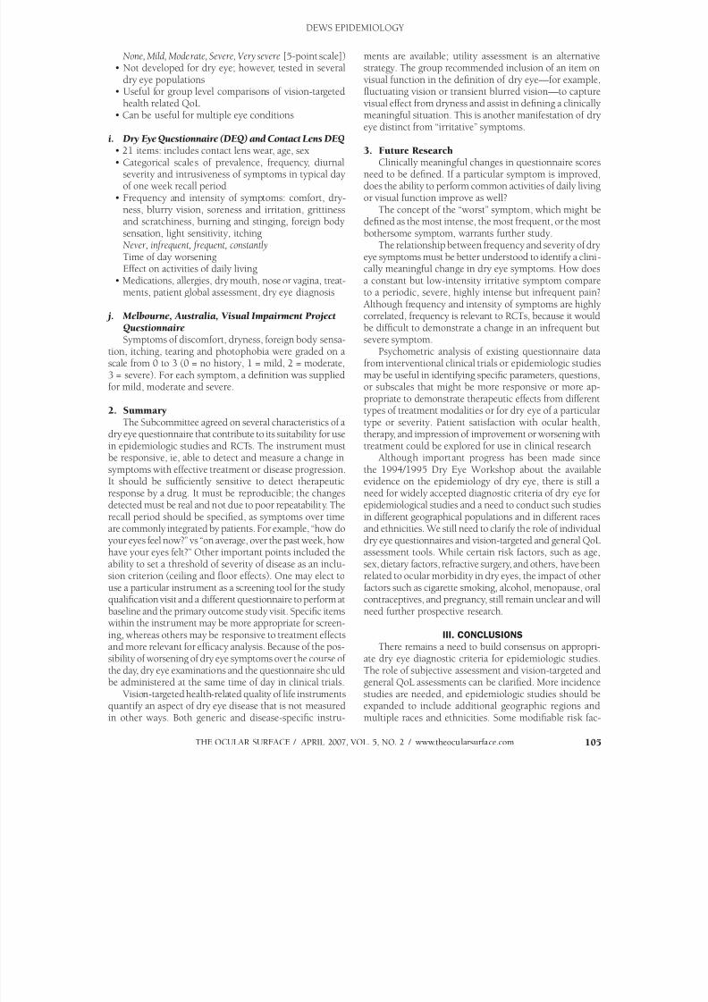
8/20/2019 dry eyes ii
http://slidepdf.com/reader/full/dry-eyes-ii 45/142
THE OCULAR SURFACE / APRIL 2007, VOL. 5, NO. 2 / www.theocularsurface.com 105
None, Mild, Moderate, Severe, Very severe [5-point scale]) • Not developed for dry eye; however, tested in several
dry eye populations • Useful for group level comparisons of vision-targeted
health related QoL• Can be useful for multiple eye conditions
i. Dry Eye Questionnaire (DEQ) and Contact Lens DEQ• 21 items: includes contact lens wear, age, sex • Categorical scales of prevalence, frequency, diurnal
severity and intrusiveness of symptoms in typical dayof one week recall period
• Frequency and intensity of symptoms: comfort, dry-ness, blurry vision, soreness and irritation, grittinessand scratchiness, burning and stinging, foreign bodysensation, light sensitivity, itchingNever, infrequent, frequent, constantly
Time of day worsening Effect on activities of daily living • Medications, allergies, dry mouth, nose or vagina, treat-
ments, patient global assessment, dry eye diagnosis
j. Melbourne, Australia, Visual Impairment ProjectQuestionnaire Symptoms of discomfort, dryness, foreign body sensa-
tion, itching, tearing and photophobia were graded on ascale from 0 to 3 (0 = no history, 1 = mild, 2 = moderate,3 = severe). For each symptom, a definition was suppliedfor mild, moderate and severe.
2. SummaryThe Subcommittee agreed on several characteristics of a
dry eye questionnaire that contribute to its suitability for usein epidemiologic studies and RCTs. The instrument mustbe responsive, ie, able to detect and measure a change insymptoms with effective treatment or disease progression.It should be sufficiently sensitive to detect therapeuticresponse by a drug. It must be reproducible; the changesdetected must be real and not due to poor repeatability. Therecall period should be specified, as symptoms over timeare commonly integrated by patients. For example, “how doyour eyes feel now?” vs “on average, over the past week, howhave your eyes felt?” Other important points included theability to set a threshold of severity of disease as an inclu-
sion criterion (ceiling and floor effects). One may elect touse a particular instrument as a screening tool for the studyqualification visit and a different questionnaire to perform atbaseline and the primary outcome study visit. Specific itemswithin the instrument may be more appropriate for screen-ing, whereas others may be responsive to treatment effectsand more relevant for efficacy analysis. Because of the pos-sibility of worsening of dry eye symptoms over the course ofthe day, dry eye examinations and the questionnaire shouldbe administered at the same time of day in clinical trials.
Vision-targeted health-related quality of life instrumentsquantify an aspect of dry eye disease that is not measured
in other ways. Both generic and disease-specific instru-
ments are available; utility assessment is an alternativestrategy. The group recommended inclusion of an item onvisual function in the definition of dry eye—for example,fluctuating vision or transient blurred vision—to capturevisual effect from dryness and assist in defining a clinicallymeaningful situation. This is another manifestation of dryeye distinct from “irritative” symptoms.
3. Future ResearchClinically meaningful changes in questionnaire scores
need to be defined. If a particular symptom is improved,does the ability to perform common activities of daily livingor visual function improve as well?
The concept of the “worst” symptom, which might bedefined as the most intense, the most frequent, or the mostbothersome symptom, warrants further study.
The relationship between frequency and severity of dryeye symptoms must be better understood to identify a clini-cally meaningful change in dry eye symptoms. How doesa constant but low-intensity irritative symptom compareto a periodic, severe, highly intense but infrequent pain? Although frequency and intensity of symptoms are highlycorrelated, frequency is relevant to RCTs, because it wouldbe difficult to demonstrate a change in an infrequent butsevere symptom.
Psychometric analysis of existing questionnaire datafrom interventional clinical trials or epidemiologic studiesmay be useful in identifying specific parameters, questions,or subscales that might be more responsive or more ap-propriate to demonstrate therapeutic effects from differenttypes of treatment modalities or for dry eye of a particulartype or severity. Patient satisfaction with ocular health,
therapy, and impression of improvement or worsening withtreatment could be explored for use in clinical research
Although important progress has been made sincethe 1994/1995 Dry Eye Workshop about the availableevidence on the epidemiology of dry eye, there is still aneed for widely accepted diagnostic criteria of dry eye forepidemiological studies and a need to conduct such studiesin different geographical populations and in different racesand ethnicities. We still need to clarify the role of individualdry eye questionnaires and vision-targeted and general QoLassessment tools. While certain risk factors, such as age,sex, dietary factors, refractive surgery, and others, have been
related to ocular morbidity in dry eyes, the impact of otherfactors such as cigarette smoking, alcohol, menopause, oralcontraceptives, and pregnancy, still remain unclear and willneed further prospective research.
III. CONCLUSIONS
There remains a need to build consensus on appropri-ate dry eye diagnostic criteria for epidemiologic studies.The role of subjective assessment and vision-targeted andgeneral QoL assessments can be clarified. More incidencestudies are needed, and epidemiologic studies should beexpanded to include additional geographic regions and
multiple races and ethnicities. Some modifiable risk fac-
DEWS EPIDEMIOLOGY
8/20/2019 dry eyes ii
http://slidepdf.com/reader/full/dry-eyes-ii 46/142
THE OCULAR SURFACE / APRIL 2007, VOL. 5, NO. 2 / www.theocularsurface.com106
tors have been identified for dry eye, and public educationresulting this regard should lead to improvement in botheye and general health, while further, prospective study isneeded to elucidate other risk factors.
Detailed templates of questionnaires can be accessedat: www.tearfilm.org.
REFERENCES
1. Lemp MA. Report of the National Eye Institute/Industry workshop onClinical Trials in Dry Eyes. CLAO J 1995;21:221-32
2. Miyawaki S, Nishiyama S. [Classification criteria for Sjogren’s syndrome-sensitivity and specificity of criteria of the Japanese Ministry of Healthand Welfare (1977) and criteria of European community (1993)]. NipponRinsho 1995;53:2371-5
3. Schein OD, Hochberg MC, Munoz B, et al. Dry eye and dry mouth in theelderly: a population-based assessment. Arch Intern Med 1999;159:1359-63
4. Schein OD, Munoz B, Tielsch JM, et al. Prevalence of dry eye among theelderly. Am J Ophthalmol 1997;124:723-8
5. Munoz B, West SK, Rubin GS, et al. Causes of blindness and visual impair-ment in a population of older Americans: The Salisbury Eye EvaluationStudy. Arch Ophthalmol 2000;118:819-25
6. Moss SE, Klein R, Klein BE. Prevalence of and risk factors for dry eyesyndrome. Arch Ophthalmol 2000;118:1264-8
7. Schaumberg DA, Sullivan DA, Buring JE, Dana MR. Prevalence of dry eye
syndrome among US women. Am J Ophthalmol 2003:136;318-26 8. Christen WG, Manson JE, Glynn RJ, et al. Low-dose aspirin and risk of
cataract and subtypes in a randomized trial of US physicians. OphthalmicEpidemiol 1998;5:133-42
9. Christen WG, Gaziano JM, Hennekens CH. Design of Physicians’ HealthStudy II—a randomized trial of beta-carotene, vitamins E and C, and mul-tivitamins, in prevention of cancer, cardiovascular disease, and eye disease,and review of results of completed trials. Ann Epidemiol 2000;10:125-34
10. Chia EM, Mitchell P, Rochtchina E, et al. Prevalence and associations ofdry eye syndrome in an older population: the Blue Mountains Eye Study.Clin Experiment Ophthalmol 2003;31:229-32
11. McCarty CA, Bansal AK, Livingston PM, et al. The epidemiology of dryeye in Melbourne, Australia. Ophthalmology 1998;105:1114-9
12. Lin PY, Tsai SY, Cheng CY, et al. Prevalence of dry eye among an elderlyChinese population in Taiwan: the Shihpai Eye Study. Ophthalmology 2003;110:1096-101
13. Lee AJ, Lee J, Saw SM, et al. Prevalence and risk factors associated withdry eye symptoms: a population based study in Indonesia. Br J Ophthalmol 2002;86:1347-51
14. Miljanovic B, Dana MR, Sullivan DA, Schaumberg DA. Prevalence andrisk factors for dry eye syndrome among older men in the United States.Invest Ophthalmol Vis Sci 2007 (ARVO abstract)
15. Ellwein LB, Urato CJ. Use of eye care and associated charges among theMedicare population:1991-1998. Arch Ophthalmol 2002;120:804-11
16. Yazdani C, McLaughlin T, Smeeding JE, Walt J. Prevalence of treated dryeye disease in a managed care population. Clin Ther 2001;23:1672-82
17. Reddy P, Grad O, et al. The economic burden of dry eye: a conceptualframework and preliminary assessment. Cornea 2004;23:751-61
18. Mertzanis P, Abetz L, Rajagopalan K, et al. The relative burden of dry eyein patients’ lives: comparisons to a US normative sample. Invest OphthalmolVis Sci 2005;46:46-50
19. Schiffman RM, Christianson MD, Jacobsen G, et al. Reliability and validity
of the Ocular Surface Disease Index. Arch Ophthalmol 2000;118:615-21 20. Brown MM, Brown GC. Utility assessment and dry eye disease. Ophthal-
mology 2004;111:852-3; author reply, 853 21. Schiffman RM, Walt JG, Jacobsen G, et al. Utility assessment among
patients with dry eye disease. Ophthalmology 2003;110:1412-9 22. Gulati A, Sullivan R, Buring JE, et al. Validation and repeatability of a short
questionnaire for dry eye syndrome. Am J Ophthalmol 2006;142:125-31 22a. Miljanovic B, Dana R, Sullivan DA, Schaumberg DA. Impact of dry eye
syndrome on vision-related quality of life. Am J Ophthalmol 2007;143:409-15. Epub 107 Jan2.
23. Rajagopalan K, Abetz L, Mertzanis P, et al. Comparing the discriminativevalidity of two generic and one disease-specific health-related quality of lifemeasures in a sample of patients with dry eye. Value Health 2005;8:168-74
24. Vitale S, Goodman LA, Reed GF, Smith JA. Comparison of the NEI-VFQand OSDI questionnaires in patients with Sjogren’s syndrome-related dryeye. Health Qual Life Outcomes 2004;2:44
25. Thomas E, Hay EM, et al. Sjogren’s syndrome: a community-based study
of prevalence and impact. Br J Rheumatol 1998;37:1069-76 26. Sullivan BD, Cermak JM, Sullivan RM, et al. Correlations between nutri-
ent intake and the polar lipid profiles of meibomian gland secretions inwomen with Sjogren’s syndrome. Adv Exp Med Biol 2002;506:441-8
27. Sutcliffe N, Stoll T, Pyke S, Isenberg DA. Functional disability and endorgan damage in patients with systemic lupus erythematosus (SLE) andSjogren’s syndrome (SS), and primary SS. J Rheumatol 1998;25:63-8
28. Strombeck B, Ekdahl C, Manthorpe R, et al. Health-related quality of life inprimary Sjogren’s syndrome, rheumatoid arthritis and fibromyalgia comparedto normal population data using SF-36. Scand J Rheumatol 2000;29:20-8
29. Bjerrum K, Prause JU. Primary Sjogren’s syndrome: a subjective descriptionof the disease. Clin Exp Rheumatol 1990;8:283-8
30. Godaert GL, Hartkamp A, Geenen R, et al. Fatigue in daily life in patientswith primary Sjogren’s syndrome and systemic lupus erythematosus. AnnN Y Acad Sci 2002;966:320-6
31. Giles I, Isenberg D. Fatigue in primary Sjogren’s syndrome: is there a linkwith the fibromyalgia syndrome? Ann Rheum Dis 2000;59: 875-8
32. Stevenson HA, Jones ME, Rostron JL, et al. UK patients with primarySjogren’s syndrome are at increased risk from clinical depression. Ger-odontology 2004;21:141-5
33. Valtysdottir ST, Gudbjornsson B, Lindtqvist U, et al. Anxiety and depressionin patients with primary Sjogren’s syndrome. J Rheumatol 2000;27:165-9
34. Begley CG, Chalmers RL, Abetz L, et al. The relationship between habitualpatient-reported symptoms and clinical signs among patients with dry eyeof varying severity. Invest Ophthalmol Vis Sci 2003;44:4753-61
35. Bjerrum KB. Test and symptoms in keratoconjunctivitis sicca and their
correlation. Acta Ophthalmol Scand 1996;74 436-41 36. Goto E, Yagi Y, Matsumoto Y, Tsubota K. Impaired functional visual acuity
of dry eye patients. Am J Ophthalmol 2002;133:181-6 37. Tutt R, Bradley A, Begley C, Thibos LN. Optical and visual impact of tear
break-up in human eyes. Invest Ophthalmol Vis Sci 2000;41:4117-23 38. Montes-Mico R, Caliz A, Alio JL. Wavefront analysis of higher order aber-
rations in dry eye patients. J Refract Surg 2004;20:243-7 39. Thibos LN, Hong X. Clinical applications of the Shack-Hartmann aber-
rometer. Optom Vis Sci 1999;76:817-25 40. Lin YY, Carrel H, Wang IJ, et al. Effect of tear film break-up on higher
order aberrations of the anterior cornea in normal, dry, and post-LASIKeyes. J Refract Surg 2005;21:S525-S529
41. Ishida R, Kojima T, Dogru M, et al. The application of a new continuousfunctional visual acuity measurement system in dry eye syndromes. Am
J Ophthalmol 2005;139:253-8 42. Nilforoushan MR, Latkany RA, Speaker MG. Effect of artificial tears on
visual acuity. Am J Ophthalmol 2005;140: 830-5 43. Liu Z, Pflugfelder SC. Corneal surface regularity and the effect of artificial
tears in aqueous tear deficiency. Ophthalmology 1999;106:939-43 44. Nichols JJ, Ziegler C, Mitchell GL, Nichols KK. Self-reported dry eye disease
across refractive modalities. Invest Ophthalmol Vis Sci 2005;46:1911–445. Nichols J J, Sinnott LT. Tear film, contact lens, and patient-related factors
associated with contact lens-related dry eye. Invest Ophthalmol Vis Sci 2006;47:1319-28
46. Albietz JM, Lenton LN, McLennan SB. Chronic dry eye and regression after la-ser in situ keratomileusis for myopia. J Cataract Refract Surg 2004;30:675-84
47. Albietz JM, Lenton LM, McLennan SG. Dry eye after LASIK: comparison ofoutcomes for Asian and Caucasian eyes. Clin Exp Optom 2005;88:89-96
48. Jones RR, Maguire LJ. Corneal complications after cataract surgery inpatients with rheumatoid arthritis. Cornea 1992;11:148-50
49. Ram J, Sharma A, Pandav SS, et al. Cataract surgery in patients with dryeyes. J Cataract Refract Surg 1998;24:1119-24
50. Ram J, Gupta A, Brar G, et al. Outcomes of phacoemulsification in patientswith dry eye. J Cataract Refract Surg 2002;28:1386-9
51. Schaumberg DA, Buring JE, Sullivan DA, Dana MR. Hormone replacementtherapy and dry eye syndrome. JAMA 2001;286:2114-9
52. Miljanovic B, Trivedi KA, Dana MR, et al. Relation between dietary n-3and n-6 fatty acids and clinically diagnosed dry eye syndrome in women.
Am J Clin Nutr 2005;82: 887-93 53. Hovanesian JA, Shah SS, et al. Symptoms of dry eye and recurrent erosion
syndrome after refractive surgery. J Cataract Refract Surg 2001;27:577-84 54. Zegans ME, Anninger W, Chapman C, Gordon SR. Ocular manifestations
of hepatitis C virus infection. Curr Opin Ophthalmol 2002;13: 423-7 55. Sommer A. Vitamin A deficiency and the global response. Forum Nutr
2003;56:33-5 56. Lee WB, Hamilton SM, et al. Ocular complications of hypovitaminosis a
after bariatric surgery. Ophthalmology 2005;112:1031-4 57. Jaworowski S, Drabkin E, Rozenman Y. Xerophthalmia and undiagnosed
eating disorder. Psychosomatics 2002;43(6): 506-7.
DEWS EPIDEMIOLOGY

8/20/2019 dry eyes ii
http://slidepdf.com/reader/full/dry-eyes-ii 47/142
THE OCULAR SURFACE / APRIL 2007, VOL. 5, NO. 2 / www.theocularsurface.com 107
58. Colev MH, Engel H, Mayers M, et al. Vegan diet and vitamin A deficiency.Clin Pediatr (Phila) 2004;43:107-9
59. Kaiserman IN, Kaiserman N, Nakar S, Vinker S. Dry eye in diabetic pa-tients. Am J Ophthalmol 2005;139: 498-503
60. Chronister CL. Review of external ocular disease associated with aids andHIV infection. Optom Vis Sci 1996;73:225-30
61. Merle HD, Smadja D, Le Hoang P, et al. Ocular manifestations in patientswith HTLV-I associated infection--a clinical study of 93 cases. Jpn J Oph-thalmol 1996;40:260-70
62. Aragona P, Cannavo SP, Boragia F, Guarneri F. Utility of studying the ocu-
lar surface in patients with acne vulgaris treated with oral isotretinoin: arandomized controlled trial. Br J Dermatol 2005;152:576-8
63. Ozdemir M, Ozdemir G, Sasmaz S, Arican O. Ocular surface disorders andtear function changes in nodulo-cystic acne. J Dermatol 2005;32:174-8
64. Hom M, De Land P. Prevalence and severity of symptomatic dry eyes inHispanics. Optom Vis Sci 2005;82:206-8
65. Thomas O, Mahe M, Campion L, et al. Long-term complications of totalbody irradiation in adults. Int J Radiat Oncol Biol Phys 2001;49:125-31
66. Bray LC, Carey PJ, Proctor SJ, et al. Ocular complications of bone marrowtransplantation. Br J Ophthalmol 1991;75:611-4
67. Ogawa Y, Kuwana M. Dry eye as a major complication associated withchronic graft-versus-host disease after hematopoietic stem cell transplanta-tion. Cornea 2003;22(7 Suppl):S19-S27
68. Suh DW, Ruttum MS, Stuckenschneider BJ, et al. Ocular findings afterbone marrow transplantation in a pediatric population. Ophthalmology 1999;106:1564-70
69. Sullivan BD, Evans JE, Dana MR, Sullivan DA. Influence of aging on thepolar and neutral lipid profiles in human meibomian gland secretions.
Arch Ophthalmol 2006;124: 1286-92 70. Sullivan BD, Evans JE, et al. Complete androgen insensitivity syndrome: effect
on human meibomian gland secretions. Arch Ophthalmol 2002;120:1689-99 71. Cermak JM, Krenzer KL, Sullivan RM, et al. Is complete androgen insen-
sitivity syndrome associated with alterations in the meibomian gland andocular surface? Cornea 2003;22:516-21
72. Sullivan DA, Belanger A, Cermak JM, et al. Are women with Sjogren’ssyndrome androgen-deficient? J Rheumatol 2003;30:2413-9
73. Smith JA, Vitale S, et al. Dry eye signs and symptoms in women withpremature ovarian failure. Arch Ophthalmol 2004;122:151-6
74. Sullivan BD, Evans JE, Krenzer KL, et al. Impact of antiandrogen treat-ment on the fatty acid profile of neutral lipids in human meibomian glandsecretions. J Clin Endocr Metab 2000;85:4866-73
75. Krenzer KL, Dana MR, Ullman MD, et al. Effect of androgen deficiency on
the human meibomian gland and ocular surface. J Clin Endocrinol Metab 2000;85:4874-82
76. Sullivan DA. Androgen deficiency and dry eye syndromes. Arch Soc EspOftalmol 2004;79:49-50
77. Connor CG, Flockencier LL, Hall CW. The influence of gender on theocular surface. J Am Optom Assoc1999;70:182-6
78. Brennan MT, Sankar V, Leakan RA, et al. Sex steroid hormones in primarySjogren’s syndrome. J Rheumatol 2003 Jun;30:1267-71
79. Schechter JE, Pidgeon M, Chang D, et al. Potential role of disruptedlacrimal acinar cells in dry eye during pregnancy. Adv Exp Med Biol2002;506(Pt A):153-7.
80. Cermak JM, Papas AS, Sullivan RM, et al. Nutrient intake in women withprimary and secondary Sjogren’s syndrome.Eur J Clin Nutr 2003;57:328-34
81. Sullivan RM, Cermak JM, Papas AS, et al. Economic and quality of lifeimpact of dry eye symptoms in women with Sjogren’s syndrome. Adv ExpMed Biol 2002;506(Pt B):1183-8
82. Skyberg K, Skulberg KR, et al. Symptoms prevalence among office employ-ees and associations to building characteristics. Indoor Air 2003;13:246-52
83. Wolkoff P, Nojgaard JK, Troiano P, Piccoli B. Eye complaints in the officeenvironment: precorneal tear film integrity influenced by eye blinkingefficiency. Occup Environ Med 2005;62:4-12
84. McCulley J P, Aronowicz JD, et al. Correlations in a change in aqueoustear evaporation with a change in relative humidity and the impact. Am
J Ophthalmol 2006;141:758-60 85. Lindgren T, Andersson K, Dammstrom BG, Norback D. Ocular, nasal,
dermal and general symptoms among commercial airline crews. Int ArchOccup Environ Health 2002;75:475-83
86. Sato M, Fukayo S, Yano E. Adverse environmental health effects of ultra-low relative humidity indoor air. J Occup Health 2003;45:133-6
87. Nakaishi H, Yamada Y. Abnormal tear dynamics and symptoms of eyestrainin operators of visual display terminals. Occup Environ Med 1999;56:6-9
88. Blehm CS, Vishnu S, Khattak A, et al. Computer vision syndrome: a review.
Surv Ophthalmol 2005;50:253-62
89. Farris RL. The dry eye: its mechanisms and therapy, with evidence thatcontact lens is a cause. CLAO J 1986;12:234-46
90. Brennan NA, Efron N. Symptomatology of HEMA contact lens wear.Optom Vis Sci 1989;66:834-8
91. Doughty MJ, Fonn D, Richter D, et al. A patient questionnaire approachto estimating the prevalence of dry eye symptoms in patients presentingto optometric practices across Canada. Optom Vis Sci 1997;74:624-31
92. Vajdic C, Holden BA, et al. The frequency of ocular symptoms dur-ing spectacle and daily soft and rigid contact lens wear. Optom Vis Sci 1999;76:705-11
93. Begley CG, Caffery B, Nichols KK, Chalmers R. Responses of contact lenswearers to a dry eye survey. Optom Vis Sci 2000;77:40-6
94. Pritchard N, Fonn D, Brazeau D. Discontinuation of contact lens wear: asurvey. Int Contact Lens Clin 1999;26:157-62
95. Richdale K, Sinnott LT, Skadahl E, Nichols JJ. Frequency of and factorsassociated with contact lens dissatisfaction and discontinuation. Cornea 2007;26:168-74
96. Toda I, Asano-Kato N, Komai-Hori Y, Tsubota K. Dry eye after laser insitu keratomileusis. Am J Ophthalmol 2001;132:1-7
97. Wilson SE. Laser in situ keratomileusis-induced (presumed) neurotrophicepitheliopathy. Opthalmology 2001;108:1082-7
98. Hammond MD, Madigan WP Jr, Bower KS. Refractive surgery in theUnited States Army, 2000-2003. Ophthalmology 2005;112:184-90
99. Battat L, Macri A, Dursum D, Pflugfelder SC. Effects of laser in situkeratomileusis on tear production, clearance, and the ocular surface.Ophthalmology 2001;108:1230-5
100. De Paiva CS, Chen Z, Koch DD, et al. The incidence and risk factors fordeveloping dry eye after myopic LASIK. Am J Ophthalmol 2006;141:438-45
101. Rajan MS, Jaycock P, et al. A long-term study of photorefractive keratec-tomy; 12-year follow-up. Ophthalmology 2004;111:1813-24
102. Benitez-del-Castillo J M, del Rio T, Iradier T, et al. Decrease in tear se-cretion and corneal sensitivity after laser in situ keratomileusis. Cornea 2001;20:30-2
103. McMonnies CW. Key questions in a dry eye history. J Am Optometric Assn1986;57:512-7
104. Nichols KK, Nichols JJ, Mitchell GL. The reliability and validity of Mc-Monnies Dry Eye Index. Cornea 2004;23:365-71
105. Schiffman R, Christianson D, Jacobsen G, et al. Reliability and validity ofthe Ocular Surface Disease Index. Arch Ophthalmol 2000;118:615-21
106. Schein OD, Tielsch JM, Munoz B, et al. Relationship between signs andsymptoms of dry eye in the elderly: a population-based perspective.Ophthalmology 1997;104:1395-1401
107. Bandeen-Roche K, Munoz B, Tielsch JM, et al. Self-reported assess-ment of dry eye in a population-based setting. Invest Ophthalmol Vis Sci 1997;38:2469-77
108. Oden NL, Lilienfeld DE, Lemp MA, et al. Sensitivity and specificity of ascreening questionnaire for dry eye. Adv Exp Med Biol 1998;438:807-20
109. Mangione CM, Lee PP, Pitts J, et al. Psychometric properties of the NationalEye Institute Visual Function Questionnaire (NEI-VFQ). Arch Ophthalmol 1998;116:1496-1504
110. Begley CG, Caffery B, Chalmers RL, Mitchell GL; Dry Eye Investigation(DREI) Study Group. Use of the dry eye questionnaire to measure symp-toms of ocular irritation in patients with aqueous tear deficient dry eye.Cornea 2002;21:664-70
111. Hays RD, Mangione CM, Ellwein L, et al. Psychometric properties ofthe NEI-Refractive Error Quality of Life instrument. Ophthalmology 2003;110:2292-301
112. Bowman SJ, Booth DA, Platts RG, et al, UK Sjogren’s Interest Group.
Validation of the Sicca Symptoms Inventory for clinical studies of Sjogren’ssyndrome. J Rheumatol 2003;30:1259-66
113. Bjerrum K. Dry eye symptoms in patients and normals. Acta Ophthalmmol(Scand) 2000:14-15
114. Bjerrum K. Study design and study populations. Acta Ophthalmol (Scand)2000:10-13
115. Shimmura S, Shimazaki J, Tsubota K. Results of a population-based ques-tionnaire on the symptoms and lifestyles associated with dry eye. Cornea 1999;18:408-11
116. Jensen JL, Bergem HO, Gilboe IM, et al. Oral and ocular sicca symptomsand findings are prevalent in systemic lupus erythematosus. J Oral PatholMed 1999;28:317-22
117. Vitali C, Bombardieri S, Jonnson R, et al. Classification criteria for Sjogren’ssyndrome: a revised version of the European criteria proposed by theAmerican-European Consensus Group. Ann Rheum Dis 2002;1:554-8
118. Ellwein L. The Eye Care Technology Forum Impacting Eye Care. Oph-
thalmology 1994;101:199-201
DEWS EPIDEMIOLOGY
8/20/2019 dry eyes ii
http://slidepdf.com/reader/full/dry-eyes-ii 48/142
THE OCULAR SURFACE / APRIL 2007, VOL. 5, NO. 2 / www.theocularsurface.com108
ABSTRACT The role of the Diagnostic Methodology Sub-
committee of the Dry Eye Workshop was 1) to identify tests
used to screen, diagnose and monitor dry eye disease, 2) to
establish criteria for test performance, and 3) to consider the
utility of tests in a variety of clinical settings. The committee
created a database of tests used to diagnose and monitor dryeye, each compiled by an expert in the field (rapporteur) and
presented within a standard template. Development of the
templates involved an iterative process between the Chairman
of the subcommittee, the rapporteurs, and, at times, an addi-
tional group of expert reviewers. This process is ongoing. Each
rapporteur was instructed on how to the complete a template,
using a proforma template and an example of a completed
template. Rapporteurs used the literature and other available
sources as the basis for constructing their assigned template.
The Chairman of the subcommittee modified the template
to produce a standardized version and reviewed it with the
rapporteur. The completed database will be searchable by analphabetical list of test names, as well as by functional group-
ings, for instance, tests of aqueous dynamics, lipid functions,
etc. The templates can be accessed on the website of the
Tear Film and Ocular Surface Society (www.tearfilm.org). This
report provides a general overview of the criteria applied in
the development of tests for screening and diagnosis.
KEY WORDS diagnosis, dry eye, Dry Eye WorkShop,
methodology for appraising dry eye tests, questionnaires,
tests for dry eye, screening, Sjogren syndrome
I. INTRODUCTION
he Diagnostic Methodology Subcommittee set outto create a detailed register of diagnostic tests usedto diagnose and monitor dry eye. The aim was
to perform a thorough review of the literature and otheravailable sources, to summarize findings in a standardizedfashion, and to provide the research community with a
searchable database of tests, including an assessment oftheir diagnostic efficacy. The committee considered thefeasibility and operational use of tests and questionnairesin a variety of settings, including general eye clinics, dryeye specialty clinics, clinical trials in dry eye, and non-trialclinical research in dry eye. The committee also sought toidentify areas in which new tests are needed, and to provideadvice on how these might be brought to clinical use.
The attempt to meet these goals has been challenged by thelongstanding lack of a uniform set of criteria for the diagnosisof dry eye, for which there has been no generally agreed “goldstandard.” Studies of test efficacy and/or performance are
influenced by the fact that subjects have often been selectedbased on the same tests that are under scrutiny. Similarly, theperformance of any “new” test may be compromised when thetest is assessed in a population of dry eye patients who havebeen diagnosed using unestablished criteria.
An additional challenge relates to the variety of settings inwhich diagnostic tests are being used. For example, tests maybe applied in everyday clinical practice, or to assess eligibilityin a clinical trial. Furthermore, tests may be used to follow thenatural history of the disorder or to quantify clinical changesover the duration of a clinical trial (ie, in monitoring). Teststhat are useful in one setting may differ from those employed
in others.
DEWS Diagnostic Methodology
©2007 Ethis Communications, Inc. The Ocular Surface ISSN: 1542-
0124. (No authors listed). Methodologies to diagnose and monitor dry
eye disease: report of the Diagnostic Methodology Subcommittee of
the International Dry Eye WorkShop (2007). 2007;5(2):108-152.
Methodologies to Diagnose and Monitor Dry Eye Disease:
Report of the Diagnostic Methodology Subcommitteeof the International Dry Eye WorkShop (2007)
Accepted for publication January 2007.
Diagnostic Methodology Subcommittee members: Anthony J. Bron,FCOphth, FMedSci (Chair), Diagnostic Tests Section: Anthony J. Bron,FCOphth (Captain); Mark B. Abelson, MD; George Ousler, BS; E. Pearce,PhD; Alan Tomlinson, PhD, DSc; Norihiko Yokoi, MD, PhD. SymptomsSection: Janine A. Smith, MD (Captain); Carolyn Begley, OD; Barbara Caf-fery, OD; Kelly Nichols, PhD; MD; Debra Schaumberg, PhD; Oliver Schein,MD, MPH, MBA. Emerging Technologies Section: Margarita Calonge, MD(Captain); Christophe Baudouin, MD, PhD; Eiki Goto, MD; Franz Grus,MD, PhD; Jerry Paugh, PhD, OD.
Writing Team Coordinator: Debra Schaumberg, PhD.Particular thanks are due Prof. Alan Tomlinson for his seminal contributionsto this report.
Proprietary interests of Subcommittee members are disclosed on pages 202and 204.
Reprints are not available. Articles can be accessed at: www.tearfilm.org.
Correspondence related to this chapter of the DEWS Report should be directedto: Anthony Bron, FMedSci, FRCS, Nuffield Laboratory of Ophthalmology,
Walton St., Oxford OX2 6AW, UK. Email: [email protected]
T
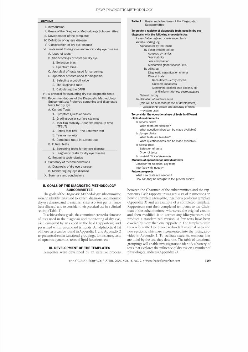
8/20/2019 dry eyes ii
http://slidepdf.com/reader/full/dry-eyes-ii 49/142
THE OCULAR SURFACE / APRIL 2007, VOL. 5, NO. 2 / www.theocularsurface.com 109
II. GOALS OF THE DIAGNOSTIC METHODOLOGY
SUBCOMMITTEE
The goals of the Diagnostic Methodology Subcommitteewere to identify tests used to screen, diagnose, and monitor
dry eye disease, and to establish criteria of test performance(test efficacy) and to consider their practical use in a clinicalsetting (Table 1).
To achieve these goals, the committee created a databaseof tests used in the diagnosis and monitoring of dry eye,each compiled by an expert in the field (rapporteur) andpresented within a standard template. An alphabetical listof these tests can be found in Appendix 1, and Appendix 2re-presents them in functional groupings, for instance, testsof aqueous dynamics, tests of lipid functions, etc.
III. DEVELOPMENT OF THE TEMPLATES
Templates were developed by an iterative process
between the Chairman of the subcommittee and the rap-porteurs. Each rapporteur was sent a set of instructions onhow to complete a template, together a proforma template
(Appendix 3) and an example of a completed template.Rapporteurs sent their completed templates to the Chair-man of the subcommittee, who saved the original versionand then modified it to correct any idiosyncrasies andproduce a standardized version. A few tests have beencovered by more than one rapporteur. The templates werethen reformatted to remove redundant material or to addnew sections, which are incorporated into the listing pro-vided in Appendix 1. To facilitate searches, template filesare titled by the test they describe. The table of functionalgroupings will enable investigators to identify a battery oftests that explores the influence of dry eye on a number of
physiological indices (Appendix 2).
OUTLINE
I. Introduction
II. Goals of the Diagnostic Methodology Subcommittee
III. Development of the templates
IV. Definition of dry eye disease
V. Classification of dry eye disease
VI. Tests used to diagnose and monitor dry eye diseaseA. Uses of tests
B. Shortcomings of tests for dry eye
1. Selection bias
2. Spectrum bias
C. Appraisal of tests used for screening
D. Appraisal of tests used for diagnosis
1. Selecting a cut-off value
2. The likelihood ratio
3. Calculating the OAPR
VII. A protocol for evaluating dry eye diagnostic tests
VIII. Recommendations of the Diagnostic Methodology
Subcommittee: Preferred screening and diagnostictests for dry eye
A. Current Tests
1. Symptom Questionnaires
2. Grading ocular surface staining
3. Tear film stability—tear film break-up time(TFBUT)
4. Reflex tear flow—the Schirmer test
5. Tear osmolarity
6. Combined tests in current use
B. Future Tests
1. Screening tests for dry eye disease
2. Diagnostic tests for dry eye disease
C. Emerging technologies
IX. Summary of recommendations
A. Diagnosis of dry eye disease
B. Monitoring dry eye disease
X. Summary and conclusions
Table 1. Goals and objectives of the DiagnosticSubcommittee
To create a register of diagnostic tests used in dry eye
diagnosis with the following characteristics:
A searchable register of referenced tests
Variable sorting, eg,
Alphabetical by test name
By organ system tested Aqueous dynamics
Tear stability
Tear composition
Meibomian gland function, etc.
By utility, eg,
Diagnostic classification criteria
Clinical trials
Recruitment—entry criteria
Outcome measures
Monitoring specific drug actions, eg,
anti-inflammatories; secretagogues
Natural history
Identification of evidence level
[this will be a second phase of development]
—validation/precison and accuracy of tests
—system used
To consider the operational use of tests in different
clinical environments
In general clinics
What tests are feasible?
What questionnaires can be made available?
In dry eye clinics
What tests are feasible?
What questionnaires can be made available?
In clinical trials
Selection of tests
Order of tests
In non-trial Clinical Research
Manuals of operation for individual tests
Consider for selected, key tests
Interface with industry
Future prospects
What new tests are needed?
How can they be brought to the general clinic?
DEWS DIAGNOSTIC METHODOLOGY
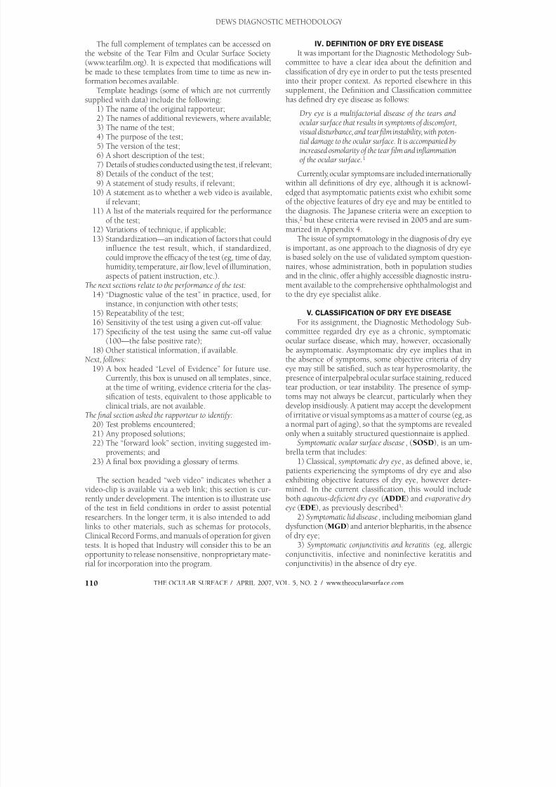
8/20/2019 dry eyes ii
http://slidepdf.com/reader/full/dry-eyes-ii 50/142
THE OCULAR SURFACE / APRIL 2007, VOL. 5, NO. 2 / www.theocularsurface.com110
The full complement of templates can be accessed onthe website of the Tear Film and Ocular Surface Society(www.tearfilm.org). It is expected that modifications willbe made to these templates from time to time as new in-formation becomes available.
Template headings (some of which are not currrentlysupplied with data) include the following:
1) The name of the original rapporteur;2) The names of additional reviewers, where available; 3) The name of the test; 4) The purpose of the test; 5) The version of the test; 6) A short description of the test; 7) Details of studies conducted using the test, if relevant; 8) Details of the conduct of the test; 9) A statement of study results, if relevant; 10) A statement as to whether a web video is available,
if relevant; 11) A list of the materials required for the performance
of the test; 12) Variations of technique, if applicable; 13) Standardization—an indication of factors that could
influence the test result, which, if standardized,could improve the efficacy of the test (eg, time of day,humidity, temperature, air flow, level of illumination,aspects of patient instruction, etc.).
The next sections relate to the performance of the test:14) “Diagnostic value of the test” in practice, used, for
instance, in conjunction with other tests; 15) Repeatability of the test; 16) Sensitivity of the test using a given cut-off value; 17) Specificity of the test using the same cut-off value
(100—the false positive rate);18) Other statistical information, if available.
Next, follows:19) A box headed “Level of Evidence” for future use.
Currently, this box is unused on all templates, since,at the time of writing, evidence criteria for the clas-sification of tests, equivalent to those applicable toclinical trials, are not available.
The final section asked the rapporteur to identify: 20) Test problems encountered; 21) Any proposed solutions;
22) The “forward look” section, inviting suggested im-
provements; and 23) A final box providing a glossary of terms.
The section headed “web video” indicates whether avideo-clip is available via a web link; this section is cur-rently under development. The intention is to illustrate useof the test in field conditions in order to assist potentialresearchers. In the longer term, it is also intended to addlinks to other materials, such as schemas for protocols,Clinical Record Forms, and manuals of operation for giventests. It is hoped that Industry will consider this to be anopportunity to release nonsensitive, nonproprietary mate-
rial for incorporation into the program.
IV. DEFINITION OF DRY EYE DISEASE
It was important for the Diagnostic Methodology Sub-committee to have a clear idea about the definition andclassification of dry eye in order to put the tests presentedinto their proper context. As reported elsewhere in thissupplement, the Definition and Classification committeehas defined dry eye disease as follows:
Dry eye is a multifactorial disease of the tears andocular surface that results in symptoms of discomfort,visual disturbance, and tear film instability, with poten-tial damage to the ocular surface. It is accompanied byincreased osmolarity of the tear film and inflammationof the ocular surface.1
Currently, ocular symptoms are included internationallywithin all definitions of dry eye, although it is acknowl-edged that asymptomatic patients exist who exhibit someof the objective features of dry eye and may be entitled tothe diagnosis. The Japanese criteria were an exception tothis,2 but these criteria were revised in 2005 and are sum-marized in Appendix 4.
The issue of symptomatology in the diagnosis of dry eyeis important, as one approach to the diagnosis of dry eyeis based solely on the use of validated symptom question-naires, whose administration, both in population studiesand in the clinic, offer a highly accessible diagnostic instru-ment available to the comprehensive ophthalmologist andto the dry eye specialist alike.
V. CLASSIFICATION OF DRY EYE DISEASE
For its assignment, the Diagnostic Methodology Sub-committee regarded dry eye as a chronic, symptomatic
ocular surface disease, which may, however, occasionallybe asymptomatic. Asymptomatic dry eye implies that inthe absence of symptoms, some objective criteria of dryeye may still be satisfied, such as tear hyperosmolarity, thepresence of interpalpebral ocular surface staining, reducedtear production, or tear instability. The presence of symp-toms may not always be clearcut, particularly when theydevelop insidiously. A patient may accept the developmentof irritative or visual symptoms as a matter of course (eg, asa normal part of aging), so that the symptoms are revealedonly when a suitably structured questionnaire is applied.
Symptomatic ocular surface disease, (SOSD), is an um-
brella term that includes:1) Classical, symptomatic dry eye, as defined above, ie,
patients experiencing the symptoms of dry eye and alsoexhibiting objective features of dry eye, however deter-mined. In the current classification, this would includeboth aqueous-deficient dry eye ( ADDE) and evaporative dryeye (EDE), as previously described3:
2) Symptomatic lid disease, including meibomian glanddysfunction (MGD) and anterior blepharitis, in the absenceof dry eye;
3) Symptomatic conjunctivitis and keratitis (eg, allergicconjunctivitis, infective and noninfective keratitis and
conjunctivitis) in the absence of dry eye.
DEWS DIAGNOSTIC METHODOLOGY
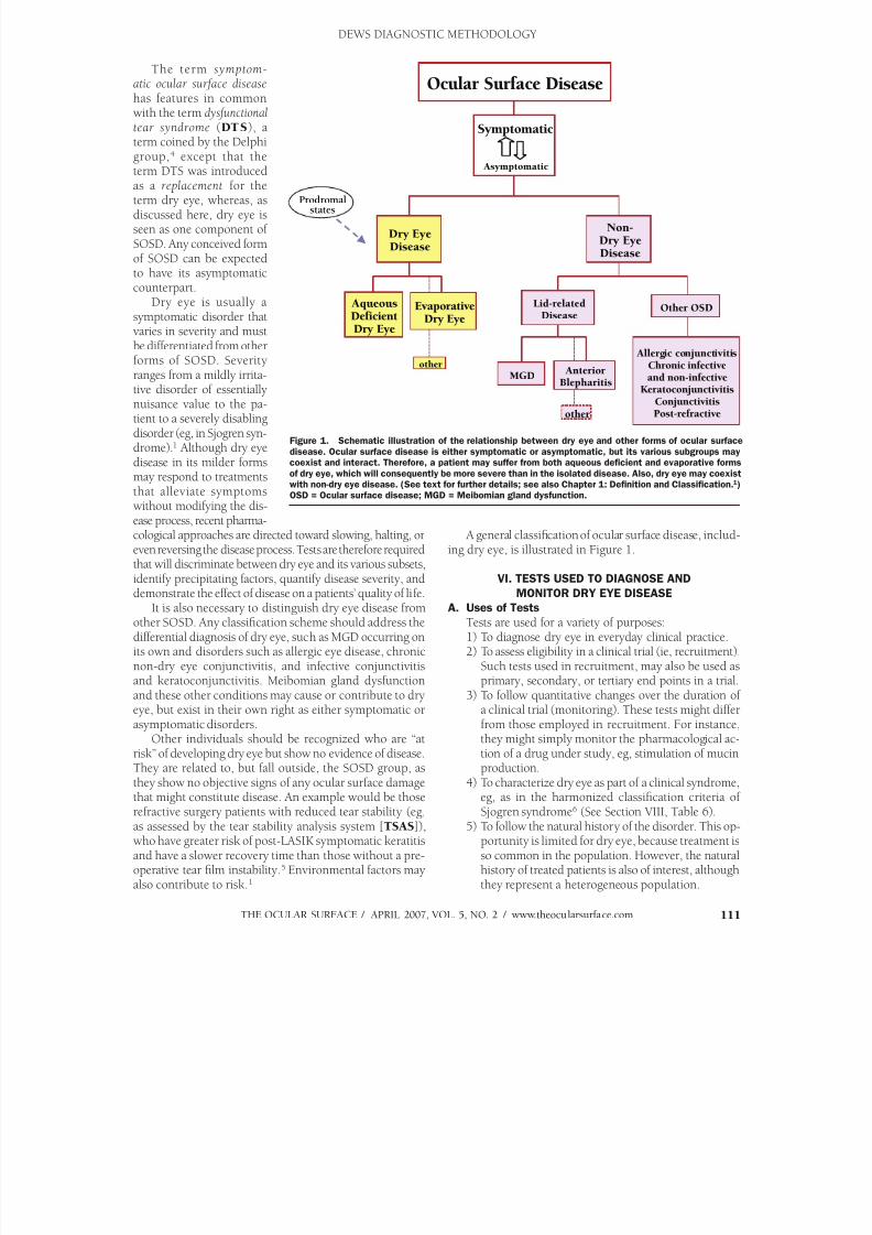
8/20/2019 dry eyes ii
http://slidepdf.com/reader/full/dry-eyes-ii 51/142
THE OCULAR SURFACE / APRIL 2007, VOL. 5, NO. 2 / www.theocularsurface.com 111
The term symptom-atic ocular surface disease has features in commonwith the term dysfunctionaltear syndrome (DTS), aterm coined by the Delphigroup,4 except that the
term DTS was introducedas a replacement for theterm dry eye, whereas, asdiscussed here, dry eye isseen as one component ofSOSD. Any conceived formof SOSD can be expectedto have its asymptomaticcounterpart.
Dry eye is usually asymptomatic disorder thatvaries in severity and mustbe differentiated from otherforms of SOSD. Severityranges from a mildly irrita-tive disorder of essentiallynuisance value to the pa-tient to a severely disablingdisorder (eg, in Sjogren syn-drome).1 Although dry eyedisease in its milder formsmay respond to treatmentsthat alleviate symptomswithout modifying the dis-ease process, recent pharma-
cological approaches are directed toward slowing, halting, oreven reversing the disease process. Tests are therefore requiredthat will discriminate between dry eye and its various subsets,identify precipitating factors, quantify disease severity, anddemonstrate the effect of disease on a patients’ quality of life.
It is also necessary to distinguish dry eye disease fromother SOSD. Any classification scheme should address thedifferential diagnosis of dry eye, such as MGD occurring onits own and disorders such as allergic eye disease, chronicnon-dry eye conjunctivitis, and infective conjunctivitisand keratoconjunctivitis. Meibomian gland dysfunctionand these other conditions may cause or contribute to dry
eye, but exist in their own right as either symptomatic orasymptomatic disorders.
Other individuals should be recognized who are “atrisk” of developing dry eye but show no evidence of disease.They are related to, but fall outside, the SOSD group, asthey show no objective signs of any ocular surface damagethat might constitute disease. An example would be thoserefractive surgery patients with reduced tear stability (eg,as assessed by the tear stability analysis system [TSAS]),who have greater risk of post-LASIK symptomatic keratitisand have a slower recovery time than those without a pre-operative tear film instability.5 Environmental factors may
also contribute to risk.1
A general classification of ocular surface disease, includ-ing dry eye, is illustrated in Figure 1.
VI. TESTS USED TO DIAGNOSE AND
MONITOR DRY EYE DISEASE
A. Uses of Tests
Tests are used for a variety of purposes: 1) To diagnose dry eye in everyday clinical practice. 2) To assess eligibility in a clinical trial (ie, recruitment).
Such tests used in recruitment, may also be used asprimary, secondary, or tertiary end points in a trial.
3) To follow quantitative changes over the duration of
a clinical trial (monitoring). These tests might differfrom those employed in recruitment. For instance,they might simply monitor the pharmacological ac-tion of a drug under study, eg, stimulation of mucinproduction.
4) To characterize dry eye as part of a clinical syndrome,eg, as in the harmonized classification criteria ofSjogren syndrome6 (See Section VIII, Table 6).
5) To follow the natural history of the disorder. This op-portunity is limited for dry eye, because treatment isso common in the population. However, the naturalhistory of treated patients is also of interest, although
they represent a heterogeneous population.
Dry EyeDisease
Ocular Surface Disease
Symptomatic
Asymptomatic
Non-Dry EyeDisease
other
AqueousDeficientDry Eye
EvaporativeDry Eye
other
Allergic conjunctivitisChronic infectiveand non-infective
KeratoconjunctivitisConjunctivitisPost-refractive
Other OSDLid-relatedDisease
MGD Anterior
Blepharitis
Prodromal
states
Figure 1. Schematic illustration of the relationship between dry eye and other forms of ocular surface
disease. Ocular surface disease is either symptomatic or asymptomatic, but its various subgroups maycoexist and interact. Therefore, a patient may suffer from both aqueous deficient and evaporative forms
of dry eye, which will consequently be more severe than in the isolated disease. Also, dry eye may coexistwith non-dry eye disease. (See text for further details; see also Chapter 1: Definition and Classification.1)OSD = Ocular surface disease; MGD = Meibomian gland dysfunction.
DEWS DIAGNOSTIC METHODOLOGY

8/20/2019 dry eyes ii
http://slidepdf.com/reader/full/dry-eyes-ii 52/142
THE OCULAR SURFACE / APRIL 2007, VOL. 5, NO. 2 / www.theocularsurface.com112
B. Shortcomings of Tests for Dry Eye
1. Selection BiasNo “gold standard” exists for the diagnosis of dry eye.
Thus, when a test, eg, Schirmer test or rose bengal stain-ing, is being evaluated for efficacy, the test population mayhave been classified as affected or non-affected based onthose same tests. Similarly, the performance of any “new”
test may be compromised when the test is assessed in apopulation of dry eye patients who have been diagnosedusing unestablished criteria.
When studies of test efficacy look at how the test definesaffected and unaffected individuals using individuals fromthe sample from which the diagnostic cut-offs were derived,this potentially results in a higher sensitivity and specificityrating than would have arisen from an independent sample. Also, because of the multi-factorial nature of dry eye, vari-able test efficacy is likely to occur from study to study.
2. Spectrum Bias When the study sample consists of patients with either
very mild or very severe disease, results are compromisedbecause the severity of the disease in the sample studiedhas been highly selected.
Certain ground rules are proposed for appraising theperformance of tests for dry eye diagnosis reported in theliterature (Table 2). 1) Accept efficacy values on samples from which the test
cut-off was derived (as is the case in most reports).2) Exclude data from studies with selection bias due to
the test being part of the original dry eye diagnosticcriteria (to avoid study results with high, ie, false,sensitivity and specificity values).
3) To avoid spectrum bias, study samples should belarge enough to include a range of dry eye patientswith various etiologies.
4) The choice of the cut-off value for diagnosis andthe test itself, unless there is some special physi-ological reason, should be based on a consider-ation of the relative consequences of having toomany false-positives or too many false-negatives.Generally, in a screening test for a serious or life-threatening condition, it is desirable to have a testof high sensitivity (high detection rate)—with fewfalse-negatives—since failure to detect the condition
early can be fatal. In a mass screening test for a lessserious condition or for one whose early detectionis not critical, it may be more desirable to have a highspecificity to avoid overburdening the health caredelivery system with too many false-positives.
5) For dry eye screening tests, it is suggested thatsensitivity and the predictive value of a positive test(PPV , see below) be maximized, ie, avoid high false-negative rates by “over-diagnosing” dry eye throughchoice of cut-off/test. This is appropriate when thepatient is to be further assessed with other tests tofinally diagnose dry eye. However, low false-negative
rates (choice of test or cut-off maximize sensitivity)
should be balanced by an acceptable PPV. 6) In diagnostic tests, optimize overall accuracy (OA)
and combine this with a high sensitivity and PPV. 7) Simplify comparisons of screening and diagnostic
tests by using single and simple terms for measuringtest efficacy.
C. Appraisal of Tests Used for Screening
The purpose of screening is prevention, and it aimsto identify people at high risk of a disorder. It is implicitin the screening process that a treatment is available thatwill reduce the morbidity of the disorder in a cost-effec-tive manner. Screening has been defined, among personswho have not sought medical attention, as the “systematicapplication of a test or enquiry to identify individualsat sufficient risk of a…disorder to benefit from furtherinvestigation or…preventive action...”26 It is implied thatthe disorder has serious consequences and that a remedyis available that could reduce morbidity.
Inclusion of symptoms within the definition of dry eyehas an awkward implication in the context of screening.To identify those at risk of developing the disorder or whohave unrecognized disease, screening is characteristicallycarried out on asymptomatic individuals who have notpresented themselves for diagnosis; those who are symp-tomatic already have the disease. This “at-risk” group islikely to be represented by asymptomatic subjects whosepathophysiological background favors the developmentof dry eye. Perhaps, their lacrimal secretory level or theirmeibomian lipid secretion or delivery is at the lower limitof normal, so that with time they will pass into a state ofinsufficiency. They may have an unstable tear film, or they
may be in the prodromal stages of a disease (eg, exhibitingnonophthalmic features of primary Sjogren syndrome),whose natural history dictates that they will eventuallydevelop dry eyes. Members of this diverse group of subjectscould be precipitated into dry eye by a number of biologi-cal, pharmacological or environmental events, ie, hormonalchanges, drug exposure, high air or wind speeds, irritants,low humidity, and high temperatures. Exposure to suchinfluences might engender dry eye symptoms in an at-riskgroup at a lower threshold than in subjects not at risk ofdry eye disease.
At-risk subjects could be identified by “stress tests,”
some of which are included among the test templates thataccompany this report and/or can be accessed at www.tearfilm.org. Whether or not such tests could or shouldbecome part of a “screening program” depends on whetherany perceived therapeutic benefits would be economically justified. One such benefit might be to identify the suit-ability of individuals to work within a particular work en-vironment, or to answer questions about the modificationsof environments to avoid inducing symptomatic disease.
To be of value, a screening test should be simple, effec-tive, applicable to a definable population, and cost-effective.In an effective screening program, a positive test ultimately
leads to diagnostic tests, which, if positive, lead to timely
DEWS DIAGNOSTIC METHODOLOGY
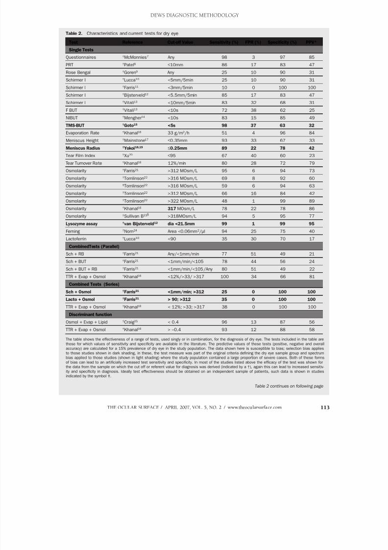
8/20/2019 dry eyes ii
http://slidepdf.com/reader/full/dry-eyes-ii 53/142
THE OCULAR SURFACE / APRIL 2007, VOL. 5, NO. 2 / www.theocularsurface.com 113
Table 2. Characteristics and current tests for dry eye
Test Reference Cut-off Value Sensitivity (%) FPR (%) Specificity (%) PPV*
Single Tests
Questionnaires †McMonnies7 Any 98 3 97 85
PRT †Patel8 ≤10mm 86 17 83 47
Rose Bengal †Goren9 Any 25 10 90 31
Schirmer I †Lucca10 <5mm/5min 25 10 90 31
Schirmer I †Farris11 <3mm/5min 10 0 100 100
Schirmer I †Bijsterveld12 <5.5mm/5min 85 17 83 47
Schirmer I †Vitali13 <10mm/5min 83 32 68 31
F BUT †Vitali13 <10s 72 38 62 25
NIBUT †Mengher14 <10s 83 15 85 49
TMS-BUT †Goto15 <5s 98 37 63 32
Evaporation Rate †Khanal16 33 g/m2/h 51 4 96 84
Meniscus Height †Mainstone17 ≤0.35mm 93 33 67 33
Meniscus Radius †Yokoi18,19 ≤0.25mm 89 22 78 42
Tear Film Index
†
Xu
20
≤95 67 40 60 23Tear Turnover Rate †Khanal16 12%/min 80 28 72 79
Osmolarity †Farris21 >312 MOsm/L 95 6 94 73
Osmolarity †Tomlinson22 >316 MOsm/L 69 8 92 60
Osmolarity ‡Tomlinson22 >316 MOsm/L 59 6 94 63
Osmolarity †Tomlinson22 >312 MOsm/L 66 16 84 42
Osmolarity †Tomlinson22 >322 MOsm/L 48 1 99 89
Osmolarity †Khanal16 317 MOsm/L 78 22 78 86
Osmolarity †Sullivan B23§ >318MOsm/L 94 5 95 77
Lysozyme assay †van Bijsterveld12 dia <21.5mm 99 1 99 95
Ferning †Norn24 Area <0.06mm2/µl 94 25 75 40
Lactoferrin †Lucca10 <90 35 30 70 17
CombinedTests (Parallel)
Sch + RB †Farris21 Any/<1mm/min 77 51 49 21
Sch + BUT †Farris21 <1mm/min/<105 78 44 56 24
Sch + BUT + RB †Farris21 <1mm/min/<105/Any 80 51 49 22
TTR + Evap + Osmol †Khanal16 <12%/>33/ >317 100 34 66 81
Combined Tests (Series)
Sch + Osmol †Farris21 <1mm/min; >312 25 0 100 100
Lacto + Osmol †Farris21 > 90; >312 35 0 100 100
TTR + Evap + Osmol †Khanal16 < 12%; >33; >317 38 0 100 100
Discriminant functionOsmol + Evap + Lipid †Craig25 < 0.4 96 13 87 56
TTR + Evap + Osmol †Khanal16 > –0.4 93 12 88 58
The table shows the effectiveness of a range of tests, used singly or in combination, for the diagnosis of dry eye. The tests included in the table arethose for which values of sensitivity and specificity are available in the literature. The predictive values of these tests (positive, negative and overallaccuracy) are calculated for a 15% prevalence of dry eye in the study population. The data shown here is susceptible to bias; selection bias appliesto those studies shown in dark shading, in these, the test measure was part of the original criteria defining the dry eye sample group and spectrumbias applied to those studies (shown in light shading) where the study population contained a large proportion of severe cases. Both of these formsof bias can lead to an artificially increased test sensitivity and specificity. In most of the studies listed above the efficacy of the test was shown forthe data from the sample on which the cut off or referent value for diagnosis was derived (indicated by a †), again this can lead to increased sensitiv-ity and specificity in diagnosis. Ideally test effectiveness should be obtained on an independent sample of patients, such data is shown in studiesindicated by the symbol ‡.
Table 2 continues on following page
DEWS DIAGNOSTIC METHODOLOGY
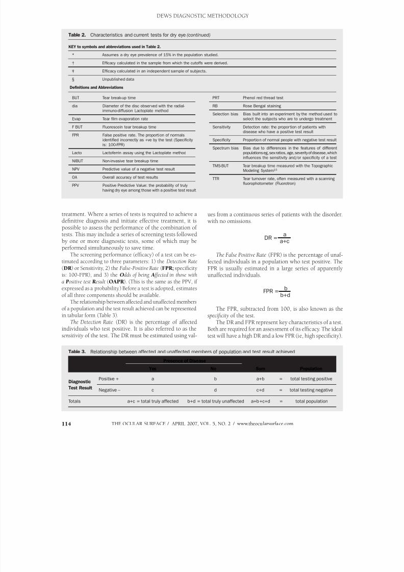
8/20/2019 dry eyes ii
http://slidepdf.com/reader/full/dry-eyes-ii 54/142
THE OCULAR SURFACE / APRIL 2007, VOL. 5, NO. 2 / www.theocularsurface.com114
Table 2. Characteristics and current tests for dry eye (continued)
KEY to symbols and abbreviations used in Table 2.
* Assumes a dry eye prevalence of 15% in the population studied.
† Efficacy calculated in the sample from which the cutoffs were derived.
‡ Efficacy calculated in an independent sample of subjects.
§ Unpublished data
Definitions and Abbreviations
BUT Tear break-up time
dia Diameter of the disc observed with the radial-immuno-diffusion Lactoplate method
Evap Tear film evaporation rate
F BUT Fluorescein tear breakup time
FPR False positive rate. The proportion of normalsidentified incorrectly as +ve by the test (Specificityis: 100-FPR)
Lacto Lactoferrin assay using the Lactoplate method
NIBUT Non-invasive tear breakup time
NPV Predictive value of a negative test result
OA Overall accuracy of test results
PPV Positive Predictive Value: the probability of truly having dry eye among those with a positive test result
PRT Phenol red thread test
RB Rose Bengal staining
Selection bias Bias built into an experiment by the method used toselect the subjects who are to undergo treatment
Sensitivity Detection rate: the proportion of patients withdisease who have a positive test result
Specificity Proportion of normal people with negative test result
Spectrum bias Bias due to differences in the features of differentpopulations eg, sex ratios, age, severity of disease, whichinfluences the sensitivity and/or specificity of a test
TMS-BUT Tear breakup time measured with the TopographicModeling System15
TTR Tear turnover rate, often measured with a scanningfluorophotometer (Fluorotron)
treatment. Where a series of tests is required to achieve adefinitive diagnosis and initiate effective treatment, it ispossible to assess the performance of the combination oftests. This may include a series of screening tests followedby one or more diagnostic tests, some of which may be
performed simultaneously to save time.The screening performance (efficacy) of a test can be es-
timated according to three parameters: 1) the Detection Rate (DR ) or Sensitivity, 2) the False-Positive Rate (FPR; specificityis: 100-FPR), and 3) the Odds of being A ffected in those witha P ositive test R esult (OAPR ). (This is the same as the PPV, ifexpressed as a probability.) Before a test is adopted, estimatesof all three components should be available.
The relationship between affected and unaffected membersof a population and the test result achieved can be representedin tabular form (Table 3).
The Detection Rate (DR) is the percentage of affected
individuals who test positive. It is also referred to as thesensitivity of the test. The DR must be estimated using val-
ues from a continuous series of patients with the disorder,with no omissions.
DR = aa+c
The False Positive Rate (FPR) is the percentage of unaf-
fected individuals in a population who test positive. TheFPR is usually estimated in a large series of apparentlyunaffected individuals.
FPR = bb+d
The FPR, subtracted from 100, is also known as thespecificity of the test.
The DR and FPR represent key characteristics of a test.
Both are required for an assessment of its efficacy. The idealtest will have a high DR and a low FPR (ie, high specificity).
Table 3. Relationship between affected and unaffected members of population and test result achieved
Presence of Disease
Yes No Sum Population
Diagnostic Positive + a b a+b = total testing positive
Test Result Negative – c d c+d = total testing negative
Totals a+c = total truly affected b+d = total truly unaffected a+b+c+d = total population
DEWS DIAGNOSTIC METHODOLOGY

8/20/2019 dry eyes ii
http://slidepdf.com/reader/full/dry-eyes-ii 55/142
THE OCULAR SURFACE / APRIL 2007, VOL. 5, NO. 2 / www.theocularsurface.com 115
The DRs and FPRs for a number of tests used in dry eyediagnosis are presented in Table 2.
The third parameter is dependent on the prevalence of thedisorder in the population studied. This is The Odds of being
A ffected in those with a P ositive test R esult (OAPR [or PPV]).This is expressed as an odds value, eg, 1:3 or 1:100, etc. Itcan also be expressed as a percent probability (which in these
cases would be: 1/4
100 = 25%, or 1/101
100 = 0.99%).
OAPR = a
a+b
D. Appraisal of Tests Used for Diagnosis
Diagnostic tests are applied to symptomatic or asymp-tomatic patients to obtain a diagnosis and, by inference, toexclude other diagnoses. A successful diagnosis can serveseveral functions, paramount of which is the opportunityfor therapy. Therapy can ameliorate the symptoms of adisease, retard its progression, or produce a cure. Arrivalat a successful diagnosis may also serve other functions, forinstance, in relation to the natural history and prognosis ofa disease, knowledge of which is of value to both patientand doctor. Also, a diagnosis, by excluding other diseases,may usefully indicate that a feared diagnosis is not presentand that other kinds of therapy are not indicated.
1. Selecting a Cut-off ValueTest data may be qualitative (categorical, eg, with or
without epiphora), semi-quantitative (ordinal, eg, gradingby corneal staining), or quantitative (continuous, eg, theSchirmer test result in mm, intraocular pressure). For a testoffering continuous data, it is appropriate to select a cut-off
value to discriminate between affected and unaffected sub- jects. This may involve a trade-off between the DR and FPR,depending on the distribution of test values between thesetwo groups. The DR and FPR are dependent on the selectedcut-off values, and this is influenced by the overlap of valuesbetween affected and unaffected subjects. For instance, ifthere is no overlap in values between unaffected and affectedsubjects, then the cut-off will lie between the two data sets.However, where there is an overlap of values, which is usu-ally the case, a cut-off must be chosen somewhere in theregion of overlap.
The concept of choosing a cut-off is illustrated in the Figures
2a and 2b, which represent the situation in a hypothetical disor-der in which the test variable is higher in the affected than in theunaffected population.27 An example might be a staining score.
When distributions are presented in this way, then the area tothe right of the cut-off under the unaffected curve, provides theFPR, while the area to the right of the cut-off under the affected curve, gives the DR. Moving the cut-off to the right (as in Figure2b) reduces the FPR but also reduces the DR.
2. The Likelihood Ratio
A useful way of expressing the interaction of DR andFPR is by calculating the Likelihood Ratio (LR ), which is the
ratio of those areas. The LR is a measure of the number of
times individuals with positive results are more likely tohave the disorder compared with individuals who have notbeen tested. A successful screening test might have an LRin the range of 5 to 25.
3. Calculating the OAPR The OAPR is a valuable parameter that represents the
average chance of being affected for all individuals witha positive result by the test. It expresses the odds of the
number of true positives to the number of false positives.For a given population, the OAPRs of different tests for thesame condition may be compared directly with one another.There are two ways to calculate the OAPR (examples takenfrom Wald26 and Wald and Cuckle27).
The first method uses a flow chart to estimate testperformance.
Considering the total number of individuals identified aspositive by a test within a defined population, a proportion willbe true positives (determined by the DR of the test), and theremainder will be the false positives (determined by the FPR).The OAPR is the ratio of these two numbers, ie, OAPR = True
Positives: False Positives.
Unaffected
Affected
Test variable (arbitrary units)
Unaffected
(a)
Affected
2 3 4 5 6 7 8 9 10 11 12
2 3 4 5 6 7 8 9 10 11 12
B
Test variable (arbitrary units)
A
(b)
Figure 2. Illustrates how selection of the cutoff value influences
the FPR and DR. See text for details.
DEWS DIAGNOSTIC METHODOLOGY
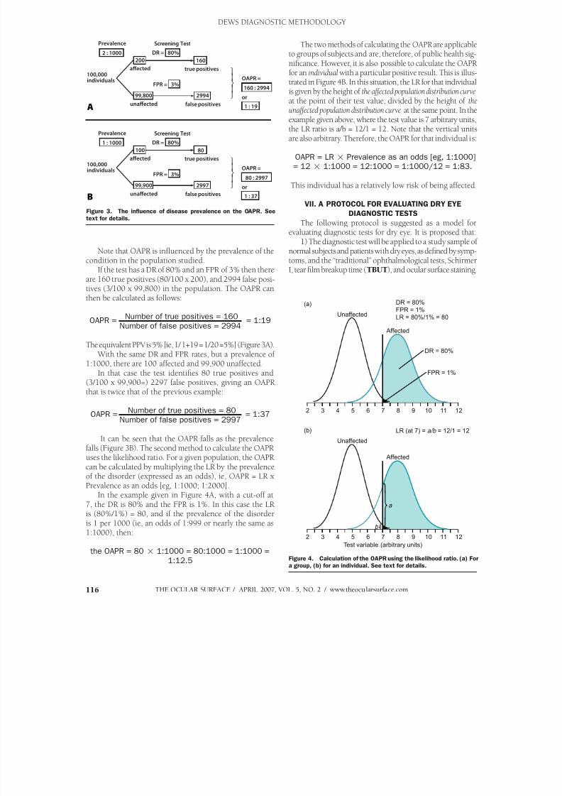
8/20/2019 dry eyes ii
http://slidepdf.com/reader/full/dry-eyes-ii 56/142
THE OCULAR SURFACE / APRIL 2007, VOL. 5, NO. 2 / www.theocularsurface.com116
2 : 1000
200 160
DR = 80%
2994
FPR = 3%
99,800
1 : 19
Prevalence Screening Test
160 : 2994
true positives
false positivesunaected
aected100,000individuals OAPR =
or
A
1 : 1000
100 80
DR = 80%
2997
FPR = 3%
99,900
1 : 37
Prevalence Screening Test
80 : 2997
true positives
false positivesunaected
aected100,000individuals OAPR =
or
B
Note that OAPR is influenced by the prevalence of thecondition in the population studied.
If the test has a DR of 80% and an FPR of 3% then thereare 160 true positives (80/100 x 200), and 2994 false posi-tives (3/100 x 99,800) in the population. The OAPR canthen be calculated as follows:
OAPR = Number of true positives = 160
= 1:19Number of false positives = 2994
The equivalent PPV is 5% [ie, 1/ 1+19 = 1/20 =5%] (Figure 3A). With the same DR and FPR rates, but a prevalence of
1:1000, there are 100 affected and 99,900 unaffected.In that case the test identifies 80 true positives and
(3/100 x 99,900=) 2297 false positives, giving an OAPRthat is twice that of the previous example:
OAPR =Number of true positives = 80
= 1:37Number of false positives = 2997
It can be seen that the OAPR falls as the prevalence
falls (Figure 3B). The second method to calculate the OAPR
uses the likelihood ratio. For a given population, the OAPRcan be calculated by multiplying the LR by the prevalenceof the disorder (expressed as an odds), ie, OAPR = LR xPrevalence as an odds [eg, 1:1000; 1:2000].
In the example given in Figure 4A, with a cut-off at7, the DR is 80% and the FPR is 1%. In this case the LRis (80%/1%) = 80, and if the prevalence of the disorderis 1 per 1000 (ie, an odds of 1:999 or nearly the same as1:1000), then:
the OAPR = 80 1:1000 = 80:1000 = 1:1000 =
1:12.5
The two methods of calculating the OAPR are applicableto groups of subjects and are, therefore, of public health sig-nificance. However, it is also possible to calculate the OAPRfor an individualwith a particular positive result. This is illus-trated in Figure 4B. In this situation, the LR for that individualis given by the height of the affected population distribution curve at the point of their test value, divided by the height of the
unaffected population distribution curve at the same point. In theexample given above, where the test value is 7 arbitrary units,the LR ratio is a/b = 12/1 = 12. Note that the vertical unitsare also arbitrary. Therefore, the OAPR for that individual is:
OAPR = LR Prevalence as an odds [eg, 1:1000]= 12 1:1000 = 12:1000 = 1:1000/12 = 1:83.
This individual has a relatively low risk of being affected.
VII. A PROTOCOL FOR EVALUATING DRY EYE
DIAGNOSTIC TESTS
The following protocol is suggested as a model forevaluating diagnostic tests for dry eye. It is proposed that:
1) The diagnostic test will be applied to a study sample ofnormal subjects and patients with dry eyes, as defined by symp-toms, and the “traditional” ophthalmological tests, SchirmerI, tear film breakup time (TBUT), and ocular surface staining.
Figure 3. The influence of disease prevalence on the OAPR. See
text for details.
Figure 4. Calculation of the OAPR using the likelihood ratio. (a) For
a group, (b) for an individual. See text for details.
DR = 80%
FPR = 1%
LR = 80%/1% = 80
DR = 80%
FPR = 1%
Unaffected
(a)
Affected
Test variable (arbitrary units)
LR (at 7) = a/b = 12/1 = 12
a
b
Unaffected
(b)
Affected
2 3 4 5 6 7 8 9 10 11 12
2 3 4 5 6 7 8 9 10 11 12
DEWS DIAGNOSTIC METHODOLOGY
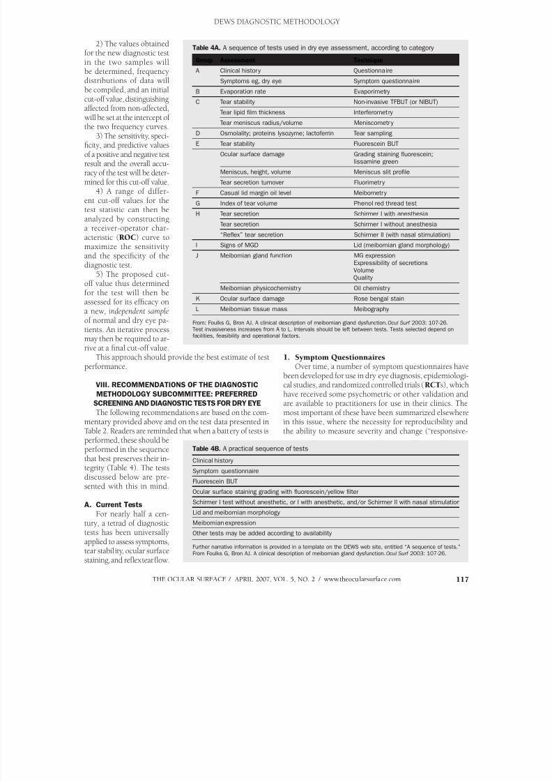
8/20/2019 dry eyes ii
http://slidepdf.com/reader/full/dry-eyes-ii 57/142
THE OCULAR SURFACE / APRIL 2007, VOL. 5, NO. 2 / www.theocularsurface.com 117
2) The values obtainedfor the new diagnostic testin the two samples willbe determined, frequencydistributions of data willbe compiled, and an initialcut-off value, distinguishing
affected from non-affected,will be set at the intercept ofthe two frequency curves.
3) The sensitivity, speci-ficity, and predictive valuesof a positive and negative testresult and the overall accu-racy of the test will be deter-mined for this cut-off value.
4) A range of differ-ent cut-off values for thetest statistic can then beanalyzed by constructinga receiver-operator char-acteristic (ROC) curve tomaximize the sensitivityand the specificity of thediagnostic test.
5) The proposed cut-off value thus determinedfor the test will then beassessed for its efficacy ona new, independent sample of normal and dry eye pa-tients. An iterative process
may then be required to ar-rive at a final cut-off value.
This approach should provide the best estimate of testperformance.
VIII. RECOMMENDATIONS OF THE DIAGNOSTIC
METHODOLOGY SUBCOMMITTEE: PREFERRED
SCREENING AND DIAGNOSTIC TESTS FOR DRY EYE
The following recommendations are based on the com-mentary provided above and on the test data presented inTable 2. Readers are reminded that when a battery of tests isperformed, these should be
performed in the sequencethat best preserves their in-tegrity (Table 4). The testsdiscussed below are pre-sented with this in mind.
A. Current Tests
For nearly half a cen-tury, a tetrad of diagnostictests has been universallyapplied to assess symptoms,tear stability, ocular surface
staining, and reflex tear flow.
Table 4B. A practical sequence of tests
Clinical history
Symptom questionnaire
Fluorescein BUT
Ocular surface staining grading with fluorescein/yellow filter
Schirmer I test without anesthetic, or I with anesthetic, and/or Schirmer II with nasal stimulation
Lid and meibomian morphology
Meibomian expression
Other tests may be added according to availability
Further narrative information is provided in a template on the DEWS web site, entitled “A sequence of tests.”From Foulks G, Bron AJ. A clinical description of meibomian gland dysfunction. Ocul Surf 2003: 107-26.
Table 4A. A sequence of tests used in dry eye assessment, according to category
Group Assessment Technique
A Clinical history Questionnaire
Symptoms eg, dry eye Symptom questionnaire
B Evaporation rate Evaporimetry
C Tear stability Non-invasive TFBUT (or NIBUT)
Tear lipid film thickness Interferometry
Tear meniscus radius/volume Meniscometry
D Osmolality; proteins lysozyme; lactoferrin Tear sampling
E Tear stability Fluorescein BUT
Ocular surface damage Grading staining fluorescein;lissamine green
Meniscus, height, volume Meniscus slit profile
Tear secretion turnover Fluorimetry
F Casual lid margin oil level Meibometry
G Index of tear volume Phenol red thread test
H Tear secretion Schirmer I with anesthesia
Tear secretion Schirmer I without anesthesia
“Reflex” tear secretion Schirmer II (with nasal stimulation)
I Signs of MGD Lid (meibomian gland morphology)
J Meibomian gland function MG expression Expressibility of secretions Volume Quality
Meibomian physicochemistry Oil chemistry
K Ocular surface damage Rose bengal stain
L Meibomian tissue mass Meibography
From: Foulks G, Bron AJ. A clinical description of meibomian gland dysfunction. Ocul Surf 2003: 107-26.Test invasiveness increases from A to L. Intervals should be left between tests. Tests selected depend on
facilities, feasibility and operational factors.
DEWS DIAGNOSTIC METHODOLOGY
1. Symptom QuestionnairesOver time, a number of symptom questionnaires have
been developed for use in dry eye diagnosis, epidemiologi-cal studies, and randomized controlled trials (RCTs), whichhave received some psychometric or other validation andare available to practitioners for use in their clinics. Themost important of these have been summarized elsewherein this issue, where the necessity for reproducibility andthe ability to measure severity and change (“responsive-
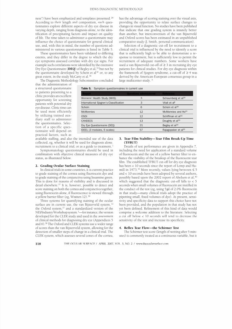
8/20/2019 dry eyes ii
http://slidepdf.com/reader/full/dry-eyes-ii 58/142
THE OCULAR SURFACE / APRIL 2007, VOL. 5, NO. 2 / www.theocularsurface.com118
ness”) have been emphasized and templates presented.28 According to their length and composition, such ques-tionnaires explore different aspects of dry eye disease invarying depth, ranging from diagnosis alone, to the iden-tification of precipitating factors and impact on qualityof life. The time taken to administer a questionnaire mayinfluence the choice of questionnaire for general clinical
use, and, with this in mind, the number of questions ad-ministered in various questionnaires is listed in Table 5.These questionnaires have been validated to differing
extents, and they differ in the degree to which the dryeye symptoms assessed correlate with dry eye signs. Forexample such correlations were identified by the extensiveDry Eye Questionnaire (DEQ ) of Begley et al,34 but not bythe questionnaire developed by Schein et al30 or, to anygreat extent, in the study McCarty et al.36
The Diagnostic Methodology Subcommittee concludedthat the administration ofa structured questionnaireto patients presenting to aclinic provides an excellentopportunity for screeningpatients with potential dryeye disease. Clinic time canbe used most efficientlyby utilizing trained aux-iliary staff to administerthe questionnaires. Selec-tion of a specific ques-tionnaire will depend onpractical factors, such asavailable staffing, and also the intended use of the data
collected, eg, whether it will be used for diagnosis alone,recruitment to a clinical trial, or as a guide to treatment.1
Symptomatology questionnaires should be used incombination with objective clinical measures of dry eyestatus, as illustrated below.
2. Grading Ocular Surface Staining In clinical trials in some countries, it is current practice
to grade staining of the cornea using fluorescein dye andto grade staining of the conjunctiva using lissamine green.This is done for reasons of visibility and is discussed indetail elsewhere.37 It is, however, possible to detect and
score staining on both the cornea and conjunctiva together,using fluorescein alone, if fluorescence is viewed througha yellow barrier filter (eg, Wratten 12).38
Three systems for quantifying staining of the ocularsurface are in current use, the van Bijsterveld system,12 the Oxford system,37 and a standardized version of theNEI/Industry Workshop system,3—for instance, the versiondeveloped for the CLEK study and used in the assessmentof clinical methods for diagnosing dry eye (Appendices 5and 6).38 The Oxford and CLEK systems use a wider rangeof scores than the van Bijsterveld system, allowing for thedetection of smaller steps of change in a clinical trial. The
CLEK system, which assesses several zones of the cornea,
has the advantage of scoring staining over the visual axis,providing the opportunity to relate surface changes tochanges in visual function. No studies have been publishedthat indicate that one grading system is innately betterthan another, but interconversion of the van Bijsterveldand Oxford scores has been estimated in an unpublishedcomparative study (J. Smith, personal communication).
Selection of a diagnostic cut-off for recruitment to aclinical trial is influenced by the need to identify a scorethat is sufficiently high to be able to demonstrate a re-sponse to treatment, but is sufficiently low to permit therecruitment of adequate numbers. Some workers haveused a van Bijsterveld cut-off of ≥ 3 in recruiting dry eyepatients for clinical studies. For dry eye diagnosis withinthe framework of Sjogren syndrome, a cut-off of ≥ 4 wasderived by the American-European consensus group in alarge multicenter study. 6
3. Tear Film Stability—Tear Film Break-Up Time
(TFBUT)Details of test performance are given in Appendix 7,
including the need for application of a standard volumeof fluorescein and the use of a yellow barrier filter to en-hance the visibility of the breakup of the fluorescent tearfilm. The established TFBUT cut-off for dry eye diagnosishas been < 10 seconds since the report of Lemp and Ha-mill in 1973.39 More recently, values lying between ≤ 5and < 10 seconds have been adopted by several authors,possibly based upon the 2002 report of Abelson et al,40 which suggested that the diagnostic cut-off falls to < 5seconds when small volumes of fluorescein are instilled in
the conduct of the test (eg, using 5µl of 2.0% fluoresceinin that study—many clinical trials adopt the practice ofpipetting small, fixed volumes of dye). At present, sensi-tivity and specificity data to support this choice have notbeen provided, and the population in that study has notyet been defined. Refinement of this kind of data wouldcomprise a welcome addition to the literature. Selectinga cut off below < 10 seconds will tend to decrease thesensitivity of the test and increase its specificity.
4. Reflex Tear Flow—the Schirmer TestThe Schirmer test score (length of wetting after 5 min-
utes) is commonly treated as a continuous variable, but it
Table 5. Symptom questionnaires in current use
Report Questions administered ReferenceWomens’ Health Study (WHS) 3 Schaumberg et al29
International Sjogren’s Classification 3 Vitali et al6
Schein 6 Schein et al30
McMonnies 12 McMonnies and Ho31
OSDI 12 Schiffman et al32
CANDEES 13 Doughty et al33
Dry Eye Questionnaire (DEQ) 21 Begley et al34
IDEEL (3 modules, 6 scales) 57 Rajagopalan et al35
DEWS DIAGNOSTIC METHODOLOGY

8/20/2019 dry eyes ii
http://slidepdf.com/reader/full/dry-eyes-ii 59/142
THE OCULAR SURFACE / APRIL 2007, VOL. 5, NO. 2 / www.theocularsurface.com 119
is more properly termed a pseudocon-tinuous variable, as wetting length val-ues are generally taken as the nearestinteger or half integer rather than ascontinuous fractions of a millimeter.
The Schirmer test without an-esthesia is a well-standardized test
that is currently performed withthe patient’s eyes closed (Appendix8).6 There is wide intrasubject, day-to-day, and visit-to-visit variation,but the variation and the absolutevalue decrease in aqueous-deficientdry eye, probably because of thedecreased reflex response with lac-rimal failure. The diagnostic cut-offemployed in the past was ≤ 5.5 mmin 5 minutes, based on the van Bi- jsterveld study,12,41 and the studies ofPflugfelder et al42,43 and others6 havemade a case for using ≤ 5 mm. Morerecently, many authors and clinicaltrialists have adopted a cut-off of< 5 mm although the basis for thisshift is unclear. Lowering the cut-offdecreases the detection rate (sensitiv-ity) but increases the specificity ofthe test. The van Bijsterveld study,although a model study in manyways, suffered from selection biasand, therefore, a refinement of thisvalue, using appropriate studies, is
needed (see above). In the meantime,it is reasonable to carry out the Schirmer test using a cut-off of ≤ 5 mm in 5 minutes.
5. Tear OsmolarityThe place of tear osmolarity measurement in dry eye
diagnosis is well established, and its adoption has severalattractions. There is considerable value in assessing a pa-rameter that is directly involved in the mechanism of dryeye. Tear hyperosmolarity may reasonably be regarded asthe signature feature that characterizes the condition of“ocular surface dryness.”1 Furthermore, in several studies,
as illustrated in Table 2, development of a diagnostic os-molar cut-off value has utilized appropriate methodology,using an independent sample of dry eye patients. Thus, therecommended cut-off value of 316 mOsm/l can be said tobe well validated.22
In the past, although the measurement of tear osmolar-ity has been offered as a “gold standard” in dry eye diagno-sis,11 its general utility as a test has been hindered by theneed for expert technical support; thus, its use has beenconfined to a small number of specialized laboratories. Thefeasibility of this objective test is greatly enhanced by theimminent availability of a commercial device that will make
the technology generally available (see below).23,45
6. Combined Tests in Current UseIn various RCT settings, different authors have adopted
different approaches to the recruitment of dry eye patients,on an ad hoc basis, usually requiring subjects to satisfyentry criteria including a symptom or symptoms togetherwith one or more positive signs (eg, a positive TFBUT test,staining grade, or Schirmer test).
The best example of the validated use of a combina-tion of tests in dry eye for diagnosis is provided by theclassification criteria of the American-European consensusgroup.6 These criteria require evidence for a single ocular
symptom and a single ocular sign for the diagnosis of dryeye as a component of Sjogren syndrome, as summarizedin Table (Table 6).
B. Future Tests
Looking to the future and based on the currently availabledata (Table 2), the use of various tests, singly or in combina-tion, can be considered as adjunctive approaches to dry eyescreening and diagnosis. They are summarized briefly below:
1. Screening Tests for Dry Eye DiseaseScreening tests should maximize sensitivity and “dry
eye overdiagnosis.” Such tests include single measures of
Table 6. Revised international classification criteria for ocular manifestationsof Sjogren syndrome
I. Ocular symptoms: a positive response to at least one of the following questions:
1. Have you had daily, persistent, troublesome dry eyes for more than 3 months?
2. Do you have a recurrent sensation of sand or gravel in the eyes?
3. Do you use tear substitutes more than 3 times a day?
II. Oral symptoms: a positive response to at least one of the following questions:
1. Have you had a daily feeling of dry mouth for more than 3 months?
2. Have you had recurrently or persistently swollen salivary glands as an adult?
3. Do you frequently drink liquids to aid in swallowing dry food?
III. Ocular signs: that is, objective evidence of ocular involvement defined as a posi-
tive result for at least one of the following two tests:
1. Schirmer’s I test, performed without anaesthesia (≤5 mm in 5 minutes)
2. Rose bengal score or other ocular dye score (≥4 according to van Bi-
jsterveld’s scoring system)
IV. Histopathology: In minor salivary glands (obtained through normal-appearing
mucosa) focal lymphocytic sialoadenitis, evaluated by an expert histopatholo-
gist, with a focus score ≥1, defined as a number of lymphocytic foci (which are
adjacent to normal-appearing mucous acini and contain more than 50 lympho-
cytes) per 4 mm2 of glandular tissue
V. Salivary gland involvement: objective evidence of salivary gland involvement
defined by a positive result for at least one of the following diagnostic tests:
1. Unstimulated whole salivary flow (≤1.5 ml in 15 minutes)
2. Parotid sialography showing the presence of diffuse sialectasias (punctate,
cavitary or destructive pattern), without evidence of obstruction in the major ducts
3. Salivary scintigraphy showing delayed uptake, reduced concentration and/or
delayed excretion of tracer
VI. Autoantibodies: presence in the serum of the following autoantibodies:
1. Antibodies to Ro(SSA) or La(SSB) antigens, or both
Reprinted with permission from: Vitali C, Bombardieri S, Jonnson R, et al. Classification criteriafor Sjogren’s syndrome: a revised version of the European criteria proposed by the American-European Consensus Group. Ann Rheum Dis 2002;1:554-8.
DEWS DIAGNOSTIC METHODOLOGY

8/20/2019 dry eyes ii
http://slidepdf.com/reader/full/dry-eyes-ii 60/142
THE OCULAR SURFACE / APRIL 2007, VOL. 5, NO. 2 / www.theocularsurface.com120
meniscus height (using ap-propriate technology), tearferning; or parallel com-binations of tear turnoverrate (TTR ) + evaporation+ osmolarity, or weightedcombinations (by discrimi-
nant function analysis) ofosmolarity + evaporation +lipid classification or TTR .
Because a screening testshould be rapid and simple,the preference might be fora meniscus height or radiusmeasure.
2. Diagnostic Tests forDry Eye DiseaseDiagnostic tests should
combine high overall ac-curacy with good sensitiv-ity. As noted above, themeasurement of tear osmo-larity may turn out to bethe single most important,objective test in the diag-nosis of dry eye disease. Alternative candidates asobjective tests include 1)the parallel combinationof TTR + evaporation +osmolarity, or the weighted
combination (by discrimi-nant function analysis) ofosmolarity + evaporation +lipid classification or TTR.
The most effective testcandidates are complex andnot easily applicable, clini-cally. This might suggestnoninvasive TFBUT as theclinical alternative.
Certain combinationsof dry eye-related tests have been used to predict
the risk of contact lens intolerance in patients pre-senting for fitting with hydrogel contact lenses. 1,44
C. Emerging Technologies
The purpose of this section is to review those diagnostictechnologies that show promise for advancing our abilityto investigate, monitor, or diagnose dry eye disease in thefuture. Many of these technologies are described withinthe web-based diagnostic test templates, and some are at anascent stage. Such tests start life as prototype instrumentsthat are used by investigators within a research environ-ment. Some of these never see broader application as inex-
pensive, easy-to-use tools that can be used in the clinical
setting. There is particular interest is in those technologies
that might be adapted and adopted for everyday clinicaluse. The tests discussed here are summarized in Table 7.The new technologies are at various stages of development.Some are elaborations of old technologies and some areentirely new.
Most technologies sample the eye in some fashion, andit is useful to consider whether that sampling process isnoninvasive, minimally invasive, or invasive. In tear sam-pling, a non- or minimally-invasive technique has the majoradvantage that it captures data from the surface of the eyewithout significantly inducing reflex tearing. Reflex tearinghas been a major obstacle to the interpretation of aqueous
tear-sourced data from the earliest days of tear research.
Table 7. A selected list of some emerging technologies
Invasiveness Comment Reference
Non-invasive Symptom questionnaires (also see Table 2)
Schein Schein et al30
OSDI Schiffman et al32
DEQ Begley et al34
IDEEL Rajagopalan et al35
Utility assessment Buchholz et al45
Non- to Minimal Optical sampling
Meniscometry (Appendix 10) Yokoi et al46
Lipid layer interferometry (Appendix 11) Yokoi et al47
Tear stability analysis system (Appendix 12) Kojima et al48
High speed video—tear film dynamics Nemeth et al49
OCT tear film and tear film imaging Wang et al50
Confocal microscopy Erdelyi51
Tear fluid sampling Strip meniscometry Dogru et al52
Sampling for proteomic analysis Grus et al53
Osmolarity eg, OcuSense (Appendix 9) Sullivan54
Moderate Meibomian sampling; Meibometry (Appendix 13) Yokoi et al55
Meibography (Appendix 14) Mathers, et al56
Invasive
non-stress
Staining: new dyes
Digital photography of surface staining
Note: Thesetechniques mayreflect steady stateconditions at the timeof sampling, eventhough they disturbthe steady state with
respect to down-stream tests.
Impression and brush cytology—coupled to
flow cytometry (Appendices 15 and 16)
Lacrimal scintigraphy
Stress Tests Functional visual acuity Ishida et al57
Controlled Adverse Environment (CAE) Ousler et al58
S-TBUD (Areal BUT while staring) Liu et al59
Forceful blink test (Korb) Korb60
DEQ = Dry Eye Questionnaire; IDEEL=Impact of Dry Eye on Everyday Life; OCT =Ocular Coherence Tomography;OSDI =Ocular Surface Disease Index; S-TBUD=Staring Tear Breakup Dynamics.
DEWS DIAGNOSTIC METHODOLOGY
8/20/2019 dry eyes ii
http://slidepdf.com/reader/full/dry-eyes-ii 61/142
THE OCULAR SURFACE / APRIL 2007, VOL. 5, NO. 2 / www.theocularsurface.com 121
There are evident advantages to the capturing of data thatrepresent the steady state, whether these are physiologicaldata or pathologic data.
The problem of reflex tearing has, of course, greatlyinfluenced the interpretation of tear compositional data.For this reason, techniques that gather information fromthe tear film by processing reflected light or images from the
tear film surface have a particular attraction as representingthe “true” state of the ocular surface. This would includetechniques such as interferometry, meniscometry, high-speed videotopography and optical coherence tomography(OCT). Some of these techniques offer the opportunity ofdelivering on-line data to a data capture system, allowingprocessing of the dynamic behavior of the tear film. Inthe same way, the capturing of images of cells and othermaterials at the ocular surface on-line seems to representan opportunity to view the steady state.
It is the view of the Diagnostic Methodology Subcom-mittee that access to the steady-state presents less of asampling problem when data are directly acquired from theocular surface (eg, sampling cells or mucin from the ocularsurface by impression cytology or brush cytology), as thesample makes an instantaneous statement about the steadystate. Here, however, there may be problems in interpret-ing the sample because of the variable and partial nature ofthe sampling procedure. These problems can be handledin part by standardization. Also, although such samplingmay take a “snapshot” of the steady state, such procedures(ie, impression cytology), because they are invasive, willinfluence subsequent sampling events. Therefore, they mayneed to be placed at the end of a series of tests.
It is our expectation that the sampling of expressed
meibomian lipid is likely to reflect the steady state condi-tion of the meibomian glands at the time of collection. Herewe encounter other kinds of difficulties; for instance, theexpressed material is all presecretory and, therefore, it doesnot fully reflect the nature of lipids delivered to the tearfilm, and in the case of meibomian gland dysfunction, theexpressed material is likely to be increasingly contaminatedwith keratinized epithelial debris. For this reason, manypublications refer to this expressed material as “meibomianexcreta” or “meibum.” Nonetheless, such expressed mate-rial, whether secretion or excreta, is likely to reflect thesteady state of the meibomian and ductular product.
In summary, the Diagnostic Methodology Subcommit-tee concludes that in studying the ocular surface, there is areasonable opportunity to obtain steady-state informationabout ocular surface cells and the meibomian gland andduct status. For studying the tear film, the greatest oppor-tunity lies in the use of noninvasive techniques involvingthe sampling of optical radiation reflected from the tearfilm. However, even with noninvasive techniques, we mustbe cautious, as a gradual change has been observed inmeniscus curvature by meniscometry in subjects sitting inapparently stable room conditions over a matter of severalminutes, suggesting that it is very easy to induce minor
degrees of reflex tearing under “test” conditions. Conse-
quently, such techniques hover in a gray zone betweennon- and minimally-invasive in character. On the otherhand, we anticipate that the designation of “minimallyinvasive” may be reasonably applied to direct sampling oftears under circumstances where sample volumes are in thelow nanolitre range. This relates to sampling for proteomicanalysis and to the depression of freezing point and “lab-on
a-chip” methods for estimating tear osmolarity.In considering noninvasiveness, it is important to notethat there have been significant advances in the devel-opment of questionnaires to diagnose dry eye, identifyprecipitating or risk factors and explore quality-of-lifeimplications. Nonetheless, even questionnaires are nottruly non-invasive, since whenever people are observedwithin a study, their behavior or performance is altered(the “Hawthorne effect”61).
Although emerging technologies have focused on thedevelopment of noninvasive techniques to observe thesteady state conditions of dry eye, there is one area wherethe invasive test plays a useful role. This relates to variousstress tests for dry eye diagnosis, which aim to subject theeye to some sort of stress that will reveal a predisposition todry eye. Such stress tests include the staring tear breakupdynamics ( S-TBUD) test, forced closure test, and use ofa controlled adverse environment (CAE).
In general, the recommended approach favors tech-nologies that allow changes in tears at the ocular surfaceto be detected while causing the least disturbance to tearfilm dynamics during sampling. Proteomic and relatedtechniques are examples of these. Such non- or minimally-invasive technologies offer improved acceptability to thepatient and the possibility of assessment at something close
to the steady-state. In addition to disturbing the tear filmand altering the accuracy of the test, an invasive test is morelikely to influence the outcome of another test performedsequentially, perhaps as part of a battery of tests. Someminimally invasive technologies are already in place andrequire only further refinement, such as the developmentof micro-processor-controlled systems to capture and re-present data. In other technologies, the induction of reflextearing at the time of tear sampling still exists as a problemto be overcome.
IX. SUMMARY OF RECOMMENDATIONS
A. Diagnosis of Dry Eye DiseaseTwo factors influence our recommendations of diagnos-
tic tests for dry eye. First, many candidate tests derive fromstudies that were subject to various forms of bias (Table2). This means that the cut-offs that they propose may beunreliable. Second, several tests with excellent credentialsare not available outside of specialist clinics. We thereforeoffer here a pragmatic approach to the diagnosis of dry eyedisease based on the quality of tests currently available andtheir practicality in a general clinic, but we ask readers toapprise themselves of the credentials of each test by refer-ring to Table 2.
1) Seven sets of validated questionnaires, of differing
DEWS DIAGNOSTIC METHODOLOGY
8/20/2019 dry eyes ii
http://slidepdf.com/reader/full/dry-eyes-ii 62/142
THE OCULAR SURFACE / APRIL 2007, VOL. 5, NO. 2 / www.theocularsurface.com122
length, are listed in Table 5 (refer to the website, www.tearfilm.org, and the report of the Epidemiology Sub-committee28 for further details). We recommend thatpractitioners adopt one of these for routine screeningin their clinics, keeping in mind the qualitative differ-ences between the tests.
2) The dry eye component of the international classifica-
tion criteria for Sjogren syndrome requires one ocularsymptom (out of three) and one ocular sign (out oftwo) to be satisfied (Table 6).6
3) Tear Evaluationa) Tear osmolarity: Although techniques to mea-
sure tear osmolarity are currently inaccessible tomost practitioners, the development of commercialinstruments may make such measurements feasiblein the near future. As an objective measure of dry eye,hyperosmolarity is attractive as a signature feature,characterizing dryness. A number of studies, includ-ing the study of an independent sample, suggest adiagnostic cut-off of ≥ 316 MOsm/L.
b) Non-invasive TFBUT: If the studies shown inTable 2 that are potentially susceptible to selectionor spectrum bias are ignored, the simple clinicalalternative for dry eye diagnosis might be nonin-vasive TFBUT measurements that give moderatelyhigh sensitivity (83%) with good overall accuracy(85%).
c) Tear function: The tear function index (TFI) hasbeen used in the diagnosis of dry eye as a componentof Sjogren syndrome. It is the quotient of the Schirm-er value and the tear clearance rate, and a standardkit is available (see web template). The sensitivity
of the test is cited as 100% with a cut off of < 40.62 4) Better test performance can be achieved when tests
are used in combination, either in series or in paral-lel and the opportunity should be taken to reviewsome of the standard tests cited above, using large,independent populations of subjects.
B. Monitoring Dry Eye Disease
Many of the tests used to diagnose dry eye are also usedto monitor its progress, either in the clinic or within clini-cal trials. Additional tests, many of them referred to in thisDEWS Report or presented on the website (www.tearfilm.
org) can be used to follow the progress of the disease. Inthe future, these may include increasingly sophisticatedtechniques applied to tiny tear volumes with minimalinvasiveness. Such tests will help to identify importantchanges in the native and inflammatory components ofthe tears in dry eye.
X. SUMMARY AND CONCLUSIONS
The purpose of this report was to review the literatureand develop a resource of tests used in dry eye disease di-agnosis and monitoring. These are displayed as templateson the TFOS website (www.tearfilm.org), which will be
updated from time to time. A selection is presented herein.
To give guidance as to their selection and interpretation,we have indicated some of their shortcomings and sourcesof bias. Our aim has been to facilitate standardization andvalidation. In general, with some exceptions, there is stilla deficiency of symptom questionnaires and objective teststhat have been adequately validated within well-definedsample populations. These deficiencies are remediable and
will be a stimulus for future research. As we emphasize here,in considering emerging technologies, the way forward willbe with new, minimally invasive techniques that sample theeye and preserve its steady state.
REFERENCES 1. Definition and Classification of Dry Eye. Report of the Diagnosis and
Classification Subcommittee of the Dry Eye WorkShop (DEWS). OculSurf 2007;5:75-92
2. Miyawaki S, Nishiyama S. Classification criteria for Sjogren’s syndrome--sensitivity and specificity of criteria of the Japanese Ministry of Healthand Welfare (1977) and criteria of European community (1993). NipponRinsho 1995;53:2371-5
3. Lemp MA. Report of National Eye Institute/Industry Workshop on clinicaltrials in dry eyes. CLAO J 1995;21:221-232
4. Behrens A, Doyle JJ, Stern L, et al. Dysfunctional tear syndrome: A Delphipproach to reatment ecommendations. Cornea 2006;25:900-7
5. Goto T, Zheng X, Klyce SD, et al. Evaluation of the tear film stability afterlaser in situ keratomileusis using the tear film stability analysis system.
Am J Ophthalmol 2004;137:116-20 6. Vitali C, Bombardieri S, Jonnson R, et al. Classification criteria for Sjogren’s
syndrome: a revised version of the European criteria proposed by theAmerican-European Consensus Group. Ann Rheum Dis 2002;1:554-8
7. McMonnies C. Responses to a dry eye questionnaire from a normalpopulation. J Am Optom Assoc 1987;58:588-9
8. Patel S, Farrell J, Blades KJ, Grierson DJ. The value of a phenol red impreg-nated thread for differentiating between the aqueous and non-aqueousdeficient dry eye. Ophthalmic Physiol Opt 1998;18:471-6
9. Goren MB, Goren SB. Diagnostic tests in patients with symptoms of kera-toconjunctivitis sicca in clinical practice. Am J Ophthalmol 1988;106:570-4
10. Lucca JA, Nunez JN, Farris RL. A comparison of diagnostic tests for kera-
toconjunctivitis sicca: lactoplate, Schirmer, and tear osmolarity. CLAO J1990;16:109-12
11. Farris RL, Stuchell RN, Mandel ID. Basal and reflex human tear analysis.I. Physical measurements. Osmolarity, basal volumes, and reflex flow rate.Ophthalmology 1981;88:852-7
12. Van Bijsterveld OP. Diagnostic tests in the sicca syndrome. Arch Ophthalmol 1969;82:10-4
13. Vitali C, Moutsopoulos HM, Bombardieri S. The European CommunityStudy Group on diagnostic criteria for Sjogren’s syndrome. Sensitivity andspecificity of tests for ocular and oral involvement in Sjogren’s syndrome.
Ann Rheum Dis 1994;5310:637-47 14. Mengher LS, Bron AJ, Tonge SR , Gilbert DJ. A non-invasive instrument
for clinical assessment of the pre-corneal tear film stability. Curr Eye Res1985;4:1-7
15. Goto T, Zheng X, Okamoto S, Ohashi Y. Tear film stability analysis system: in-troducing a new application for videokeratography.Cornea 2004;23:S65-S70
16. Khanal S. Diagnosis and management of dry eye. PhD thesis, Glasgow-Caledonian University, submitted Dec 2006
17. Mainstone JC, Bruce AS, Golding TR. Tear meniscus measurement in thediagnosis of dry eye. Curr Eye Res 1996;15:653-61
18. Yokoi N, Komuro A. Non-invasive methods of assessing the tear film.ExpEye Res 2004;78:399-407
19. Yokoi N, Bron AJ, Tiffany JM, Maruyama K, Komuro A, Kinoshita S.Relationship between tear volume and tear meniscus curvature. ArchOphthalmol 2004;122:1265-1269
20. Xu KP, Yagi Y, Toda I, Tsubota K. Tear Function Index A New Measure ofDry Eye. Arch Ophthalmol 1995a;113:84-88
21. Farris RL. Tear osmolarity--a new gold standard? Adv Exp Med Biol1994;350:495-503
22. Tomlinson A, Khanal S, Ramaesh K, et al. Tear filmosmolarity: determi-nation of a referent value for dry eye diagnosis. Invest Ophthalmol Vis Sci2006;47:4309-15
23. Sullivan B. 4th International Conference on the Lacrimal Gland, Tear Film
DEWS DIAGNOSTIC METHODOLOGY

8/20/2019 dry eyes ii
http://slidepdf.com/reader/full/dry-eyes-ii 63/142
THE OCULAR SURFACE / APRIL 2007, VOL. 5, NO. 2 / www.theocularsurface.com 123
& Ocular Surface and Dry Eye Syndromes. Adv Exp Med Biol 2004 24. Norn M. Quantitative tear ferning. Clinical investigations. Acta Ophthalmol
(Copenh) 1994;72:369-72 25. Craig JP, Blades K, Patel S. Tear lipid layer structure and stability following ex-
pression of the meibomian glands. Ophthalmic Physiol Opt1995;15:569-7426. Wald NJ: The epidemiological approach. London, Royal Society of Medi-
cine, 2004 27. Wald N, Cuckle H. Reporting the assessment of screening and diagnostic
tests. Br J Obstet Gynaecol 1989;96:389-9628. Epidemiology of dry eye. Report of the Epidemiology Subcommittee of
the Dry Eye WorkShop (DEWS). Ocul Surf 2007;5:93-107 29. Schaumberg DA, Sullivan DA, Buring JE, Dana MR. Prevalence of dry eye
syndrome among US women. Am J Ophthalmol 2003:136;318-26 30. Schein OD, Tielsch JM, Munoz B, et al. Relationship between signs and
symptoms of dry eye in the elderly: a population-based perspective.Ophthalmology 1997;104:1395-401
31. McMonnies C, Ho A. Marginal dry eye diagnosis, in Holly F (ed). Thepreocular tear film in health, disease and contact lens wear. 1986, LubbockTX, Dry Eye Institute, pp 32-38
32. Schiffman RM, Christianson MD, Jacobsen G, et al. Reliability and validityof the Ocular Surface Disease Index. Arch Ophthalmol 2000;118:615-21
33. Doughty MJ, Fonn D, Richter D, et al. A patient questionnaire approachto estimating the prevalence of dry eye symptoms in patients presentingto optometric practices across Canada. Optom Vis Sci 1997;74:624-31
34. Begley CB, Caffrey B, Chalmers RL, Mitchell GL. Use of the dry eyequestionnaire to measure symptoms of ocular irritation in patients with
aqueous tear deficient dry eye. Cornea 2002;21:664-70 35. Rajagopalan K, Abetz L, Mertzanis P, et al. Comparing the discriminative
validity of two generic and one disease-specific health-related quality of lifemeasures in a sample of patients with dry eye. Value Health 2005;8:68-74
36. McCarty C, Bansal AK, Livingston PM, et al. The epidemiology of dry eyein Melbourne, Australia. Ophthalmology 1998;105:1114-9
37. Bron AJ, Evans VE, Smith JA. Grading of corneal and conjunctival stainingin the context of other dry eye tests. Cornea 2003;22:640-50
38. Nichols, KK, Mitchell GL, Zadnik K. The repeatability of clinical measure-ments of dry eye. Cornea 2004;23:272-85
39. Lemp MA, Hamill JR. Factors affecting tear film breakup in normal eyes. Arch Ophthalmol 1973; 89:103-5
40. Abelson M, Ousler G 3rd, Nally LA, et al. Alternate reference values fortear film break-up time in normal and dry eye populations. Adv Exp MedBiol 2002;506(Part B):1121-5
41. Mackie IA, Seal DV. The questionably dry eye.Br J Ophthalmol 1981;65:2-9
42. Pflugfelder SC, Tseng SC, Yoshino K, et al. Correlation of goblet celldensities and mucosal epithelial membrane mucin (MEM) expressionwith rose bengal staining in patients with ocular irritation. Ophthalmology 1997;104:223-5
43. Pflugfelder SC, Tseng SC, Sanabria O, et al. Evaluation of subjective as-sessments and objective diagnostic tests for diagnosing tear-film disordersknown to cause ocular irritation. Cornea 1998;17:38-56
44. Glasson MJ, Stapleton F, Leay L, et al. Differences in clinical parameters andtear film of tolerant and intolerant contact lens wearers. Invest OphthalmolVis Sci 2003;44:5116-24
45. Buchholz P, Steeds CS, Stern LS, et al. Utility assessment to measure theimpact of dry eye disease. Ocul Surf 2006;4:155-61
46. Yokoi N, Bron AJ, Tiffany JM, Brown NAP, Hsuan JD, Fowler CW. Reflec-tive meniscometry: a non-invasive method to measure tear meniscuscurvature. Br J Ophthalmol 1999; 83: 92-97
47. Yokoi N, Takehisa Y, Kinoshita S. Correlation of tear lipid layer interfer-ence patterns with the diagnosis and severity of dry eye. Am J Ophthalmol
1996;122:818-24 48. Kojima T, Ishida R, Dogru M, et al. A new noninvasive tear stability
analysis system for the assessment of dry eyes. Invest Ophthalmol Vis Sci2004;45:1369-74
49. Nemeth J, Erdelyi B, Csakany B, et al. High-speed videotopographicmeasurement of tear film build-up time. Invest Ophthalmol Vis Sci2002;43:1783-90
50. Wang J, Aquavella J, Palakuru J, et al. Relationships between central tearfilm thickness and tear menisci of the upper and lower eyelids. InvestOphthalmol Vis Sci 2006;47:4349-55
51. Erdelyi B, Kraak R, Zhivov A, et al. In vivo confocal laser scanningmicroscopy of the cornea in dry eye. Graefes Arch Clin Exp Ophthalmol 2007;245:39-44
52. Dogru M, Ishida K, Matsumoto Y, et al. Strip meniscometry: a new andsimple method of tear meniscus evaluation. Invest Ophthalmol Vis Sci2006;47:1895-901
53. Grus FH, Podust VN, Bruns K, et al. SELDI-TOF-MS ProteinChip arrayprofiling of tears from patients with dry eye. Invest Ophthalmol Vis Sci 2005;46:863-76
54. Sullivan B. Clinical results of a first generation lab-on-chip nanolitre tearfilm osmometer (abstract). Ocul Surf 2005;3:S31
55. Yokoi N, Mossa F, Tiffany JM, Bron AJ. Assessment of meibomian glandfunction in dry eye using meibometry. Arch Ophthalmol 1999b;117:723-9
56. Mathers W, Shields WJ, Sachdev MS, et al. Meibomian gland dysfunctionin chronic blepharitis. Cornea 1991;10:277-85
57. Ishida R, Kojima T, Dogru M, et al. The application of a new continuousfunctional visual acuity measurement system in dry eye syndromes. Am
J Ophthalmol 2005;139:253-8 58. Ousler GW, Gomes PJ, Welch D, Abelson MB. Methodologies for the study
of ocular surface disease. Ocul Surf 2005;3:143-54 59. Liu H, Begley CG, Chalmers R, et al. Temporal progression and spatial
repeatability of tear breakup. Optom Vis Sci 2006;83:723-30
60. Korb DR. Survey of preferred tests for diagnosis of the tear film and dryeye. Cornea 2000;19:483-6
61. Adair G. The Hawthorne effect: A reconsideration of the methodologicalartifact J Appl Psychol 1984;69:334-45
62. Kaye SB, Sims G, Willoughby C, et al. Modification of the tear functionindex and its use in the diagnosis of Sjogren’s syndrome. Br J Ophthalmol2001;85:193-99
DEWS DIAGNOSTIC METHODOLOGY
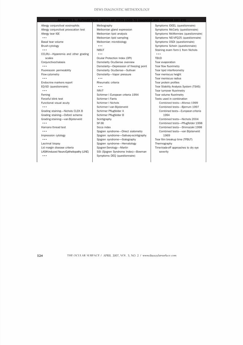
8/20/2019 dry eyes ii
http://slidepdf.com/reader/full/dry-eyes-ii 64/142
THE OCULAR SURFACE / APRIL 2007, VOL. 5, NO. 2 / www.theocularsurface.com124
APPENDIX 1. ALPHABETICAL LISTING OF TESTS USED TO DIAGNOSE AND MONITOR DRY EYE
Allergy conjunctival eosinophils
Allergy conjunctival provocation test
Allergy tear IGE
x x x
Basal tear volume
Brush cytology
x x x
CCLRU—Hyperemia and other grading
scales
Conjunctivochalasis
x x x
Fluorescein permeability
Flow cytometry
x x x
Endocrine markers report
EQ-SD (questionnaire)
x x x
Ferning
Forceful blink testFunctional visual acuity
x x x
Grading staining—Nichols CLEK B
Grading staining—Oxford scheme
Grading staining—van Bijsterveld
x x x
Hamano thread test
x x x
Impression cytology
x x x
Lacrimal biopsy
Lid margin disease criteriaLASIK-induced Neuro-Epitheliopathy (LINE)
x x x
Symptoms IDEEL (questionnaire)
Symptoms McCarty (questionnaire)
Symptoms McMonnies (questionnaire)
Symptoms NEI-VFQ25 (questionnaire)
Symptoms OSDI (questionnaire)
Symptoms Schein (questionnaire)
Staining exam form-1 from Nichols
x x x
TBUD
Tear evaporation
Tear flow fluorimetry
Tear lipid interferometry
Tear meniscus height
Tear meniscus radius
Tear protein profiles
Tear Stability Analysis System (TSAS)
Tear turnover fluorimetry
Tear volume fluorimetry
Tests used in combination Combined tests—Afonso 1999
Combined tests—Bjerrum 1997
Combined tests—European criteria
1994
Combined tests—Nichols 2004
Combined tests—Pflugfelder 1998
Combined tests—Shimazaki 1998
Combined tests—van Bijsterveld
1969
Tear film breakup time (TFBUT)
Thermography
Time-trade-off approaches to dry eyeseverity
Meibography
Meibomian gland expression
Meibomian lipid analysis
Meibomian lipid sampling
Meibomian microbiology
x x x
NIBUT
x x x
Ocular Protection Index (OPI)
Osmolarity OcuSense overview
Osmolarity—Depression of freezing point
Osmolarity OcuSense—Sullivan
Osmolarity—Vapor pressure
x x x
Rheumatic criteria
x x x
SBUT
Schirmer I European criteria 1994
Schirmer I FarrisSchirmer I Nichols
Schirmer I van Bijsterveld
Schirmer Pflugfelder A
Schirmer Pflugfelder B
Scintigraphy
SF-36
Sicca index
Sjogren syndrome—Direct sialometry
Sjogren syndrome—Salivary-scintigraphy
Sjogren syndrome—Sialography
Sjogren syndrome—Hematology
Sjogren Serology—MartinSSI (Sjogren Syndrome Index)—Bowman
Symptoms DEQ (questionnaire)
DEWS DIAGNOSTIC METHODOLOGY

8/20/2019 dry eyes ii
http://slidepdf.com/reader/full/dry-eyes-ii 65/142
THE OCULAR SURFACE / APRIL 2007, VOL. 5, NO. 2 / www.theocularsurface.com 125
APPENDIX 2. F UNCTIONAL GROUPINGS OF TESTS USED IN THE ASSESSMENT OF DRY EYE
1. Symptoms tests
Questionnaires
NEI-VFQ25
McMonnies
Schein
McCarty
OSDI DEQ
IDEEL
Visual function
LogMar acuity
Contrast sensitivity
Functional visual acuity
2. Aqueous tears
Tear volume
Fluorimetry
Hamano thread
Periotron test—“basal tear volume”
Tear meniscus
Radius of curvature
Height Area of cross-section
Tear film thickness
Tear flow
Fluoroimetry
Schirmer test
Schirmer I
Dynamic Schirmer
Schirmer II
Reflex Schirmer
Tear turnover
Dye dilution
Tear clearance
Fluorimetry
Tear evaporation Evaporimetry
3. Tear stability and visual function
Visual acuity
ETDRS
Functional visual acuity
Tear stability
Breakup time (BUT)
SBUT: Symptomatic BUT
Tear film BUT fluorescein
Noninvasive BUT (NIBUT)
Tear thinning time
Topographic analysis
Tear stability analysis system
Wavefront analysis4. Tear composition
Biological fluids
Aqueous tears
Lactoferrin
Lysozyme
Peroxidase
Immunoglobulin A
Ceruloplasmin
Inflammatory mediators
Matrix metalloproteinases
Other proteins
Mucins
Lipids
Cells in biofluids
Inflammatory cells
Epithelial cells
Tear debris
Surface cells
Impression cytology
Flow cytometry Brush cytology
Confocal microscopy
Meibomian lipids
Evaporimetry
Interferometry
Thickness
Grading
Meibometry
Meibography
Morphology in MGD
Expressed oil quality
Lipid chemistry
Tears: physical
Osmolarity Depression of freezing point
Vapor pressure osmometry
Conductivity OcuSense
Electrolyte composition
Tear ferning
Surface damage
Grading staining
Fluorescein stain
Rose Bengal stain
Lissamine green
Double staining
5. Other criteria
Tear function index (TFI)
Ocular protection index (OPI) Conjunctivochalasis score
Blink characteristics
Distinction from allergy
Lid margin disease criteria
Microbiology and lid disease
6. Sjogren syndrome
Serological tests
Anti-Ro
Anti-La
Anti-M3 receptor
Anti-fodrin
Minor salivary gland biopsy
Lacrimal gland biopsy
Systemic endocrine findings Tests of salivary function
Biscuit test
Sialography
7. Tests for assorted disorders
Wegener’s: Positive ANCA
Rheumatoid arthritis: Positive Rh-F
Systemic lupus erythematosus
LASIK-Induced Neuro Epitheliopathy
DEWS DIAGNOSTIC METHODOLOGY
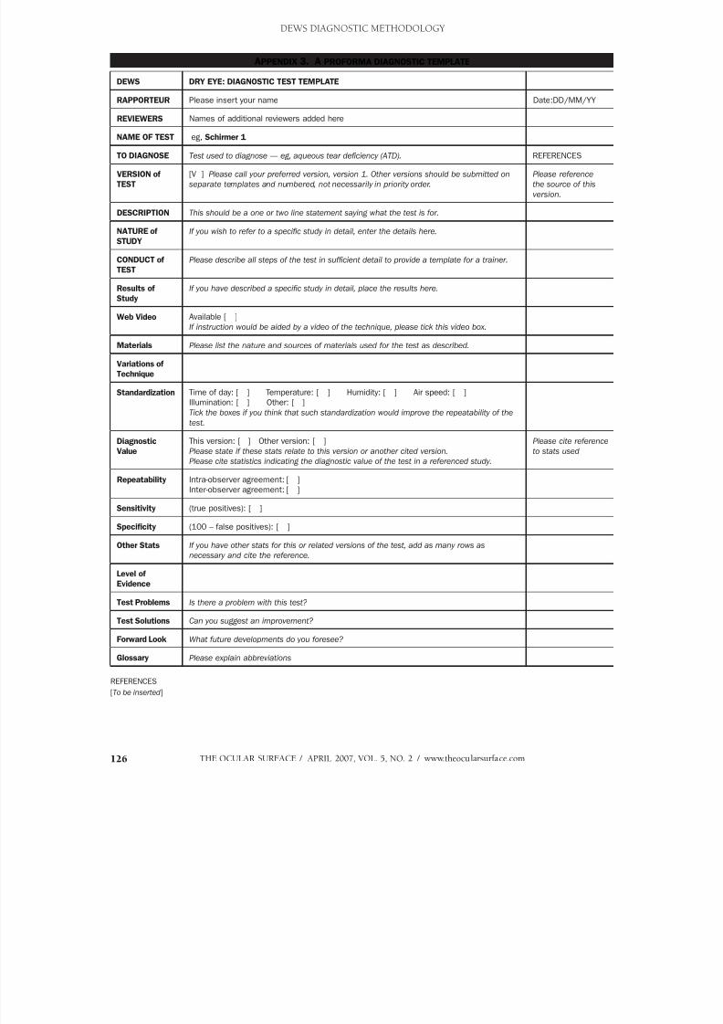
8/20/2019 dry eyes ii
http://slidepdf.com/reader/full/dry-eyes-ii 66/142
THE OCULAR SURFACE / APRIL 2007, VOL. 5, NO. 2 / www.theocularsurface.com126
DEWS DIAGNOSTIC METHODOLOGY
APPENDIX 3. A PROFORMA DIAGNOSTIC TEMPLATE
DEWS DRY EYE: DIAGNOSTIC TEST TEMPLATE
RAPPORTEUR Please insert your name Date:DD/MM/YY
REVIEWERS Names of additional reviewers added here
NAME OF TEST eg, Schirmer 1
TO DIAGNOSE Test used to diagnose — eg, aqueous tear deficiency (ATD). REFERENCES
VERSION of
TEST
[V ] Please call your preferred version, version 1. Other versions should be submitted on
separate templates and numbered, not necessarily in priority order.
Please reference
the source of this
version.
DESCRIPTION This should be a one or two line statement saying what the test is for.
NATURE of
STUDY
If you wish to refer to a specific study in detail, enter the details here.
CONDUCT of
TEST
Please describe all steps of the test in sufficient detail to provide a template for a trainer.
Results of
Study
If you have described a specific study in detail, place the results here.
Web Video Available [ ]
If instruction would be aided by a video of the technique, please tick this video box.
Materials Please list the nature and sources of materials used for the test as described.
Variations of
Technique
Standardization Time of day: [ ] Temperature: [ ] Humidity: [ ] Air speed: [ ]
Illumination: [ ] Other: [ ]
Tick the boxes if you think that such standardization would improve the repeatability of the
test.
DiagnosticValue
This version: [ ] Other version: [ ]Please state if these stats relate to this version or another cited version.
Please cite statistics indicating the diagnostic value of the test in a referenced study.
Please cite referenceto stats used
Repeatability Intra-observer agreement: [ ]
Inter-observer agreement: [ ]
Sensitivity (true positives): [ ]
Specificity (100 – false positives): [ ]
Other Stats If you have other stats for this or related versions of the test, add as many rows as
necessary and cite the reference.
Level of
Evidence
Test Problems Is there a problem with this test?
Test Solutions Can you suggest an improvement?
Forward Look What future developments do you foresee?
Glossary Please explain abbreviations
REFERENCES
[To be inserted]

8/20/2019 dry eyes ii
http://slidepdf.com/reader/full/dry-eyes-ii 67/142
THE OCULAR SURFACE / APRIL 2007, VOL. 5, NO. 2 / www.theocularsurface.com 127
APPENDIX 4. A NOTE ON THE JAPANESE CRITERIA FOR DRY EYE DIAGNOSIS
The previous Japanese dry eye diagnostic criteria were revised by the Japanese Dry Eye Research Society after the 1994-95 NEI/
Industry workshop (Miyawaki S, Nishiyama S. Classification criteria for Sjogren’s syndrome—sensitivity and specificity of criteria of
the Japanese Ministry of Health and Welfare (1977) and criteria of European community (1993). Nippon Rinsho 1995;53:2371-5).
The criteria, unpublished in the English literature, omitted symptoms from the diagnostic criteria at that time, because objective and
subjective findings did not appear to correlate. Following the DEWS meeting of 2004, the importance of symptoms was accepted in
Japan and the criteria have been modified.
The Japanese criteria prior to the 2004 DEWS meeting were:1) Qualitative or quantitative disturbance of the tear film (quantity: Schirmer test less than 5 mm or phenol red thread test less than
10 mm; quality: BUT less than 5 sec)
2) Conjunctivocorneal epithelial damage (excluding all other etiologies other than that listed under number 1)
Fluorescein staining greater than 1 point
RB staining greater than 3 points
(The presence of either fluorescein or RB staining is finding sufficient to satisfy criterion number 2)
The presence of both 1 and 2 = Definite dry eye. Presence of 1 or 2 = Probable dry eye
The Japanese diagnostic criteria have been revised by the Japan Dry Eye Research Society in August 2005, to include symptoms,
as follows.
New Diagnostic Criteria of the Japan Dry Eye Research Society: Revised in August 2005
Definite DE Probable DE
Symptoms Yes Yes Yes No
Tear film quality/quantity—disturbed Yes No Yes Yes
Epithelial damage Yes Yes No Yes
The phenol red thread test has been removed from the diagnostic criteria.
A fluorescein staining score of above 3 points is now required as positive staining (instead of 1 point).
DEWS DIAGNOSTIC METHODOLOGY

8/20/2019 dry eyes ii
http://slidepdf.com/reader/full/dry-eyes-ii 68/142
THE OCULAR SURFACE / APRIL 2007, VOL. 5, NO. 2 / www.theocularsurface.com128
APPENDIX 5
DEWS DRY EYE: DIAGNOSTIC TEST TEMPLATE
RAPPORTEUR A.J. Bron 22nd Oct 2004
TEST GRADING STAINING: CLEK Schema
TO DIAGNOSE The scheme is used to estimate surface damage in dry eye. REFERENCES
VERSION of TEST [ V1 ] [CLEK study] Barr et al 1999
Lemp 1995DESCRIPTION Surface damage to the exposed eye, assessed by staining, is graded against
standard charts.
NATURE of STUDY Nature of study
In this study, 75 patients regarded as having mild to moderate dry eye were assessed
for symptoms, MGD, tear quality, meniscus height, blink quality, TBUT F and BR
staining, phenol red test and Schirmer.
70.7% female.
61% using ATS
21.9% met European Criteria for moderate to severe dry eye.
About 30% were CL wearers.
Nichols et al 2004
CONDUCT of TEST Fluorescein instillation:
Fluorescein strip wetted with buffered saline. Drop instilled on inferior palpebral
conjunctiva. Blink several times.
Rose Bengal Staining: A Rosets Rose Bengal Ophthalmic Strip is wetted with sterile
buffered saline and instilled on the inferior bulbar conjunctiva. (“care taken to instill
adequate dye”)
STAINING: 5 corneal regions and 4 conjunctival regions as described in the CLEK
study (Barr et al. 1999).
The staining scale was 0-4, with 0.5 unit steps in each of the 5 corneal regions.
Photos were used as examples of severity.
The “total score” could either be summed, or averaged.
Nichols et al 2004
Barr et al 1999
[CLEK study]
OD OS
C I N T S = Central Inferior Nasal Temporal Superior
0–4 scale in 0.5 unit steps
circle location Check appropriate box
OD Location Cornea/Conj. Punctate FB Coalesced Full-Thickness Other
Stain 1 C I N T S
Stain 2 C I N T S
Stain 3 C I N T S
Stain 4 C I N T S
Stain 5 C I N T S
Stain 6 C I N T S
Stain 7 C I N T S
Stain 8 C I N T S
Stain 9 C I N T S
continued
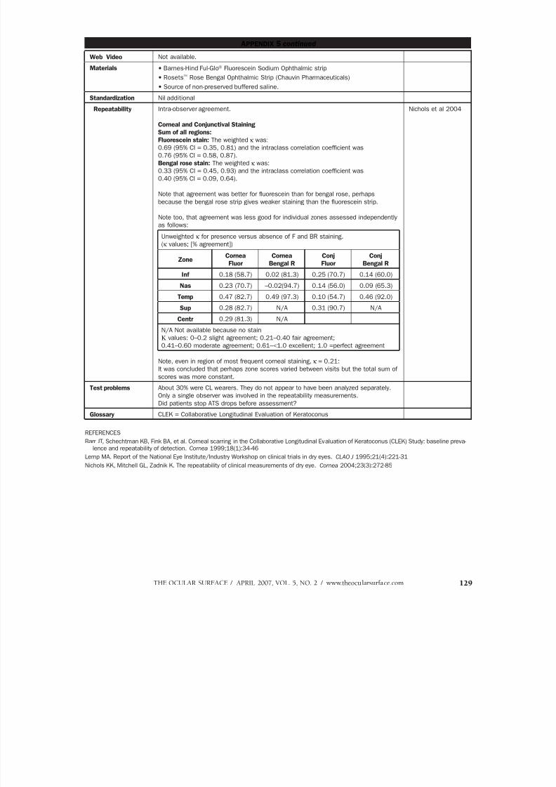
8/20/2019 dry eyes ii
http://slidepdf.com/reader/full/dry-eyes-ii 69/142
THE OCULAR SURFACE / APRIL 2007, VOL. 5, NO. 2 / www.theocularsurface.com 129
APPENDIX 5 continued
Web Video Not available.
Materials • Barnes-Hind Ful-Glo Fluorescein Sodium Ophthalmic strip
• Rosets Rose Bengal Ophthalmic Strip (Chauvin Pharmaceuticals)
• Source of non-preserved buffered saline.
Standardization Nil additional
Repeatability Intra-observer agreement.
Corneal and Conjunctival Staining
Sum of all regions:
Fluorescein stain: The weighted κ was:
0.69 (95% CI = 0.35, 0.81) and the intraclass correlation coefficient was
0.76 (95% CI = 0.58, 0.87).
Bengal rose stain: The weighted κ was:
0.33 (95% CI = 0.45, 0.93) and the intraclass correlation coefficient was
0.40 (95% CI = 0.09, 0.64).
Note that agreement was better for fluorescein than for bengal rose, perhaps
because the bengal rose strip gives weaker staining than the fluorescein strip.
Note too, that agreement was less good for individual zones assessed independently
as follows:
Unweighted κ for presence versus absence of F and BR staining.
(κ values; [% agreement])
ZoneCornea
Fluor
Cornea
Bengal R
Conj
Fluor
Conj
Bengal R
Inf 0.18 (58.7) 0.02 (81.3) 0.25 (70.7) 0.14 (60.0)
Nas 0.23 (70.7) –0.02(94.7) 0.14 (56.0) 0.09 (65.3)
Temp 0.47 (82.7) 0.49 (97.3) 0.10 (54.7) 0.46 (92.0)
Sup 0.28 (82.7) N/A 0.31 (90.7) N/A
Centr 0.29 (81.3) N/A
N/A Not available because no stain
Κ values: 0–0.2 slight agreement; 0.21–0.40 fair agreement;0.41–0.60 moderate agreement; 0.61–<1.0 excellent; 1.0 =perfect agreement
Note, even in region of most frequent corneal staining, κ = 0.21:
It was concluded that perhaps zone scores varied between visits but the total sum of
scores was more constant.
Nichols et al 2004
Test problems About 30% were CL wearers. They do not appear to have been analyzed separately.
Only a single observer was involved in the repeatability measurements.
Did patients stop ATS drops before assessment?
Glossary CLEK = Collaborative Longitudinal Evaluation of Keratoconus
REFERENCES
Barr JT, Schechtman KB, Fink BA, et al. Corneal scarring in the Collaborative Longitudinal Evaluation of Keratoconus (CLEK) Study: baseline preva-lence and repeatability of detection. Cornea 1999;18(1):34-46
Lemp MA. Report of the National Eye Institute/Industry Workshop on clinical trials in dry eyes. CLAO J 1995;21(4):221-31
Nichols KK, Mitchell GL, Zadnik K. The repeatability of clinical measurements of dry eye. Cornea 2004;23(3):272-85

8/20/2019 dry eyes ii
http://slidepdf.com/reader/full/dry-eyes-ii 70/142
THE OCULAR SURFACE / APRIL 2007, VOL. 5, NO. 2 / www.theocularsurface.com130
APPENDIX 6
DEWS DRY EYE: DIAGNOSTIC TEST TEMPLATE
RAPPORTEUR A.J.Bron 21st Oct 04
TEST GRADING STAINING: Oxford Schema
TO DIAGNOSE The scheme is used to estimate surface damage in dry eye. REFERENCES
VERSION of TEST [ V 1 ]
DESCRIPTION Surface damage to the exposed eye, assessed by staining, is graded against standard charts.
CONDUCT of
TEST
Grading Schema:
Staining is represented by punctate dots on a series of panels (A-E). Staining ranges from
0-5 for each panel and 0-15 for the total exposed inter-palpebral conjunctiva and cornea. The
dots are ordered on a log scale
PANEL Grade Criteria
A 0 Equal to or less than panel A
B I Equal to or less than panel B, greater than A
C II Equal to or less than panel C, greater than B
D III Equal to or less than panel D, greater than C
E IV Equal to or less than panel E, greater than D
>E V Greater than panel E
Conduct of Test:
• Dye is instilled.• Slit-lamp is set (eg, 16 magnification with x10 oculars with Haag-Streit).
• Cornea: The upper eyelid is lifted slightly to grade the whole corneal surface,
• Conjunctiva: To grade the temporal zone, the subject looks nasally; to grade the nasal
zone the subject looks temporally.
• (The upper and lower conjunctiva can also be graded).
Selection of dyes:
A list dyes and filters can be found in the original paper.
With fluorescein, staining must be graded as quickly as possible after instillation, since the
dye then diffuses rapidly into the tissue and its high luminosity blurring the stain margin.
Staining after rose bengal or lissamine green, persists at high contrast and may therefore be
observed for a considerable period. This is convenient for both grading and photography.
Fluorescein sodium
1. Quantified drop instillation
eg 2 µl of 2% sterile fluorescein instilled into each conjunctival sac with a micro-pipette
(using a sterile tip). In very dry eye, larger volumes risk the possibility of inadequate dilution
into the fluorescent range.
2. Unquantified instillation — impregnated paper strips
This is a convenient approach in the clinic using the following method of application:
• A single drop of unit dose saline is instilled onto a fluorescein-impregnated strip.
• When the drop has saturated the impregnated tip, the excess is shaken into a waste bin
with a sharp flick.
• The right lower lid is then pulled down and the strip is tapped onto the lower tarsal
conjunctiva. A similar procedure is carried out on the left.
If too large a volume is delivered then the concentration in the tear film will be too high, and
the tear film and staining pattern will be non-fluorescent.
Bron Evans Smith
2003
continued
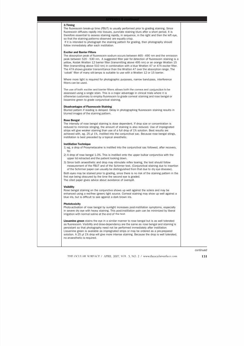
8/20/2019 dry eyes ii
http://slidepdf.com/reader/full/dry-eyes-ii 71/142
THE OCULAR SURFACE / APRIL 2007, VOL. 5, NO. 2 / www.theocularsurface.com 131
APPENDIX 6 continued
3.Timing
The fluorescein break-up time (FBUT) is usually performed prior to grading staining. Since
fluorescein diffuses rapidly into tissues, punctate staining blurs after a short period. It is
therefore essential to assess staining rapidly, in sequence, in the right and then the left eye,
so that the staining patterns observed are equally crisp.
If it is intended to photograph the staining pattern for grading, then photography should
follow immediately after each instillation.
Exciter and Barrier Filters
The absorption peak of fluorescein sodium occurs between 465 - 490 nm and the emission
peak between 520 - 530 nm. A suggested filter pair for detection of fluorescein staining is a
yellow, Kodak Wratten 12 barrier filter (transmitting above 495 nm) or an orange Wratten 15
filter (transmitting above 510 nm) in combination with a blue Wratten 47 or 47A exciter filter.
The 47A shows greater transmittance than the Wratten 47 over the absorption range. The
‘cobalt’ filter of many slit-lamps is suitable to use with a Wratten 12 or 15 barrier.
Where more light is required for photographic purposes, narrow band-pass, interference
filters can be used.
The use of both exciter and barrier filters allows both the cornea and conjunctiva to be
assessed using a single stain. This is a major advantage in clinical trials where it is
otherwise customary to employ fluorescein to grade corneal staining and rose bengal or
lissamine green to grade conjunctival staining.
Disadvantages of Fluorescein Staining
Blurred pattern if reading is delayed. Delay in photographing fluorescein staining results in
blurred images of the staining pattern.
Rose Bengal
The intensity of rose bengal staining is dose dependent. If drop size or concentration is
reduced to minimize stinging, the amount of staining is also reduced. Use of impregnated
strips will give weaker staining than use of a full drop of 1% solution. Best results are
achieved with, eg. 25 µl 1%, instilled into the conjunctival sac. Because rose bengal stings,
instillation is best preceded by a topical anesthetic.
Instillation Technique
1) eg, a drop of Proxymetacaine is instilled into the conjunctival sac followed, after recovery,by;
2) A drop of rose bengal 1.0%. This is instilled onto the upper bulbar conjunctiva with the
upper lid retracted and the patient looking down.
3) Since both anaesthetic and drop may stimulate reflex tearing, the test should follow
measurement of the FBUT and of the Schirmer test. (Conjunctival staining due to insertion
of the Schirmer paper can usually be distinguished from that due to dry eye disease).
Both eyes may be stained prior to grading, since there is no risk of the staining pattern in the
first eye being obscured by the time the second eye is graded.
The cited paper gives advice about avoidance of overspill.
Visibility
Rose bengal staining on the conjunctiva shows up well against the sclera and may be
enhanced using a red-free (green) light source. Corneal staining may show up well against a
blue iris, but is difficult to see against a dark brown iris.
Phototoxicity
Photo-activation of rose bengal by sunlight increases post-instillation symptoms, especially
in severe dry eye with heavy staining. This post-instillation pain can be minimized by liberal
irrigation with normal saline at the end of the test.
Lissamine green stains the eye in a similar manner to rose bengal but is as well tolerated
as fluorescein. Visibility and dose-dependency are the same as rose bengal and staining is
persistant so that photography need not be performed immediately after instillation.
Lissamine green is available as impregnated strips or may be ordered as a pre-prepared
solution. A 25 µl 1% drop will give more intense staining. Because the drop is well tolerated,
no anaesthetic is required.
continued
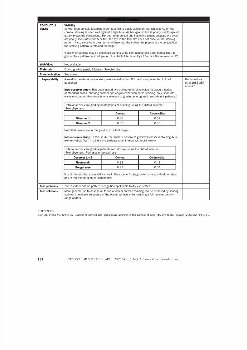
8/20/2019 dry eyes ii
http://slidepdf.com/reader/full/dry-eyes-ii 72/142
THE OCULAR SURFACE / APRIL 2007, VOL. 5, NO. 2 / www.theocularsurface.com132
APPENDIX 6 continued
CONDUCT of
TESTS
Visibility
As with rose bengal, lissamine green staining is easily visible on the conjunctiva. On the
cornea, staining is seen well against a light blue iris background but is poorly visible against
a dark brown iris background. For both rose bengal and lissamine green, because the dyes
are poorly seen within the tear film, the dye in the tear film does not obscure the staining
pattern. Also, since both dyes do not diffuse into the substantia propria of the conjunctiva,
the staining pattern is retained for longer.
Visibility of staining may be enhanced using a white light source and a red barrier filter, to
give a black pattern on a red ground. A suitable filter is a Hoya 25A, or a Kodak Wratten 92.
Web Video Not available
Materials Oxford grading panel; Slit-lamp; Selected dye.
Standardization See above.
Repeatability A small intra-inter observer study was carried out in 1986 and was presented but not
published:
Intra-observer study: This study asked two trained ophthalmologists to grade a series
of standard slides, showing corneal and conjunctival fluorescein staining, on 2 separate
occasions. [note: -this study is only relevant to grading photographic records not patients.]
Intra-observer κ for grading photographs of staining, using the Oxford scheme.
Two observers.
Cornea Conjunctiva
Observer 1 0.86 0.69
Observer 2 0.65 0.83
Note that values are in the good to excellent range.
Inter-observer study: In this study, the same 2 observers graded fluorescein staining (blue
exciter; yellow filter) in 13 dry eye patients at an interval within 2-3 weeks.
Inter-observer κ for grading patients with dry eye, using the Oxford scheme.Two observers. Fluorescein; bengal rose
Observer 1 v 2 Cornea Conjunctiva
Fluorescein 0.88 0.48
Bengal rose 0.87 0.54
It is of interest that observations are in the excellent category for cornea, with either stain
and in the fair category for conjunctiva.
Hardman Lea
et al 1986 AER
abstract.
Test problems The test depends on pattern recognition applicable to dry eye states.
Test solutions More general use to assess all forms of ocular surface staining can be achieved by scoring
staining in multiple segments of the ocular surface while retaining a full number density
range of dots
REFERENCES
Bron AJ, Evans VE, Smith JA. Grading of corneal and conjunctival staining in the context of other dry eye tests. Cornea 2003;22(7):640-50.

8/20/2019 dry eyes ii
http://slidepdf.com/reader/full/dry-eyes-ii 73/142
THE OCULAR SURFACE / APRIL 2007, VOL. 5, NO. 2 / www.theocularsurface.com 133
APPENDIX 7
DEWS DRY EYE: DIAGNOSTIC TEST TEMPLATE
RAPPORTEUR Mark B. Abelson and George W. Ousler III 5th Nov 2004
Reviewers –J Paugh 27th Dec 2007
TEST Tear Film Break-Up Time (TFBUT)
also: BUT (Break-up Time) and FBUT (Fluorescein Break-Up Time )
TO DIAGNOSE Tear Film Stability VERSION Version I
DESCRIPTION The tear film break-up time is defined as the interval between the last complete blink and
the first appearance of a dry spot, or disruption in the tear film.
Lemp 1970
Lemp 1995
STUDY 100 subjects with normal ocular health and 100 patients with ‘a history of dry eye’. 5 µl
of 2% fluorescein were instilled. Average of 3 readings.
Abelson et al 2002
CONDUCT of
TEST [V1]
Standardization of the volume instilled is important. Johnson and Murphy 2005 found that
increasing the volume of fluorescein instilled from 1–2.7 µl, increased the TFBUT, but that
increasing to 7.4 µl was not associated with further change.
1. Instill 1 to 5 micro-liters of non-preserved, 2% sodium fluorescein onto the bulbar
conjunctiva without inducing reflex tearing by using a micro-pipette or D.E.T. strip;
2. The patient is instructed to blink naturally, without squeezing, several times to
distribute the fluorescein3. Within 10 - 30 seconds of the fluorescein instillation, the patient is asked to stare
straight ahead without blinking, until told otherwise;
4. Set slit-lamp magnification at 10X, keep the background illumination intensity constant
(cobalt blue light) and use a Wratten 12 yellow filter to enhance observation of the tear
film over the entire cornea;
5. Use stopwatch to record time between last complete blink and first appearance of
growing micelle;
6. Once TFBUT is observed, instruct patient to blink freely.
Various authors advocate the use of a yellow barrier filter (Kodak Wratten 12) to enhance
the visibility of the break in the fluorescent tear film. (Eliason and Maurice 1990; Cho and
Brown 1993; Nichols et al. 2003; Bron et al 2003.
Johnson et al 2005).
Johnson and Murphy
2005
CONDUCT of
TEST [V2]
2.5 µl 1.0% fluorescein Vitale et al 1994
Results of
study
The mean TFBUT for normal subjects was 7.1 s (range 4.7 to 11.4 s) and for dry eye
patients 2.2 s (range (0.9 to 5.2 s). On the basis of this, a cut-off for dry eye diagnosis of
≤ 5 s was recommended.
Abelson et al 2002
Video *Slit-lamp, on-line video camera may be used to capture TFBUT. Video capture with an
on-screen timer allows for precise measurement of the time between the last complete
blink and the appearance of the first, growing micelle. This also allows masking for clinical
trials purposes
Welch et al 2003
Web video Not available
Materials • Non-preserved, 2% sodium fluorescein;
• Micro-pipette;
• Or D.E.T. strip.
• Slit-lamp
• Timer
• Kodak Wratten filter 12. See variations, below.
Variations of
technique
Historically, the technique for evaluating TFBUT has lacked consistency. Large and
varying amounts of sodium fluorescein (up to 50 µl) were used, times were determined
by counting aloud and using less sophisticated instrumentation. Such techniques yield
varying results.
Standardization Time of day [√] Temperature [√] Humidity [√] Air speed [√] Illumination [√]
• Patient instruction;
• Slit-lamp magnification;
• Barrier filter.
continued
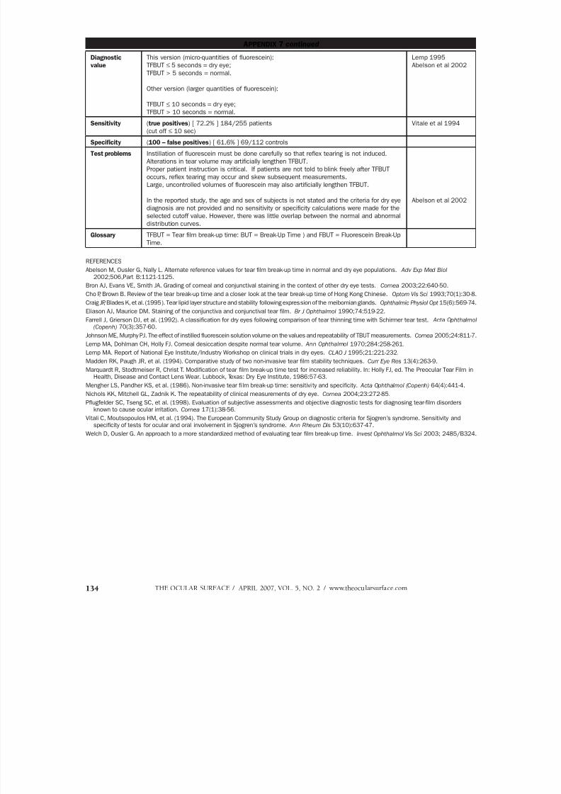
8/20/2019 dry eyes ii
http://slidepdf.com/reader/full/dry-eyes-ii 74/142
THE OCULAR SURFACE / APRIL 2007, VOL. 5, NO. 2 / www.theocularsurface.com134
APPENDIX 7 continued
Diagnostic
value
This version (micro-quantities of fluorescein):
TFBUT ≤ 5 seconds = dry eye;
TFBUT > 5 seconds = normal.
Other version (larger quantities of fluorescein):
TFBUT ≤ 10 seconds = dry eye;
TFBUT > 10 seconds = normal.
Lemp 1995
Abelson et al 2002
Sensitivity (true positives) [ 72.2% ] 184/255 patients
(cut off ≤ 10 sec)
Vitale et al 1994
Specificity (100 – false positives) [ 61.6% ] 69/112 controls
Test problems Instillation of fluorescein must be done carefully so that reflex tearing is not induced.
Alterations in tear volume may artificially lengthen TFBUT.
Proper patient instruction is critical. If patients are not told to blink freely after TFBUT
occurs, reflex tearing may occur and skew subsequent measurements.
Large, uncontrolled volumes of fluorescein may also artificially lengthen TFBUT.
In the reported study, the age and sex of subjects is not stated and the criteria for dry eye
diagnosis are not provided and no sensitivity or specificity calculations were made for the
selected cutoff value. However, there was little overlap between the normal and abnormal
distribution curves.
Abelson et al 2002
Glossary TFBUT = Tear film break-up time: BUT = Break-Up Time ) and FBUT = Fluorescein Break-Up
Time.
REFERENCES
Abelson M, Ousler G, Nally L. Alternate reference values for tear film break-up time in normal and dry eye populations. Adv Exp Med Biol
2002;506,Part B:1121-1125.
Bron AJ, Evans VE, Smith JA. Grading of corneal and conjunctival staining in the context of other dry eye tests. Cornea 2003;22:640-50.
Cho P, Brown B. Review of the tear break-up time and a closer look at the tear break-up time of Hong Kong Chinese. Optom Vis Sci 1993;70(1):30-8.
Craig JP, Blades K, et al. (1995). Tear lipid layer structure and stability following expression of the meibomian glands. Ophthalmic Physiol Opt 15(6):569-74.
Eliason AJ, Maurice DM. Staining of the conjunctiva and conjunctival tear film. Br J Ophthalmol 1990;74:519-22.
Farrell J, Grierson DJ, et al. (1992). A classification for dry eyes following comparison of tear thinning time with Schirmer tear test. Acta Ophthalmol
(Copenh) 70(3):357-60.
Johnson ME, Murphy PJ. The effect of instilled fluorescein solution volume on the values and repeatability of TBUT measurements. Cornea 2005;24:811-7.
Lemp MA, Dohlman CH, Holly FJ. Corneal desiccation despite normal tear volume. Ann Ophthalmol 1970;284:258-261.
Lemp MA. Report of National Eye Institute/Industry Workshop on clinical trials in dry eyes. CLAO J 1995;21:221-232.
Madden RK, Paugh JR, et al. (1994). Comparative study of two non-invasive tear film stability techniques. Curr Eye Res 13(4):263-9.
Marquardt R, Stodtmeiser R, Christ T. Modification of tear film break-up time test for increased reliability. In: Holly FJ, ed. The Preocular Tear Film inHealth, Disease and Contact Lens Wear. Lubbock, Texas: Dry Eye Institute, 1986:57-63.
Mengher LS, Pandher KS, et al. (1986). Non-invasive tear film break-up time: sensitivity and specificity. Acta Ophthalmol (Copenh) 64(4):441-4.
Nichols KK, Mitchell GL, Zadnik K. The repeatability of clinical measurements of dry eye. Cornea 2004;23:272-85.
Pflugfelder SC, Tseng SC, et al. (1998). Evaluation of subjective assessments and objective diagnostic tests for diagnosing tear-film disordersknown to cause ocular irritation. Cornea 17(1):38-56.
Vitali C, Moutsopoulos HM, et al. (1994). The European Community Study Group on diagnostic criteria for Sjogren’s syndrome. Sensitivity andspecificity of tests for ocular and oral involvement in Sjogren’s syndrome. Ann Rheum Dis 53(10):637-47.
Welch D, Ousler G. An approach to a more standardized method of evaluating tear film break-up time. Invest Ophthalmol Vis Sci 2003; 2485/B324.
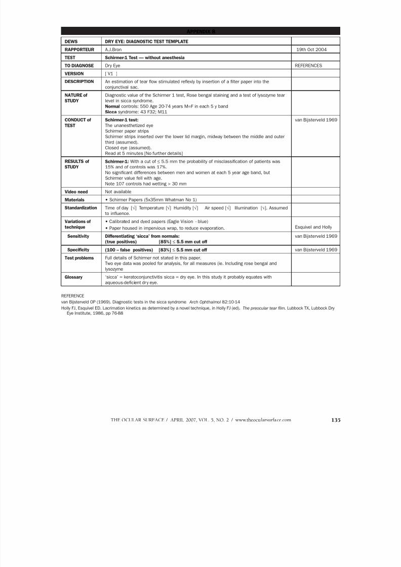
8/20/2019 dry eyes ii
http://slidepdf.com/reader/full/dry-eyes-ii 75/142
THE OCULAR SURFACE / APRIL 2007, VOL. 5, NO. 2 / www.theocularsurface.com 135
APPENDIX 8
DEWS DRY EYE: DIAGNOSTIC TEST TEMPLATE
RAPPORTEUR A.J.Bron 19th Oct 2004
TEST Schirmer-1 Test — without anesthesia
TO DIAGNOSE Dry Eye REFERENCES
VERSION [ V1 ]
DESCRIPTION An estimation of tear flow stimulated reflexly by insertion of a filter paper into theconjunctival sac.
NATURE of
STUDY
Diagnostic value of the Schirmer 1 test, Rose bengal staining and a test of lysozyme tear
level in sicca syndrome.
Normal controls: 550 Age 20-74 years M=F in each 5 y band
Sicca syndrome: 43 F32; M11
CONDUCT of
TEST
Schirmer-1 test:
The unanesthetized eye
Schirmer paper strips
Schirmer strips inserted over the lower lid margin, midway between the middle and outer
third (assumed).
Closed eye (assumed).
Read at 5 minutes [No further details]
van Bijsterveld 1969
RESULTS ofSTUDY
Schirmer-1: With a cut of ≤ 5.5 mm the probability of misclassification of patients was15% and of controls was 17%.
No significant differences between men and women at each 5 year age band, but
Schirmer value fell with age.
Note 107 controls had wetting > 30 mm
Video need Not available
Materials • Schirmer Papers (5x35mm Whatman No 1)
Standardization Time of day [√] Temperature [√] Humidity [√] Air speed [√] Illumination [√]. Assumed
to influence.
Variations of
technique
• Calibrated and dyed papers (Eagle Vision - blue)
• Paper housed in impervious wrap, to reduce evaporation. Esquivel and Holly
Sensitivity Differentiating ‘sicca’ from normals:
(true positives) [85%] ≤ 5.5 mm cut off
van Bijsterveld 1969
Specificity (100 – false positives) [83%] ≤ 5.5 mm cut off van Bijsterveld 1969
Test problems Full details of Schirmer not stated in this paper.
Two eye data was pooled for analysis, for all measures (ie. Including rose bengal and
lysozyme
Glossary ‘sicca’ = keratoconjunctivitis sicca = dry eye. In this study it probably equates with
aqueous-deficient dry eye.
REFERENCE
van Bijsterveld OP (1969). Diagnostic tests in the sicca syndrome. Arch Ophthalmol 82:10-14
Holly FJ, Esquivel ED. Lacrimation kinetics as determined by a novel technique, in Holly FJ (ed). The preocular tear film. Lubbock TX, Lubbock DryEye Institute, 1986, pp 76-88

8/20/2019 dry eyes ii
http://slidepdf.com/reader/full/dry-eyes-ii 76/142
THE OCULAR SURFACE / APRIL 2007, VOL. 5, NO. 2 / www.theocularsurface.com136
APPENDIX 9
DEWS DRY EYE: DIAGNOSTIC TEST TEMPLATE
RAPPORTEUR Michael A. Lemp 16th Oct 2004;
15th March 2006
TEST Tear Osmolarity
TO DIAGNOSE Global test for dry eye Sullivan 2004
VERSION of TEST OcuSense Volume Independent Tear Osmometer
DESCRIPTION This “lab-on-a-chip” test uses a combination of impedance information with
sophisticated mathematics to derive tear film osmolarity. A small nanoliter tear sample
is obtained with a standard micropipette and is then automatically transferred to a chip
surface. A precise readout is obtained in seconds after the transfer.
CONDUCT of TEST 1. Snap microchip in place
2. Touch lower lid with microcapillary
3. Let capillary action draw a few nL
4. Place capillary in machine
5. Read osmolarity
Web video Available:[No]
Materials • 1-lambda microcapillary
• microchip• Both available from OcuSense
Standardization Time of day [√ ] Temperature [√ ] Humidity [√ ] Air speed [ √ ] Illumination [ √ ]
Assumed to influence
Other: [ Avoid reflex tearing ]
White et. al. Showed that use of a slit lamp has upwards of a 7 mOsm/kg effect on
the value of osmolality due to the induction of reflex tearing.
Overstimulation during collection is discouraged. Reflex tears have far lower
osmolality (≈ 5%, Nelson, 1986) than basal tears.
White et al 1993
Nelson et al 1986
Repeatability Intra-observer agreement. [ ]
Inter-observer agreement. [< 2.6% 1st prototype]
Sullivan B 2004
Sensitivity (true positives) [ projected 94%]
≥ 318 mOsm: –provisional
Sullivan B 2004
Specificity (100 – false positives) [ projected 84%] Sullivan B 2004
Test problems Limited availability
Test solutions Commercial development
FORWARD LOOK This is a high throughput test that can be performed by a technician, and currently
carries a miscellaneous CPT.
REFERENCES
Farris RL. Tear osmolarity--a new gold standard? Adv Exp Med Biol 350:495-503, 1994
Nelson JD, Wright JC. Tear film osmolality determination: an evaluation of potential errors in measurement. Curr Eye Res Sep;5(9):677-81, 1986
Sullivan B, et al. 4th International Conference on the Lacrimal Gland, Tear Film & Ocular Surface and Dry Eye Syndromes, 11/20/04
White KM, Benjamin WJ, Hill RM. Human basic tear fluid osmolality. I. Importance of sample collection strategy. Acta Ophthalmol (Copenh) Aug;71(4):524-9, 1993
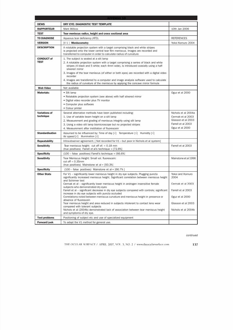
8/20/2019 dry eyes ii
http://slidepdf.com/reader/full/dry-eyes-ii 77/142
THE OCULAR SURFACE / APRIL 2007, VOL. 5, NO. 2 / www.theocularsurface.com 137
APPENDIX 10
DEWS DRY EYE: DIAGNOSTIC TEST TEMPLATE
RAPPORTEUR Mark Willcox 10th Jan 2006
TEST Tear meniscus radius, height and cross sectional area
TO DIAGNOSE Aqueous tear deficiency (ATD). REFERENCES
VERSION [V 1 ] Meniscometry Yokoi Komuro 2004
DESCRIPTION A rotatable projection system with a target comprising black and white stripesis projected onto the lower central tear film meniscus. Images are recorded and
transferred to computer in order to calculate radius of curvature
CONDUCT of
TEST
1. The subject is seated at a slit lamp
2. A rotatable projection system with a target comprising a series of black and white
stripes (4 black and 5 white; each 4mm wide), is introduced coaxially using a half-
silvered mirror
3. Images of the tear meniscus (of either or both eyes) are recorded with a digital video
recorder
4. Images are transferred to a computer and image analysis software used to calculate
the radius of curvature of the meniscus by applying the concave mirror formula
Web Video Not available
Materials: • Slit lamp
• Rotatable projection system (see above) with half silvered mirror
• Digital video recorder plus TV monitor
• Computer plus software
• Colour printer
Oguz et al 2000
Variations of
technique
Several alternative methods have been published including:
1. Use of variable beam height on a slit lamp
2. Measurement and grading of meniscus integrity using slit lamp
3. Using a video slit lamp biomicroscope but no projected stripes
4. Measurement after instillation of fluorescein
Nichols et al 2004a
Cermak et al 2003
Glasson et al 2003
Farrell et al 2003
Oguz et al 2000
Standardisation Assumed to be influenced by: Time of day [√] Temperature [√] Humidity [√]
Air speed [√] Illumination [√]
Repeatability Intra-observer agreement. [ Not recorded for V1 – but poor in Nichols et al system]Sensitivity Tear meniscus height: cut off of: < 0.18 mm
(true positives) Farrell et al’s technique = [72.8%]
Farrell et al 2003
Specificity (100 – false positives) Farrell’s technique = [66.6%]
Sensitivity Tear Meniscus Height: Small vol. fluorescein:
cut off < 0.35mm
(true positives) Mainstone et al = [93.3%]
Mainstone et al 1996
Specificity (100 – false positives) Mainstone et al = [66.7% ]
Other Stats For V1 – significantly lower meniscus height in dry eye subjects. Plugging puncta
significantly increased meniscus height. Significant correlation between meniscus height
and Schirmer test
Cermak et al – significantly lower meniscus height in androgen insensitive female
subjects who demonstrated dry eyes
Farrell et al – significant decrease in dry eye subjects compared with controls; significantincrease in dry eye subjects with puncta occluded
Correlations noted between meniscus curvature and meniscus height in presence or
absence of fluorescein
Tear meniscus height and area reduced in subjects intolerant to contact lens wear
compared with tolerant subjects
Nichols et al (2004b) demonstrated lack of association between tear meniscus height
and symptoms of dry eye.
Yokoi and Komuro
2004
Cermak et al 2003
Farrell et al 2003
Oguz et al 2000
Glasson et al 2003
Nichols et al 2004b
Test problems Positioning of subject etc and use of specialized equipment
Forward Look To adapt the V1 method for general use.
continued
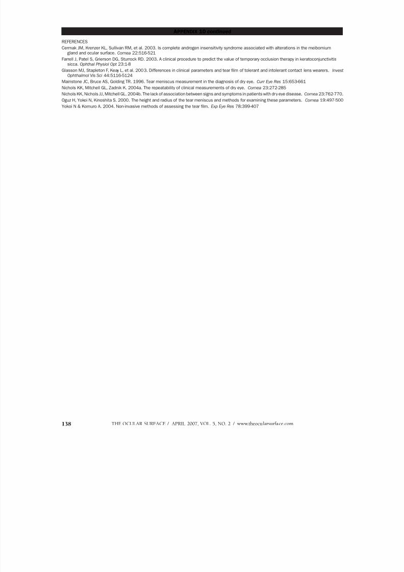
8/20/2019 dry eyes ii
http://slidepdf.com/reader/full/dry-eyes-ii 78/142
THE OCULAR SURFACE / APRIL 2007, VOL. 5, NO. 2 / www.theocularsurface.com138
REFERENCES
Cermak JM, Krenzer KL, Sullivan RM, et al. 2003. Is complete androgen insensitivity syndrome associated with alterations in the meibomiumgland and ocular surface. Cornea 22:516-521
Farrell J, Patel S, Grierson DG, Sturrock RD. 2003. A clinical procedure to predict the value of temporary occlusion therapy in keratoconjunctivitissicca. Ophthal Physiol Opt 23:1-8
Glasson MJ, Stapleton F, Keay L, et al. 2003. Differences in clinical parameters and tear film of tolerant and intolerant contact lens wearers. Invest
Ophthalmol Vis Sci 44:5116-5124
Mainstone JC, Bruce AS, Golding TR. 1996. Tear meniscus measurement in the diagnosis of dry eye. Curr Eye Res 15:653-661Nichols KK, Mitchell GL, Zadnik K. 2004a. The repeatability of clinical measurements of dry eye. Cornea 23:272-285
Nichols KK, Nichols JJ, Mitchell GL. 2004b. The lack of association between signs and symptoms in patients with dry eye disease. Cornea 23:762-770.
Oguz H, Yokoi N, Kinoshita S. 2000. The height and radius of the tear meniscus and methods for examining these parameters. Cornea 19:497-500
Yokoi N & Komuro A. 2004. Non-invasive methods of assessing the tear film. Exp Eye Res 78:399-407
APPENDIX 10 continued
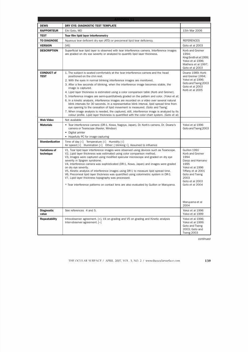
8/20/2019 dry eyes ii
http://slidepdf.com/reader/full/dry-eyes-ii 79/142
THE OCULAR SURFACE / APRIL 2007, VOL. 5, NO. 2 / www.theocularsurface.com 139
APPENDIX 11
DEWS DRY EYE: DIAGNOSTIC TEST TEMPLATE
RAPPORTEUR Eiki Goto, MD 15th Mar 2006
TEST Tear film lipid layer interferometry
TO DIAGNOSE Aqueous tear deficient dry eye (ATD) or precorneal lipid tear deficiency. REFERENCES
VERSION [V6] Goto et al 2003
DESCRIPTION Superficial tear lipid layer is observed with tear interference camera. Interference imagesare graded on dry eye severity or analyzed to quantify lipid layer thickness.
Korb and Greiner1994;
King-Smith et al 1999;
Yokoi et al 1996;
Mathers et al 1997;
Goto et al 2003
CONDUCT of
TEST
1. The subject is seated comfortably at the tear interference camera and the head
positioned on the chin rest.
2. With the eyes in normal blinking interference images are monitored.
3. After a few seconds of blinking, when the interference image becomes stable, the
image is captured.
4. Lipid layer thickness is estimated using a color comparison table (Korb and Greiner).
5. Interference images are semi-quantitatively graded on the pattern and color. (Yokoi et al)
6. In a kinetic analysis, interference images are recorded on a video over several naturalblink intervals for 30 seconds. In a representative blink interval, lipid spread time from
eye opening to the cessation of lipid movement is measured. (Goto and Tseng)
7. When image analysis is needed, the captured, still, interference image is analyzed by its
colour profile. Lipid layer thickness is quantified with the color chart system. (Goto et al)
Doane 1989; Korb
and Greiner 1994;
Yokoi et al 1996;
Goto and Tseng 2003
Goto et al 2003
Korb et al 2005
Web Video Not available
Materials • Tear interference camera (DR-1, Kowa, Nagoya, Japan), Dr. Korb’s camera, Dr. Doane’s
camera or Tearscope (Keeler, Windsor)
• Digital printer
• Hopefully PC for image capturing
Yokoi et al 1996
Goto and Tseng 2003
Standardization Time of day [√] Temperature [√] Humidity [√]
Air speed [√] Illumination [√] Other: [ blinking √]. Assumed to influence
Variations oftechnique V1, Tear lipid layer interference images were observed using devices such as Tearscope.V2, Lipid layer thickness was estimated using color comparison method.
V3, Images were captured using modified specular microscope and graded on dry eye
severity in Sjogren syndrome.
V4, Interference camera was sophisticated (DR-1, Kowa, Japan) and images were graded
on dry eye severity.
V5, Kinetic analysis of interference images using DR-1 to measure lipid spread time.
V6, Precorneal lipid layer thickness was quantified using colorimetric system in DR-1.
V7, Lipid layer thickness topography was processed.
* Tear interference patterns on contact lens are also evaluated by Guillon or Maruyama.
Guillon 1992Korb and Greiner
1994
Danjo and Hamano
1995
Yokoi et al 1996
Tiffany et al 2001
Goto and Tseng
2003
Goto et al 2003
Goto et al 2004
Maruyama et al2004
Diagnostic
value
See references 4 and 5. Yokoi et al 1996
Yokoi et al 1999
Repeatability Intra-observer agreement. [+], V4 on grading and V5 on grading and Kinetic analysis
Inter-observer agreement. [–]
Yokoi et al 1996;
Yokoi et al 1999;
Goto and Tseng
2003; Goto and
Tseng 2003
continued
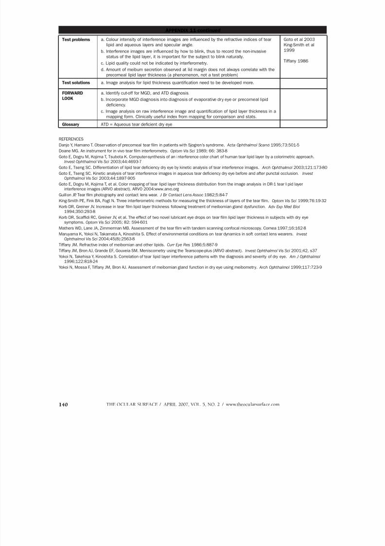
8/20/2019 dry eyes ii
http://slidepdf.com/reader/full/dry-eyes-ii 80/142
THE OCULAR SURFACE / APRIL 2007, VOL. 5, NO. 2 / www.theocularsurface.com140
APPENDIX 11 continued
Test problems a. Colour intensity of interference images are influenced by the refractive indices of tear
lipid and aqueous layers and specular angle.
b. Interference images are influenced by how to blink, thus to record the non-invasive
status of the lipid layer, it is important for the subject to blink naturally.
c. Lipid quality could not be indicated by interferometry.
d. Amount of meibum secretion observed at lid margin does not always correlate with the
precorneal lipid layer thickness (a phenomenon, not a test problem)
Goto et al 2003
King-Smith et al
1999
Tiffany 1986
Test solutions a. Image analysis for lipid thickness quantification need to be developed more.
FORWARD
LOOK
a. Identify cut-off for MGD, and ATD diagnosis.
b. Incorporate MGD diagnosis into diagnosis of evaporative dry eye or precorneal lipid
deficiency.
c. Image analysis on raw interference image and quantification of lipid layer thickness in a
mapping form. Clinically useful index from mapping for comparison and stats.
Glossary ATD = Aqueous tear deficient dry eye
REFERENCES
Danjo Y, Hamano T. Observation of precorneal tear film in patients with Sjogren’s syndrome. Acta Ophthalmol Scand 1995;73:501-5
Doane MG. An instrument for in vivo tear film interferometry. Optom Vis Sci 1989; 66: 383-8
Goto E, Dogru M, Kojima T, Tsubota K. Computer-synthesis of an interference color chart of human tear lipid layer by a colorimetric approach.Invest Ophthalmol Vis Sci 2003;44:4693-7
Goto E, Tseng SC. Differentiation of lipid tear deficiency dry eye by kinetic analysis of tear interference images. Arch Ophthalmol 2003;121:173-80
Goto E, Tseng SC. Kinetic analysis of tear interference images in aqueous tear deficiency dry eye before and after punctal occlusion. Invest
Ophthalmol Vis Sci 2003;44:1897-905
Goto E, Dogru M, Kojima T, et al. Color mapping of tear lipid layer thickness distribution from the image analysis in DR-1 tear lipid layerinterference images (ARVO abstract). ARVO 2004:www.arvo.org
Guillon JP. Tear film photography and contact lens wear. J Br Contact Lens Assoc 1982;5:84-7
King-Smith PE, Fink BA, Fogt N. Three interferometric methods for measuring the thickness of layers of the tear film. Optom Vis Sci 1999;76:19-32
Korb DR, Greiner JV. Increase in tear film lipid layer thickness following treatment of meibomian gland dysfunction. Adv Exp Med Biol 1994;350:293-8
Korb DR, Scaffidi RC, Greiner JV, et al. The effect of two novel lubricant eye drops on tear film lipid layer thickness in subjects with dry eyesymptoms. Optom Vis Sci 2005; 82: 594-601
Mathers WD, Lane JA, Zimmerman MB. Assessment of the tear film with tandem scanning confocal microscopy. Cornea 1997;16:162-8
Maruyama K, Yokoi N, Takamata A, Kinoshita S. Effect of environmental conditions on tear dynamics in soft contact lens wearers. InvestOphthalmol Vis Sci 2004;45(8):2563-8
Tiffany JM. Refractive index of meibomian and other lipids. Curr Eye Res 1986;5:887-9
Tiffany JM, Bron AJ, Grande EF, Gouveia SM. Meniscometry using the Tearscope-plus (ARVO abstract). Invest Ophthalmol Vis Sci 2001;42, s37
Yokoi N, Takehisa Y, Kinoshita S. Correlation of tear lipid layer interference patterns with the diagnosis and severity of dry eye. Am J Ophthalmol 1996;122:818-24
Yokoi N, Mossa F, Tiffany JM, Bron AJ. Assessment of meibomian gland function in dry eye using meibometry. Arch Ophthalmol 1999;117:723-9

8/20/2019 dry eyes ii
http://slidepdf.com/reader/full/dry-eyes-ii 81/142
THE OCULAR SURFACE / APRIL 2007, VOL. 5, NO. 2 / www.theocularsurface.com 141
APPENDIX 12
DEW DRY EYE: DIAGNOSTIC TEST TEMPLATE
RAPPORTEUR Murat Dogru 24th Oct 2004
TEST Tear Stability Analyses System (TSAS)
TO DIAGNOSE Test used to diagnose –Tear Instability
Refs:
Kojima 2004
Goto 2004a,b
VERSION [TMS-2N] Kojima 2004DESCRIPTION Noninvasive and objective test for tear film stability analysis
Study To compare the sensitivity and specificity of TSAS with the BUT (based on slit-lamp
examination and use of fluorescein), 48 volunteers without any eye disease, surgery or
drug use within 1 year of study were recruited. See below.
Goto 2004a
CONDUCT of
TEST
Subject seated in front of TMS-2N corneal topography unit.
Subject asked not to blink for 10 seconds with test initiation
Device automatically captures corneal topograms each second for 11 consecutive
seconds, displayed as time plot curves of SRI, SAI, BUT area
Results of
Study
See study, above.
42.5% (34 eyes) of the 80 eyes of the volunteers studied had a normal BUT and 57.5%
had an abnormal BUT. On the basis of the subjects’ dry eye symptoms such as FBS,
soreness, dryness etc, the sensitivity and specificity of the BUT were 75% and 60%
respectively. Among 34 eyes with a normal BUT, 11 (32.35%) were found to have anabnormal TMS BUT. Of these eyes, 9 (81.8%) were from 6 subjects who had dry eye
symptoms in their questionnaires. On the basis of symptomatology, the sensitivity and
specificity of TMS BUT was 97.5 and 62.5% respectively. The difference of sensitivity
between SLE BUT and TMS BUT was significant; however, the difference in specificity was
not.
Web Video Not available
Materials TMS-2N corneal topography device
TSAS software( Tomey Inc)
Standardization Time of day [√] Temperature [√] Humidity [√] Air speed [√] Illumination [√] . Assumed
to influence.
Sensitivity (true positives) [97.5% ] Goto 2004a
Specificity (100 – false positives) [62.5 % ]
Test problems Although the test appears to be a promising, non-invasive method to test tear stability, it is
not known whether the test is evaluating tear stability due to lipid layer or overall tear film
changes.
Only one study compares the test with the invasive fluorescein aided BUT measurement.
Normal values of this test and age-specific cut off values on a large set of subjects not yet
established.
Comparative studies with other invasive and non-invasive tests of tear stability do not exist
as yet.
Needs a corneal topography device and the software which makes it expensive compared
to fluorescein aided BUT testing.
Test solutions The above mentioned studies will prepare this test for general clinical prime time.
Forward Look The device is still being furnished with novel parameters such as BUT area. For dynamic
analyses of tear functions in dry eye syndromes and ocular surface disorders, I believe
that this new system is set to play an important role in the future.
Glossary TSAS: Tear Stability Analyses System
REFERENCES
Goto T, Zheng X, Okamoto S, Ohashi Y. Tear Film Stability Analysis System: Introducing a new application for videokeratography. Cornea 2004aNov;23(8):S65-S70
Goto T, Zheng X, Klyce SD, et al. Evaluation of the tear film stability after laser in situ keratomileusis using the tear film stability analysis system. Am J Ophthalmol 2004b Jan;137(1):116-20
Kojima T, Ishida R, Dogru M, et al. A new noninvasive tear stability analysis system for the assessment of dry eyes. Invest Ophthalmol Vis Sci 2004May;45(5):1369-74

8/20/2019 dry eyes ii
http://slidepdf.com/reader/full/dry-eyes-ii 82/142
THE OCULAR SURFACE / APRIL 2007, VOL. 5, NO. 2 / www.theocularsurface.com142
APPENDIX 13
DEWS DRY EYE: DIAGNOSTIC TEST TEMPLATE
RAPPORTEUR John M. Tiffany 12th Nov 2004
TEST MEIBOMETRY
TO DIAGNOSE Meibomian Gland Dysfunction — (MGD) REFERENCES
VERSION of TEST [ V1 ] Komuro et al 2002
DESCRIPTION Lipid on the lower central lid margin is blotted onto a plastic tape and the amount takenup read by optical densitometry. This provides an indirect measure of the steady state
level of meibomian lipid.
CONDUCT of
TEST
1. The subject is seated, with the head resting comfortably at the slit-lamp.
2. With the eyes in upgaze, the right lower lid is drawn down lightly without pressure on the
tarsal plate.
3. A standard loop of plastic tape, held in an applanation or ultrasonography probe holder,
is applied to the central third of the everted lid margin for 3 seconds, at 0 mmHg
exerted pressure.
4. The tape is air dried for 3 minutes to allow tear evaporation if necessary.
5. The increase in transparency induced by the lipid blot, is read in the laser meibometer.
6. The Casual Lipid level (expressed as arbitrary optical density units) is calculated as (C-B),
where C is the casual reading, B is the reading from the untouched tape (background).
Komuro et al 2002
Video need Not available.
Materials • Plastic tape: 8 mm wide (Courage and Khazaka, Köln)
• Tape Holder:(eg. NIDEK ultrasonographic probe holder.
• Laser Meibometer. Window size (2.5 x 5.0 mm2)
Standardization Time of day [ x ]
The level is highest in the first hour after waking, but thereafter settles to a constant level
through most of the day
Variations of
technique
In the original version, [V2 ] optical density was read using an adaptation of the Courage
and Khazaka sebumeter. A point reading was taken at the centre of the blot.
Other methods exist in which the blot is scanned and the increase in transparency is
integrated over the length of the blot . The spring-clip holding the loop of tape can be
mounted with wax, modeling clay or “Blu-Tack” to the end of a thin wooden rod (eg, a
bamboo kitchen skewer) held upright by a lump of wax to the ultrasonography mounting-
plate; this also exerts zero pressure on the eyelid.After blotting, the loop is opened and attached to a highly-reflective surface (mirror or
polished metal) for scanning.
Chew et al 1993a,b
Yokoi et al 1999
Test problems a. In normal subjects the lipid blot is uniform and results can be extrapolated to the total
lid length.
In MGD, focal gland obstruction may vary along the lid length so that central readings
may not truly reflect the overall picture.
b. Calibrations and assumptions are required to convert raw densitometry readings into
meibomian lipid equivalent values.
Test solutions a. Measurement should be made along the whole of the lower lid length in order to reflect
variation in MGD.
b. If the scanning method is used, either a maximally-wide or a very narrow area across
the blot should be integrated, to give either an averaged reading including regions with
non-functional glands, or a reading only from a selected area of full blotting.
Forward Look a. Develop a system to integrate lipid along full lid length.
b. Identify cut-off for MGD diagnosis.
c. Incorporate MGD diagnosis into diagnosis of evaporative dry eye.
Glossary MGD: Meibomian gland dysfunction
REFERENCES
Chew CKS, Jansweijer C, Tiffany JM, et al. An instrument for quantifying meibomian lipid on the lid margin: the Meibometer. Curr Eye Res 1993a;12:247-254
Chew CKS, Hykin PG, Jansweijer C, et al. The casual level of meibomian lipids in humans. Current Eye Research 1993b;12:255-259
Komuro A, Yokoi N, Kinoshita S, et al. Assessment of meibomian gland function by a newly developed laser meibometer. Adv Exp Med Biol2002;506:517-520
Yokoi N, Mossa F, Tiffany JM, et al. Assessment of meibomian gland function in dry eye using meibometry. Arch Ophthalmol 1999;117:723-729

8/20/2019 dry eyes ii
http://slidepdf.com/reader/full/dry-eyes-ii 83/142
THE OCULAR SURFACE / APRIL 2007, VOL. 5, NO. 2 / www.theocularsurface.com 143
APPENDIX 14
DEWS DRY EYE: DIAGNOSTIC TEST TEMPLATE
RAPPORTEUR Gary N. Foulks 19th Oct 04
TEST MEIBOGRAPHY/MEIBOSCOPY REFERENCES
TO DIAGNOSE Meibomian gland morphology and density and drop out.
Diagnosis of Meibomian gland dysfunction (MGD)
Robin et al 1985
Jester et al 1982
VERSION [V1 ] reference 1 aboveDESCRIPTION Meiboscopy is the visualization of the meibomian gland by transillumination of the eyelid.
Meibography implies photographic documentation
Mathers et al 1994
CONDUCT of
TEST
Meiboscopy: The most basic version uses white light from a Finoff transilluminator. This
is applied to the cutaneous side of the everted eyelid and allows observation from the
conjunctival surface The presence and morphology of the glands can be observed and
gland loss, or “drop out” quantified.
Meibography is the photographic documentation of the image of the gland under
such illumination. Variations on the theme include the use of infrared photography or
videophotography.
Web Video Not available
Materials • Finoff head light, slit lamp biomicroscope
• (variation: infrared light source and sensor; videography)
Variations of
technique
1) infrared photography 2) videography
Variations in scoring systems.
Pflugfelder 1998
Shimazaki 1998
Yokoi 2007
Standardization Illumination [ √ ]
Diagnostic
value
This version : [x] Most reliable test in patients with ectodermal dysplasia syndrome
Other version: [ ]
Kaercher et al 2004
Other Stats Greatest value is determining presence or absence of gland. Morphological variations,
while interesting, are more difficult to quantify.
Test problems The limitation is the subjective nature of the observation.
Test solutions An improvement could be standardized photographs as reference.
Forward Look Improved photographic documentation.
Glossary MGD: Meibomian gland dysfunction
REFERENCES
Kaercher R. Ocular symptoms and signs in patients with ectodermal dysplasia symdromes. Grafes Arch Clin Exp Ophthalmol 2004;495-500
Jester JV, Rife L, Luttrull JK, et al. In vivo biomcroscopy and photography of meibomian glands in a rabbit model of meibomian gland dysfunction.Invest Ophthalmol Vis Sci 1982;22:660-7
Mathers WD, Daley T, Verdick R. Video imaging of the meibomian gland. Arch Ophthalmol 1994;112:448-9
Pflugfelder SC, Tseng SC, et al. Evaluation of subjective assessments and objective diagnostic tests for diagnosing tear-film disorders known tocause ocular irritation. Cornea 1998;17(1):38-56
Robin JB, Jester JV, Nobe J, et al. In vivo transillumination biomicroscopy and photography of meibomian gland dysfunction. Ophthalmology 1985;92:1423-6
Shimazaki J, Goto E, et al. Meibomian gland dysfunction in patients with Sjogren syndrome. Ophthalmology 1998;105(8):1485-8
Yokoi N, Komuro A, Yamada H, et al. A newly developed video-meibography system featuring a newly designed probe. Jpn J Ophthalmol 2007; 51:53-6
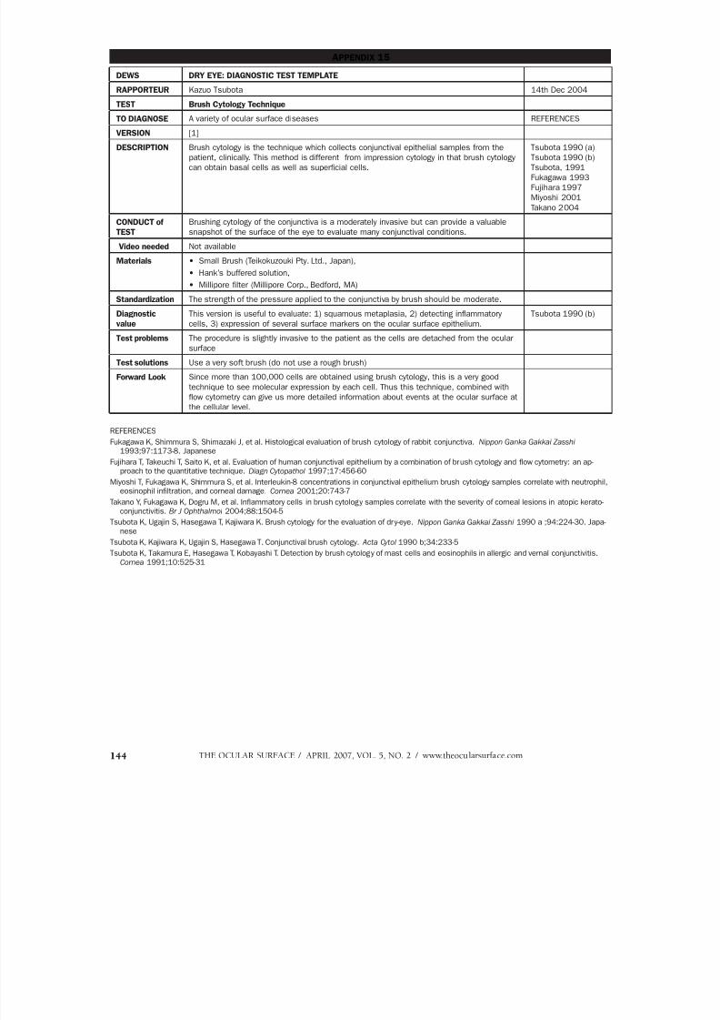
8/20/2019 dry eyes ii
http://slidepdf.com/reader/full/dry-eyes-ii 84/142
THE OCULAR SURFACE / APRIL 2007, VOL. 5, NO. 2 / www.theocularsurface.com144
APPENDIX 15
DEWS DRY EYE: DIAGNOSTIC TEST TEMPLATE
RAPPORTEUR Kazuo Tsubota 14th Dec 2004
TEST Brush Cytology Technique
TO DIAGNOSE A variety of ocular surface diseases REFERENCES
VERSION [1]
DESCRIPTION Brush cytology is the technique which collects conjunctival epithelial samples from thepatient, clinically. This method is different from impression cytology in that brush cytology
can obtain basal cells as well as superficial cells.
Tsubota 1990 (a)Tsubota 1990 (b)
Tsubota, 1991
Fukagawa 1993
Fujihara 1997
Miyoshi 2001
Takano 2004
CONDUCT of
TEST
Brushing cytology of the conjunctiva is a moderately invasive but can provide a valuable
snapshot of the surface of the eye to evaluate many conjunctival conditions.
Video needed Not available
Materials • Small Brush (Teikokuzouki Pty. Ltd., Japan),
• Hank’s buffered solution,
• Millipore filter (Millipore Corp., Bedford, MA)
Standardization The strength of the pressure applied to the conjunctiva by brush should be moderate.
Diagnostic
value
This version is useful to evaluate: 1) squamous metaplasia, 2) detecting inflammatory
cells, 3) expression of several surface markers on the ocular surface epithelium.
Tsubota 1990 (b)
Test problems The procedure is slightly invasive to the patient as the cells are detached from the ocular
surface
Test solutions Use a very soft brush (do not use a rough brush)
Forward Look Since more than 100,000 cells are obtained using brush cytology, this is a very good
technique to see molecular expression by each cell. Thus this technique, combined with
flow cytometry can give us more detailed information about events at the ocular surface at
the cellular level.
REFERENCES
Fukagawa K, Shimmura S, Shimazaki J, et al. Histological evaluation of brush cytology of rabbit conjunctiva. Nippon Ganka Gakkai Zasshi 1993;97:1173-8. Japanese
Fujihara T, Takeuchi T, Saito K, et al. Evaluation of human conjunctival epithelium by a combination of brush cytology and flow cytometry: an ap-proach to the quantitative technique. Diagn Cytopathol 1997;17:456-60
Miyoshi T, Fukagawa K, Shimmura S, et al. Interleukin-8 concentrations in conjunctival epithelium brush cytology samples correlate with neutrophil,eosinophil infiltration, and corneal damage. Cornea 2001;20:743-7
Takano Y, Fukagawa K, Dogru M, et al. Inflammatory cells in brush cytology samples correlate with the severity of corneal lesions in atopic kerato-conjunctivitis. Br J Ophthalmol 2004;88:1504-5
Tsubota K, Ugajin S, Hasegawa T, Kajiwara K. Brush cytology for the evaluation of dry-eye. Nippon Ganka Gakkai Zasshi 1990 a ;94:224-30. Japa-nese
Tsubota K, Kajiwara K, Ugajin S, Hasegawa T. Conjunctival brush cytology. Acta Cytol 1990 b;34:233-5
Tsubota K, Takamura E, Hasegawa T, Kobayashi T. Detection by brush cytology of mast cells and eosinophils in allergic and vernal conjunctivitis.Cornea 1991;10:525-31

8/20/2019 dry eyes ii
http://slidepdf.com/reader/full/dry-eyes-ii 85/142
THE OCULAR SURFACE / APRIL 2007, VOL. 5, NO. 2 / www.theocularsurface.com 145
APPENDIX 16
DEWS DRY EYE: DIAGNOSTIC TEST TEMPLATE
RAPPORTEUR Christophe Baudouin 7th Nov 2004
TEST Flow cytometry in impression cytology
TO DIAGNOSE Conjunctival inflammation / apoptosis REFERENCES
VERSION of TEST [V 1 ]
[V2] Also available: Brush cytology for cell collection before flow cytometryprocedures (Fujihara et al., 1997).
Baudouin et al 1997
Fujihara et al 1997
DESCRIPTION This technique is highly sensitive and specific for analyzing expression of any marker
by conjunctival epithelial cells, or identification of inflammatory and goblet cells.
HLA-DR normally not or weakly expressed. Strongly overexpressed in case of ocular
surface inflammation
NATURE of STUDY Technique specially relevant in dry eye, allergy or assessment of antiglaucoma
eyedrops
Brignole et al 2000,
2001
CONDUCT of TEST 1. Without or under topical anesthesia with one drop of 0.04% oxibuprocaine, one or
more filters, 13 x 6.5 mm in size, are gently applied to the conjunctival surface.
2. After removal, the membranes are dipped into tubes containing 0.05%
paraformaldehyde. The tubes have to be kept at 4°C before and after impression
collection in order to avoid sample degradation during the phase of fixation.
Under this condition the filters with the conjunctival specimens can be stored
several days and sent to the laboratory in cold-conditioned containers before
being processed for flow cytometry analyses.
3. Cell extraction is manually conducted by gentle agitation. After centrifugation in
PBS, conjunctival cells are then immunostained and analyzed by flow cytometry.
4. Indirect or direct immunofluorescence procedures may be used. Simple or
multi-color analysis can be performed commonly using 2 to 4 antibodies
conjugated with different fluorochromes. A nonimmune isotype-matched mouse
immunoglobulin has to be used as a negative isotypic control, fluorochrome-
conjugated or not, according to direct or indirect immunofluorescence procedure.
5. At the end of incubation with specific antibodies, cells are centrifuged in PBS
(1600 rpm, 5 minutes), resuspended in PBS and analysed on a flow cytometer.
Intracytoplasmic markers can also be detected by using specific permeabilization
techniques, such as 0.5% saponin, X100 triton X or ethanol.
6. Many markers available giving relevant information on ocular surface disorders;HLA DR expression by epithelial cells, gold standard for inflammatory
assessment
Brignole et al 2004
Web Video Not available
Materials 1. Polyethersulfone filters (Supor, Gelman Sciences Ann Arbor, MI, USA), 13 mm in
diameter with pores of 0.20 µm
2. Paraformaldehyde freshly prepared and preserved at 4°C, monoclonal antibodies
and material for immunostaining
3. Flow cytometer
Variations of
technique
[V2] Brush cytology for cell collection before flow cytometry procedures. Fujihara et al 1997
Diagnostic value This version : [√]
HLA DR inferior to 45% of positive cells and 18,000 MESF (molecular equivalent of
soluble fluorochrome) in normal eyes. Widely above these values in inflammatoryocular surface disorders
Please cite statistics indicating the diagnostic value of the test.
Brignole et al 2004
Repeatability Standardized technique reliable over time and from one laboratory to another
Test problems This procedure is highly technical and requires a laboratory equipped with a flow
cytometer and a staff familiar with immunostaining processing and flow cytometry
analysis on paucicellular specimens
FORWARD LOOK Many markers for a large variety of applications have yet to be tested with further
improvement of pathophysiological knowledge of ocular surface diseases
Glossary HLA-DR: Major leukocyte antigen, human histocompatibility complex, class II cell
surface receptor
continued

8/20/2019 dry eyes ii
http://slidepdf.com/reader/full/dry-eyes-ii 86/142
THE OCULAR SURFACE / APRIL 2007, VOL. 5, NO. 2 / www.theocularsurface.com146
APPENDIX 16 continued
REFERENCES
Baudouin C, Brignole F, Becquet F, et al. (1997a). Flow cytometry in impression cytology specimens. A new method for evaluation of conjunctivalinflammation. Invest Ophthalmol Vis Sci 38:1458-1464
Bourcier T, De Saint-Jean M, Brignole F, et al. (2000). Expression of CD40 and CD40 ligand in the human conjunctival epithelium. Invest
Ophthalmol Vis Sci 41:120-126
Brignole F, Becquet F, Pisella PJ, et al. (1998). Expression of Fas antigen (CD95) in the human conjunctival epithelium. Positive correlation withclass II HLA DR expression in inflammatory conditions. Exp Eye Res 67:687-697
Brignole F, Pisella PJ, Goldschild M, et al. (2000) Flow cytometric analysis of inflammatory markers in conjunctival epithelial cells of patients withdry eyes. Invest Ophthalmol Vis Sci 41:1356-1363
Brignole F, Pisella PJ, De Saint Jean M, et al. and Baudouin C. (2001) Flow cytometric analysis of inflammatory markers in KCS: 6-monthtreatment with topical cyclosporin A. Invest Ophthalmol Vis Sci 42:90-95
Brignole F, Ott AC, Warnet JM, Baudouin C. (2004) Flow cytometry in conjunctival impression cytology: a new tool for exploring ocular surfacepathologies. Exp Eye Res 78:473-481
Fujihara T, Takeuchi T, Saito K, et al. (1997) Evaluation of human conjunctival epithelium by a combination of brush cytology and flow cytometry: anapproach to the quantitative technique. Diagn Cytopathol 17:456-460
Pisella PJ, Brignole F, Debbasch C, et al. (2000) Flow cytometric analysis of conjunctival epithelium in ocular rosacea and keratoconjunctivitissicca. Ophthalmology 107:1841-1849
Pisella, PJ, Debbasch C, Hamard P, et al. (2004) Conjunctival proinflammatory and proapoptotic effects of latanoprost, preserved timolol andunpreserved timolol: an ex vivo and in vitro study. Invest Ophthalmol Vis Sci 45:1360-1368
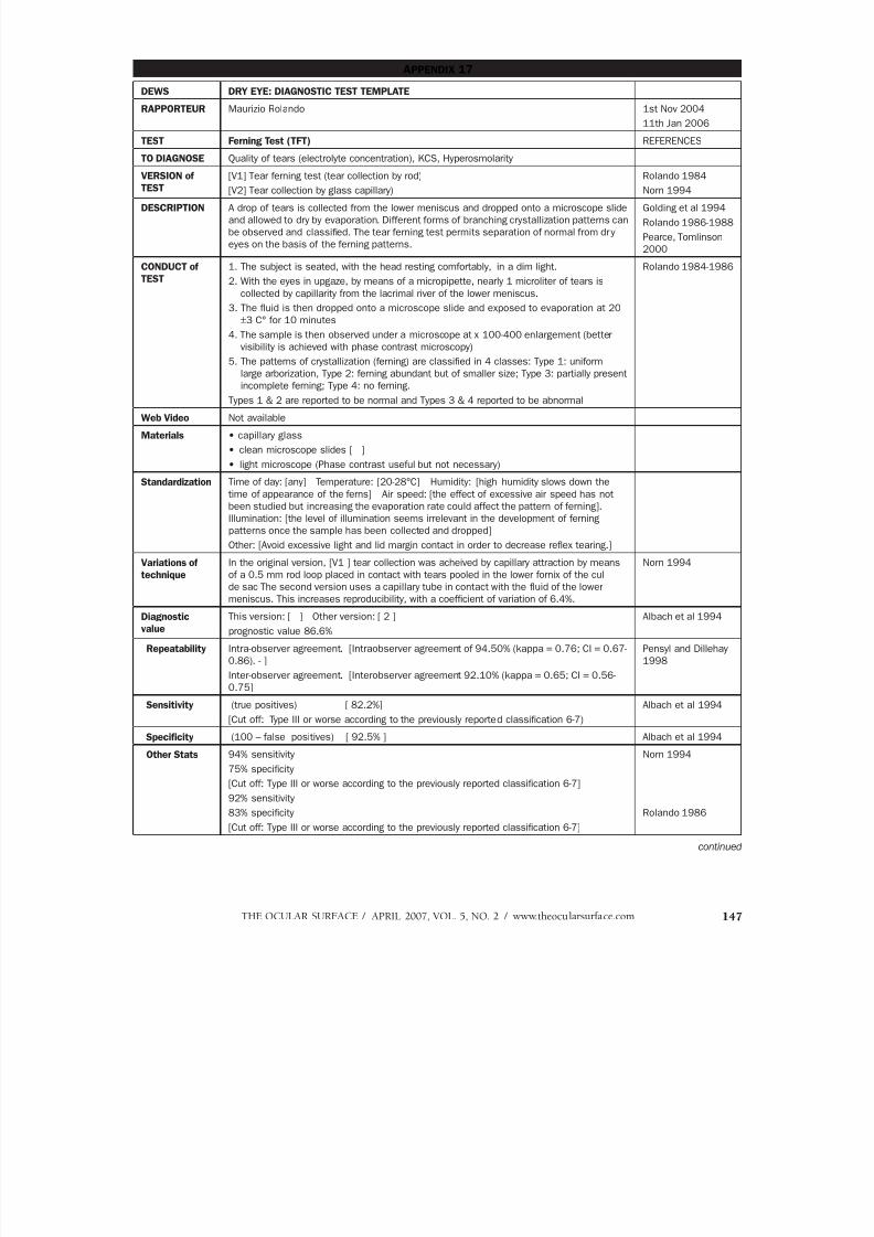
8/20/2019 dry eyes ii
http://slidepdf.com/reader/full/dry-eyes-ii 87/142
THE OCULAR SURFACE / APRIL 2007, VOL. 5, NO. 2 / www.theocularsurface.com 147
APPENDIX 17
DEWS DRY EYE: DIAGNOSTIC TEST TEMPLATE
RAPPORTEUR Maurizio Rolando 1st Nov 2004
11th Jan 2006
TEST Ferning Test (TFT) REFERENCES
TO DIAGNOSE Quality of tears (electrolyte concentration), KCS, Hyperosmolarity
VERSION ofTEST
[V1] Tear ferning test (tear collection by rod)[V2] Tear collection by glass capillary)
Rolando 1984Norn 1994
DESCRIPTION A drop of tears is collected from the lower meniscus and dropped onto a microscope slide
and allowed to dry by evaporation. Different forms of branching crystallization patterns can
be observed and classified. The tear ferning test permits separation of normal from dry
eyes on the basis of the ferning patterns.
Golding et al 1994
Rolando 1986-1988
Pearce, Tomlinson
2000
CONDUCT of
TEST
1. The subject is seated, with the head resting comfortably, in a dim light.
2. With the eyes in upgaze, by means of a micropipette, nearly 1 microliter of tears is
collected by capillarity from the lacrimal river of the lower meniscus.
3. The fluid is then dropped onto a microscope slide and exposed to evaporation at 20
±3 C° for 10 minutes
4. The sample is then observed under a microscope at x 100-400 enlargement (better
visibility is achieved with phase contrast microscopy)
5. The patterns of crystallization (ferning) are classified in 4 classes: Type 1: uniform
large arborization, Type 2: ferning abundant but of smaller size; Type 3: partially present
incomplete ferning; Type 4: no ferning.
Types 1 & 2 are reported to be normal and Types 3 & 4 reported to be abnormal
Rolando 1984-1986
Web Video Not available
Materials • capillary glass
• clean microscope slides [ ]
• light microscope (Phase contrast useful but not necessary)
Standardization Time of day: [any] Temperature: [20-28°C] Humidity: [high humidity slows down the
time of appearance of the ferns] Air speed: [the effect of excessive air speed has not
been studied but increasing the evaporation rate could affect the pattern of ferning].
Illumination: [the level of illumination seems irrelevant in the development of ferning
patterns once the sample has been collected and dropped]Other: [Avoid excessive light and lid margin contact in order to decrease reflex tearing.]
Variations of
technique
In the original version, [V1 ] tear collection was acheived by capillary attraction by means
of a 0.5 mm rod loop placed in contact with tears pooled in the lower fornix of the cul
de sac The second version uses a capillary tube in contact with the fluid of the lower
meniscus. This increases reproducibility, with a coefficient of variation of 6.4%.
Norn 1994
Diagnostic
value
This version: [ ] Other version: [ 2 ]
prognostic value 86.6%
Albach et al 1994
Repeatability Intra-observer agreement. [Intraobserver agreement of 94.50% (kappa = 0.76; CI = 0.67-
0.86). - ]
Inter-observer agreement. [Interobserver agreement 92.10% (kappa = 0.65; CI = 0.56-
0.75]
Pensyl and Dillehay
1998
Sensitivity (true positives) [ 82.2%][Cut off: Type III or worse according to the previously reported classification 6-7)
Albach et al 1994
Specificity (100 – false positives) [ 92.5% ] Albach et al 1994
Other Stats 94% sensitivity
75% specificity
[Cut off: Type III or worse according to the previously reported classification 6-7]
92% sensitivity
83% specificity
[Cut off: Type III or worse according to the previously reported classification 6-7]
Norn 1994
Rolando 1986
continued

8/20/2019 dry eyes ii
http://slidepdf.com/reader/full/dry-eyes-ii 88/142
THE OCULAR SURFACE / APRIL 2007, VOL. 5, NO. 2 / www.theocularsurface.com148
Test problems Care should be taken not to elicit reflex tearing during collections
Light microscopy is often unavailable in the office.
In spite of a good clinical ability of separating normal from dry eyes, the real meaning of
the results is not known
[Test affected by extreme conditions of temperature and humidity]
Forward Look It would be interesting to explore the correlation between the patterns of crystallization
(test types I to IV) and the level of tear film osmolarity Glossary TFT: Tear ferning test
REFERENCES
Albach KA, Lauer M, Stolze HH. Diagnosis of keratoconjunctivitis sicca in rheumatoid arthritis. The value of various tests Ophthalmologe 1994Apr;91(2):229-34
Golding TR, Baker AT, Rechberger J, Brennan NA. X-ray and scanning electron microscopic analysis of the structural composition of tear ferns.Cornea 1994 Jan;13(1):58-66
Norn M. Quantitative tear ferning. Clinical investigations. Acta Ophthalmol (Copenh) 1994 Jun;72(3):369-72
Pearce EI, Tomlinson A. Spatial location studies on the chemical composition of human tear ferns. Ophthalmic Physiol Opt 2000 Jul;20(4):306-13
Pensyl CD, Dillehay SM. The repeatability of tear mucus ferning grading. Optom Vis Sci 1998 Aug;75(8):600-4
Rolando M. Tear mucus ferning test in normal and keratoconjunctivitis sicca eyes. Chibret Int J Ophthalmol 1984;2(4):32-41
Rolando M, Baldi F, Calabria G. Tear mucus ferning test in keratoconjunctivitis sicca. In: Holly FJ, Lamberts DW, MacKeen DL (eds.): The preoculartear film in health, disease, and contact lens wear. 1st Intern Tear Film Symposium. Lubbok (Texas, USA), Dry Eye Institute, 1986, 203-210
Rolando M, Baldi F, Zingirian M. The effect of hyperosmolarity on tear mucus ferning. Fortschr Ophthalmol 1986;83:644-646Rolando M, Baldi F, Calabria G. Tear mucus crystallization in children with cystic fibrosis. Ophthalmologica 1988;197(4):202-6
APPENDIX 17 continued
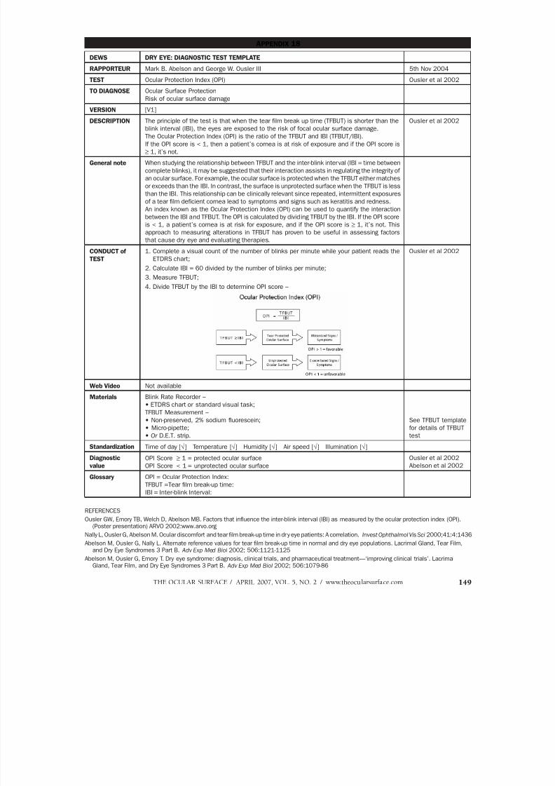
8/20/2019 dry eyes ii
http://slidepdf.com/reader/full/dry-eyes-ii 89/142
THE OCULAR SURFACE / APRIL 2007, VOL. 5, NO. 2 / www.theocularsurface.com 149
APPENDIX 18
DEWS DRY EYE: DIAGNOSTIC TEST TEMPLATE
RAPPORTEUR Mark B. Abelson and George W. Ousler III 5th Nov 2004
TEST Ocular Protection Index (OPI) Ousler et al 2002
TO DIAGNOSE Ocular Surface Protection
Risk of ocular surface damage
VERSION [V1]DESCRIPTION The principle of the test is that when the tear film break up time (TFBUT) is shorter than the
blink interval (IBI), the eyes are exposed to the risk of focal ocular surface damage.
The Ocular Protection Index (OPI) is the ratio of the TFBUT and IBI (TFBUT/IBI).
If the OPI score is < 1, then a patient’s cornea is at risk of exposure and if the OPI score is
≥ 1, it’s not.
Ousler et al 2002
General note When studying the relationship between TFBUT and the inter-blink interval (IBI = time between
complete blinks), it may be suggested that their interaction assists in regulating the integrity of
an ocular surface. For example, the ocular surface is protected when the TFBUT either matches
or exceeds than the IBI. In contrast, the surface is unprotected surface when the TFBUT is less
than the IBI. This relationship can be clinically relevant since repeated, intermittent exposures
of a tear film deficient cornea lead to symptoms and signs such as keratitis and redness.
An index known as the Ocular Protection Index (OPI) can be used to quantify the interaction
between the IBI and TFBUT. The OPI is calculated by dividing TFBUT by the IBI. If the OPI score
is < 1, a patient’s cornea is at risk for exposure, and if the OPI score is ≥ 1, it’s not. Thisapproach to measuring alterations in TFBUT has proven to be useful in assessing factors
that cause dry eye and evaluating therapies.
CONDUCT of
TEST
1. Complete a visual count of the number of blinks per minute while your patient reads the
ETDRS chart;
2. Calculate IBI = 60 divided by the number of blinks per minute;
3. Measure TFBUT;
4. Divide TFBUT by the IBI to determine OPI score –
Ousler et al 2002
Web Video Not available
Materials Blink Rate Recorder –
• ETDRS chart or standard visual task;
TFBUT Measurement –
• Non-preserved, 2% sodium fluorescein;
• Micro-pipette;
• Or D.E.T. strip.
See TFBUT template
for details of TFBUT
test
Standardization Time of day [√] Temperature [√] Humidity [√] Air speed [√] Illumination [√]
Diagnostic
value
OPI Score ≥ 1 = protected ocular surface
OPI Score < 1 = unprotected ocular surface
Ousler et al 2002
Abelson et al 2002
Glossary OPI = Ocular Protection Index:
TFBUT =Tear film break-up time:
IBI = Inter-blink Interval:
REFERENCES
Ousler GW, Emory TB, Welch D, Abelson MB. Factors that influence the inter-blink interval (IBI) as measured by the ocular protection index (OPI).(Poster presentation) ARVO 2002:www.arvo.org
Nally L, Ousler G, Abelson M. Ocular discomfort and tear film break-up time in dry eye patients: A correlation. Invest Ophthalmol Vis Sci 2000;41:4:1436
Abelson M, Ousler G, Nally L. Alternate reference values for tear film break-up time in normal and dry eye populations. Lacrimal Gland, Tear Film,and Dry Eye Syndromes 3 Part B. Adv Exp Med Biol 2002; 506:1121-1125
Abelson M, Ousler G, Emory T. Dry eye syndrome: diagnosis, clinical trials, and pharmaceutical treatment—‘improving clinical trials’. Lacrimal
Gland, Tear Film, and Dry Eye Syndromes 3 Part B. Adv Exp Med Biol 2002; 506:1079-86

8/20/2019 dry eyes ii
http://slidepdf.com/reader/full/dry-eyes-ii 90/142
THE OCULAR SURFACE / APRIL 2007, VOL. 5, NO. 2 / www.theocularsurface.com150
APPENDIX 19
DEW DRY EYE DIAGNOSTIC TEST TEMPLATE
RAPPORTEUR Alan Tomlinson 10th Jan 2006
TEST Fluorophotometry (Fluorimetry) – Tear Flow
DIAGNOSES Changes in tear flow in aqueous tear deficiency (ATD). REFERENCES
VERSION of TEST [Version 1] Scanning automated fluorophotometry (Fluorotron Master, Coherent Inc, Palo, Alto, CA)
DESCRIPTION To calculate tear flow from measurements of tear volume and turnover.
CONDUCT of
TEST
Tear Turnover Rate
1) Subject is seated at the chin rest of the Fluorotron (with the anterior segment adapter fitted).
Horizontal and vertical adjustments are made to align the subject’s eye in the instrument’s
optic beam.
2) Three scans are conducted to establish the intrinsic corneal autofluorescence.
3) A 1 µl drop of 2% sodium fluorescein is instilled into the lower fornix with a pipette.
4) Initial scans are taken 1 minute post instillation, then at 2 minute intervals for a further 20 minutes.
5) The intrinsic corneal autofluorescence value is substracted from all values obtained from tear
film fluorescence, prior to data analysis.
6) Fluoroscein concentration at each time point is calculated from the Fluorotron scans obtained at
all time points beyond 4 minute post instillation, to avoid initial reflex tearing caused by instillation.
7) The decay in fluorescence is calculated from the log of the curve obtained from the formula:
T0(t0) = 100 [Ct(t0) – Ct(t0+1] ( %/min)Ct(t0)
Where Ct(t) = fluorescein concentration in tear film at time t(min).
Assuming a monophasic decay of fluorescence from 5 mins post instillation with a decay time
constant β (min–1):
Ct(t) = Ct (0).eβt (ng/ml)
the following is obtained:
Tt(t0) = 100 (1 – eβt) ( %/min)
This calculation can be carried out using the software package ANT_SEGMENT tear.
Tear Volume
1) Subject is seated at the chin rest of the Fluorotron (with the anterior segment adapter fitted).Horizontal and vertical adjustments are made to align the subject’s eye in the instrument’soptic beam.
2) Three scans are conducted to establish the intrinsic corneal autofluorescence.
3) One µl of 2% sodium fluorescein is instilled into the lower fornix with a pipette.
4) Initial scans are taken 1 minute post instillation, then at 1 minute intervals for a further 4 minutes.
5) The intrinsic corneal autofluorescence value is substracted from all values obtained from tearfilm fluorescence, prior to data analysis.
6) Fluorescein concentration at each time point is calculated from all the Fluorotron scans obtained.
7) The decay in fluorescence is calculated from the log of the curve obtained from the formula:
T0(t0) = 100 [Ct(t0) – Ct(t0+1] ( %/min)
Ct(t0)
Where Ct(t) = fluorescein concentration in tear film at time t(min).
Assuming a monophasic decay of fluorescence from 5 mins post instillation with a decay time
constant β (min–1):
Ct(t) = Ct (0).eβt (ng/ml)
the following is obtained:
Tt(t0) = 100 (1 – eβt) ( %/min)
This calculation can be carried out using the software package ANT_SEGMENT tear.
Tear volume is then calculated from:
Vt = (Cd.Cm–1.k–1–1) Vd
Where
Cd = fluorescein concentration in the drop
Cm = initial fluoroscein concentration calculated by back extrapolation with the Fluorotron in ng/ml
k = correction factor (k = 250) for the limited spatial resolution of the Fluorotron and
Vd = drop volume in ml
Calculation of tear flow:
Tear flow = Vt ( µl/min)
T0(t0)
Kuppens 1992
Van Best 1995
Van Best 1995
Kuppens 1992
Van Best 1995
Kuppens 1992
Mishima 1965
continued

8/20/2019 dry eyes ii
http://slidepdf.com/reader/full/dry-eyes-ii 91/142
THE OCULAR SURFACE / APRIL 2007, VOL. 5, NO. 2 / www.theocularsurface.com 151
APPENDIX 19 continued
Web Video Not available
Materials Fluorotron Master
2% sodium fluorescein Mimims (Chauvin, UK)
Air displacement pipette P2 Pipetman (Gilson, Villiers-le-Bel, France)
Disposable sterile tips (Gilson, Villiers-le-Bel, France)
Variations of
technique
Varying concentrations and instillation volumes of fluorescein can be used, eg, 1% and 0.5
-2 µl.
Standardization Time of day [X] Temperature [ ] Humidity [ ] Air speed [still] Illumination [low ambient]
Other: [Blink is initiated immediately prior to scan to ensure uniform tear thickness]
Pearce et al 2000
Diagnostic
value
This version: [ ] Determination of tear flow an indication of aqueous tear deficiency. To
obtain estimate of tear drainage from eye.
Other version: [ ]
Mathers, Daley
1996
Mathers et al 1996
Gobbels et al 1992
Repeatability Intra-observer variation. [Not significant]
Inter-observer variation. [Not significant]
Mishima et al 1966
Van Best 1995
Test problems High cost of basic equipment.Time required for measurement.
Indirect (surrogate) measures of tear outflow and volume as it is assumed that fluorescein
and aqueous tear are eliminated at the same rate from the eye.
Absorption of fluorescein into the ocular tissue may be a factor in dry eye patients and
may decrease apparent rate of decay.
Test solutions Use of high molecular weight conjugates. McNamara et al 1998
Forward Look Production of a cheaper automated scanning fluorophotometer.
Development of reduced test incorporating 6 measurements for total of 10 minutes (tear
turnover).
Combination of tear flow (µl/min) with evaporation rate (µl/min) gives a value of “total
tear flow” in the eye and an estimate of total tear production. This allows analysis of the
proportion of tears eliminated by evaporation and/or drainage in various forms of dry eye.
Pearce et al 2000
Mathers, Daley 1996
Mathers 2004
REFERENCES
Gobbels M, Goebels G, et al. Tear secretion in dry eyes as assessed by objective fluorophotometry. Ger J Ophthalmol 1992; 1:350-353
Kuppens EV, Stolwijk TR, et al. Basal tear turnover and topical timolol in glaucoma patients and healthy controls by Fluorophotometry. Invest Oph-
thalmol Vis Sci 1992; 33:3442-3448
Mishima S. Some physiological aspects of the precorneal tear film. Arch Ophthalmol 1965;73:233-241
Mishima S, et al. Determination of tear volume and tear flow. Invest Ophthalmol 1966; 5:264-275
Mathers WD, Daley TE. Tear film and evaporation in patients with and without dry eye. Ophthalmology 1996; 103:664-669
Mathers WD, Lane J, Zimmerman M. Tear film changes associated with normal aging. Cornea 1996; 15:229-334
Mathers WD. Evaporation from the ocular surface. Exp Eye Res 2004; 78:389-394
Van Best JA, et al. Measurement of basal tear turnover using a standardised protocol. Graefe’s Arch Clin Exp Ophthalmol 1995; 233:1-7
McNamara NA, et al. Fluorometry in contact lens research: The next step. Optom Vis Sci 1998; 75:316-322
Pearce EI, Keenan BP, McRory C. An improved fluorophotometric method for tear turnover assessment. Optom Vis Sci 2001; 78:30-36

8/20/2019 dry eyes ii
http://slidepdf.com/reader/full/dry-eyes-ii 92/142
THE OCULAR SURFACE / APRIL 2007, VOL. 5, NO. 2 / www.theocularsurface.com152
APPENDIX 20
DEW DRY EYE: DIAGNOSTIC TEST TEMPLATE
RAPPORTEUR Stephen Kaye 18th April 2006
TEST Tear Function Index (Liverpool modification)
Email: [email protected]
TO DIAGNOSE To evaluate the tear dynamics of production and drainage and detect subjects suffering
from dry eye
Ono et al 1991
Xu et al 1995(a)
Xu et al 1995(b)Kaye et al 2001
VERSION of TEST The test is a modification of that described by Xu et al. (1995) and depends on using
prepared filter paper strips containing fluorescein. The test has been designed to allow
direct measurement of the TFI using prepared tear strips.
Kaye et al 2001
DESCRIPTION TFI is the quotient of the Schirmer test value and the Tear clearance rate (TCR).
CONDUCT of
TEST
A fluorescein-coated tear strip is placed over the lower lid margin at the junction of the
middle and lateral third of the lid.
1. The eye is closed and the strip is left in place for 3 minutes
2. On removal, the distance from the strip notch to the wetted dye front is recorded, using
the scale provided.
3. The strip is air dried and
4. The intensity of staining is compared with that of the calibrated panel of dilutions,
(ranging from 1:1 to 1:128), to determine the TCR.
5. The TFI is defined as the quotient of the Schirmer test and the TCR.
Web Video Not available
Materials • The standard kit provides a cardboard envelope, containing a docket with 4 see-through pouches.
• Each pouch contains 4 sterile, single-use, fluorescein-coated tear-strips together with a
calibrated colour scale for reference.
• A ruled measurement scale is printed on the envelope, together with
• a nomogram and
• a set of instructions
The kit, containing the prepared strips, together with instructions and calibrated measuring
scale and colour scale are provided by the Dept. Clinical Engineering of the Royal Liverpool
University Hospital, Prescot Street Liverpool L7 8XP. For further information:
Email: [email protected]
Variations of
technique
TFI as described by Xu et al (1995)
Standardization The procedure is standardised. Strips are calibrated for use in each pack.
Diagnostic value Identification of subjects suffering from aqueous tear deficiency, for instance in Sjögrens
syndrome.
Sensitivity A TFI of less than 40 is 100% sensitive for patients with SS dry eye Kaye et al 2001
Specificity Patients with Sjögren’s syndrome have a TFI upper 95% confidence interval of 15 (12 if
anaesthetic has been used)
Kaye et al 2001
Other Stats Less inter-ocular difference and less variability than the original method Kaye et al 2001
Test problems As with the Schirmer’s test, it is uncomfortable. Also, staining of the ocular surface at the
sites of strip contact with the conjunctiva occur after using fluorescein or Rose Bengal.
FORWARD
LOOK
Performing the TFI using prepared filter paper strips with the matched colour dilution
is very sensitive for detecting patients with SS dry eye. The test can be used by non-
ophthalmically trained personel. Subjects with a TFI of less than 40 can then be referred
for an ophthalmic assessment.
Glossary TFI: Tear function index
REFERENCES
Kaye SB, Sims G, Willoughby C, et al. Modification of the tear function index and its use in the diagnosis of Sjögren’s syndrome. Br J Ophthalmol 2001;85;193-199
Ono M, Yoshino K, Ogawa Y, et al. Tear clearance rate in normal and dry eye patients. Invest Opthalmol Vis Sci (Suppl) 1991;32:1113
Xu KP, Yagi Y, Toda I, Tsubota K. Tear Function Index. A new measure of dry eye. Arch Ophthalmol 1995a;113:84-88
Xu KP, Tsubota K. Correlation of tear clearance rate and fluorophotometric assessment of tear turnover. Br J Ophthalmol 1995b;79:1042-1045

8/20/2019 dry eyes ii
http://slidepdf.com/reader/full/dry-eyes-ii 93/142
THE OCULAR SURFACE / APRIL 2007, VOL. 5, NO. 2 / www.theocularsurface.com 153
ABSTRACT This report summarizes some universal concepts
with regard to clinical trials in general and other issues per-
taining to clinical trials specifically tailored to the study of
therapeutic intervention in dry eye disease. The report also
makes recommendations for logistical design and imple-
mentation of such trials. It identifies peculiarities of dry eye
disease that complicate clinical trial design, such as the lack
of correlation of signs and symptoms, as well as the likelihood
of control interventions having a lubricant (placebo) effect.
Strategies for environmental trials and controlled adverse
environment trials are reviewed.
KEY WORDS clinical trials, DEWS, dry eye, Dry Eye WorkShop
I. INTRODUCTION
linical trials in dry eye disease represent a chal-lenge to clinicians, epidemiologists, and biostat-isticians, as well as to those seeking regulatory
approval for medications or other therapies.1 This reportsummarizes some universal concepts with regard to clinicaltrials in general and addresses other issues pertaining toclinical trials specifically tailored to the study of therapeuticintervention in dry eye disease. The level of evidence for
supporting data from clinical trials is identified in the bib-liography, according to the modified American Academy ofOphthalmology Preferred Practices guidelines. The reportalso makes recommendations for logistical design andimplementation of such trials.
II. GOALS OF THE CLINICAL TRIALS SUBCOMMITTEE
The goals of the Clinical Trials Subcommittee were tosystematically review literature, procedures, and conceptsrelated to clinical trials in general, to consider special issuesrelated to clinical trials involving therapeutic interventionsin dry eye disease, and to present guidelines for successfulconduct of clinical trials.
III. GUIDELINES FOR CLINICAL TRIALS IN GENERAL
Before a clinical trial is initiated, a state of equipoisemust exist. In other words, there must be sufficient doubtabout the effectiveness of the particular intervention under
consideration to justify withholding it from a portion of thestudy subjects, and, at the same time, there must be suf-ficient belief in the therapeutic potential of the interventionto justify its exposure to the remaining portion of willingand eligible study participants. If these conditions are met,then a number of additional issues need to be consideredin the design and conduct of the clinical trial so that validresults can be obtained (Table 1). Important processes in-clude formulation of a concise and specific study question,specification of the primary outcome measure, statisticalestimation of the necessary sample-size, specification of thelength of follow-up and specific schedule for baseline and
follow-up evaluations, selection of the study population,definition of the primary outcome measure, random allo-cation of the intervention(s)/treatment(s), establishment ofstrategies for maintenance of compliance with the allocatedintervention(s)/treatment(s) and for achievement of highand balanced rates of follow-up. In addition, it is impor-tant to establish an organizational and decision-makingstructure and specific procedures for intake of data, andfor patient safety monitoring.
A. Design
The most desirable design of a clinical trial is a prospec-
tive, randomized, double-masked, placebo- or vehicle- con-
DEWS Clinical Trials
©2007 Ethis Communications, Inc. The Ocular Surface ISSN: 1542-
0124. (No authors listed). Design and conduct of clinical trials:
report of the Clinical Trials Subcommittee of the International Dry
Eye WorkShop (2007). 2007;5(2):153-162.
Design and Conduct of Clinical Trials:
Report of the Clinical Trials Subcommittee of the International Dry Eye WorkShop (2007)
Accepted for publication January 2007.
Clinical Trials Subcommittee members: Kazuo Tsubota, MD (Chair);Penny Asbell, MD; Murat Dogru, MD; Desmond Fonn, OD; Gary Foulks,MD (captain); Debra Schaumberg, ScD, OD, MPH; Oliver Schein, MD, MPH;Hitoshi Watanabe, MD.
Proprietary interests of Subcommittee members are disclosed on pages 202and 204.
Reprints are not available. Articles can be accessed at: www.tearfilm.org
Correspondence in regard to the DEWS Report this chapter should be ad-dressed to Kazuo Tsubota MD, Ophthalmology, Keio University School ofMedicine, 35 Shinanomachi, Shinjuku-ku, Tokyo 160-8582, Japan. Tel: 81-3-3353-1211. Fax: 81-3-3352-8703. Email: [email protected]
C
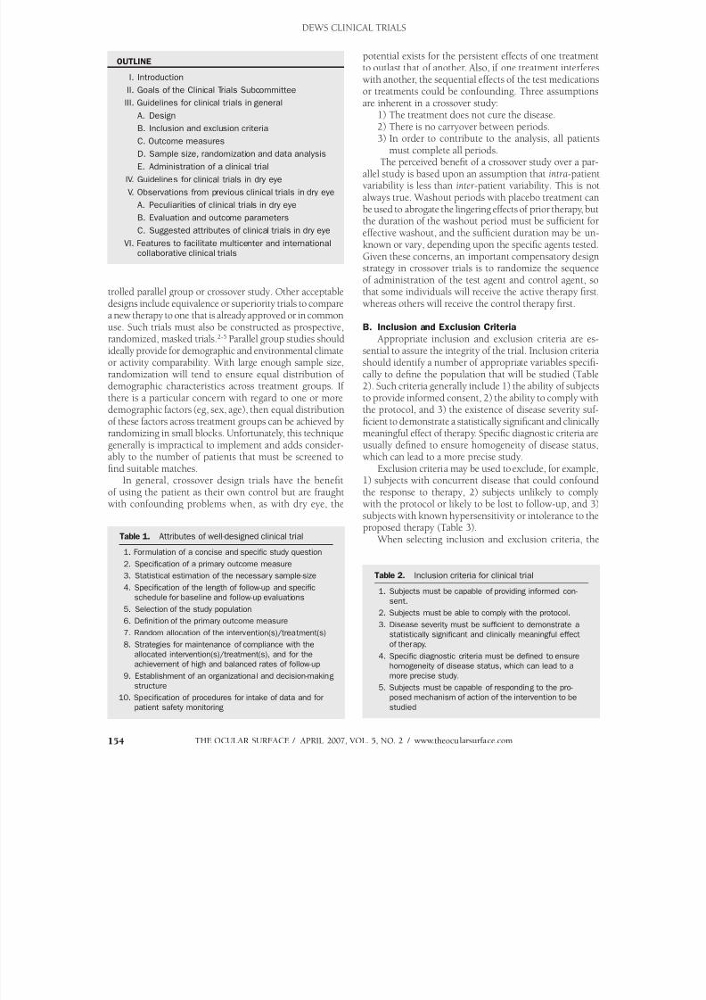
8/20/2019 dry eyes ii
http://slidepdf.com/reader/full/dry-eyes-ii 94/142
THE OCULAR SURFACE / APRIL 2007, VOL. 5, NO. 2 / www.theocularsurface.com154
trolled parallel group or crossover study. Other acceptabledesigns include equivalence or superiority trials to comparea new therapy to one that is already approved or in commonuse. Such trials must also be constructed as prospective,randomized, masked trials.2-5 Parallel group studies shouldideally provide for demographic and environmental climateor activity comparability. With large enough sample size,randomization will tend to ensure equal distribution ofdemographic characteristics across treatment groups. Ifthere is a particular concern with regard to one or moredemographic factors (eg, sex, age), then equal distributionof these factors across treatment groups can be achieved by
randomizing in small blocks. Unfortunately, this techniquegenerally is impractical to implement and adds consider-ably to the number of patients that must be screened tofind suitable matches.
In general, crossover design trials have the benefitof using the patient as their own control but are fraughtwith confounding problems when, as with dry eye, the
potential exists for the persistent effects of one treatmentto outlast that of another. Also, if one treatment interfereswith another, the sequential effects of the test medicationsor treatments could be confounding. Three assumptionsare inherent in a crossover study: 1) The treatment does not cure the disease. 2) There is no carryover between periods.
3) In order to contribute to the analysis, all patientsmust complete all periods. The perceived benefit of a crossover study over a par-
allel study is based upon an assumption that intra-patientvariability is less than inter -patient variability. This is notalways true. Washout periods with placebo treatment canbe used to abrogate the lingering effects of prior therapy, butthe duration of the washout period must be sufficient foreffective washout, and the sufficient duration may be un-known or vary, depending upon the specific agents tested.Given these concerns, an important compensatory designstrategy in crossover trials is to randomize the sequenceof administration of the test agent and control agent, sothat some individuals will receive the active therapy first,whereas others will receive the control therapy first.
B. Inclusion and Exclusion Criteria
Appropriate inclusion and exclusion criteria are es-sential to assure the integrity of the trial. Inclusion criteriashould identify a number of appropriate variables specifi-cally to define the population that will be studied (Table2). Such criteria generally include 1) the ability of subjectsto provide informed consent, 2) the ability to comply withthe protocol, and 3) the existence of disease severity suf-ficient to demonstrate a statistically significant and clinically
meaningful effect of therapy. Specific diagnostic criteria areusually defined to ensure homogeneity of disease status,which can lead to a more precise study.
Exclusion criteria may be used to exclude, for example,1) subjects with concurrent disease that could confoundthe response to therapy, 2) subjects unlikely to complywith the protocol or likely to be lost to follow-up, and 3)subjects with known hypersensitivity or intolerance to theproposed therapy (Table 3).
When selecting inclusion and exclusion criteria, the
OUTLINE
I. Introduction
II. Goals of the Clinical Trials Subcommittee
III. Guidelines for clinical trials in general
A. Design
B. Inclusion and exclusion criteria
C. Outcome measuresD. Sample size, randomization and data analysis
E. Administration of a clinical trial
IV. Guidelines for clinical trials in dry eye
V. Observations from previous clinical trials in dry eye
A. Peculiarities of clinical trials in dry eye
B. Evaluation and outcome parameters
C. Suggested attributes of clinical trials in dry eye
VI. Features to facilitate multicenter and internationalcollaborative clinical trials
Table 1. Attributes of well-designed clinical trial
1. Formulation of a concise and specific study question
2. Specification of a primary outcome measure
3. Statistical estimation of the necessary sample-size
4. Specification of the length of follow-up and specific
schedule for baseline and follow-up evaluations
5. Selection of the study population
6. Definition of the primary outcome measure
7. Random allocation of the intervention(s)/treatment(s)
8. Strategies for maintenance of compliance with the
allocated intervention(s)/treatment(s), and for the
achievement of high and balanced rates of follow-up
9. Establishment of an organizational and decision-making
structure
10. Specification of procedures for intake of data and for
patient safety monitoring
Table 2. Inclusion criteria for clinical trial
1. Subjects must be capable of providing informed con-
sent.
2. Subjects must be able to comply with the protocol.
3. Disease severity must be sufficient to demonstrate a
statistically significant and clinically meaningful effect
of therapy.
4. Specific diagnostic criteria must be defined to ensure
homogeneity of disease status, which can lead to a
more precise study.
5. Subjects must be capable of responding to the pro-
posed mechanism of action of the intervention to be
studied
DEWS CLINICAL TRIALS

8/20/2019 dry eyes ii
http://slidepdf.com/reader/full/dry-eyes-ii 95/142
THE OCULAR SURFACE / APRIL 2007, VOL. 5, NO. 2 / www.theocularsurface.com 155
investigator should be aware of the inherent trade-offsbetween the internal validity of the trial and its generaliz-ability to the larger population of people with the diseaseof interest. Minimally restrictive inclusion and exclusioncriteria make recruitment easier and provide a wider basisfor generalization of the study findings, but treatment effectsmay be obscured by heterogeneity of disease status.
C. Outcome Measures
The outcome measure used to compare treatments maybe either a clinical event or a surrogate outcome measure.The primary outcome measure should be selected priorto the start of data collection, as its rate of occurrence willaffect various aspects of the study design, including thelength of the study and the sample size. Although someclinical trials have employed post-hoc analysis of outcomevariables, regulatory agencies are often reluctant to acceptsuch analyses in pivotal trials. However, it is appropriate
for most trials additionally to collect and analyze informa-tion on a number of secondary outcome measures. Thesecan provide further information that may contribute to theoverall evaluation of the study treatments.
Surrogate outcome measures are measurable features ofthe disease that reliably reflect an outcome parameter thatis clinically relevant but difficult to precisely determine. Forexample, measurement of frequency of required instillationof comfort drops can be a quantifiable surrogate subjectivemeasure of frequency/duration of discomfort occurringduring the day. Similarly, an objective surrogate measureof tear film osmolarity could be the electrical conductivity
of a tear sample. The surrogate outcome measure must bevalidated as a reliable and relevant monitor of outcome, butit may be of special value in a condition such as dry eye,where the correlation of signs and symptoms is weak, andobjective evidence of change in disease is needed.
D. Sample Size, Randomization and Data Analysis
The sample size of a clinical trial should be sufficientto allow for a statistically powerful analysis of the primarystudy hypothesis. It may also provide for statistical com-parisons within subgroups, if this is considered desirableor necessary to clarify the therapeutic response. It is es-
sential that the trial be of sufficient size to provide power
to detect a clinically meaningful treatment effect, as wellas a statistically significant effect. Statistical analysis mustbe appropriate for the size, design, outcome measure(s),and duration of the study. The power to detect a givendifference between treatments is directly proportional tothe sample size and treatment difference, and indirectlyproportional to the alpha level and variability. A key factor
is the study planners’ selection of a clinically significantdifference. Then, they can determine the required numberof patients to detect a difference that is at least that large,given that it exists.
Randomization to test or control treatment is generallythe best strategy available in clinical trials to guard againsttreatment selection bias. There are numerous methods forestablishing randomization. Today, most researchers usecomputer-generated randomization lists, which may befurther stratified by study site and a pre-study characteristic(eg, disease severity). A written description of the ran-domization scheme used to generate treatment allocationsshould be recorded. This description should include suf-ficient detail to allow a person to reproduce the allocationschedule, and the assignment process should establish aclear audit trail.
Treatment assignments should be masked to the patient,physician, and the person issuing the assignment, until thepatient has been officially enrolled and randomized into thestudy. Preferably, the study should be masked for patientsand physicians until it is completed. This may be easiest toimplement if assignments are issued by a person or grouplocated outside of the clinic. Investigators should also beaware, particularly in small studies, that a randomizationbias could occur that must be controlled or evaluated.
The baseline characteristics of the study groups may alsovary by chance, and if large enough, such differences canimpact treatment comparisons. The strategy for the analysisof clinical trial data must be outlined in advance and mustaccommodate the form of the specified outcome variable(s)with appropriate methods of analysis.
The key feature in the analysis of clinical trials is adher-ence to the principle of “intention-to-treat.” That is, theprimary analysis of data in a trial must be conducted byclassifying study subjects based on the original treatment towhich they were assigned, regardless of the treatment theyactually received or their adherence to the study protocol
(Table 4). Good clinical practice dictates that assessmentof qualifying patients and visits be made by the clinicalmanagement (ie, organization team) prior to unmasking ofthe treatment assignment. Furthermore, it should be stateda priori in the protocol and statistical analysis plan which
Table 3. Exclusion criteria for clinical trial
1. Subjects have concurrent disease that could confound
the response to therapy.
2. Subjects are unlikely to comply with the protocol or
likely to be lost to follow-up.
3. Subjects have known hypersensitivity or intolerance to
the proposed therapy.
4. Subjects use concomitant therapy that affects eithertear function or ocular surface integrity.
5. Subjects have had surgical or other manipulation of the
eye that could confound the outcome parameters or
interfere with the mechanism of action of the proposed
intervention to be studied.
Table 4. Data analysis: populations to analyze
1. Intent to Treat (ITT): All subjects randomized.
2. Modified Intent to Treat (Mod ITT): All subjects random-
ized who received at least one dose of medication
3. Per Protocol (PP): All subjects randomized who com-
pleted the treatment according to protocol
DEWS CLINICAL TRIALS
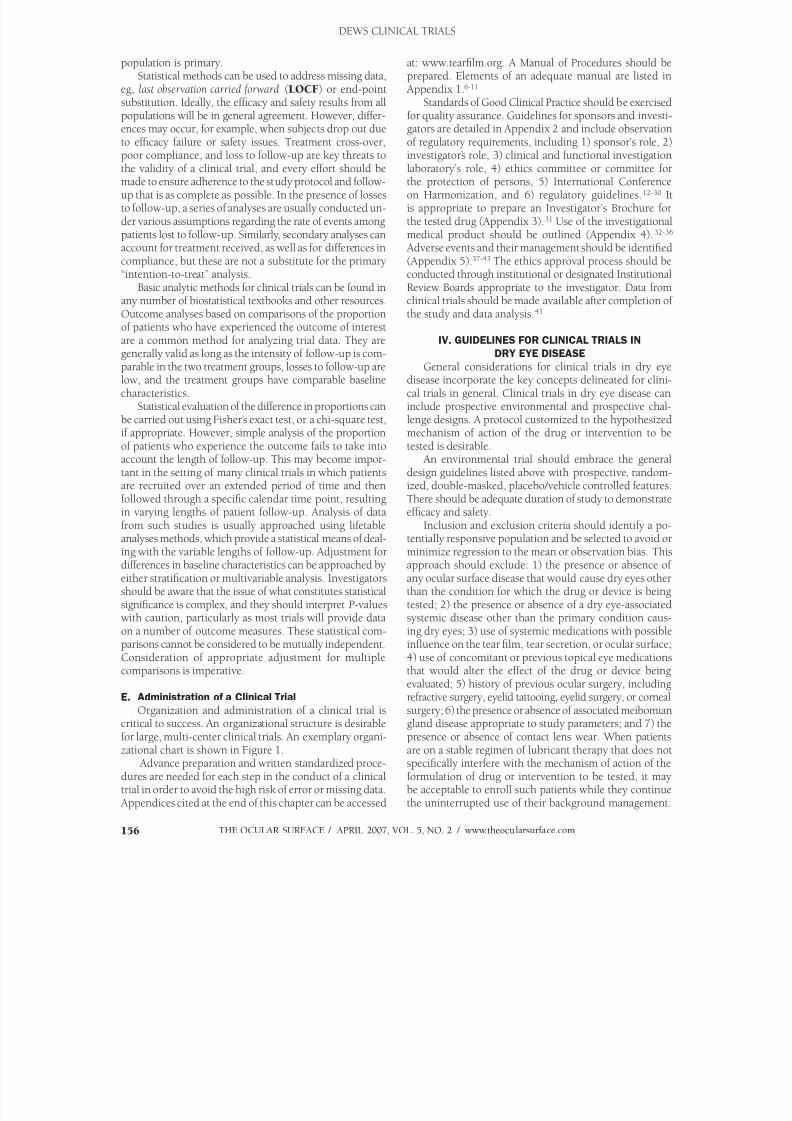
8/20/2019 dry eyes ii
http://slidepdf.com/reader/full/dry-eyes-ii 96/142
THE OCULAR SURFACE / APRIL 2007, VOL. 5, NO. 2 / www.theocularsurface.com156
population is primary.Statistical methods can be used to address missing data,
eg, last observation carried forward (LOCF) or end-pointsubstitution. Ideally, the efficacy and safety results from allpopulations will be in general agreement. However, differ-ences may occur, for example, when subjects drop out dueto efficacy failure or safety issues. Treatment cross-over,
poor compliance, and loss to follow-up are key threats tothe validity of a clinical trial, and every effort should bemade to ensure adherence to the study protocol and follow-up that is as complete as possible. In the presence of lossesto follow-up, a series of analyses are usually conducted un-der various assumptions regarding the rate of events amongpatients lost to follow-up. Similarly, secondary analyses canaccount for treatment received, as well as for differences incompliance, but these are not a substitute for the primary“intention-to-treat” analysis.
Basic analytic methods for clinical trials can be found inany number of biostatistical textbooks and other resources.Outcome analyses based on comparisons of the proportionof patients who have experienced the outcome of interestare a common method for analyzing trial data. They aregenerally valid as long as the intensity of follow-up is com-parable in the two treatment groups, losses to follow-up arelow, and the treatment groups have comparable baselinecharacteristics.
Statistical evaluation of the difference in proportions canbe carried out using Fisher’s exact test, or a chi-square test,if appropriate. However, simple analysis of the proportionof patients who experience the outcome fails to take intoaccount the length of follow-up. This may become impor-tant in the setting of many clinical trials in which patients
are recruited over an extended period of time and thenfollowed through a specific calendar time point, resultingin varying lengths of patient follow-up. Analysis of datafrom such studies is usually approached using lifetableanalyses methods, which provide a statistical means of deal-ing with the variable lengths of follow-up. Adjustment fordifferences in baseline characteristics can be approached byeither stratification or multivariable analysis. Investigatorsshould be aware that the issue of what constitutes statisticalsignificance is complex, and they should interpret P-valueswith caution, particularly as most trials will provide dataon a number of outcome measures. These statistical com-
parisons cannot be considered to be mutually independent.Consideration of appropriate adjustment for multiplecomparisons is imperative.
E. Administration of a Clinical Trial
Organization and administration of a clinical trial iscritical to success. An organizational structure is desirablefor large, multi-center clinical trials. An exemplary organi-zational chart is shown in Figure 1.
Advance preparation and written standardized proce-dures are needed for each step in the conduct of a clinicaltrial in order to avoid the high risk of error or missing data.
Appendices cited at the end of this chapter can be accessed
at: www.tearfilm.org. A Manual of Procedures should beprepared. Elements of an adequate manual are listed in Appendix 1.6-11
Standards of Good Clinical Practice should be exercisedfor quality assurance. Guidelines for sponsors and investi-gators are detailed in Appendix 2 and include observationof regulatory requirements, including 1) sponsor’s role, 2)
investigator’s role, 3) clinical and functional investigationlaboratory’s role, 4) ethics committee or committee forthe protection of persons, 5) International Conferenceon Harmonization, and 6) regulatory guidelines.12-30 Itis appropriate to prepare an Investigator’s Brochure forthe tested drug (Appendix 3).31 Use of the investigationalmedical product should be outlined (Appendix 4).32-36 Adverse events and their management should be identified(Appendix 5).37-43 The ethics approval process should beconducted through institutional or designated InstitutionalReview Boards appropriate to the investigator. Data fromclinical trials should be made available after completion ofthe study and data analysis.43
IV. GUIDELINES FOR CLINICAL TRIALS IN
DRY EYE DISEASE
General considerations for clinical trials in dry eyedisease incorporate the key concepts delineated for clini-cal trials in general. Clinical trials in dry eye disease caninclude prospective environmental and prospective chal-lenge designs. A protocol customized to the hypothesizedmechanism of action of the drug or intervention to betested is desirable.
An environmental trial should embrace the generaldesign guidelines listed above with prospective, random-
ized, double-masked, placebo/vehicle controlled features.There should be adequate duration of study to demonstrateefficacy and safety.
Inclusion and exclusion criteria should identify a po-tentially responsive population and be selected to avoid orminimize regression to the mean or observation bias. Thisapproach should exclude: 1) the presence or absence ofany ocular surface disease that would cause dry eyes otherthan the condition for which the drug or device is beingtested; 2) the presence or absence of a dry eye-associatedsystemic disease other than the primary condition caus-ing dry eyes; 3) use of systemic medications with possible
influence on the tear film, tear secretion, or ocular surface;4) use of concomitant or previous topical eye medicationsthat would alter the effect of the drug or device beingevaluated; 5) history of previous ocular surgery, includingrefractive surgery, eyelid tattooing, eyelid surgery, or cornealsurgery; 6) the presence or absence of associated meibomiangland disease appropriate to study parameters; and 7) thepresence or absence of contact lens wear. When patientsare on a stable regimen of lubricant therapy that does notspecifically interfere with the mechanism of action of theformulation of drug or intervention to be tested, it maybe acceptable to enroll such patients while they continue
the uninterrupted use of their background management.
DEWS CLINICAL TRIALS
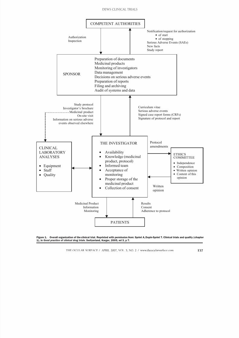
8/20/2019 dry eyes ii
http://slidepdf.com/reader/full/dry-eyes-ii 97/142
THE OCULAR SURFACE / APRIL 2007, VOL. 5, NO. 2 / www.theocularsurface.com 157
Study protocol
Investigator’s brochure
Medicinal product
On-site visit
Information on serious adverse
events observed elsewhere
COMPETENT AUTHORITIES
Preparation of documentsMedicinal products
Monitoring of investigatorsData managementSPONSOR Decisions on serious adverse eventsPreparation of reports
Filing and archivingAudit of systems and data
Authorization
Inspection
Notification/request for authorization
of start
of stopping
Serious Adverse Events (SAEs)
New facts
Study report
CLINICALLABORATORY
ANALYSES
Equipment Staff
Quality
ETHICS
COMMITTEE
Independence
Composition
Written opinion Content of this
opinion
Protocol
amendments
Written
opinion
THE INVESTIGATOR
Availability Knowledge (medicinal
product, protocol) Informed team
Acceptance of monitoring
Proper storage of themedicinal product
Collection of consent
Curriculum vitae
Serious adverse events
Signed case report forms (CRFs)
Signature of protocol and report
PATIENTS
Medicinal Product
Information
Monitoring
Results
Consent
Adherence to protocol
Figure 1. Overall organization of the clinical trial. Reprinted with permission from: Spriet A, Dupin-Spriet T. Clinical trials and quality (chapter
1), in Good practice of clinical drug trials. Switzerland, Karger, 2005, ed 3, p 7.
DEWS CLINICAL TRIALS
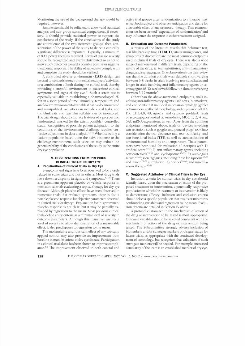
8/20/2019 dry eyes ii
http://slidepdf.com/reader/full/dry-eyes-ii 98/142
THE OCULAR SURFACE / APRIL 2007, VOL. 5, NO. 2 / www.theocularsurface.com158
Monitoring the use of the background therapy would berequired, however.
Sample size should be sufficient to allow valid statisticalanalysis and sub-group statistical comparisons, if neces-sary. It should provide statistical power to support theconclusions of the study. If the conclusions of the studyare equivalence of the two treatment groups, then con-
sideration of the power of the study to detect a clinicallysignificant difference is important. Typically, a minimumof 80% power (beta) is required. Levels of disease severityshould be recognized and evenly distributed so as not toskew study outcomes toward a possible positive or negativetherapeutic response. The ability of subjects to comply withand complete the study should be verified.
A controlled adverse environment (CAE) design canbe used to control the environment, the subjects’ activities,or a combination of both during the clinical trial, therebyproviding a stressful environment to exacerbate clinicalsymptoms and signs of dry eye.44 Such a stress test isespecially valuable in establishing a pharmacological ef-fect in a short period of time. Humidity, temperature, andair-flow are environmental variables that can be monitoredand manipulated. Activities can include visual tasks, andthe blink rate and tear film stability can be monitored.The trial design should embrace features of a prospective,randomized, masked (to the extent possible), controlledstudy. Recognition of possible patient adaptation to theconditions of the environmental challenge requires cor-rective adjustment in data analysis.45,46 When selecting apatient population based upon the naive response to thechallenge environment, such selection may reduce thegeneralizability of the conclusions of the study to the entire
dry eye population.
V. OBSERVATIONS FROM PREVIOUS
CLINICAL TRIALS IN DRY EYE
A. Peculiarities of Clinical Trials in Dry Eye
Symptoms and signs have been observed to be closelyrelated in some trials and not in others. Most drug trialshave shown a disparity in signs and symptoms.47-76 Thereis a prominent apparent placebo or vehicle response inmost clinical trials evaluating a topical therapy for dry eyedisease.1 Although placebo effects have been observed innumerous trials that evaluate symptoms, there is also a
notable placebo response for objective parameters observedin clinical trials for dry eye. Explanation for this prominentplacebo response is not clear, but it may be partially ex-plained by regression to the mean. Most previous clinicaltrials define entry criteria as a minimal level of severity inoutcome parameters. Although this maneuver assures alevel of severity to allow demonstration of a measurableeffect, it also predisposes to regression to the mean.
The moisturizing and lubricant effect of any topicallyapplied control may also provide an improvement frombaseline in manifestations of dry eye disease. Participationin a clinical trial alone has been shown to improve compli-
ance.3,5 The improvement observed in both control and
active trial groups after randomization to a therapy mayreflect both subject and observer anticipation and desire fora favorable effect of any proposed therapy. This phenom-enon has been termed “expectation of randomization” andmay influence the response to either treatment assigned.
B. Evaluation and Outcome Parameters
A review of the literature reveals that Schirmer test,tear film breakup time (TFBUT), vital staining scores, andsymptoms of discomfort are the most common endpointsused in clinical trials of dry eyes. There was also a widerange of markers used in different trials, depending on thenature of the drug, ie, tear substitutes, anti-inflammatorydrugs, and secretagogues. One observation from this reviewwas that the duration of trials was relatively short, varyingbetween 6-8 weeks in trials involving tear substitutes andlonger in trials involving anti-inflammatory agents or se-cretagogues (8-12 weeks with follow-up durations varyingbetween 3-12 months).
Other than the above-mentioned endpoints, trials in-volving anti-inflammatory agents used tests, biomarkers,and endpoints that included impression cytology (gobletcell numbers, epithelial morphology, and expression of HLADR, CD3,4,8, 40, Apo2.7, and cytokine profiles). Trialsof secretagogues looked at osmolarity, MUC 1, 2, 4 and5AC mRNA expressions, as well. Apart from the commonendpoints mentioned above, trials on devices involvingtear retention, such as goggles and punctal plugs, took intoconsideration the tear clearance rate, tear osmolarity, andtear functional index (TFI), as well as standardization ofenvironmental humidity and temperature. These param-eters have been used for evaluation of therapies with 1)
artificial tears47-52; 2) anti-inflammatory agents, includingcorticosteroids53,54 and cyclosporine55-61; 3) autologousserum 62-66; secretagogues, including those for aqueous67-72 and mucin73-78 stimulation; 4) devices79-86; and miscella-neous therapy.87-88
C. Suggested Attributes of Clinical Trials in Dry Eye
Inclusion criteria for clinical trials in dry eye shouldidentify, based upon the mechanism of action of the pro-posed treatment or intervention, a potentially responsivepopulation in which the treatment or intervention is likelyto demonstrate efficacy. Inclusion and exclusion criteria
should select a specific population that avoids or minimizesconfounding variables and regression to the mean. Exclu-sion criteria are detailed in Section IV above.
A protocol customized to the mechanism of action ofthe drug or intervention to be tested is most appropriate.Outcome variables should be selected consistent with themechanism of action of the drug or intervention beingtested. The Subcommittee strongly advises inclusion ofbiomarkers and/or surrogate markers of disease status forfuture trials, as appropriate with the continued develop-ment of technology, but recognizes that validation of suchsurrogate markers will be needed. For example, increased
osmolarity of the tears is an established marker of dry eye,
DEWS CLINICAL TRIALS

8/20/2019 dry eyes ii
http://slidepdf.com/reader/full/dry-eyes-ii 99/142
THE OCULAR SURFACE / APRIL 2007, VOL. 5, NO. 2 / www.theocularsurface.com 159
and there are several possible methods of measurement.Surrogate markers may be direct or correlative. Direct
surrogate markers are those that derive from the samephysical or chemical properties as the primary marker,eg, tear conductivity as a measure of tear osmolarity. Cor-relative surrogate markers are those that correlate with theprimary marker but can be produced by other mechanisms
as well, eg, a single inflammatory cytokine level as a markerof inflammation.In dry eye disease, in which variability of a sign or symp-
tom can be greatly influenced by environmental or visualtask activities at any given point in time, the measurementof reliable, durable surrogate markers of disease activityshould be considered as a valid measure of effectiveness ofany given therapy or intervention. The outcome measuresshould be measurable with adequate accuracy and repro-ducibility. Measurement of the primary outcome parametershould be accomplished with a well-validated test. This istrue for clinical signs of disease and surrogate measures,as well as for symptoms of discomfort and visual distur-bance.89-96 The primary outcome variable may be a symp-tom or a sign for valid outcome analysis, but regulatoryapproval may require both in some countries. Symptomsshould be graded in a well-defined scoring system, such asthe visual analog scale ( VAS) or with Likert scores.2,97
In recognition of the prominence of placebo and vehicleresponse in clinical trials in dry eye, the Subcommitteemade several observations. Because a true placebo has notbeen found that lacks inherent lubricant effect, consider-ation of a non-treatment arm could be considered. Althoughsuch a design has limitations of possible institutional reviewboard constraints, and given that patients may be prone to
intermittent use of over-the-counter lubricants that couldconfound the outcome, consideration of such a design hasmerit. In the absence of such a protocol, the Subcommitteerecommends consideration of 1) a randomized, maskedtrial, in which the initiation of treatment is also maskedboth to investigator and subject, or 2) a withdrawal study,in which all patients initially receive active medication, fol-lowed by randomization to vehicle. One benefit of such adesign is that all subjects receive active medication at somepoint in the trial, and this may serve to improve willingnessof subjects to enroll in a well-designed trial.
The Subcommittee recommends inclusion of the fol-
lowing outcome parameters:1. An objective measure of visual function (eg, Functional
Visual Acuity);2. Determination of tear volume and production (eg,
Schirmer test or fluorescein dilution test);3. Determination of tear stability (eg, tear breakup with
fluorescein TFBUT or a non-invasive TFBUT devicesuch as videokeratography)96;
4. Measurement of tear composition (eg, osmolarity, de-termination of specific protein content, or the measure-ment of inflammatory mediators in tears);
5. Measurement of ocular surface integrity.
There is consensus that the determination of ocular
surface integrity is at this time best performed by stainingof the ocular surface with fluorescein and lissamine greenor rose bengal (see parameters from the Diagnostic Method-ology Subcommittee Report in this issue for appropriateconcentrations and use of barrier filters),98 although thelimitations of such evaluation have been documented inprevious clinical trials.58,69,76 A standardized grading system
should separately grade corneal and conjunctival stainingand record individual area scores, as well as combinedarea scores, for analysis (see the Diagnostic MethodologySubcommittee Report for appropriate grading system).98
The grading system should allow for one or two dots ofstaining in the inferonasal quadrant of the cornea, becausesuch staining may occur in normal subjects.99-107 Stainingof the conjunctival caruncle and semilunar fold shouldnot be counted, as this occurs in a majority of normalsubjects.101
Other tests that could be used as outcome measures inspecific protocols might include impression cytology andflow cytometry (for selected trials, see parameters fromthe Diagnostic Methodology Subcommittee Report for ap-propriate method and staining procedure).98 Technologicaladvances in measurement of tear film stability, measurementof the tear meniscus volume, or measurement of ocularsurface protection and epithelial permeability may in thefuture allow more precise determination of tear functionand ocular surface integrity. However, at present, they arenot well validated in clinical trials.
Outcome analysis in a multi-factorial disease withseveral clinical parameters of tear film abnormality, ocularsurface damage, and functional impairment may be ame-nable to composite indices of disease severity. This approach
has been utilized in evaluation of rheumatic disease, withconsensus development of the American Congress of Rheu-matology (ACR) indices (ACR50 and ACR70) that evaluatemultiple descriptors of disease severity. Currently, therehas been inadequate evaluation of such composite indicesin dry eye disease, and validated indices are not available.The committee identifies as a need and an area for futuredeliberation the development and validation of such indicesfor evaluation of dry eye disease.
Appropriate and carefully planned statistical analysis iscritical in evaluating clinical trial data. The analysis strategywill depend on the primary outcome variable selected for
the trial, and it must be chosen prior to the beginning ofdata collection. The general principle of the intent-to-treatanalysis should be adhered to for the primary analysis ofdata.
VI. FEATURES TO FACILITATE MULTICENTER
AND INTERNATIONAL COLLABORATIVE
CLINICAL TRIALS
The Subcommittee recommends the development ofcriteria to be used in multinational venues. Importantaspects to consider for such international trials are the useof uniform terminology. This may require that terms are
translated and back-translated for clarity and accuracy.
DEWS CLINICAL TRIALS

8/20/2019 dry eyes ii
http://slidepdf.com/reader/full/dry-eyes-ii 100/142
THE OCULAR SURFACE / APRIL 2007, VOL. 5, NO. 2 / www.theocularsurface.com160
It is necessary to resolve cultural or ethnic connotationsor implications in terminology. There should be uniforminterpretation of outcome variables with standardizedprotocols for measurement and recording of data. Testingprocedures should be uniform, with use of standardizedreagents, standardized protocols, and consistent recordingof results. It is necessary to maintain skill levels of data col-
lectors and observers, including certification of investigatorsand research coordinators and technicians. Attempts shouldbe made to reduce biases related to population differences(race, ethnicity, climatic).
These appendices can be accessed at www.tearfilm.org: x Appendix 1. Outline of a manual of procedures x Appendix 2. Guidelines for Good Clinical Practices x Appendix 3. Writing the Investigator’s Brochure for the
tested drug x Appendix 4. Using the investigational medicinal product x Appendix 5. Adverse events and management issues
REFERENCES
(Parenthetical codes following some references indicate level of evidence accordingto the American Academy of Ophthalmology Preferred Practices guidelines.)
1. Foulks GN. Challenges and pitfalls in clinical trials of treatments for dryeye. Ocul Surf 2003;1:21-36
2. Chow SC, Liu J-P. Design and analysis of clinical trials:concept and meth-odologies. John C Wiley and Co, New York, 1998. pp5,6,180
3. Hrobjartsson A, Gotzsche PC. Is the placebo powerless? An analysisof clinical trials comparing placebo with no treatment. N Engl J Med2001;345:1277-8
4. Weihrauch TR, Gauler TC. Placebo—efficacy and adverse effects incontrolled clinical trials. Arzneimittel-Forschung 1999;49:385-93
5. Shapiro AK, Shapiro E. The powerful placebo. Johns Hopkins UniversityPress, Baltimore, 1997, p149
6. Belorgey C, Demarez JP, Jaillon P, Giens XVII. Good clinical practice in
clinical trials: training physicians, incentives to apply good clinical practiceand assessment of compliance. Therapie 2002;57:339-46
7. Bohaychuk W, Ball G.Conducting GDP-compliant clinical research. Chichester, John Wiley and Sons: 1999
8. Bohaychuk W, Ball G. Standard operating procedures for clinical researchpersonnel. Quality Assurance J Part 1, 1998;3:137-50. Part 2, 2000;4:31-46. Part 3, 2000;4:95-106. Part 4, 2000;4:151-68. Part 5, 2000;4:211-23.Part 6, 2001;5:37-47. Part 7, 2001;5:91-112. Part 8, 2001;5:163-73. Part9, 2001;5:221-40. Part 10, 2002;6:23-32. Part 11, 2002;6:33-43. Part 12,2002;6:103-11. Part 13, 2002;6:187-95. Part 14, 2002;6:235-44. Part 15,2003;7:41-9. Part 16, 2003;7:100-12
9. Gerlis L. Sample standard operating procedures for good clinical practice.Richmond, PJB Publications, 1988
10. Spiet A, Dupin-Spriet T. Evaluation des therapeutiques, in Herve P, RifleG, Vuitton DA, et al (eds). Transplantation d’organes et greffes de tissus.Paris, INSERM, John Libbey, 1994;77-97
11. Spriet A, Dupin-Spriet T, Simon P. Methodology of clinical drug trials.Basel, Karger, 1994
12. DeAngelis C, Fontanarosa PB, Flanagin A. Reporting financial conflictsof interest and relationships between investigators and research sponsors.
JAMA 2001;286:89-91 13. Decoster G, Wahl M. Insuring subjects in clinical trials: sponsors need to
work through a regulatory maze. Appl Clin Trials 1998 ;7 :30-6 14. Dupin-Spriet T, Spriet A. Des essais cliniques pour un dossier international.
Quelles difficultes? STP Pharma Pratiques 1993;3:1-5 15. European Parliament and Council of the European Union. Directive
2001/20/EC of the European Parliament and of the Council of April 2001on the approximation of the laws, regulations and administrative provisionsof the Member States relating to the implementation of good clinical prac-tice in the conduct of clinical trials on medicinal products for human use.Official Journal of the European Communities 1.5.2001;L121/34-44/34-44.
16. European Agency for the Evaluation of Medicinal Products. Explana-
tory note and comments to the ICH harmonized tripartite guideline
E6: Note for Guidance on Good Clinical Practice (CPMP/ICH/135/95,CPMP/768/97. 8 September 1997
17. European Commission Enterprise Directorate-General. The rules govern-ing medicinal products in the European Union. Volume 4, Good Manu-facturing Practices, Annex 13, Manufacture of investigational medicinalproducts. July 2003
18. Horton R. Conflicts of interest in clinical research: opprobrium or obses-sion? Lancet 1997:349;1112-3
19. Hvidberg EF. Achieving investigator compliance. Drug Information J1993;27:1107-10
20. Imada S. An update on good clinical practice legislation in Japan. Quality Assurance J 1998;3:37-42
21. Inoue K, Hasebe S. The quality assurance and quality control issue forsponsors on GCP compliance in Japan. Quality Assurance J 1999;3:65-9
22. International Conference on Harmonisation of Technical Requirements forRegistration of Pharmaceuticals for Human Use. ICH topic E3. Structureand content of clinical study report. November 1995
23. International Conference on Harmonisation of Technical Requirementsfor Registration of Pharmaceuticals for Human Use. ICH topic E6. Notefor guidance on good clinical practice. July 1996
24. International Conference on Harmonisation of technical requirements forregistration of pharmaceuticals for human use. ICH topic E9. Statisticalprinciples for clinical trials. February 1998
25. International Conference on Harmonisation of Technical Requirementsfor Registration of Pharmaceuticals for Human Use. ICH topic S4A. Notefor guidance on duration of chronic toxicity in animals (rodent and non
rodent toxicity testing). November 1998 26. European Agency for Evaluation of Medicinal Products. ICH topic M3(M).
Note for guidance on non-clinical safety studies for the conduct of humanclinical trials for pharmaceuticals. November 2000
27. Lacombe D. Insuring international clinical trials. An academic viewpoint.Appl Clin Trials 1998;7:24-9
28. Moses H, Martin JB. Academic relationships with industry. A model forbiomedical research. JAMA 2001;285:933-5
29. US Code of Federal Regulations. Title 21: Food and Drugs. Part 50: Pro-tection of human subjects. Part 56: Institutional review boards. Part 312:Investigational new drug application. April 2003
30. US Food and Drug Administration. Guidance for industry. Financialdisclosure by clinical investigators. March 20, 2001
31. International Conference on Harmonisation of Technical Requirementsfor Registration of Pharmaceuticals for Human Use. ICH topic E6. Goodclinical practice consolidated guideline. Section 7: Investigator’s brochure.
July 1996 32. Dupin-Spriet T, Spriet A. Les bonnes pratiques de fabrication des medica-
ments pour essai clinque, in Medicament, Ethique et Realite Industrielle.La reglementation pharmaceutique—Principes et philosophie. Paris,Editions de Sante 1996
33. International Air Transport Association. IATA dangerous goods regulations44th edition, 2003
34. International Conference on Harmonisation of Technical Requirementsfor Registration of Pharmaceuticals for Human Use. ICH topic Q1A (R2).Stability testing of new drug substances and products. August 2003
35. Lantz RJ Jr. Stability aspects of clinical supplies and scale-up studies. DrugDevelopment Industrial Pharmacy 1984;10:1425-32
36. US Food and Drug Administration. Center for Drugs and Biologics. Of-fice of Drug. Department of Health and Human Services. Guideline forsubmitting documentation for the stability of human drugs and biologics.Rockville, Food and Drug Administration, 1987
37. Begaud B, Evreux JC, Jouglard J, Lagier G. [Imputation of the unexpectedor toxic effects of drugs. Actualization of the method used in France].Therapie 1985;40:111-8
38. European Commission Enterprise Directorate-General. Detailed guid-ance on the European database of suspected unexpected serious adversereactions (Eudravigilance—clinical trial module). April 2003
39. European Commission Enterprise Directorate-General. Detailed guidancefor the request for authorization of a clinical trial on a medicinal productfor human use to the competent authorities, notification of substantialamendments and declaration of the end of the trial. April 2003
40. International Conference on Harmonisation of Technical Requirements forRegistration of Pharmaceuticals for Human Use. ICH topic E2A. Clinicalsafety data management: definitions and standards for expedited reporting:availability, notice. November 1994
41. International Conference on Harmonisation of Technical Requirementsfor Registration of Pharmaceuticals for Human Use. ICH topic E2B (M).
Maintenance of the ICH guideline on clinical safety data management:
DEWS CLINICAL TRIALS
8/20/2019 dry eyes ii
http://slidepdf.com/reader/full/dry-eyes-ii 101/142
THE OCULAR SURFACE / APRIL 2007, VOL. 5, NO. 2 / www.theocularsurface.com 161
data elements for transmission of individual case safety reports. February2001.
42. Karch FE, Lasagna L. Toward the operational identification of adversedrug reactions. Clin Pharmacol Ther 1977;21:247-54
43. US Code of federal regulations title 21, volume 5. Part 312 InvestigationalNew Drug application. Subpart B Section 312.32 IND safety reports.
44. Ousler G, Gomes PJ, Welch D, Abelson MB. Methodologies for the studyof of ocular surface disease. Ocul Surf 2005;3:143-54
45. Ousler GW 3rd, Abelson MB, Nally LA, et al: Evaluation of time to ‘naturalcompensation’ in normal and dry eye subject populations during expo-
sure to a controlled adverse environment. Adv Exp Med Biol 2002;506(PtB):1057-63
46. Abelson MB, Ousler GW3rd, Nally LA, et al: Alternative reference valuesfor tear film break up time in normal and dry eye populations. Adv ExpMed Biol 2002;506(Pt B):1121-5
47. Lopez Bernal D, Ubels JL. Quantitative evaluation of the corneal epithelialbarrier: effect of artificial tears and preservatives. Curr Eye Res 1991;10:645-56 (LA1)
48. Lopez Bernal D, Ubels JL. Artificial tear composition and promotion ofrecovery of the damaged corneal epithelium. Cornea 1993;12:115-20(LA1)
49. Gilbard JP, Farris RL.Tear osmolarity and ocular surface disease in kera-toconjunctivitis sicca. Arch Ophthalmol 1979;97:1642-6 (LA1)
50. Christensen MT, Cohen S, Rinehart J, et al. Clinical evaluation of an HP-guar gellable lubricant eye drop for the relief of dryness of the eye. CurrEye Res 2004;28:55-62 (LA1)
51. Sand BB, Marner K, Norn MS. Sodium hyaluronate in the treatment ofkeratoconjunctivitis sicca. A double masked clinical trial. Acta Ophthalmol(Copenh) 1989;67:181-3 (LA1)
52. Nelson JD, Farris RL. Sodium hyaluronate and polyvinyl alcohol artificialtear preparations. A comparison in patients with keratoconjunctivitis sicca.
Arch Ophthalmol 1988;106:484-7 (LA1) 53. Pflugfelder SC, Maskin SL, Anderson B, et al. A randomized, double-
masked, placebo-controlled, multicenter comparison of loteprednoletabonate ophthalmic suspension, 0.5%, and placebo for treatment ofkeratoconjunctivitis sicca in patients with delayed tear clearance. Am JOphthalmol 2004;138:444-57 (LA1)
54. Avunduk AM, Avunduk MC, Varnell ED, Kaufman HE. The comparisonof efficacies of topical corticosteroids and nonsteroidal anti-inflammatorydrops on dry eye patients: a clinical and immunocytochemical study. Am
J Ophthalmol 2003;136:593-602 (LA1) 55. Gunduz K, Ozdemir O. Topical cyclosporin treatment of keratoconjunc-
tivitis sicca in secondary Sjogren’s syndrome. Acta Ophthalmol (Copenh) 1994;72:438-42 (LA1)
56. Laibovitz RA, Solch S, Andriano K, et al. Pilot trial of cyclosporine 1%ophthalmic ointment in the treatment of keratoconjunctivitis sicca. Cornea 1993;12:315-23 (LA1)
57. Stevenson D, Tauber J, Reis BL. Efficacy and safety of cyclosporin A oph-thalmic emulsion in the treatment of moderate-to-severe dry eye disease: adose-ranging, randomized trial. The Cyclosporin A Phase 2 Study Group.Ophthalmology 2000;107:967-74 (LA1)
58. Sall K, Stevenson OD, Mundorf TK, Reis BL. Two multicenter, randomizedstudies of the efficacy and safety of cyclosporine ophthalmic emulsion inmoderate to severe dry eye disease. CsA Phase 3 Study Group. Ophthal-mology 2000;107:631-9 (LA1)
59. Kunert KS, Tisdale AS, Gipson IK. Goblet cell numbers and epithelialproliferation in the conjunctiva of patients with dry eye syndrome treatedwith cyclosporine. Arch Ophthalmol 2002;120:330-7 (LA1)
60. Brignole F, Pisella PJ, De Saint Jean M, et al. Flow cytometric analysis ofinflammatory markers in KCS: 6-month treatment with topical cyclosporinA. Invest Ophthalmol Vis Sci 2001;42:90-5 (LA1)
61. Kunert KS, Tisdale AS, Stern ME, et al. Analysis of topical cyclosporinetreatment of patients with dry eye syndrome: effect on conjunctival lym-phocytes. Arch Ophthalmol 2000;118:1489-96 (LA1)
62. Poon AC, Geerling G, Dart JK, et al. Autologous serum eyedrops fordry eyes and epithelial defects: clinical and in vitro toxicity studies. Br JOphthalmol 2001;85:1188-97 (LB2)
63. Tananuvat N, Daniell M, Sullivan LJ, et al. Controlled study of the use ofautologous serum in dry eye patients. Cornea 2001;20:802-6 (LA2)
64. Kojima T, Ishida R, Dogru M, et al. The effect of autologous serum eye-drops in the treatment of severe dry eye disease: a prospective randomizedcase-control study. Am J Ophthalmol 2005;139:242-6 (LA1)
65. Noble BA, Loh RS, MacLennan S, et al. Comparison of autologous serumeye drops with conventional therapy in a randomised controlled crossover
trial for ocular surface disease. Br J Ophthalmol 2004;88:647-52 (LA1)
66. Tsubota K, Goto E, Fujita H, et al. Treatment of dry eye by autologousserum application in Sjogren’s syndrome. Br J Ophthalmol 1999;83:390-5(LB2)
67. Gilbard JP, Rossi SR, Heyda KG, Dartt DA. Stimulation of tear secretionby topical agents that increase cyclic nucleotide levels. Invest OphthalmolVis Sci 1990;31:1381-8 (LA1)
68. Gilbard JP, Rossi SR, Heyda KG, Dartt DA. Stimulation of tear secretionand treatment of dry-eye disease with 3-isobutyl-1-methylxanthine. ArchOphthalmol 1991;109:67;2-6 (LA1)
69. Tauber J, Davitt WF, Bokosky JE, et al. Double-masked, placebo-controlled
safety and efficacy trial of diquafosol tetrasodium (INS365) ophthalmicsolution for the treatment of dry eye. Cornea 2004;23:784-92 (LA1)
70. Mundasad MV, Novack GD, Allgood VE, et al. Ocular safety of INS365ophthalmic solution: a P2Y(2) agonist in healthy subjects. J Ocul PharmacolTher 2001;17:173-9
71. Yerxa BR, Mundasad M, Sylvester RN, et al. Ocular safety of INS365ophthalmic solution, a P2Y2 agonist, in patients with mild to moderatedry eye disease. Adv Exp Med Biol 2002;506(Pt B):1251-7 (LB2)
72. Fujihara T, Murakami T, Fujita H, et al. Improvement of corneal barrierfunction by the P2Y(2) agonist INS365 in a rat dry eye model. InvestOphthalmol Vis Sci 2001;42:96-100
73. Fujihara T, Murakami T, Nagano T, et al. INS365 suppresses loss of cornealepithelial integrity by secretion of mucin-like glycoprotein in a rabbitshort-term dry eye model. J Ocul Pharmacol Ther 2002;18:363-70
74. Yerxa BR, Douglass JG, Elena PP, et al. Potency and duration of action ofsynthetic P2Y2 receptor agonists on Schirmer scores in rabbits. Adv Exp
Med Biol 2002;506(Pt A):261-5 75. Nakamura M, Endo K, Nakata K, Hamano T. Gefarnate stimulates secre-
tion of mucin-like glycoproteins by corneal epithelium in vitro and protectscorneal epithelium from desiccation in vivo. Exp Eye Res 1997;65:569-74(LA1)
76. Hamano T. Dry eye treatment with eye drops that stimulate mucin produc-tion. Adv Exp Med Biol 1998;438:965-8 (LA2)
77. Jumblatt JE, Cunningham LT, Li Y, Jumblatt MM. Characterizationof human ocular mucin secretion mediated by 15(S)-HETE. Cornea 2002;21:818-24 (LA1)
78. Jackson RS 2nd, Van Dyken SJ, McCartney MD, Ubels JL. The eicosanoid,15-(S)-HETE, stimulates secretion of mucin-like glycoprotein by thecorneal epithelium. Cornea 2001;20:516-21 (LA1)
79. Tuberville AW, Frederick WR, Wood TO. Punctal occlusion in tear defi-ciency syndromes. Ophthalmology 1982;89:1170-2 (LA1)
80. Willis RM, Folberg R, Krachmer JH, Holland EJ. The treatment of aque-
ous-deficient dry eye with removable punctal plugs. A clinical and impres-sion-cytologic study. Ophthalmology 1987;94:514-8 (LA1)
81. Yen MT, Pflugfelder SC, Feuer WJ. The effect of punctal occlusion ontear production, tear clearance, and ocular surface sensation in normalsubjects. Am J Ophthalmol 2001;131:314-23 (LA1)
82. Balaram M, Schaumberg DA, Dana MR. Efficacy and tolerability outcomesafter punctal occlusion with silicone plugs in dry eye syndrome. Am JOphthalmol 2001;131:30-6 (LA1)
83. Tsubota K, Yamada M, Urayama K. Spectacle side panels and moist insertsfor the treatment of dry-eye patients. Cornea 1994;13:197-201 (LA2)
84. Tsubota K. The effect of wearing spectacles on the humidity of the eye. Am J Ophthalmol 1989;108:92-3 (LA2)
85. Gresset J, Simonet P, Gordon D. Combination of a side shield with anocular moisture chamber. Am J Optom Physiol Opt 1984;61:610-2 (LA2)
86. Korb DR, Greiner JV, Glonek T, et al. Effect of periocular humidity on thetear film lipid layer. Cornea 1996;15:129-34 (LA2)
87. Sommer A. Treatment of corneal xerophthalmia with topical retinoic acid. Am J Ophthalmol 1983;95:349-52 (LA1)
88. Nelson JD, Gordon JF. Topical fibronectin in the treatment of keratocon- junctivitis sicca. Chiron Keratoconjunctivitis Sicca Study Group. Am JOphthalmol 1992;114:441-7 (LA1)
89. Begley CG, Chalmers RL, Abetz L, et al. The relationship between habitualpatient-reported symptoms and clinical signs among patients with dry eyeof varying severity. Invest Ophthalmol Vis Sci 2003;44:4753-61 (LA1)
90. Begley CG, Caffery B, Chalmers RL, Mitchell GL. Dry Eye Investigation(DREI) Study Group. Use of the dry eye questionnaire to measure symp-toms of ocular irritation in patients with aqueous tear deficient dry eye.Cornea 2002;21:664-70 (LA1)
91. Nichols KK, Begley CG, Caffery B, Jones LA. Symptoms of ocular irrita-tion in patients diagnosed with dry eye. Optom Vis Sci 1999;76:838-44(LA1)
92. Schein OD, Tielsch JM, Munoz B, et al. Relation between signs and
symptoms of dry eye in the elderly. A population-based perspective.
DEWS CLINICAL TRIALS

8/20/2019 dry eyes ii
http://slidepdf.com/reader/full/dry-eyes-ii 102/142
THE OCULAR SURFACE / APRIL 2007, VOL. 5, NO. 2 / www.theocularsurface.com162
Ophthalmology 1997;104:1395-401 (LA1) 93. Bjerrum KB. Test and symptoms in keratoconjunctivitis sicca and their
correlation. Acta Ophthalmol Scand 1996;74:436-41 (LA1) 94. Bowman SJ, Booth DA, Platts RG, et al; UK Sjogren’s Interest Group.
Validation of the Sicca Symptoms Inventory for clinical studies of Sjogren’ssyndrome. J Rheumatol 2003;30:1259-66 (LA1)
95. Nichols KK. Patient-reported symptoms in dry eye disease. Ocul Surf 2006;4:137-145
96. de Paiva CS, Lindsey JL, Pflugfelder SC. Assessing the severity of keratitissicca with videokeratoscopic indices. Ophthalmology 2003;110:1102-9
(LA1) 97. www.socialreseachmethods.net/kb/scallik.htm (Accessed July 24,
2006). 98. (No authors listed). Methodologies to diagnose and monitor dry eye:
report of the Diagnostic Methodology Subcommittee of the InternationalDry Eye WorkShop (2007). Ocul Surf 2007;5:108-152
99. Bron AJ, Evans VE, Smith JA. Grading of corneal and conjunctival staining
in the context of other dry eye tests. Cornea 2003;22:640-50 100. Norn MS: Vital staining of cornea and conjunctiva. Acta Ophthal 1972;Supp
113:3-66 101. Norn MS. Lissamine green vital staining of cornea and conjunctiva. Acta
Ophthalmol 1973;51:483-91 102. Norn MS. Vital staining of cornea and conjunctiva. Acta Ophthal (Kbh)
1962;40:389-401 103. Norn MS. External eye: methods of evaluation. Copenhagen, Scriptor,
1974:51-5 104. Korb DR, Korb JM: Corneal staining prior to contact lens wearing. J Am
Optom Assn 1970;41:228-32 105. Schwallie JD, McKenney CD, Long WD, McNeil A. Corneal staining pat-
terns in normal non-contact lens wearers. Optom Vis Sci 1997;74:92-8 106. Josephson JE, Caffery BE. Corneal staining characteristics after sequential
instillations of fluorescein. Optom Vis Sci 1992;69:570-3 107. Caffery BE, Josephson JE. Corneal staining after sequential instillation of
fluorescein over 30 days. Optom Vis Sci 1991;68:881-9
DEWS CLINICAL TRIALS
8/20/2019 dry eyes ii
http://slidepdf.com/reader/full/dry-eyes-ii 103/142
THE OCULAR SURFACE / APRIL 2007, VOL. 5, NO. 2 / www.theocularsurface.com 163
DEWS Management and Therapy
©2007 Ethis Communications, Inc. The Ocular Surface ISSN: 1542-
0124. (No authors listed). Management and therapy of dry eye
disease: report of the Management and Therapy Subcommittee of
the International Dry Eye WorkShop (2007). 2007;5(2):163-178.
Management and Therapy of Dry Eye Disease:
Report of the Management and Therapy Subcommitteeof the International Dry Eye WorkShop (2007)
ABSTRACT The members of the Management and Therapy
Subcommittee assessed current dry eye therapies. Each mem-
ber wrote a succinct evidence-based review on an assigned
aspect of the topic, and the final report was written after
review by and with consensus of all subcommittee members
and the entire Dry Eye WorkShop membership. In addition to
its own review of the literature, the Subcommittee reviewed
the Dry Eye Preferred Practice Patterns of the American
Academy of Ophthalmology and the International Task Force
(ITF) Delphi Panel on Dry Eye. The Subcommittee favored the
approach taken by the ITF, whose recommended treatments
were based on level of disease severity. The recommenda-
tions of the Subcommittee are based on a modification of
the ITF severity grading scheme, and suggested treatments
were chosen from a menu of therapies for which evidence of
therapeutic effect had been presented.
KEYWORDS DEWS, dry eye disease, Dry Eye WorkShop,
management, therapy
I. INTRODUCTION
his report summarizes the management and thera-peutic options for treating dry eye disease. The levelof evidence for supporting data from the literature
is evaluated according to the modified American Academyof Ophthalmology Preferred Practices guidelines (Table 1).
II. GOALS OF THE MANAGEMENT AND THERAPY
SUBCOMMITTEE
Goals of this committee were to identify appropriatetherapeutic methods for the management of dry eye diseaseand recommend a sequence or strategy for their application,based on evidence-based review of the literature.
The quality of the evidence in the literature was gradedaccording to a modification of the scheme used in the American Academy of Ophthalmology Preferred PracticePatterns series. When possible, peer-reviewed full publica-tions, not abstracts, were used. The report was reviewed
Accepted for publication January 2007.
Management and Therapy Subcommittee members: Stephen C. Pflugfelder,MD (Chair); Gerd Geerling, MD; Shigero Kinoshita, MD; Michael A. Lemp,MD; James McCulley, MD; Daniel Nelson, MD; Gary N. Novack, PhD; JunShimazaki, MD; Clive Wilson, PhD.
Proprietary interests of Subcommittee members are disclosed on pages 202and 204.
Reprints are not available. Articles can be accessed at:www.tearfilm.org.
Correspondence in regard to the this chapter should be addressed to StephenC. Pflugfelder MD, Ophthalmology-Ocular Surf Ctr, Cullen Eye Institute,6565 Fannin Street NC 205, Houston, TX 77030. Tel: 713-798-4732. Fax:713-798-1457. Email: [email protected]
Table 1. Evidence grading scheme
Clinical Studies
Level 1. Evidence obtained from at least one properly
conducted, well-designed, randomized, controlled trial,
or evidence from well-designed studies applying rigorous
statistical approaches.
Level 2. Evidence obtained from one of the following: a
well-designed controlled trial without randomization,
a well-designed cohort or case-control analytic study,
preferably from one or more center, or a well-designed
study accessible to more rigorous statistical analysis.
Level 3. Evidence obtained from one of the following:
descriptive studies, case reports, reports of expert
committees, expert opinion.
Basic Science Studies
Level 1. Well-performed studies confirming a hypothesis with
adequate controls published in a high-impact journal.
Level 2. Preliminary or limited published study.
Level 3. Meeting abstracts or unpublished presentations.
This evidence grading scheme is based on that used in the AmericanAcademy of Ophthalmology Preferred Practice Pattern series.
T
8/20/2019 dry eyes ii
http://slidepdf.com/reader/full/dry-eyes-ii 104/142
THE OCULAR SURFACE / APRIL 2007, VOL. 5, NO. 2 / www.theocularsurface.com164
by all subcommittee members and by the entire Dry Eye WorkShop membership. Comments and suggested revi-sions were discussed by the subcommittee members andincorporated into the report where deemed appropriate
by consensus.
III. ASSESSMENT OF CURRENT DRY EYE THERAPIES
A. Tear Supplementation: Lubricants
1. General Characteristics and EffectsThe term “artificial tears” is a misnomer for most prod-
ucts that identify themselves as such, because they do notmimic the composition of human tears. Most function aslubricants, although some more recent formulations mimic
the electrolyte composition of human tears (TheraTears®
[Advanced Vision Research, Woburn, MA]).1,2 The ocularlubricants presently available in the United States are ap-proved based on the US Food and Drug Administration(FDA) monograph on over-the-counter (OTC) products(21 CFR 349) and are not based on clinical efficacy. Themonograph specifies permitted active ingredients (eg,demulcents, emulsifiers, surfactants, and viscosity agents)and concentrations, but gives only limited guidance oninactive additives and solution parameters. Certain inac-tive ingredients that are used in artificial tears sold in theUS (eg, castor oil in Endura™ [Allergan, Inc., Irvine, CA]and guar in Systane® [Alcon, Ft Worth, TX]) are not listedin the monograph.
It is difficult to prove that any ingredient in an ocularlubricant acts as an active agent. If there is an active in-gredient, it is the polymeric base or viscosity agent, butthis has proved difficult to demonstrate. This is eitherbecause it is not possible to detect the effects or differencesin clinical trials with presently available clinical tests orbecause the currently available agents do not have anydiscernable clinical activity beyond a lubrication effect. Although certain artificial tears have demonstrated moresuccess than others in reducing symptoms of irritationor decreasing ocular surface dye staining in head-to-head
comparisons, there have been no large scale, masked,comparative clinical trials to evaluate the wide variety ofocular lubricants.
What is the clinical effect of ocular lubricants or artificialtears? Do they lubricate, replace missing tear constituents,reduce elevated tear film osmolarity, dilute or wash outinflammatory or inflammation-inducing agents? Do they,in some instances, actually wash out essential substancesfound in normal human tears? These questions remain tobe answered as more sensitive clinical tests become avail-able to detect changes in the ocular surface.
The foremost objectives in caring for patients with dry
eye disease are to improve the patient’s ocular comfort andquality of life, and to return the ocular surface and tear filmto the normal homeostatic state. Although symptoms canrarely be eliminated, they can often be improved, leadingto an improvement in the quality of life. It is more difficultto demonstrate that topical lubricants improve the ocularsurface and the tear film abnormalities associated with dryeye. Most clinical studies fail to demonstrate significantcorrelation between symptoms and clinical test valuesor between the clinical test values themselves.3-5 It is notunusual for a dry eye with only mild symptoms to showsignificant rose bengal staining. Until agents are developed
that can restore the ocular surface and tear film to their
OUTLINE
I. Introduction
II. Goals of the Management and TherapySubcommittee
III. Assessment of current dry eye therapies
A. Tear supplementation: lubricants
1. General characteristics and effects
2. Preservatives
3. Electrolyte composition
4. Osmolarity
5. Viscosity agents
6. Summary
B. Tear Retention
1. Punctal occlusion
a. Rationale
b. Types
c. Clinical studies
d. Indications and contraindications
e. Complications
f. Summary
2. Moisture chamber spectacles
3. Contact lenses
C. Tear stimulation: secretagogues
D. Biological tear substitutes
1. Serum
2. Salivary gland autotransplantation
E. Anti-inflammatory therapy
1. Cyclosporine
2. Corticosteroids
a. Clinical studies
b. Basic research
3. Tetracyclines
a. Properties of tetracyclines and theirderivatives
1) Antibacterial properties
2) Anti-inflammatory
3) Anti-angiogenic properties
b. Clinical applications of tetracycline
1) Acne Rosacea
2) Chronic posterior blepharitis:meibomianitis, meibomian gland
dysfunction
3) Dosage and safety
F. Essential fatty acids
G. Environmental strategies
IV. Treatment recommendations
V. Unanswered questions and future directions
DEWS MANAGEMENT AND THERAPY
8/20/2019 dry eyes ii
http://slidepdf.com/reader/full/dry-eyes-ii 105/142
THE OCULAR SURFACE / APRIL 2007, VOL. 5, NO. 2 / www.theocularsurface.com 165
normal homeostatic state, the symptoms and signs of dryeye disease will continue.
Ocular lubricants are characterized by hypotonic orisotonic buffered solutions containing electrolytes, surfac-tants, and various types of viscosity agents. In theory, theideal artificial lubricant should be preservative-free, containpotassium, bicarbonate, and other electrolytes and have a
polymeric system to increase its retention time.1,6-8
Physicalproperties should include a neutral to slightly alkaline pH.Osmolarities of artificial tears have been measured to rangefrom about 181 to 354 mOsm/L.9 The main variables in theformulation of ocular lubricants regard the concentrationof and choice of electrolytes, the osmolarity and the typeof viscosity/polymeric system, the presence or absence ofpreservative, and, if present, the type of preservative.
2. PreservativesThe single most critical advance in the treatment of dry
eye came with the elimination of preservatives, such as benzal-konium chloride (BAK ), from OTC lubricants. Becauseof the risk of contamination of multidose products, mosteither contain a preservative or employ some mechanismfor minimizing contamination. The FDA has required thatmultidose artificial tears contain preservatives to preventmicrobial growth.10 Preservatives are not required in unitdose vials that are discarded after a single use. The wide-spread availability of nonpreserved preparations allowspatients to administer lubricants more frequently withoutconcern about the toxic effects of preservatives. For patientswith moderate-to-severe dry eye disease, the absence ofpreservatives is of more critical importance than the particu-lar polymeric agent used in ocular lubricants. The ocular
surface inflammation associated with dry eye is exacerbatedby preserved lubricants; however, nonpreserved solutionsare inadequate in themselves to improve the surface inflam-mation and epithelial pathology seen in dry eye disease.11
Benzalkonium chloride is the most frequently usedpreservative in topical ophthalmic preparations, as well asin topical lubricants. Its epithelial toxic effects have beenwell established.12-17 The toxicity of BAK is related to itsconcentration, the frequency of dosing, the level or amountof tear secretion, and the severity of the ocular surfacedisease. In the patient with mild dry eye, BAK-preserveddrops are usually well tolerated when used 4-6 times a day
or less. In patients with moderate-to-severe dry eye, thepotential for BAK toxicity is high, due to decreased tearsecretion and decreased turnover.17 Some patients may beusing other topical preparations (eg, glaucoma medications)that contain BAK, increasing their exposure to the toxiceffects of BAK. Also, the potential for toxicity exists withpatient abuse of other OTC products that contain BAK,such as vasoconstrictors.
BAK can damage the corneal and conjunctival epithe-lium, affecting cell-to-cell junctions and cell shape andmicrovilli, eventually leading to cell necrosis with sloughingof 1-2 layers of epithelial cells.17 Preservative-free formula-
tions are absolutely necessary for patients with severe dry
eye with ocular surface disease and impairment of lacrimalgland secretion, or for patients on multiple, preservedtopical medications for chronic eye disease. Patients withsevere dry eye, greatly reduced tear secretion, and punctalocclusion are at particular risk for preservative toxicity. Insuch patients, the instilled agent cannot be washed out; ifthis risk has not been appreciated by the clinician, preserved
drops might be used at high frequency. Another additive used in OTC formulations is disodium(EDTA). It augments the preservative efficacy of BAK andother preservatives, but, by itself, it is not a sufficient pre-servative. Used in some nonpreserved solutions, it mayhelp limit microbial growth in opened unit-dose vials. Although use of EDTA may allow a lower concentration ofpreservative, EDTA may itself be toxic to the ocular surfaceepithelium. A study comparing two preservative-free solu-tions, Hypotears PF® (Novartis Ophthalmics, East Hanover,NJ) containing EDTA and Refresh® (Allergan, Inc., Irvine,CA) without EDTA, showed that both formulations hadidentical safety profiles and were completely nontoxic tothe rabbit corneal epithelium.18 Other studies found thatEDTA-containing preparations increased corneal epithelialpermeability.19,20 The potential exists that patients withsevere dry eye will find that EDTA-containing preparationsincrease irritation.
Nonpreserved, single unit-dose tear substitutes aremore costly for the manufacturer to produce, morecostly for the patients to purchase, and less convenientto use than bottled ocular lubricants. For these reasons,reclosable unit dose vials (eg, Refresh Free [Allergan Inc.,Irvine, CA]; Tears Natural Free® [Alcon, Fort Worth,TX]) were introduced. Less toxic preservatives, such as
polyquad (polyquaternium-1), sodium chlorite (Purite®),and sodium perborate were developed to allow the useof multidose bottled lubricants and to avoid the knowntoxicity of BAK-containing solutions.21,22 The “vanishing”preservatives were sodium perborate and sodium chlorite(TheraTears® [Advanced Vision Research, Woburn, MA],Genteal® [Novartis, East Hanover, NJ], and Refresh Tears® [Allergan Inc., Irvine, CA]).
Sodium chlorite degrades to chloride ions and waterupon exposure to UV light after instillation. Sodium perbo-rate is converted to water and oxygen on contact with thetear film. For patients with severe dry eye, even vanishing
preservatives may not totally degrade, due to a decrease intear volume, and may be irritating. Patients prefer bottledpreparations for reasons of both cost and ease of use. Theideal lubricant would come in a multidose, easy-to-usebottle that contains a preservative that completely dissipatesbefore reaching the tear film, or is completely nontoxic andnonirritating and maintains absolute sterility with frequentuse. One such multi-use, preservative-free product hasbeen introduced to the market (Visine Pure-Tears® [Pfizer,Inc, NJ]).
Ocular ointments and gels are also used in treatment ofdry eye disease. Ointments are formulated with a specific
mixture of mineral oil and petrolatum. Some contain lanolin,
DEWS MANAGEMENT AND THERAPY

8/20/2019 dry eyes ii
http://slidepdf.com/reader/full/dry-eyes-ii 106/142
THE OCULAR SURFACE / APRIL 2007, VOL. 5, NO. 2 / www.theocularsurface.com166
which can be irritating to the eye and delay corneal woundhealing.23 Individuals with sensitivity to wool may also besensitive to lanolin.23 Some ointments contain parabens aspreservatives, and these ointments are not well toleratedby patients with severe dry eye. In general, ointments donot support bacterial growth and, therefore, do not requirepreservatives. Gels containing high molecular weight cross-
linked polymers of acrylic acid (carbomers) have longerretention times than artificial tear solutions, but have lessvisual blurring effect than petrolatum ointments.
3. Electrolyte CompositionSolutions containing electrolytes and or ions have been
shown to be beneficial in treating ocular surface damagedue to dry eye.1,6,20,24,25 To date, potassium and bicarbon-ate seem to be the most critical. Potassium is important tomaintain corneal thickness.7 In a dry-eye rabbit model, ahypotonic tear-matched electrolyte solution (TheraTears® [Advanced Vision Research, Woburn, MA]) increased con- junctival goblet cell density and corneal glycogen content,and reduced tear osmolarity and rose bengal staining after 2weeks of treatment.25 The restoration of conjunctival gobletcells seen in the dry-eye rabbit model has been corroboratedin patients with dry eye after LASIK.26
Bicarbonate-containing solutions promote the recoveryof epithelial barrier function in damaged corneal epitheliumand aid in maintaining normal epithelial ultrastructure.They may also be important for maintaining the mucin layerof the tear film.6 Ocular lubricants are available that mimicthe electrolyte composition of human tears, eg, TheraTears® (Advanced Vision Research, Woburn, MA) and BION Tears® (Alcon, Fort Worth, TX).1,2 These also contain bicarbonate,
which is critical for forming and maintaining the protec-tive mucin gel in the stomach.27 Bicarbonate may play asimilar role for gel-forming mucins on the ocular surface.Because bicarbonate is converted to carbon dioxide whenin contact with air and can diffuse through the plastic unitdose vials, foil packaging of the plastic vials is required tomaintain stability.
4. OsmolarityTears of patients with dry eye have a higher tear film
osmolarity (crystalloid osmolarity) than do those of normalpatients.28,29 Elevated tear film osmolarity causes mor-
phological and biochemical changes to the corneal andconjunctival epithelium18,30 and is pro-inflammatory.31 Thisknowledge influenced the development of hypo-osmoticartificial tears such as Hypotears® (230 mOsm/L [NovartisOphthalmics, East Hanover, NJ]) and subsequently Thera-Tears® (181 mOsm/L [Advance Vision Research, Woburn,MA]).32
Colloidal osmolality is another factor that varies inartificial tear formulations. While crystalloid osmolarityis related to the presence of ions, colloidal osmolality isdependent largely on macromolecule content. Colloidalosmolarity, also known as oncotic pressure, is involved in the
control of water transport in tissues. Differences in colloidal
osmolality affect the net water flow across membranes, andwater flow is eliminated by applying hydrostatic pressureto the downside of the water flow. The magnitude of thisosmotic pressure is determined by osmolality differenceson the two sides of the membrane. Epithelial cells swelldue to damage to their cellular membranes or due to adysfunction in the pumping mechanism. Following the
addition of a fluid with a high colloidal osmolality to thedamaged cell surface, deturgescence occurs, leading to areturn of normal cell physiology. Theoretically, an artificialtear formulation with a high colloidal osmolality may be ofvalue. Holly and Esquivel evaluated many different artificialtear formulations and showed that Hypotears® (NovartisOphthalmics, East Hanover, NJ) had the highest colloidalosmolality of all of the formulations tested.33 Formulationswith higher colloidal osmolality have since been marketed(Dwelle® [Dry Eye Company, Silverdale, WA]).
Protection against the adverse effects of increased os-molarity (osmoprotection) has led to development of OTCdrops incorporating compatible solutes (such as glycerin,erythritol, and levocarnitine (Optive® [Allergan Inc., Irvine,CA]). It is thought that the compatible solutes distribute be-tween the tears and the intracellular fluids to protect againstpotential cellular damage from hyperosmolar tears.34
5. Viscosity AgentsThe stability of the tear film depends on the chemical-
physical characteristics of that film interacting with theconjunctival and corneal epithelium via the membrane-spanning mucins (ie, MUC-16 and MUC-4). In the classicalthree-layered tear film model, the mucin layer is usuallythought of as a surfactant or wetting agent, acting to lower
the surface tension of the relatively hydrophobic ocularsurface, rendering the corneal and conjunctival cells “wet-table.”33 Currently, the tear film is probably best describedas a hydrated, mucin gel whose mucin concentrationdecreases with distance from the epithelial cell surface. Itmay have a protective role similar to that of mucin in thestomach.35 It may also serve as a “sink” or storage vehiclefor substances secreted by the main and accessory lacrimalglands and the ocular surface cells. This may explain whymost of the available water-containing lubricants are onlyminimally effective in restoring the normal homeostasisof the ocular surface. In addition to washing away and
diluting out irritating or toxic substances in the tear film,artificial lubricants hydrate gel-forming mucin. While somepatients with dry eye have decreased aqueous lacrimal glandsecretion, alterations or deficiencies involving mucin alsocause dry eye.
Macromolecular complexes added to artificial lubricantsact as viscosity agents. The addition of a viscosity agent in-creases residence time, providing a longer interval of patientcomfort. For example, when a viscous, anionic chargedcarboxymethyl-cellulose (CMC, 100,000 mw) solution wascompared with a neutral hydroxymethylcellulose (HPMC)solution, CMC was shown to have a significantly slower rate
of clearance from the eye.36 Viscous agents in active drug
DEWS MANAGEMENT AND THERAPY

8/20/2019 dry eyes ii
http://slidepdf.com/reader/full/dry-eyes-ii 107/142
THE OCULAR SURFACE / APRIL 2007, VOL. 5, NO. 2 / www.theocularsurface.com 167
formulations may also prolong ocular surface contact, in-creasing the duration of action and penetration of the drug.
Viscous agents may also protect the ocular surfaceepithelium. It is known that rose bengal stains abnormalcorneal and conjunctival epithelial cells expressing an al-tered mucin glycocalyx.37 Agents such as hydroxymethycel-lulose (HMC), which decrease rose bengal staining in dry
eye subjects,38
may either “coat and protect” the surfaceepithelium or help restore the protective effect of mucins.In the US, carboxymethyl cellulose is the most com-
monly used polymeric viscosity agent (IRI Market ShareData, Chicago, IL), typically in concentrations from 0.25%to 1%, with differences in molecular weight also contrib-uting to final product viscosity. Carboxymethyl cellulosehas been found to bind to and be retained by human epi-thelial cells.39 Other viscosity agents included in the FDAmonograph (in various concentrations) include polyvinylalcohol, polyethylene glycol, glycol 400, propylene glycolhydroxymethyl cellulose and hydroxypropyl cellulose.
The blurring of vision and esthetic disadvantages of cak-ing and drying on eyelashes are drawbacks of highly viscousagents that patients with mild to moderate dry eye willnot tolerate. Lower molecular-weight viscous agents helpto minimize these problems. Because patient compliance,comfort, and convenience are important considerations, arange of tear substitute formulations with varying viscosi-ties are needed.
Hydroxypropyl-guar (HP-guar) has been used as a gel-ling agent in a solution containing glycol 400 and propyl-ene glycol (Systane®, Alcon, Fort Worth, TX). It has beensuggested that HP-guar preferentially binds to the morehydrophobic, desiccated or damaged areas of the surface
epithelial cells, providing temporary protection for thesecells.40,41 Several commercial preparations containing oil inthe form of castor oil (Endura™ [Allergan Inc., Irvine, CA])or mineral oil (Soothe® [Bausch & Lomb, Rochester, NY])are purported to aid in restoring or increasing the lipid layerof the tear film.42,43 Hyaluronic acid is a viscosity agent thathas been investigated for years as an “active” compoundadded to tear substitute formulations for the treatment ofdry eye. Hyaluronic acid (0.2%) has significantly longerocular surface residence times than 0.3 percent HPMCor 1.4 percent polyvinyl alcohol.44 Some clinical studiesreported improvement in 44-48 dry eye in patients treated
with sodium hyaluronate-containing solutions comparedto other lubricant solutions, whereas others did not.48 Although lubricant preparations containing sodium hyal-uronate have not been approved for use in the US, they arefrequently used in some countries.
6. Summary Although many topical lubricants, with various viscos-
ity agents, may improve symptoms and objective findings,there is no evidence that any agent is superior to another.Most clinical trials involving topical lubricant preparationswill document some improvement (but not resolution) of
subjective symptoms and improvement in some objective
parameters.4 However, the improvements noted are notnecessarily any better than those seen with the vehicle orother nonpreserved artificial lubricants. The eliminationof preservatives and the development of newer, less toxicpreservatives have made ocular lubricants better toleratedby dry eye patients. However, ocular lubricants, whichhave been shown to provide some protection of the ocular
surface epithelium and some improvement in patient symp-toms and objective findings, have not been demonstratedin controlled clinical trials to be sufficient to resolve theocular surface disorder and inflammation seen in most dryeye sufferers.
B. Tear Retention
1. Punctal Occlusiona. Rationale
While the concept of permanently occluding the lacri-mal puncta with cautery to treat dry eye extends back 70years,49 and, although the first dissolvable implants wereused 45 years ago,50 the modern era of punctal plug usebegan in 1975 with the report by Freeman.51 Freeman de-scribed the use of a dumbbell-shaped silicone plug, whichrests on the opening of the punctum and extends into thecanaliculus. His report established a concept of punctal oc-clusion, which opened the field for development of a varietyof removable, long-lasting plugs to retard tear clearancein an attempt to treat the ocular surface of patients withdeficient aqueous tear production. The Freeman style plugremains the prototype for most styles of punctal plugs.
b. TypesPunctal plugs are divided into two main types: absorb-
able and nonabsorbable. The former are made of collagenor polymers and last for variable periods of time (3 daysto 6 months). The latter nonabsorbable “permanent” plugsinclude the Freeman style, which consists of a surface collarresting on the punctal opening, a neck, and a wider base. Incontrast, the Herrick plug (Lacrimedics [Eastsound,WA])is shaped like a golf tee and is designed to reside withinthe canaliculus. It is blue for visualization; other variationsare radiopaque. A newly designed cylindrical Smartplug™ (Medennium Inc [Irvine, CA]) expands and increases indiameter in situ following insertion into the canaliculusdue to thermodynamic properties of its hydrophilic acrylic
composition.
c. Clinical Studies A variety of clinical studies evaluating the efficacy of
punctal plugs have been reported.52-56 These series generallyfall into Level II evidence. Their use has been associatedwith objective and subjective improvement in patientswith both Sjogren and non-Sjogren aqueous tear deficientdry eye, filamentary keratitis, contact lens intolerance,Stevens-Johnson disease, severe trachoma, neurotrophickeratopathy, post-penetrating keratoplasty, diabetic kera-topathy, and post-photorefractive keratectomy or laser in
situ keratomileusis. Several studies have been performed
DEWS MANAGEMENT AND THERAPY

8/20/2019 dry eyes ii
http://slidepdf.com/reader/full/dry-eyes-ii 108/142
THE OCULAR SURFACE / APRIL 2007, VOL. 5, NO. 2 / www.theocularsurface.com168
to evaluate the effects of punctal plugs on the efficacy ofglaucoma medications in reducing intraocular pressure,and these studies have reported conflicting results.57,58
Beneficial outcome in dry eye symptoms has been reportedin 74-86% of patients treated with punctal plugs. Objectiveindices of improvement reported with the use of punctalplugs include improved corneal staining, prolonged tear
film breakup time (TFBUT), decrease in tear osmolarity,and increase in goblet cell density. Overall, the clinical util-ity of punctal plugs in the management of dry eye diseasehas been well documented.
d. Indications and ContraindicationsIn a recent review on punctal plugs, it was reported
that in a major eye clinic, punctal plugs are consideredindicated in patients who are symptomatic of dry eyes,have a Schirmer test (with anesthesia) result less than 5mm at 5 minutes, and show evidence of ocular surfacedye staining.56
Contraindications to the use of punctal plugs includeallergy to the materials used in the plugs to be implanted,punctal ectropion, and pre-existing nasolacrimal duct ob-struction, which would, presumably, negate the need forpunctal occlusion. It has been suggested that plugs maybe contraindicated in dry eye patients with clinical ocularsurface inflammation, because occlusion of tear outflowwould prolong contact of the abnormal tears contain-ing proinflammatory cytokines with the ocular surface.Treatment of the ocular surface inflammation prior toplug insertion has been recommended. Acute or chronicinfection of the lacrimal canaliculus or lacrimal sac is alsoa contraindication to use of a plug.
e. ComplicationsThe most common complication of punctal plugs is
spontaneous plug extrusion, which is particularly commonwith the Freeman-style plugs. Over time, an extrusion rateof 50% has been reported, but many of these extrusionstook place after extensive periods of plug residence. Mostextrusions are of small consequence, except for incon-venience and expense. More troublesome complicationsinclude internal migration of a plug, biofilm formation andinfection,59 and pyogenic granuloma formation. Removal ofmigrated canalicular plugs can be difficult and may require
surgery to the nasolacrimal duct system.60,61
f. Summary The extensive literature on the use of punctal plugs in
the management of dry eye disease has documented theirutility. Several recent reports, however, have suggestedthat absorption of tears by the nasolacrimal ducts into sur-rounding tissues and blood vessels may provide a feedbackmechanism to the lacrimal gland regulating tear produc-tion.62 In one study, placement of punctal plugs in patientswith normal tear production caused a significant decreasein tear production for up to 2 weeks after plug insertion.63
This cautionary note should be considered when deciding
whether to incorporate punctal occlusion into a dry eyedisease management plan.
2. Moisture Chamber SpectaclesThe wearing of moisture-conserving spectacles has for
many years been advocated to alleviate ocular discomfortassociated with dry eye. However, the level of evidence sup-
porting its efficacy for dry eye treatment has been relativelylimited. Tsubota et al, using a sensitive moisture sensor,reported an increase in periocular humidity in subjectswearing such spectacles.64 Addition of side panels to thespectacles was shown to further increase the humidity.65 The clinical efficacy of moisture chamber spectacles hasbeen reported in case reports.66,67 Kurihashi proposed arelated treatment for dry eye patients, in the form of a wetgauze eye mask.68 Conversely, Nichols et al recently report-ed in their epidemiologic study that spectacle wearers weretwice as likely as emmetropes to report dry eye disease.69 The reason for this observation was not explained.
There have been several reports with relatively highlevel of evidence describing the relationship betweenenvironmental humidity and dry eye. Korb et al reportedthat increases in periocular humidity caused a significantincrease in thickness of the tear film lipid layer.70 Dry eyesubjects wearing spectacles showed significantly longerinterblink intervals than those who did not wear spectacles,and duration of blink (blinking time) was significantlylonger in the latter subjects.70 Instillation of artificial tearscaused a significant increase in the interblink interval anda decrease in the blink rate.71 Maruyama et al reported thatdry eye symptoms worsened in soft contact lens wearerswhen environmental humidity decreased.72
3. Contact LensesContact lenses may help to protect and hydrate the
corneal surface in severe dry eye conditions. Several differ-ent contact lens materials and designs have been evaluated,including silicone rubber lenses and gas permeable scleral-bearing hard contact lenses with or without fenestration.73-77 Improved visual acuity and comfort, decreased cornealepitheliopathy, and healing of persistent corneal epithelialdefects have been reported.73-77 Highly oxygen-permeablematerials enable overnight wear in appropriate circum-stances.75 There is a small risk of corneal vascularization
and possible corneal infection associated with the use ofcontact lenses by dry eye patients.
C. Tear Stimulation: Secretogogues
Several potential topical pharmacologic agents maystimulate aqueous secretion, mucous secretion, or both.The agents currently under investigation by pharmaceuti-cal companies are diquafosol (one of the P2Y2 receptoragonists), rebamipide, gefarnate, ecabet sodium (mucoussecretion stimulants), and 15(S)-HETE (MUC1 stimulant). Among them, a diquafosol eye drop has been favorablyevaluated in clinical trials. 2% diquafosol (INS365, DE-089
[Santen, Osaka, Japan]; Inspire [Durham, NC]) proved to
DEWS MANAGEMENT AND THERAPY
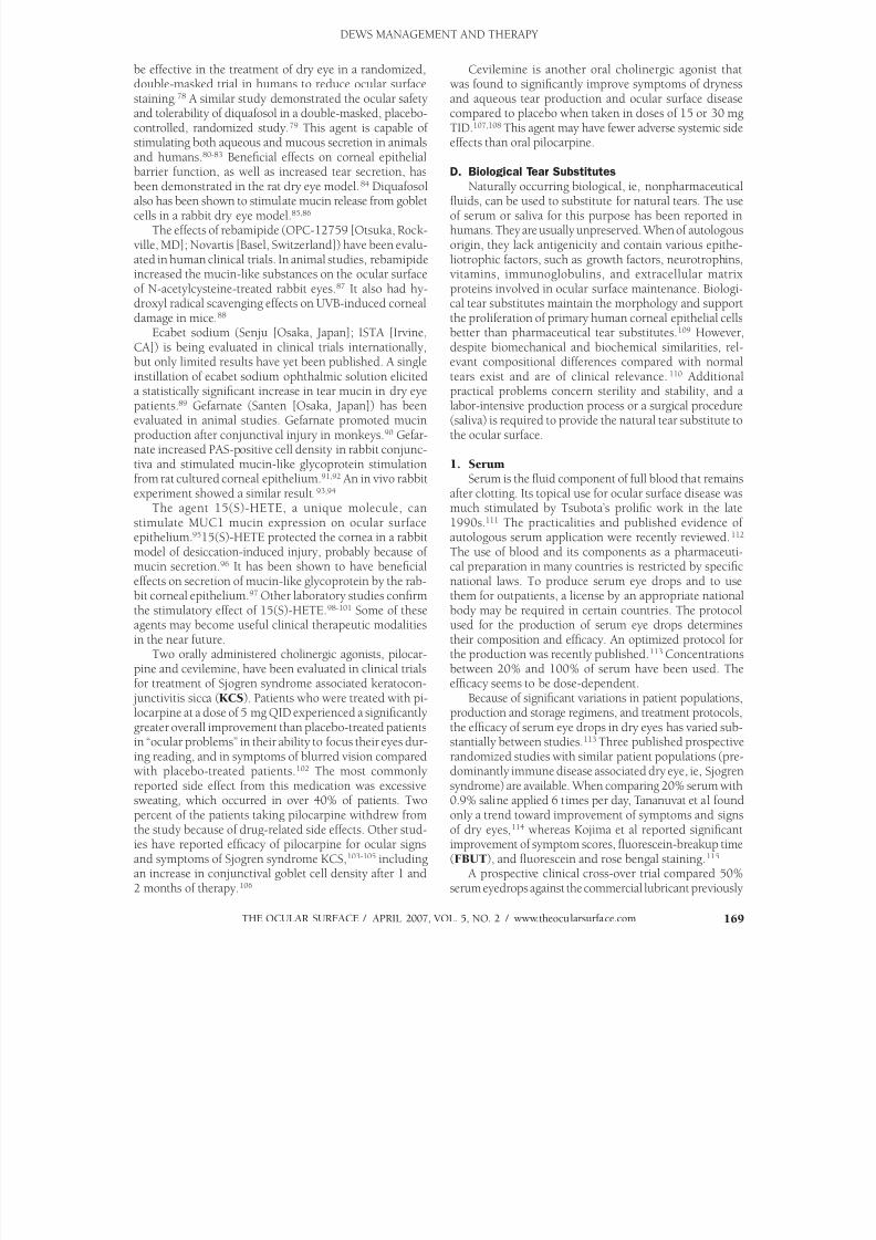
8/20/2019 dry eyes ii
http://slidepdf.com/reader/full/dry-eyes-ii 109/142
THE OCULAR SURFACE / APRIL 2007, VOL. 5, NO. 2 / www.theocularsurface.com 169
be effective in the treatment of dry eye in a randomized,double-masked trial in humans to reduce ocular surfacestaining.78 A similar study demonstrated the ocular safetyand tolerability of diquafosol in a double-masked, placebo-controlled, randomized study.79 This agent is capable ofstimulating both aqueous and mucous secretion in animalsand humans.80-83 Beneficial effects on corneal epithelial
barrier function, as well as increased tear secretion, hasbeen demonstrated in the rat dry eye model.84 Diquafosolalso has been shown to stimulate mucin release from gobletcells in a rabbit dry eye model.85,86
The effects of rebamipide (OPC-12759 [Otsuka, Rock-ville, MD]; Novartis [Basel, Switzerland]) have been evalu-ated in human clinical trials. In animal studies, rebamipideincreased the mucin-like substances on the ocular surfaceof N-acetylcysteine-treated rabbit eyes.87 It also had hy-droxyl radical scavenging effects on UVB-induced cornealdamage in mice.88
Ecabet sodium (Senju [Osaka, Japan]; ISTA [Irvine,CA]) is being evaluated in clinical trials internationally,but only limited results have yet been published. A singleinstillation of ecabet sodium ophthalmic solution eliciteda statistically significant increase in tear mucin in dry eyepatients.89 Gefarnate (Santen [Osaka, Japan]) has beenevaluated in animal studies. Gefarnate promoted mucinproduction after conjunctival injury in monkeys.90 Gefar-nate increased PAS-positive cell density in rabbit conjunc-tiva and stimulated mucin-like glycoprotein stimulationfrom rat cultured corneal epithelium.91,92 An in vivo rabbitexperiment showed a similar result.93,94
The agent 15(S)-HETE, a unique molecule, canstimulate MUC1 mucin expression on ocular surface
epithelium.9515(S)-HETE protected the cornea in a rabbitmodel of desiccation-induced injury, probably because ofmucin secretion.96 It has been shown to have beneficialeffects on secretion of mucin-like glycoprotein by the rab-bit corneal epithelium.97 Other laboratory studies confirmthe stimulatory effect of 15(S)-HETE.98-101 Some of theseagents may become useful clinical therapeutic modalitiesin the near future.
Two orally administered cholinergic agonists, pilocar-pine and cevilemine, have been evaluated in clinical trialsfor treatment of Sjogren syndrome associated keratocon- junctivitis sicca (KCS). Patients who were treated with pi-
locarpine at a dose of 5 mg QID experienced a significantlygreater overall improvement than placebo-treated patientsin “ocular problems” in their ability to focus their eyes dur-ing reading, and in symptoms of blurred vision comparedwith placebo-treated patients.102 The most commonlyreported side effect from this medication was excessivesweating, which occurred in over 40% of patients. Twopercent of the patients taking pilocarpine withdrew fromthe study because of drug-related side effects. Other stud-ies have reported efficacy of pilocarpine for ocular signsand symptoms of Sjogren syndrome KCS,103-105 includingan increase in conjunctival goblet cell density after 1 and
2 months of therapy.106
Cevilemine is another oral cholinergic agonist thatwas found to significantly improve symptoms of drynessand aqueous tear production and ocular surface diseasecompared to placebo when taken in doses of 15 or 30 mgTID.107,108 This agent may have fewer adverse systemic sideeffects than oral pilocarpine.
D. Biological Tear Substitutes
Naturally occurring biological, ie, nonpharmaceuticalfluids, can be used to substitute for natural tears. The useof serum or saliva for this purpose has been reported inhumans. They are usually unpreserved. When of autologousorigin, they lack antigenicity and contain various epithe-liotrophic factors, such as growth factors, neurotrophins,vitamins, immunoglobulins, and extracellular matrixproteins involved in ocular surface maintenance. Biologi-cal tear substitutes maintain the morphology and supportthe proliferation of primary human corneal epithelial cellsbetter than pharmaceutical tear substitutes.109 However,despite biomechanical and biochemical similarities, rel-evant compositional differences compared with normaltears exist and are of clinical relevance.110 Additionalpractical problems concern sterility and stability, and alabor-intensive production process or a surgical procedure(saliva) is required to provide the natural tear substitute tothe ocular surface.
1. SerumSerum is the fluid component of full blood that remains
after clotting. Its topical use for ocular surface disease wasmuch stimulated by Tsubota’s prolific work in the late1990s.111 The practicalities and published evidence of
autologous serum application were recently reviewed.112 The use of blood and its components as a pharmaceuti-cal preparation in many countries is restricted by specificnational laws. To produce serum eye drops and to usethem for outpatients, a license by an appropriate nationalbody may be required in certain countries. The protocolused for the production of serum eye drops determinestheir composition and efficacy. An optimized protocol forthe production was recently published.113 Concentrationsbetween 20% and 100% of serum have been used. Theefficacy seems to be dose-dependent.
Because of significant variations in patient populations,
production and storage regimens, and treatment protocols,the efficacy of serum eye drops in dry eyes has varied sub-stantially between studies.113 Three published prospectiverandomized studies with similar patient populations (pre-dominantly immune disease associated dry eye, ie, Sjogrensyndrome) are available. When comparing 20% serum with0.9% saline applied 6 times per day, Tananuvat et al foundonly a trend toward improvement of symptoms and signsof dry eyes,114 whereas Kojima et al reported significantimprovement of symptom scores, fluorescein-breakup time(FBUT), and fluorescein and rose bengal staining.115
A prospective clinical cross-over trial compared 50%
serum eyedrops against the commercial lubricant previously
DEWS MANAGEMENT AND THERAPY

8/20/2019 dry eyes ii
http://slidepdf.com/reader/full/dry-eyes-ii 110/142
THE OCULAR SURFACE / APRIL 2007, VOL. 5, NO. 2 / www.theocularsurface.com170
used by each patient. Symptoms improved in 10 out 16patients, and impression cytological findings improved in12 out of 25 eyes.116 Noda-Tsuruya and colleagues foundthat 20% autologous serum significantly improved TFBUTand decreased conjunctival rose bengal and cornea fluo-rescein staining 1-3 months postoperatively, compared totreatment with artificial tears, which did not change these
parameters.117
Additional reports of successful treatmentof persistent epithelial defects—where success is moreclearly defined as “healing of the defect”—with autologousserum substantiate the impression that this is a valuabletherapeutic option for ocular surface disease.118
2. Salivary Gland AutotransplantationSalivary submandibular gland transplantation is capable
of replacing deficient mucin and the aqueous tear filmphase. This procedure requires collaboration between anophthalmologist and a maxillofacial surgeon. With appro-priate microvascular anastomosis, 80% of grafts survive.In patients with absolute aqueous tear deficiency, viablesubmandibular gland grafts, in the long-term, providesignificant improvement of Schirmer test FBUT, and rosebengal staining, as well as reduction of discomfort and theneed for pharmaceutical tear substitutes. Due to the hypo-osmolarity of saliva, compared to tears, excessive salivarytearing can induce a microcystic corneal edema, which istemporary, but can lead to epithelial defects.110 Hence, thisoperation is indicated only in end-stage dry eye disease withan absolute aqueous tear deficiency (Schirmer-test wettingof 1 mm or less), a conjunctivalized surface epithelium, andpersistent severe pain despite punctal occlusion and at leasthourly application of unpreserved tear substitutes. For this
group of patients, such surgery is capable of substantiallyreducing discomfort, but often has no effect on vision.119,120
E. Anti-Inflammatory Therapy
Disease or dysfunction of the tear secretory glands leadsto changes in tear composition, such as hyperosmolarity,that stimulate the production of inflammatory mediators onthe ocular surface.31,121 Inflammation may, in turn, causedysfunction or disappearance of cells responsible for tearsecretion or retention.122 Inflammation can also be initiatedby chronic irritative stress (eg, contact lenses) and systemicinflammatory/autoimmune disease (eg, rheumatoid arthri-
tis). Regardless of the initiating cause, a vicious circle ofinflammation can develop on the ocular surface in dry eyethat leads to ocular surface disease. Based on the conceptthat inflammation is a key component of the pathogenesisof dry eye, the efficacy of a number of anti-inflammatoryagents for treatment of dry eye disease has been evaluatedin clinical trials and animal models.
1. CyclosporineThe potential of cyclosporine-A (CsA) for treating dry
eye disease was initially recognized in dogs that developspontaneous KCS.123 The therapeutic efficacy of CsA for
human KCS was then documented in several small, single-
center, randomized, double-masked clinical trials.124,125 CsA emulsion for treatment of KCS was subsequentlyevaluated in several large multicenter, randomized, double-masked clinical trials.
In a Phase 2 clinical trial, four concentrations of CsA(0.05%, 0.1%, 0.2%, or 0.4%) administered twice dailyto both eyes of 129 patients for 12 weeks was compared
to vehicle treatment of 33 patients.126
CsA was found tosignificantly decrease conjunctival rose bengal staining,superficial punctate keratitis, and ocular irritation symp-toms (sandy or gritty feeling, dryness, and itching) in asubset of 90 patients with moderate-to-severe KCS. Therewas no clear dose response; CsA 0.1% produced the mostconsistent improvement in objective endpoints, whereasCsA 0.05% gave the most consistent improvement in pa-tient symptoms (Level I).
Two independent Phase 3 clinical trials comparedtwice-daily treatment with 0.05% or 0.1% CsA or vehiclein 877 patients with moderate-to-severe dry eye disease.127 When the results of the two Phase 3 trials were combinedfor statistical analysis, patients treated with CsA, 0.05% or0.1%, showed significantly (P < 0.05) greater improvementin two objective signs of dry eye disease (corneal fluoresceinstaining and anesthetized Schirmer test values) compared tothose treated with vehicle. An increased Schirmer test scorewas observed in 59% of patients treated with CsA, with15% of patients having an increase of 10 mm or more. Incontrast, only 4% of vehicle-treated patients had this mag-nitude of change in their Schirmer test scores (P < 0.0001).
CsA 0.05% treatment also produced significantly greaterimprovements (P < 0.05) in three subjective measures of dryeye disease (blurred vision symptoms, need for concomitant
artificial tears, and the global response to treatment). Nodose-response effect was noted. Both doses of CSA exhib-ited an excellent safety profile with no significant systemicor ocular adverse events, except for transient burningsymptoms after instillation in 17% of patients. Burning wasreported in 7% of patients receiving the vehicle. No CsA wasdetected in the blood of patients treated with topical CsAfor 12 months. Clinical improvement from CsA that wasobserved in these trials was accompanied by improvementin other disease parameters. Treated eyes had an approxi-mately 200% increase in conjunctival goblet cell density.128 Furthermore, there was decreased expression of immune
activation markers (ie, HLA-DR), apoptosis markers (ie,Fas), and the inflammatory cytokine IL-6 by the conjunc-tival epithelial cells.129,130The numbers of CD3-, CD4-, andCD8-positive T lymphocytes in the conjunctiva decreasedin cyclosporine-treated eyes, whereas vehicle-treated eyesshowed an increased number of cells expressing thesemarkers.131 After treatment with 0.05% cyclosporine, therewas a significant decrease in the number of cells expressingthe lymphocyte activation markers CD11a and HLA-DR,indicating less activation of lymphocytes compared withvehicle-treated eyes.
Two additional immunophilins, pimecrolimus and ta-
crolimus, have been evaluated in clinical trials of KCS.
DEWS MANAGEMENT AND THERAPY

8/20/2019 dry eyes ii
http://slidepdf.com/reader/full/dry-eyes-ii 111/142
THE OCULAR SURFACE / APRIL 2007, VOL. 5, NO. 2 / www.theocularsurface.com 171
2. Corticosteroidsa. Clinical Studies
Corticosteroids are an effective anti-inflammatorytherapy in dry eye disease. Level I evidence is publishedfor a number of corticosteroid formulations. In a 4-week,double-masked, randomized study in 64 patients withKCS and delayed tear clearance, loteprednol etabonate
0.5% ophthalmic suspension (Lotemax [Bausch and Lomb,Rochester, NY]), q.i.d., was found to be more effective thanits vehicle in improving some signs and symptoms.132
In a 4-week, open-label, randomized study in 32 pa-tients with KCS, patients receiving fluorometholone plusartificial tear substitutes ( ATS) experienced lower symptomseverity scores and lower fluorescein and rose bengal stain-ing than patients receiving either ATS alone or ATS plusflurbiprofen.133
A prospective, randomized clinical trial compared theseverity of ocular irritation symptoms and corneal fluores-cein staining in two groups of patients, one treated withtopical nonpreserved methylprednisolone for 2 weeks,followed by punctal occlusion (Group 1), with a groupthat received punctal occlusion alone (Group 2).134 After 2months, 80% of patients in Group 1 and 33% of patients inGroup 2 had complete relief of ocular irritation symptoms.Corneal fluorescein staining was negative in 80% of eyes inGroup 1 and 60% of eyes in Group 2 after 2 months. Nosteroid-related complications were observed in this study.
Level III evidence is also available to support the efficacyof corticosteroids. In an open-label, non-comparative trial,extemporaneously formulated nonpreserved methylpred-nisolone 1% ophthalmic suspension was found to be clini-cally effective in 21 patients with Sjogren syndrome KCS.135
In a review, it was stated that “…clinical improvement ofKCS has been observed after therapy with anti-inflamma-tory agents, including corticosteroids.”136
In the US Federal Regulations, ocular corticosteroidsreceiving “class labeling” are indicated for the treatment“…of steroid responsive inflammatory conditions of thepalpebral and bulbar conjunctiva, cornea and anteriorsegment of the globe such as allergic conjunctivitis, acnerosacea, superficial punctate keratitis, herpes zoster kerati-tis, iritis, cyclitis, selected infective conjunctivitides, whenthe inherent hazard of steroid use is accepted to obtain anadvisable diminution in edema and inflammation.” We in-
terpret that KCS is included in this list of steroid-responsiveinflammatory conditions.137-140
b. Basic ResearchCorticosteroids are the standard anti-inflammatory
agent for numerous basic research studies of inflamma-tion, including the types that are involved in KCS. Thecorticosteroid methylprednisolone was noted to preservecorneal epithelial smoothness and barrier function in anexperimental murine model of dry eye.141 This was at-tributed to its ability to maintain the integrity of cornealepithelial tight junctions and decrease desquamation of
apical corneal epithelial cells.142 A concurrent study showed
that methylprednislone prevented an increase in MMP-9protein in the corneal epithelium, as well as gelatinaseactivity in the corneal epithelium and tears in response toexperimental dry eye.141
Preparations of topically applied androgen and es-trogen steroid hormones are currently being evaluatedin randomized clinical trials. A trial of topically applied
0.03% testosterone was reported to increase the percent-age of patients that had meibomian gland secretions withnormal viscosity and to relieve discomfort symptoms after6 months of treatment compared to vehicle.143 TFBUT andlipid layer thickness were observed to increase in a patientwith KCS who was treated with topical androgen for 3months.144 Tear production and ocular irritation symptomswere reported to increase following treatment with topical17 beta-oestradiol solution for 4 months.145
3. Tetracyclinesa. Properties of Tetracyclines and Their Derivatives1) Antibacterial Properties
The antimicrobial effect of oral tetracycline treatmentanalogues (eg, minocycline, doxycline) has previously beendiscussed by Shine et al,146 Dougherty et al,147 and Ta etal.148 It is hypothesized that a decrease in bacterial flora pro-ducing lipolytic exoenzymes146,148 and inhibition of lipaseproduction147 with resultant decrease in meibomian lipidbreakdown products146 may contribute to improvement inclinical parameters in dry eye-associated diseases.
2) Anti-Inflammatory PropertiesThe tetracyclines have anti-inflammatory as well as
antibacterial properties that may make them useful for
the management of chronic inflammatory diseases. Theseagents decrease the activity of collagenase, phospholipase A2, and several matrix metalloproteinases, and they de-crease the production of interleukin (IL)-1 and tumornecrosis factor (TNF)-alpha in a wide range of tissues,including the corneal epithelium.149-151 At high concentra-tions, tetracyclines inhibit staphylococcal exotoxin-inducedcytokines and chemokines.152,153
3) Anti-angiogenic Properties Angiogenesis, the formation of new blood vessels, oc-
curs in many diseases. These include benign conditions (eg,
rosacea) and malignant processes (eg, cancer). Minocyclineand doxycycline inhibit angiogenesis induced by implantedtumors in rabbit cornea.154 The anti-angiogenic effect oftetracycline may have therapeutic implications in inflamma-tory processes accompanied by new blood vessel formation. Well-controlled studies must be performed, at both thelaboratory and clinical levels, to investigate this potential.155
b. Clinical Applications of Tetracycline 1) Acne Rosacea
Rosacea, including its ocular manifestations, is an in-flammatory disorder, occurring mainly in adults, with peak
severity in the third and fourth decades. Current recom-
DEWS MANAGEMENT AND THERAPY

8/20/2019 dry eyes ii
http://slidepdf.com/reader/full/dry-eyes-ii 112/142
THE OCULAR SURFACE / APRIL 2007, VOL. 5, NO. 2 / www.theocularsurface.com172
mendations are to treat rosacea with long-term doxycycline,minocycline, tetracycline, or erythromycin.156 These recom-mendations may be tempered by certain recent reports thatin women, the risk of developing breast cancer and of breastcancer morbidity increases cumulatively with duration ofantibiotic use, including tetracyclines.157,158 Another largestudy did not substantiate these findings.159
Tetracyclines and their analogues are effective in thetreatment of ocular rosacea,160,161 for which a single dailydose of doxycycline may be effective.162 In addition to theanti-inflammatory effects of tetracyclines, their ability toinhibit angiogenesis may contribute to their effectiveness inrosacea-related disorders. Factors that promote angiogen-esis include protease-triggered release of angiogenic factorsstored in the extracellular matrix, inactivation of endothelialgrowth factor inhibitors, and release of angiogenic factorsfrom activated macrophages.155,163
Tetracyclines are also known to inhibit matrix metal-loproteinase expression, suggesting a rationale for their usein ocular rosacea.164 Although tetracyclines have been usedfor management of this disease, no randomized, placebo-controlled, clinical trials have been performed to assesstheir efficacy.153
2) Chronic Posterior Blepharitis: Meibomianitis,Meibomian Gland DysfunctionChronic blepharitis is typically characterized by inflam-
mation of the eyelids. There are multiple forms of chronicblepharitis, including staphylococcal, seborrheic (alone,mixed seborrheic/staphylococcal, seborrheic with meibo-mian seborrhea, seborrheic with secondary meibomitis),primary meibomitis, and others, like atopic, psoriatic, and
fungal infections.165 Meibomian gland dysfunction (MGD)has been associated with apparent aqueous-deficient dryeye. Use of tetracycline in patients with meibomianitis hasbeen shown to decrease lipase production by tetracycline-sensitive as well as resistant strains of staphylococci. Thisdecrease in lipase production was associated with clinicalimprovement.147 Similarly, minocycline has been shown todecrease the production of diglycerides and free fatty acids inmeibomian secretions. This may be due to lipase inhibitionby the antibiotic or a direct effect on the ocular flora.146 Onerandomized, controlled clinical trial of tetracycline in ocularrosacea compared symptom improvement in 24 patients
treated with either tetracycline or doxycycline.166 All but onepatient reported an improvement in symptoms after 6 weeksof therapy. No placebo group was included in this trial.
A prospective, randomized, double-blind, placebo-controlled, partial crossover trial compared the effect ofoxytetracycline to provide symptomatic relief of blepharitiswith or without rosacea. Only 25% of the patients withblepharitis without rosacea responded to the antibiotic,whereas 50% responded when both diseases were pres-ent.167 In another trial of 10 patients with both acne rosa-cea and concomitant meibomianitis, acne rosacea withoutconcomitant ocular involvement, or seborrheic blepharitis,
minocycline 50 mg daily for 2 weeks followed by 100 mg
daily for a total of 3 months significantly decreased bacte-rial flora (P = 0.0013). Clinical improvement was seen inall patients with meibomianitis.148
Because of the improvement observed in small clinicaltrials of patients with meibomianitis, the American Acad-emy of Ophthalmology recommends the chronic use ofeither doxycycline or tetracycline for the management of
meibomianitis.165
Larger randomized placebo-controlledtrials assessing symptom improvement rather than surro-gate markers are needed to clarify the role of this antibioticin blepharitis treatment.153 Tetracycline derivatives (eg,minocycline, doxycycline) have been recommended astreatment options for chronic blepharitis because of theirhigh concentration in tissues, low renal clearance, long half-life, high level of binding to serum proteins, and decreasedrisk of photosensitization.168
Several studies have described the beneficial effects ofminocycline and other tetracycline derivatives (eg, doxy-cycline) in the treatment of chronic blepharitis.146,147,168,169 Studies have shown significant changes in the aqueous tearparameters, such as tear volume and tear flow, followingtreatment with tetracycline derivatives (eg, minocycline).One study also demonstrated a decrease in aqueous tear pro-duction that occurred along with clinical improvement.170
A recently published randomized, prospective studyby Yoo Se et al compared different doxycycline doses in150 patients (300 eyes) who had chronic meibomian glanddysfunction and who did not respond to lid hygiene andtopical therapy for more than 2 months.171 All topicaltherapy was stopped for at least 2 weeks prior to begin-ning the study. After determining the TFBUT and Schirmertest scores, patients were divided into three groups: a high
dose group (doxycycline, 200 mg, twice a day), a low dosegroup (doxycycline, 20 mg, twice a day) and a control group(placebo). After one month, TFBUT, Schirmer scores, andsymptoms improved. Both the high- and low-dose groupshad statistically significant improvement in TFBUT aftertreatment. This implies that low-dose doxycycline (20mg twice a day) therapy may be effective in patients withchronic meibomian gland dysfunction.
3) Dosage and SafetySystemic administration of tetracyclines is widely recog-
nized for the ability to suppress inflammation and improve
symptoms of meibomianitis.172,173 The optimal dosingschedule has not been established; however, a variety ofdose regimens have been proposed including 50 or 100 mgdoxycycline once a day,174 or an initial dose of 50 mg a dayfor the first 2 weeks followed by 100 mg a day for a periodof 2.5 months, in an intermittent fashion.146-148,170 Othershave proposed use of a low dose of doxycycline (20 mg)for treatment of chronic blepharitis on a long-term basis.171 The safety issues associated with long-term oral tetracyclinetherapy, including minocycline, are well known. Manymanagement approaches have been suggested for the use oftetracycline and its derivatives; however, a safe but adequate
option in management needs to be considered because of
DEWS MANAGEMENT AND THERAPY

8/20/2019 dry eyes ii
http://slidepdf.com/reader/full/dry-eyes-ii 113/142
THE OCULAR SURFACE / APRIL 2007, VOL. 5, NO. 2 / www.theocularsurface.com 173
the new information regarding the potentially hazardouseffects of prolonged use of oral antibiotics. A recent studysuggested that a 3-month course of 100 mg of minocyclinemight be sufficient to bring significant meibomianitis under
control, as continued control was maintained for at least 3months after cessation of therapy.170
In an experimental murine model of dry eye, topicallyapplied doxycycline was found to preserve corneal epithe-lial smoothness and barrier function.141 It also preservedthe integrity of corneal epithelial tight junctions in dry eyes,leading to a marked decrease in apical corneal epithelial celldesquamation.142 This corresponded to a decrease in MMP-9 protein in the corneal epithelium and reduced gelatinaseactivity in the corneal epithelium and tears.141
F. Essential Fatty Acids
Essential fatty acids are necessary for complete health.They cannot be synthesized by vertebrates and must beobtained from dietary sources. Among the essential fattyacids are 18 carbon omega-6 and omega-3 fatty acids. Inthe typical western diet, 20-25 times more omega-6 thanomega-3 fatty acids are consumed. Omega-6 fatty acids areprecursors for arachidonic acid and certain proinflamma-tory lipid mediators (PGE2 and LTB4). In contrast, certainomega-3 fatty acids (eg, EPA found in fish oil) inhibit thesynthesis of these lipid mediators and block production ofIL-1 and TNF-alpha.175,176
A beneficial clinical effect of fish oil omega-3 fatty ac-
ids on rheumatoid arthritis has been observed in several
double-masked, placebo-controlled clinical trials.177,178 In aprospective, placebo-controlled clinical trial of the essentialfatty acids, linoleic acid and gamma-linolenic acid adminis-tered orally twice daily produced significant improvement
in ocular irritation symptoms and ocular surface lissaminegreen staining.179 Decreased conjunctival HLA-DR stainingalso was observed.
G. Environmental Strategies
Factors that may decrease tear production or increasetear evaporation, such as the use of systemic anticholiner-gic medications (eg, antihistamines and antidepressants)and desiccating environmental stresses (eg, low humid-ity and air conditioning drafts) should be minimizedor eliminated.180-182 Video display terminals should belowered below eye level to decrease the interpalpebral
aperture, and patients should be encouraged to take pe-riodic breaks with eye closure when reading or workingon a computer.183 A humidified environment is recom-mended to reduce tear evaporation. This is particularlybeneficial in dry climates and high altitudes. Nocturnallagophthalmos can be treated by wearing swim goggles,taping the eyelid closed, or tarsorrhapy.
IV. TREATMENT RECOMMENDATIONS
In addition to material presented above, the subcom-mittee members reviewed the Dry Eye Preferred PracticePatterns of the American Academy of Ophthalmology and
the International Task Force (ITF) Delphi Panel on dry
Table 2. Dry eye severity grading scheme
Dry Eye SeverityLevel 1 2 3 4*
Discomfort, severity
& frequency
Mild and/or episodic
occurs under environ
stress
Moderate episodic or
chronic, stress or no
stress
Severe frequent or
constant without
stress
Severe and/or
disabling and constant
Visual symptomsNone or episodic mild
fatigue
Annoying and/or activity
limiting episodic
Annoying, chronic and/
or constant limiting
activity
Constant and/or
possibly disabling
Conjunctival injection None to mild None to mild +/– +/++
Conjunctival staining None to mild Variable Moderate to marked Marked
Corneal staining
(severity/location)None to mild Variable Marked central
Severe punctate
erosions
Corneal/tear signs None to mild Mild debris, ↓ meniscus
Filamentary keratitis,
mucus clumping,
↑ tear debris
Filamentary keratitis,
mucus clumping,
↑ tear debris, ulceration
Lid/meibomian glands MGD variably present MGD variably present FrequentTrichiasis, keratinization,
symblepharon
TFBUT (sec) Variable ≤ 10 ≤ 5 Immediate
Schirmer score
(mm/5 min)Variable ≤ 10 ≤ 5 ≤ 2
*Must have signs AND symptoms. TBUT: fluorescein tear break-up time. MGD: meibomian gland disease
Reprinted with permission from Behrens A, Doyle JJ, Stern L, et al. Dysfunctional tear syndrome. A Delphi approach to treatment recommendations.Cornea 2006;25:90-7
DEWS MANAGEMENT AND THERAPY
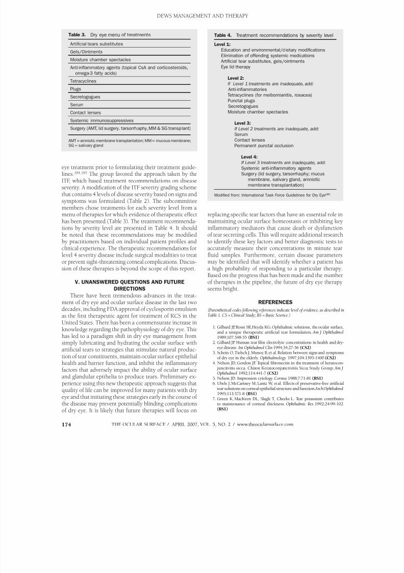
8/20/2019 dry eyes ii
http://slidepdf.com/reader/full/dry-eyes-ii 114/142
THE OCULAR SURFACE / APRIL 2007, VOL. 5, NO. 2 / www.theocularsurface.com174
eye treatment prior to formulating their treatment guide-lines.184,185 The group favored the approach taken by theITF, which based treatment recommendations on diseaseseverity. A modification of the ITF severity grading schemethat contains 4 levels of disease severity based on signs andsymptoms was formulated (Table 2). The subcommitteemembers chose treatments for each severity level from amenu of therapies for which evidence of therapeutic effecthas been presented (Table 3). The treatment recommenda-tions by severity level are presented in Table 4. It shouldbe noted that these recommendations may be modifiedby practitioners based on individual patient profiles and
clinical experience. The therapeutic recommendations forlevel 4 severity disease include surgical modalities to treator prevent sight-threatening corneal complications. Discus-sion of these therapies is beyond the scope of this report.
V. UNANSWERED QUESTIONS AND FUTURE
DIRECTIONS
There have been tremendous advances in the treat-ment of dry eye and ocular surface disease in the last twodecades, including FDA approval of cyclosporin emulsionas the first therapeutic agent for treatment of KCS in theUnited States. There has been a commensurate increase in
knowledge regarding the pathophysiology of dry eye. Thishas led to a paradigm shift in dry eye management fromsimply lubricating and hydrating the ocular surface withartificial tears to strategies that stimulate natural produc-tion of tear constituents, maintain ocular surface epithelialhealth and barrier function, and inhibit the inflammatoryfactors that adversely impact the ability of ocular surfaceand glandular epithelia to produce tears. Preliminary ex-perience using this new therapeutic approach suggests thatquality of life can be improved for many patients with dryeye and that initiating these strategies early in the course ofthe disease may prevent potentially blinding complications
of dry eye. It is likely that future therapies will focus on
replacing specific tear factors that have an essential role inmaintaining ocular surface homeostasis or inhibiting keyinflammatory mediators that cause death or dysfunctionof tear secreting cells. This will require additional researchto identify these key factors and better diagnostic tests to
accurately measure their concentrations in minute tearfluid samples. Furthermore, certain disease parametersmay be identified that will identify whether a patient hasa high probability of responding to a particular therapy.Based on the progress that has been made and the numberof therapies in the pipeline, the future of dry eye therapyseems bright.
REFERENCES
(Parenthetical codes following references indicate level of evidence, as described inTable 1. CS = Clinical Study; BS = Basic Science.)
1. Gilbard JP, Rossi SR, Heyda KG. Ophthalmic solutions, the ocular surface,and a unique therapeutic artificial tear formulation. Am J Ophthalmol 1989;107:348-55 (BS1)
2. Gilbard JP. Human tear film electrolyte concentrations in health and dry-eye disease. Int Ophthalmol Clin 1994;34:27-36 (CS2)
3. Schein O, Tielsch J, Munoz B, et al. Relation between signs and symptomsof dry eye in the elderly. Ophthalmology 1997;104:1395-1400 (CS2)
4. Nelson JD, Gordon JF. Topical fibronectin in the treatment of keratocon- junctivitis sicca. Chiron Keratoconjunctivitis Sicca Study Group. Am JOphthalmol 1992;114:441-7 (CS2)
5. Nelson JD. Impression cytology. Cornea 1988;7:71-81 (BS1) 6. Ubels J, McCartney M, Lantz W, et al. Effects of preservative-free artificial
tear solutions on corneal epithelial structure and function. Arch Ophthalmol 1995;113:371-8 (BS1)
7. Green K, MacKeen DL, Slagle T, Cheeks L. Tear potassium contributesto maintenance of corneal thickness. Ophthalmic Res 1992;24:99-102
(BS1)
Table 4. Treatment recommendations by severity level
Level 1:
Education and environmental/dietary modifications
Elimination of offending systemic medications
Artificial tear substitutes, gels/ointments
Eye lid therapy
Level 2:
If Level 1 treatments are inadequate, add:
Anti-inflammatories
Tetracyclines (for meibomianitis, rosacea)
Punctal plugs
Secretogogues
Moisture chamber spectacles
Level 3:
If Level 2 treatments are inadequate, add:
Serum
Contact lenses
Permanent punctal occlusion
Level 4:
If Level 3 treatments are inadequate, add:Systemic anti-inflammatory agents
Surgery (lid surgery, tarsorrhaphy; mucus
membrane, salivary gland, amniotic
membrane transplantation)
Modified from: International Task Force Guidelines for Dry Eye185
Table 3. Dry eye menu of treatments
Artificial tears substitutes
Gels/Ointments
Moisture chamber spectacles
Anti-inflammatory agents (topical CsA and corticosteroids,
omega-3 fatty acids)
TetracyclinesPlugs
Secretogogues
Serum
Contact lenses
Systemic immunosuppressives
Surgery (AMT, lid surgery. tarsorrhaphy, MM & SG transplant)
AMT = amniotic membrane transplantation; MM = mucous membrane;SG = salivary gland
DEWS MANAGEMENT AND THERAPY
8/20/2019 dry eyes ii
http://slidepdf.com/reader/full/dry-eyes-ii 115/142
THE OCULAR SURFACE / APRIL 2007, VOL. 5, NO. 2 / www.theocularsurface.com 175
8. Holly F, Lemp M. Surface chemistry of the tear film: Implications for dryeye syndromes, contact lenses, and ophthalmic polymers. Contact LensSoc Am J 1971;5:12-9 (BS2)
9. Perrigan DM, Morgan A, Quintero S, et al. Comparison of osmolarityvalues of selected ocular lubricants. ARVO 2004 poster session 449
10. Kaufman B, Novack GD. Compliance issues in manufacturing of drugs.Ocul Surf 2003;1:80-5
11. Albietz J, Bruce A. The conjunctival epithelium in dry eye subtypes:Effect of preserved and nonpreserved topical treatments. Curr Eye Res 2001;22:8-18 (CS2)
12. Gasset AR, Ishii Y, Kaufman H, Miller T. Cytotoxicity of ophthalmicpreservatives. Am J Ophthalmol 1974;78:98-105 (BS1)
13. Wilson F. Adverse external effects of topical ophthalmic medications. SurvOphthalmol 1979;24:57-88 (CS3)
14. Burstein N. Corneal cytotoxicity of topically applied drugs, vehicles andpreservatives.Surv Ophthalmol 1980;25:15-30 (CS3)
15. Burstein N. The effects of topical drugs and preservatives on the tears andcorneal epithelium in dry eye. Trans Ophthalmol Soc UK. 1985;104:402-9(CS3)
16. Brubaker R, McLaren J. Uses of the fluorophotometer in glaucoma research.Ophthalmology 1985;92:884-90 (BS1)
17. Smith L, George M, Berdy G, Abelson M. Comparative effects of pre-servative free tear substitutes on the rabbit cornea: a scanning electronmicroscopic evaluation ( ARVO abstract). Invest Ophthalmol Vis Sci 1991;32(Suppl):733 (BS1)
18. Gilbard JP, Farris RL, Santamaria J 2nd. Osmolarity of tear microvolumes
in keratoconjunctivitis sicca. Arch Ophthalmol 1978;96:677-81 (BS2) 19. Lopez Bernal D, Ubels JL. Quantitative evaluation of the corneal epithelial
barrier: effect of artificial tears and preservatives. Curr Eye Res1991;10:645-56 (BS1)
20. Bernal DL, Ubels JL. Artificial tear composition and promotion of recoveryof the damaged corneal epithelium. Cornea 1993;12:115-20 (BS1)
21. Noecker R: Effects of common ophthalmic preservatives on ocular health. Adv Ther 2001;18:205-15 (CS1)
22. Tripathi BJ, Tripathi RC, Kolli SP: Cytotoxicity of ophthalmic preserva-tives on human corneal epithelium. Lens Eye Toxicity Res 1992;9:361-75(BS1)
23. Herrema J, Friedenwald J. Retardation of wound healing in the cornealepithelium by lanolin. Am J Ophthalmol 1950;33:1421(CS3)
24. Nelson J, Drake M, Brewer J, Tuley M. Evaluation of physiologic tearsubstitute in patients with keratoconjunctivitis sicca. Adv Exp Med Biol1994;350:453-7 (CS2)
25. Gilbard JP, Rossi SR. An electrolyte-based solution that increases cornealglycogen and conjunctival goblet-cell density in a rabbit model for kera-toconjunctivitis sicca. Ophthalmology 1992;99:600-4 (BS1)
26. Lenton LM, Albietz JM: Effect of carmellose-based artificial tears on theocular surface in eyes after laser in situ keratomileusis. J Refract Surg1999;15(2 Suppl):S227-S231 (CS2)
27. Slomiany BL, Slomiany A. Role of mucus in gastric mucosal protection. J Physiol Pharmacol 1991; 42:147-61 (BS1)
28. Gilbard JP. Tear film osmolarity and keratoconjunctivitis sicca. CLAO J1985;11:243-50 (CS1)
29. Gilbard J. Tear film osmolarity and keratoconjuncitivitis sicca. LubbockTX,Dry Eye Institute, 1986 (CS3)
30. Gilbard J, Carter J, Sang D, et al. Morphologic effect of hyperosmolarityon rabbit corneal epithelium. Ophthalmology 1984;91:1205-12 (BS1)
31. Luo L, Li D, Corrales R, Pflugfelder S. Hyperosmolar saline is a proinflam-matory stress on the mouse ocular surface. Eye Contact Lens 2005;31:186-
93 (BS1) 32. Gilbard JP, Kenyon KR. Tear diluents in the treatment of keratoconjunc-
tivitis sicca. Ophthalmology 1985;92:646-50 (CS2) 33. Holly F, Esquivel E. Colloid osmotic pressure of artificial tears. J Ocul
Pharmacol 1985;1:327-36 (BS1) 34. Yancey PH: Organic osmolytes as compatible, metabolic and counter-
acting cryoprotectants in high osmolarity and other stresses .J Exp Biol2005;208:2819-30 (BS2)
35. Holly F, Lemp M. Wettability and wetting of corneal epithelium. Exp EyeRes 1971;11:239-50 (BS1)
36. Hawi A, Smith T, Digenis G. A quantitiative comparison of artificial tearclearance rates in humans using gamma scintigraphy (ARVO abstract).Invest Ophthalmol Vis Sci 1990;31 (Suppl):517 (BS1)
37. Argueso P, Tisdale A, Spurr-Michaud S, et al. Mucin characteristics of hu-man corneal-limbal epithelial cells that exclude the rose bengal anionicdye. Invest Ophthalmol Vis Sci 2006;47:113-9 (BS1)
38. Versura P, Maltarello M, Stecher F, et al. Dry eye before and after therapy
with hydroxypropylmethylcellulose. Ophthalmologica 1989;198:152-62(CS3)
39. Simmons PA, Garrett Q, Xu S, et al. Interaction of carboxymethylcellulosewith human corneal cells. ARVO 2006, E-Abstract 2759 (BS1)
40. Christiansen M, Cohen S, Rinehart J,et al. Clinical evaluation of an HP-guar gellable lubricant eye drop for the relief of dryness of the eye. CurrEye Res 2004;28:55-62 (CS2)
41. Di Pascuale MA, Goto E, Tseng SC. Sequential changes of lipid tear filmafter the instillation of a single drop of a new emulsion eye drop in dryeye patients. Ophthalmology 2004;111:783-91 (CS2)
42. Korb DR, Scaffidi RC, Greiner JV, et al. The effect of two novel lubricant eyedrops on tear film lipid layer thickness in subjects with dry eye symptoms.Optom Vis Sci 2005;82:594-601 (CS2)
43. Snibson GR, Greaves JL, Soper ND, et al. Precorneal residence times ofsodium hyaluronate solutions studied by quantitative gamma scintigraphy.Eye 1990;4:594-602 (CS3)
44. Polack F, McNiece M. The treatment of dry eyes with NA hyaluronate(Healon). Cornea 1982;1:1333 (CS3)
45. Stuart JC, Linn JG. Dilute sodium hyaluronate (Healon) in the treatmentof ocular surface disorders. Ann Ophthalmol 1985;17:190-2 (CS3)
46. DeLuise V, Peterson W. The use of topical Healon tears in the managementof refractory dry-eye syndrome. Ann Ophthalmol 1984;16:823-4 (CS3)
47. Sand B, Marner K, Norn M. Sodium hyaluronate in the treatment ofkeratoconjuctivitis sicca. Acta Ophthalmol 1989; 67:181-3 (CS3)
48. Nelson JD, Farris RL. Sodium hyaluronate and polyvinyl alcohol artificialtear preparations a comparison in patients with keratoconjunctivitis sicca.
Arch Ophthalmol 1988;106:484-7 (CS2) 49. Beetham WP. Filamentary keratitis. Trans Am Ophthalmol Soc 1936:33:413-
35 (CS1) 50. Foulds WS. Intracanalicular gelatin implants in the treatment of kerato-
conjunctivitis sicca. Br J Ophthalmol 1961:45:625-7 (CS2) 51. Freeman JM. The punctum plug: evaluation of a new treatment for the dry
eye. Trans Am Acad Ophthalmol Otolaryngol 1975:79:OP874-9 (CS2) 52. Tuberville AW, Frederick WR, Wood TO. Punctal occlusion in tear defi-
ciency syndromes. Ophthalmology 1982;89:1170-2 (CS2) 53. Willis RM, Folberg R, Krachmer JH, et al. The treatment of aqueous-de-
ficient dry eye with removable punctal plugs. A clinical and impression-cytological study. Ophthalmology 1987;94:514-8 (CS2)
54. Gilbard JP, Rossi SR, Azar DT, Gray KL. Effect of punctal occlusion byFreeman silicone plug insertion on tear osmolarity in dry eye disorders.CLAO J 1989;15:216-8 (CS2)
55. Balaram M, Schaumberg DA, Dana MR. Efficacy and tolerability outcomes
after punctal occlusion with silicone plugs in dry eye syndrome. Am JOphthalmol 2001;131:30-6 (CS1)
56. Baxter SA, Laibson PR. Punctal plugs in the management of dry eyes OculSurf 2004;2:255-65 (CS3)
57. Bartlett JD, Boan K, Corliss D, Gaddie IB. Efficacy of silicone punctalplugs as adjuncts to topical pharmacotherapy of glaucoma--a pilot study.Punctal Plugs in Glaucoma Study Group. J Am Optom Assoc 1996;67:664-8(CS2)
58. Huang TC, Lee DA. Punctal occlusion and topical medications for glau-coma. Am J Ophthalmol 1989;107:151-5 (CS2)
59. Sugita J, Yokoi N, Fullwood NJ, et al. The detection of bacteria and bacte-rial biofilms in punctal plug holes. Cornea 2001;20: 362-5 (CS3)
60. Gerding H, Kuppers J, Busse H. Symptomatic cicatrizial occlusion ofcanaliculi after insertion of Herrick lacrimal plugs. Am J Ophthalmol 2003;136:926-8 (CS3)
61. Lee J, Flanagan JC. Complications associated with silicone intracanalicular
plugs. Ophthal Plast Reconstr Surg 2001;17:465-9 (CS3) 62. Paulsen F. The human lacrimal glands. Adv Anat Embryol Cell Biol
2003;170:III-XI,1-106 (BS1) 63. Yen MT, Pflugfelder SC, Feuer WJ. The effect of punctal occlusion on
tear production, tear clearance, and ocular surface sensation in normalsubjects. Am J Ophthalmol 2001;131:314-23 (CS2)
64. Tsubota K. The effect of wearing spectacles on the humidity of the eye. Am J Ophthalmol 1989;15;108:92-3 (BS2)
65. Tsubota K, Yamada M, Urayama K. Spectacle side panels and moist insertsfor the treatment of dry-eye patients. Cornea 1994;13:197-201 (BS1)
66. Gresset J, Simonet P, Gordon D. Combination of a side shield with anocular moisture chamber. Am J Optom Physiol Opt 1984;61:610-2 (CS3)
67. Savar DE. A new approach to ocular moisture chambers. J Pediatr Oph-thalmol Strabismus 1978;15:51-3 (CS3)
68. Kurihashi K. Moisture aid during sleep for the treatment of dry eye: wetgauze eye mask. Ophthalmologica 1994;208:216-9 (CS3)
69. Nichols JJ, Ziegler C, Mitchell GL, Nichols KK. Self-reported dry eye dis-
DEWS MANAGEMENT AND THERAPY

8/20/2019 dry eyes ii
http://slidepdf.com/reader/full/dry-eyes-ii 116/142
THE OCULAR SURFACE / APRIL 2007, VOL. 5, NO. 2 / www.theocularsurface.com176
ease across refractive modalities. Invest Ophthalmol Vis Sci 2005;46:1911-4(CS2)
70. Korb DR, Greiner JV, Glonek T, et al. Effect of periocular humidity on thetear film lipid layer. Cornea 1996;15:129-34 (BS2)
71. Tsubota K, Hata S, Okusawa Y, et al. Quantitative videographic analysisof blinking in normal subjects and patients with dry eye. Arch Ophthalmol 1996;114:715-20 (BS1)
72. Maruyama K, Yokoi N, Takamata A, Kinoshita S. Effect of environmentalconditions on tear dynamics in soft contact lens wearers. Invest OphthalmolVis Sci 2004;45:2563-8 (BS1)
73. Bacon AS, Astin C, Dart JK. Silicone rubber contact lenses for the com-promised cornea. Cornea 1994;13:422-8 (CS3)
74. Pullum KW, Whiting MA, Buckley RJ. Scleral contact lenses: the expandingrole. Cornea 2005;24:269-77 (CS3)
75. Tappin MJ, Pullum KW, Buckley RJ. Scleral contact lenses for overnightwear in the management of ocular surface disorders. Eye 2001;15(Pt2):168-72 (CS3)
76. Romero-Rangel T, Stavrou P, Cotter J, et al. Gas-permeable scleral contactlens therapy in ocular surface disease. Am J Ophthalmol 2000;130:25-32(CS3)
77. Rosenthal P, Cotter JM, Baum J. Treatment of persistent corneal epithelialdefect with extended wear of a fluid-ventilated gas-permeable scleralcontact lens. Am J Ophthalmol 2000;130:33-41 (CS3)
78. Tauber J, Davitt WF, Bokosky JE, et al. Double-masked, placebo-controlledsafety and efficacy trial of diquafosol tetrasodium (INS365) ophthalmicsolution for the treatment of dry eye. Cornea 2004;23:784-92 (CS1)
79. Mundasad MV, Novack GD, Allgood VE, et al. Ocular safety of INS365ophthalmic solution: a P2Y(2) agonist in healthy subjects. J Ocul Pharmacol Ther 2001;17:173-9 (CS1)
80. Murakami T, Fujihara T, Horibe Y, Nakamura M. Diquafosol elicits in-creases in net Cl- transport through P2Y2 receptor stimulation in rabbitconjunctiva. Ophthalmic Res 2004;36:89-93 (BS1)
81. Li DQ, Lokeshwar BL, Solomon A, et al. Regulation of MMP-9 in humancorneal epithelial cells. Exp Eye Res 2001;73:449-59 (BS1)
82. Murakami T, Fujita H, Fujihara T, et al. Novel noninvasive sensitivedetermination of tear volume changes in normal cats. Ophthalmic Res 2002;34:371-4 (BS1)
83. Yerxa BR, Mundasad M, Sylvester RN, et al. Ocular safety of INS365ophthalmic solution, a P2Y2 agonist, in patients with mild to moderatedry eye disease. Adv Exp Med Biol 2002;506(Pt B):1251-7 (BS2)
84. Fujihara T, Murakami T, Fujita H, et al. Improvement of corneal barrierfunction by the P2Y(2) agonist INS365 in a rat dry eye model. Invest
Ophthalmol Vis Sci 2001;42:96-100 (BS1) 85. Fujihara T, Murakami T, Nagano T, et al. INS365 suppresses loss of
corneal epithelial integrity by secretion of mucin-like glycoprotein in arabbit short-term dry eye model. J Ocul Pharmacol Ther 2002;18:363-70(BS1)
86. Yerxa BR, Douglass JG, Elena PP, et al. Potency and duration of action ofsynthetic P2Y2 receptor agonists on Schirmer scores in rabbits Adv ExpMed Biol 2002;506(Pt A):261-5 (BS2)
87. Urashima H, Okamoto T, Takeji Y, et al. Rebamipide increases the amountof mucin-like substances on the conjunctiva and cornea in the N-acetyl-cysteine-treated in vivo model. Cornea 2004;23:613-9 (BS1)
88. Tanito M, Takanashi T, Kaidzu S, et al. Cytoprotective effects of rebamipideand carteolol hydrochloride against ultraviolet B-induced corneal damagein mice. Invest Ophthalmol Vis Sci 2003; 44:2980-5 (BS3)
89. Masuda K, Tokushige H, Ogawa T, et al. Effect of topical ecabet sodium onmucin levels in the tear fluid of patients with dry eye. SERI-ARVO2003.
90. Toshida H, Nakata K, Hamano T, et al. Effect of gefarnate on the ocularsurface in squirrel monkeys Cornea 2002;21:292-9 (BS3)
91. Nakamura M, Endo K, Nakata K, Hamano T. Gefarnate increases PASpositive cell density in rabbit conjunctiva. Br J Ophthalmol 1998;82:1320-3(BS3)
92. Nakamura M, Endo K, Nakata K, Hamano T. Gefarnate stimulates secre-tion of mucin-like glycoproteins by corneal epithelium in vitro and protectscorneal epithelium from desiccation in vivo.Exp Eye Res 1997;65:569-74(BS3)
93. Toshida H, Nakata K, Hamano T, et al. Gefarnate stimulates goblet cellrepopulation following an experimental wound to the tarsal conjunctivain the dry eye rabbit. Adv Exp Med Biol 2002;506(Pt A):353-7 (BS 3)
94. Hamano T. Dry eye treatment with eye drops that stimulate mucin produc-tion. Adv Exp Med Biol1998;438:965-8 (CS3)
95. Jumblatt JE, Cunningham L, Jumblatt MM. Effects of 15(S)-HETE onhuman conjunctival mucin secretion. Adv Exp Med Biol 2002;506(Pt
A):323-7 (BS1)
96. Gamache DA, Wei ZY, Weimer LK, et al. Corneal protection by the ocularmucin secretagogue 15(S)-HETE in a rabbit model of desiccation-inducedcorneal defect. J Ocul Pharmacol Ther 2002;18:349-61 (BS2)
97. Jackson RS 2nd, Van Dyken SJ, McCartney MD, Ubels JL. The eicosanoid,15-(S)-HETE, stimulates secretion of mucin-like glycoprotein by thecorneal epithelium Cornea 2001;20:516-21 (BS2)
98. Azar RG, Edelhauser HF. Evaluation of the effects of 15(S)-HETE oncorneal epithelial cells: an electrophysiological and cytochemical study.
Adv Exp Med Biol 2002; 506(Pt A):329-33 (BS3) 99. Ubels JL, Aupperlee MD, Jackson RS 2nd, et al. Topically applied 15-(S)-
HETE stimulates mucin production by corneal epithelium. Adv Exp MedBiol 2002;506(Pt A):317-21 (BS2)
100. Gamache DA, Wei ZY, Weimer LK, et al. Preservation of corneal integrityby the mucin secretagogue 15(S)-HETE in a rabbit model of desiccation-induced dry eye. Adv Exp Med Biol 2002;506(Pt A):335-40 (BS2)
101. Jumblatt JE, Cunningham LT, Li Y, Jumblatt MM. Characterizationof human ocular mucin secretion mediated by 15(S)-HETE. Cornea 2002;21:818-24 (BS3)
102. Vivino FB, Al-Hashimi I, Khan K, et al. Pilocarpine tablets for the treatmentof dry mouth and dry eye symptoms in patients with Sjogren’s syndrome.
Arch Intern Med 1999;159:174-81 (CS1) 103. Takaya M, Ichikawa Y, Yamada C, et al. Treatment with pilocarpine
hydrochloride for sicca symptoms in Sjogren’s syndrome. Ryumachi 1997;37:453-7 (CS2)
104. Tsifetaki N, Kitsos G, Paschides CA, et al. Oral pilocarpine for the treatmentof ocular symptoms in patients with Sjogren’s syndrome: a randomised
12-week controlled study. Ann Rheum Dis 2003;62:1204-7 (CS2) 105. Papas AS, Sherrer YS, Charney M, et al. Successful treatment of dry
mouth and dry eye symptoms in Sjogren’s syndrome patients with oralpilocarpine: A randomized, placebo-controlled, dose-adjustment study. JClin Rheumatol 2004;4:169-77 (CS1)
106. Aragona P, Di Pietro R, Spinella R, Mobrici M. Conjunctival epitheliumimprovement after systemic pilocarpine in patients with Sjogren’s syn-drome. Br J Ophthalmol 2006;90:166-70 (CS2)
107. Petrone D, Condemi JJ, Fife R, et al. Double-blind randomized placebo-controlled study of cevimeline in Sjogren’s syndrome patients with xerosto-mia and keratoconjunctivitis sicca. Arthritis Rheum 2002;46:748-54 (CS1)
108. Ono M, Takamura E, Shinozaki K, et al. Therapeutic effect of cevimelineon dry eye in patients with Sjogren’s syndrome: a randomized, double-blind clinical study. Am J Ophthalmol 2004;138:6-17 (CS1)
109. Geerling G, Daniels JT, Dart JK, et al. Toxicity of natural tear substitutesin a fully defined culture model of human corneal epithelial cells. Invest
Ophthalmol Vis Sci 2001;42948-56 (BS1) 110. Geerling G, Honnicke K, Schroder C, et al. Quality of salivary tears fol-
lowing autologous submandibular gland transplantation for severe dryeye. Graefes Arch Clin Exp Ophthalmol 2000;238:45-52 (BS1)
111. Tsubota K, Goto E, Fujita H, et al. Treatment of dry eye by autologousserum application in Sjogren’s syndrome. Br J Ophthalmol 1999;83:390-5(CS2)
112. Geerling G, Hartwig D. Autologous serum eyedrops for ocular surfacedisorders, in Reinhard T, Larkin F (eds). Cornea and external eye disease.Berlin, Heidelberg, Springer, 2005, pp 2-19
113. Liu L, Hartwig D, Harloff S, et al. An optimised protocol for the produc-tion of autologous serum eyedrops. Graefes Arch Clin Exp Ophthalmol 2005;243:706-14 (BS1)
114. Tananuvat, N, Daniell M, Sullivan LJ, et al. Controlled study of the use ofautologous serum in dry eye patients. Cornea 2001;20:802-6 (CS1)
115. Kojima T, Ishida R, Dogru M, et al. The effect of autologous serum eye-
drops in the treatment of severe dry eye disease: a prospective randomizedcase-control study. Am J Ophthalmol 2005;139:242-6 (CS1)
116. Noble BA, Loh RS, MacLennan S, et al. Comparison of autologous serumeye drops with conventional therapy in a randomised controlled crossovertrial for ocular surface disease. Br J Ophthalmol 2004;88:647-52 (CS1)
117. Noda-Tsuruya T, Asano-Kato N, Toda I, Tsubota K. Autologous serumeye drops for dry eye after LASIK. J Refract Surg 2006;22:61-6 (CS1)
118. Schulze SD, Sekundo W,Kroll P.Autologous serum for the treatment ofcorneal epithelial abrasions in diabetic patients undergoing vitrectomy.
Am J Ophthalmol 2006;142:207-11 (BS1) 119. Geerling G, Sieg P, Bastian GO, Laqua H. Transplantation of the autologous
submandibular gland for most severe cases of keratoconjunctivitis sicca.Ophthalmology 1998;105:327-35 (CS2)
120. Schroder, Sieg P, Framme C, et al. [Transplantation of the submandibulargland in absolute dry eyes. Effect on the ocular surface]. Klin Monatsbl
Augenheilkd 2002;219:494-501(CS2)
121. Luo L, Li DQ, Doshi A, et al. Experimental dry eye stimulates production
DEWS MANAGEMENT AND THERAPY
8/20/2019 dry eyes ii
http://slidepdf.com/reader/full/dry-eyes-ii 117/142
THE OCULAR SURFACE / APRIL 2007, VOL. 5, NO. 2 / www.theocularsurface.com 177
of inflammatory cytokines and MMP-9 and activates MAPK signalingpathways on the ocular surface. Invest Ophthalmol Vis Sci 2004;45:4293-301 (BS1)
122. Niederkorn JY, Stern ME, Pflugfelder SC, et al. Desiccating stress inducesT cell-mediated Sjogren‘s syndrome-like lacrimal keratoconjunctivitis. JImmunol 2006;176:3950-7 (BS1)
123. Kaswan RL, Salisbury MA, Ward DA. Spontaneous canine keratoconjuncti-vitis sicca. A useful model for human keratoconjunctivitis sicca: treatmentwith cyclosporine eye drops. Arch Ophthalmol 1989;107:1210-16 (BS2)
124. Gunduz K, Ozdemir O. Topical cyclosporin treatment of keratoconjuncti-
vitis sicca in secondary Sjogren’s syndrome. Acta Ophthalmol 1994;72:38-42 (CS2)
125. Laibovitz RA, Solch S, Andrianao J. Pilot trial of cyclosporin 1% oph-thalmic ointment in the treatment of keratoconjunctivitis sicca. Cornea 1993;12:315-23 (CS1)
126. Stevenson D, Tauber J, Reis BL. Efficacy and safety of cyclosporin A oph -thalmic emulsion in the treatment of moderate-to-severe dry eye disease. Adose-ranging, randomized trial. Ophthalmology 2000;107:967-74 (CS1)
127. Sall K, Stevenson OD, Mundorf TK, Reis BL. Two multicenter, randomizedstudies of the efficacy and safety of cyclosporine ophthalmic emulsionin moderate to severe dry eye disease. Ophthalmology 2000;107:631-9(CS1)
128. Kunert KS, Tisdale AS, Gipson IK. Goblet cell numbers and epithelialproliferation in the conjunctiva of patients with dry eye syndrome treatedwith cyclosporine. Arch Ophthalmol 2002;120:330-7 (BS1)
129. Brignole F, Pisella PJ, De Saint Jean M, et al. Flow cytometric analysis of
inflammatory markers in KCS: 6-month treatment with topical cyclosporinA. Invest Ophthalmol Vis Sci 2001;42:90-5 (BS1)
130. Turner K, Pflugfelder SC, Ji Z, et al. Interleukin-6 levels in the conjuncti-val epithelium of patients with dry eye disease treated with cyclosporineophthalmic emulsion. Cornea 2000;19:492-6 (BS1)
131. Kunert KS, Tisdale AS, Stern ME, Smith JA. Analysis of topical cyclospo-rine treatment of patients with dry eye syndrome: effect on conjunctivallymphocytes. Arch Ophthalmol 2000;118:1489-96 (BS1)
132. Pflugfelder SC, Maskin SL, Anderson B, et al. A randomized, double-masked, placebo-controlled, multicenter comparison of loteprednoletabonate ophthalmic suspension, 0.5%, and placebo for treatment ofkeratoconjunctivitis sicca in patients with delayed tear clearance. Am JOphthalmol 2004;138:444-57 (CS1)
133. Avunduk AM, Avunduk MC, Varnell ED, Kaufman HE. The comparisonof efficacies of topical corticosteroids and nonsteroidal antiinflammatorydrops on dry eye patients: A clinical and immunocytochemical study. Am
J Ophthalmol 2003;136:593-602 (CS1) 134. Sainz de la Maza Serra SM, Simon Castellvi C, Kabbani O. Nonpreserved
topical steroids and punctal occlusion for severe keratoconjunctivitis sicca. Arch Soc Esp Oftalmol 2000;75:751-56 (CS1)
135. Marsh P, Pflugfelder SC. Topical nonpreserved methylprednisolonetherapy for keratoconjunctivitis sicca in Sjogren syndrome.Ophthalmology 1999;106:811-6 (CS3)
136. Pflugfelder SC. Antiinflammatory therapy for dry eye. Am J Ophthalmol 2004;137:337-42 (CS3)
137. Anonymous. Certain ophthalmic antibiotic combination drugs for humanuse; Drug efficacy study implementation. Fed Reg 1982;47:21296
138. Anonymous. Certain steroid preparations for ophthalmic and/or otic use.Fed Reg 1980a;45:57776-80
139. Anonymous. Certain ophthalmic antibiotic combination drugs for humanuse; Drug efficacy study implementation. Fed Reg 1980b;45:57780-3
140. Anonymous. Certain steroid preparations for ophthalmic or otic use. Fed
Reg 1976;41:34340-2 141. De Paiva CS, Corrales RM, Villarreal AL, et al. Apical corneal barrier disrup-
tion in experimental murine dry eye is abrogated by methylprednisoloneand doxycycline. Invest Ophthalmol Vis Sci 2006;47:2847-56 (RS1)
142. De Paiva CS, Corrales RM, Villarreal AL, et al. Corticosteroid and doxy-cycline suppress MMP-9 and inflammatory cytokine expression, MAPKactivation in the corneal epithelium in experimental dry eye. Exp Eye Res 2006;83:526-35 (RS1)
143. Schiffman RM, Bradford R, Bunnell B, et al. A multicenter, double-masked,randomized, vehicle-controlled, parallel-group study to evaluate thesafety and efficacy of testosterone ophthalmic solution in patients withmeibomian gland dysfunction. ARVO 2006, E-Abstract 5608 (CS3)
144. Worda C, Nepp J, Huber JC, Sator MO. Treatment of keratoconjunctivitissicca with topical androgen. Maturitas 2001;37:209-12 (CS3)
145. Sator MO, Joura EA, Golaszewski T, et al. Treatment of menopausalkeratoconjunctivitis sicca with topical oestradiol. Br J Obstet Gynaecol
1998;105:100-2 (CS2)
146. Shine WE, McCulley JP, Pandya AG. Minocycline effect on meibomiangland lipids in meibomianitis patients. Exp Eye Res 2003;76:417–20(CS2)
147. Dougherty JM, McCulley JP, Silvany RE, et al. The role of tetracycline inchronic blepharitis. Invest Ophthalmol Vis Sci 1991;32:2970–5 (CS2)
148. Ta CN, Shine WE, McCulley JP, et al. Effects of minocycline on the ocu-lar flora of patients with acne rosacea or seborrheic blepharitis. Cornea 2003;22:545–8 (CS2)
149. Solomon A, Rosenblatt M, Li DQ, et al. Doxycycline inhibition of interleu-kin-1 in the corneal epithelium. Invest Ophthalmol Vis Sci 2000;41:2544-57
(CS2) 150. Li Y, Kuang K, Yerxa B, et al. Rabbit conjunctival epithelium transports
fluid, and P2Y2(2) receptor agonists stimulate Cl(-) and fluid secretion. Am J Physiol Cell Physiol 2001;281:C595-602 (BS1)
151. Li DQ, Luo L, Chen Z, et al. JNK and ERK MAP kinases mediate induc-tion of IL-1beta, TNF-alpha and IL-8 following hyperosmolar stress inhuman limbal epithelial cells. Exp Eye Res 2006;82:588-96. Epub 2005Oct 3 (BS1)
152. Krakauer T, Buckley M. Doxycycline is anti-inflammatory and inhibitsstaphylococcal exotoxin-induced cytokines and chemokines. Antimicrob
Agents Chemother 2003;47:3630-3 (BS1) 153. Voils SA, Evans ME, Lane MT, et al. Use of macrolides and tetracyclines for
chronic inflammatory diseases. Ann Pharmacother 2005;39:86-94 (CS3) 154. Tamargo RJ, Bok RA, Brem H. Angiogenesis inhibition by minocycline.
Cancer Res 1991;51:672-5 (BS1) 155. Sapadin AN, Fleischmajer R. Tetracyclines: Nonantibiotic properties and
their clinical implications J Am Acad Dermatol 2006;54:258-65 (CS3) 156. Habif TP. Clinical dermatology, 4th ed. St Louis:Mosby-Year Book, 2004,
pp 162-89 (CS3) 157. Velicer CM, Heckbert SR, Lampe JW, et al. Antibiotic use in relation to
the risk of breast cancer. JAMA 2004;291:827–35158. Velicer CM, Heckbert SR, Rutter C, et al. Association between antibiotic
use prior to breast cancer diagnosis and breast tumour characteristics(United States). Cancer Causes Control (Netherlands) 2006;17:307-13
159. Garcıa Rodrıguez LA , Gonzalez-Perez A. Use of antibiotics and risk ofbreast cancer. Am J Epidemiology 2005;161:616-9
160. Macdonald A, Feiwel M. Perioral dermatitis: aetiology and treatment withtetracycline. Br J Dermatol 1972;87:315-9 (CS3)
161. Jansen T, Plewig G. Rosacea: classification and treatment. J R Soc Med 1997;90:144-50 (CS3)
162. Frucht-Pery J, Chayet AS, Feldman ST, et al. The effect of doxycycline onocular rosacea. Am J Ophthalmol 1989;107:434-5 (CS2)
163. Wilkin JK. Rosacea. pathophysiology and treatment. Arch Dermatol 1994;130:359-62 (BS1)
164. Stone DU, Chodosh J. Oral tetracyclines for ocular rosacea: an evidence-based review of the literature. Cornea 2004;23:106-9 (CS1)
165. McCulley JP, Dougherty JM, Deneau DG. Classification of chronic blepha-ritis. Ophthalmology 1982;89:1173-80 (CS2)
166. Frucht-Pery J, Sagi E, Hemo I, Ever-Hadani P. Efficacy of doxycycline andtetracycline in ocular rosacea. Am J Ophthalmol 1993;116:88-92 (CS1)
167. Seal DV, Wright P, Picker L, et al. Placebo controlled trial of fusidic acid geland oxytetracycline for recurrent blepharitis and rosacea. Br J Ophthalmol 1995;79:42-5 (CS1)
168. Hoeprich PD, Warshauer DM. Entry of four tetracyclines into saliva andtears. Antimicob Agents Chemother 1974;3:330–6 (BS1)
169. Gulbenkian A, Myers J, Freis D. Hamster flank organ hydrolase and lipaseactivity. J Invest Dermatol 1980;75:289–92 (BS1)
170. Aronowicz JD, Shine WE, Oral D, et al. Short term oral minocycline
treatment of meibomianitis. Br J Ophthalmol 2006;90:856-60 (CS2) 171. Yoo SE, Lee DC, Chang MH. The effect of low-dose doxycycline therapy in
chronic meibomian gland dysfunction. Korean J Ophthalmol 2005;19:258-63 (CS2)
172. Browning DJ, Proia AD. Ocular rosacea. Surv Ophthalmol 1986;31:145– 58(CS3)
173. Esterly NB, Koransky JS, Furey NL, et al. Neutrophil chemotaxis inpatients with acne receiving oral tetracycline therapy. Arch Dermatol 1984;120:1308-13 (BS1)
174. Gilbard JP. The scientific context and basis of the pharmacologic manage-ment of dry eyes. Ophthalmol Clin North Am 2005;18:475-84,v (CS3)
175. James MJ, Gibson RA, Cleland LG. Dietary polyunsaturated fatty acidsand inflammatory mediator production. Am J Clin Nutr 2000;71(1Suppl):343S-8S (BS2)
176. Endres S, Ghorbani R, Kelley VE, et al. The effect of dietary supplemen-tation with n-3 polyunsaturated fatty acids on the synthesis of inter-
leukin-1 and tumor necrosis factor by mononuclear cells. N Engl J Med
DEWS MANAGEMENT AND THERAPY

8/20/2019 dry eyes ii
http://slidepdf.com/reader/full/dry-eyes-ii 118/142
THE OCULAR SURFACE / APRIL 2007, VOL. 5, NO. 2 / www.theocularsurface.com178
1989;320:265-71 (BS1) 177. James MJ, Cleland LG. Dietary n-3 fatty acids and therapy for rheumatoid
arthritis. Semin Arthritis Rheum 1997;27:85-97 (CS3) 178. Kremer JM. n-3 fatty acid supplements in rheumatoid arthritis. Am J Clin
Nutr 2000;71(1 Suppl):349S-51S (CS1) 179. Barabino S, Rolando M, Camicione P, et al. Systemic linoleic and gamma-
linolenic acid therapy in dry eye syndrome with an inflammatory com-ponent. Cornea 2003;22:97-101 (CS2)
180. Seedor JA, Lamberts D, Bergmann RB, Perry HD. Filamentary keratitisassociated with diphenhydramine hydrochloride (Benadryl). Am J Oph-
thalmol 1986;101:376-7 (CS3)
181. Moss SE, Klein R, Klein BE. Prevalence of and risk factors for dry eyesyndrome. Arch Ophthalmol 2000;118:1264-68
182. Mader TH, Stulting RD. Keratoconjunctivitis sicca caused by diphenox-ylate hydrochloride with atropine sulfate (Lomotil). Am J Ophthalmol1991;111:377-8 (CS2)
183. Tsubota K, Nakamori K. Dry eyes and video display terminals. N Engl JMed 1993;25;328:584 (CS2)
184. Matoba AY, Harris DJ, Mark DB, et al. Dry eye syndrome, AmericanAcademy of Ophthalmology, 2003
185. Behrens A, Doyle JJ, Stern L, et al. Dysfunctional tear syndrome: A Delphi
approach to treatment recommendations. Cornea 2006;25:900-7
DEWS MANAGEMENT AND THERAPY
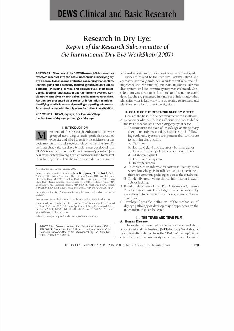
8/20/2019 dry eyes ii
http://slidepdf.com/reader/full/dry-eyes-ii 119/142
THE OCULAR SURFACE / APRIL 2007, VOL. 5, NO. 2 / www.theocularsurface.com 179
DEWS Clinical and Basic Research
©2007 Ethis Communications, Inc. The Ocular Surface ISSN:
1542-0124. (No authors listed). Research in dry eye: report of the
Research Subcommittee of the International Dry Eye WorkShop
(2007). 2007;5(2):179-193.
Research in Dry Eye:
Report of the Research Subcommittee ofthe International Dry Eye WorkShop (2007)
ABSTRACT Members of the DEWS Research Subcommittee
reviewed research into the basic mechanisms underlying dry
eye disease. Evidence was evaluated concerning the tear film,
lacrimal gland and accessory lacrimal glands, ocular surface
epithelia (including cornea and conjunctiva), meibomian
glands, lacrimal duct system and the immune system. Con-
sideration was given to both animal and human research data.
Results are presented as a series of information matrices,
identifying what is known and providing supporting references.
An attempt is made to identify areas for further investigation.
KEY WORDS DEWS, dry eye, Dry Eye WorkShop,
mechanisms of dry eye, pathology of dry eye
I. INTRODUCTION
embers of the Research Subcommittee weregrouped according to their particular areas ofexpertise and asked to review the evidence for the
basic mechanisms of dry eye pathology within that area. Tofacilitate this, a standardized template was developed (theDEWS Research Committee Report Form—Appendix 1 [ac-cess at: www.tearfilm.org]), which members used to presenttheir findings. Based on the information derived from the
returned reports, information matrices were developed.Evidence related to the tear film, lacrimal gland and
accessory lacrimal glands, ocular surface epithelia (includ-ing cornea and conjunctiva), meibomian glands, lacrimalduct system, and the immune system was evaluated. Con-sideration was given to both animal and human researchdata. Results are presented in a matrix of information thatidentifies what is known, with supporting references, andidentifies areas for further investigation.
II. GOALS OF THE RESEARCH SUBCOMMITTEE
Goals of the Research Subcommittee were as follows: A. To consider whether there is sufficient evidence to define
the basic mechanisms underlying dry eye disease.1. To summarize the state of knowledge about primary
alterations and/or secondary responses of the follow-ing ocular and systemic components that contributeto tear film dysfunction.
a. Tear filmb. Lacrimal gland and accessory lacrimal glandsc. Ocular surface epithelia, cornea, conjunctiva
d. Meibomian glande. Lacrimal duct systemf. Immune system
2. To construct an information matrix to identify areaswhere knowledge is insufficient and to determine ifthere are common pathologies across the syndrome.
3. To identify areas where clinical information is avail-able or lacking.
B. Based on data derived from Part A, to answer Question
2: Is the state of basic knowledge on mechanisms of dryeye sufficient to determine how these give rise to diseasesymptoms?
C. Develop, if possible, definitions of the mechanism ofdry eye pathology or develop major hypotheses on themechanism that can be tested.
III. THE TEARS AND TEAR FILM
A. Human Disease
The evidence presented at the last dry eye workshopreport (National Eye Institute [NEI]/Industry Workshop of1995, hereafter referred to as the “1995 Workshop”) indi-
cated that tear film osmolarity is increased in all forms of
Accepted for publication January 2007.
Research Subcommittee members: Ilene K. Gipson, PhD (Chair); Pablo Argüeso, PhD; Roger Beuerman, PhD; Stefano Bonini, MD; Igor Butovich,PhD; Reza Dana, MD, MPH; Darlene Dartt, PhD; Dan Gamache, PhD; BryanHam, PhD; Marcia Jumblatt, PhD; Donald Korb, OD; Friederich Kruse, MD;
Yoko Ogawa, MD; Friedrich Paulsen, MD, PhD; Michael Stern, PhD; DeborahF. Sweeney, PhD; John Tiffany, PhD; John Ubels, PhD; Mark Willcox, PhD.
Proprietary interests of Subcommittee members are disclosed on pages 202and 204.
Reprints are not available. Articles can be accessed at: www.tearfilm.org
Correspondence related to this chapter of the DEWS Report should be directedto: Ilene K. Gipson PhD, Schepens Eye Research Inst, 20 Staniford Street,Boston, MA 02114-2500. Tel: 617-912-0210. Fax: 617-912-0126. Email:[email protected]
Pablo Argüeso participated in the writing of the manuscript.
M

8/20/2019 dry eyes ii
http://slidepdf.com/reader/full/dry-eyes-ii 120/142
THE OCULAR SURFACE / APRIL 2007, VOL. 5, NO. 2 / www.theocularsurface.com180
dry eye (DE) and that tear volume and certain lacrimal tearproteins, such as lysozyme and lactoferrin, are decreased inaqueous-deficient dry eye.1 An evaporative form of dry eyewas also recognized, caused, for example, by a decreasedintegrity of the tear film lipid layer.
New evidence since the 1995 Workshop indicates thatmeibomian lipid composition and distribution is altered inDE and a number of bioactive tear proteins, including plas-min, matrix metalloproteinases (MMPs), defensive mol-ecules, and phospholipase A2 IIa in DE are increased. Thereis also an increase in inflammatory cytokines in non-Sjogrensyndrome (NSS) dry eye, as well as in Sjogren syndrome(SS) dry eye, and a decrease in goblet cell mucin MUC5ACin keratoconjunctivitis sicca (KCS) and SS (Table 1).
Given the sparsity of information available about thechanges in the composition of the tear film listed above, it isunclear how the changes in human tear composition relate
to tear dysfunction. To better understand the mechanismof dry eye disease, there is need for proteomic, lipidomic,and glycomic analyses of the tears from large, well-defined,staged, and age-matched patients or subject populations, todevelop biomarkers specific to dry eye disease. Progress hasbeen made in developing proteomic baseline studies of tearproteins, but studies comparing normal and dry eye tearsare lacking.41-44 Mass spectrometry is a powerful analyticaltool for identification45 of molecules and compounds, and itis being used to develop a standard lipid profile of normaltears and to identify specific component differences in thetears from DE models.
The application of mass spectrometry to the character-
ization and identification of the lipids of the meibomiangland secretions is demonstrating that the previouslyreported compositions are in need of revision. Compli-cating these efforts is the observation that the lipids arevery diverse in class and functionality. Different analyticalapproaches for isolation and detection are needed to dif-ferentiate lipid classes.
High throughput mass spectroscopic and glycan arraymethodologies are now available for glycomic analysis,and these could be used to analyze tear glycans in normaland DE patients. Similarly, determination of ratios andamounts of membrane-associated and secreted mucins intear film is necessary. It will also be important to determinethe relationship between various measures of tear stability(eg, tear film breakup time [TFBUT]) and the mucin andlipid quantity and character of the tears.
OUTLINE
I. Introduction
II. Goals of the Research Subcommittee
III. The tears and tear film
A. Human disease
B. Animal models of dry eye
IV. Ocular surfaceA. Human disease
B. In vitro and animal models
V. Immune system
A. Human disease
B. In vitro/animal models of dry eye—immunesystem
VI. Hypothesis of the mechanism of acute and chronicinflammation in dry eye disease
VII. Lacrimal/accessory lacrimal glands/nasolacrimalduct
A. Human disease
B. In vitro/animal models VIII. Meibomian gland
A. Human disease
B. In vitro/animal models
IX. Mechanisms underlying dry eye pathology
Abbreviations used in text and tables
↑ = Increase in/increased↓ = Decrease in/decreased
∆ = Change in/changes to
–/– = Homozygous null mouse
– = totally depleted
ACAT-1 = Acyl-CoA:cholesterol acyltransferase-1
Auto-AG = Autoantigen
BUT = Breakup time
CALT = Conjunctiva-associated lymphoid tissue
Chr Bleph = Chronic blepharitis
CIC = Cicatrizing disease
Conj = Conjunctiva/conjunctival
Cont lens = Contact lens
DE = Dry eyeDES = Dry eye syndrome
EDA = Ectodermal dysplasia
ENV STR = Environmental stress
epi = Epithelia/epithelial
Epi. Diff/sq metaplasia = Epithelial differentiation/squamous
metaplasia
GVHD = Graft vs host disease
KCS = Keratoconjunctivitis sicca
Lac = Lacrimal
Meibom = Meibomian
↓MG = Loss of meibomian glands
MGD = Meibomian gland dysfunction
NSS = Non-Sjogren syndromeNSS/ACQ = Aqueous-deficient non-Sjogren syndrome
Nasolac = Nasolacrimal
NLD = Nasolacrimal duct
RA-MGD = Retinoic acid induced MGD
SCOP = Scopolamine
siRNA = Small interfering RNA
Spont DE = Spontaneous dry eye
SS = Sjogren syndrome
TALT = Tear duct-associated lymphoid tissue
TBUT = Tear breakup time
Undif KCS = undifferentiated keratoconjunctivitis sicca
↓Vit A = Vitamin A deficient
–Vit A = Vitamin A totally depleted
DEWS CLINICAL AND BASIC RESEARCH

8/20/2019 dry eyes ii
http://slidepdf.com/reader/full/dry-eyes-ii 121/142
THE OCULAR SURFACE / APRIL 2007, VOL. 5, NO. 2 / www.theocularsurface.com 181
B. Animal Models of Dry Eye
Animal models discussed at the 1995 Workshop in-cluded a rabbit model in which the meibomian and lacrimalglands and the nictitans were ablated, which caused tearhyperosmolarity and ocular surface damage, mimickingthe features of human DE.
New models and find-ings since the 1995 Work-shop include: 1) mouse
models of DE that employscopolamine and environ-mental, dessicating stressthat show increases in in-flammatory cytokines andosmolarity in their tears;2) neurturin-deficient micethat develop DE and haveincreased inflammatorymediators in their tear film;3) a rabbit lacrimal glandablation model that shows
that treatment with dexa-
methasone reverses the decreased TFBUT and ocular surfacedamage; and 4) rabbit lacrimal gland denervation modelsthat produce altered tear protein and lipid profiles (Table 2).
One critical area of investigation with respect to theexisting evidence presented regards the need to correlatetear osmolarity, tear breakup, and the inflammatory stress
Table 1. Information matrix: human tear film
Androgen Contact Refs
KCS* NSS SS MGD Deficiency Lens/DE Refs
Tear Volume/Osmolarity:
↑ Osmolarity, ↓ Volume ✓ ✓ ✓ ✓ ✓ ✓ 2-6
↑ Evaporation ✓ ✓ 1, 7-9
↓Meniscus ✓ ✓ ✓ ✓ ✓ ✓ 5, 10-13 Correlation: Evaporation to
osmolarity & lipid layer✓ 14, 15
↓ BUT, ↑ Surface tension ✓ ✓ ✓ ✓ ✓ ✓ 5, 12, 16-20
Mucins:
↓ Glycoproteins, MUC5AC ✓ ✓ ✓ 21-23
Lipids:
∆ Lipid patterns, Distribution ✓ ✓ 24, 25
↓ Polar lipids ✓ 26
↓ Lipid layer, ↑ Evaporation ✓ 14
Proteins:
∆ Proteins ✓ 27, 28
↑ Plasmin levels ✓ 29
↑MMPs ✓ 30, 31
↑ Inflammation markers, PRPs ✓ ✓ 32
↓ Lactoferrin 33
↑ Nine defensive molecules ✓ 34
↓ Lysozyme, Lactoferrin 35
↑ Phospholipase A2 IIa ✓ ✓ 36, 37
Inflammatory Mediators:
Proinflammatory cytokines: IL-1, IL-6, IL-8, TNF-α ✓ ✓ 38-40
*Type not defined
Table 2. Information matrix: animal tear film
Rabbit Mouse RefsTear Vol/Osmolarity
↑ Osmolarity + ↓ Tear volume –Meibomian glands Scop & Env Str 48-49
↑ Osmolarity, ↑ surface injury –Lacrimal gland 50
↓ BUT, ↓ surface injury with dexamethasone –Lacrimal gland 51
Lipids
↑ Acylglycerols –Lacrimal gland/nictitans 45
Lipids in rabbit/human match –Lacrimal gland/nictitans 45
Proteins
↓ Protein –Nerves 52
↑ IL-1β –Neurturin 53
DEWS CLINICAL AND BASIC RESEARCH

8/20/2019 dry eyes ii
http://slidepdf.com/reader/full/dry-eyes-ii 122/142
THE OCULAR SURFACE / APRIL 2007, VOL. 5, NO. 2 / www.theocularsurface.com182
response. To that end, immortalized human corneal andconjunctival epithelial cell lines are now available that havedifferentiation characteristics of native epithelia.46,47 Theywill be useful to study effects of tear osmolarity, inflamma-tory mediators, and DE tears on surface epithelia.
Mass spectrometry, lipidomics, and proteomics inanimal models of dry eye should be done to provide in-
sight into the DE condition. Comparison of animal tearproteomes, lipidomes, and glycomes will help ascertainthe most appropriate human-relevant models (eg, totalchloroform extractables of rabbit tears match closely thoseof human tears).45
IV. OCULAR SURFACE
A. Human Disease
Aspects of dry eye surface pathology discussed at the1995 Workshop included the lack of epithelial barrier func-tion as demonstrated by increased dye uptake (with no dataavailable on mechanism), an increased tear film osmolaritycausing ocular surface damage, a loss of conjunctival gobletcells, and an increased squamous metaplasia of the surfaceepithelial cells (morphological observations).
New evidence since that report indicates that thereare alterations in cell-surface and secreted mucins and inkeratinization-related proteins expressed by epithelial cells.There also are alterations in corneal innervation densityand sensitivity. Studies document increased conjunctivalepithelial cell turnover. Evidence indicates that conjunctival
epithelial cells are active in the immune response and are asource of inflammatory mediators85 (Table 3).
Despite what is known, information about the tear filmand ocular surface in dry eye disease is still deficient. Itwould be of value to determine the conjunctival epithelialproteome and glycome in a well-defined, staged, dry eyepopulation compared to age- and sex-matched controls to
identify common changes in apical surface componentswith disease. It is desirable to determine if age and sex,or a combination thereof, influence the effects of environ-mental stress on ocular surface epithelia. It is importantto determine any genetic predictors of susceptibility toDE. Finally, a comparison of early intermittent stages ofthe disease to chronic disease may distinguish primarypathways causing DE from secondary responses associatedwith the disease.
B. In Vitro and Animal Models
Information gathered from in vitro and animal modelsas of the 1995 Workshop identified lack of barrier functionas demonstrated by dye uptake in several animal modelsof dry eye, loss of goblet cells in several animal models ofdry eye, and keratinization of ocular surface epithelium invitamin A deficiency.
Since the 1995 Workshop, investigations have identifiedthe role of membrane-associated mucins as a protective bar-rier (human epithelial cells in vitro), increased cell turnover(mouse experimental dry eye), and increased expression
Table 3. Information matrix: human ocular surface
Undif KCS NSS/ACQ SS CIC ↓ Vit A Cont Lens LASIK Refs
Corneal and conj. epi. cell
damage as indicated by✓ ✓ ✓ ✓ ✓ ✓ ✓ Well established
dye penetrance — Fluorescein,
lissamine green, rose bengal
Mucins:
↓ Goblet cells ✓ ✓ ✓ ✓ ✓ ↑ ✓ 54-61
↓MUC5AC ✓ ✓ 22, 23
Mucin glycosylation altered ✓ ✓ 62-65
∆ Glycosyltransferases ✓ 66
∆ Membrane-associated mucins ✓ ✓ 22, 57, 65, 67
∆ Conj. Cell-Epithelial:
↓Microplicae ✓ 68
Filamentary keratitis ✓ 69
↑ Stratification ✓ ✓ 66, 70
Epi proliferation ✓ 71
∆ Nuclear/chromatin structure ✓ ✓ 72-74
↑ Apoptosis ✓ ✓ ✓ 75
∆ Innervation ✓ ✓ ✓ 76-80
↑ Infection ✓ 35, 81
↑ Keratinization related proteins ✓ ✓ 82-84
Inflammatory markers on
conj. epi. cells✓ ✓ ✓ 75, 85
DEWS CLINICAL AND BASIC RESEARCH
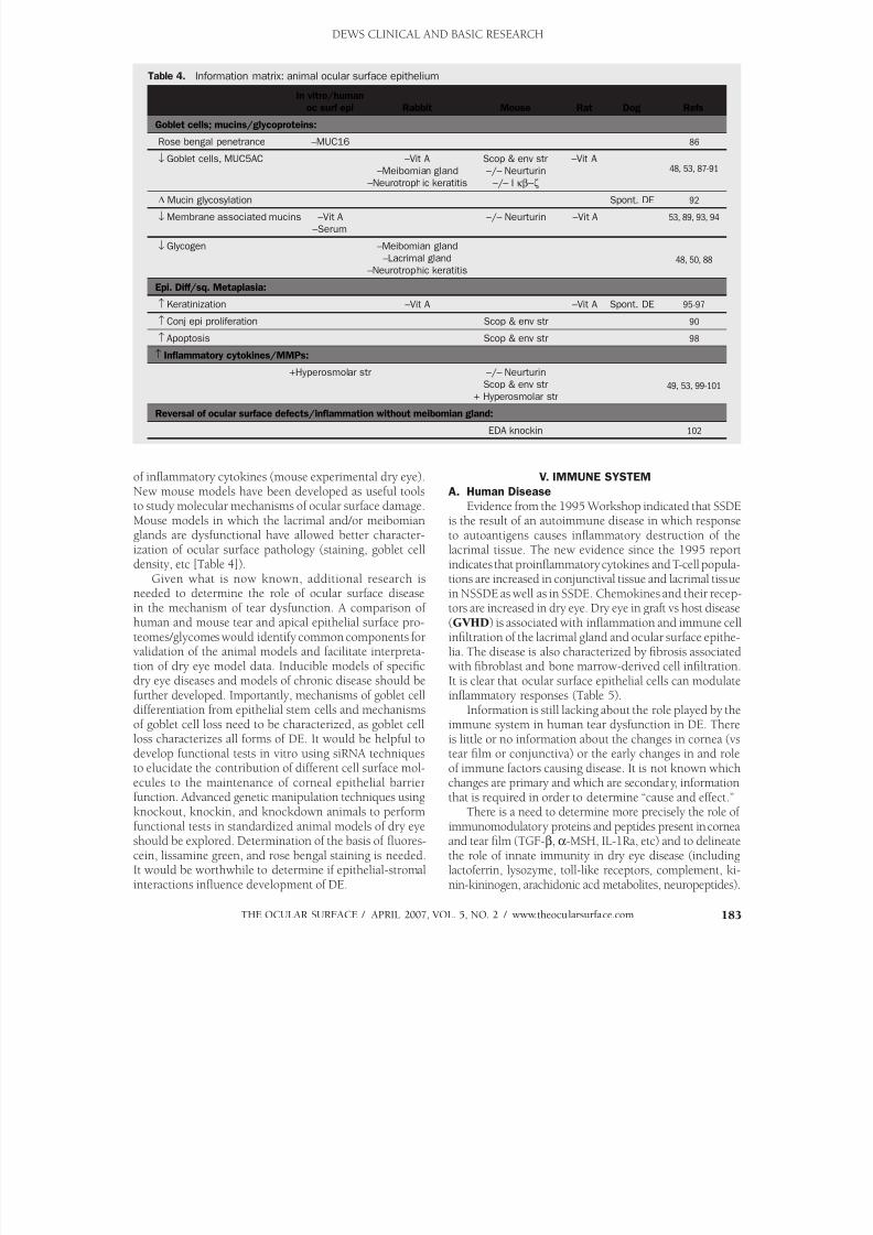
8/20/2019 dry eyes ii
http://slidepdf.com/reader/full/dry-eyes-ii 123/142
THE OCULAR SURFACE / APRIL 2007, VOL. 5, NO. 2 / www.theocularsurface.com 183
of inflammatory cytokines (mouse experimental dry eye).New mouse models have been developed as useful toolsto study molecular mechanisms of ocular surface damage.Mouse models in which the lacrimal and/or meibomian
glands are dysfunctional have allowed better character-ization of ocular surface pathology (staining, goblet celldensity, etc [Table 4]).
Given what is now known, additional research isneeded to determine the role of ocular surface diseasein the mechanism of tear dysfunction. A comparison ofhuman and mouse tear and apical epithelial surface pro-teomes/glycomes would identify common components forvalidation of the animal models and facilitate interpreta-tion of dry eye model data. Inducible models of specificdry eye diseases and models of chronic disease should befurther developed. Importantly, mechanisms of goblet cell
differentiation from epithelial stem cells and mechanismsof goblet cell loss need to be characterized, as goblet cellloss characterizes all forms of DE. It would be helpful todevelop functional tests in vitro using siRNA techniquesto elucidate the contribution of different cell surface mol-ecules to the maintenance of corneal epithelial barrierfunction. Advanced genetic manipulation techniques usingknockout, knockin, and knockdown animals to performfunctional tests in standardized animal models of dry eyeshould be explored. Determination of the basis of fluores-cein, lissamine green, and rose bengal staining is needed.It would be worthwhile to determine if epithelial-stromal
interactions influence development of DE.
V. IMMUNE SYSTEM
A. Human Disease
Evidence from the 1995 Workshop indicated that SSDEis the result of an autoimmune disease in which response
to autoantigens causes inflammatory destruction of thelacrimal tissue. The new evidence since the 1995 reportindicates that proinflammatory cytokines and T-cell popula-tions are increased in conjunctival tissue and lacrimal tissuein NSSDE as well as in SSDE. Chemokines and their recep-tors are increased in dry eye. Dry eye in graft vs host disease(GVHD) is associated with inflammation and immune cellinfiltration of the lacrimal gland and ocular surface epithe-lia. The disease is also characterized by fibrosis associatedwith fibroblast and bone marrow-derived cell infiltration.It is clear that ocular surface epithelial cells can modulateinflammatory responses (Table 5).
Information is still lacking about the role played by theimmune system in human tear dysfunction in DE. Thereis little or no information about the changes in cornea (vstear film or conjunctiva) or the early changes in and roleof immune factors causing disease. It is not known whichchanges are primary and which are secondary, informationthat is required in order to determine “cause and effect.”
There is a need to determine more precisely the role ofimmunomodulatory proteins and peptides present in corneaand tear film (TGF-β, α-MSH, IL-1Ra, etc) and to delineatethe role of innate immunity in dry eye disease (includinglactoferrin, lysozyme, toll-like receptors, complement, ki-
nin-kininogen, arachidonic acd metabolites, neuropeptides).
Table 4. Information matrix: animal ocular surface epithelium
In vitro/human
oc surf epi Rabbit Mouse Rat Dog Refs
Goblet cells; mucins/glycoproteins:
Rose bengal penetrance –MUC16 86
↓ Goblet cells, MUC5AC –Vit A Scop & env str –Vit A48, 53, 87-91 –Meibomian gland –/– Neurturin
–Neurotrophic keratitis –/– I κβ−ζ
∆ Mucin glycosylation Spont. DE 92
↓Membrane associated mucins –Vit A –/– Neurturin –Vit A 53, 89, 93, 94
–Serum
↓ Glycogen –Meibomian gland
–Lacrimal gland
–Neurotrophic keratitis48, 50, 88
Epi. Diff/sq. Metaplasia:
↑ Keratinization –Vit A –Vit A Spont. DE 95-97
↑ Conj epi proliferation Scop & env str 90
↑ Apoptosis Scop & env str 98
↑ Inflammatory cytokines/MMPs:
+Hyperosmolar str –/– Neurturin
Scop & env str
+ Hyperosmolar str49, 53, 99-101
Reversal of ocular surface defects/inflammation without meibomian gland:
EDA knockin 102
DEWS CLINICAL AND BASIC RESEARCH
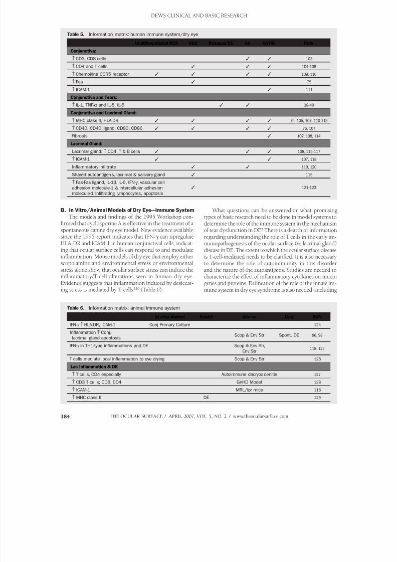
8/20/2019 dry eyes ii
http://slidepdf.com/reader/full/dry-eyes-ii 124/142
THE OCULAR SURFACE / APRIL 2007, VOL. 5, NO. 2 / www.theocularsurface.com184
B. In Vitro/Animal Models of Dry Eye---Immune System
The models and findings of the 1995 Workshop con-firmed that cyclosporine A is effective in the treatment of aspontaneous canine dry eye model. New evidence availablesince the 1995 report indicates that IFN-g can upregulate
HLA-DR and ICAM-1 in human conjunctival cells, indicat-ing that ocular surface cells can respond to and modulateinflammation. Mouse models of dry eye that employ eitherscopolamine and environmental stress or environmentalstress alone show that ocular surface stress can induce theinflammatory/T-cell alterations seen in human dry eye.Evidence suggests that inflammation induced by desiccat-ing stress is mediated by T-cells126 (Table 6).
What questions can be answered or what promisingtypes of basic research need to be done in model systems todetermine the role of the immune system in the mechanismof tear dysfunction in DE? There is a dearth of informationregarding understanding the role of T cells in the early im-
munopathogenesis of the ocular surface (vs lacrimal gland)disease in DE. The extent to which the ocular surface diseaseis T-cell-mediated needs to be clarified. It is also necessaryto determine the role of autoimmunity in this disorderand the nature of the autoantigens. Studies are needed tocharacterize the effect of inflammatory cytokines on mucingenes and proteins. Delineation of the role of the innate im-mune system in dry eye syndrome is also needed (including
Table 5. Information matrix: human immune system/dry eye
Undifferentiated KCS NSS Rosacea DE SS GVHD Refs
Conjunctiva:
↑ CD3, CD8 cells ✓ ✓ 103
↑ CD4 and T cells ✓ ✓ ✓ 104-108
↑ Chemokine CCR5 receptor ✓ ✓ ✓ ✓ 109, 110
↑ Fas ✓ 75
↑ ICAM-1 ✓ 111
Conjunctiva and Tears:
↑ IL-1, TNF-α and IL-8, IL-6 ✓ ✓ 38-40
Conjunctiva and Lacrimal Gland:
↑MHC class II, HLA-DR ✓ ✓ ✓ ✓ 75, 105, 107, 110-113
↑ CD40, CD40 ligand, CD80, CD86 ✓ ✓ ✓ ✓ 75, 107
Fibrosis ✓ 107, 108, 114
Lacrimal Gland:
Lacrimal gland: ↑ CD4, T & B cells ✓ ✓ ✓ 108, 115-117
↑ ICAM-1✓
✓
107, 118 Inflammatory infiltrate ✓ ✓ 119, 120
Shared autoantigens, lacrimal & salivary gland ✓ 115
↑ Fas-Fas ligand, IL-1β, IL-6, IFN-g , vascular cell
adhesion molecule-1 & intercellular adhesion
molecule-1 Infiltrating lymphocytes, apoptosis
✓ 121-123
Table 6. Information matrix: animal immune system
In vitro Animal Rabbit Mouse Dog Refs
IFN-g ↑ HLA-DR, ICAM-1 Conj Primary Culture 124
Inflammation ↑ Conj,
lacrimal gland apoptosisScop & Env Str Spont. DE 96, 98
IFN-g in TH1-type inflammations and DE Scop & Env Str,
Env Str118, 125
T cells mediate local inflammation to eye drying Scop & Env Str 126
Lac Inflammation & DE
↑ T cells, CD4 especially Autoimmune dacryoadenitis 127
↑ CD3 T cells; CD8, CD4 GVHD Model 128
↑ ICAM-1 MRL/lpr mice 118
↑ MHC class II DE 129
DEWS CLINICAL AND BASIC RESEARCH
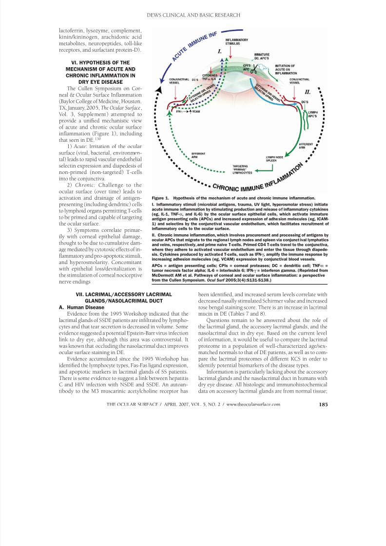
8/20/2019 dry eyes ii
http://slidepdf.com/reader/full/dry-eyes-ii 125/142
THE OCULAR SURFACE / APRIL 2007, VOL. 5, NO. 2 / www.theocularsurface.com 185
lactoferrin, lysozyme, complement,kinin/kininogen, arachidonic acidmetabolites, neuropeptides, toll-likereceptors, and surfactant protein-D).
VI. HYPOTHESIS OF THE
MECHANISM OF ACUTE AND
CHRONIC INFLAMMATION IN
DRY EYE DISEASE
The Cullen Symposium on Cor-neal & Ocular Surface Inflammation(Baylor College of Medicine, Houston.TX, January, 2005, The Ocular Surface, Vol. 3, Supplement) attempted toprovide a unified mechanistic viewof acute and chronic ocular surfaceinflammation (Figure 1), includingthat seen in DE.130
1) Acute: Irritation of the ocularsurface (viral, bacterial, environmen-tal) leads to rapid vascular endothelialselectin expression and diapedesis ofnon-primed (non-targeted) T-cellsinto the conjunctiva.
2) Chronic: Challenge to theocular surface (over time) leads toactivation and drainage of antigen-presenting (including dendritic) cellsto lymphoid organs permitting T-cellsto be primed and capable of targetingthe ocular surface.
3) Symptoms correlate primar-
ily with corneal epithelial damage,thought to be due to cumulative dam-age mediated by cytotoxic effects of in-flammatory and pro-apoptotic stimuli,and hyperosmolarity. Concomitantwith epithelial loss/devitalization isthe stimulation of corneal nociceptivenerve endings
VII. LACRIMAL/ACCESSORY LACRIMAL
GLANDS/NASOLACRIMAL DUCT
A. Human Disease
Evidence from the 1995 Workshop indicated that thelacrimal glands of SSDE patients are infiltrated by lympho-cytes and that tear secretion is decreased in volume. Someevidence suggested a potential Epstein-Barr virus infectionlink to dry eye, although this area was controversial. Itwas known that occluding the nasolacrimal duct improvesocular surface staining in DE.
Evidence accumulated since the 1995 Workshop hasidentified the lymphocyte types, Fas-Fas ligand expression,and apoptotic markers in lacrimal glands of SS patients.There is some evidence to suggest a link between hepatitisC and HIV infection with NSDE and SSDE. An autoan-
tibody to the M3 muscarinic acetylcholine receptor has
been identified, and increased serum levels correlate withdecreased nasally stimulated Schirmer value and increasedrose bengal staining score. There is an increase in lacrimal
mucin in DE (Tables 7 and 8).Questions remain to be answered about the role of
the lacrimal gland, the accessory lacrimal glands, and thenasolacrimal duct in dry eye. Based on the current levelof information, it would be useful to compare the lacrimalproteome in a population of well-characterized age/sex-matched normals to that of DE patients, as well as to com-pare the lacrimal proteomes of different KCS in order toidentify potential biomarkers of the disease types.
Information is particularly lacking about the accessorylacrimal glands and the nasolacrimal duct in humans withdry eye disease. All histologic and immunohistochemical
data on accessory lacrimal glands are from normal tissue;
Figure 1. Hypothesis of the mechanism of acute and chronic immune inflammation.
I. Inflammatory stimuli (microbial antigens, trauma, UV light, hyperosmolar stress) initiate
acute immune inflammation by stimulating production and release of inflammatory cytokines(eg, IL-1, TNF-α, and IL-6) by the ocular surface epithelial cells, which activate immatureantigen presenting cells (APCs) and increased expression of adhesion molecules (eg, ICAM-
1) and selectins by the conjunctival vascular endothelium, which facilitates recruitment ofinflammatory cells to the ocular surface.
II. Chronic immune inflammation, which involves procurement and processing of antigens byocular APCs that migrate to the regional lymph nodes and spleen via conjunctival lymphaticsand veins, respectively, and prime naive T-cells. Primed CD4 T-cells travel to the conjunctiva,
where they adhere to activated vascular endothelium and enter the tissue through diapede-sis. Cytokines produced by activated T-cells, such as IFN-g , amplify the immune response byincreasing adhesion molecules (eg, VCAM) expression by conjunctival blood vessels.
APCs = antigen presenting cells; CPIs = corneal proteases; DC = dendritic cell; TNF-α =tumor necrosis factor alpha; IL-6 = interleukin 6; IFN-g = interferon gamma. (Reprinted from
McDermott AM et al. Pathways of corneal and ocular surface inflammation: a perspectivefrom the Cullen Symposium. Ocul Surf 2005;3(4):S131-S138.)
DEWS CLINICAL AND BASIC RESEARCH
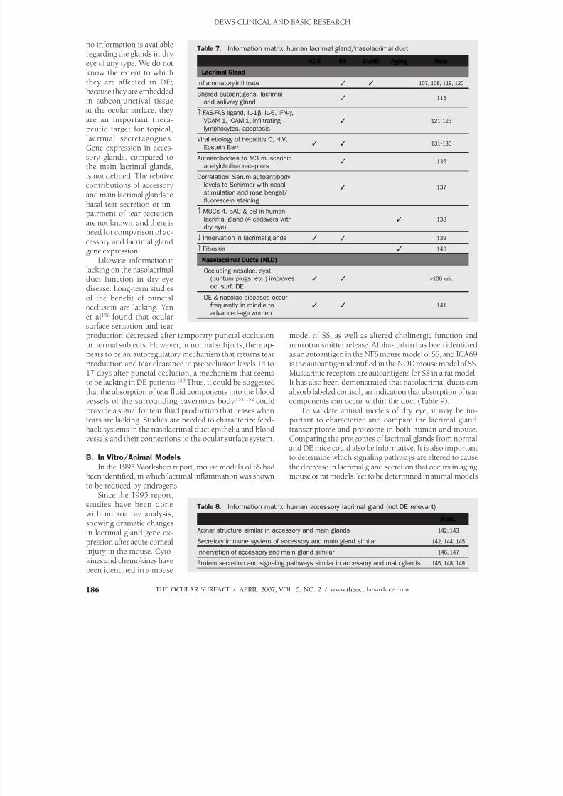
8/20/2019 dry eyes ii
http://slidepdf.com/reader/full/dry-eyes-ii 126/142
THE OCULAR SURFACE / APRIL 2007, VOL. 5, NO. 2 / www.theocularsurface.com186
no information is availableregarding the glands in dryeye of any type. We do notknow the extent to whichthey are affected in DE;because they are embeddedin subconjunctival tissue
at the ocular surface, theyare an important thera-peutic target for topical,lacrimal secretagogues.Gene expression in acces-sory glands, compared tothe main lacrimal glands,is not defined. The relativecontributions of accessoryand main lacrimal glands tobasal tear secretion or im-pairment of tear secretionare not known, and there isneed for comparison of ac-cessory and lacrimal glandgene expression.
Likewise, information islacking on the nasolacrimalduct function in dry eyedisease. Long-term studiesof the benefit of punctalocclusion are lacking. Yenet al150 found that ocularsurface sensation and tearproduction decreased after temporary punctal occlusion
in normal subjects. However, in normal subjects, there ap-pears to be an autoregulatory mechanism that returns tearproduction and tear clearance to preocclusion levels 14 to17 days after punctal occlusion, a mechanism that seemsto be lacking in DE patients.150 Thus, it could be suggestedthat the absorption of tear fluid components into the bloodvessels of the surrounding cavernous body151,152 couldprovide a signal for tear fluid production that ceases whentears are lacking. Studies are needed to characterize feed-back systems in the nasolacrimal duct epithelia and bloodvessels and their connections to the ocular surface system.
B. In Vitro/Animal ModelsIn the 1995 Workshop report, mouse models of SS had
been identified, in which lacrimal inflammation was shownto be reduced by androgens.
Since the 1995 report,studies have been donewith microarray analysis,showing dramatic changesin lacrimal gland gene ex-pression after acute cornealinjury in the mouse. Cyto-kines and chemokines have
been identified in a mouse
model of SS, as well as altered cholinergic function and
neurotransmitter release. Alpha-fodrin has been identifiedas an autoantigen in the NFS mouse model of SS, and ICA69is the autoantigen identified in the NOD mouse model of SS.Muscarinic receptors are autoantigens for SS in a rat model.It has also been demonstrated that nasolacrimal ducts canabsorb labeled cortisol, an indication that absorption of tearcomponents can occur within the duct (Table 9).
To validate animal models of dry eye, it may be im-portant to characterize and compare the lacrimal glandtranscriptome and proteome in both human and mouse.Comparing the proteomes of lacrimal glands from normaland DE mice could also be informative. It is also important
to determine which signaling pathways are altered to causethe decrease in lacrimal gland secretion that occurs in agingmouse or rat models. Yet to be determined in animal models
Table 7. Information matrix: human lacrimal gland/nasolacrimal duct
KCS SS GVHD Aging Refs
Lacrimal Gland
Inflammatory infiltrate ✓ ✓ 107, 108, 119, 120
Shared autoantigens, lacrimal
and salivary gland✓ 115
↑ FAS-FAS ligand, IL-1β, IL-6, IFN-g , VCAM-1, ICAM-1, Infiltrating
lymphocytes, apoptosis
✓ 121-123
Viral etiology of hepatitis C, HIV,
Epstein Barr✓ ✓ 131-135
Autoantibodies to M3 muscarinic
acetylcholine receptors✓ 136
Correlation: Serum autoantibody
levels to Schirmer with nasal
stimulation and rose bengal/
fluorescein staining
✓ 137
↑ MUCs 4, 5AC & 5B in human
lacrimal gland (4 cadavers with
dry eye)
✓ 138
↓ Innervation in lacrimal glands ✓ ✓ 139
↑ Fibrosis ✓ 140
Nasolacrimal Ducts (NLD)
Occluding nasolac. syst.
(puntum plugs, etc.) improves
oc. surf. DE
✓ ✓ >100 refs.
DE & nasolac diseases occur
frequently in middle to
advanced-age women
✓ ✓ 141
Table 8. Information matrix: human accessory lacrimal gland (not DE relevant)
Refs.
Acinar structure similar in accessory and main glands 142, 143
Secretory immune system of accessory and main gland similar 142, 144, 145
Innervation of accessory and main gland similar 146, 147
Protein secretion and signaling pathways similar in accessory and main glands 145, 148, 149
DEWS CLINICAL AND BASIC RESEARCH

8/20/2019 dry eyes ii
http://slidepdf.com/reader/full/dry-eyes-ii 127/142
THE OCULAR SURFACE / APRIL 2007, VOL. 5, NO. 2 / www.theocularsurface.com 187
is the role of myoepithelial cells in lacrimal gland dysfunc-tion. It may be useful to determine, using the autologouslymphocyte rabbit model, if exposure of cryptic antigens
through errors in recycling initiates SS. Determination of thecellular mechanisms used to induce autoimmune diseasein the lacrimal gland could also employ the autologouslymphocyte rabbit model. This model could also be usedto determine if the exocytotic process for protein secretionis a target for lacrimal gland dysfunction and to determinethe role of lacrimal gland duct cells in lacrimal gland dys-function through laser capture microdissection.
With regard to the nasolacrimal ducts, information islacking regarding cells of the ducts, and cell lines of naso-lacrimal duct epithelium are not currently available. Ques-tions to be answered in animal models include whether the
absorption of tear fluid components into the blood vessels
of the cavernous body surrounding the nasolacrimal ductschanges or ceases in dry eye models, and what happens todrained tear fluid in the nasolacrimal passage.
VIII. MEIBOMIAN GLAND
A. Human Disease
The 1995 Workshop report documented decreased and/ or altered meibomian lipids in DE, as well as morphologicabnormalities of the gland acini and tubules.
New evidence since the 1995 report identifies keratini-zation of ductal epithelium, orifice metaplasia, and reducedquality of meibomian gland secretions in people duringaging, in patients taking antiandrogen therapy, and/or inwomen with Complete Androgen Insensitivity Syndrome(Androgen Deficiency). Correlations have been made be-
tween nutrient intake (eg, omega 3 fatty acids, vitamin B6,
Table 9. Information matrix: animal lacrimal gland/nasolacrimal duct
In Vitro Rabbit Mouse Rat Dog Refs
Lacrimal Gland:
Coculture of lacrimal acinar cells/ Lacrimal gland ✓ 153-157
lymphocytes activates lymphocytes
and cause inflammation in host
lacrimal gland
↑ Lymphocytic infiltration, CD4, CD8; MRL/lpr mouse 158-166
↑ Fas, Fas-Ligand & cytokine NOD mouse
model of SS
Androgens ↓ inflammation, are MRL/Mp-lpr/ Exp. Dog DE 161,
immunosuppressive & decrease lpr mice; autoimmune 167-176
androgen receptors NZB/NZW dacryoadenitis
F1 Mouse
Lacrimal gland autoantigen or extract Mouse in vivo Rat in vivo 172, 173,
causes lymphocytic infiltration in 177, 178
lacrimal gland
Cholinergic function altered Sjögren’s NOD mouse 179, 180
syndrome ICA69 is autoantigen model of SS
Lymphocytic infiltration blocks lacrimal MRL/lpr 181
gland secretion by preventing nerve mouse model
release of neurotransmitters in of SS
Sjögren’s syndrome
α-fodrin is an autoantigen for the lacrimal NFS Mouse 182
gland and causes Sjögren’s syndrome model of SS
↑ vulnerability to herpes infection Cells of female 174
lacrimal gland
∆ Lacrimal gland gene express. Normal mouse 183
in corneal injury
Nasolacrimal duct (NLD):
3H-cortisol incorporated from NLDs into Absorpt. of No absorption
rabbit blood lipophillic of lipophillic
substances fr. substance from 184, 185 tear fluid by tears by epi.
epi. of NLDs of NLDs
Anatomy useful for investigating NLDs Comparative Comparative
studies studies184-186
↓ Secretion
↓ Innervation
↑ Lipofusci
Aging model 187
DEWS CLINICAL AND BASIC RESEARCH
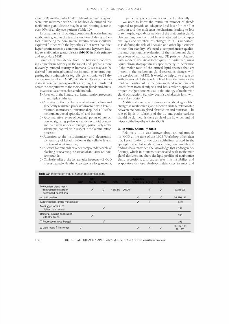
8/20/2019 dry eyes ii
http://slidepdf.com/reader/full/dry-eyes-ii 128/142
THE OCULAR SURFACE / APRIL 2007, VOL. 5, NO. 2 / www.theocularsurface.com188
vitamin D) and the polar lipid profiles of meibomian glandsecretions in women with SS. It has been determined thatmeibomian gland disease may be a contributing factor inover 60% of all dry eye patients (Table 10).
Information is still lacking about the role of the humanmeibomian gland in the tear dysfunction of dry eye. Fac-tors influencing meibomian duct keratinization should be
explored further, with the hypothesis (not new) that ducthyperkeratinization is a common factor and key event lead-ing to meibomian gland disease (MGD) in both primaryand secondary MGD.
Some clues may derive from the literature concern-ing epinephrine toxicity in the rabbit and, perhaps morerelevantly, retinoid toxicity in humans. Clues may also bederived from an insubstantial but interesting literature sug-gesting that conjunctivitis (eg, allergic, chronic) or SS dryeye are associated with MGD, with the implication that me-diators (proinflammatory or otherwise) might be transferredacross the conjunctiva to the meibomian glands and ducts.
Investigative approaches could include:1) A review of the literature of keratinization processes
in multiple epithelia; 2) A review of the mechanism of retinoid action and
genetically regulated processes involved with kerati-nization, in mucosae, transitional epithelia (like themeibomian ductal epithelium) and in skin;
3) A comparative review of potential points of interac-tion of signaling pathways under retinoid controland pathways under adrenergic, particularly alphaadrenergic, control, with respect to the keratinizationprocess;
4) Attention to the histochemistry and electronhis-
tochemistry of keratinization at the cellular levels,markers of keratinization;
5) A search for retinoids or other compounds capable ofblocking or reversing the action of anti-acne retinoidcompounds;
6) Clinical studies of the comparative frequency of MGDin eyes treated with adrenergic agonists for glaucoma,
particularly where agonists are used unilaterally. We need to know the minimum number of glands
required to provide an adequate lipid layer for tear filmfunction and the molecular mechanisms leading to lossor to morphologic abnormalities of the meibomian gland.Determining how the lipid layer is attached to the aque-ous layer and whether this changes in DE is important,
as is defining the role of lipocalin and other lipid carriersin tear film stability. We need a comprehensive qualita-tive and quantitative evaluation of the meibomian glandsecretions of normal subjects and DE patients, obtainedwith modern analytical techniques, in particular, usingliquid chromatography/mass spectrometry to determineif the molar ratio of the critical lipid species that arepresent in the meibomian gland secretions changes withthe development of DE. It would be helpful to create anartificial model of the tear film lipid layer that mimics thelipid composition of the meibomian gland secretions col-lected from normal subjects and has similar biophysicalproperties. Questions exist as to the etiology of meibomiangland obstruction, eg, why doesn’t a chalazion form withevery obstruction?
Additionally, we need to know more about age-relatedchanges in meibomian gland function and the relationshipbetween meibomian gland obstruction and nutrition. Therole of lipids in lubricity of the lid and ocular surfacesshould be clarified. Is there a role of the lid wiper and lidwiper epitheliopathy within MGD?
B. In Vitro/Animal Models
Relatively little was known about animal modelsfor MGD at the time of the 1995 Workshop other than
that keratinization of the duct epithelium existed in theepinephrine rabbit models. Since then, new models andfindings have provided the knowledge that androgen de-ficiency, which in humans is associated with meibomiangland dysfunction, alters the lipid profiles of meibomiangland secretions, and causes tear film instability andevaporative dry eye. Androgen deficiency in mice and
Table 10. Information matrix: human meibomian gland
Chr Androgen Cont
KCS Bleph MGD NSS SS Deficiency Aging Lens Refs
Meibomian gland loss/✓ ✓ ✓18.5% ✓60% ✓ ✓ 6, 188-195 obstruction/distortion
decreased secretions
∆ Lipid profiles ✓ ✓ ✓ 36, 196-198
Keratinization, orifice metaplasia ✓ ✓ 5, 10
Melting pt. of lipid 3°
higher than normal✓ 199
Bacterial strains associated
with Chr Bleph✓ 200
↑ Fluorescein, rose bengal ✓ 195
∆ Lipid layer; ↑ Thickness ✓ ✓ ✓ ✓ 36, 197, 198,
201, 202
DEWS CLINICAL AND BASIC RESEARCH
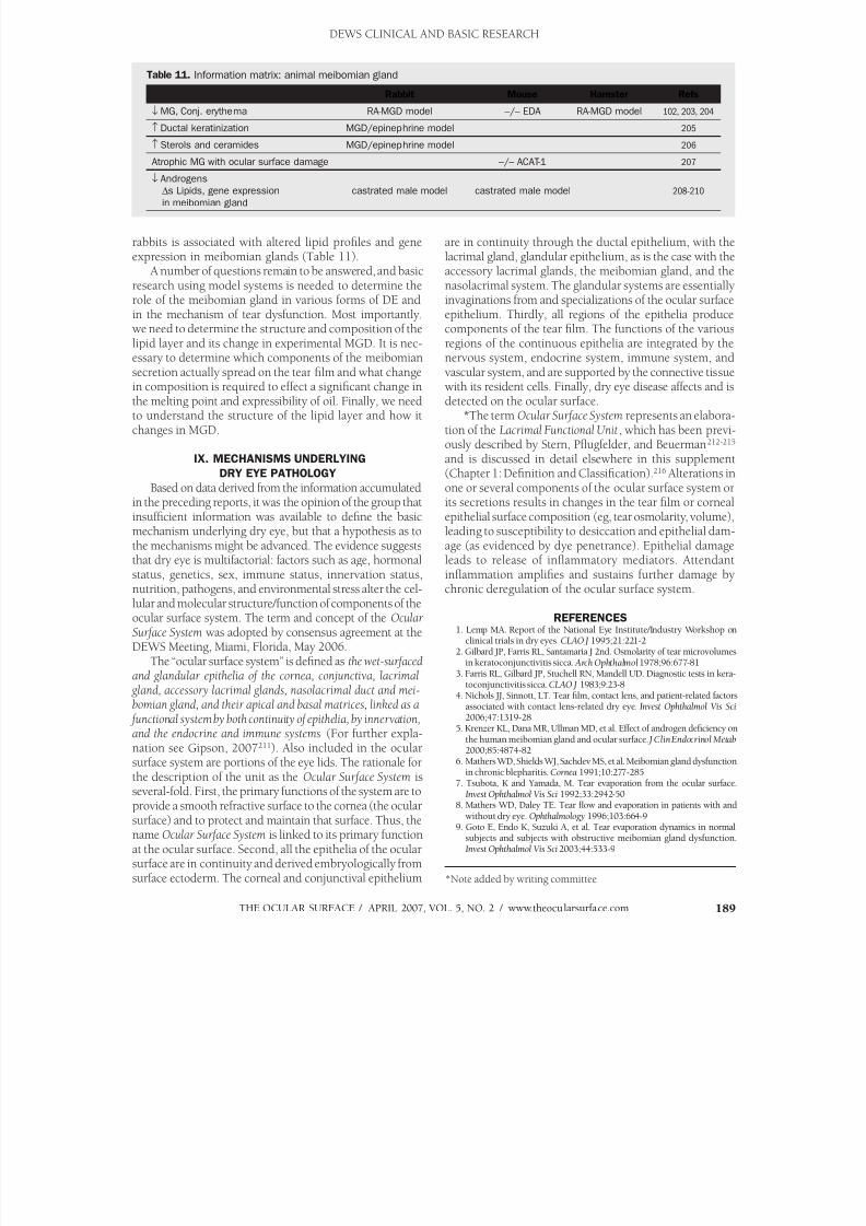
8/20/2019 dry eyes ii
http://slidepdf.com/reader/full/dry-eyes-ii 129/142
THE OCULAR SURFACE / APRIL 2007, VOL. 5, NO. 2 / www.theocularsurface.com 189
rabbits is associated with altered lipid profiles and geneexpression in meibomian glands (Table 11).
A number of questions remain to be answered, and basicresearch using model systems is needed to determine therole of the meibomian gland in various forms of DE andin the mechanism of tear dysfunction. Most importantly,we need to determine the structure and composition of thelipid layer and its change in experimental MGD. It is nec-essary to determine which components of the meibomiansecretion actually spread on the tear film and what changein composition is required to effect a significant change inthe melting point and expressibility of oil. Finally, we needto understand the structure of the lipid layer and how itchanges in MGD.
IX. MECHANISMS UNDERLYING
DRY EYE PATHOLOGY
Based on data derived from the information accumulatedin the preceding reports, it was the opinion of the group thatinsufficient information was available to define the basic
mechanism underlying dry eye, but that a hypothesis as tothe mechanisms might be advanced. The evidence suggeststhat dry eye is multifactorial: factors such as age, hormonalstatus, genetics, sex, immune status, innervation status,nutrition, pathogens, and environmental stress alter the cel-lular and molecular structure/function of components of theocular surface system. The term and concept of the OcularSurface System was adopted by consensus agreement at theDEWS Meeting, Miami, Florida, May 2006.
The “ocular surface system” is defined as the wet-surfacedand glandular epithelia of the cornea, conjunctiva, lacrimal
gland, accessory lacrimal glands, nasolacrimal duct and mei-
bomian gland, and their apical and basal matrices, linked as a functional system by both continuity of epithelia, by innervation,and the endocrine and immune systems (For further expla-nation see Gipson, 2007211). Also included in the ocularsurface system are portions of the eye lids. The rationale forthe description of the unit as the Ocular Surface System isseveral-fold. First, the primary functions of the system are toprovide a smooth refractive surface to the cornea (the ocularsurface) and to protect and maintain that surface. Thus, thename Ocular Surface System is linked to its primary functionat the ocular surface. Second, all the epithelia of the ocularsurface are in continuity and derived embryologically from
surface ectoderm. The corneal and conjunctival epithelium
are in continuity through the ductal epithelium, with thelacrimal gland, glandular epithelium, as is the case with theaccessory lacrimal glands, the meibomian gland, and thenasolacrimal system. The glandular systems are essentiallyinvaginations from and specializations of the ocular surfaceepithelium. Thirdly, all regions of the epithelia producecomponents of the tear film. The functions of the variousregions of the continuous epithelia are integrated by thenervous system, endocrine system, immune system, andvascular system, and are supported by the connective tissuewith its resident cells. Finally, dry eye disease affects and isdetected on the ocular surface.
*The term Ocular Surface System represents an elabora-tion of the Lacrimal Functional Unit, which has been previ-ously described by Stern, Pflugfelder, and Beuerman212-215 and is discussed in detail elsewhere in this supplement(Chapter 1: Definition and Classification).216 Alterations inone or several components of the ocular surface system orits secretions results in changes in the tear film or cornealepithelial surface composition (eg, tear osmolarity, volume),
leading to susceptibility to desiccation and epithelial dam-age (as evidenced by dye penetrance). Epithelial damageleads to release of inflammatory mediators. Attendantinflammation amplifies and sustains further damage bychronic deregulation of the ocular surface system.
REFERENCES 1. Lemp MA. Report of the National Eye Institute/Industry Workshop on
clinical trials in dry eyes. CLAO J 1995;21:221-22. Gilbard JP, Farris RL, Santamaria J 2nd. Osmolarity of tear microvolumes
in keratoconjunctivitis sicca. Arch Ophthalmol 1978;96:677-813. Farris RL, Gilbard JP, Stuchell RN, Mandell UD. Diagnostic tests in kera-
toconjunctivitis sicca. CLAO J 1983;9:23-84. Nichols JJ, Sinnott, LT. Tear film, contact lens, and patient-related factors
associated with contact lens-related dry eye. Invest Ophthalmol Vis Sci2006;47:1319-28
5. Krenzer KL, Dana MR, Ullman MD, et al. Effect of androgen deficiency onthe human meibomian gland and ocular surface. J Clin Endocrinol Metab 2000;85:4874-82
6. Mathers WD, Shields WJ, Sachdev MS, et al. Meibomian gland dysfunctionin chronic blepharitis. Cornea 1991;10:277-285
7. Tsubota, K and Yamada, M. Tear evaporation from the ocular surface.Invest Ophthalmol Vis Sci 1992;33:2942-50
8. Mathers WD, Daley TE. Tear flow and evaporation in patients with andwithout dry eye. Ophthalmology 1996;103:664-9
9. Goto E, Endo K, Suzuki A, et al. Tear evaporation dynamics in normalsubjects and subjects with obstructive meibomian gland dysfunction.Invest Ophthalmol Vis Sci 2003;44:533-9
Table 11. Information matrix: animal meibomian gland
Rabbit Mouse Hamster Refs
↓ MG, Conj. erythema RA-MGD model –/– EDA RA-MGD model 102, 203, 204
↑ Ductal keratinization MGD/epinephrine model 205
↑ Sterols and ceramides MGD/epinephrine model 206
Atrophic MG with ocular surface damage –/– ACAT-1 207
↓ Androgens ∆s Lipids, gene expression
in meibomian gland
castrated male model castrated male model 208-210
DEWS CLINICAL AND BASIC RESEARCH
*Note added by writing committee

8/20/2019 dry eyes ii
http://slidepdf.com/reader/full/dry-eyes-ii 130/142
THE OCULAR SURFACE / APRIL 2007, VOL. 5, NO. 2 / www.theocularsurface.com190
10. Cermak JM, Krenzer KL, Sullivan RM, et al. Is complete androgen insen-sitivity syndrome associated with alterations in the meibomian gland andocular surface? Cornea 2003;22:516-21
11. Mainstone JC, Bruce AS, Golding TR. Tear meniscus measurement in thediagnosis of dry eye. Curr Eye Res 1996;15:653-61
12. Kallarackal GU, Ansari EA, Amos N, et al. A comparative study to assessthe clinical use of fluorescein meniscus time (FMT) with tear break uptime (TBUT) and Schirmer’s tests (ST) in the diagnosis of dry eyes. Eye 2002;16:594-600
13. Savini G, Barboni P, Zanini M. Tear meniscus evaluation by optical coher-
ence tomography. Ophthalmic Surg Lasers Imaging 2006;37:112-814. Craig JP, Tomlinson A. Importance of the lipid layer in human tear film
stability and evaporation. Optom Vis Sci 1997;74:8-1315. Craig JP, Singh I, Tomlinson A, et al. The role of tear physiology in ocular
surface temperature. Eye 2000;14 ( Pt 4):635-4116. Tiffany JM, Winter N, Bliss G. Tear film stability and tear surface tension.
Curr Eye Res 1989;8:507-1517. Nagyova, B, Tiffany JM. Components responsible for the surface tension
of human tears. Curr Eye Res 1999;19:4-11.18. Glasson MJ, Stapleton F, Keay L, et al. Differences in clinical parameters and
tear film of tolerant and intolerant contact lens wearers. Invest OphthalmolVis Sci 2003;44:5116-24
19. Begley CG, Chalmers RL, Abetz L, et al. The relationship between habitualpatient-reported symptoms and clinical signs among patients with dry eyeof varying severity. Invest Ophthalmol Vis Sci 2003;44:4753-61
20. Yamada M, Mochizuki H, Kawai M, et al. Decreased tear lipocalin con-
centration in patients with meibomian gland dysfunction. Br J Ophthalmol 2005;89:803-5
21. Dohlman CH, Friend J, Kalevar V, et al. The glycoprotein (mucus) contentof tears from normals and dry eye patients. Exp Eye Res 1976;22:359-65
22. Argueso P, Balaram M, Spurr-Michaud S, et al. Decreased levels of thegoblet cell mucin MUC5AC in tears of patients with Sjogren’s syndrome.Invest Ophthalmol Vis Sci 2002;43:1004-11
23. Zhao H, Jumblatt JE , Wood TO, Jumblatt MM. Quantification of MUC5ACprotein in human tears. Cornea 2001;20:873-7
24. Goto E, Tseng SC. Differentiation of lipid tear deficiency dry eye by ki-netic analysis of tear interference images. Arch Ophthalmol 2003;121:173-80
25. Danjo Y, Hamano T. Observation of precorneal tear film in patientswith Sjogren’s syndrome. Acta Ophthalmol Scand 1995;73:501-5
26. Shine WE, McCulley JP. Keratoconjunctivitis sicca associated with meibo-mian secretion polar lipid abnormality. Arch Ophthalmol 1998;116:849-52
27. Grus FH, Sabuncuo P, Herber S, Augustin AJ. Analysis of tear protein pat-
terns for the diagnosis of dry eye. Adv Exp Med Biol 2002;506:1213-628. Ohashi Y, Ishida R, Kojima T, et al. Abnormal protein profiles in tears with
dry eye syndrome. Am J Ophthalmol 2003;136:291-929. Virtanen T, Konttinen YT, Harkonen M, Tervo T. Tear fluid plasmin activ-
ity of dry eye patients with Sjogren’s syndrome. Acta Ophthalmol Scand 1997;75:137-41
30. Afonso AA, Sobrin L, Monroy DC, et al. Tear fluid gelatinase B activitycorrelates with IL-1alpha concentration and fluorescein clearance in ocularrosacea. Invest Ophthalmol Vis Sci 1999;40:2506-12
31. Sobrin L, Liu A, Monroy DC, et al. Regulation of MMP-9 activity in humantear fluid and corneal epithelial culture supernatant.Invest Ophthalmol VisSci 2000;41:1703-9
32. Grus FH, Podust VN, Bruns K, et al. SELDI-TOF-MS ProteinChip arrayprofiling of tears from patients with dry eye. Invest Ophthalmol Vis Sci 2005;46: 863-76
33. Danjo Y, Lee M, Horimoto K, Hamano T. Ocular surface damage and tear
lactoferrin in dry eye syndrome. Acta Ophthalmol (Copenh) 1994;72:433-734. Koo BS, Lee DY, Ha HS, et al. Comparative analysis of the tear protein
expression in blepharitis patients using two-dimensional electrophoresis. J Proteome Res 2005;4:719-24
35. Seal DV. The effect of aging and disease on tear constituents. Trans Oph-thalmol Soc UK 1985;104 ( Pt 4):355-62
36. Glasson, M, Stapleton F, Willcox M. Lipid, lipase and lipocalin differ-ences between tolerant and intolerant contact lens wearers. Curr Eye Res2002;25:227-35
37. Aho VV, Nevalainen TJ, Paavilainen V, Saari KM. Group IIA phospholipaseA2 content of tears in patients with keratoconjunctivitis sicca. Graefes
Arch Clin Exp Ophthalmol 2002 Jul;240(7):521-3 38. Tishler M, Yaron I, Geyer O, et al. Elevated tear interleukin-6 levels in
patients with Sjogren syndrome. Ophthalmology 1998;105:2327-939. Pflugfelder SC, Jones D, Ji Z, et al. Altered cytokine balance in the tear fluid
and conjunctiva of patients with Sjogren’s syndrome keratoconjunctivitis
sicca. Curr Eye Res 999;19:201-11
40. Solomon A, Dursun D, Liu Z, et al. Pro- and anti-inflammatory forms ofinterleukin-1 in the tear fluid and conjunctiva of patients with dry-eyedisease. Invest Ophthalmol Vis Sci 2001;42:2283-92
41. Molloy MP, Bolis S, Herbert BR, et al. Establishment of the human reflextear 2D-PAGE reference map: a first step towards the diagnosis of disease.Electrophoresis 1997;18: 2811-5
42. Tsai PS, Evans JE, Green KM, et al. Proteomic analysis of human meibo-mian gland secretions. Br J Ophthalmol 2006;90:372-7
43. Mulvenna I, Stapleton F, Hains PG, et al. Low molecular weight analysisof tears using matrix assisted laser desorption ionization-time of flight
mass spectrometry. Clin Experiment Ophthalmol 2000;28:205-744. Fung K, Morris C, Duncan M. Mass spectrometric techniques applied to
the analysis of human tears: a focus on the peptide and protein constitu-ents. Adv Exp Med Biol 2002;506(PtA):601-5
45. Ham BM, Jacob JT, Keese MM, Cole RB. Identification, quantificationand comparison of major non-polar lipids in normal and dry eye tearlipidomes by electrospray tandem mass spectrometry. J Mass Spectrom 2004;39:1321-36
46. Gipson IK, Spurr-Michaud SJ, Argueso P, et al. Mucin gene expression inimmortalized human corneal-limbal and conjunctival epithelial cell lines.Invest Ophthalmol Vis Sci 2003;44:2496-506
47. Robertson DM, Li L, Fisher S, et al. Characterization of growth and dif-ferentiation in a telomerase-immortalized human corneal epithelial cellline. Invest Ophthalmol Vis Sci 2005;46:470-8
48. Gilbard JP, Rossi SR, Heyda KG. Tear film and ocular surface changesafter closure of the meibomian gland orifices in the rabbit.Ophthalmology
1989;96:1180-649. Stewart P, Chen Z, Farley W, et al. Effect of experimental dry eye on tear
sodium concentration in the mouse. Eye Contact Lens 2005;31:175-850. Gilbard JP, Rossi SR, Gray KL, et al. Tear film osmolarity and ocular
surface disease in two rabbit models for keratoconjunctivitis sicca. InvestOphthalmol Vis Sci 1988;29:374-8
51. Nagelhout TJ, Gamache DA, Roberts L, et al. Preservation of tear filmintegrity and inhibition of corneal injury by dexamethasone in a rabbitmodel of lacrimal gland inflammation-induced dry eye. J Ocul PharmacolTher 2005;21:139-48.
52. Salvatore MF, Pedroza L, Beuerman RW. Denervation of rabbit lacrimalgland increases levels of transferrin and unidentified tear proteins of 44and 36 kDa. Curr Eye Res 1999;18:455-66.
53. Song XJ, Li DQ, Farley W, et al. Neurturin-deficient mice develop dry eyeand keratoconjunctivitis sicca. Invest Ophthalmol Vis Sci 2003;44:4223-9
54. Poon AC, Geerling G, Dart JK, et al. Autologous serum eyedrops for
dry eyes and epithelial defects: clinical and in vitro toxicity studies. Br JOphthalmol 2001;85:1188-97
55. Ralph RA. Conjunctival goblet cell density in normal subjects and in dryeye syndromes. Invest Ophthalmol Vis Sci 1975;14:299-302
56. Nelson JD, Havener VR, Cameron JD. Cellulose acetate impressions ofthe ocular surface. Dry eye states. Arch Ophthalmol 1983;101:1869-72
57. Pflugfelder SC, Tseng SC, Yoshino K, et al. Correlation of goblet cell densityand mucosal epithelial membrane mucin expression with rose bengalstaining in patients with ocular irritation. Ophthalmology 1997;104:223-35
58. Blodi BA, Byrne KA, Tabbara KF. Goblet cell population among patientswith inactive trachoma. Int Ophthalmol 1988;12:41-5
59. Sommer A. Treatment of corneal xerophthalmia with topical retinoic acid. Am J Ophthalmol 1983;95:349-52
60. Lievens CW, Connor CG, Murphy H. Comparing goblet cell densitiesin patients wearing disposable hydrogel contact lenses versus siliconehydrogel contact lenses in an extended-wear modality. Eye Contact Lens
2003;29:241-4.61. Albietz JM, McLennan SG, Lenton LM. Ocular surface management of
photorefractive keratectomy and laser in situ keratomileusis. J Refract Surg 2003;19:636-44
62. Versura P, Maltarello MC, Cellini M, et al. Detection of mucus glycocon- jugates in human conjunctiva by using the lectin-colloidal gold techniquein TEM. II. A quantitative study in dry-eye patients. Acta Ophthalmol (Copenh) 1986;64:451-5
63. Garcher C, Bron AJ, Baudouin C, et al. CA 19-9 ELISA test: a new methodfor studying mucus changes in tears. Br J Ophthalmol 1998;82:88-90
64. Versura P, Maltarello MC, Cellini M, et al. Detection of mucus glycocon- jugates in human conjunctiva by using the lectin-colloidal gold techniquein TEM. III. A quantitative study in asymptomatic contact lens wearers.
Acta Ophthalmol (Copenh) 1987;65:661-765. Hayashi Y, Kao WW, Kohno N, et al. Expression patterns of sialylated
epitope recognized by KL-6 monoclonal antibody in ocular surface
epithelium of normals and dry eye patients. Invest Ophthalmol Vis Sci
DEWS CLINICAL AND BASIC RESEARCH

8/20/2019 dry eyes ii
http://slidepdf.com/reader/full/dry-eyes-ii 131/142
THE OCULAR SURFACE / APRIL 2007, VOL. 5, NO. 2 / www.theocularsurface.com 191
2004;45:2212-766. Argueso P, Tisdale A, Mandel U, et al. The cell-layer- and cell-type-specific
distribution of GalNAc-transferases in the ocular surface epithelia is alteredduring keratinization. Invest Ophthalmol Vis Sci 2003;44:86-92
67. Danjo Y, Watanabe H, Tisdale AS, et al. Alteration of mucin in humanconjunctival epithelia in dry eye. Invest Ophthalmol Vis Sci1998;39:2602-9
68. Koufakis DI, Karabatsas CH, Sakkas LI, et al. Conjunctival surface changesin patients with Sjogren’s syndrome: a transmission electron microscopystudy. Invest Ophthalmol Vis Sci 2006;47:541-4
69. Albietz J, Sanfilippo P, Troutbeck R, Lenton LM. Management of filamen-
tary keratitis associated with aqueous-deficient dry eye. Optom Vis Sci 2003;80:420-30
70. Murube J, Rivas L. Biopsy of the conjunctiva in dry eye patients estalishesa correlation between squamous metaplasia and dry eye clinical severity.Eur J Ophthalmol 2003;13:246-56
71. Kunert KS, Tisdale AS, Gipson IK. Goblet cell numbers and epithelialproliferation in the conjunctiva of patients with dry eye syndrome treatedwith cyclosporine. Arch Ophthalmol 2002;120:330-7
72. Meller D. The fine structure of chromatin alterations in conjunctivalepithelial cells in keratoconjunctivitis sicca. Cornea 1999;18:225-32
73. Albietz JM, Bruce AS. The conjunctival epithelium in dry eye subtypes:effect of preserved and non-preserved topical treatments. Curr Eye Res 2001;22:8-18
74. Bjerrum KB. Snake-like chromatin in conjunctival cells of normal elderlypersons and of patients with primary Sjogren’s syndrome and other con-nective tissue diseases. Acta Ophthalmol Scand 1995;73:33-6
75. Brignole F, Pisella PJ, De Saint Jean M, et al. Flow cytometric analysis ofinflammatory markers in conjunctival epithelial cells of patients with dryeyes. Invest Ophthalmol Vis Sci 2000;41:1356-63
76. Benitez del Castillo JM, Wasfy MA, Fernandez C, Garcia-Sanchez J. An invivo confocal masked study on corneal epithelium and subbasal nervesin patients with dry eye. Invest Ophthalmol Vis Sci 2004;45:3030-5
77. Xu KP, Yagi Y, Tsubota K. Decrease in corneal sensitivity and change intear function in dry eye. Cornea 1996;15:235-9
78. Horwath-Winter J, Vidic B, Schantzer G, Schmut O. Early changes incorneal sensation, ocular surface integrity, and tear-film function after laser-assisted subepithelial keratectomy. J Cataract Refract Surg 2004;30:2316-21
79. Hovanesian JA, Shah SS, Maloney RK. Symptoms of dry eye and recur-rent erosion syndrome after refractive surgery. J Cataract Refract Surg 2001;27:577-84
80. Wilson SE. Laser in situ keratomileusis-induced (presumed) neurotrophicepitheliopathy. Ophthalmology 2001;108:1082-7
81. Scott IU, Flynn HW Jr, Feuer W, et al. Endophthalmitis associated withmicrobial keratitis. Ophthalmology 1996;103:1864-70
82. Kawasaki S, Kawamoto S, Yokoi N, et al. Up-regulated gene expressionin the conjunctival epithelium of patients with Sjogren’s syndrome. ExpEye Res 2003;77:17-26
83. Nakamura T, Nishida K, Dota A, et al. Elevated expression of transglu-taminase 1 and keratinization-related proteins in conjunctiva in severeocular surface disease. Invest Ophthalmol Vis Sci 2001;42:549-56
84. Nakamura T, Nishida K, Dota A, Kinoshita S. Changes in conjunctivalclusterin expression in severe ocular surface disease. Invest OphthalmolVis Sci 2002;43:1702-7
85. Narayanan S, Miller WL, McDermott AM. Conjunctival cytokine expres-sion in symptomatic moderate dry eye subjects. Invest Ophthalmol Vis Sci 2006;47:2445-50
86. Argueso P, Tisdale A, Spurr-Michaud S, et al. Mucin characteristics ofhuman corneal-limbal epithelial cells that exclude the rose bengal anionic
dye. Invest Ophthalmol Vis Sci 2006;47:113-987. Hatchell DL, Sommer A. Detection of ocular surface abnormalities in
experimental vitamin A deficiency. Arch Ophthalmol 1984;102:1389-9388. Gilbard JP, Rossi SR. Tear film and ocular surface changes in a rabbit model
of neurotrophic keratitis. Ophthalmology 1990;97:308-1289. Tei M, Spurr-Michaud SJ, Tisdale AS, Gipson IK. Vitamin A deficiency
alters the expression of mucin genes by the rat ocular surface epithelium.Invest Ophthalmol Vis Sci 2000;41:82-8
90. Dursun D, Wang M, Monroy D, et al. A mouse model of keratoconjunc-tivitis sicca. Invest Ophthalmol Vis Sci 2002;43:632-8
91. Ueta M, Hamuro A, Yamamoto M, et al. Spontaneous ocular surface in-flammation and goblet cell disappearance in I kappa B zeta gene-disruptedmice. Invest Ophthalmol Vis Sci 2005;46:579-88
92. Corfield AP, Donapaty SR, Carrington SD, et al. Identification of 9-O-acetyl-N-acetylneuraminic acid in normal canine pre-ocular tear filmsecreted mucins and its depletion in keratoconjunctivitis sicca. Glycoconj
J 2005;22:409-16
93. Tsubota, K, Goto E, Fujita H, et al. Treatment of dry eye by autologousserum application in Sjogren’s syndrome. Br J Ophthalmol 1999;83:390-5
94. Hori Y, Spurr-Michaud S, Russo CL, et al. Differential regulation of mem-brane-associated mucins in the human ocular surface epithelium. InvestOphthalmol Vis Sci 2004;45:114-22
95. Tseng SC, Hatchell D, Tierney N, et al. Expression of specific keratinmarkers by rabbit corneal, conjunctival, and esophageal epithelia duringvitamin A deficiency. J Cell Biol 1984;99:2279-86
96. Gao J, Schwalb TA, Addeo JV, et al. The role of apoptosis in the pathogenesisof canine keratoconjunctivitis sicca: the effect of topical Cyclosporin A
therapy. Cornea 1998;17:654-6397. Toshino A, Shiraishi A, Zhang W, et al. Expression of keratinocyte transglu-
taminase in cornea of vitamin A-deficient rats. Curr Eye Res 2005;30:731-998. Yeh S, Song XJ, Farley W, et al. Apoptosis of ocular surface cells in experi-
mentally induced dry eye. Invest Ophthalmol Vis Sci 2003;44:124-999. Luo L, Li DQ, Doshi A, et al. Experimental dry eye stimulates production
of inflammatory cytokines and MMP-9 and activates MAPK signaling path-ways on the ocular surface. Invest Ophthalmol Vis Sci 2004;45:4293-301
100. Li DQ, Chen Z, Song XJ, et al. Stimulation of matrix metalloproteinasesby hyperosmolarity via a JNK pathway in human corneal epithelial cells.Invest Ophthalmol Vis Sci 2004;45:4302-11
101. Li DQ, Luo L, Chen Z, et al. JNK and ERK MAP kinases mediate inductionof IL-1beta, TNF-alpha and IL-8 following hyperosmolar stress in humanlimbal epithelial cells. Exp Eye Res 2006;82:588-96
102. Cui CY, Smith JA, Schlessinger D, Chan CC. X-linked anhidrotic ectoder-mal dysplasia disruption yields a mouse model for ocular surface disease
and resultant blindness. Am J Pathol 2005;167:89-95103. Rojas B, Cunha R, Zafirakis P, et al. Cell populations and adhesion mol-
ecules expression in conjunctiva before and after bone marrow transplanta-tion. Exp Eye Res 2005;81:313-25
104. Raphael M, Bellefgih S, Piette JC, et al. Conjunctival biopsy in Sjogren’ssyndrome: Correlations between histological and immunohistochemicalfeatures. Histopathology 1988;13:191-202
105. Stern ME, Gao J, Schwalb TA, et al. Conjunctival T-cell subpopulationsin Sjogren’s and non-Sjogren’s patients with dry eye. Invest Ophthalmol VisSci 2002;43:2609-14
106. Pflugfelder SC, Huang AJ, Feuer W, et al. Conjunctival cytologic featuresof primary Sjögren’s syndrome. Ophthalmology 1990;97:985-91
107. Ogawa Y, Kuwana M, Yamazaki K, et al. Periductal area as the primary sitefor T-cell activation in lacrimal gland chronic graft-versus-host disease.Invest Ophthalmol Vis Sci 2003;44:1888-96
108. Ogawa Y, Yamazaki K, Kuwana M, et al. A significant role of stromal
fibroblasts in rapidly progressive dry eye in patients with chronic GVHD.Invest Ophthalmol Vis Sci 2001;42:111-9
109. Gulati A, Sacchetti M, Bonini A, Dana MR. Chemokine receptor CCR5expression in conjunctival epithelium of patients with dry eye syndrome.
Arch Ophthalmol 2006;124:710-6 110. Baudouin C, Liang H, Bremond-Gignac D, et al. CCR 4 and CCR 5 expres-
sion in conjunctival specimens as differential markers of T(H)1/T(H)2 inocular surface disorders. J Allergy Clin Immunol 2005;116:614-9
111. Aronni S, Cortes M, Sacchetti M, et al. Upregulation of ICAM-1 expressionin the conjunctiva of patients with chronic graft-versus-host disease. Eur
J Ophthalmol 2006;16:17-23 112. Tsubota K, Fujihara T, Saito K, Takeuchi T. Conjunctival epithelium ex-
pression of HLA-DR in dry eye patients. Ophthalmologica 1999;213:16-9113. Jones DT, Monroy D, Ji Z, et al. Sjogren’s syndrome: cytokine and Epstein-
Barr viral gene expression within the conjunctival epithelium. InvestOphthalmol Vis Sci 1994;35:3493-3504
114. Ogawa Y, Kodama H, Kameyama K, et al. Donor fibroblast chimerism inthe pathogenic fibrotic lesion of human chronic graft-versus-host disease.Invest Ophthalmol Vis Sci 2005;46:4519-27
115. Matsumoto I, Tsubota K, Satake Y, et al. Common T cell receptor clonotypein lacrimal glands and labial salivary glands from patients with Sjogren’ssyndrome. J Clin Invest 1996;97:1969-77
116. Pepose JS, Akata RF, Pflugfelder SC, Voight W. Mononuclear cell pheno-types and immunoglobulin gene rearrangements in lacrimal gland biopsiesfrom patients with Sjogren’s syndrome. Ophthalmology 1990;97:1599-605
117. Williamson J, Gibson AA, Wilson T, et al. Histology of the lacrimal glandin keratoconjunctivitis sicca. Br J Ophthalmol 1973;57:852-8
118. Gao J, Morgan G, Tieu D, et al. ICAM-1 expression predisposes oculartissues to immune-based inflammation in dry eye patients and Sjogrenssyndrome-like MRL/lpr mice. Exp Eye Res 2004;78:823-35
119. Sullivan D, et al. Aqueous tear deficiency in Sjogren’s syndrome: Possiblecauses and potential treatment, in Pleyer U, et al (eds). Oculodermal dis-
eases--immunology of bullous oculo-muco-cutaneous disorders. Buren,
DEWS CLINICAL AND BASIC RESEARCH

8/20/2019 dry eyes ii
http://slidepdf.com/reader/full/dry-eyes-ii 132/142
THE OCULAR SURFACE / APRIL 2007, VOL. 5, NO. 2 / www.theocularsurface.com192
The Netherlands, Aeolus Press, 1997, pp 95-152 120. Zoukhri D. Effect of inflammation on lacrimal gland function. Exp Eye
Res 2006;82:885-98121. Tsubota K, Fujita H, Tsuzaka K, Takeuchi T. Quantitative analysis of
lacrimal gland function, apoptotic figures, Fas and Fas ligand expressionof lacrimal glands in dry eye patients. Exp Eye Res 2003;76:233-40
122. Saito I, Terauchi K, Shimuta M, et al. Expression of cell adhesion moleculesin the salivary and lacrimal glands of Sjogren’s syndrome. J Clin Lab Anal 1993;7:180-7
123. Ono M, Yoshino K, Tsubota K, Saito I. Subclass expression of IgA in
lacrimal glands of patients with Sjogren’s syndrome. Adv Exp Med Biol 1994;350:185-8
124. Zhan H, Towler HM, Calder VL. The immunomodulatory role of humanconjunctival epithelial cells. Invest Ophthalmol Vis Sci 2003;44:3906-10
125. Barabino S, Shen L, Chen L, et al. The controlled-environment chamber:a new mouse model of dry eye. Invest Ophthalmol Vis Sci 2005;46:2766-71
126. Niederkorn J, Stern ME, Pflugfelder SC, et al. Desiccating stress inducesT cell-mediated Sjogren’s syndrome-like lacrimal keratoconjunctivitis. JImmunol 2006;176:3950-57
127. van Blokland SC, Versnel MA. Pathogenesis of Sjogren’s syndrome: char-acteristics of different mouse models for autoimmune exocrinopathy. ClinImmunol 2002;103:111-24
128. Hassan AS, Clouthier SG, Ferrara JL, et al. Lacrimal gland involvementin graft-versus-host disease: a murine model. Invest Ophthalmol Vis Sci 2005;46:2692-7
129. Yang T, Zeng H, Zhang J, et al. MHC class II molecules, cathepsins, and
La/SSB proeins in lacrimal acinar cell endomembranes. Am J Physiol1999;277:C994-C1007
130. McDermott A, Perez V, Huang AJ, et al. Pathways of corneal and ocularsurface inflammation: A perspecive from the Cullen Symposium. OculSurf 2005;3:S131-S138
131. Lee-Wing MW, Hodge WG, Diaz-Mitoma F. Investigating a viral etiologyfor keratoconjunctivitis sicca among patients who are positive for humanimmunodeficiency virus. Cornea 1999;18:671-4
132. Matoba AY. Ocular disease associated with Epstein-Barr virus infection.Surv Ophthalmol 1990;35:145-50
133. Tsubota K, Fujishima H, Toda I, et al. Increased levels of Epstein-Barr virusDNA in lacrimal glands of Sjogren’s syndrome patients. Acta OphthalmolScand 1995;73:425-30
134. Zegans ME, Anninger W, Chapman C, Gordon SR. Ocular manifestationsof hepatitis C virus infection. Curr Opin Ophthalmol 2002;13:423-7
135. Merayo-Lloves J, Baltatzis S, Foster CS. Epstein-Barr virus dacryoad-
enitis resulting in keratoconjunctivitis sicca in a child. Am J Ophthalmol 2001;132:922-3
136. Bacman S, Perez-Leiros C, Sterin-Borda L, et al. Autoantibodies againstlacrimal gland M3 muscarinic acetylcholine receptors in patients withprimary Sjogren’s syndrome. Invest Ophthalmol Vis Sci 1998;39:151-6
137. Tsubota K, Fujihara T, Takeuchi T. Soluble interleukin-2 receptors andserum autoantibodies in dry eye patients: correlation with lacrimal glandfunction. Cornea 1997;16:339-44
138. Paulsen F, Langer G, Hoffman W, Berry M. Human lacrimal gland mucins.Cell Tissue Res 2004;316:167-77
139. Rivas L, Murube J, Toledano A. [Innervation of the lachrymal gland inpatients with primary Sjogren’s syndrome. An immunohistopathologicalstudy]. Arch Soc Esp Oftalmol 2002;77:623-29
140. Obata H, Yamamoto S, Horiuchi H, Machinami R. Histopathologic studyof human lacrimal gland. Statistical analysis with special reference to ag-ing. Ophthalmology 1995;102:678-86
141. McCarty CA, Bansal AK, Livingston PM, et al. The epidemiology of dryeye in Melbourne, Australia. Ophthalmology 1998;105:1114-9
142. Gillette T, Allansmith MR, Greiner JV, Janusz M. Histologic and im-munohistologic comparison of main and accessory lacrimal tissue. Am JOphthalmol 1980;89:724-30
143. Seifert P, Spitznas M, Koch F, Cusumano A. The architecture of humanaccessory lacrimal glands. Ger J Ophthalmol 1993;2:444-54
144. Allansmith MR, Kajiyama G, Abelson MB, Simon MA. Plasma cell contentof main and accessory lacrimal glands and conjunctiva. Am J Ophthalmol 1976;82:819-26
145. Hunt S, Spitznas M, Seifert P, Rauwolf M. Organ culture of human mainand accessory lacrimal glands and their secretory behaviour. Exp Eye Res 1996;62:541-54
146. Seifert P, Spitznas M. Vasoactive intestinal polypeptide (VIP) innervationof the human eyelid glands. Exp Eye Res 1999;68:685-92
147. Esmaeli-Gutstein B, Hewlett BR, Harvey JT. Characterization of adrenergic
receptors in the accessory lacrimal glands of the upper eyelid. Ophthal
Plast Reconstr Surg 1999;15:245-51148. Gillette TE, Greiner JV, Allansmith MR. Immunohistochemical localization
of human tear lysozyme. Arch Ophthalmol 1981;99:298-300149. Obata H, Horiuchi H, Dobashi Y, et al. Immunohistochemical localization
of epidermal growth factor in human main and accessory lacrimal glands. Jpn J Ophthalmol 1993;37:113-21
150. Yen MT, Pflugfelder SC, Feuer WJ. The effect of punctal occlusion ontear production, tear clearance, and ocular surface sensation in normalsubjects. Am J Ophthalmol 2001;131:314-23.
151. Ayub M, Thale AB, Hedderich J, et al. The cavernous body of the human
efferent tear ducts contributes to regulation of tear outflow. Invest Oph-thalmol Vis Sci 2003;44:4900-7
152. Paulsen FP, Thale AB, Hallman UJ, et al. The cavernous body of the humanefferent tear ducts: function in tear outflow mechanism. Invest OphthalmolVis Sci 2000;41:965-70
153. Guo Z, Azzarolo AM, Schecter JE, et al. Lacrimal gland epithelial cellsstimulate proliferation in autologous lymphocyte preparations. Exp EyeRes 2000;71:11-22
154. Guo Z, Song D, Azzarolo AM, et al. Autologous lacrimal-lymphoid mixed-cell reactions induce dacryoadenitis in rabbits. Exp Eye Res 2000;71:23-31
155. Zhu Z. Lacrimal histopathology and ocular surface disease in a rabbitmodel of autoimmune dacryoadenitis. Cornea 2003;22:25-32
156. Franklin R, McGee DW, Shepard KF. Lacrimal gland-directed B cell re-sponses. J Immunol 1985;135:95-99
157. Liu S, Zhou DH, Franklin RM. Lacrimal gland-derived lymphocyte pro-liferation potentiating factor. Invest Ophthalmol Vis Sci 1993;34:650-7
158. Jabs DA, Enger C, Prendergast RA. Murine models of Sjogren’s syndrome.Evolution of the lacrimal gland inflammatory lesions. Invest OphthalmolVis Sci 1991;32:371-80
159. Jabs DA, Prendergast RA, Rorer EM, et al. Cytokines in autoimmunelacrimal gland disease in MRL/MpJ mice. Invest Ophthalmol Vis Sci 2001;42:2567-71
160. Akpek EK, Jabs DA, Gerard HC, et al. Chemokines in autoimmune lacrimalgland disease in MRL/MpJ mice. Invest Ophthalmol Vis Sci 2004;45:185-90
161. Sato EH, Ariga H, Sullivan DA. Impact of androgen therapy in Sjogren’ssyndrome: hormonal influence on lymphocyte populations and Ia expres-sion in lacrimal glands of MRL/Mp-lpr/lpr mice. Invest Ophthalmol Vis Sci 1992;33:2537-45
162. Rocha E, Wickham LA, Huang Z, et al. Presence and testosteroe influenceon the levels of anti- and pro-inflammatory cytokines in lacrimal tissues ofa mouse model of Sjogren’s syndrome. Adv Exp Med Biol 1998;438:485-91
163. Toda I, Wickham LA, Sullivan DA. Gender and androgen treatment
influence the expression of proto-oncogenes and apoptotic factors inlacrimal and salivary tissues of MRL/lpr mice. Clin Immunol Immunopathol 1998;86:59-71
164. Toda I, Sullivan BD, Wickham LA, Sullivan DA. Gender- and androgen-related influence on the expression of proto-oncogene and apoptotic factormRNAs in lacrimal glands of autoimmune and non-autoimmune mice. J Steroid Biochem Mol Biol 1999;71:49-61
165. Tornwall J, Lane TE, Fox RI, Fox HS. T cell attractant chemokine expres-sion initiates lacrimal gland destruction in nonobese diabetic mice. LabInvest 1999;79:1719-26
166. Robinson CP, Cornelius J, Bounous DE, et al. Characterization of thechanging lymphocyte populations and cytokine expression in the exocrinetissues of autoimmune NOD mice. Autoimmunity 1998;27:29-44
167. Vendramini AC, Soo C, Sullivan DA. Testosterone-induced suppressionof autoimmune disease in lacrimal tissue of a mouse model (NZB/NZWF1) of Sjogren’s syndrome. Invest Ophthalmol Vis Sci 1991;32:3002-6
168. Sullivan DA, Edwards JA. Androgen stimulation of lacrimal gland func-tion in mouse models of Sjogren’s syndrome. J Steroid Biochem Mol Biol 1997;60:237-45
169. Ariga H, Edwards J, Sullivan DA. Androgen control of autoimmuneexpression in lacrimal glands of MRL/Mp-lpr/lpr mice. Clin ImmunolImmunopathol 1989;53:499-508
170. Sato E, Sullivan D. Comparative influence of steroid hormones and im-munosuppressive agents on autoimmune expression in lacrimal glandsof a female mouse model of Sjogren’s syndrome. Invest Ophthalmol Vis Sci1994;35:2632-42
171. Rocha F, et al. Effect of androgen analogue treatment and androgenwithdrawal on lacrimal gland inflammation in a mouse model (MRL/Mp-lpr/lpr) of Sjogren’s syndrome. Reg Immunol 1994;6:270-7
172. Liu SH, Prendergast RA, Silverstein AM. Experimental autoimmunedacryoadenitis. I. Lacrimal gland disease in the rat. Invest Ophthalmol VisSci 1987;28:270-5
173. Mizejewski G. Studies of autoimmune induction in the rat lacrimal gland.
DEWS CLINICAL AND BASIC RESEARCH

8/20/2019 dry eyes ii
http://slidepdf.com/reader/full/dry-eyes-ii 133/142
THE OCULAR SURFACE / APRIL 2007, VOL. 5, NO. 2 / www.theocularsurface.com 193
Experientia 1978;34:1093-5174. Huang Z, Lambert RW, Wickham LA, Sullivan DA. Analysis of cytomega-
lovirus infection and replication in acinar epithelial cells of the rat lacrimalgland. Invest Ophthalmol Vis Sci 1996;37:1174-86
175. Stern M. Ocular surface inflammation: a causative factor in dry eye. JRheumatol 1997;50(Suppl):42
176. Gao J, Stern M. Modulators of apoptosis in the lacrimal gland of dry eyedogs. J Rheumatol 1997;50(Suppl):43
177. Liu SH, Zhou DH, Hess AD. Adoptive transfer of experimental autoim-mune dacryoadenitis in susceptible and resistant mice. Cell Immunol
1993;150:311-20178. Ono M, Rocha FJ, Sullivan DA. Immunocytochemical location and
hormonal control of androgen receptors in lacrimal tissues of the fe-male MRL/Mp-lpr/lpr mouse model of Sjogren’s syndrome. Exp Eye Res 1995;61:659-66
179. Yamamoto H, Sims NE, Maccauley SP, et al. Alterations in the secretoryresponse of non-obese diabetic (NOD) mice to muscarinic receptor stimu-lation. Clin Immunol Immunopathol 1996;78:245-55
180. Winer S, Astsaturov I, Cheung R, et al. Primary Sjogren’s syndrome anddeficiency of ICA69. Lancet 2002;360:1063-9
181. Zoukhri D, Kublin CL. Impaired neurotransmitter release from lacrimaland salivary gland nerves of a murine model of Sjogren’s syndrome. InvestOphthalmol Vis Sci 2001;42:925-32
182. Haneji N, Nakamura T, Takio K, et al. Identification of alpha-fodrinas a candidate autoantigen in primary Sjogren’s syndrome. Science 1997;276:604-7
183. Fang Y, Choi D, Searles RP, Mathers WD.A time course microarray studyof gene expression in the mouse lacrimal gland after acute corneal trauma.Invest Ophthalmol Vis Sci 2005;46:461-9
184. Paulsen FP, Foge M, Thale AB, et al. Animal model for the absorption oflipophilic substances from tear fluid by the epithelium of the nasolacrimalducts. Invest Ophthalmol Vis Sci 2002;43:3137-43
185. Paulsen F, Thale AB, Mentlein R.What happens to tears inside the efferentlacrimal passage? An animal experimental study. Graefes Arch Clin ExpOphthalmol 2000;238:496-9
186. Hirt RA. Comparative anatomy of the canine efferent tear duct systemwith regard to mucin production. Ann Anat Supp 2003;185:259-260
187. Rios JD, Horikawa Y, Chen LL, et al. Age-dependent alterations in mouseexorbital lacrimal gland structure, innervation and secretory response. Exp Eye Res 2005;80:477-91
188. Sullivan BD, Cermak JM, Sullivan RM, et al. Correlations between nutrientintake and the polar lipid profiles of meibomian gland secretions in women
with Sjogren’s syndrome. Adv Exp Med Biol 2002;506(Pt A):441-7189. Robin JB, Jester JV, Nobe J, et al. In vivo transillumination biomicroscopy
and photography of meibomian gland dysfunction. A clinical study. Oph-thalmology 1985;92:1423-6
190. Gutgesell VJ, Stern GA, Hood CI. Histopathology of meibomian glanddysfunction. Am J Ophthalmol 1982;94:383-7
191. Matsuoka T. Video-meibographic observations of the meibomian gland. Jpn J Clin Ophthalmol 1996;50:351-4
192. Yokoi N, Mossa F, Tiffany JM, Bron AJ. Assessment of meibomian glandfunction in dry eye by meibometry. Arch Ophthalmol 1999;117:723-9
193. Hykin PG, Bron AJ. Age-related morphological changes in lid margin andmeibomian gland anatomy. Cornea 1992;11:334-42
194. Shimazaki J, Goto E, Ono M, et al. Meibomian gland dysfunction inpatients with Sjögren syndrome. Ophthalmology 1998;105:1485-8
195. Shimazaki J, Sakata M, Tsuboto K. Ocular surface changes and dis-
comfort in patients with meibomian gland dysfunction. Arch Ophthalmol 1995;113:1266-70
196. Sullivan BD, Evans JE, Dana MR, Sullivan DA. Impact of androgen defi-ciency on the lipid profiles in human meibomian gland secretions. AdvExp Med Biol 2002;506(Pt A):449-58
197. Sullivan BD, Evans JE, Krenzer KL, et al. Impact of antiandrogen treat-ment on the fatty acid profile of neutral lipids in human meibomian glandsecretions. J Clin Endocrinol Metab 2000;85:4866-73
198. Sullivan BD, Evans JE, Cermak JM, et al. Complete androgen insensitivitysyndrome: effect on human meibomian gland secretions. Arch Ophthalmol
2002;120:1689-99199. Ong BL, Larke JR. Meibomian gland dysfunction: some clinical, biochemi-
cal and physical observations. Ophthalmic Physiol Opt 1990;10:144-8200. Dougherty JM, McCulley JP. Bacterial lipases and chronic blepharitis.
Invest Ophthalmol Vis Sci 1986;27:486-91201. Isreb MA, Greiner VJ, Korb DR, et al. Correlation of lipid layer thickness
measurements with fluorescein tear film break-up time and Schirmer’stest. Eye 2003;17:79-83
202. Sullivan BD, Evans JE, Dana MR, Sullivan DA. Influence of aging on thepolar and neutral lipid profiles in human mebomian gland secretions.
Arch Ophthalmol 2006;124:1286-92 203. Lambert RW, Smith RE. Pathogenesis of blepharoconjunctivitis complicat-
ing 13-cis-retinoic acid (isotretinoin) therapy in a laboratory model. InvestOphthalmol Vis Sci 1988;29:1559-64
204. Lambert RW, Smith RE. Effects of 13-cis-retinoic acid on the hamstermeibomian gland. J Invest Dermatol 1989;92:321-5
205. Jester JV, Nicolaides N, Kiss-Palvolgyi I, Smith RE. Meibomian glanddysfunction. II. The role of keratinization in a rabbit model of MGD.Invest Ophthalmol Vis Sci 1989;30:936-45
206. Nicolaides N, Santos EC, Smith RE, Jester JV. Meibomian glanddysfunction. III. Meibomian gland lipids. Invest Ophthalmol Vis Sci 1989;30:946-51
207. Yagyu H, Kitamine T, Osuga J, et al. Absence of ACAT-1 attenuates ath-erosclerosis but causes dry eye and cutaneous xanthomatosis in mice withcongenital hyperlipidemia. J Biol Chem 2000;275:21324-30
208. Steagall R, Yamagami H, Wickham LA, Sullivan DA. Androgen controlof gene expression in the rabbit mebomian gland. Adv Exp Med Biol 2002;506(Pt A):465-476.
209. Sullivan DA, Sullivan BD, Ullman MD, et al. Androgen influence on themeibomian gland. Invest Ophthalmol Vis Sci 2000;41:3732-42
210. Schirra F, Suzuki T, Richards SM, et al. Androgen control of gene expressionin the mouse meibomian gland. Invest Ophthalmol Vis Sci 2005;46:3666-75
211. Gipson IK. Friedenwald Lecture: The ocular surface: the challenge toenable and protect vision. Invest Ophthalmol Vis Sci (2007, in press)
212. Stern ME, Beuerman RW, Fox RI, et al. The pathology of dry eye: theinteraction between the ocular surface and lacrimal glands. Cornea 1998;17:584-9
213. Pflugfelder SC, Solomon A, Stern ME. The diagnosis and management ofdry eye: a twenty-five year review. Cornea 2000;19:644-9
214. Beuerman RW, Mircheff AK, Pflugfelder SC, Stern ME. The lacrimal func-tional unit, in Pflugfelder SC, Stern ME, Beuerman RW. Dry eye and theocular surface—a unified approach. New York, Marcel Dekker, 2004
215. Stern ME, Gao J, Siemasko KF, et al. The role of the lacrimal functionalunit in the pathophysiology of dry eye. Exp Eye Res 2004;78:409-16
216. (No authors listed) Definition and classification of dry eye: Report of theDefinition and Classification Subcommittee of the 2007 Dry Eye Work-Shop. Ocul Surf 2007;5:75-92
DEWS CLINICAL AND BASIC RESEARCH

8/20/2019 dry eyes ii
http://slidepdf.com/reader/full/dry-eyes-ii 134/142
THE OCULAR SURFACE / APRIL 2007, VOL. 5, NO. 2 / www.theocularsurface.com194

8/20/2019 dry eyes ii
http://slidepdf.com/reader/full/dry-eyes-ii 135/142
THE OCULAR SURFACE / APRIL 2007, VOL. 5, NO. 2 / www.theocularsurface.com 195
A
abbreviations, 74, 180accessory lacrimal glands, 185-87ACE (angiotensin converting enzyme) inhibitors, 82acne rosacea, tetracycline for, 171-72ACR50, ACR70, 73
acute ocular surface inflammation, 185ADDE. See aqueous deficient dry eye (ADDE)affected population, 115-16age and aging, 78, 96age-related dry eye (ARDE), 73, 80AIDS, 80AKC. See atopic keratoconjunctivitis (AKC)allergic conjunctivitis, 86allergic eye disease, 87Allgrove syndrome, 80allogeneic bone marrow transplantation, 100alpha-fodrin, 186American Academy of Ophthalmology Preferred
Practice Patterns, 163American Congress of Rheumatology (ACR)
indices, 159
American-European Consensus Group, 103tandrogen deficiency, 78, 83-84, 187-89 levels, 100 topically applied, 171androgen insensitivity syndrome, 78angiogenesis, tetracyclines and, 171animal models for dry eye disease, 181-82 meibomian gland, 188-89 ocular surface, 182-83 tear film, 181tantiandrogen therapy, 78, 187-88anti-inflammatory therapy, 170-73 corticosteroids, 171 cyclosporine-A (CsA), 170 tetracyclines, 171-73aqueous deficient dry eye (ADDE), 76, 110 classification, 77 f , 79 defined, 73, 78 lacrimal secretory response and, 87aqueous tear deficiency (ATD), 73, 85f ARDE, 73, 80artificial tears, 73, 171 characteristics and effects of, 164-65 hypo-osmotic, 166 ocular surface disorders and, 84 osmolarities of, 165 preservatives, 165-66asymptomatic dry eye disease, 110, 112atopic keratoconjunctivitis (AKC), 73at-risk population, screening, 112ATS. See artificial tear substitute
autoantigens, 183autoimmune acinar damage, 79autoimmune disease, research, 183-85autologous serum, 169-70
B
BAC. See benzalkonium chloride (BAC)basic science studies, 163tBaudoin, Christophe, 65, 71Baudouin, C., 86Beaver Dam Eye Study, 81, 86, 93, 95tBegley, C. B., 118, 120tBehrens, A., 173benzalkonium chloride (BAC), 165 ocular surface disorders and, 84 tear film instability and, 87-88
Beuerman, R. W., 189bicarbonate-containing solutions, 166bilateral sensory loss, 81biological tear substitutes, 169-70 saliva, 169, 170 serum, 169-70Bjerrum questionnaire, 103tblepharitis chronic posterior, 172 posterior, 82-83blinking time, 168blink rate, 78, 83-84blood, as biological tear substitute, 169-70Blousse, V., 83Blue Mountains Eye Study, 95t, 100Bombardieri, S., 119tbone marrow transplantation, 100Brewitt, H., 88Bron, Anthony J., 66, 69, 71Brush Cytology Technique, 144tBUT. See fluorescein break-up time (test)
C
CAE. See Controlled Adverse EnvironmentCanadian Dry Eye Epidemiology Study
(CANDEES), 102t, 104, 118tcancer, 100canine models, immune system, 184carboxymethyl-cellulose (CMC) solution, 166castor oil, 167cataract surgery, in patients with dry eye, 98-99CCLR. See Centre for Contract Lens Research
(CCLR)cevilemine, 169challenge clinical trials, 73chemical burns, 81cholinergic agonists, 169chronic ocular surface inflammation, 185chronic posterior blepharitis, 172CIC. See Conjunctival impression cytology
(CIC)cicatricial pemphigoid, 81classification, 76-88 aqueous tear-deficient dry eye, 78-82 causative mechanisms of dry eye, 86-88 etiopathogenic, 77-86 evaporative dry eye, 82-86 severity, 77, 89 symptoms, 88 systems, 76-77CLEK Schema, 73, 118, 128-29tclinical studies, 163tclinical trials, 153-60 administration, 156 challenges in, 153
collaborative, 159-60 controlled adverse environment (CAE), 158 data analysis, 155-56 design, 153-54 evaluation and outcome parameters, 158 exclusion criteria, 154-55, 156, 158 goals for, 153 guidelines for, 153-58 inclusion criteria, 154-55, 156, 158 observations from, 158-59 organization of, 157t outcome analysis, 155, 156, 158-59 peculiarities of, 158 placebo effects, 158, 159 primary outcome measures, 159 randomized, 74, 154, 155
sample size, 155, 158 surrogate outcome measures, 155, 159collaborative clinical trials, 159-60colloidal osmolarity, 166Complete Androgen Insensitivity Syndrome, 187-88computer use, 100computer vision syndrome (CVS), 73, 100congenital alacrima, 80Congress of the European Society of
Ophthalmology, 77conjunctiva, 118, 184tContact Lens Dry Eye Questionnaire
(CLDERQ), 84, 103tcontact lenses corneal sensitivity and, 81 dry eye and, 84-86, 98, 100-101 hydrogel, 84 intolerance, 84 protection of corneal surface by, 168 soft, 85 tear film and, 84, 88 visual performance and, 85-86controlled adverse environment (CAE), 73, 121, 158
corneal surface contact lenses and, 168 fluorescein staining, 118, 171 irregularity, 98 sensitivity, 87correlative surrogate markers, 159corticosteroids, 171CPT, 73, 96Craig, J. P., 80cranial nerve VII, 82crossover design trials, 154crystalloid osmolarity, 166Cuckle, H., 115Cullen Symposium on Corneal & Ocular
Surface Inflammation, 185current procedure terminology (CPT), 73, 96
cut-off values, 115, 117, 119CVS. See computer vision syndrome (CVS)cyclosporine-A (CsA), 170, 184
D
Damato, B. E., 80data analysis, for clinical trials, 155-56“Definition and Classification of Dry Eye
Disease, The” (Definition and ClassificationSubcommittee), 75-89
Definition and Classification Subcommittee,75-89, 110
goals, 75Delphi group, 111Delphi Panel, 76, 77demographics, clinical trials and, 154
De Paiva, C. S., 101depression, in Sjogren syndrome (SS), 98DEQ. See Dry Eye Questionnaire (DEQ)DES. See dry eye syndrome (DES)Detection Rate (DR), 114, 115DEWS report authorship, 70 glossary, 73-74 Introduction, 69-70DEWS Research Committee Report Form, 179diabetes mellitus, 81-82
DEWS Report Index*
*Index compiled by Marilyn Rowland.
f = figure; t = table
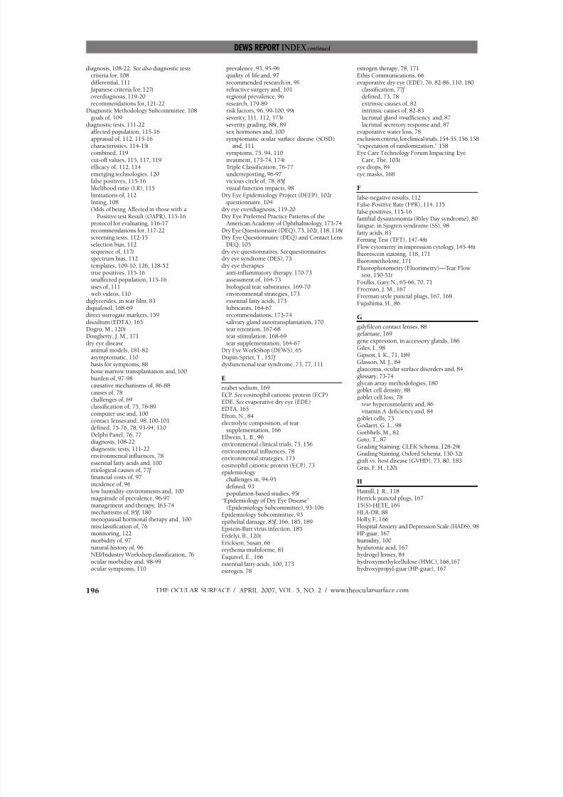
8/20/2019 dry eyes ii
http://slidepdf.com/reader/full/dry-eyes-ii 136/142
THE OCULAR SURFACE / APRIL 2007, VOL. 5, NO. 2 / www.theocularsurface.com196
diagnosis, 108-22. See also diagnostic tests criteria for, 108 differential, 111 Japanese criteria for, 127t overdiagnosis, 119-20 recommendations for, 121-22Diagnostic Methodology Subcommittee, 108 goals of, 109diagnostic tests, 111-22 affected population, 115-16 appraisal of, 112, 115-16 characteristics, 114-15t combined, 119 cut-off values, 115, 117, 119 efficacy of, 112, 114 emerging technologies, 120 false positives, 115-16 likelihood ratio (LR), 115 limitations of, 112 listing, 108 Odds of being Affected in those with a
Positive test Result (OAPR), 115-16 protocol for evaluating, 116-17
recommendations for, 117-22 screening tests, 112-15 selection bias, 112 sequence of, 117t spectrum bias, 112 templates, 109-10, 126, 128-52 true positives, 115-16 unaffected population, 115-16 uses of, 111 web videos, 110diglycerides, in tear film, 83diquafosol, 168-69direct surrogate markers, 159disodium (EDTA), 165Dogru, M., 120tDougherty, J. M., 171
dry eye disease animal models, 181-82 asymptomatic, 110 basis for symptoms, 88 bone marrow transplantation and, 100 burden of, 97-98 causative mechanisms of, 86-88 causes of, 78 challenges of, 69 classification of, 75, 76-89 computer use and, 100 contact lenses and, 98, 100-101 defined, 75-76, 78, 93-94, 110 Delphi Panel, 76, 77 diagnosis, 108-22 diagnostic tests, 111-22
environmental influences, 78 essential fatty acids and, 100 etiological causes of, 77 f financial costs of, 97 incidence of, 96 low humidity environments and, 100 magnitude of prevalence, 96-97 management and therapy, 163-74 mechanisms of, 85 f , 180 menopausal hormonal therapy and, 100 misclassification of, 76 monitoring, 122 morbidity of, 97 natural history of, 96 NEI/Industry Workshop classification, 76 ocular morbidity and, 98-99
ocular symptoms, 110
prevalence, 93, 95-96 quality of life and, 97 recommended research in, 99 refractive surgery and, 101 regional prevalence, 96 research, 179-89 risk factors, 96, 99-100, 99t severity, 111, 112, 173t severity grading, 88t, 89 sex hormones and, 100 symptomatic ocular surface disease (SOSD) and, 111 symptoms, 75, 94, 110 treatment, 173-74, 174t Triple Classification, 76-77 underreporting, 96-97 vicious circle of, 78, 85 f visual function impacts, 98Dry Eye Epidemiology Project (DEEP), 102t questionnaire, 104dry eye overdiagnosis, 119-20Dry Eye Preferred Practice Patterns of the
American Academy of Ophthalmology, 173-74
Dry Eye Questionnaire (DEQ), 73, 102t, 118, 118tDry Eye Questionnaire (DEQ) and Contact Lens
DEQ, 105dry eye questionnaires. See questionnairesdry eye syndrome (DES), 73dry eye therapies anti-inflammatory therapy, 170-73 assessment of, 164-73 biological tear substitutes, 169-70 environmental strategies, 173 essential fatty acids, 173 lubricants, 164-67 recommendations, 173-74 salivary gland autotransplantation, 170 tear retention, 167-68 tear stimulation, 168-69
tear supplementation, 164-67Dry Eye WorkShop (DEWS), 65Dupin-Spriet, T., 157 f dysfunctional tear syndrome, 73, 77, 111
E
ecabet sodium, 169ECP. See eosinophil cationic protein (ECP)EDE. See evaporative dry eye (EDE)EDTA, 165Efron, N., 84electrolyte composition, of tear
supplementation, 166Ellwein, L. B., 96environmental clinical trials, 73, 156environmental influences, 78
environmental strategies, 173eosinophil cationic protein (ECP), 73epidemiology challenges in, 94-95 defined, 93 population-based studies, 95t“Epidemiology of Dry Eye Disease”
(Epidemiology Subcommittee), 93-106Epidemiology Subcommittee, 93epithelial damage, 85 f , 166, 185, 189Epstein-Barr virus infection, 185Erdelyi, B., 120tErickson, Susan, 66erythema multiforme, 81Esquivel, E., 166essential fatty acids, 100, 173
estrogen, 78
estrogen therapy, 78, 171Ethis Communications, 66evaporative dry eye (EDE), 76, 82-86, 110, 180 classification, 77 f defined, 73, 78 extrinsic causes of, 82 intrinsic causes of, 82-83 lacrimal gland insufficiency and, 87 lacrimal secretory response and, 87evaporative water loss, 78exclusion criteria, for clinical trials, 154-55, 156, 158“expectation of randomization,” 158Eye Care Technology Forum Impacting Eye
Care, The, 103teye drops, 84eye masks, 168
F
false-negative results, 112False-Positive Rate (FPR), 114, 115false positives, 115-16familial dysautonomia (Riley Day syndrome), 80
fatigue, in Sjogren syndrome (SS), 98fatty acids, 83Ferning Test (TFT), 147-48tFlow cytometry in impression cytology, 145-46tfluorescein staining, 118, 171fluorometholone, 171Fluorophotometry (Fluorimetry)—Tear Flow
test, 150-51tFoulks, Gary N., 65-66, 70, 71Freeman, J. M., 167Freeman style punctal plugs, 167, 168Fujishima, H., 86
G
galyfilcon contact lenses, 88gefarnate, 169
gene expression, in accessory glands, 186Giles, I., 98Gipson, I. K., 71, 189Glasson, M. J., 84glaucoma, ocular surface disorders and, 84glossary, 73-74glycan array methodologies, 180goblet cell density, 88goblet cell loss, 78 tear hyperosmolarity and, 86 vitamin A deficiency and, 84goblet cells, 73Godaert, G. L., 98Goebbels, M., 82Goto, T., 87Grading Staining: CLEK Schema, 128-29tGrading Staining: Oxford Schema, 130-32tgraft vs. host disease (GVHD), 73, 80, 183Grus, F. H., 120t
H
Hamill, J. R., 118Herrick punctal plugs, 16715(S)-HETE, 169HLA-DR, 88Holly, F., 166Hospital Anxiety and Depression Scale (HADS), 98HP-guar, 167humidity, 100hyaluronic acid, 167hydrogel lenses, 84hydroxymethylcellulose (HMC), 166,167
hydroxypropyl-guar (HP-guar), 167
DEWS REPORT INDEX continued
8/20/2019 dry eyes ii
http://slidepdf.com/reader/full/dry-eyes-ii 137/142
THE OCULAR SURFACE / APRIL 2007, VOL. 5, NO. 2 / www.theocularsurface.com 197
hyperosmolarity, 86-87, 122hypoosmolarity, of salivary glands, 170hypo-osmotic artificial tears, 166
I
ICAM-1, 73, 88
ICDM-9-CM codes, 73, 96immune system information matrix, 184t research, 183-85Impact of Dry Eye on Everyday Life (IDEEL),
73, 97, 102t, 103, 118t features, 104incidence, 73, 96inclusion criteria, for clinical trials, 154-55, 156,
158inflammation anti-inflammatory therapy for, 170-73 information matrix, 184t mechanism of, 185 f research, 185inflammatory cytokines, 180
inflammatory lacrimal damage, 85 f inflammatory markers, 88inflammatory mediators, 78“intention-to-treat” principle, 155interblink intervals, 168International Classification of Disease, Ninth Revision, Clinical Modification (ICD-9 CM), 96International Dry Eye WorkShop (DEWS) abbreviations, 74 defined, 73 glossary, 73-74 membership, 70 Report Introduction, 69-70 subcommittee members, 70 subcommittees, 69International Sjogren’s Classification, 118tInternational Task Force (ITF), 173-74in vitro models, 182-83Isenberg, D., 98Ishida, R., 120tisotretinoin, 83
J
Japanese criteria for diagnosis, 127t Japanese dry eye awareness study, 103t Johnson, R., 119t
K
keratoconjunctivitis sicca (KCS), 73, 78, 180Knop, E., 88Kojima, T., 120tKorb, D. R., 120t, 168
Kurihashi, K., 168
L
lacrimal acinar damage, 84Lacrimal Functional Unit (LFU), 65, 189damage to, 76defined, 73disturbance of, 76lacrimal gland, 76 ablation, 81 denervation, 81 duct obstruction, 81 excessive reflex stimulation of, 87 hyposecretion, 79 infiltration, 78, 80 information matrix, 184t, 186t, 187t
insufficiency, 86-87
primary deficiencies, 80 reflex stimulation of, 86 research, 185-87 secondary deficiencies, 80-81lacrimal secretory compensation, 87lacrimal secretory response, 87lacrimal tear deficiency. See aqueous deficient
dry eye (ADDE)lacrimal tear secretion drug-related reduction of, 78 failure of, 78lanolin, 165-66laser assisted in situ keratomileusis (LASIK)
surgery. See LASIK surgeryLASIK-Induced NeuroEpitheliopathy (LINE),
73, 101LASIK surgery defined, 73 dry eye following, 81, 101 post-LASIK symptomatic keratitis, 111 tear film instability and, 87last observation carried forward (LOCF), 73, 156Lemp, M. A., 69, 71, 118
lid oil, 78-79, 85 f lids, 76 aperture disorders, 83 commensal organisms, 83 lid/globe congruity, 83likelihood ratio (LR) for diagnostic tests, 115lipids, 181t, 188tlissamine green staining, 118Liu, H., 120tlow-humidity environments, 100low-income populations, 96-97lubricants characteristics and effects of, 164-67 electrolyte composition, 166 osmolarity, 166
preservatives, 165-66 viscosity agents, 166-67lymphoma, 80
M
macromolecular complexes, 166Magalhaes, M., 84Management and Therapy Subcommittee, 163 goals, 163Maruyama, K., 168mass spectrometry, 180Mathers, W. D., 80, 120tMcCarty, C., 118McCulley, J. P., 83McMonnies Dry Eye Questionnaire, 84, 102t, 118tfeatures, 104
Medical Outcome Study Short Form-36, 97meibography, 83, 143tmeibometry, 83, 142tmeibomian excreta (meibum), 121meibomian foam, 83meibomian gland, 121 animal models, 188-89 atrophy, 83 information matrix, 188t, 189t lipids, 180 obstruction, 82-83 research, 187-89meibomian gland dysfunction (MGD), 82-83,
85 f , 111, 121, 172, 188 allergic conjunctivitis and, 86 amount of oil in lid margin reservoir, 83
causing evaporative dry eye, 82t
cicatricial, 83 degree of, 83 degree of gland dropout, 83 simple, 83 tear hyperosmolarity and, 87meibomianitis, 172meibomian lipids, 121, 180meiboscopy, 143tMelbourne Visual Impairment Project
Questionnaire, 93, 95t, 103t, 105membrane-spanning mucins, 166menopausal hormone therapy (MHT), 73, 100Mertzanis, P., 97methylprednisolone, 171MGD. See meibomian gland dysfunction (MGD)milieu exterieur, 77 f , 78milieu interieur, 77 f, 78mineral oil, 167minority populations, 96-97MMP-9 protein, 171moisture chamber spectacles, 168monoglycerides, 83morbidity, 97
motor nerves, 76mouse models dry eye disease, 181 immune system, 184 lacrimal gland, 186 ocular surface, 183MUC-4, 73, 166MUC-16, 166MUC5AC, 88, 180mucin markers, 88mucins, 73, 166, 180, 181t, 182tmucous membrane pemphigoid, 81multi-dimensional fatigue inventory (MFI), 73, 98multidose artificial tears, 165multifactorial diseases, 159multinational clinical trials, 159-60
muscarinic receptors, 186
N
nasolacrimal duct information matrix, 186t, 187t research, 185-87National Eye Institute (NEI), 69, 75, 93National Eye Institute (NEI) 42-Item Refractive
Error Questionnaire, 103tNational Eye Institute (NEI)/Industry Workshop
classification, 76National Eye Institute (NEI)-Visual Function Questionnaire (NEI-VFQ), 74, 97, 98, 102t, 104-5Nemeth, J., 120tnervus intermedius, 82neurogenic inflammatory cytokine response, 87
neurotrophic keratitis, 82neurturin-deficient mice, 181Nichols, J. J., 86, 168Noda-Tsuruya, T., 170“non-autoimmune” dry eye, 78non-invasive TFBUT, 121, 122non-Sjogren syndrome dry eye (NSSDE), 180 age-related, 80 classification, 77 f defined, 74, 80 forms of, 80, 80t IDEEL questionnaire and, 97 lacrimal gland duct obstruction, 81 lid oil and, 78-79 primary lacrimal gland deficiencies, 80 reflex hyposecretion, 81-82
secondary lacrimal gland deficiencies, 80-81
DEWS REPORT INDEX continued
8/20/2019 dry eyes ii
http://slidepdf.com/reader/full/dry-eyes-ii 138/142
THE OCULAR SURFACE / APRIL 2007, VOL. 5, NO. 2 / www.theocularsurface.com198
“normal-cholesterol absent” group (N[CA]), 83“normal-cholesterol present” group (N[CP]), 83nutritional deficiencies, 79
O
ocular allergy, 86
ocular comfort moisture chamber spectacles and, 168 tear supplementation and, 164ocular irritation, 76ocular lubricants. See lubricantsocular morbidity, 98-99ocular ointments and gels, 165-66Ocular Protection Index (OPI), 87, 149tocular sensory loss, 81tocular surface, 76 animal models, 182-83 chronic inflammation, 185 disorders, 84 dryness, 79t, 119 epithelial cell hyperosmolarity, 78 hyperosmolarity, 85 f
inflammation, 76, 168, 185 information matrix, 182t, 183t protection of, 166 research, 182-83, 182t staining, 118ocular surface disease, 86 classification, 110-11, 111 f research, 183Ocular Surface Disease Index (OSDI), 97, 102t, 118t defined, 74 features, 104ocular surface system (OSS), 74, 189ocular symptoms, 110Odds of being Affected in those with a Positive
test Result (OAPR) calculating, 116 for diagnostic tests, 114, 115-16 prevalence and, 116 f oncotic pressure, 166optical aberrations, 98osmolarity colloidal, 166 crystalloid, 166 of tear supplementation, 166osmoprotection, 166Ousler, G. W., 120toutcome analysis, for clinical trials, 155, 156, 158-59overdiagnosis, 119-20Oxford Schema, 118, 130-32t
P
palpebral aperture natural height of, 78 width of, 78parallel group studies, 154Parkinson disease (PD), 83Pflugfelder, S. C., 71, 119, 189PharMetrics’ Integrated Outcomes, 96photorefractive keratoplasty (PRK), 74, 101Physicians’ Health Study (PHS), 74, 93, 95, 95tpilocarpine, 169Pisella, P. J., 88placebo effects, 74, 158, 159polychlorinated biphenyls, 83polymacon contact lenses, 88posterior blepharitis, 82-83post-LASIK symptomatic keratitis, 111potassium, 166predictive value of a positive test (PPV), 114
pre-lens tear film (PLTF), 84preservatives elimination of, 165-66 ocular surface disorders and, 84 tear film instability and, 87-88 in tear supplements, 165-66 “vanishing,” 165prevalence, 93, 95-97 defined, 74 magnitude of, 96-97 Odds of being Affected in those with a
Positive test Result (OAPR) and, 116 f regional, 96 underreporting, 96-97 in women, 84, 95Prichard, N., 100-101primary lacrimal disease, 80primary lacrimal gland deficiencies, 80primary outcome measures, 155, 159primary Sjogren syndrome, 79proteins, 181tpunctal occlusion, 167-68, 171, 186punctal plugs
absorbable, 167 clinical studies, 167-68 complications, 168 contraindications for, 168 Freeman style, 167, 168 Herrick, 167 indications for, 168 nonabsorbable, 167 Smartplug, 167 tear production and, 168punctate keratoconjunctivitis, 86
Q
quality of life (QoL) defined, 74 in dry eye disease, 97 in Sjogren syndrome, 98 tear supplementation and, 164questionnaires characteristics, 113 in current use, 118 evaluation, 105 features of, 104-5 recommendations for, 117-18 research needed, 105 review of, 101-5 symptoms and quality of life instruments, 102-3t
R rabbit models androgen deficiency, 188-89 lacrimal gland, 181, 187t
Rajagopalan, K., 120trandomized clinical trials, 74, 154, 155rebamipide, 169receiver-operator characteristic (ROC) curve, 117reflex hyposecretion, 81-82reflex sensory block, 81reflex tearing, 76, 118-19, 120-21reflex trigeminal activity, 86refractive surgery, 101. See also LASIK surgeryresearch, 179-89 accessory lacrimal glands, 185-87 dry eye mechanisms, 189 immune system, 183-85 inflammation, 185 lacrimal gland, 185-87 meibomian gland, 187-89
nasolacrimal duct, 185-87
ocular surface, 182-83 tear film, 179-82Research Subcommittee, 65, 179 goals of, 179Riley Day syndrome, 80rose bengal staining, 167
S
S. aureus, 83Salisbury Eye Evaluation Questionnaire, 93, 102t features, 104Salisbury Eye Study, 95tsaliva, as tear substitute, 169, 170salivary gland autotransplantation, 170sample size, for clinical trials, 155, 158sarcoidosis, 80Schein, O. D., 82, 118, 120tSchein questionnaire, 118tSchiffman, R. M., 120tSchirmer test, 118-19, 126-27t defined, 74 without anesthesia, 74, 135t
screening tests appraisal of, 112-15 defined, 112 recommendations for, 117-21, 119-20seasonal allergic conjunctivitis, 87secondary lacrimal gland deficiencies, 80-81secondary Sjogren syndrome, 79secretagogues, 74, 168-69selection bias, in diagnostic tests, 112sensitivity, of diagnostic tests, 74, 114sensory nerves, 76serum, as tear substitute, 169-70severity grading, 77, 89sex hormones, 78, 100sex steroid deficiency, 100Shack-Hartmann aberrometer, 98Shihpai study, 95t, 100Shimazaki, J., 87Shine, W. E., 83, 171Sicca/SLE Questionnaire, 103tSicca/SS Questionnaire, 103tsilicone hydrogel contact lenses, 88single unit-dose tear substitutes, 165Sjogren syndrome (SS), 111 classification, 76, 77 f, 79 f criteria for ocular manifestations of, 119t quality of life in, 98Sjogren syndrome dry eye (SSDE), 79-80, 180 autoantibodies, 79t defined, 79 forms of, 79 histopathology, 79t nutritional deficiencies and, 79
ocular dryness, 79 ocular signs, 79t ocular symptoms, 79t oral syndromes, 79t primary, 79 salivary gland involvement, 79t secondary, 79Sjogren syndrome-related KCS IDEEL questionnaire and, 97 tear stimulation and, 169Smartplug, 167Smith, J., 71, 118sodium chlorite, 165sodium perborate, 165soft contact lenses, 85specificity, in diagnostic tests, 74, 114-15
spectrum bias, in diagnostic tests, 112
DEWS REPORT INDEX continued
8/20/2019 dry eyes ii
http://slidepdf.com/reader/full/dry-eyes-ii 139/142
THE OCULAR SURFACE / APRIL 2007, VOL. 5, NO. 2 / www.theocularsurface.com 199
Spriet, A., 157 f Standards of Good Clinical Practice, 156staring tear breakup dynamics (S-TBUD), 121Stern, J. J., 173Stern, M. E., 189Stevenson, H. A., 98Strombeck, B., 98Sullivan, Amy G., 65, 70Sullivan, B., 120tSullivan, David A., 65, 69, 70, 71Sullivan, Rose M., 65, 70Sumatra study, 95tsurrogate markers, 74, 155, 159Survey Manual and Interpretation Guide, 97Sutcliffe, N., 98symptomatic conjunctivitis, 110symptomatic dry eye, 110symptomatic keratitis, 110symptomatic lid disease, 110symptomatic ocular surface disease (SOSD) classification of, 110-11 defined, 111 dry eye disease and, 111
symptomatology, 110symptom questionnaires, 94systemic drug use, 82
T
Tamer, C., 83tear clearance rate (TCR), 74tear-deficient dry eye. See aqueous deficient dry
eye (ADDE)tear film animal, 181t contact lenses and, 84 destabilization of, 76 hyperosmolarity, 78, 85 f, 86-87 information matrix, 181t Lacrimal Functional Unit and, 76 model, 166 noninvasive study techniques, 121, 122 osmolarity, 119, 122, 166, 179-80, 181t research, 179-82, 182 thinning rates, 86tear film analysis system (TMS), 87tear film breakup time (TFBUT), 74, 116, 118,
133-34t blink rate and, 83 optical aberrations and, 98 tear film instability and, 87tear film instability, 85 f, 86, 87-88 allergic conjunctivitis and, 86 LASIK surgery and, 87
tear film lipid layer interferometry, 139-40tTear Film & Ocular Surface Society, The
(TFOS), 65, 69 officers and staff, 70 website, 70tear film stability test performance, 118 threats to, 76 viscosity agents and, 166Tear Function Index (Liverpool modification), 152ttear function index (TFI), 122Tear meniscus radius, height, and cross sectional
area, 137-38tTear Osmolarity test, 136ttear retention, 167-68 moisture chamber spectacles, 168 punctual occlusion, 167-68tears composition, 120-21, 180 deficiency, 94 evaporation rate, 87 flow obstruction, 85 f flow reduction, 87
sampling, 120 stimulation, 168-69 volume, 181ttear stability analysis system (TSAS), 111, 141ttear supplementation characteristics and effects of, 164-67 electrolyte composition, 166 osmolarity, 166 preservatives in, 165-66 viscosity agents, 166-67tear turnover rate (TTR), 120templates, 179 development of, 109-10 headings, 110 “web video” section, 110testosterone, topically applied, 171
tetracyclines, 171-73 for acne rosacea, 171-72 anti-angiogenic properties, 171 antibacterial properties, 171 anti-inflammatory properties, 171 for chronic posterior blepharitis, 172 clinical applications, 171-73 dosage and safety, 172-73 for meibomian gland dysfunction (MGB), 172Thai, L. C., 84thermal burns, 81Thomas, E., 98Tomlinson, A., 71, 80topic anesthesia, 84
trachoma, 81treatment assignments, for clinical trials, 155triple A syndrome (Allgrove syndrome), 80Triple Classification, 76-77true positives, 115-16Tsubota, K., 69, 71, 168, 169
U
unaffected population, 115-16upgaze, 78U.S. Food and Drug Administration (FDA), 164, 165Utility assessment questionnaire, 103t
V
Valtysdottir, S. T., 98van Bijsterveld study, 118, 119“vanishing” preservatives, 165vernal keratoconjunctivitis, 74, 87video display terminals, 78, 100viscosity agents, 166-67Vision-Targeted Health-Related Quality of Life
(VT-HRQ), 74, 98
visual function contact lenses and, 85-86 dry eye and, 98 vision, 167Vitale, S., 98Vitali, C., 76, 119tvitamin A deficiency, 84, 87
W
waking tear flow, 76 Wald, N. J., 115 Wang, J., 120tweb videos, 110women dry eye prevalence, 84, 95, 100 dry eye risk factors, 96
menopausal hormone therapy (MHT), 100 Women’s Health Study, 74, 93, 95, 95t questionnaire, 102t, 104, 118t
X
xerophthalmia, 74, 84, 87
Y
Yazdani, C., 96yellow barrier filers, 118Yen, M. T., 186Yokoi, N., 120tYoo, S. E., 172
DEWS REPORT INDEX continued

8/20/2019 dry eyes ii
http://slidepdf.com/reader/full/dry-eyes-ii 140/142
THE OCULAR SURFACE / APRIL 2007, VOL. 5, NO. 2 / www.theocularsurface.com208
STEERING COMMITTEE MEMBERS
Christophe Baudouin has received financial
support from Alcon, Allergan, Pfizer, Santen,and Thea. He has been a consultant to Alcon,Allergan, Bausch & Lomb, Novagali, Pfizer,Santen, and Laboratoires Théa and is currentlyconsulting for Allergan, Novartis, Pfizer andNovagali.
Anthony J. Bron has a personal financial inter-est in OcuSense. He has been a consultant toAcucela, Alcon, Novartis, OcuSense, Proctor andGamble, Senju, and Takeda, and he has receivedgifts from Alcon, Allergan, and Takeda. Cur-rently, Professor Bron is a consultant for Actelion,Acucela, Canfite Novagali, OcuSense, Proctorand Gamble and Takeda. He receives meetingsupport from Alcon, and Allergan.
Murat Dogru has no proprietary interest in any
company with products or interests related todry eye.
Gary N. Foulks has received financial supportfrom Alacrity, Allergan, Inspire, Lantibio,Novartis, and Otsuka and he has a personalfinancial interest in Inspire. He is, or has been,a consultant to Alacrity, Alcon, Alimera Sciences,Allergan, Bausch & Lomb, Fovea, Inspire, ISTA,Lantibio, Nascent, Novagali, Novartis, OcuSense,Otsuka, and Pfizer, Santen, Sarcode and XTLBiopharmaceuticals. He has received gifts fromAlacrity, Alcon, Bausch & Lomb, Inspire, Otsuka,and Pfizer.
Ilene K. Gipson has received financial supportfrom Alcon.
Michael A. Lemp has received financial supportfrom Addition Technologies, Alcon, Allergan,Bausch & Lomb, Inspire, Novagali, Novartis,Pfizer, OcuSense, and Santen. He is a consultantfor Otsuka Research Corp. and Fovea Pharma-ceuticals. He has a personal financial interest inInspire and OcuSense, and he is Chief MedicalOfficer of OcuSense.
J. Daniel Nelson has no proprietary interest in anycompany with products or interests related todry eye. He is listed on a patent for dry eye thatis held by the University of Illinois.
Kelly K. Nichols has a personal financial interestin Inspire, and she is a consultant to Alcon, Al-lergan, and Inspire.
Stephen C. Pflugfelder has received support from
Allergan, and he is a consultant to Allergan. Heis listed on two patents held by the University ofMiami that relate to dry eye disease.
Debra A. Schaumberg has received financialsupport from DSM Pharmaceuticals, Daiichi,and Pfizer Consumer Healthcare, Inc. She has apersonal financial interest in OcuSense, and sheis a consultant to OcuSense.
Janine A. Smith is named on a patent held by theUS government.
David A. Sullivan has received financial supportfrom Allergan, and has serverd as a consultantto Allergan and Novartis. He is named as theinventor on patents for Sjogren Syndrome andKeratoconjunctivitis Sicca treatments held by theSchepens Eye Research Institute.
Alan Tomlinson has received financial supportfrom Allergan, Renaissance Pharmaceuticals,and Pfizer. He has a personal financial interestin Bausch & Lomb.
Kazuo Tsubota has received financial supportfrom Etech and Santen, and he is a consultantto Nidek and Rainbow. He is named on multiplepatents both in the U.S. and Japan that relate todry eye disease.
SUBCOMMITTEE MEMBERS
Mark Abelson is president of Ophthalmic Re-search Associates, a company that providesconsulting and research services to many oph-thalmic industries.
Julie Albietz has no proprietary interest in anycompany with products or interests related todry eye.
Pablo Argüeso has received financial supportfrom Alcon.
Penny Asbell serves as a Speaker Bureau consul-tant to Alcon, Allergan, Bausch & Lomb, Inspire,Novagali, Novartis, Otsuka, Pfizer, Santen, andVistakon Pharma.
Jules Baum has no proprietary interest in anycompany with products or interests related todry eye.
Carolyn Begley has received financial supportfrom Alcon Research, Allergan, and Vistakon.She is a consultant to Alcon and Novartis.
Roger Beuerman has received financial supportfrom Advanced Ocular Systems and Allergan,and he is a consultant to both companies.
Stefano Bonini has received financial supportfrom Alcon, Allergan, Bausch & Lomb, Novartis,SIFI and SOOFT. He is a member of the AnabasisSociety, SrL, which holds rights to a patent forthe use of NGF as it relates to the treatment dryeye disease.
Igor Butovich has no proprietary interest in anycompany with products or interests related todry eye.
Barbara Caffery has no proprietary interest inany company with products or interests relatedto dry eye.
Margarita Calonge is a consultant to Allergan.
Reza Dana has received financial support fromAllergan, Biogen-Idec, and Johnson & Johnson.
He is a consultant to Johnson & Johnson.Darlene Dartt has received financial support
from Johnson & Johnson and Otsuka. She is aconsultant to Johnson & Johnson, and holds apatent with that company. She has received giftsfrom Johnson & Johnson and Alcon.
Desmond Fonn has received financial supportfrom Advanced Medical Optics, Alcon, Allergan,Bausch & Lomb, Ciba Vision, CooperVision,and Johnson & Johnson Vision Care. He is aconsultant to Ciba Vision, and he has receivedgifts from Ciba Vision and CooperVision.
Daniel Gamache is employed by Alcon and isnamed on many of its patents related to dryeye disease.
Gerd Geerling is a consultant to Pfizer Health
Care and Novartis Pharma. He holds a patentapplication for a dry eye product. The patenthas not been assigned.
Eiki Goto has no proprietary interest in anycompany with products or interests related todry eye.
Franz Grus has no proprietary interest in anycompany with products or interests related todry eye.
Bryan Ham has no proprietary interest in anycompany with products or interests related todry eye.
Marcia Jumblatt has no proprietary interest inany company with products or interests relatedto dry eye.
Shigeru Kinoshita has received financial supportfrom Alcon Japan, Alblast Ltd, Otsuka, Senju,and Santen. He is a consultant to AdvancedMedical Optics, Otsuka and Senju.
Donald Korb has a personal financial interest inOcular Research of Boston and Kolis Scientific.He is an employee of and holds patents withboth companies.
Friedrich Kruse has no proprietary interest inany company with products or interests relatedto dry eye.
Peter Laibson has no proprietary interest in anycompany with products or interests relatedto dry eye. He is on the Speakers Bureau forBausch & Lomb.
James McCulley is a consultant for Alcon Labo-ratories.
Juan Murube has no proprietary interest in any
company with products or interests related todry eye disease.
Gary Novack owns and operates PharmaLogic,which provides editorial and consulting servicesto industry.
George Ousler is employed by OphthalmicResearch Associates, a company that providesconsulting and research services to many oph-thalmic industries.
Jerry Paugh’s employing institution has receivedfinancial support from Alcon, Allergan, andBausch & Lomb.
Friedrich Paulsen has no proprietary interest inany company with products or interests relatedto dry eye.
Ian E. Pearce has received support from Allergan,Pfizer, and Sequani, Ltd. He is a consultant toPfizer, and he has received gifts from Pfizer.
Maurizio Rolando has received gifts from Alconand Pfizer.
Oliver Schein has received financial support fromBausch & Lomb, Novartis, and he has receivedfinancial support from CIBA (inactive). He is aconsultant to Bausch & Lomb.
Jun Shimazaki has no proprietary interest inany company with products or interests relatedto dry eye.
Michael Stern is employed by Allergan.
DEWS Disclosures
Disclosures of financial/proprietary relationships of DEWS participants with companies with products or interests related to dry eye
continued on page 204
202

8/20/2019 dry eyes ii
http://slidepdf.com/reader/full/dry-eyes-ii 141/142
THE OCULAR SURFACE / APRIL 2007, VOL. 5, NO. 2 / www.theocularsurface.com 209

8/20/2019 dry eyes ii
http://slidepdf.com/reader/full/dry-eyes-ii 142/142
Deborah Sweeney has no proprietary interest in any company with productsor interests related to dry eye.
John Tiffany is a consultant to Pfizer Consumer Healthcare. He holds a patentfor lipid-containing eyedrops (no longer active).
Ikuko Toda has no proprietary interest in any company with products orinterests related to dry eye.
John Ubels has no proprietary interest in any company with products orinterests related to dry eye. He is a consultant for Alcon Research, Ltd.
Hitoshi Watanabe has no proprietary interest in any company with productsor interests related to dry eye.
Mark Willcox has received financial support from CIBA, Alcon, Allergan,Clearlab International Pte, and Ocular Sciences (CooperVision). He cur-rently receives funding from Alcon, Allergan, Bausch & Lomb, and CIBAVision.
Clive G. Wilson has received financial support from industries in the fieldsof gastroenterology and ocular drug delivery unrelated to dry eye. He isa consultant to Allergan (retinal drug delivery) and to GlaxoSmithKline(biopharamaceutics).
Norihiko Yokoi has no proprietary interest in any company with products orinterests related to dry eye.
INDUSTRY LIAISON COMMITTEE
Fouad Amer is employed by Novartis.
Michael J. Brubaker is employed by Alcon.
Timothy Comstock is employed by Bausch & Lomb.
David Eveleth is employed by Pfizer.
William Florida formerly employed by Novartis.
Fulvio Foschini is employed by SOOFT.
Sherryl Frisch formerly employed by Pfizer, now Johnson & Johnson, McNeilConsumer Health Care Group
Jeff Gilbard is employed by Advanced Vision Research and is a consultantto that company.
Kate Kline is employed by Allergan.
Ami Shah formerly of Bausch & Lomb
M N k i l d b S
DEWS DISCLOSURES continued