Embed Size (px)
Citation preview

DR. HAMAD AL-QAHTANI , MD , CABS , FRCS
ASSOCIATE PROFESSOR & CONSULTANT HEPATOBILIARY
SURGEON

THE PANCREAS

Surgical anatomy of the pancreas


Pancreatitis

Acute pancreatitis
It is the acute inflammation of the pancreas

Etiology1- Gallstone2- Alcohol3- Hyperlipidemia4- Hypercalcemia5- Heridetary6- Autoimmun7- Infection ( mumps and coxsaki B viral infection)8- Trauma (blunt , penetrating , surgical , ERCP)9- Pancreatic duct obstruction ( neoplasm , worms , pancreatic divism )10- Medications ( thiazid , steroid , azathioprin)11- Idioipathic
Clinical features
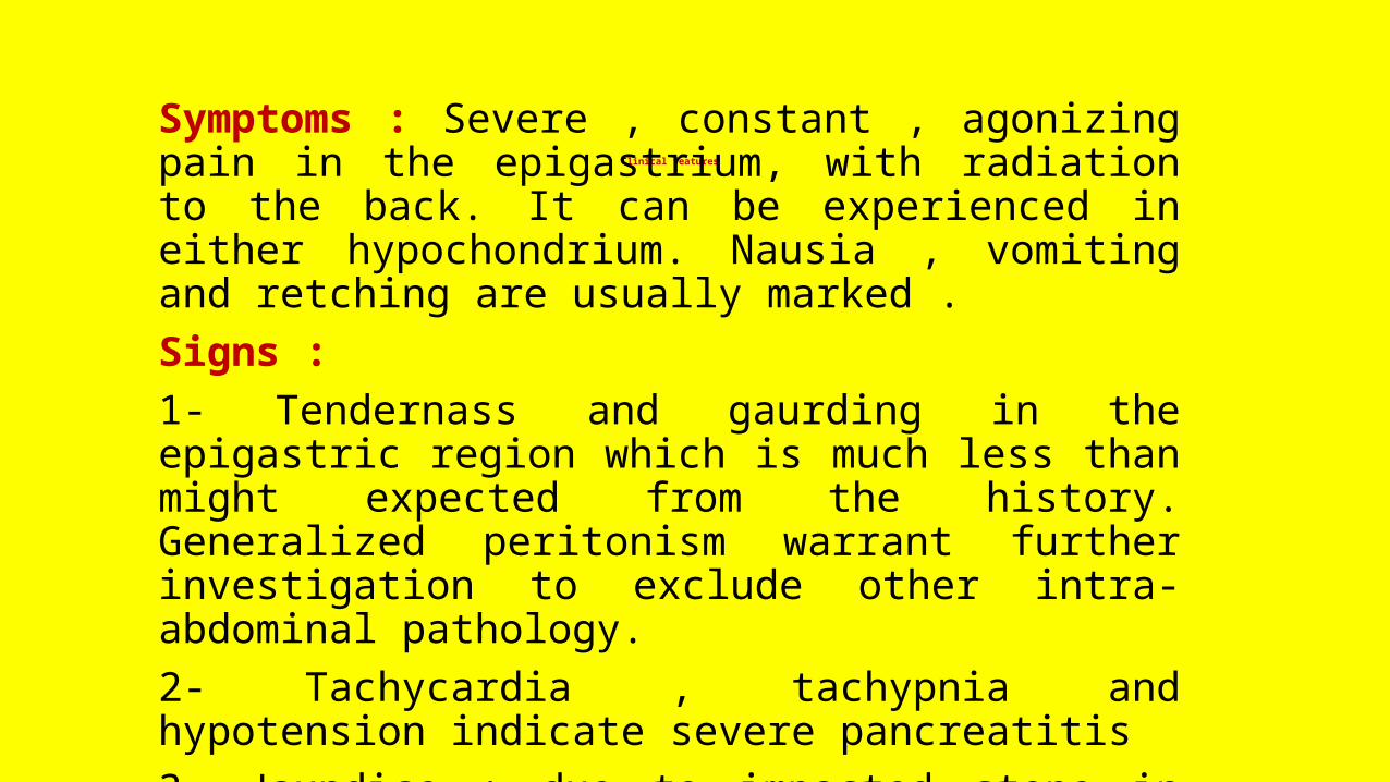
Symptoms : Severe , constant , agonizing pain in the epigastrium, with radiation to the back. It can be experienced in either hypochondrium. Nausia , vomiting and retching are usually marked .Signs :1- Tendernass and gaurding in the epigastric region which is much less than might expected from the history. Generalized peritonism warrant further investigation to exclude other intra-abdominal pathology.2- Tachycardia , tachypnia and hypotension indicate severe pancreatitis3- Jaundice : due to impacted stone in the ampulla or due to the pressure of edematous pancreas on the distal common bile duct. It should raise the possibility of co-existing cholangitis.

Gallstone blocking the CBD and pancreatic duct in gallstone pancreatitis

Investigations 1- The key to diagnosis of acute pancreatitis is a high index of suspision and measurment of the serum amylase concentration. The usuall diagnostic cut-off for serum amylase is three times the upper reference limit. Serum lipase is alternative and more specific.
2- Other underlying causes of hyperamylasemia in patients with abdominal pain include : mesentric vascular ischemia , small bowel strangulation , perforated doudenal ulcer , rupture aortic aneurysm , ruptured ectopic pregnancy , acute cholecystitis.

3- Complete blood count , urea , electrolytes , liver function test , LDH , lipid profile , coagulation profile, serum calcium, C-reactive protein, blood glucose.
4- Imaging : Chest X-ray should be done to look for pleural effusion due to acute pancreatitis , and it may show air under the diaphragm in cases of perforated peptic ulcer. Ultrasound abdomen to look for the presence of gallstone as underlying cause of acute pancreatitis . Abdominal computed tomography is indicated to clarify the diagnosis if the diagnosis of acute pancreatitis still in doubt , to look for the complications of acute pancreatitis or to assess for any evidence of necrotizing pancreatitis.

Assessment of severity of acute pancreatitis
The aim of severity assessment is the early recognition of the patients with severe pancreatitis and to ensure that they admitted in high dependency unit or critical
care unit for intensive management .

Ranson’s Criteria

1 (For non-gallstone pancreatitis, the parameters are:
At admission:
1- Age in years > 55 years
2- White blood cell count > 16000 cells/mm3
3- Blood glucose > 10 mmol/L (> 200 mg/dL)
4- Serum AST > 250 IU/L
5- Serum LDH > 350 IU/L
Within 48 hours:
1- Serum calcium < 2.0 mmol/L (< 8.0 mg/dL)
2- Hematocrit fall > 10%
3- Oxygen (hypoxemia PaO2 < 60 mmHg)
4- BUN increased by 1.8 or more mmol/L (5 or more mg/dL) after IV fluid hydration
5- Base deficit (negative base excess) > 4 mEq/L
6- Sequestration of fluids > 6 L

The criteria for point assignment is that a certain breakpoint be met at anytime during that 48 hour period, so that in some situations it can be calculated shortly after admission. It is applicable to non-gallstone
pancreatitis.
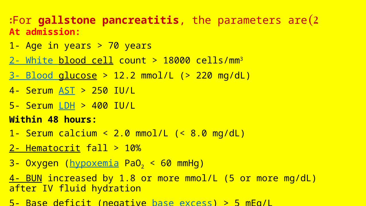
2 (For gallstone pancreatitis, the parameters are:At admission:
1- Age in years > 70 years
2- White blood cell count > 18000 cells/mm3
3- Blood glucose > 12.2 mmol/L (> 220 mg/dL)
4- Serum AST > 250 IU/L
5- Serum LDH > 400 IU/L
Within 48 hours:
1- Serum calcium < 2.0 mmol/L (< 8.0 mg/dL)
2- Hematocrit fall > 10%
3- Oxygen (hypoxemia PaO2 < 60 mmHg)
4- BUN increased by 1.8 or more mmol/L (5 or more mg/dL) after IV fluid hydration
5- Base deficit (negative base excess) > 5 mEq/L
6- Sequestration of fluids > 4 L

Interpretation of Ranson's criteria
- If the score ≥ 3, severe pancreatitis is likely.- If the score < 3, severe pancreatitis is unlikelyOr- Score 0 to 2 : 2% mortality- Score 3 to 4 : 15% mortality- Score 5 to 6 : 40% mortality
-Score 7 to 8 : 100% mortality..

Alternatively, pancreatitis severity can be assessed by
any of the following :
- APACHE II score ≥ 8 - Organ failure- Substantial pancreatic necrosis (at least 30% glandular necrosis according to contrast-enhanced CT)

TreatmentMost of attacks of acute pancreatitis will settle with conservative treatment including :
1. Pain relief : opiates administration
2. Fluid resuscitation : patients with severe pancreatitis require large volumes of fluid to maintain adequate urine output and blood pressure. Adequate early resuscitation in such cases is the most important consideration in early treatment .
3. Antibiotics prophylaxis: it is indicated in severe necrotizing pancreatitis and the recommended antibiotics is the imipenem or meropenem

4. Nutritional support : patients with severe pancreatitis who is unable to resume normal oral diets within 72 hours require nutritional support. This is best delivered by an enteral rather than parenteral route.
5. Endoscopic treatment: gallstone pancreatitis is due to the transient impaction of a stone at the papilla causing pancreatic duct obstruction. ERCP with sphincterotomy is indicated in patient with acute pancreatitis with persistent obstructive jaundice with or without cholangitis.
6. Surgical treatment: patients with gallstone related acute pancreatitis should undergo cholecystectomy ( mild pancreatitis during the index admission while severe pancreatitis interval cholecystectomy in 8 – 12 weeks after resolution of the attach of severe acute pancreatitis). Note: Surgery is indicated in patient with infected necrotizing pancreatitis or in patient with sterile necrotizing pancreatitis who deteriorate e and develop progressive multi-organ failure.

Complications of acute pancreatitis
1 .Infected pancreatic necrosis It is a serious complication which can develops in acute
necrotizing pancreatitis with high mortality rate. It can be diagnosed by presence of gas bubbles in CT scan at the area of pancreas or by fine needle aspiration under imaging guidance. Such patients need intensive management with antibiotics coverage and possibly surgical intervention ( surgical debridement ).

2 .Pancreatic pseudocyst It is a collection of pancreatic secretions and inflammatory exudate enclosed in a wall of fibrous or granulomatous tissue. It is differs from a true cyst in that collection has no epithelial lining. It form commonly in the lesser sac near the pancreas and persist for 4 weeks or more from the onset of acute pancreatitis. It need drainage if it persists more than 6 week after the attack of pancreatitis , the size > 6 cm and symptomatic

CT scan of pancreatic pseudocyst

3 .Pancreatic abscess It is a circumscribed intra-abdominal collection of pus , usually in proximity to the pancreas. This is a result of
infection of a pseudocyst. It need urgent drainage .
CT scan of pancreatic abscess

4. Gastrointestinal bleeding Severe pancreatitis may be complicated by bleeding from gastritis , erosion or duodenal ulceration. Thrombosis of splenic vein can lead to splenomegaly , gastric fundal varices which can result in massive upper gastrointestinal bleeding ( sinistral , left sided , compartmental portal hypertension) .
5. Progressive jaundice
due to impacted stone in the papilla or compression of distal common bile duct by enlarged edematous pancreas.

sinistral , left sided , compartmental portal hypertension

Chronic pancreatitis
It is a chronic inflammatory condition characterized by fibrosis and destruction of exocrine pancreatic tissue .

Etiology
1. Alcohol
2. Hereditary pancreatitis
3. Idiopathic

Pathophysiology
The secretion of an viscid pancreatic secretion may allow protein plugs to form in the duct system and these plugs subsequently calcify to form duct stones. Impaired flow of pancreatic juice then leads to inflammation , stricture formation in duct system , and progressive replacement of gland by fibrous tissue. Loss of acinar tissue is reflected by steatorrhea and in time loss of islet tissue may lead to diabetes mellitus.

Clinical features
1. Pain is the outstanding feature in most cases. It characteristically epigastric with marked radiation through to the back and is eased by leaning forward.
2. Weight loss is usual and reflects a combination of inadequate intake , poor diets and malabsorption.
3. Steatorrhea is common , the bowel motion is pale , bulky , offensive ,floating on water , and difficult to flush.
4. Diabetes mellitus develop in about one third of patients
5. Other less common manifestation of chronic pancreatitis include : transient or intermittent obstructive jaundice , duodenal obstruction , and splenic vein thrombosis ( leading to splenomegaly , hypersplenism , gastric and esophageal varices : compartmental, left sided or sinistral portal hypertension that may cause massive upper gastrointestinal bleeding)
Complications of chronic pancreatitis
1. Exocrine insufficiency
2. Endocrine insufficiency
3. Malignant transformation
4. Jaundice due to compression of distal CBD or tumor formation in the head of pancreas
5. Left sided ( sinistral or compartmental ) portal hypertension due to splenic vein compression or thrombosis
6. Gastric outlet obstruction due to duodenal compression

Investigation and diagnosis
1 .X-ray abdomen may show the scattered calcification in the area of pancreas.

2. CT scan abdomen : It may show the speckled calcifications typical of chronic pancreatitis , inflammatory changes , tumor , pancreatic duct dilatation or pseudocyst.
3. MRCP : to show the architecture of pancreatic duct , especially if surgery or endoscopic intervention is required.
4. Pancreatic endocrine function is assessed by measurement of fasting and postprandial blood glucose levels that may be supplemented by glucose tolerance test.
5. Pancreatic exocrine function can be assessed by measurement of fecal fat contents while the patient on fat controlled fat contents at 100g/day

TreatmentA. Conservative treatment
1. Pain relief
2. Alcohol abstinence
3. Exocrine replacement enzymes
4. Endocrine treatment with insulin or oral hypoglycemic drugs
5. Nutritional support

B. Endoscopic treatment
Pancreatic duct stenting is indicated sometimes when there is dominant pancreatic duct stricture or with disrupted pancreatic duct with pseudocyst or ascites formation.
C. Surgical treatment
Drainage or resective surgical intervention is indicated for :1. intractable pain2. Development of complications ( pseudocyst , compression of bile duct , duodenum ,portal vein or splenic vein that produce symptoms)3. Tumor formation

Neoplasms of the pancreas

Neoplasms of endocrine pancreas1. Insulinoma2. Glucagonoma3. Gastronome 4. VIPoma
Neoplasms of exocrine pancreasBenign tumors1. Serous cystadenoma2. Mucinous cystadenoma3. Intraductal papillary mucinous neoplasmMalignant tumors1. Ductal adenocarcinoma ( the commonest )2. acinar adenocarcinoma3. Mucinous cystadenocarcinoma4. Intraductal papillary mucinous neoplasm
Note : periampullary tumor could originate from the head of pancreas , distal CBD ,or from the Ampullary mucosa of duodenum and commonly the patient present early with jaundice due to the early obstruction of CBD.

Periampullary tumor

Periampullary tumor (endoscopic view )

Clinical features of pancreatic neoplasms
Presenting symptoms dependent on the site of the tumor within the pancreas.
1. Tumor in the head of pancreas : painless obstructive jaundice associated with weight loss is the classical presentation due to obstruction of the CBD. Obstruction of the flow of bile to the intestine will disrupt the enterohepatic circulation which results in dark urine and pale stool. Patient also may have intense itching. Gallbladder may become dilated and palpable ( Courvoisier's sign or law).
2. Tumor in the body and tail of pancreas : Biliary obstruction occur late , and symptoms are vague , with anorexia, weight loss , and with subsequent involvement of retroperitoneum , the development of back pain. New onset diabetes may predate the diagnosis. A late manifestation is a malignant-associated hypercoagulable state , resulting in intravascular clots with vasculitis , named thrombophlebitis migrans ( Trousseau's sign)

Investigations of pancreatic tumors
Laboratory tests : Liver function teste to confirm the cholestasis, tumor markers especially CA19-9 to help in diagnosis of malignant tumor of pancreas
Imaging
1. Ultrasound abdomen is the initial imaging which will show intra and extrahepatic biliary dilatation in jaundiced patient and will asses for gallstone.
2. CT scan and MRI can assess the site of the tumor in the pancreas and the evidence of metastases or local invasion by the tumor ( staging)
3. Upper gastrointestinal endoscopy and endoscopic ultrasound guidance can help in taking biopsy from the tumor

Tumor in the head of pancreas

Treatment A. Curative treatment Surgical resection currently offers the only potential for cure in pancreatic tumors. Tumors localized to the pancreatic parenchyma , or with limited involvement of peripancreatic fat or lymph nodes may be considered for resection
1. Tumor involving the head of pancreas treated by pancreaticoduodenectomy ( Whipple's procedure ) , which entails block resection of the head of pancreas , the distal half of the stomach , the duodenum , gallbladder and common bile duct.
2 .Tumor involving the body or tail of pancreas removed by distal pancreatectomy and splenectomy.3. Pruritus treatment with cholestyramine
4. Good operative risk patient may undergo triple bypass to relieve the obstructive jaundice and duodenal obstruction

Triple bypass procedure

Whipple's procedure

Distal pancreatectomy
B. Palliative treatment
The aim is optimization of life in patient with non-resectable tumor. Such patients require histological diagnosis before starting any chemotherapy.
1. Obstructive jaundice : can be relieved by ERCP with biliary stenting
2. Pain relief with analgesia or splanchnic nerve block ( percutaneous , surgical or endoscopic ultrasound guidance).
3. Pruritus treatment with cholestyramine
4. Good operative risk patient may undergo triple bypass to relieve the obstructive jaundice and duodenal obstruction

Triple bypass procedure

THANK YOU