Embed Size (px)
Citation preview

DNA & RNA Structure
Reading Assignment: Chapter 2, pgs 12-29, Chapter 12, pgs 315-323
BCH 5413
Dr. Yang
Fall 2013
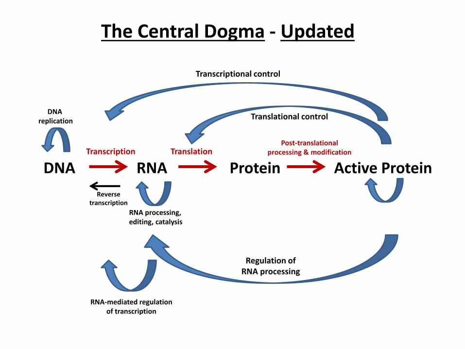
The Central Dogma - Updated
DNA RNA Protein Active Protein Transcription Translation
Post-translational processing & modification
Reverse transcription
DNA replication
RNA processing, editing, catalysis
Transcriptional control
Translational control
Regulation of RNA processing
RNA-mediated regulation of transcription

Nucleic Acid Bases
*
RNA-DNA Sugars
DNA Trinucleotide Phosphodiester bonds

Hydrogen Bonding in DNA
Watson-Crick Base Pairing
-- Chargaff’s rules: A=T and C=G
-- Propeller twist about 1o
-- Each base pair presents unique chemical
groups to the major groove
Major
Minor
Major Minor

DNA Structure


Semi-Conservative Replication of DNA (The Meselson & Stahl Expt.)

Alternative Double-Stranded DNA Conformations
A-DNA:
-- dehydrated form
-- right-handed helix
-- 11 bases per helical turn
-- base pairs tilted 19o
-- major groove narrow and deep
Z-DNA
-- >1% of cellular DNA
-- favored by G-C repeats
-- left-handed helix
-- 12 base pairs per helical turn
-- base pairs tilted 9o
-- major groove flattened, nearly gone
-- Poxvirus E3L virulence factor binds
Z-DNA down-regulating apoptosis genes
A-DNA B-DNA Z-DNA
RNA-DNA hybrids
RNA double helices
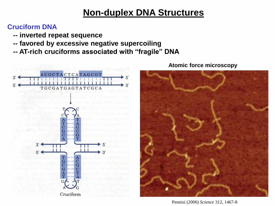
Non-duplex DNA Structures
Cruciform DNA
-- inverted repeat sequence
-- favored by excessive negative supercoiling
-- AT-rich cruciforms associated with “fragile” DNA
Atomic force microscopy
Pennisi (2006) Science 312, 1467-8
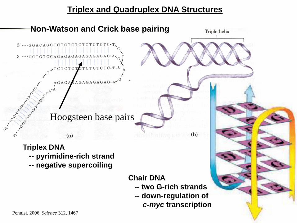
Triplex and Quadruplex DNA Structures
Triplex DNA
-- pyrimidine-rich strand
-- negative supercoiling
Chair DNA
-- two G-rich strands
-- down-regulation of
c-myc transcription
Hoogsteen base pairs
Non-Watson and Crick base pairing
Pennisi. 2006. Science 312, 1467

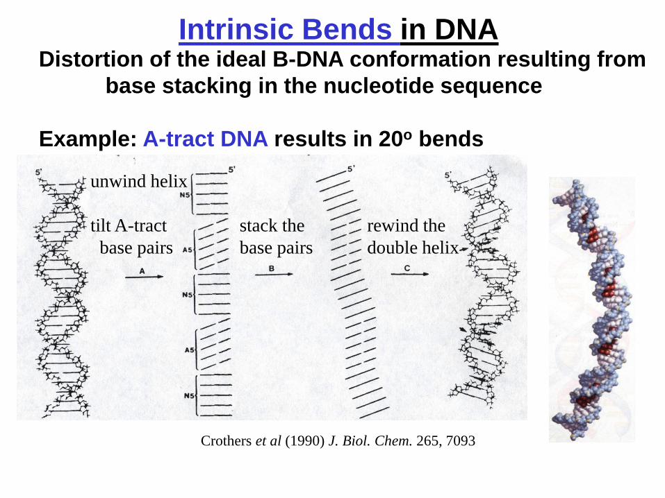
Intrinsic Bends in DNA Distortion of the ideal B-DNA conformation resulting from
base stacking in the nucleotide sequence
Example: A-tract DNA results in 20o bends
unwind helix
tilt A-tract
base pairs
stack the
base pairs
rewind the
double helix
Crothers et al (1990) J. Biol. Chem. 265, 7093
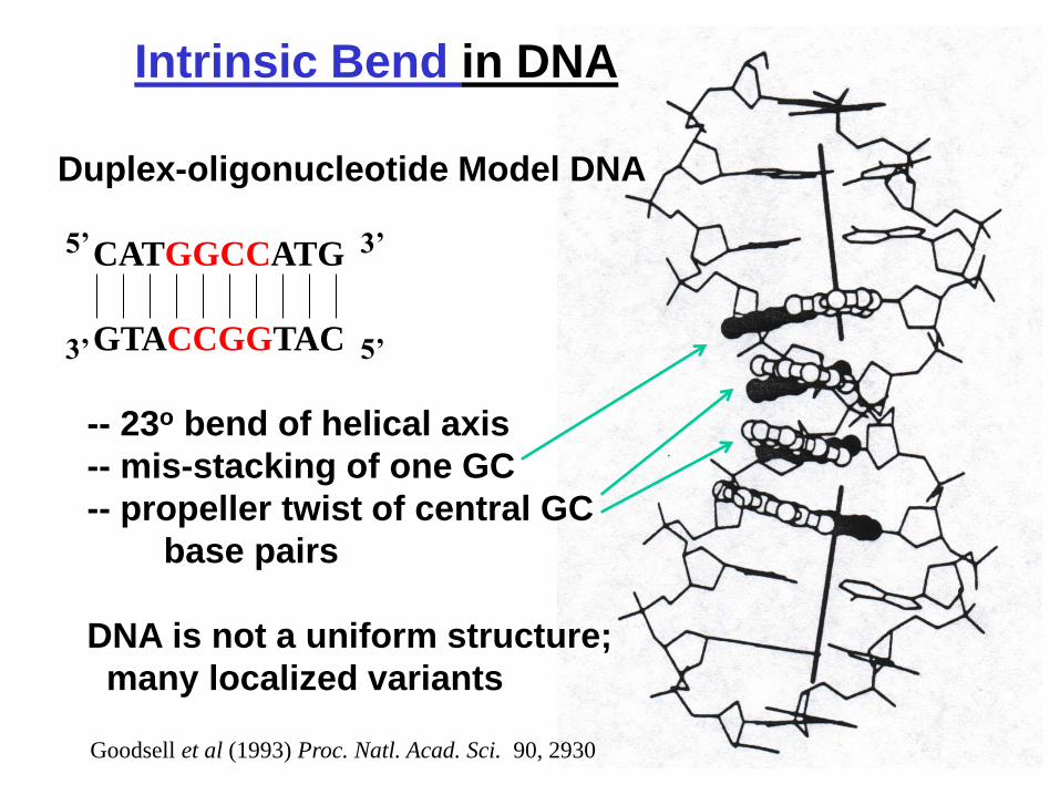
Intrinsic Bend in DNA
Duplex-oligonucleotide Model DNA
CATGGCCATG
GTACCGGTAC
-- 23o bend of helical axis
-- mis-stacking of one GC
-- propeller twist of central GC
base pairs
DNA is not a uniform structure;
many localized variants
Goodsell et al (1993) Proc. Natl. Acad. Sci. 90, 2930
5’ 3’
3’ 5’

Supercoiled DNA
Left-handed under-twisted DNA
is in a Negative Supercoil.
Right-handed over-twisted DNA
is in a Positive Supercoil.
Most biological DNA is negatively
supercoiled.
Topoisomerases alleviate
supercoils in DNA.
Relaxed Supercoiled
Electron Microscopy
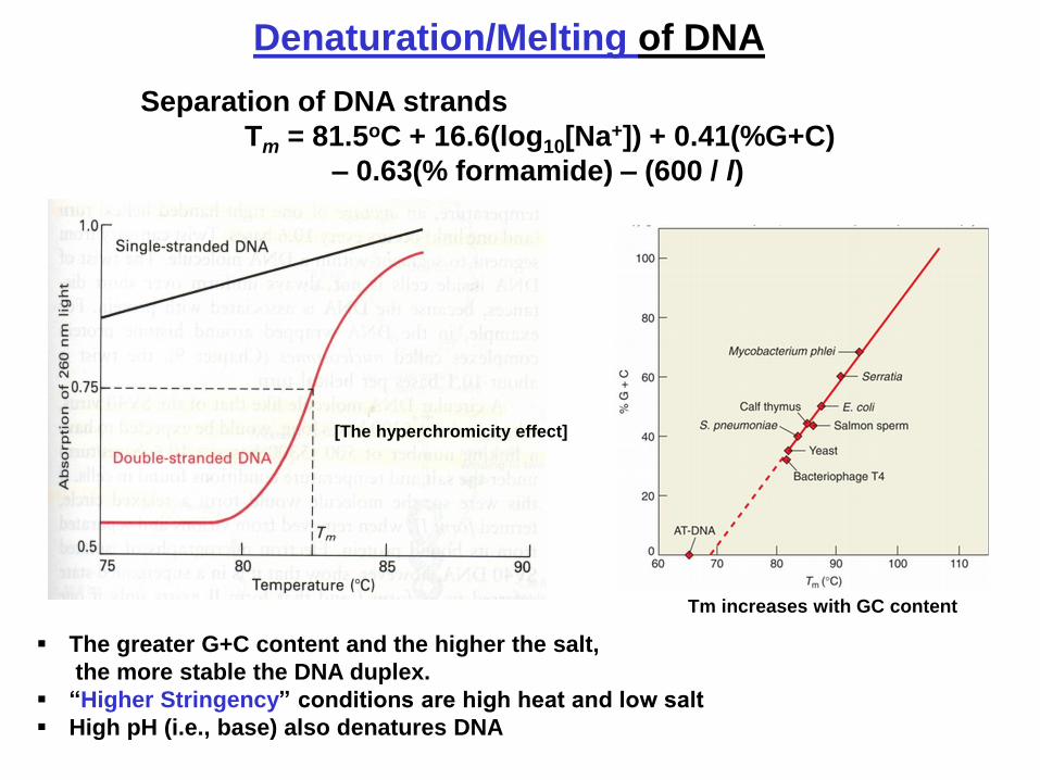
Denaturation/Melting of DNA
Separation of DNA strands
Tm = 81.5oC + 16.6(log10[Na+]) + 0.41(%G+C)
– 0.63(% formamide) – (600 / l)
The greater G+C content and the higher the salt,
the more stable the DNA duplex.
“Higher Stringency” conditions are high heat and low salt
High pH (i.e., base) also denatures DNA
[The hyperchromicity effect]
Tm increases with GC content

DNA Renaturation and Hybridization
Renature
(or DNA)

How Do Sequence-Specific DNA-Binding Proteins Bind
in a Sequence-Specific Manner?
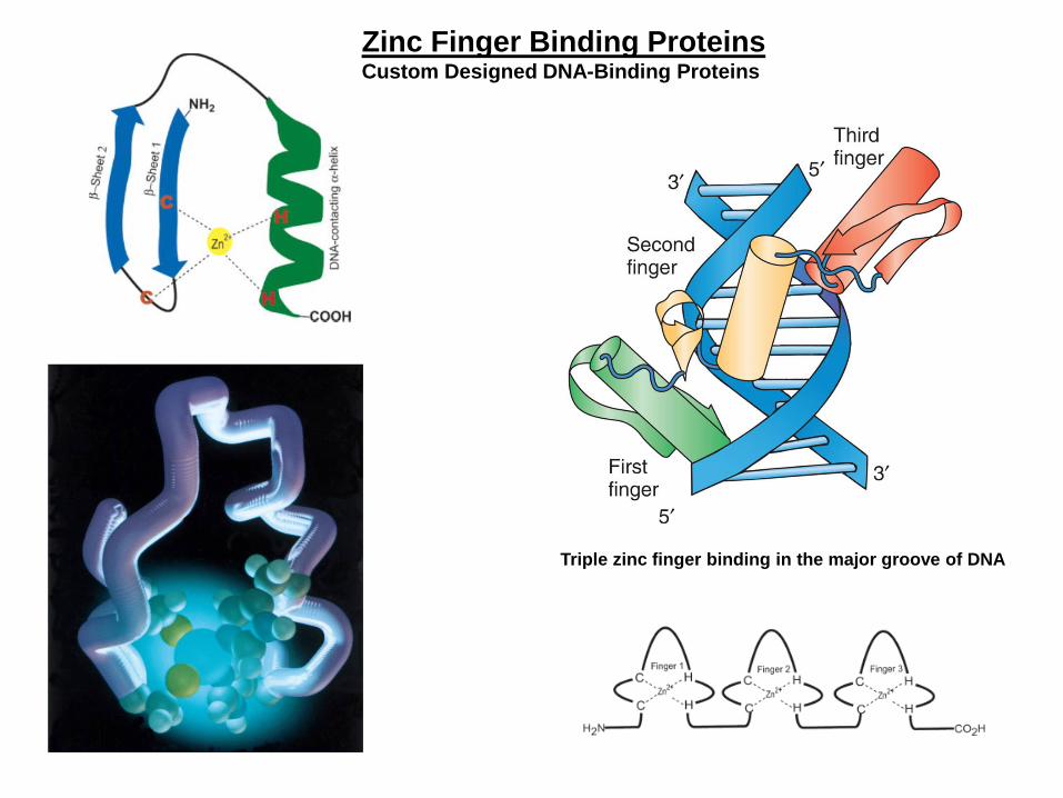
Triple zinc finger binding in the major groove of DNA
Zinc Finger Binding Proteins Custom Designed DNA-Binding Proteins

DHANASEKARAN et al.
Acc. Chem. Res. 2006, 39, 45-52
Swapping of the α-helix and β-hairpin regions alters the DNA binding properties of Sp1
Zinc Fingers (cont.)

• Crystallographic studies
– A single zinc finger protein can recognize 3 bp of DNA
Zinc Finger Properties
(Pabo, Science 1991) Courtesy of J Barrow & J Bungert
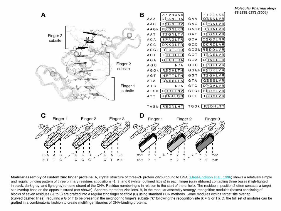
Modular assembly of custom zinc finger proteins. A, crystal structure of three-ZF protein Zif268 bound to DNA (Elrod-Erickson et al., 1996) shows a relatively simple
and regular binding pattern of three primary residues at positions -1, 3, and 6 (white, outlined labels) in each finger (gray ribbons) contacting three bases (high-lighted
in black, dark gray, and light gray) on one strand of the DNA. Residue numbering is in relation to the start of the α-helix. The residue in position 2 often contacts a target
site overlap base on the opposite strand (not shown). Spheres represent zinc ions. B, in the modular assembly strategy, recognition modules (boxes) consisting of
blocks of seven residues (-1 to 6) are grafted into a regular zinc finger scaffold (C) using standard PCR methods. Some modules exhibit target site overlap
(curved dashed lines), requiring a G or T to be present in the neighboring finger's subsite (“k” following the recognition site [k = G or T]). D, the full set of modules can be
grafted in a combinatorial fashion to create multifinger libraries of DNA-binding proteins.
Molecular Pharmacology
66:1361-1371 (2004)
Custom Designer DNA-Binding Proteins!
Potential for designing a DNA-binding protein to bind
to a single site (DNA sequence) in the genome

RNA Structure
-- Single-stranded
-- 2’ –OH
-- Uracil (not T)
-- 5’ to 3’ orientation
Secondary and Tertiary Structure
from long range base pairing
-- Watson and Crick
-- G-to-U
-- others
Stem-Loop Hairpin Pseudoknot
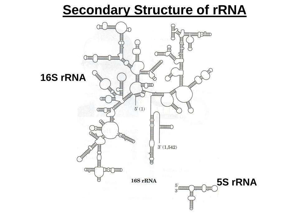
Secondary Structure of rRNA
16S rRNA
5S rRNA
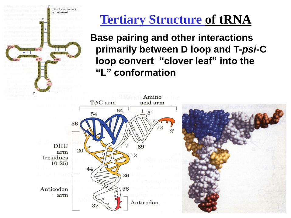
Tertiary Structure of tRNA
Base pairing and other interactions
primarily between D loop and T-psi-C
loop convert “clover leaf” into the
“L” conformation