Embed Size (px)
Citation preview

DNA replication: enzymology and mechanisms Zvi Kelman and Mike O'Donnell
Cornel l Universi ty Medica l Col lege, N e w York, U S A
Research into the enzymology of DNA replication has seen a multitude of highly significant advances during the past year, in both prokaryotic and eukaryotic systems. The scope of this article is limited to chromosomal replicases and origins of initiation. The multiprotein chromosomal replicases of prokaryotes and eukaryotes appear to be strikingly similar in structure and function, although future work may reveal their differences. Recent developments, elaborating the activation of origins in several systems, have begun to uncover mechanisms of regulation. The enzymology of eukaryotic origins has, until now, been limited to viral systems, but over the past few
years, enzymology has caught a grip on the cellular origins of yeast.
Current Opinion in Genetics and Development 1994, 4:185-195
I n t r o d u c t i o n
The structure of duplex DNA is so simple and elegant that one may have thought it would be simple to du- plicate. However, since the isolation of DNA polymer- ase I first delivered replication mechanisms into the hands of enzymologists, the process has been shown to be far from simple. Over 20 proteins are utilized in Es- cherichia coil and probably more in higher organisms [1]. In fact, the individual functions of many of these proteins are still unknown and even more proteins re- main to be identified. In overview, the process begins at a specific sequence called an origin upon which proteins bind and locally unwind the duplex, allow- ing invasion of a helicase. The helicase couples ATP hydrolysis to melt the duplex, and then the single- stranded DNA (ssDNA) is coated with ssDNA-bind- ing protein (SSB). This SSB-ssDNA nucleofilament has no secondary structure and serves as the most efficient template for chromosomal replicases, multiprotein ma- chineries characterized by their rapid and highly pro- cessive DNA synthesis. One strand of the chromosome is synthesized continuously, but because of the antipar- allel structure of the duplex, the other strand is copied discontinuously as a series of fragments (Okazaki frag- ments). This discontinuous mode of replication neces- sitates frequent reinitiation of DNA chains, which are primed by short RNA primers synthesized by a pri- mase. Most of the above proteins are thought to work together in one large replisome assembly for coordi- nated synthesis of both strands of the chromosome.
In this review, we will focus on recent developments in the study of origin activation and the mechanism of action of chromosomal replicases. These areas encom- pass only a subset of the recent advances in the enzy- mology of DNA replication.
Repl icat ion origins
The first (and still the only) cellular origin to have been activated in vitro was oriC, the origin of the E. coil chromosome [1]. Since that breakthrough, sev- eral phage and viral origins have been activated in vitro. Origin activation requires multiple copies of an origin-binding protein and usually additional ori- gin accessory proteins, some of which have secondary functions as transcriptional activators. As proposed by Hatch Echols [2], the requirement for multiple proteins in site-specific processes, such as activation of origins and promoters, is imposed by the need for a high fi- delity of action on specific sequences embedded in the bulk of chromosome DNA. These multiprotein-DNA complexes are termed specialized nucleoprotein struc- tures (snups) [2]. Origin initiation events in E. coli, phage ~ and simian virus 40 (SV40) are similar and appear to apply also to the more recently characterized systems of phage P1, bovine papilloma virus (BPV), herpes simplex virus 1 (HSV-1), and yeast (see Fig. 1). In most systems, the origin-binding protein is pre-
Abbreviations ACS~ARS core consensus sequence; ARS.--autonomous replication sequence; BPV--bovine papilloma virus; HSV-l--herpes simplex virus 1 ; MCM--minichromosome maintenance; ORC---origin recognition complex;
PCNA--proliferating cell nuclear antigen; pol/PoI~DNA polymerase; PP2A--protein phosphatase 2A; RF-C--replication factor C; RP-A--replication protein A; snup---specialized nucleoprotein structure; SSB~single-stranded DNA-binding protein; ssDNA--single-stranded DNA; SV40--simian virus 40.
© Current Biology Lid ISSN 0959-437X 185

186 Chromosomes and expression mechanisms
pared for origin activation and, for this review, we de- fine this as stage I. Multiple copies of the origin-binding protein then assemble onto the origin with the acces- sory proteins to form the snup (stage II). This untwists a section of AT-rich DNA to form an 'open complex' (stage III), providing a point of entry for the helicase (stage IV). After helicase entry, the chromosome can be extensively unwound and replication forks can be assembled. Origin activation systems differ in the de- tails of these steps and also in their regulation. In the following section, we will focus on these differences, with particular attention to recent developments.
Prokaryotic origins Recent studies show that the activation of oriC is reg- ulated at each stage (Fig. 1). In stage I, the DnaA origin-binding protein is inactive when bound to acidic phospholipids, but this repression can be relieved, and DnaA prepared for origin binding, by the DnaK heat shock protein or by phospholipase treatment [3]. In stage II, the origin snup is nucleated at four DnaA- binding sites in oriC, upon which 20 or more DnaA monomers assemble, along with several origin acces- sory proteins [1]. These accessory proteins include HU and IHF, which bend DNA, and they may act by help- ing to wrap DNA around the DnaA subunits [4°',5]. Other accessory proteins include Fis and the recently identified Rob protein [6"°]. They are not essential for the activation of the origin, and how they may modu-
late the process is not yet clear. In stage III, the ori- gin snup unwinds DNA within three AT-rich 13-mer repeats to form the open complex and this reaction requires a supercoiled template [1]. Formation of the open complex is highly regulated [1]. Negative regula- tion is achieved by the inhibitor of cellular initiation, IciA, which competes with DnaA for interaction with the 13-mers and prevents the formation of the open complex [7"°,8"°]. ATP also regulates the open com- plex formation. DnaA can bind either ATP or ADP tightly, but only the ATP form is active in unwinding the 13-mers [11. The DnaA protein becomes inactive upon slowly hydrolyzing the ATP to ADP, and the ADP remains bound, thus preventing subsequent reinitia- tions. The DnaA protein can be readily reactivated by acidic phospholipids, which catalyze the exchange of ATP for the bound ADP [1,9"'], but only when DnaA is bound to or/C, this may underlie the early obser- vations of replicon attachment to the membrane [10]. RNA polymerase provides yet another level of reg- ulation in determining the ability of R loops (RNA paired with one strand of the DNA duplex) to pro- mote the open complex, and although the transcript need not enter on'C, it must be close [11]. In stage IV, the DnaB helicase enters the open complex, but its ac- cess absolutely requires the DnaC protein, a molecu- lar matchmaker that delivers the hexameric DnaB into the origin in an ATP-dependent reaction, after which DnaC departs from the DNA [1]. Origin unwinding is bidirectional; therefore, two hexamers of DnaB must
Stage I Stage I1 DNA ~
Prepare
Stage III
r - f Open
Stage IV
origin-binding Open complex Origin- prote in ~ formation binding | I~ Replication fork
System protein II + - II II + - II Helicase proteins E. coil DnaA D n a K Phospholipids HU. IHF, ATP ADP DnaB Dna(- Excess'~
Phospholipase Fis, Rob Acidic phospholipids IciA DnaC -- | R loops / Dna(;-Prima~e
Phage ~. ~. O HU RNA pol DnaB ~. p ~. p ~ SSB Dnal. DnaK ~ DNA po] III hoh,enzyme Gq)E
Phage PI RepA DnaJ, DnaK, DnaA DnaB GrpE
Yeast ORC ATP, ABFI -~ i)okx-Prin'=ase
SV40 T-antigen Cdc2 psi ATP banligen RP-A(SSBI p53 ~ RP-A(SSB] PP2A Spl PP2A pol 8, t" (+RF-C, PCNA)
BPV E1 E2 E!
HSV-I UL9 UL5,8,52 ICP8 Helle ase--Primase (UL5,8.521 ICP8 (SSB) HSV-1 Pol q+LJt42)
@1994 Currenl Opinion In Genetics and Developmenl
Fig. 1. Regulation at different stages of origin activation in a number of systems. Origin activation has been subdivided into four stages: preparation of the origin-binding protein, assembly of the origin snup, local unwinding of origin DNA (the open complex), and entry of the helicase. Molecules that act on each stage are divided into positive (+) and negative (-) effectors and are shown for E. colt, phage )~, phage P1, yeast, SV40, BPV and HSV-1. A blank entry indicates that effectors of that step have not (yet) been identified. At the far right are listed the replication proteins needed to advance the replication fork.

DNA replication: enzymolo~/and mechanisms Kelman and O'Donnell 187
be delivered to the open complex. Negative regula- tion is achieved by the presence of too much DnaC protein, which binds to and inhibits the DnaB helicase activity, possibly by preventing its translocation along DNA; therefore, a 'fine balance' of DnaC and DnaB is essential to the productivity of this stage [12].
Phage ~. uses mainly host replication proteins for origin activation, with the exception of the phage-encoded O and P proteins, analogs of DnaA and DnaC [1]. The ;k origin snup is composed of four dimers of ~. O pro- tein, which do not require pre-activation to assem- ble onto the origin. Formation of the open complex at three AT-rich l 1-met repeats requires the DNA to be supercoiled, but does not require ATP [13,14]. In stage IV, the ~. P protein binds DnaB and delivers it to the origin, but the P protein remains tightly associ- ated with DnaB and prevents its helicase activity [15]. In this regard, the P protein acts as a negative regulator, like excess DnaC, to prevent translocation of DnaB. Al- though ATP is not required for P protein to bring DnaB to the origin, it is required to break the interaction of P protein with DnaB, thus freeing the helicase for replication; this step is mediated by heat shock pro- teins DnaJ, DnaK and GrpE [16,17]. Transcription from the rightward promoter is needed for ~. origin activa- tion in vivo, and in vitro studies show that transcription disrupts HU-mediated negative repression (presumably of stage II or III) [18].
Recent studies on the activation of the lysogenic origin of phage P 1 show that the phage-encoded RepA origin- binding protein is a native dimer, but the monomer strongly associates with the origin. In stage I, RepA is prepared for origin binding by the DnaJ, DnaK and GrpE heat shock proteins, which couple ATP hydroly- sis to monomerize RepA [19"']. The origin snup com- prises RepA bound at five sites and the host DnaA pro- tein at five other sites. At least two of the DnaA-binding sites are essential for origin function, and in vitro stud- ies show that the ADP form of DnaA protein is capable of activating the origin in combination with RepA (re- viewed in [20]). Although stages III and IV have not been studied in detail, it seems likely that the open complex forms at five slightly AT-rich 7-met repeats and then DnaC mediates delivery of DnaB to the open complex.
Eukaryotic viral origins In the SV40 system (reviewed in [21]), the stage I prepa- ration of the vitally encoded T-antigen for origin bind- ing is regulated in both positive and negative fash- ion. T-antigen must be phosphorylated at Thr124 for productive origin binding, and this modification can be performed by the cdc2 kinase [22]. Phosphoryla- tion at other sites can inhibit T-antigen, and protein phosphatase 2A (PP2A) has been shown to activate T-antigen by removing phosphates from specific ser- ine residues implicated in DNA-binding activity [23]. The p53 tumor suppressor protein is a negative reg- ulator of stage I; it binds to T-antigen preventing its association with the origin [24]. Stage II, formation of
the origin snup, is facilitated by ATP binding, which effects the assembly of two hexamers of T-antigen onto the origin (reviewed in [25]). The two hexam- ers are thought to encircle the origin DNA in SV40, because if the hexamers are pre-assembled in solution they do not bind the origin [26"']. Moreover, in elec- tron micrographs the double hexamer of T-antigen on the origin does not appear to wrap DNA around it as with other origin snups, but rather the DNA appears to follow a straight path through the protein [27]. The transcriptional activator Spl acts as an origin accessory protein that perturbs the local histone distribution to make the origin accessible for initiation proteins [28]. However, the identity of the transcriptional activator is not important, as Spl can be exchanged for other activators [28], but the DNA-binding domain must be accompanied by an activation domain for origin func- tion [29"'].
Formation of the open complex in stage III is coinci- dent with T-antigen binding to the origin, which results in the melting of 8 bp in the early palindrome and un- twisting of an AT tract [25]. These events require bind- ing, but not hydrolysis, of ATP and do not require a supercoiled template [25]. In SV40, unlike the other sys- tems discussed above, the stage IV helicase invasion is unique in that T-antigen itself is the helicase and thus encompasses the functions of the E. coli DnaA, B and C proteins. Stage IV is still regulated, however; for ex- ample, it has been shown recently that PP2A-treated T-antigen provides a cooperative interaction between the two hexamers, which facilitates stage IV [30"]. In addition, the human SSB, replication protein A (RP-A), consists of three non-identical subunits (p70, p32 and p14) and has been shown to be a positive effector of stage IV, as RP-A is needed for T-antigen to unwind the bulk of DNA [25]. A second point at which p53 may regulate replication has been identified recently in an interaction of p53 with the p70 subunit of RP-A (the ssDNA-binding subunit) that inactivates its ability to bind ssDNA [31"']. Also, the p32 subunit of RP-A is phosphorylated in a cell cycle dependent manner in the G1 to S phase transition by members of the cy- clin cdc2 kinase family, and addition of this kinase to the SV40 replication system stimulates DNA synthesis [32"']. Phosphorylation of p32 is also performed by the DNA-activated protein kinase (GS Brush, CW Ander- son, TJ Kelly, abstract 191, Eukaryotic DNA Replication Meeting, Cold Spring Harbor, September 1993).
The BPV snup is composed of two viraUy encoded proteins, E1 and E2. E1 is the origin-binding protein and a helicase (like T-antigen) [33",34"'] and E2 is an origin accessory protein (and transcriptional activator) that binds specific sequences and helps in the delivery of E1 to the origin via direct protein contacts [35",36]. In this regard, E2 fulfills a stage IV function analogous to that of E. coli DnaC in delivery of the helicase into the origin. Regulatory aspects, exact functions of ATP and identification of an open complex remain for future studies, but the in vitro replication system and avail- ability of pure E1 and E2 should yield this information in the near future.

188 Chromosomes and expression mechanisms
In the HSV-1 system, the identification of all seven of the essential virally encoded replication proteins and the cloning and production of each of them in quan- tity has enabled a number of illuminating studies, al- though replication in vitro of a plasmid containing the origin has yet to be achieved. The origin snup consists of at least two dimers of the UL9 protein, which pro- duces the open complex in an AT-rich region even in the absence of ATP [37]. UL9 has helicase activity, but it does not act catalytically like the other helicases dis- cussed thus far, Instead, it must bind DNA stoichiomet- rically for unwinding and requires a stretch of ssDNA for duplex-unwinding activity [38,39]. The UL9 helicase is stimulated by the virally encoded SSB (called ICP8) and it seems likely they function together at the origin to enlarge the open complex for the future replication fork.
Yeast chromosomal origin In the past two years, rapid and exciting advances have been made in the study of the biochemistry of yeast cellular replication. Yeast origins are known as au- tonomous replication sequences (ARS) for their ability to confer autonomous replication on plasmids. Recent studies [40 "°] of the ARS1 origin show that it contains four sequence elements: the A element, which contains an 11 bp ARS core consensus sequence (ACS) common to all ARS elements, the B3 element, which is the bind- ing site of the ABF1 transcription factor, and the B1 and B2 elements, which may interact with (as yet) uniden- tiffed origin accessory proteins. A large origin recogni- tion complex (ORC), which binds to ARS1 at the A site, has now been purified from Saccharomyces cerevisiae [41"], and genomic footprinting experiments indicate the presence of the ORC on ARS1 in vivo [42"']. The ORC contains six subunits and requires ATP to bind the ARS. The exact function of the ORC is not certain, but a number of observations confirm that it plays a central role in replication. First, the ORC does not bind to single-site mutant forms of ACS that lack ARS activity [41"']. Second, mutations affecting the 70 kDa subunit of the ORC, identified because they produce a defect in function of the HMR silencer, show that the gene is es- sential for cell viability, and a conditional lethal allele is defective in chromosome replication [43"]. Third, each of the six genes encoding the ORC subunits are essen- tial to yeast (SP Bell, R Kobayashi, B Stillman, abstract 15, Eukaryotic DNA Replication Meeting, Cold Spring Harbor, September 1993).
Recent developments in the study of the genetics of yeast replication have given researchers clues as to the roles of important replication proteins. Several genes of S. cerevisiae have been isolated, the products of which are needed for minichromosome maintenance (MCM) of ARS-containing plasmids. Three of these, MCM2, MCM3 and MCM5, are homologous in sequence and each is essential for cell viability [44"]. The intracellu- lar location of MCM2, 3 and 5 is cell cycle regulated, they are moved into the nucleus upon completion of S phase and then moved into the cytoplasm at the start of
S phase [44",45"']. It is hypothesized that in the nucleus the MCM proteins activate the ARS and are then trans- ferred to the cytoplasm to prevent reinitiation [44",45"']; however, whether they act on origins directly and in a positive fashion must await further studies. In another exciting development, the cdcl8 gene of Scbizosac- charomycespombe has been shown to suppress muta- tions of cdclO, a transcriptional activator that is needed for entry into S phase [46"']. The cdcl8 gene may be a major target of cdclO and, consistent with this, cdcl8 mutants fail to enter S phase [46"']. Further, cdcl8 may play a role in checkpoint control, as cdcl8 mutants are unable to prevent mitosis and rapid cell division even though the chromosomes are not fully duplicated. The cdcl8 gene sequence reveals a nucleotide-binding site, but whether the encoded protein plays a direct role (e.g. helicase) or an indirect role in replication must await future isolation and characterization of the gene product.
Events after helicase entry
Once the helicase has entered the chromosome, it pre- sumably nucleates assembly of replication forks con- mining the primase and two replicative polymerases, one for each strand of DNA [1]. The leading and lag- ging strand polymerases in eukaryotes appear to be different polymerases (polymerases 8 and e), whereas prokaryotes use two copies of the same polymerase. The mechanism of replicase function is the second topic of this essay.
Chromosomal replicases
Replicases of E. coli, phage T4, yeast and humans Organisms that span the evolutionary spectrum have been shown to possess replicases that appear to be similar in function and also in their actual structure. These are the E. coli DNA polymerase III holoen- zyme (Pol III holoenzyme), the phage T4 replicase, and the polymerase 15 (pol 8) of both yeast and hu- mans. In each of these systems, the replicase encom- passes several proteins that use ATP to initiate rapid and highly processive DNA synthesis. It can be thought of as having three components (see Table 1): the catalytic component, which contains the DNA poly- merase and proofreading 3"-5" exonuclease activities, and the two categories of polymerase accessory pro- teins, a complex of accessory proteins and a single subunit processivity factor. The accessory proteins are needed to confer high processivity onto the catalytic polymerase. Over the past two years, a deeper under- standing of the action of these accessory proteins has been developed and appears to apply generally to all four systems. Studies have shown that the single sub- unit accessory protein is a DNA-sliding clamp, which tethers the polymerase to DNA for high processivity.
DNA replication: enzymology and mechanisms Kelman and O'Donnell 189
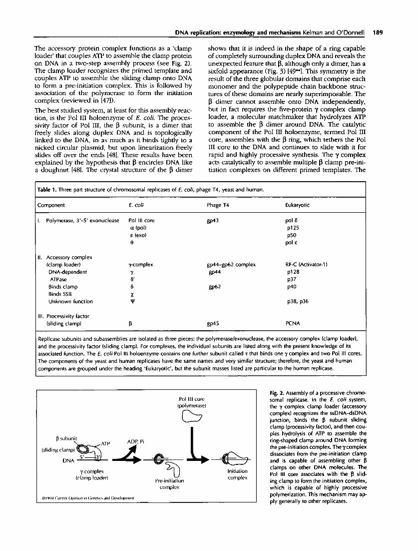
The accessory protein complex functions as a 'clamp loader' that couples ATP to assemble the clamp protein on DNA in a two-step assembly process (see Fig. 2). The clamp loader recognizes the primed template and couples ATP to assemble the sliding clamp onto DNA to form a pre-initiation complex. This is followed by association of the polymerase to form the initiation complex (reviewed in [47]).
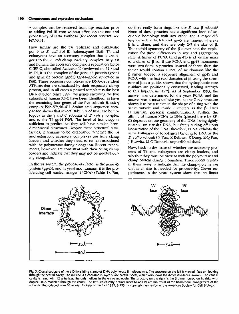
The best studied system, at least for this assembly reac- tion, is the Pol IIl holoenzyme of E. colL The proces- sivity factor of Pol III, the [3 subunit, is a dimer that freely slides along duplex DNA and is topologically linked to the DNA, in as much as it binds tighdy to a nicked circular plasmid, but upon linearization freely slides off over the ends [48]. These results have been explained by the hypothesis that [3 encircles DNA like a doughnut [48]. The crystal structure of the [3 dimer
shows that it is indeed in the shape of a ring capable of completely surrounding duplex DNA and reveals the unexpected feature that [3, although only a dimer, has a sixfold appearance (Fig. 3) [49*']. This symmetry is the result of the three globular domains that comprise each monomer and the polypeptide chain backbone struc- tures of these domains are nearly superimposable. The [3 dimer cannot assemble onto DNA independently, but in fact requires the five-protein y complex clamp loader, a molecular matchmaker that hydrolyzes ATP to assemble the [3 dimer around DNA. The catalytic component of the Pol Ill holoenzyme, termed Pol III core, assembles with the [3 ring, which tethers the Pol III core to the DNA and continues to slide with it for rapid and highly processive synthesis. The 7 complex acts catalytically to assemble multiple ~ clamp pre-ini- tiation complexes on different primed templates. The
Table 1. Three part structure of chromosomal replicases of E. co~i, phage T4, yeast and human.
Component E. colt Phage T4 Eukaryotic
I. Polymerase, 3'-5' exonuclease Pol III core gp43 pol 8 a (pol) p125 ¢ (exo) p50 B pol¢
II. Accessory complex (clamp loader) y-complex DNA-dependent 7 ATPase 8' Binds clamp 8 Binds SSB X Unknown function v2
gp44-gp62 complex RF-C (A~ivato~l) gp44 p12B
p37 gp62 p40
III. Processivity factor (sliding clamp) 13 81>45
p38, p36
PCNA
Replicase subunits and subassemblies are isolated as three pieces: the polymerase/exonuclease, the accessory complex (clamp loader), and the processivity factor (sliding clamp). For complexes, the individual subunits are listed along with the present knowledge of its associated function. The E. coli Pol III holoenzyme contains one further subunit called ~ that binds one 7 complex and two Pol III cores. The components of the yeast and human replicases have the same names and very similar structure; therefore, the yeast and human components are grouped under the heading 'Eukaryotic', but the subunit masses listed are particular to the human replicase.
Pol !11 core (polymerase)
[3 subuni,~e4....,~AT P ADP, Pi
(slidingclanlp)~s'~3'DNA 7 complex ~ " i ~ - ~ )
(clamp loader) Pre-initiation complex
© I 994 Current Opin ion in (,enel i t • and l~.,vc, lol)n~.-nl
Iniliation complex
Fig. 2. Assembly of a processive chromo- somal replicase. In the E. colt system, the 7 complex clamp loader (accessory complex) recognizes the ssDNA-dsDNA junction, binds the [3 subunit sliding clamp (processivity factor), and then cou- ples hydrolysis of ATP to assemble the ring-shaped clamp around DNA forming the pre-initiation complex. The 7 complex dissociates from the pre-initiation clamp and is capable of assembling other I], clamps on other DNA molecules. The Pol III core associates with the 13 slid- ing clamp to form the initiation complex, which is capable of highly processive polymerization. This mechanism may ap- ply generally to other replicases.
190 Chromosomes and expression mechanisms
7 c o m p l e x can be r e m o v e d from ttle reaction prior to adding Pol III core without effect on the rate and processivity of DNA synthesis (for recent reviews, see [47,50,511.
How similar are the 3"4 replicase and eukaryotic pol 8 to E. coli Pol III holoenzyme? Both "I"4 and eukaryotes have an accessory complex that is analo- gous to the E. coli clamp loader T complex. In yeast and human, the accessory complex is replication factor C (RF-C, also called Activator-l) (reviewed in [52]) and in "I"4, it is the complex of the gene 44 protein (gp44) and gene 62 protein (gp62) (gp44--gp62, reviewed in [53]). These accessory complexes are DNA-dependent ATPases that are stimulated by their respective clamp protein, and in all cases a primed template is the best DNA effector. Since 1992, the genes encoding the five subunits of human RF-C have been identified, as have the remaining four genes of the five-subunit E. coli T complex [54"-57",58--611. Amino acid sequence com- parison shows that several subunits of RF-C are homo- logous to the 7 and 8' subunits of E. coli ~l complex and to the T4 gp44 [56"]. The level of homology is sufficient to predict that they will have similar three- dimensional structures. Despite these structural simi- larities, it remains to be established whether the T4 and eukaryotic accessory complexes are truly clamp loaders and whether they need to remain associated with the polymerase during elongation. Recent experi- ments, however, are consistent with their being clamp loaders and indicate that they may not be needed dur- ing elongation.
In the "1"4 system, the processivity factor is the gene 45 protein (gp45), and in yeast and humans, it is the pro- liferating cell nuclear antigen (PCNA) (Table 1). But,
do they really form rings like the E. coli ~ subunit? None of these proteins has a significant level of se- quence homology with any other, and a major dif- ference is that PCNA and gp45 are trimers, whereas 13 is a dimer, and they are only 2/3 the size of 13. The sixfold synmaetry of the 13 dimer held the expla- nation for these differences in size and aggregation state. A trinaer of PCNA (and gp45) is of similar mass to a dinaer of I~ so, if the PCNA and gp45 monomers were two-domain proteins, instead of three, then the trimer would contain a total of six domains like the 13 dimer. Indeed, a sequence alignment of gp45 and PCNA with the first two domains of 1~, using the struc- ture of 13 as a guide, shows that the hydrophobic core residues are positionally conserved, lending strength to this hypothesis 149"']. As of September 1993, the answer was determined for the yeast PCNA, and the answer was a most definite yes, as the X-ray structure shows it to be a trinaer in the shape of a ring with the same outside and inside diameters as the 13 dimer (J Kuriyan, personal communication). Further, the affinity of human PCNA to DNA (placed there by RF- C) depends on the geometry of the DNA, being tightly retained on circular DNA, but freely sliding off upon linearization of the DNA; therefore, PCNA exhibits the same hallmarks of topological binding to DNA as the E. colf ~ subunit (N Yao, Z Kelman, Z Dong, Z-Q Pan, J Hurwitz, M O'Donnell, unpublished data).
Now, back to the issue of whether the accessory pro- teins of "I"4 and eukaryotes are clamp loaders, and whether they must be present with the polymerase and clamp protein during elongation. Three recent reports in these systems indicate that the clamp--polymerase unit is all that is needed for processivity. Clever ex- periments in the yeast system show that on linear
Dimer ._~
Interface . ~
3
"A . . . . B"
Fig. 3. Crystal structure of the 13 DNA-sliding clamp of DNA polymerase III holoenzyme. The structure on the left is viewed 'face on' looking through the central cavity. The outside is a continuous layer of antiparallel sheet, which also forms the dimer interfaces (arrows). The central cavity is lined with 12 o. helices, the only helices in the entire molecule. The structure on the right is the 13 dimer turned on its side, with duplex DNA modeled through the center. The two structurally distinct faces (A and B) are the result of the head-to-tail arrangement of the subunits. Reproduced from Molecular Biology of the Cell 1992, 3:955 by copyright permission of the American Society for Cell Biology.

DNA replication: enzymology and mechanisms Kelman and O'Donnell 191
DNA (but not circular) PCNA confers processivity onto pol 8 in the complete absence of ATP and RF-C [62"]. This result was interpreted as showing that the PCNA ring threads itself onto the end of linear DNA and then couples with pol 8 for processive synthesis, thus cir- cumventing the need to open the ring, an action that requires the RF-C complex and ATP. In the T4 system, evidence that the gp44-gp62 complex is not needed during elongation has come from the obser- vation that supply of a large excess of gp45 increases the processivity of the polymerase in the absence of the gp44-gp62 complex, a result that is similar to those from previous studies in the E. coli system and which enforces the idea of the clamp-polymerase as the processive unit [63"]. Furthermore, an elegant elec- tron microscopy study has shown that the T4 sliding clamp on DNA appears as a 'hash-mark' (with simi- lar dimensions to the ~ ring) through which DNA is threaded, and these 'hash-marks' appear in clusters indicating that they can slide [64"']. The size of the 'hash-mark' is insufficient to acconmlodate the mass of both the gp44-gp62 complex and the gp45 trimer and in light of similarity to other systems it is probably a gp45 clamp.
The actual mechanism by which the clamp loader as- sembles the clamp around DNA is a fascinating issue and is being addressed in all these systems, but detailed mechanisms are still unknown. Most of the work has been in the T4 and E. coli systems, in which the indi- vidual subunits of the clamp loaders are available in pure form. No individual subunit of either of these clamp loaders can assemble their respective clamp onto DNA; presumably this reaction is too compli- cated for just one protein. The five subunits of the E. coli ~l complex are % 8, 8", X and "q. Although only T and 8 are essential to place ~ onto DNA, the 8' subunit stimulates this reaction considerably [65,66]. DNA-de- pendent ATPase activity is produced by a mixture of T and 8' [65] and T is the presumed site of hydrolysis, as it is known to bind ATP [1]. The 8 subunit forms a protein-protein complex with ~ [67]. Hence, it appears that ~ ' recognizes the primed template, and the 8 sub- unit functions to bring ~ into the structure for assembly around DNA and hydrolysis of ATP. At elevated ionic strength, such as exists in the cell, the X and ~t subunits of the T complex are also needed to initiate processive synthesis [68]. This is probably rooted in the fact that the T complex associates with SSB-coated DNA [69"], an interaction mediated by X that may give the T com- plex the added grip that it needs to bind the template in elevated salt (Z Kelman, M O'Donnell, unpublished data).
The T4 clamp loader is composed of a tetramer of gp44 tighdy associated with one protomer of gp62 [53]. By itself, the gp44 is a DNA-dependent ATPase (like E. coli "#'), implying that it harbors the DNA-binding and ATPase sites [70]. The DNA-dependent ATPase of the gp44-gp62 complex is stimulated by gp45, whereas the gp44 ATPase is not, implying that gp62 interacts with gp45 (like E. coli 8) [70]. The gp44-gp62 complex in- teracts with gp32 (T4 SSB) bound to ssDNA (like E. coli
7,.), but it is not known whether the interaction is medi- ated by gp44 or gp62 [71]. Using footprinting methods, laser-induced UV-crosslinking of protein to DNA, and novel DNA-protein cross-linking agents, it has been found that the 1"4 gp44-gp62 complex needs only to bind ATP to bring the gp45 clamp to DNA, although it is not known whether the clamp surrounds DNA at this point [72,73"',74]. Further, these studies show that the gp44-gp62 complex works on the primed template junction to place the gp45 clamp on DNA, and then ATP hydrolysis induces a movement or the dissociation of gp44-gp62 from the DNA, presumably to make room for the polymerase [74,75].
Much less is known about the RF-C subunits, although with the recent identification of the genes encoding them we can expect much more information in the near future. The human p128 is known to bind DNA, as it was identified in 1993 in a south-western analy- sis as a DNA-binding protein called PO-GA [57"]. At the same time, a similar screen resulted in the isola- tion of the analogous RF-C subunit of the mouse [76"]. The p37 and p40 proteins are the only RF-C subunits to be obtained in pure form thus far and studies have shown that p37 binds DNA, suggesting that p128 and p37 may be similar to ~/and 8" (and T4 gp44) [77"]. The p40 binds ATP and PCNA, which suggests a functional analogy to 8 (and T4 gp62) [77"]. A summary of these individual subunit functions is included in Table 1.
In all these systems, the three replicase components have only weak interactions with one another, at least in the absence of DNA. In the E. coli system, however, there is another subunit called ~ that firmly binds one ~/complex molecule and two molecules of Pol III core, thereby acting as ~i scaffold to hold all of these pro- teins tightly together in a 'holoenzyme' particle [67]. A dimeric polymerase complete with one clamp loader within a single molecular structure fits nicely with the need to synthesize two strands of DNA, one of which is synthesized discontinuously and requires multiple clamps to be loaded onto it (lagging strand). At present, the T4 and eukaryotic replicases have been purified as the three separable components and whether, in these systems, the clamp loaders maintain an association with the polymerase and clamp or have a functional equivalent of "t to hold them together remains to be determined.
Replicases of HSV-1 and phage T7 The other two replicases that are highly processive are those of HSV-1 and phage T7 [1]. These replicases re- quire only one accessory protein for high processivity, UL42 for HSV-1 and E. coli thioredoxin for T7, and they do not need ATP. Thus, they lack a clamp loader. The crystal structure of thioredoxin bears no resemblance to a ring, so the molecular basis by which the acces- sory protein provides processivity to the polymerase is not clear. One simple possibility is that the polymerase has a cleft into which the DNA fits and the accessory protein seals off this cleft to trap DNA inside.

192 Chromosomes and expression mechanisms
Interaction of sliding clamps with. other proteins
The clamp proteins have recently been shown to share a common property, in that they interact with pro- teins other than the replicative polymerase. The best studied case is that of T4, in which the gp45 clamp interacts with RNA polymerase. Phage T4, as well as several viruses (e.g. SV40, adenovirus and HSV), has an early-to-late switch in gene expression, so that late g e n e s (eg c a p s i d p ro te ins ) are not e x p r e s s e d until the viral g e n o m e has b e e n repl ica ted . Elegant s tud ies b y Peter G e i d u s c h e k ' s r e sea rch g r o u p have r evea l ed that the unde r ly ing m e c h a n i s m of the ear ly- to- la te swi tch is an in te rac t ion b e t w e e n the T4 repl icase acces so ry p ro te ins a n d t h e E. co l i RNA p o l y m e r a s e (mod i f i ed by "1"4 gp33 a n d gp55). In 1992, a con t inua t ion o f these s tudies was p u b l i s h e d s h o w i n g that this switch is the resul t o f a t r ack ing mechan i sm, w h e r e b y the accesso ry p ro t e ins a s s e m b l e at a n ick and s l ide a long DNA, thus ac t ing as a ' m o b i l e enhance r ' for late gene ac t iva t ion [78"']. P r e s u m a b l y the in terac t ion is th rough the gp45 c l a m p a n d the mod i f i ed RNA po lymerase , a l t hough a role for the g p 4 4 - g p 6 2 c o m p l e x canno t b e ru led out, as it was no t r e m o v e d from the sys tem after f o rma t ion o f the s l id ing c lamp. This m e c h a n i s m may a p p l y to euka ryo t i c v i ruses in genera l , as a bacu lov i rus p ro t e in wi th 42% ident i ty to PCNA is also n e e d e d for ac t iva t ion o f late g e n e s [79].
Use o f the s l id ing c l a m p for o the r DNA metabo l i c p ro - cesses a p p e a r s to be the rule ra ther than the excep t ion , as the c l a m p s o f E. co l i a n d o f h u m a n also in teract wi th o the r pro te ins . In E. coli, t h e ~ c l a m p is u t i l ized b y Pol III a n d DNA p o l y m e r a s e II, a n d in h u m a n s and yeast , the PCNA c l a m p is u t i l ized b y bo th po l 8 a n d po l [1]. In fact, the h u m a n PCNA has recen t ly b e e n s h o w n to b i n d to m e m b e r s o f the cycl in D and cyc l i n -de pe n - den t k inase famil ies [80"']. The funct ion o f the lat ter in te rac t ion is u n k n o w n . Would the PCNA ring n e e d to be o n o r off DNA to manifes t the b io logica l activity? It is t e m p t i n g to specu la t e that the PCNA ring may te ther cycl ins a n d thei r a s soc ia ted k inases to DNA for s o m e a spec t o f cell cyc le cont ro l (e.g. mitot ic checkpo in t ) .
Conclusions
In s u m m a r y , k n o w l e d g e is e x p a n d i n g rapid ly a b o u t rep l i ca t ion m e c h a n i s m s in severa l systems, too many , in fact, for t h e m all to have b e e n m e n t i o n e d here . C h r o m o s o m a l rep l icases a p p e a r to be qui te s imilar in their m e c h a n i s m for a t ta in ing high processivi ty. H o w - ever, l i tde is k n o w n a b o u t h o w they a s s e m b l e at the or ig in o r c o m m u n i c a t e wi th the he l icase and pr imase , o r h o w these p rocess ive e n z y m e s re lease DNA rap id ly a n d r e b i n d n e w pr imers du r ing d i scon t inuous syn the- sis o f the l agg ing strand. Likewise, the out l ine o f even t s in ac t iva t ion o f or igins has b e e n c o n s e r v e d in evo lu t ion f rom p r o k a r y o t e s to eukaryo tes , bu t impor tan t de ta i l s in the m a n i p u l a t i o n o f these s e q u e n c e s by prote ins , the roles o f ATP, a n d regula t ion o f ini t iat ion in different sys-
tems is r evea l ing fundamen ta l diversif icat ion. The n e w in format ion a b o u t yeas t or ig in s tructure, and the p ro - te ins that act o n it, l ead us to an t i c ipa te future d i scov- er ies c o n c e r n i n g the m e c h a n i s m s b y w h i c h eukaryo t i c ce l lu lar or igins a re act ivated, r egu l a t ed and eventua l ly in tegra ted wi th s ignal t ransduct ion .
References and recommended reading
Papers of particular interest, published within the annual perkxt of review, have been highlighted as: • of special interest • . of outstanding interest
1. KORNBERG A, BAKER TA: DNA Replication. New York: WH Freeman; 1991.
2. ECHOLS H: Multiple DNA-Protein Interactions Govern- ing High-Precision DNA Transactions. Science 1986, 233:1050--1056.
3. HWANG DS, CROOKE E, KORNBERG A: Aggregated DnaA Protein is Dissociated and Activated for DNA Replication by Phospholipase or DnaK Protein. J BIol Chem 1990, 265:19244-19248.
4. HWANG DS, KORNBERG A: Open ing of the Replication Origin • . of Escberlcbla coil by DnaA Protein With Protein HU or
IHE J Btol Cbem 1992, 267:23083-23086. Important demonstration that either protein HU or IHF will suffice to form the open complex on negatively supercoiled plasmid filled with DnaA protein complexed with ATP.
5. SCHMID MB: More Than Just 'Histone-Like' Proteins. Cell 1990, 63:451--453.
6. SKARSTAD K, THONY 13, HWANG DS, KORNBERG A: A NO- ** vel Binding Protein of the Origin of the Escherfcbla coil
Chromosome. J Btol Cbem 1993, 268:5365-5370. Identification of a novel origin-binding protein in E. coil The Rob protein is present at 5000 copies per cell, contains a helix-turn-helix motif and binds at the right border of ortC.
7. HWANG DS, KORNBERG A: Opposed Actions of Regulatory • * Proteins, DnaA and IciA, in Opening of the Replication Ori-
gin of E$cherlchla coll. J Blol Chem 1992, 267:23087-23091. First demonstration that DnaA protein interacts with the middle and right 13-mers of orICwith sequence specificity, and that lciA protein blocks this interaction by binding all three 13-mers.
8. HWANG DS, THONY B, KORNBERG A: IdA Protein, a Spe- • - cific Inhibitor of Initiation of Escberlcbla coil Chromosomal
Replication. J Btol Cbem 1992, 267:2209-2213. Elevation of the intracellular concentration of IciA protein produces a lag in cell growth upon transfer to fresh medium. This correlates with a fourfold increase of IciA protein in the stationary phase.
9. CROOKE EC, CASTUMA CE, KORNBERG A: The Chromosome • - Origin of Escberlcbla coil Stabilizes DnaA Protein Dur-
ing Rejuvenation by Phospholipids. J Bfol Cbem 1992, 267:16779-16782.
First demonstration that not all of the 20 or more DnaA molecules bound to orfC need to be in the ATP-bound form for origin activa- tion; some can remain complexed with ADP.
10. JACOB F, BRENNER S, CtJZBN F: On the Regulation of DNA Replication in Bacteria. Cold Sprtng Harb Syrup Quant BIol 1963, 28:329-348.
11. BAKER T, KORNBERG A: Transcriptional Activation of Initia- tion of Replication from the E. coil Chromosomal Origin: an RNA-DNA Hybrid Near orlC. Cell 1988, 55:113-123.
12. ALLEN GC, KORNBERG A: Fine Balance in the Regulation of DnaB Helicase by DnaC Protein in Replication in Es- cherlchla coll. J Btol Cbem 1991, 266:22096-22101.

DNA replication: enzymology and mechanisms Kelman and O'Donnell 193
13. SCHNOS M, ZAHN K, INMAN RB, BI.A'FI'NER FR: Initiation Protein Induced Helix Destabilization at the ~. Origin: a Prepriming Step in DNA Replication. Cell 1988, 52:385-395.
14. ALFANO C, MCMACKEN R: Ordered Assembly of Nucleopro- rein Structures at the Bacteriophage ~. Replication Origin During the Initiation of DNA Replication. J Blol Chem 1989, 264:10699-10708.
15. MALLOTY JB, ALFANO C, MCMACKEN R: Host Virus Interac- tions in the Initiation of Bacteriophage ~. DNA Replication. Recruitment of Escherichla coil DnaB Helicase by ~. P Repli- cation Protein. J Btol Chem 1990, 265:13297-13307.
16. ALFANO C, McMACKEN R: Heat Shock Protein-Mediated Dis. assembly of Nucleoprotein Structures Required for the Ini- tiation of Bacteriophage ~. DNA Replication. J Btol Chem 1989, 264:10709-10718.
17. LIBEREK K, GEORGOPOULOS C, ZYLICZ M: Role of the Es- cherlchla coil DnaK and DngJ Heat Shock Proteins in the Initiation of Bacteriophage ~. DNA Replication. Proc Nail Acad Set USA 1988, 85:6632--6636.
18. MENSA-WILMOT K, CARROLL K, MCMACKEN R: Transcriptional Activation of Bacteriophage ~. DNA Replication In Vitro: Regulatory Role of Histone-Like Protein HU of Escberichla coll. EMBO J 1989, 8:2393-2402,
19. WICKNER S, SKOWYRA D, HOSKINS J, MCKENNEY K: DnaJ, s s DnaK, and GrpE Heat Shock Proteins are Required in orlP1
DNA Replication Solely at the RepA Monomerization Step. Proc Nail Acad Sol USA 1992, 89:10345--10349.
Urea treatment monomerizes RepA and completely bypasses the re- quirement for heat shock proteins for in t~tro replication of an oriP1 plasmid, showing that heat shock proteins are required only at the monomerization step.
20. BAKER TA, WICKNER SH: Genetics and Enzymology of DNA Replication in Escherlchla coll. Annu Rex, Genet 1992, 26:447-477.
21. CHALLH, ERG MD, KELLY TJ: Animal Virus DNA Replication. Annu Rev Blochem 1989, 58:671-717.
22. MCVEY D, BRIZUELA L, MOHR l, MAILSHAK DR, GLUZMAN Y, BEACH D: Phosphorylation of Large Tumor Antigen by cdc2 Stimulates SV40 DNA Replication. Nature 1989, 341:503-507.
23. SCHEII)MANN KH, VIRSHUP DM, KELLY TJ: Protein Phos- phatase 2A Dephosphorylates Siman Virus 40 Large T Antigen Specifically at Residues Involved in Regulation of DNA-Binding Activity. J Virol 1991, 65:2098-2101.
24. WANG EH, FRIEDMAN PN, PRIVE.'; C: The Murine p53 Protein Blocks Replication of SV40 DNA in Vitro by Inhibiting the Initiation Functions of SV40 Large T Antigen. Cell 1989, 57:379-392.
25. lk.)ROWIEC JA, DEAN Fig, BULLOCK PA, HURWI'I'Z J: Binding and Unwinding - - How T Antigen Engages the SV40 Origin of DNA Replication. Cell 1990, 60:181-184.
26. DEAN FB, BOROWIEC JA, EKI T, HURWITZ J: The Simian s. Virus 40 T Antigen Double Hexamer Assembles Around
the DNA at the Replication Origin. J Blol Chem 1992, 267:14129-14137.
In the absence of DNA, ATP promotes hexamerization of T-antigen. This hexamer is stable to isolation on a glycerol gradient, but cannot bind the origin. Incubation without ATP monomerizes T-antigen and activates it for proper assembly onto the origin upon adding ATP and DNA. This suggests that the hexamers assemble to encircle the origin DNA.
27. MAS'I'RANGELO 1A, HOUGH PVC, WALL JS, DOt)SON M, DEAN FB, HURWI'IX J: ATP-Dependent Assembly of Double Hex- amer of SV40 T Antigen at the Viral Origin of DNA Replication. Nature 1989, 338: 658-662.
28. CHENG L, KELLY TJ: Transcriptional Activator Nuclear Factor I Stimulates the Replication of SV40 Minichromosomes In Vlvo and in Vitro. Cell 198% 59:541-551.
29. CHENG L, WORKMAN JL, KINGSTON RE, KEI.LY mJ: Regulation ,,. of DNA Replication In Vitro by the Transcriptional Activa-
tion Domain of GAL4-VP16. Proc Nail Acad Scl USA 1992, 89: 589--593.
It is hypothesized that transcription fhctors either interfere with as- sembly of chromatin near the origin or facilitate binding of initiation factors at the origin. Whatever the mechanism, it may be similar to the role these factors play in transcription activation.
30. VIRSHUP DM, RUS.¢,O AAR, KELLY TJ: Mechanism of Activation ss of Siman Virus 40 DNA Replication by Protein Phosphatase
2A. Mol Cell Btol 1992, 12:4883--4895. The two hexamers of T-antigen assemble on the origin in two dis- tinct stages. This paper demonstrates that this is regulated by pro- tein phosphatase 2A. The major influence of protein phosphatase 2A treatment is that it enhances co-operative binding of the .second hexamer to the origin.
31. DLrITA A, ROPPERT JM, A.q'I'ER JC, WINCHF.STER E: Inhibition ss of DNA Replication Factor RPA by p53. Nature 1993,
365:79--82. The authors show that p53 can bind both T-antigen and RP-A at the .same time and, consistent with this, a different region of p53 is needed to bind replication protein A than to bind T-antigen.
32. DtfITA A, S'nLLMAN B: cdc2 Family Kinases Phosphorylate • . a Human Cell DNA Replication Factor, RPA, and Activate
DNA Replication. EMBO J 1992, 11:2189-2199. Stimulation of SV40 replication by cdc2 kinase was performed in cell extracts. The origin unwinding reaction was "also stimulated. An unidentified factor in the extract is required for stimulation.
33. SEO Y-S, MULLER F, LUSKY M, HORWITZ J: Bovine Papilloma s. Virus (BPV)-Encoded E1 Protein Contains Multiple Activities
Required for BPV DNA Replication. Proc Nail Acad Set USA 1993, 90:702-706.
This paper shows that the E1 helicase is most efficient on a forked DNA template and is supported by all eight nucleoside triphospates.
34. WANG L, MOHR I, Fom.x E, LIM DA, NOHAILE M, BOTCHAN • * M: The E1 Protein of Bovine Papilloma Virus 1 is an ATP-
Dependent DNA Helicase. Proc Naa Acad Sol USA 1993, 90:5086-5090.
Important demonstration that at high levels of El, and in the absence of E2, plasmids lacking the origin are unwound and replicated. This paper discusses how this promiscuous origin-independent replica- tion relates to earlier observations in some eukaryotic systems (e.g. Xenopus), which implied that cellular chromosomes do not have de- fined origins.
35. LUSKY M, HURWrlX J, SEO Y-S: Cooperative Assembly of the • Bovine Papilloma Virus El and E2 Proteins on the Replica-
tion Origin Requires an Intact E2 Binding Site. J Blol Chem 1993, 268:15795-15803.
The authors demonstrate that proper spacing between the El and E2 sites is required for cooperative binding of these proteins to DNA.
36. WANG L, MOHR l, LI R, NOTI'OLI T, SUN S, BOTCHAN M: Transcription Factor E2 Regulates BPV-1 DNA Replication in Vitro by Direct Protein-Protein Interaction. Cold Spring Harb Syrup Quant Biol 1991, 56:335-346.
37. KOFF A, SCHWEDI-2S JF, TFGTMEYER P: Herpes Simplex Virus Origin-Binding Protein (UL9) Loops and Distorts the Viral Replication Origin. J Vtrol 1991, 65:3284-3292.
38. BRUCKNER RC, CRUTE JJ, DODSON MS, LEHMAN IR: The Herpes Simplex Virus 1 Origin Binding Protein: a DNA Helicase. J Blol Chem 1991, 266:2669-2674.
39. FtERER D, CHALLBERG MD: Purification and Characterization of ULg, the Herpes Simplex Virus Type 1 Origin-Binding Protein. J Virol 1992, 66:3986-3995.
40. MARAHRENS Y, STILLMAN 13: A Yeast Chromosomal Origin of s s DNA Replication Defined by Multiple Functional Elements.
Science 1992, 255:817-823. An important study in which thirty-four overlapping linker insertion mutants were used to define the precise boundaries of the four ele-

194 Chromosomes and expression mechanisms
ments of ARSl. The multi-element nature of 0J~S1 was correlated to the known complexity of transcriptional promoters.
41. BELL SP, SllLLMAN B: ATP-Dependent Recognition of Eukary- • . otic Origins of DNA Replication by a Multiprotein Complex.
Nature 1992, 357:128-!34. This paper describes the purification of the origin recognition com- plex (ORC) through preparation of yeast nuclear extract. DNA-bind- ing activity was detectable only after ion exchange cl~'omatography, and even then ATP was needed.
42. DIm.EY JFX, COCKER JH: Protein-DNA Interactions at a Yeast • * Replication Origin. Nature 1992, 357:169-172. The genomic footprint of ARS1 indicates that ORC is situated on the A element and ABF1 on B3. An additional footprint over element B2 may indicate that another protein is present. The results also suggest that ORC may remain bound to AKS1 throughout the cell cycle.
43. MICKLEM G, ROWLEY A, HARWOOD J, NA.SM~'I'H K, DIFFLEY • JFX: Yeast Origin Recognition Complex is Involved in
DNA Replication and Transcriptional Silencing. Natttre 1993, 366:87-89.
This study provided the first genetic evidence that ORC is needed for replication.
44. YAN H, MERCHANT AM, TYE BK: Cell Cycle-Regulated Nuclear • Localization of MCM2 and MCM3, which are Required for
the Initiation of DNA Synthesis at Chromosomal Replication Origins in Yeast. Genes Dev 1993, 7:2149-2160.
This report describes studies showing that defective MCM2 and MCM3 proteins result in defects in origin-specific replication.
45. CHEN Y, HENNESSY KM, BO'I~'rEtN D, TYE B-K: CDC46/MCM5, • - a Yeast Protein whose Subcellular Locafization is Cell Cycle-
Regulated, is Involved in DNA Replication at Autonomously Repficating Sequences, Proc Natl Acad Set USA 1992, 89:10459-10463.
MCM5 is shown to be identical to CDC46, and mutants are shown to be defective in maintaining minichromosomes. The paper describes the cell cycle regulated subcellular localization of MCM5.
46. KELLY TJ, MARTIN GS, FORSBURG SL, S'IEPHEN RJ, RUSSO A, • * NURSE P: The Fission Yeast cdcl8 • Gene Product Couples
S Phase to START and Mitosis. Cell 1993, 74:371-382. Checkpoint control is a process that prevents mitosis until after the completion ors phase. Thus, the cdc18 product functions at initiation and termination of S phase.
47. O'DONNELL M, KURIYAN J, KONG X-P, STUKENBERG PT, ONRUST R: The Sliding Clamp of DNA Polymerase llI Hoioen- zyme Encircles DNA. Moi BIol Cell 1992, 3:953-957.
48. STUKENBERG PT, STUDWEI.L-VAUGHAN PS, O'DONNELL M: Mechanism of the ]3-Clamp of DNA Polymerase IIi Holoen- zyme. J Biol Chem 1991, 266:11328-11334.
49. KONG X-P, ONRUST R, O'DONNELL M, KURIYAN J: Three Di- • • mensional Structure of the ]3 Subunit of Escherlchla coil
DNA Polymerase III Holoenzyme: a Sliding DNA Clamp. Cell 1992, 69:425-437.
This report shows that the [3 subunit is in the shape of a ring, as predicted [48], and the authors present the new hypothesis that pro- liferating cell nuclear antigen (PCNA) and gp45 are trimers with a similar shape to that of ]3, based on amino acid sequence compar- isons and the structure of ~.
50. O'DONNELL M, KUPJYAN J, KONG X-P, STUKENBERG PT, ONRUST R, YAO N: The ]3 Sliding Clamp of E. coil DNA Polymerase Ill Balances Opposing Functions. Nucleic Acids Mol Bfol 1993, 8:in press.
51. KURIYAN J, O'DONNELL M: Sliding Clamps of DNA Poly- merases. J Mol BIol 1993, 234:915-925.
52. DOWNEY KM, TAN C-K, SO AG: DNA Polymera~ Delta: a Second Eukaryotic DNA Replicase. Bide•says 1990, 12:231-236.
53. YOUNG MC, REDDY MK, YON HIPPLE PH: Structure and Func- tion of the Bacteriophage T4 DNA Polymerase Holoenzyme. Biochemistry 1992, 31:8675--8690.
~i. CHEN M, PAN Z-Q, HUR~aTZ J: Studies of the Cloned 37-kDa • Subunit of Activator 1 (Replication Factor C) of Hela Cells.
Proc Nail Acad Sol USA 1992, 89:5211-5215. The sequence of p37 of RF-C has a high degree of homology to that of p40 of RF-C.
55. CHEN M, PAN Z-Q, HURWI'I-L J: Sequence and Expression • in Escberlchla coil of the 40-kDa Subunit of Activator 1
(Replication Factor C) of Hela Cells. Pro(: Naa Acad Sol USA 1992, 89:2516-2520.
This paper reports limited .sequence homology between the p40 sub- unit of RF-C, "1"4 gp44 and E. cull ~/:rod 3.
56. O'DONNELL M, ONRUS'I" R, DEAN FB, CHEN M, HURWI'IZ J: • Homology in Accessory Proteins of Replicativ¢ Polymerases
E. coil to Humans. Nucleic Acids Re• 1993, 21:1-3. The amino acid sequences of E. coital, "¢ and 8', T4 gp44 and human RF-C subunits 1340, p38, 1337 and p36 are all homologous, implying a common ancestor.
57. Lu Y, ZEVl" AS, RIEGEL T: Cloning and Expression of a Novel • Human DNA Binding Protein, PO.GA. Blocbem Btophys Re•
Cumin 1993, 193: 779-786. The PO-GA protein (p120 of RF-C) sequence has homology to E. colt and yeast DNA ligases.
58. CAR'IER JR, FRANDEN MA, AEBERSOLD R, MCHENRY CS: Molecular Cloning, Sequencing, and Overexpression of the Structural Gene Encoding the 8 Subunit of Escherlchla coil DNA Polymerase Ill Holoenzyme. J Bacterlol 1992, 174:7013-7025.
59. CARTER JR, FRANDEN MA, AI-'BERSOLD R, MCHENRY CS: Identification, Isolation, and Characterization of the Struo rural Gene Encoding the 8' Subunit of Escherichla coil DNA Polymerase Ill Holoenzyme. J Bacteriol 1993, 175:3812-3822.
60. DUNG Z, ONRUST R, SKANGALIS M, O'DONNELL M: DNA Poly- merase III Accessory Proteins. i. holA and holB Encoding 8 and 5'. J BIol Cbem 1993, 268:11758-11765.
61. XIAO H, CROMBIE R, DONG Z, ONRUSW R, O'DONNELL M: DNA Polymerase Iii Accessory Proteins. 111. holC and ho ld Encoding X and ~. J Btol Chem 1993, 268:11758--11765.
62. BURGERS PM, YODER BL: ATP-Independent Loading of the • * Proliferating Cell Nuclear Antigen Requires DNA Ends. J BIol
Chem 1993, 268:19923-19926. PCNA was shown to be capable of loading omo the end of a long stretch of duplex DNA (0.55 kb), implying that it can slide long dis- tances, but it could not load over ssDNA coated with SSB. These are similar mobility characteristics to those of the E. c-off ]3 sliding clamp.
63. REDDY MK, WEITZEL SE, VON HIPPLE PH: Assembly of a Func- • tional Replication Complex without ATP Hydrolysis: a Direct
Interaction of Bacteriophage "1"4 gp45 with T4 DNA Poly- merase. Proc Natl Acad Set USA 1993, 90:3211-3215.
An interesting observation that use of a large amount of gp45 re- sults in clamp assembly on a circular DNA molecule without the gp44-gp62 complex or ATP.
64. GOGOL EP, YOUNG MC, KUBASEK WL, JARVIS TC, VON HIPPEL • . PH: Cryoclectron Microscopic Visualization of Functional
Subassemblies of the Bacteriophage T4 DNA Replication Complex. J Mol Blol 1992, 224:395--412.
The observed T4 DNA replication complex structures (hash-marks) were not formed on supercoiled DNA, but only on DNA with a nick or a gap. In some instances several hash-marks were observed in clusters on one DNA molecule. It is not yet certain whether the observed hash-mark structure is the gp45 trimer or the gp44--gp62 complex.
65. ONRUST R, O'DONNELL M: DNA Polymerase IlI Accessory Proteins. H. Characterization of 8 and 8". J BIol Chem 1993, 268:11766-11772.
66. ONRUST R, STLIKENBERG PT, O'DONNELL M: Analysis of the ATPase Subassembly which Initiates Processive DNA Synthe- sis by DNA Polymerase III Holoenzyme. J BIol Chem 1991, 266:21(-~1-21686.

DNA replication: enzymology and mechanisms Kelman and O'Donnell 195
67. ONRUST R: The Structure and Function of the Accessory Proteins of the E. coil DNA Polymerase Ill Holoenzyme. [PhD Thesis]. New York: Comell University Medical College; 1993.
68. XIAO H, DUNG Z, O'DONNELL M: DNA Polymerase III Acces- sory Proteins. VI. Characterization of c and y. J Btol Chem 1993, 268:11779-11784.
69. FRADKIN LG, KORNBERG A: Prereplicative Complex of Corn- • ponents of DNA Polymerase III Holoenzyme of Escherlchla
coil. J BIol Chem 1992, 267:10318-10322. This study identifies an association between y complex and ssDNA that requires SSB. The paper also shows that the "y complex can place [3 onto DNA without SSB.
70. RUSH J, LIN T-C, QUINONES M, SPICER EK, DOUGLAS I, WILLIAMS KR, KONIGSBERG WH: The 44p Subunit of the "1"4 DNA Polymerase Accessory Protein Complex Catalyzes ATP Hydrolysis. J Biol Chem 1989, 264:10943-10953.
71. CHA T-A, ALBER'I~ BM: In Vltro Studies of the T4 Bacterio- phage DNA Replication System. Cancer Celb 1988, 6:1-10.
72. MUNH MM. ALBER'I.'S BM: The T4 DNA Polymerase Accessory Proteins Form an ATP-Dependent Complex on a Primer-Template Junction. J Blol Chem 1991, 266:20024-20033.
73. HOCKENSMrI'H J'~/, KUBASEK WL, EVER'I~Z EM, MESNER LD, • • VON HIPPEL PH: Laser Cross-Linking of Proteins to Nucleic
Acids. II. Interactions of the Bacteriophage T4 DNA Repli- cation Polymerase Accessory Proteins Complex with DNA. J B~ol Chem 1993, 268:15721-15730.
This paper shows that UV-induced cross-links between DNA and gp62 are increased upon adding gp45 to the gp44-gp62 complex, indicative of a conformational change of gp44--gp62 relative to DNA upon placing gp45 on DNA. The authors present models by which gp45 is placed on DNA by gp44-gp62 complex.
74. CAPSON TL, BENKOVIC SJ, NOSSAL NG: Protein-DNA Cross- Linking Demonstrates Stepwis¢ ATP-Dependent Assembly of T4 DNA Polymerasc and its Accessory Proteins on the Primer-Template. Cell 1991, 65:249-258.
75. MUNN MM, ALBERTS BM: DNA Footprinting Studies of the Complex Formed by the T4 DNA Polymerase Holoen-
zyme at a Primer-Template Junction. J Biol Chem 1991, 266:20034--20044.
76. BURBELO PD, UTANI A, PAN Z-Q, YAMADA Y: Cloning of the • Large Subunit of Activator 1 (Repfication Factor C) Reveals
Homology with Bacterial DNA I igases. Proc Nail Acad Scl USA 1993, 90:11543--11547.
This paper describes the cloning of the gene encoding RF-C. The au- thors note regions of homology between the carboxy-terminal por- tion of the large subunit of murine RF-C and the four smaller subunits of human RF-C.
77. PAN Z-Q, CHEN M. HuRwrrz J: The Subunits of Activator 1 • (Replication Factor C') Carry Out Multiple Functions Essen-
tial for Proliferating-Cell Nuclear Antigen-Dependent DNA Synthesis. Proc Nail Acad Sci USA 1993, 90:6--10.
The authors show that under some conditions p37 stimulates pol 8, but not pol 8. They also show that p40 inhibits pol 5, suggesting a p40-pol 8 interaction.
78. HERENDEEN DR, KASSAVE'nS GA, GEIDUSCHEK EP: A Transcrip- • . tional Enhancer whose Function Imposes a Require-
ment that Proteins Track Along DNA. Science 1992, 256:1298-1303.
The nicked site for assembly of the T4 sliding clamp was placed on one DNA ring of a two ring catenane; the other ring contained the T4 late promoter. The sliding clamp on one ring was unable to enhance transcription on the other ring.
79. O'REILLY DR, CRAWFORD AM, MILLER LK: Viral Proliferating Cell Nuclear Antigen. Nature 1989, 337:606.
80. XIONG Y, ZHANG H, BEACH D: D Type Cyclins Associated • - with Multiple Protein Kinases and the DNA Replication and
Repair Factor PCNA. Cell 1992, 71:505-514. It is proposed that PCNA is a member of multiprotein complexes containing p21 and combinatorial variations of cyclins D1 and 3 and cyclin-dependent kinases 2, 4 and 5.
Z Kelman and M O'Donnell, Department of Microbiology, Comell University Medical College, 1300 York Avenue, New York, New York 10021, USA.