Embed Size (px)
DESCRIPTION
Thesis with data from 2012
Citation preview

Dental Analysis of a Peruvian Highland Population in the
Huari-Ancash Region: A Comparative Study
By
Ashley J. Remy
A Thesis submitted to the Honors College in partial fulfillment of the
requirements for graduation with University Honors.
Major: Anthropology
Minor: Human Biology
Thesis Advisor: Dr. B. Benefit
New Mexico State University
Las Cruces, New Mexico
May 2013
Approved by:
_____________________________ ______________________________
Dean, Honors College Thesis Adviser

I
Acknowledgments
I would like to thank the following organizations and people:
• Core University Resource Research Labs (CURRL) for the opportunity to learn and use
the scanning electron microscope and broadband confocal.
• The NMSU Honors College for sponsorship and support of this research.
• The Huari-Ancash project for their support in allowing this research to take place.
• The valuable and priceless support of my advisors at NMSU, especially to Dr. Brenda
Benefit from the Anthropology Department and Dr. Peter Cook at CURRL.
II
Abstract
The site of Marcajirca, located in the northern highlands of Peru, preserves information about the
lifestyle and mortuary practices of its inhabitants between 1000 and 1600 AD. The purpose of
this study was to assess macroscopic evidence of dental health and microscopic evidence of diet
to determine whether individuals with culturally modified (conical shaped) and unmodified
crania might have belonged to different social groups, eaten different foods, or lived different
lifestyles. Teeth from a total of sixty individuals representing each group were examined in Peru
and high precision molds of their teeth were brought to NMSU for Scanning Electron
Microscope and Broadband Confocal analysis. Macrowear analysis revealed that the population,
including both individuals with and without cranial modification, has a rare pattern of dental
wear known as the reverse monsoon curve, similar to Maori populations. Both sets of individuals
(33% of total sample) show high percentages of cavities, developmental hypoplasias, bone
absorption and abscesses around tooth roots. Preservation of dental enamel microwear on the
surface of many teeth was poor, presumably due to high soil acidity in the Andes. Preliminary
analyses did not find differences in dental macrowear or microwear between those with and
without cranial deformation, suggesting they lived the same lifestyle.

III
Table of Contents
List of Figures ……………………………………………………………………… VIII
Background Information …………………………………………………………………… 1
1- Purpose of Research and Definitions ………………………………………………….. 5
Cranial Modification …………………………………………………………........ 5
Dental Macrowear Analysis ……………………………………………………..... 6
Dental Microwear Analysis ……………………………………………………….. 6
Taphonomical Effects on Bones …………………………………………………… 7
Coca Leaf ………………………………………………………………………….. 8
2- Methods ………………………………………………………………………………... 10
3- Results …………………………………………………………………………………. 16
Problems Encountered …………………………………………………………….. 16
Macrowear Analysis ………………………………………………………………. 21
Microwear Analysis ……………………………………………………………….. 39
Maori Comparison ………………………………………………………………… 48
4- Conclusion …………………………………………………………………………….. 49
Considerations of Methodology ………………………………………………….. 49
Inferences ………………………………………………………………………… 51
What Else Can Be Researched? …………………………………………………… 52
Current Questions …………………………………………………………………. 53
Bibliography ……………………………………………………………………………….. 55
Appendix …………………………………………………………………………………… 57
Tooth Inventory ……………………………………………………………………. 57
Maxillary Measurements ………………………………………………………….. 66

IV
Macrowear Table of Traits ………………………………………………………… 67
Microwear Table of Traits …………………………………………………………. 68
Soil Composition Charts …………………………………………………………… 69

V
List of Figures
Figure 1: Chullpas at the site…………………………………………………………… 3
Figure 2. Impressions made of a sample at site………………………………………… 12
Figure 3. Coca leaf under SEM at 500 magnification …………………………………. 13
Figure 4. Chart 1 of mandible 2180 ……………………………………………………. 18
Figure 5. Chart 2 of mandible 2180 ……………………………………………………. 19
Figure 6. Comparision of real tooth, caste, and mold of Anthro 2- LAB indv………….. 21
Figure 7. Chullpa 7; mandible 2148 ……………………………………………………. 23
Figure 8. Cave 3, mandible 1319 ……………………………………………………….. 24
Figure 9. Chullpa 13, mandible 455 ……………………………………………………. 25
Figure 10. Chullpa 26, mandible 361 …………………………………………………... 26
Figure 11. Modified Indv 4, 497C84 …………………………………………………… 26
Figure 12. Cave 3, mandible 489 ………………………………………………………. 27
Figure 13. Chullpa 7, mandible 218 …………………………………………………… 28
Figure 14. Modified 49CF3D41 ……………………………………………………….. 28
Figure 15. Chullpa 7, mandible 2180 ………………………………………………….. 29
Figure 16. Structure 7, Indv 10 ………………………………………………………… 30
Figure 17. Chullpa 13, mandible 455 ………………………………………………….. 31
Figure 18. cave 3, mandible 68 …………………………………………………………. 32
Figure 19. Chullpa 8, mandible 528 ……………………………………………………. 33

VI
Figure 20. Chullpa 7, mandible 317 …………………………………………………... 33
Figure 21. Chullpa 7, mandible 317 …………………………………………………… 34
Figure 22. Chullpa 8, mandible 251 …………………………………………………… 35
Figure 23. Cave 3, mandible 489 ……………………………………………………… 36
Figure 24. cave 3, mandible 8811 ……………………………………………………… 37
Figure 25. Chullpa 7, mandible 2180 ………………………………………………….. 37
Figures 26 and 27. Modified 49C7B4, maxilla RM2, Mesial Lingual ………………… 41
Figures 28 and 29. Modified 49FC3D41, Maxillary RM2, mesial lingual …………….. 42
Figures 30 and 31. Modified 49C19C294, maxilla, RM2, mesial lingual ……………… 43
Figues 32 and 33. Non-modified, Indv 1, maxillary LM1 mesial lingual ………………. 44
Figures 34 and 35. Non-modified, Indv 17, maxilla M2, mesial lingual (sputter) ……… 45
Figures 36 and 37. Non-modified, Indv 16, Maxilla RM2, mesial lingual ……………… 46
Figures 38 and 39. mandible 1992, M2, mesial buccal …………………………………. 47

1
Background Information
The site is located in Marcajirca, Peru in the Huari-Ancash region at an elevation of over
13,500 feet and dating between the 11th
-16th
centuries. The overall region is considered as part
of the Northern Highlands of Peru near the Andes mountain range. The time range discussed
covers the end of the Wari Empire (600-1000CE) and goes through the Late Intermediate Period
(1260-1400CE), leading to the colonial time marking the pre and post-Spanish conquest of the
Latin Americas. The Late Intermediate Period witnessed the decline of the Wari Empire along
with formation of states and growth as the Incas began to persist and rise (Covey, 2008). The
site, also referred to as a huaca, is not entirely mortuary, as evidence of a tower, amphitheatre,
and defensive wall structure laid in ruins among the Chullpa. A characteristic of many sites in
this region is how nucleated they are within communities and their ability of staying protected
(Covey, 2008). Currently the project of Huari-Ancash of the Marcajirca site is beginning its
eleventh year and has mainly focused on bone pathologies and mortuary rituals, including the
relationships of cranial modifications and coca leaf chewing.
Bone pathologies were studied on the skeletons from Marcajirca. Such pathologies cover
a range of diseases and osteological characteristics that provide insight about the behavior and
health of the people of Marcajirca. Evidence of tuberculosis, osteoporosis, fractures, and
fusions/warping of bones were especially evident on long bones and vertebrae. Tuberculosis is
found throughout the region and the frequency is not known at this site. Evidence of fractures,
trepanations, and modifications are found on the crania and have been recorded. Two cases of
severe fractures occur on the frontal portion of the skull in the sinus region (which has led to a

2
suggestion of violence in the area). Trepanations are defined as intentional holes made during
surgery to help ‘cure’ an individual of cranial trauma, headaches, or mental diseases. Many
individuals (approximately 26 percent in Peruvian highlands; Verano, 1997) were found with
either etched or rounded trepanations on the skull. Seven at the Marcajirca site had multiple
trepanations with evidence of healing of the bone.
The caves, generally dating 1020CE, were among the first things to be excavated since
they consisted of individuals with modified crania (discussed further below in purpose of
research). Majority of individuals were uncovered inside Chullpas, dating more into the Late
Intermediate Period. Chullpas are defined here as above ground tomb structures that became
popular in the Wari empire and led into the Late Intermediate Period. A few typologies exist,
however these structures are always made of stone and are usually one roomed and single
storied. At the site of Marcajirca, the orientations of the entrances of these structures were
random, suggesting the directions were not significant when these structures were built. The
stones reflect local resources; however the materials for pigmentation and the roof were carried
from a few miles away up the mountain. There is no known hierarchy base for the burials in
Chullpas as they were generally filled with many bodies comprising of both skeletal and
mummified remains. The Chullpas excavated at Marcajirca were labeled as Chullpa 7, Chullpa
8, Chullpa 13, Chullpa 14, and Chullpa 26. All represent the same timeframe.

3
Figure 1: Chullpas at the site
Structure 10, a residential structure buried a meter deep, reflects the colonial era at about
13th century CE and extends the chronology of the site into the Late Intermediate Period. This
wide range of time is often considered a rare opportunity in archaeology to peek into the
transitions of mortuary practices; these burials have changed through time, possibly as a
consequence of the conquistadores arriving to the area mid 16th
century. As Covey, 2008 states,
“Mortuary treatment in this region ranges from individual in-ground graves to group burials of a
variety of forms. Tombs in rock shelters or cliffs have been identified in some agricultural
elevations, while herding elevations contain above ground mortuary structures and sub-surface
tombs.” With this consideration, the Marcajirca site would reflect a herding population with its
Chullpas, above ground structures, characterizing many of these mortuary practices. More
evidence is found on the site with terraces covering the scenery and an emphasis of using sheep
and goats to graze the lands to aerate the soils. The entire population sample age reflects from
infant to old adult and were found in these Chullpas.
4
Dental aspects, the primary focus of this paper, have given many different interpretations
on both macro and micro level. Due to the large focus of the Marcajirca site being based on
bone pathologies, a lack of dental analysis existed. By examining the teeth in this region would
allow for a growing understanding of general health patterns and diets in this region.
Furthermore, a comparison within the sample population excavated at this site between
individuals with cranial modifications and those without could help narrow the theories as to
why cranial modification existed in Latin America (specifically in the highlands of Peru).
The chewing of coca leaf is recognized here as an herbal remedy as well as a way to gain
energy. The remedy itself is used for stomach illnesses and in the past, along with the present
date, the coca leaf is chewed into a wad and then allowed to sit on the side of the cheek, buccal
side of the molars, to slowly access the circulatory system. By this means, the coca helps to calm
the stomach and gastrointestinal system to provide relief from high elevations. In ritual settings,
the coca leaf was used in a different form to relax an individual, possibly to enter a state of
hypnosis or to lull into a peaceful death. See Coca Leaf under purposes.

5
1. Purpose of Research and Definitions
a. Cranial modification
Cranial modifications serve as one of the more interesting topics within the Latin
American region. Often referred to resemble ‘aliens’ or ‘cone-heads’, these physical
modifications were part of the peoples’ lives and culture. However, due to a lack of consensus
as to why these cranial modifications occurred has led to many theories and investigations
leaving many of these inquiries yet to be solved (Verano, 1997; Duncan and Hofling, 2011). Part
of this project was to delve into supporting the idea of a hierarchy that existed among this
population at Marcajirca. According to Verano, 1997, “cranial deformation, both intentional and
unintentional, was by far the most widely distributed from of body modification among
prehistoric Andean populations.” Why would this feature be so desired and done in the region?
The investigation of differences in microwear and macrowear could potentially produce results
distinguishing the two groups of individuals with cranial modification and those without. If there
is a marked difference between the different groups, it would suggest a different diet, as well as
different health was experienced between these groups. This difference would imply these
groups ate different foods and possibly had a different lifestyle which would occur in a stratified
society. However, if there are no marked differences between the groups, it would suggest that
these individuals lived similar lives. This would mean the individuals with cranial modifications
were not special or ‘high society’ if they are eating and living similar to the other individuals of
their society. Due to this, the cranial modification may be more of a cultural decision, such as
beautification purposes.

6
b. Dental Macrowear Analysis
Macrowear analysis is defined here as the investigation of dental features observable without
the aid of a microscope that generally observe overall wear of teeth and health problems
including cavities, hypoplasias, caries, and abscesses. The association of these traits to diets,
such as caries being closely related to carbohydrate consumption (Larson, 2002; Asencios, 2008;
Bernell et.al 2007), can further an understanding of what this population may have experienced
and ate. These macro-level observations then can allow for a comparison of general dental
hygiene an individual had in their lifetime as well as if there were periods of malnutrition the
individual may have experienced. This can be generally found by investigating hypoplasias
which show periods of nutritional stresses a person may have experienced in their lifetime. These
periods of malnutrition may also allow the possibility, if seen in many individuals, of inferring if
there were periods of less food access in the region as a result of seasonality (i.e. drought). Other
interesting comparisons can be made by seeing the build-up of calculus, which may lead on to
the development of cavities, caries, and abscesses due to lack of dental hygiene that would get
rid of calculus.
c. Dental Microwear Analysis
Microwear analysis is defined as investigating the observable effects of diet and taphonomy
on teeth that are not visible to the human eye. This can be done in a multiple of ways including
the use of a scanning electron microscope, reflective microscope, broadband Confocal
microscope, and a profiler Confocal microscope. These features help to determine dietary
patterns by allowing the viewing of striations and pitting of teeth caused by the mastication

7
process of different types of food. The effects of taphonomy include, but are not limited to, the
effects of acidic soil, erosion, and possibly animals using the teeth to gnaw on. According to
King et. al (1999), “sedimentary abrasion, weathering, and exposure to an acidic environment are
just a few of the taphonomic agents that have the potential to alter or obliterate existing
microscopic features and they need to be taken into consideration by researchers.” These
taphonomical effects on teeth were taken into caution when the molds were examined by use of
the scanning electron microscope as many times dirt sediments were found on the molds
suggesting the possible results the soils may bring during research.
Striations and pits are the main topics of research with dental microwear analysis. Striations
are here defined as the result of a mastication process, usually shearing on leaves, that brings
about a mark on enamel that is longer than it is wide. Pits are defined as the result of a
mastication process, usually a chomping action due to hard foods, which brings about a mark on
enamel that is as long as it is wide.
d. Taphonomical Effects on Bones
Taphonomy plays a large role in the excavation of human remains. It is the collection of
factors that affect an individual post-mortem. According to Naji, 2012, taphonomy is considered
as the investigation of the evolution of bones in the ground, which differs relation to
archeothanatology that aims to “reconstruct the attitude of ancient populations towards death by
focusing the study of the human skeleton and analyzing the acts linked to the management and
treatment of bones”. Taphonomy begins with the decaying process of the body. The body’s
temperature and volume increases, creating a bloating effect. Necrosis is often observed on the

8
skin as the expansion of the abdomen results in an ‘explosion’ due to the rising pressure of the
body internally. During decay, many factors must be considered during excavation, including the
determination of the initial position of the bones. If the body was laid outside, scavengers, such
as birds and canines can carry bones away and accelerate the decomposition process. Afterwards,
the bones are exposed to the elements if initially laid outside. Such elements including winds,
rain, sun, and soils play a role in the preservation of bones. A high pH, or acidic soil, may play a
large role in decaying and eroding bones away while as a low pH, or alkaline soils, may help
preserve bones better. Sun helps to dry out bones and reduce bacteria living inside the bone
marrow which leads to a better preservation for bones. Swamps and fluctuations of water can
erode bones more quickly and render them useless for research. However, this may not always
be the case for human skeletal remains. Cases such as burials and tomb structures to house the
individuals keep bones in place and reduce the elements of erosion and disturbance. These
elements still play a role in the decay process and will affect each bone, including teeth.
Corrosion of teeth due to the elements and acidity of soils can make the recognition of
microwear, and perhaps macrowear, difficult.
e. Coca leaf
Coca leaf has been a cultural item in Peru for hundreds of years prior to Inca times. The use
of coca leaf is mainly medicinal, although it is also used for ritual purposes (Chaves and
Reinhard, 2003). The chewing of coca leaf is done throughout the day and is done by placing a
wad of dry leaves into the mouth, chewing the leaves into a wad, and leaving the leaves in the
cheek pocket inside the mouth. Minor chewing takes place to keep the juices of the leaves fresh.

9
This sort of chewing would not leave a lot of microwear due to the lack of emphasized
mastication. Furthermore, it has been found that coca leaf phytoliths are not very abrasive.
Because of this, the coca leaf, although very interesting as a cultural dynamic of the population,
became regarded as a less influential item on microwear. Despite this, it would still produce a
wearing effect caused by the heavy chewing and could potentially result in macrowear traits.
10
2. Methods
While participating in the field school located in Marcajirca, Peru, directed by Dr. Bebal
Ibarra, I began my work by taking dental impressions of various individuals excavated at the site.
The dental materials included a Quala brand vinyl Polysoloxane impression and dispenser often
used by dentists. Both mandibles and maxillas with an existing first and/or second molar were
taken aside, recorded accordingly to their site identification number, measured mesio-distally
and lingual-buccally by use of a Diamax calipers, photographed with use of a personal Kodak
digital camera, and cleaned with rubbing alcohol, brushes, and Q-tips. Two mold impressions
were made afterward (the first mold intended to pick up left over dirt) and placed in an
individual clear bag that was labeled with the matching identification number. Approximately
sixty individuals have been recorded in this method.
The first portion of this project was to collect a large and varied sample size that can be
adequate enough to observe general wear patterns through time and between possible class
differences at the Marcajirca site.
In taking the samples, the objective was to fulfill the observation and understanding of wear
patterns of teeth between modified and non-modified skulls. By examining these wear patterns
not only allows inferences about diet, but also overall health. Wear patterns were emphasized on
the second molar, as this molar is predominately in existence throughout an individual’s life and
would reflect the entire mastication process and chewing habits of the individual.
The samples were taken from specific pit structures, a cave, and five Chullpa (Chulpas 7, 8,
13, 14, and 26). At least five different individuals were taken from each structure to allow for

11
consistency and a fair sample size to reflect the population of the site in general. The addition of
nine individuals with cranial modifications increases the variability for analysis and was not
excavated in this season. Instead they were from the lab from a cave excavated from the project’s
previous excavations.
The samples were selected on a basis of quality, meaning how well the wear pattern was able
to be determined, that there was not too many taphonomical/post-mortem effects on the teeth
based on macro observation, and a bias toward selecting molars for the overall sample (although
other teeth such as canines and incisors were sampled for practice purposes). Of these selections,
the teeth were cleaned with a cotton swab mainly with rubbing alcohol to avoid creating
scratches on the teeth and avoid later confusion in reading striations on the teeth. After cleaning
of the teeth with the cotton swabs and cleaning surrounding dirt with soft paint brushes, the
molars were measured by length and width with single measurements by use of calipers. The
measurements were made in the widest portion of the teeth that could be measured (if the tooth
was not isolated, the measurement was given an average). Once measurements were made, a
photograph of the buccal, occlusal, lingual, and mesial side of each sample were taken to create a
database of observable traits in order to confirm pathologies of teeth including dental abscesses
and collection of calculus as well as any gum diseases. In general, the photographs served as a
database to recollect the characteristics of the teeth sampled as well as to show/demonstrate
pathologies of the region. Once photographed, the teeth, if not set in a stable mandible/maxilla,
were set in clay to be prepared for impression with the vinyl soloxene material. The vinyl
soloxene is dispersed to create a dental impression that can be read by a Confocal and Scanning
Electron Microscope (SEM). Two dental impressions may be made of each sample judging by
how much dirt and bubbles are found on the first impression.

12
Figure 2. Impressions made of a sample at site.
Returning to New Mexico State University (NMSU), I begun to work and do research at
the Core University Resource Research Lab (CURRL) with direction under Dr. Peter Cooke and
with advising from Dr. Brenda Benefit. The work at the lab included use of a Scanning Electron
Microscope (SEM) to begin examination of dental microwear. Selections of the impressions
with priority viewing was made through reading an excel spreadsheet made of the entire
collection that listed the sample number, location, and tooth sampled (either maxillary,
mandibular, left, right, and if first, second, or third molar). It was decided maxillary samples
were to receive priority viewing as they included the entire portion of individuals with cranial
modifications.
The SEM is emphasized as a practical instrument for dental microwear analysis as it
allows for large sized samples to be placed in the machine and viewed under large
magnifications. Magnifications used were 500, 350, 250, 150, 100 and 50 with occasional 800-
950 magnifications to get detailed shots of possible phytoliths and dirt attached to the dental
molds. The vinyl soloxene material does not charge very easily in the SEM and can be viewed
for long periods of time. The views of the samples were focused on the molars on both lingual
and buccal sides mainly on phase II facets. Maps/drawings of the impression were made prior to

13
placement in the SEM to help identify areas were possible microwear was present. Striations are
defined as being longer than wide, and pitting defined as equally long in width and length (as
pictographs can be drawn over these possible microwear structures). After use of the SEM, the
dental images were placed on a Microsoft PowerPoint document and, by use of drawing tools in
the program, possible striations and pitting were marked.
To investigate coca leaf a broadband confocal microscope using laser fluorescence
settings as well as the SEM was used. Some of the dental samples and the coca leaf sample
received the sputter technique to improve imaging and detailing for analysis. Analysis of coca
leaf was done under a broadband confocal microscope in the CURRL in order to investigate cell
and leaf structure. The main purpose of this was to look at phytolith structure and see if
phytoliths would be abrasive enough to leave striations on teeth. To understand abrasive wear
was to see the overall structure of a phytolith. After many readings and imaging, the phytoliths
on coca leaf were found to not be very abrasive.
Figure 3. Coca leaf under SEM at 500 magnification
The dental molds were then casted using an Epotek 301 material with a hardener. Molds
were set in a hood and 15ml of Epotek was used to fill in molds at a time. Many sets of molds
were used to create castes. After casting, the molds were re-examined with the SEM. Under new
perspective the castes demonstrated corrosion and less microwear. After researching further,

14
acidic soil gave an explanation as to why the molds and castes often presented corrosion and
anomalies in wear patterns (See problems encountered below).
With this new presented problem sputter coating of the castes helped to lead and narrow
down where specific microwear could be found. The sputter coat became very helpful as it
limited charging in the SEM. More castes were later made after using a reflective microscope to
pick out molds based on how shiny they were. The shine produced by the molds helped to
separate the individuals whose teeth may have been eroded and those who did not.
Teeth from the lab also helped to further experiment the accuracy of the molds and castes
made. Concerns rose when castes viewed were porous and did not produce the desired results.
This may have been caused by a lack of a vacuum to pull out the bubbles observed during the
casting process. See problems encountered below. Since the original teeth from Peru could not
be obtained, teeth from NMSU Anthropology were used.
This comparison of molds versus caste was done by first taking aside three isolated teeth
from the NMSU Anthropology lab. The teeth were molded in the same way as the samples in
Peru. Afterward, the molds were viewed in the SEM to find striations and pitting patterns. The
molds demonstrated a high accuracy of capturing microwear features from the original teeth,
approximately 90%. After examing the molds on each cuspule, molds were created and these
molds were then viewed in the SEM to determine similarity and accuracy in comparison to the
original teeth. The castes did not reflect as high as an accuracy to the original teeth as the molds,
creating a concern about the use of castes in examing microwear features of a sample population
for research. Again this problem may be due to lack of available equipment to ensure no bubbles
were present in the molding material in the casting process.

15
Dirt pieces were also found attached to the molds. An analysis of the elemental
composition was also measured to see if acidity could be determined. The overall composition
did prove the material to be organic with iron and magnesium imprints.
Macrowear analysis was made by looking at the photographs made of each sample. The
photographs represented the mesial, distal, occlusal, lingual, and buccal sides of the sample. The
following traits were observed: caries, cavities, abcesses, hypoplasias, and general wear patterns.
These traits were selected as since they were observed in previous years of the project at
Marcajirca and would help show general health trends of the population sample. Wear patterns
was selected as one of these traits to see if there was a trend in mastication habits the population
had. This included observing if a monson curve or reverse monson curve occurred in this
population. The build-up of calculus was also recorded since it causes caries, cavities, and
abscesses and would allow me to create a record of health trends. After recording the traits,
comparisons were made of individuals with cranial modifications and of those without.
16
3. Results
a. Problems encountered
In any situation of conducting and tackling a scientific project there will be problems and
unintended circumstances unforeseen that will be encountered. This is no exception to the work
presented here.
One of the first problems encountered was the sample size between individuals with
modified crania and those without along with commonalities of the time each individual
represented. The uncovered individuals represented different time eras and the reflection of diet
may not have been fully accurate. Furthermore, about twenty-five percent of the entire sample
were individuals with modified crania. Of these individuals, dental samples were only found on
the maxilla, no mandibles from individuals with modified crania were found or available at the
site. This in turn reduced the rest of the sample that reflected non-modified crania to those with
maxillas uncovered to also about twenty-five percent. In total, the total count of individuals with
maxillas in the sample were about twenty individuals, not including juvenile individuals, an
additional two individuals.
i. Acidic soils
According to Bromley (1995), elevations and humidity generally induce acidity in soil
composition. As elevation increases, the soil changes pH conformity in comparison to lower
elevations. This may be worth consideration in examing microwear and macrowear.
By examing specs of dirt left on the molds and examing the content of the organic
material, the soil composition was found to be representative of what would be considered

17
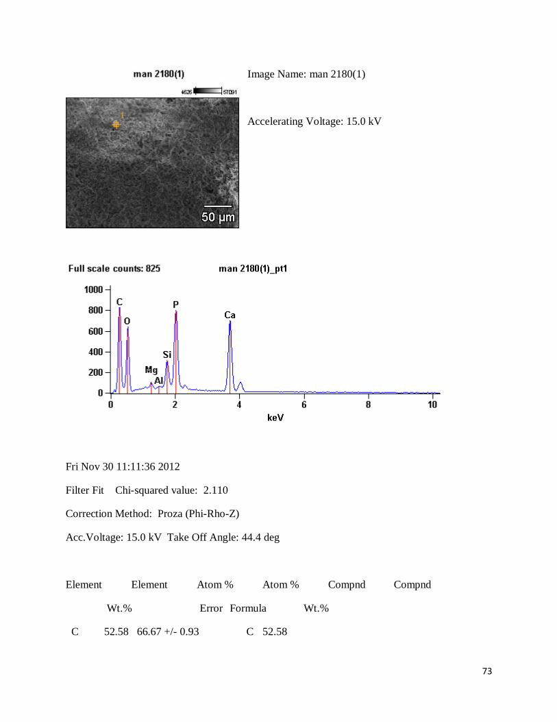
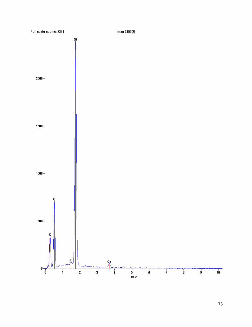
acidic. Below are the following graphs (from a Noran System Six 300 Nanotrace software)
showing the count levels of elements found in the organic material. The first chart below is from
mandible 2180 of an individual without cranial modification. The second was form the mold in
order to compare the molding material to the dirt specs found on the mold. The first chart shows
high counts for phosphorus, calcium, and oxygen with traces of carbon, magnesium, and
aluminum. With the exception of aluminum, these elements are not very acidic. However, the
calcium trace may be from the tooth itself that left traces on the mold. To have a fairly acidic soil
composition would require more aluminum, iron, and hydrogen counts and less calcium and
magnesium. With only specs of dirt to analyze, possible soil acidity is very difficult to determine,
if at all.

18
Figure 4. chart 1 of mandible 2180

19
Figure 5. Chart 2 of mandible 2180

20
ii. Casting materials and accuracy thereof
The casting materials first used was an Epotek apoxy resin which did not allow for proper
analysis of the original teeth as could be inferred upon from the molds. This was due to the
porous appearance of the molds possibly in part of a lack of a vacuum to take the bubbles out of
the mold. Later, another resin was used, Epotek 301 with a hardener. These molds did not
produce viable results. They too were porous and very difficult to work with in the SEM. The
castes charged quickly and proper viewing was limited as after the mold was casted it would take
a period of time to gain a refreshed image, which did not further any good results. Sputter coat
was used on a few of the castes, resulting in a less charging effect, however the pores of the
castes still made microwear analysis very difficult to discern.
In a comparative analysis of the molds verses castes, it was found the molds did reflect a
higher accuracy of microwear of the real tooth verses the caste. The caste would only capture
large gashes and had a lower frequency of accuracy to the real tooth. Further support is provided
by another study done by Galbany et. al (2006) who stated the use of coltene and 3M low
viscosity body polyvinylsiloxane impression is accurate to about the fourth caste made and
presidents microsystem material is widely used due to its accuracy. The Quala brand used in this
study is of similar viscosity and produced quality images reflecting similar accuracy. This was
observed by comparing images, side by side, taken from the actual tooth, mold, and caste see
how similar the images were to each other. The molds and actual tooth images were almost
identical while the molds did not.

21
b. Dental Macrowear Analysis
Macrowear allows for interpretations of general lifestyles within the population including
health and mastication tendencies. The main macrowear traits observed were cavities, caries,
abscesses, hypoplasias, build-up of calculus, and general wear-patterns. These observations were
based off photographs taken of each sampled individual. The photographs were taken in a
mesial, distal, lingual, and buccal view. The traits were selected to see the prevalence of gum
diseases, as generally made by calculus/plaque build-up on the teeth as well as effects of the
mastication process on wear. If calculus was present on the sample along with other traits leads
to the inference of poor hygiene and possibly less prepared foods. This would be because
Figure 6. Comparision of real
tooth (upper left), caste
(lower left), and mold (lower
right) of Anthro 2- LAB
individual.
22
calculus would build up and create various gum diseases, such as Periodontitis, and lead to
observable characteristics of caries and abscesses. Cavities are a bit different as they are caused
more by bacterial build-up which can increase in severity (into caries) if not treated. Many
causes can create these problems, such as diets with a high carbohydrate and sugar intake.
Hypoplasias also allow a way to see if health problems concerning nutrition were prevalent
throughout periods of an individual’s life. Poor nutrition could be a result of a lack of resources
due to seasonality or even class structure within a society.
Below are the traits with pictures to present the characteristics found within this
population. Majority of the pictures below are from mandibles of individuals without cranial
modification since many of these traits were more prevalent in this group. Individuals with
cranial modification are labeled accordingly to compare when possible.
23
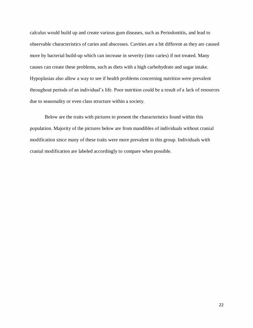
Examples of Hypoplasias
Hypoplasias, defined by lines on teeth, occur due to lack of proper nutrition and life
stresses and can be seen by finding lines on the tooth enamel.
Below are two of the three individuals from the population found with hypoplasia. All
three were found on the mandible of non-modified individuals on the left M3. The percent from
the total population is .055, and in non-modified individuals only the percent is .065.
Figure 7. Chullpa 7; mandible 2148

24
Figure 8. Cave 3, mandible 1319
25
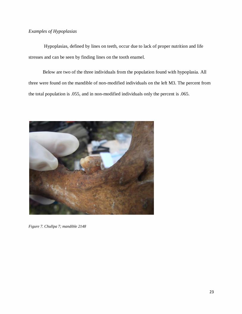
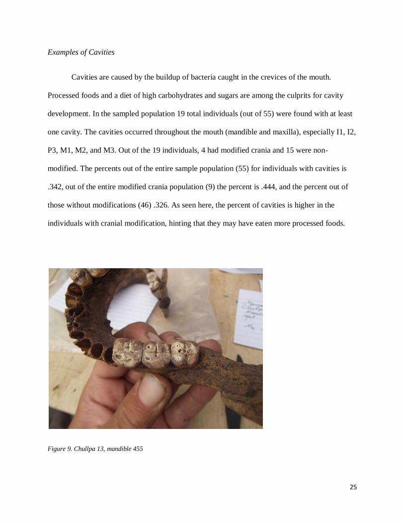
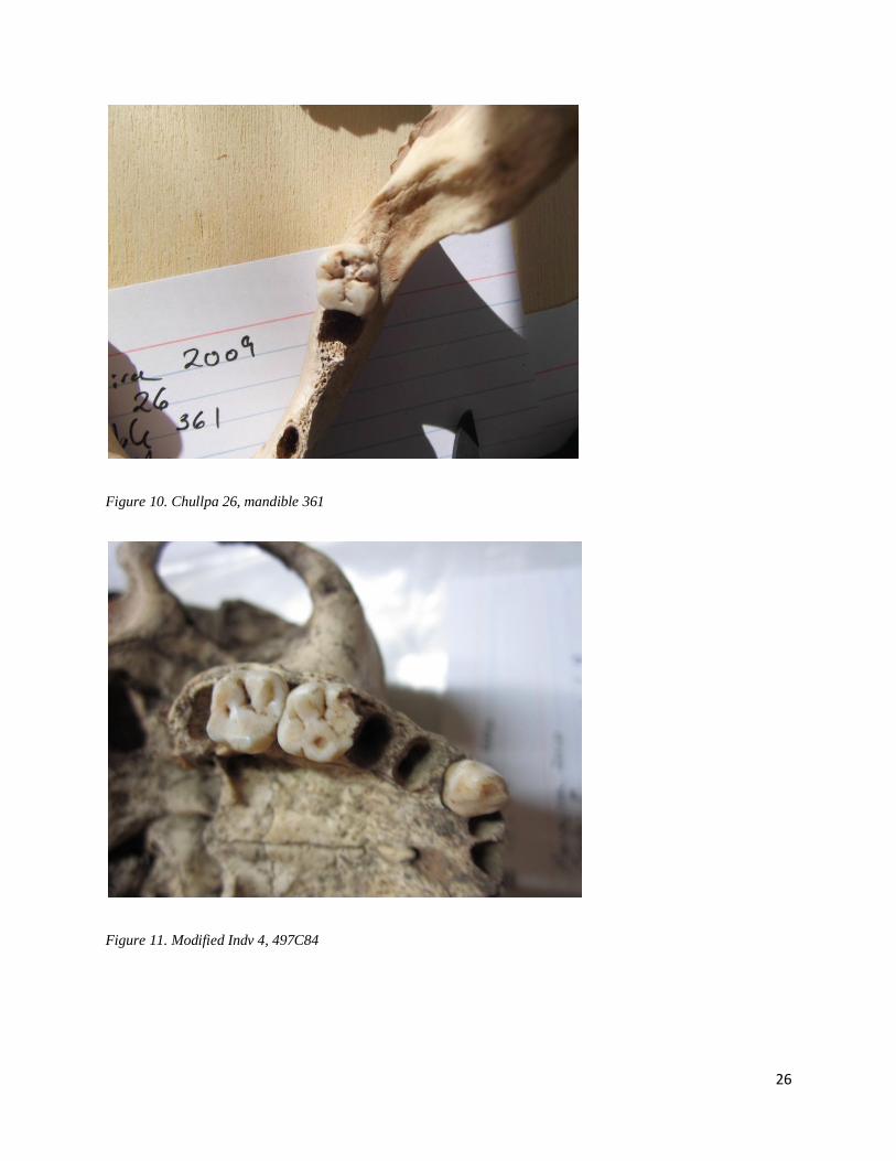
Examples of Cavities
Cavities are caused by the buildup of bacteria caught in the crevices of the mouth.
Processed foods and a diet of high carbohydrates and sugars are among the culprits for cavity
development. In the sampled population 19 total individuals (out of 55) were found with at least
one cavity. The cavities occurred throughout the mouth (mandible and maxilla), especially I1, I2,
P3, M1, M2, and M3. Out of the 19 individuals, 4 had modified crania and 15 were non-
modified. The percents out of the entire sample population (55) for individuals with cavities is
.342, out of the entire modified crania population (9) the percent is .444, and the percent out of
those without modifications (46) .326. As seen here, the percent of cavities is higher in the
individuals with cranial modification, hinting that they may have eaten more processed foods.
Figure 9. Chullpa 13, mandible 455
26
Figure 10. Chullpa 26, mandible 361
Figure 11. Modified Indv 4, 497C84

27
Examples of Caries
Caries are more extreme in severity in comparison to cavities. They are usually caused by
gum diseases and build-up of calculus and plaque. In the sample population, 13 individuals were
identified with carious lesions giving a percent of .236 out of the entire sample. One of these
individuals was from the modified cranial group and represented .111 percent of the group with a
caries occurring on the incisal region. The 12 non-modified individuals gave a percent of .261 in
their group with caries mainly in the molar region. The percent is higher in the group without
cranial modifications.
Figure 12. Cave 3, mandible 489

28
Figure 13. Chullpa 7, mandible 218
Figure 14. Modified 49CF3D41

29
Examples of Abscesses
Abscesses are more severe than caries. They generally result from gum diseases. Out of
the sample population, 3 individuals, each non-modified, were identified to have abscesses. The
occurrence found of the abscesses was generally below the incisors, and one below the M1. The
total percent is .055, and out of the group of individuals without cranial modification, .007.
Figure 15. Chullpa 7, mandible 2180

30
Figure 16. Structure 7, Indv 10

31
Wear Patterns
Wear patterns can define numerous things, such as a mastication preference for one side
or how tough foods were to wear on teeth quickly. There are many types of curvatures wear
patterns can create. The two most prevalent in the sample population are known as the Monson
Curve and Reverse Monson Curve.
- Slope in (Monson Curve)
Figure 17. Chullpa 13, mandible 455

32
Figure 18. cave 3, mandible 68
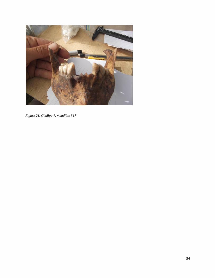
- Slope out (Reverse Monson Curve)
A reverse monson curve resembles a sloping down wear on the molars. This may more of
an orthodontic trait due to the possibility the maxilla is larger than the mandible.

33
Figure 19. Chullpa 8, mandible 528
Figure 20. Chullpa 7, mandible 317
34
Figure 21. Chullpa 7, mandible 317

35
General Wear of Teeth
The general wear of the teeth were found to be heavy on the molars. This created an
upside-down –shape with little wear on the incisors. Below are two more examples in addition to
the pictures given above showing this phenomena. As Mahoney (2006) stated in his research,
“human bite force increases on the more posterior molars”. As seen, the wear does increase
dramatically in the molar region.
Figure 22. Chullpa 8, mandible 251

36
Figure 23. Cave 3, mandible 489

37
Abnormal Traits
In addition to the traits described above, a few irregulars were found in the population
and are pictured below.
Figure 24. cave 3, mandible 8811
Figure 25. Chullpa 7, mandible 2180

38
After reviewing the photos of fifty-five individuals, it was found that majority of the
individuals without cranial modifications had a higher frequency of caries, abscesses, and
hypoplasias along with more instances of calculus build-up. The individuals without cranial
modifications tended to have a higher frequency of cavities and less instances of the other
observed traits. Part of this could be due to how the only sample available for individuals with
cranial modification was maxillas; the sample for individuals without cranial modifications was
majority mandibles. However, in the purpose of observing and creating inferences on health,
calculus build-up exists on maxillary teeth at the same rate as on mandibular. If there was a lack
of calculus on maxillary teeth, it can be assumed there may have also been a lack of calculus on
mandibular teeth. Considering this, there is a marked difference between individuals with cranial
modifications and those without.
By looking at the general wear patterns, the process of mastication was more observable
in the mandibles, as more of their teeth, including incisors, were still in the mandible. The
maxillas still produced results too. The two patterns investigated were the sloping of teeth, if
they sloped inward toward the lingual side, known as a monson curve, or outwards toward the
buccal side, referred to as a reverse monson curve. Again, as the entire sample of the individuals
with cranial modifications were maxillas, it was difficult to infer these patterns. However, the
inverse of the maxillas, meaning how the maxillary teeth were sloped and shaped in a way to
produce a mandibular and similar slope of the teeth, could be inferred on. These would make a
fair guess, but not entirely accurate due to other exposures that the teeth can experience post-
mortem in regards to taphonomy. Despite this, it was found in the sample of individuals without
cranial modifications that about twenty percent of them demonstrated the Reverse Monson
Curve. The rest were simply heavily worn down in the molar region into a Monson Curve.

39
As found in the pictures above, the individual represented by mandible 2180 suffered
from severe health problems that resulted in caries, abscesses, and inflammation within the jaw.
The inflammation is a bit irregular since it is not normally found, nor is it erupting into an
abscess. The picture for mandible 8811 shows a sixth cusp on the first molar. This individual was
the only one found with this trait.
c. Dental Microwear Analysis
The microwear of the molds was accurate in determining dietary features in the maxilla and
mandible. One main observation made was the differences both groups presented, meaning either
the individual demonstrated a microwear patter of mostly striations or mostly of pits. There was
a lack of cases in which an individual without cranial modification demonstrated an even
distribution of striations and pits.
Individuals with cranial modifications had more cases of striations on their teeth or
combination of striations and pitting. This was mainly observed in the mesial side of the tooth
viewed in the SEM. However, it is important to keep in mind that these individuals with cranial
modification were only sampled with maxillary teeth due to the lack of mandibular teeth.
Individuals without cranial modification had a lack of maxillary teeth viewed due to lack of
maxillas available. This brings to point that findings at excavations are indeed unpredictable.
Different magnifications were used throughout the process, ranging from 15 to 550. It was
found that 150 and 250 magnifications produced optimal results, with re-evaluations at 50
magnifications to ensure which area was being viewed. Photos from 9 modified and 5 non-
modified individuals with a sampled maxillary M2 were used in the SEM and focused in the

40
mesial-lingual and mesial-buccal sides. A few mandibular samples from other individuals were
viewed as well, but majority of these were experimental and included castes.
The corrosion on the teeth was most frequent in the individuals with cranial modification.
This may be due to the preservation and taphonomical processes in the duration of the
excavation of these crania. Another possibility of this corrosion is due to taphonomy. Many of
the individuals with cranial modifications were found in caves while majority of the individuals
without cranial modifications were found in Chullpas.
Below, in the following figures are examples of individuals with cranial modifications and
those without on maxillary samples. The photos are marked to show where the striations and pits
are identified with numbers identifying how many in each photo.

41
Figures 26 and 27. Modified 49C7B4, maxilla RM2, Mesial Lingual

42
Figures 28 and 29. Modified 49FC3D41, Maxillary RM2, mesial lingual

43
Figures 30 and 31. Modified 49C19C294, maxilla, RM2, mesial lingual

44
Figues 32 and 33. Non-modified, Indv 1, maxillary LM1 mesial lingual

45
Figures 34 and 35. Non-modified, Indv 17, maxilla M2, mesial lingual (sputter coat)
46
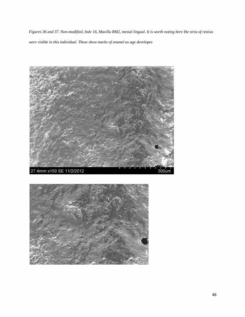
Figures 36 and 37. Non-modified, Indv 16, Maxilla RM2, mesial lingual. It is worth noting here the stria of retzius
were visible in this individual. These show marks of enamel as age developes

47
Examples from mandible:
Figures 38 and 39. mand 1992, M2, mesial buccal

48
d. Maori comparison
The Reverse Monson Curve was found in the Maori Culture in New Zealand, and thought to
have been a fairly unique trait to this region. According to Keiser et. al 2001, this may have been
more of an orthodontic effect stating, “If, for instance, the maxillary arch is wider than the
mandible, then the wear plane will tend to be ad palatum while if the mandible arch is wider, an
accentuated Monson, ad lingum, will result.” The Maori were known to be tall with large faces
that featured a robust mandible despite their teeth being moderate in size (Keiser et. al, 2001). It
is difficult to compare the robusticity of the individuals from Maori to Marcajirca due to the lack
of the entire crania of the same individual being excavated. However, robusticity in the jaw
could create a larger bite force, especially if the maxilla was slightly larger than the mandible to
produce a more buccal wear on mandibular teeth. If this was a cultural trait on either population,
the appearance of the wear may be slightly different in regards to a preference of a side used in
mastication. Although this is sometimes the case in the Marcajirca individuals, it is very difficult
to determine a proper comparison of cultural dynamics of these two populations.

49
4. Conclusion
a. Considerations of Methodology
As in any archaeological excavation, methodology is far from perfect. Conditions at a site
and what is encountered during and after can hardly be planned for. This happened to be the case
in this investigation as problems encountered during researched occurred in sequence, on after
the other. It began with the uncertainty of corrosion and quality of microwear found on the molds
via SEM. Part of this problem was the lack of investigation in the acidity and quality of the soils.
The Chullpas, caves, and pits at the site may have had different compositions, but it is difficult to
tell at this time due to the lack of information. The available information was found on the dirt
pieces adhered to the molds that came from the teeth of the individuals. Although they were
small, the composition of the dirt pieces could be determined; however the sample size was too
small to determine pH of the soil. It is suggested here, in reference to future excavations, that soil
samples be analyzed from various parts of a site. They are essential to research. It is also
important to keep in mind the burial environments may have changed over time (Turner-Walker,
2008) possibly due to seasonality changes or even unnatural causes. Soil testing would allow
investigations of bones and teeth to be referenced in a new dynamic related to taphonomy. Bones
can degrade in specific soils, specifically acidic soils, or as stated (Child, 1995) “The chemical
and physical deterioration of buried bone depends solely upon the chemistry (and biochemistry)
of the surrounding soil environment. Acidic soils will dissolve hydroxyapitite, but the threshold
of acidity required is not clear.” Furthermore, it is stated by Child, 1995, that bones do not
disintegrate easily in neutral/alkaline soils and soils with low phosphate levels tend to
demineralize bones. If soil pH can be tested and known prior to excavations of bones, it would

50
help investigations and create a more accurate expectation of what is found on the bones. After
having had examined the dirt composition from the SEM, there were a few traces of alkali
elements such as Magnesium, that may have led to the bone disintegration found on the enamel.
This would have to be further investigated due to the lack of a good sample size of soil available.
The sample size of the population is small and very little can be done about sample size. You
cannot predict what is found and in what quality it is found in. However, this should not deter
from the quality of the results. According to Covey (2008) the Wari empire was not entirely
uniform in the highlands and according to Isbell and Schreiber (1978) the Wari empire was the
largest site of the Andes in 700CE. A further dental investigation as to how diverse the empire is
throughout the highland region may help elaborate these statements.
A large portion of methodology practices is to create a way to translate results in a universal
manner that allows for replication. Most of this depends on availability of resources and access
to lab equipment. This project used a scanning electron microscope, yet the new direction of
dental analysis is examining texture by means of a confocal profiler. A Confocal profiler is
currently not an easily accessible machine. Due to this, it is suggested here that results to include
an analysis of a universal basis, such as specific measurements, magnifications, and facets. This
may not solve the issue of resources, but would narrow results to areas that can be cross
referenced.
On the macrowear level a suggestion for definitions to be made for bioarchaeologists would
be helpful. Many of the definitions found and used here were from dental journals. These
definitions may be limiting to bioarchaeologists as many times taphonomy plays a large role in
preservation of teeth. It would be useful to create a definition of traits found on the macro level

51
that refers to taphonomical effects in order to avoid possible confusion of what was pre and post
mortem.
b. Inferences
After going through the many traits found within the population on a micro and macro level,
it may be said that individuals with cranial modifications had a different diet consisting of more
processed foods. On a macro level, a large difference between the groups, modified verses non-
modified, were found. In general, there was a lack of poor health characteristics in individuals
with cranial modification. The only trait that occurred, and was more frequent than individuals
without cranial modifications, was cavities. Cavities occur due to a buildup of bacteria and are
not as severe as caries or abscesses. They are caused by softer foods and sugars, many times
considered a luxury food item. The individuals without cranial modifications suffered the more
extreme versions of caries, abscesses, and a higher frequency of calculus build-up. Caries are
more common in diets of high carbohydrates based on cultured plants, and less common in diets
that are rich in meat (Bernall et. al, 2007). This again points to the inference of an elite class in
the population represented by individuals with cranial modifications. The high occurrence of
hypoplasias and calculus in the individuals without cranial modification also suggests a poorer
quality of diet and less proper hygiene which would create these dental problems the individuals
experienced pre-mortem.
The differences in frequencies of the striations verses pits found on the molds differed on a
micro level. More striations, or combinations of striations and pits, were found on the maxillary
M2 of individuals with cranial modification. Individuals without cranial modification had a

52
higher frequency of pits on their, ranging from small to large pits. A higher frequency of
striations generally means a diet with softer foods that require a sliding mastication process.
Such foods could be leaves and meat which generally can relate to an elite diet (Klaus and Tam,
2010; Ascencios, 2008). This makes sense in considering the use of coca leaf. Coca leaf is seen
as a ritual and respectful commodity, although it is chewed to help fight altitude sickness. If
considered special, it may have been reserved for the individuals with more authority. A diet that
results in more pits generally would reflect harder or less processed foods and would thus belong
to a lesser class. In conclusion, it is very possible this population demonstrated a social status by
use of cranial modification.
c. What Else Can Be Researched?
At this point in the study, a lot of work has still to be done with regard to cross cultural
comparisons in the Latin American region. What may be a direction is to gather more maxillas
and mandibles to increase the sample population. Other sites along the highlands of Peru can
expand this sample greatly and further comparisons of tribes can be made in the future. Another
direction would be to mold teeth of individuals from the lowland and coastal sites of Peru. This
would allow a significant comparison of environmental and regional differences in the sub-
populations of the Peru country.
On the macro level, many abscesses and evidence of cavities are present throughout the
population sample. Nutritional stresses are also evident in few through hypoplasias found on
incisors. Wearing of the teeth was among the more interesting aspects. The wearing of the
molars was heavier than on the incisors, forming what is known as a reversed Monson curve.

53
This curve, upon simple observation looks as if the mandible forms an upside-down U-shape and
a buccal slope on the teeth. The cause of this is poorly understood and more research as to what
parts of the process during mastication could create a more diagnostic definition of it.
On a micro level, the molds and castes showed hints of striations and pitting, with a variance
between individuals with modified crania and those without modifications. The diet consists of
carbohydrates, mainly revolving around potatoes (remnants found at the site as well as potatoes
still growing), fish, and rice along with habitual coca leaf chewing. The amounts available year
round could allow why combinations of striations and pitting occurs more in individuals with
cranial modification verses those without. Also if there were cultural practices, such as rope
pulling, done with the use of teeth as part of a reason as why there are such differences in
microwear traits.
It may also be interesting to see the differences based on sex in order to recognize if males
and females were treated differently by means of foods available to them. If females were given
slightly harder and less process foods; had a higher frequency of hypoplasias, cavities, and caries
or vice versa if such marked differences do exist.
d. Current Questions
Many questions still remain about this population. One of the first ones is how related is this
population to other highland populations in dietary aspects? How similar/dissimilar are the
coastal groups? How related to groups outside of the entire region?
How much does taphonomy play a role in teeth?

54
What other populations represent a reverse monson curve?
How similar is coca leaf chewing to tobacco chewing and can this information be used to
help determine wear patterns of past populations?
What other factors could result in a reverse-monson curve other than orthodontic?

55
Bibliography
Ascencios, B.I. (2008) The Bioarchaeology of Health and Social Organization at Marcajirca,
North-Central Highlands, Peru. (Project Review) Centre de research d’ Archéologie
Précolombienne. Université Paris I. Surbonne. Paris. Institiuto Cultural RVNA. Huari-Ancash
Archaeological Project.
Bernal, V. Novellino, P., Gonzalez, P.N., and Perez, S.I. (2007). Role of Wild Plant Foods Among Late
Holocene Hunter-Gatherers From Central and North Patagonia (South America): An Approach
from Dental Evidence. Journal of Physical Anthropology. 133:1047-1059
Bromley, P. (1995). The Effect of Elevation Gain on Soil. Environmental Studies 102.
(Unpublished Dissertation, no institution marked).
Child, A.M. (1995). Microbial Taphonomy of Archaeological Bone. Studies in Conservation.
40:19-30.
Chaves, S.A. and Reinhard, K.J. (2003) Paleopharmacology and Pollen: Theory, Method, and
Application. Mem Inst Oswaldo Cruz, Rio de Janeiro. 98:207-211.
Covey, R. A. (2008). Multiregional Perspectives on the Archaeology of the Andes During the
Late Intermediate Period (c.A.D. 1000-1400). Journal of Archaeological Research. 16:287-338.
Duncan, W.N. and Hofling, C.A. (2011). Why the Head? Cranial Modification as Protection and
Ensoulment Among the Maya. Ancient Mesoamerica. 22:199-210.
Galbany, J., Estebaranz, F., Martínez, L.M., Romero, A., De Juan, J., Turbón, D., and Pérez-Pérez, A.
(2006). Comparitive Analysis of Dental Enamel Polyvinylsiloxane Impression and
Polyurethane Casting Methods for SEM Research. Microscopy Research and Technique. 69:246-
252.
Isbell, W.H. and Schrieber, K.J. (1978). Was Huari a State? American Antiquity. 43:372-389.
Kieser, J.A., Dennison, K.J., Kaidonis, J.A., Huang, D., Herbison, P. G. P., and Tayles, N.G. (2001).
Patterns of Dental Wear in the Early Maori Dentition. International Jouranal of Osteology.
11:206-217.
Kieser, J.A., Kelsen, A., Love, R., Herbison, P.G.P., and Dennison, K.J. (2001). Periapical Lesions and
Dental Wear in the Early Maori. International Journal of Osteology. 11:290-297.
King, T., Andrews, P., and Boz, B. (1999). Effect of Taphonomical Processes on Dental Microwear.
American Journal of Physical Anthropology. 108:359-373.
Klaus, H.D. and Tam, M.E. (2010). Oral Health and the Postcontact Adaptive Transistion: A Contextual
Reconstruction of Diet in Mórrope, Peru. American Journal of Physical Anthropology. 141. 594-
609.
56
Larsen, C.S. (2002). Bioarchaeology: The Lives and Lifestyles of Past People. Journal of Archaeological
Research. 10:119-166.
Mahoney, P. (2006). Brief Communication: Intertooth and Intrafacet Dental Microwear Variation in an
Archaeological Sample of Modern Humans From the Jordan Valley. American Journal of
Physical Anthropology. 129:39-44.
Naji, S. (2012). Introduction to Archaeothanotology. Lecture at Huari-Ancash Project. 2012 Season.
Turner-Walker, G., 2008. The Chemical and Microbial Degradation of Bones and Teeth. In: Pinhasi, R., Mays, S. (Eds.), Advances in Human Paleopathology. Wiley & Sons,
Chichester, pp. 1–29
Verano, J.W. (1997). Advances in the Paleopathology of Andean South America. Journal of World
Prehistory. 11:237-268.

57
APPENDIX
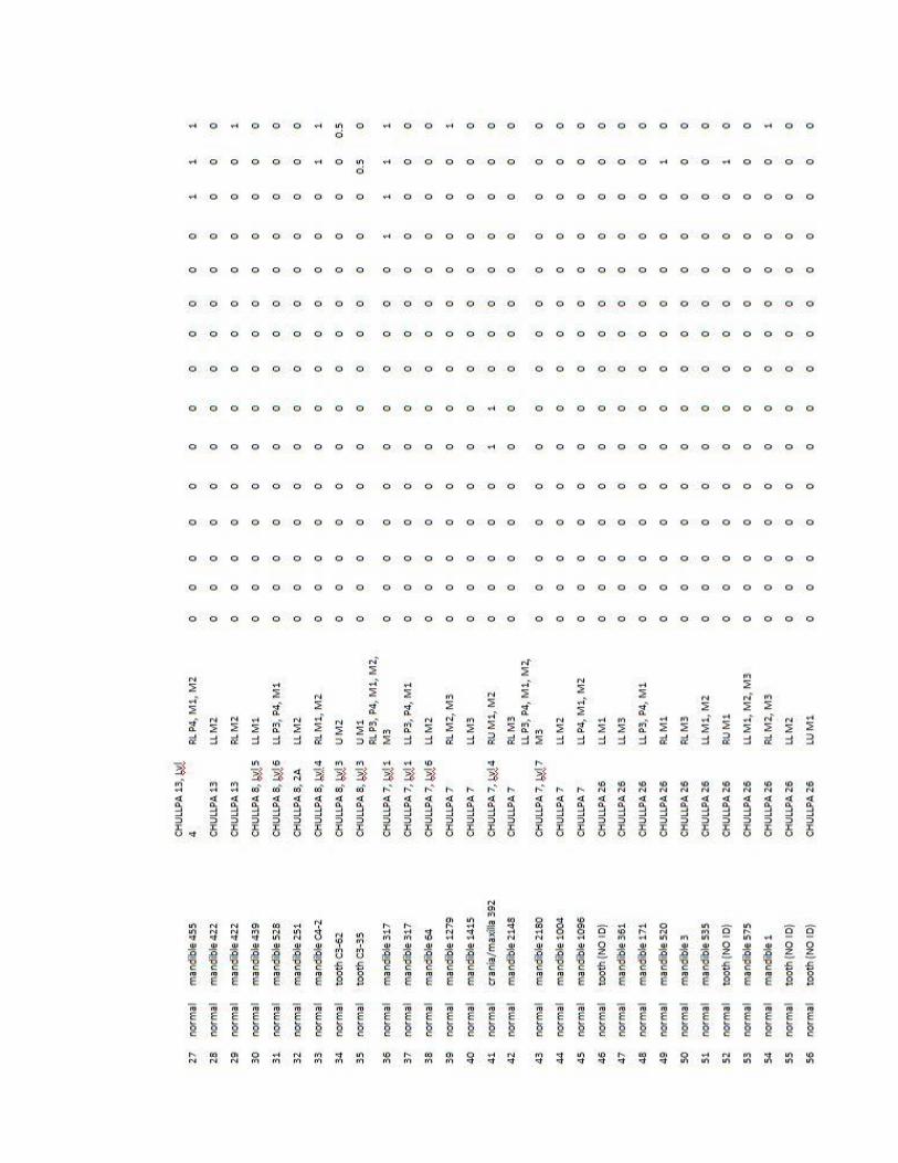
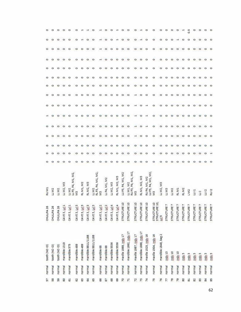
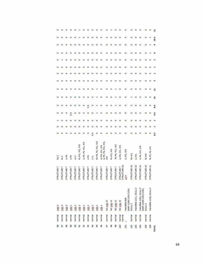
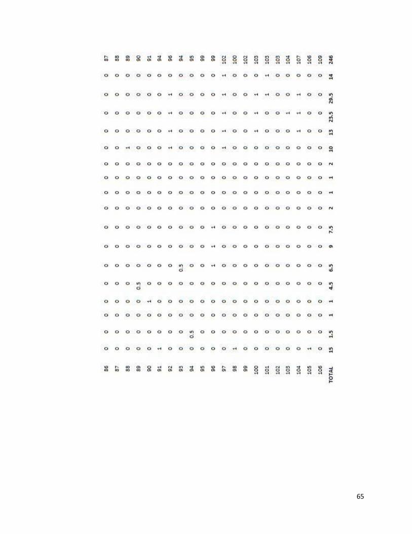
Tooth Inventory
On the following pages is the entire sampled population used for this research. The abbreviations
are listed below. The decimal, .5, represents an indeterminate side.
ABBREVIATIONS
RU Right Upper
RL Right Lower
LU Left Upper
LL Left Lower
Indv Individual
Lvl Level
J Juvenille
I Incisor
C Canine
P Premolar
M Molar

58

59
60

61
62

63
64
65

66
Maxillary Measurements
Type Identification Side
M1
length M1 width M2 length M2 width
Modified 49-C19-B2-1028 L 10.8 11 - -
Modified(J) 49F-C7B-1-2 R - - 10.2 10.6
Modified 49F-C3-D4-1 L - - 9.4 10.4
Modified 49-C7B-4 L - - 11.2 12.2
Modified 49-C3-F3-1043 R - - 10.6 11.8
Modified 49F-C3-D-1 R - - 9.7 10.4
Modified 49-C7B-1 R - - 10.1 12.1
Modified (J) 49-C7B-1-2 L - - 10.2 10.4
Modified 49-C19-A2-1 R - - 9.6 11.3
Modified (J) 49-C19-C2-94 R - - 9.5 11.3
Modified 49-C7B-4 R - - 10.7 12.2
reg Chullpa 26 R 8.9 9.9 - -
reg indv 16, stc 10, 1504 L - - 9.7 10.7
reg stc F, 1249/1251/1252 R 11.1 11.7 - -
reg (J) no # ? - - 13.3 11.6
reg Chullpa 26 R 9.1 10.9 - -
reg Chullpa 26 L 11.1 10.2 - -
reg Indv 1, sector 7 R -
11 13.5
reg Indv 1, sector 7 L 11.6 13.2 - -
reg C3-62 ? - - 10.2 11.6
reg Indv 16, stc 10, 1525 R - - 9.5 10.5
reg Chullpa 26 L 8.7 10.2 - -
reg Indv 17, Stc 10, 1987 R - - 9.7 11.6
reg Chullpa 8, C3-35 ? 10 10.4 - -
reg Indv 17, stc 10, 1989 L - - 10 11.2
AVGS Modified
10.12 11.27
Modified- juvi
10.3 11.486
non mod (reg)
10.48 11.52

67
Macrowear
Features
Observed caries location abscess location cavity location hypoplasia location calculus location
Slope
out wear
(Reverse
Monson)
Molars;
M1,
M2, and
mainly
M3.
One P3
In
modifie
d, found
on incisors
and
canine
only
below
incisors
and M1
I1, I2,
P3, M1,
M2, M3
M3 generally
througho
ut indv
TOTAL
INDVS (out
of 55) 13 3 19 3 15 11
Percent
Total (out of
55) 0.236 0.056 0.352 0.055 0.278 0.2
Total in
Non-
modified
Indvs (out of
46) 12 3 15 3 15 11
Percent total
in non-
modified 0.261 0.007 0.326 0.065 0.326 0.239
Total in
Modified
Indvs (out of
9) 1 0 4 0 0 0
Percent total
in modified 0.11 0 0.444 0 0 0
notes- modified were only maxillary, all sloping inwards. The compliment of this would be a mandible that slopes out.
in this regards, it may be possible to infer all mandibles for modified individuals were sloped outward

68
Microwear
magnification striations pits
total
pics pits/striations
Modified 150 435 139 26 0.319
250 313 68 29 0.217
Non-mod 150 102 65 10 0.637
250 70 63 15 0.9
notes
non-mods show more combination of pits
and striations, while modified show more
striations in general
size of pits varied

69
Soil Analysis from Molds (with compositional counts):

70

71
Fri Nov 30 11:06:39 2012
Filter Fit Chi-squared value: 1.754
Correction Method: Proza (Phi-Rho-Z)
Acc.Voltage: 15.0 kV Take Off Angle: 44.4 deg
Element Element Atom % Atom % Compnd Compnd
Wt.% Error Formula Wt.%
C 22.06 35.19 +/- 0.97 C 22.06
O 34.51 41.32 +/- 0.51 O 34.51
Mg 0.69 0.54 +/- 0.07 Mg 0.69
Al 0.14 0.10 +/- 0.04 Al 0.14
Si 2.59 1.77 +/- 0.05 Si 2.59
P 13.97 8.64 +/- 0.09 P 13.97
Ca --- --- --- ---
Ca 26.04 12.44 +/- 0.12 Ca 26.04
---------- ---------- ----------
Total 100.00 100.00 100.00

72
Image Name: man 2180(1)
Accelerating Voltage: 15.0 kV
Magnification: 422
73
Fri Nov 30 11:11:36 2012
Filter Fit Chi-squared value: 2.110
Correction Method: Proza (Phi-Rho-Z)
Acc.Voltage: 15.0 kV Take Off Angle: 44.4 deg
Element Element Atom % Atom % Compnd Compnd
Wt.% Error Formula Wt.%
C 52.58 66.67 +/- 0.93 C 52.58
Image Name: man 2180(1)
Accelerating Voltage: 15.0 kV
Magnification: 422

74
O 24.59 23.41 +/- 0.30 O 24.59
Mg 0.36 0.22 +/- 0.02 Mg 0.36
Al 0.12 0.07 +/- 0.02 Al 0.12
Si 1.83 0.99 +/- 0.02 Si 1.83
P 7.55 3.71 +/- 0.04 P 7.55
Ca --- --- --- ---
Ca 12.97 4.93 +/- 0.05 Ca 12.97
---------- ---------- ----------
Total 100.00 100.00 100.00
75

76
Fri Nov 30 11:15:06 2012
Filter Fit Chi-squared value: 2.136
Correction Method: Proza (Phi-Rho-Z)
Acc.Voltage: 15.0 kV Take Off Angle: 44.4 deg
Element Element Atom % Atom % Compnd Compnd
Wt.% Error Formula Wt.%
C 45.87 58.45 +/- 0.95 C 45.87
O 29.69 28.39 +/- 0.31 O 29.69
Al 0.20 0.11 +/- 0.02 Al 0.20
Si 23.22 12.65 +/- 0.09 Si 23.22
Ca 1.02 0.39 +/- 0.02 Ca 1.02
Ca --- --- --- ---
---------- ---------- ----------
Total 100.00 100.00 100.00

77
78
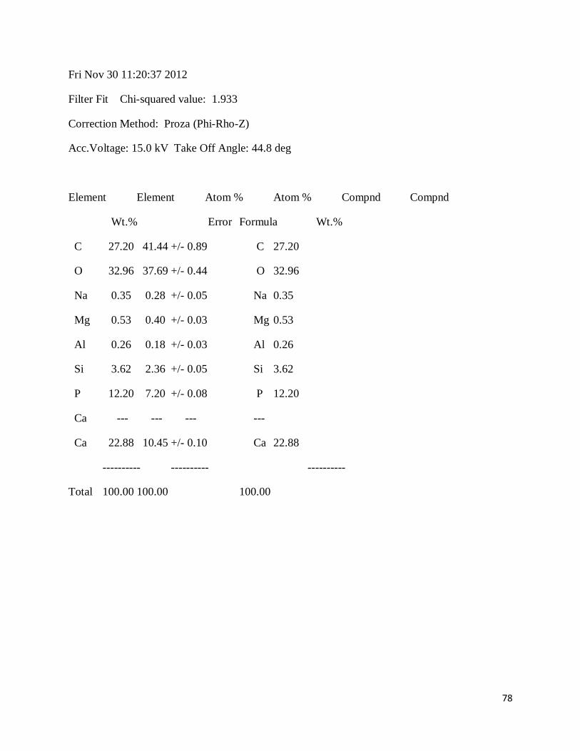
Fri Nov 30 11:20:37 2012
Filter Fit Chi-squared value: 1.933
Correction Method: Proza (Phi-Rho-Z)
Acc.Voltage: 15.0 kV Take Off Angle: 44.8 deg
Element Element Atom % Atom % Compnd Compnd
Wt.% Error Formula Wt.%
C 27.20 41.44 +/- 0.89 C 27.20
O 32.96 37.69 +/- 0.44 O 32.96
Na 0.35 0.28 +/- 0.05 Na 0.35
Mg 0.53 0.40 +/- 0.03 Mg 0.53
Al 0.26 0.18 +/- 0.03 Al 0.26
Si 3.62 2.36 +/- 0.05 Si 3.62
P 12.20 7.20 +/- 0.08 P 12.20
Ca --- --- --- ---
Ca 22.88 10.45 +/- 0.10 Ca 22.88
---------- ---------- ----------
Total 100.00 100.00 100.00

79
80
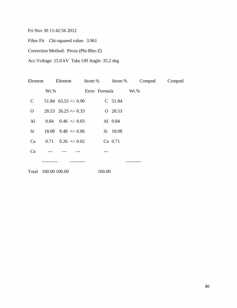
Fri Nov 30 11:42:56 2012
Filter Fit Chi-squared value: 3.961
Correction Method: Proza (Phi-Rho-Z)
Acc.Voltage: 15.0 kV Take Off Angle: 35.2 deg
Element Element Atom % Atom % Compnd Compnd
Wt.% Error Formula Wt.%
C 51.84 63.55 +/- 0.90 C 51.84
O 28.53 26.25 +/- 0.33 O 28.53
Al 0.84 0.46 +/- 0.03 Al 0.84
Si 18.08 9.48 +/- 0.06 Si 18.08
Ca 0.71 0.26 +/- 0.02 Ca 0.71
Ca --- --- --- ---
---------- ---------- ----------
Total 100.00 100.00 100.00

81

82
Fri Nov 30 11:48:58 2012
Filter Fit Chi-squared value: 2.538
Correction Method: Proza (Phi-Rho-Z)
Acc.Voltage: 15.0 kV Take Off Angle: 35.2 deg
Element Element Atom % Atom % Compnd Compnd
Wt.% Error Formula Wt.%
C 38.30 49.97 +/- 0.98 C 38.30
O 39.57 38.76 +/- 0.38 O 39.57
Mg 0.12 0.08 +/- 0.02 Mg 0.12
Al 3.56 2.07 +/- 0.03 Al 3.56
Si 14.74 8.23 +/- 0.05 Si 14.74
P 0.24 0.12 +/- 0.02 P 0.24
Ca --- --- --- ---
Ca 1.20 0.47 +/- 0.02 Ca 1.20
In --- --- --- ---
In 2.27 0.31 +/- 0.03 In 2.27
---------- ---------- ----------
Total 100.00 100.00 100.00