Embed Size (px)
Citation preview

Neuron
Review
Cytokines and CNS Development
Benjamin E. Deverman1 and Paul H. Patterson1,*1Division of Biology, California Institute of Technology, 1200 East California Boulevard M/C 216-76, Pasadena, CA 91125, USA*Correspondence: [email protected] 10.1016/j.neuron.2009.09.002
Cytokines are pleotrophic proteins that coordinate the host response to infection as well as mediate normal,ongoing signaling between cells of nonimmune tissues, including the nervous system. As a consequence ofthis dual role, cytokines induced in response to maternal infection or prenatal hypoxia can profoundly impactfetal neurodevelopment. The neurodevelopmental roles of individual cytokine signaling pathways are beingelucidated through gain- and loss-of-function studies in cell culture and model organisms. We review thiswork with a particular emphasis on studies where cytokines, their receptors, or components of their signalingpathways have been altered in vivo. The extensive and diverse requirements for properly regulated cytokinesignaling during normal nervous system development revealed by these studies sets the foundation forongoing and future work aimed at understanding how cytokines induced normally and pathologically duringcritical stages of fetal development alter nervous system function and behavior later in life.
IntroductionCytokines are small, mostly secreted proteins that were origi-
nally characterized as immune modulators but have subse-
quently been found to mediate a diverse array of functions in
nonimmune tissues, including the central nervous system
(CNS). Cytokines are of particular importance during neural
development and function at all stages, starting with induction
of the neuroepithelium. Subsequently, cytokines, in particular
the neuropoietic, or gp130 family cytokines, regulate the self-
renewal of the neuroepithelial/radial glia cells (RGC), which func-
tion both as the scaffolds for radially migrating neurons and as
the precursors for all neurons, macroglia (astrocytes and oligo-
dendrocytes), and adult progenitors. RGCs give rise to neurons
first and then glia, and several cytokines, including the gp130
cytokines and the bone morphogenetic proteins (BMPs), have
a central role in this shift to gliogenesis. In addition, chemokines,
a subclass of small cytokines with chemoattractant properties,
function as cues for the migration of newly generated neurons
and glia and are modulators of axon pathfinding. Furthermore,
as a general rule, neurons and glia are produced in excess,
and cytokines have roles as both neurotrophic factors that
promote the survival of cells that make the appropriate connec-
tions and as signals that trigger the apoptosis of cells that fail to
compete for these connections.
Not all cells in the CNS have neural origins. Microglia, which
are immune surveillance cells of the hematopoietic lineage,
colonize the early embryonic neuroepithelium and phagocytose
developmental apoptotic debris. These immune cells also
modulate vascularization, neuronal survival, and synapse forma-
tion and function, in part through their response to, and secretion
of, a wide repertoire of cytokines.
Although cytokines primarily act locally, they can also have
endocrine effects. Thus, cytokine induction in response to
maternal infection or fetal injury may adversely affect neurode-
velopment. Indeed, epidemiological evidence points to maternal
infection as a cause of neurodevelopmental abnormalities
that increase the risk for schizophrenia, autism, and cerebral
palsy in the offspring (for more information, see accompanying
article from Ellman and Susser, 2009 [this issue of Neuron]).
Recent findings from animal models of maternal infection
support this hypothesis and provide evidence that dysregulation
of individual cytokines can induce striking behavioral deficits in
the offspring. Further insight into how cytokine dysregulation
interferes with normal neural development will come from
animal models of maternal infection as well as a continued
focus on the normal roles that cytokines have during CNS
development.
Neural Induction and Primitive NSCsNeural induction in the vertebrate embryo is repressed by BMPs,
which are members of the transforming growth factor beta
(TGFb) cytokine superfamily (reviewed in Gaulden and Reiter,
2008). Active inhibition of BMP signaling is required for normal
neural development in mice as demonstrated by the lack of fore-
brain development in the absence of Noggin and Chordin, two
BMP antagonists (Bachiller et al., 2000). These findings have
been largely recapitulated in culture. Embryonic stem cells
(ESCs) can give rise to cells with the characteristics of primitive
neural stem cells (NSCs) in the absence of exogenous factors,
but do so more efficiently in the presence of Noggin, or if BMP
signaling is blocked by deletion of SMAD4, the common down-
stream signaling effector for the TGFb family of cytokines (Tro-
pepe et al., 2001). In addition, leukemia inhibitory factor (LIF),
a gp130 family cytokine well known for its role promoting mouse
ESC self-renewal, functions as a permissive survival factor
during the transformation of ESCs into cells with the characteris-
tics of embryonic NSCs. This role for LIF is likely culture-specific
since the neuroepithelium forms in vivo in the absence of LIF or
gp130, the common receptor subunit required for signaling by
LIF and related gp130 cytokines (Escary et al., 1993; Yoshida
et al., 1996).
Maintaining the Neural Progenitor PoolNeuroepithelial cells directly or indirectly give rise to all of the
neurons, astrocytes, and oligodendrocytes in the adult brain.
Initially, neuroepithelial cells divide symmetrically, expanding
Neuron 64, October 15, 2009 ª2009 Elsevier Inc. 61

Neuron
Review
Notch
LIF/CNTF/NP/?
Jak
STAT3
neurogenic genes
gliogenesis adultneural specificationIL-1β
OPCs& OLs
IL-6LIF
neurons
SHP-2
gp130
BMPs
neurogenesis
basalprogenitors CT-1
LIF/CNTF/NP/?
STAT3 pSTAT3
gp130Notch
JakHes
Notch Hes
EGF
gp130
JakSTAT3pSTAT3
Jak
self-renewalradialglia
Notch Hes
EGF
gp130
JakSTAT3pSTAT3
Jak
self-renewal
Ngn1SMAD1p300/CBP
xgliogenic genes
BMPs
Dll1 Dll1
methylation
pSTAT3
SMAD1p300/CBP
pSTAT3gliogenic genesneurogenic genes
x
BMPs
NogginChordin
neuro-epithelium
OPCs
adultSVZ & SGZprogenitors
LIF CNTF
radialglia
GFAP+
?
A B C D
CNTF
glialprogenitors
Figure 1. gp130 Family Cytokines Cooperate with Notch and BMP Signaling to Regulate Radial Glial Cell Self-Renewal and ProgenitorDifferentiation(A) BMP antagonists Noggin and Chordin are required for neural specification. At the onset of neurogenesis, neuroepithelial cells acquire radial glial cell (RGC)characteristics in a Notch-dependent manner.(B) gp130-STAT3 signaling cooperates with Notch to promote RGC self-renewal (bottom box). Activation of STAT3 can promote Notch activation through theupregulation of expression of both Notch1 and the Notch ligand, Dll1. In addition, Notch promotes STAT3 phosphorylation (pSTAT3) through the Notch effectorproteins HES1/5, which facilitate JAK mediated-STAT3 phosphorylation in response to EGF. During neurogenesis (top box), gp130/STAT3 signaling componentsare expressed, but STAT3 activation is inhibited by the SHP-2 phosphatase. STAT3-mediated transcriptional activation of gliogenic genes is further kept in checkby promoter methylation and by competition with the neurogenic Ngn transcription factor for binding the p300/CBP-SMAD1 coactivator complex.(C) Newly generated neurons (in green) produce the gp130 family cytokine CT-1, which signals through gp130 to induce the phosphorylation and activation ofSTAT3. pSTAT3, in the presence of BMP signaling, forms a complex with SMAD1, a downstream effector of the BMPs, and the transcriptional coactivator p300/CBP. This complex induces transcription of gliogenic genes such as GFAP and S100b (top box). While BMPs promote astrogliogenesis, they inhibit oligoden-drocyte specification. IL-1b expression in the CNS peaks during gliogenesis, and IL-1 can promote astrocyte differentitation in vitro.(D) A subpopulation of RGCs transforms into astrocyte-like cells that retain the capacity to make new neurons and glia throughout adulthood. These cells persistin the adult brain, and give rise to neurons and oligodendrocyte lineage cells (oligodendrocyte progenitor cells, OPCs; oligodendrocytes, OL). In the adult brain,LIF promotes the self-renewal of SVZ progenitors, and CNTF can enhance the proliferation of GFAP+ SVZ progenitors and stimulate neurogenesis in vivo while LIFand IL-6 inhibit neurogenesis from the SVZ and subgranular zone (SGZ), respectively.
the population. They then take on characteristics of RGCs,
a conversion that coincides with the onset of neurogenesis
(E9–E10 in mice) and requires Notch activation (Hatakeyama
et al., 2004). RGCs extend processes to the ventricle and to
the pia, support the radial migration of developing neurons,
and act as precursors for all macroglia, including adult neural
stem cells (NSCs), and all but the earliest born neurons (reviewed
in Pinto and Gotz, 2007).
In cooperation with Notch and growth factors such as fibro-
blast growth factor 2 (FGF2), signaling by the gp130 family of
cytokines maintains the RGC pool by promoting RGC self-
renewal (Figure 1). Importantly, key components of the signaling
pathway, including the receptor subunits LIFRb, ciliary neurotro-
phic factor receptor (CNTFRa) and gp130, and the signal trans-
ducer and activator of transcription 3 (STAT3), a transcription
factor that is a primary mediator of gp130 signaling, are all
expressed in the early ventricular zone (VZ) populated by
RGCs (Alfonsi et al., 2008; Gregg and Weiss, 2005; Hatta et al.,
2002; Ip et al., 1993; Yoshimatsu et al., 2006). Several gp130
family cytokines are expressed in the embryonic brain, including
62 Neuron 64, October 15, 2009 ª2009 Elsevier Inc.
cardiotrophin-1 (CT-1) in newly generated neurons, neuropoietin
(NP) in the early neuroepithelium, and LIF and CNTF in the
choroid plexus (Barnabe-Heider et al., 2005; Derouet et al.,
2004; Gregg and Weiss, 2005), although the latter two have
not been universally detected in the embryonic brain (Barnabe-
Heider et al., 2005; Stockli et al., 1991). Multiple studies point
to an important role for gp130 family cytokine signaling in the
regulation of RGC self-renewal during embryogenesis. First,
the number of mitotic RGCs in the VZ is reduced in the forebrain
of gp130 KO mice at E15 (Hatta et al., 2002), and mice null for
LIFRb, a coreceptor used by a subset of gp130 cytokines
including LIF, CNTF, and CT-1, show a similar deficit at E12.5
(Gregg and Weiss, 2005). Second, treatment of E14-E15 brain
explant cultures with CNTF or LIF increases VZ cell division
(Gregg and Weiss, 2005; Hatta et al., 2002). Third, deletion of
the downstream signaling molecule STAT3 in E14.5 progenitors
by in utero electroporation of vectors expressing Cre into
STAT3fl/fl embryos increases the expression of a neuronal
marker (bIII-tubulin) and decreases expression of several RGC/
progenitor markers (Yoshimatsu et al., 2006).

Neuron
Review
In vitro studies using the neurosphere assay further support
the hypothesis that gp130 family cytokines enhance embryonic
forebrain NSC self-renewal. Both exogenous LIF and CNTF
potentiate secondary neurosphere formation (Gregg and Weiss,
2005; Pitman et al., 2004), an in vitro correlate of self-renewing
potential, and LIF maintains the long-term growth of human
embryonic NSCs (Carpenter et al., 1999). Interestingly, LIF is
secreted by neurospheres, suggesting that LIF may promote
self-renewal through an autocrine/paracrine mechanism (Chang
et al., 2004). On the other hand, primary and secondary neuro-
spheres can be generated from the E14.5 forebrain of LIFRb
KO and WT mice with equal efficiency, and the same appears
to be true of cells from gp130 mutant mice (Gregg and Weiss,
2005; Ohtani et al., 2000; Pitman et al., 2004; Shimazaki et al.,
2001). Surprisingly, cells dissociated from LIFRb KO mice can
be maintained in epidermal growth factor (EGF) and FGF2 as
multipotent, neurosphere-forming cells for at least 15 passages
(Pitman et al., 2004). While the cause of this apparent contradic-
tion with the in vivo findings is not clear, it is important to note
that the sphere-forming potential of the LIFRb KO cells, as
compared to WT cells, is reduced when cultured at low density
(Pitman et al., 2004) and is lost after several passages when
grown in EGF-supplemented media lacking FGF2 (Shimazaki
et al., 2001). Thus, perhaps only under in vivo conditions, where
growth factor availability, paracrine effects and cell-cell interac-
tions are tightly controlled, are the full consequences of disrup-
ted LIFRb/gp130 signaling apparent.
Investigation into the mechanism by which gp130 cytokine
signaling promotes cortical RGC self-renewal has focused
largely on interactions with the Notch pathway. Notch is neces-
sary for RGC self-renewal and its activation occurs, at least in
part, by cell-cell contact with newly generated neurons that
express the Notch ligands Dll1 and Dll3 (Campos et al., 2001).
gp130-STAT3 activation by CNTF increases Notch1 expression
in cooperation with EGF, although whether this upregulation is
required for the CNTF-induced enhancement of neurosphere
formation was not demonstrated in this study (Chojnacki et al.,
2003). In addition, STAT3 activation upregulates Dll1 in neuro-
spheres, and knockdown of Dll1 by RNAi blocks the potentiation
of neurosphere formation by activated STAT3, suggesting that
the promotion of self-renewal by STAT3 requires Dll1-mediated
Notch activation (Yoshimatsu et al., 2006). Interestingly, the
upregulation of Dll1 by STAT3 provides a potential explanation
for the surprising finding by these authors that enforced STAT3
expression in vivo promotes the RGC fate in a cell-non-autono-
mous manner. Together these results suggest that STAT3-
induced Dll1 expression signals to adjacent RGCs to promote
their self-renewal and prevents their precocious neuronal differ-
entiation by maintaining Notch activation, which is known to
enforce the RGC fate at the expense of neurogenesis. Further-
more, the crosstalk between STAT3 and Notch signaling
appears to be bidirectional. The maintenance of the RGC pheno-
type by activated Notch is blocked in vivo by coexpression of
a dominant-negative mutant of STAT3 (Kamakura et al., 2004).
This may be related to the finding that the Notch effectors
HES1 and HES5 enhance STAT3 phosphorylation, at least in
culture (Kamakura et al., 2004). Thus, gp130-STAT3 signaling
inhibits neurogenesis and maintains a pool of self-renewing
RGCs through both cell-autonomous and cell-non-autonomous
mechanisms that involve Notch pathway activation and cooper-
ation with growth factor signaling.
In contrast to the gp130 cytokines, little is known about the
role that other cytokines might have on RGC precursors,
although other cytokines and cytokine receptors are expressed
in the VZ. For example, the granulocyte colony stimulating factor
(G-CSF) receptor is expressed in RGCs from E11 onward (Kirsch
et al., 2008). Intraventricular infusion of G-CSF in the adult brain
enhances neurogenesis from SVZ and subgranular zone (SGZ)
progenitors (Jung et al., 2006), but whether this cytokine has
similar effects on embryonic progenitors in vivo is not known.
Another cytokine, interleukin-1b (IL-1b) is expressed in the
embryonic spinal cord neuroepithelium in both rats and chicken,
and ectopic delivery of IL-1b to the chick spinal cord in vivo
increases the number of BrdU+ proliferating cells in the dorsal
spinal cord, while decreasing the number in the ventral cord
(de la Mano et al., 2007). Conversely, delivery of an IL-1b
blocking antibody induces a slight but significant reduction in
proliferation in the dorsal spinal cord, suggesting that endoge-
nous IL-1b regulates progenitor proliferation in vivo. In the adult,
IL-1b and other inflammatory cytokines also act on neural stem
and progenitor cells influencing proliferation and neurogenesis
in response to stress, disease and injury (see Carpentier and
Palmer, 2009 [this issue of Neuron]).
The Temporal Regulation of Neurogenesisand GliogenesisNeurogenesis largely precedes gliogenesis during mammalian
brain development. Specific subtypes of neurons and then
astrocytes and oligodendrocytes are generated from spatially
and temporally segregated pools of RGCs that become progres-
sively more restricted with time. Key aspects of this shift from
producing neurons to glia are recapitulated in vitro using progen-
itors isolated from the early rodent cortex (Qian et al., 2000).
Initially, during expansion in FGF2, these cells generate clones
that give rise to neurons and a small population of multipotent
progenitors. Only after 10 days in vitro (DIV) do they begin to
produce astrocytes and oligodendrocyte progenitors. Results
from studies of cultured embryonic progenitors implicate both
intrinsic and extrinsic factors in the timely shift to gliogenesis,
and particular attention has been focused on the role of gp130
family cytokines.
Exogenous LIF or CNTF can induce premature astrocyte
generation, as assessed by upregulation of glial fibrillary acidic
protein (GFAP) expression, in late (after E15), but not early
(E12), cultures of embryonic cortical progenitors through the
activation of the JAK/STAT pathway (Bonni et al., 1997; He
et al., 2005; Molne et al., 2000). The change in competency to
interpret LIF/CNTF-induced gp130-STAT3 signaling as astro-
gliogenic has been attributed to several factors: (1) the inhibition
of STAT3 signaling by the protein tyrosine phosphatase SHP-2
during neurogenesis (Gauthier et al., 2007), (2) the competition
between STAT3 and the proneuronal bHLH protein neurogenin 1
(Ngn1) for binding to the CBP-Smad1 transcriptional coactivator
complex (Sun et al., 2001), (3) the timely developmental
increase in EGFR expression, the forced expression of which
accelerates the ability of precursors to interpret gp130 cytokines
Neuron 64, October 15, 2009 ª2009 Elsevier Inc. 63

Neuron
Review
as astrogliogenic signals (Viti et al., 2003), and (4) inhibitory
methylation in the vicinity of the STAT3 binding sites within the
promoters of the glial-specific genes, GFAP and S100b, which
prevents their precocious expression (Song and Ghosh, 2004).
In addition, STAT3 signaling directly induces the expression of
several components of the JAK-STAT pathway, creating a
positive, autoregulatory loop that potentiates JAK-STAT induced
astrogliogenesis over time (He et al., 2005). Furthermore, gp130-
STAT3 signaling cooperates with both the Notch pathway and
BMP signaling to further reinforce the commitment to glial fate
(Ge et al., 2002; Nakashima et al., 1999b; Taylor et al., 2007).
In vivo, deletion of LIF or CNTF has little effect on astrogliogen-
esis, although adult LIF KO mice have regional decreases in
GFAP expression but not astrocyte number (Bugga et al.,
1998). In contrast, astrogliogenesis, as measured by GFAP
expression, is severely impaired in late-stage embryonic gp130
or LIFRb KO mice (Koblar et al., 1998; Nakashima et al.,
1999a), and electroporation of siRNA against gp130 in the E14/
E15 embryonic cortex reduces the percentage of transfected
cells that express GFAP (Barnabe-Heider et al., 2005). Thus,
while LIF and CNTF are the most widely used gp130 cytokines
for in vitro gliogenesis studies, they may not be the key family
members involved in astrogliogenesis in vivo. Indeed, this role
appears to be filled in part by the related gp130 family member
CT-1, as early postnatal CT-1 KO mice exhibit reduced expres-
sion of GFAP and the early astrocyte marker CD44 (Barnabe-
Heider et al., 2005). Interestingly, CT-1 is secreted from newly
generated neurons, suggesting that astrogliogenesis is triggered
in part by the accumulation of CT-1-expressing neurons. Other
gp130 family cytokines including NP and the cardiotrophin-like
cytokine (CLC; also known as novel neurotrophin 1 and B cell
stimulating factor 3), which binds CNTFRa together with cyto-
kine-like factor 1 (CLF1) (Elson et al., 2000), could also have
a role in gliogenesis since the gliogenesis phenotype in the
CT-1 mice is apparently not as severe as that seen in gp130 or
LIFRb KO mice.
Surprisingly, other evidence suggests that gp130 is dispens-
able for astrogliogenesis. Conditional deletion of gp130 in late
RGC and astrocytes, by crossing gp130 floxed mice to mice
expressing Cre from the human GFAP promoter, does not lead
to an obvious loss of astrocytes or GFAP expression in the adult,
although gp130 deletion does increase the sensitivity of these
mice and their astrocytes to adult Toxoplasma encephalitis
(Drogemuller et al., 2008). While the role of gp130 signaling
during astrogliogenesis was not the focus of this study, the pres-
ence of apparently normal numbers of astrocytes in these mice
suggests that gp130 is not necessary for their differentiation or
adult GFAP expression. One potential explanation for the
discrepancy between this finding and the dramatic loss of
GFAP expression in the gp130 and LIFRb KO mice is that the
loss of gp130 occurs later in the conditional KOs, raising the
possibility that an early function of gp130 (e.g., the promotion
of RGC self-renewal) underlies the deficit in astrogliogenesis.
The reduced number of GFAP+ cells observed after knockdown
of gp130 later during development (E14/E15) by in utero electro-
poration of gp130 siRNA argues against this hypothesis.
However, a caveat of this and other experiments using electro-
poration or viral vectors to manipulate gene expression in utero
64 Neuron 64, October 15, 2009 ª2009 Elsevier Inc.
is the possibility that these perturbations induce an injury
response involving altered cytokine expression. Since injury
induces GFAP expression in the adult in a STAT3-dependent
manner (Herrmann et al., 2008), injury-induced gp130-STAT3
cytokine signaling might contribute to the level of GFAP expres-
sion in the control transfected embryos. Alternatively, astrocyte
differentiation and GFAP expression may be delayed in the
absence of gp130, a possibility that could not be investigated
in the gp130 KO mice, which die perinatally. Thus, the require-
ments for gp130 cytokine signaling during astrocyte specifica-
tion and differentiation are still unclear and warrant further
developmental studies using conditional gp130 KO mice.
Furthermore, while it is clear that gp130-STAT3 signaling
stimulates GFAP expression, this does not necessarily represent
a switch to a committed astrocyte fate since cells stimulated to
express GFAP by LIF remain multipotent and self-renew, at least
in vitro (Bonaguidi et al., 2005). This is in stark contrast to the
GFAP-expressing cells generated in the presence of BMPs,
which cease dividing and develop a stellate morphology similar
to adult astrocytes (Bonaguidi et al., 2005). Thus, distinguishing
between differentiated astrocytes and multipotent progenitors
requires more than an analysis of GFAP expression. LIF/CNTF-
stimulated astrogliosis may represent a step in the progression
to a more adult-like, GFAP+, multipotent astroglial population,
similar to that which persists in the SVZ throughout life. Interest-
ingly, these adult GFAP+ SVZ progenitors are direct descen-
dants of RGCs (Merkle et al., 2004), and it is possible that
gp130 signaling is required for maintenance of SVZ progenitors
throughout development and into adulthood, a possibility that
has yet to be demonstrated, but is suggested by the finding
that LIF can support their self-renewal in the adult as well (Bauer
and Patterson, 2006). In addition, transgenes driven by the
human GFAP (hGFAP) promoter are expressed in late, but not
early, RGCs in the mouse (Anthony and Heintz, 2008) and
hGFAP-driven, cre-dependent lineage tracing labels cortical
projection neurons as well as astrocytes, oligodendrocytes and
adult SVZ progenitors (Malatesta et al., 2003) demonstrating
the multipotentiality of the GFAP+ RGCs. Considered together,
these findings indicate that gp130 family cytokines such as LIF
and CNTF induce GFAP expression, but not astrocyte differenti-
ation. Instead, these cytokines support the maintenance of
a pool of self-renewing, multipotent progenitors that exhibit
certain astroglial characteristics, while other cytokines such as
the BMPs may promote the generation of stellate parenchymal
astrocytes. Indeed, mice lacking the BMP receptors BMPR1a
and BMPR1b in the CNS have reduced GFAP+ and S100b+
astrocytes in the P0 spinal cord (See et al., 2007).
While most studies have focused exclusively on the role of
gp130 family cytokines as promoters of astrocyte-specific
genes, comparatively little is known about how these cytokines
influence oligodendrocyte specification during gliogenesis. In
antagonism with BMP-2, LIF promotes oligodendrocyte lineage
elaboration in vitro from fetal cortical progenitors (Adachi et al.,
2005), and several gp130 family cytokines (e.g., LIF, CNTF,
and IL-11) promote oligodendrocyte differentiation and survival
(Barres et al., 1993; Mayer et al., 1994; Zhang et al., 2006).
Whether gp130 family cytokines have a role in specifying astro-
cyte versus oligodendrocyte lineage fate in vivo is unclear.

Neuron
Review
Neuronal Fate Specification and DifferentiationWhile it is known that cytokines such as LIF can control neuro-
transmitter and neuropeptide phenotype in the adult nervous
system (Bauer et al., 2007) and that many cytokines can influ-
ence neural identity in vitro (Mehler and Kessler, 1995), only
a few studies have identified roles for cytokines in the control
of neuronal identity in the embryonic brain. One recent study
implicates LIFRb signaling in the determination of facial bran-
chiomotor (fbm) neuron subtype identity. LIFRb expression is
first detected at E11.5 in the rhombencephalon VZ, where
fbm neuron progenitors arise, and it is also expressed, starting
1 day later, in postmitotic fbm neurons after they have reached
the developing facial nucleus (Alfonsi et al., 2008). Fbm neurons,
marked by Isl1 expression, are present in equivalent numbers in
LIFRb KO and WT mice, but expression of Phox2b, another
marker of all fbm neurons, is strongly reduced in the KO. In addi-
tion, fbm neurons segregate into several subnuclei that innervate
specific muscle targets. One subpopulation that expresses the
ETS gene family transcription factor ER81 is expanded by 85%
at E16.5 in LIFRb KO mice, while another subpopulation marked
by Lhx4, a LIM homeobox-containing transcription factor, is
unchanged (Alfonsi et al., 2008). These findings suggest that
LIFR signaling influences fbm neuronal subtype identity but not
initial fbm specification.
Another example is TGFb signaling, which is required for the
differentiation of mouse mesencephalic progenitors into tyrosine
hydroxylase (TH)+ dopaminergic neurons in vivo (Roussa et al.,
2006). TGFb2/TGFb3 double KO mice have reduced numbers
of TH+ neurons in the ventral mesencephalon, but not in the locus
coeruleus, demonstrating the importance of TGFb signaling for
ventral midbrain dopaminergic neuron development. In support
of this, exogenous TGFb induces ectopic TH+ cell generation
from dorsal mesencephalic neurospheres in a Shh- and FGF8-
independent manner, an effect that results from the promotion
of the TH+ phenotype by TGFb rather than from an increase in
survival or proliferation (Roussa et al., 2006). Moreover, ventral
dopaminergic neurons do not develop in zebrafish with muta-
tions that disrupt TGFb/nodal signaling (Farkas et al., 2003;
Holzschuh et al., 2003).
Chemokines in Progenitor Migration, Proliferation,and Axon PathfindingChemokines, a family of small proteins that are best known for
their control of cell trafficking during immune surveillance and
for inflammatory cell recruitment following infection or injury,
are dynamically or constitutively expressed in the developing
and adult CNS, and several are implicated the migration, prolif-
eration, or differentiation of neurons and glia. Most chemokines
and their receptors are promiscuous with respect to their binding
partners; many receptors bind multiple chemokine ligands,
and conversely, many chemokines bind several receptors. This
promiscuity likely results from the extensive expansion of this
family of cytokines by gene duplication during vertebrate evolu-
tion (DeVries et al., 2006). As a result, the lack of binding speci-
ficity has limited the understanding of the individual role that
each chemokine and its receptor has during development and
in the adult. In contrast, analysis of animals deficient for the che-
mokine CXCL12 (SDF-1) and its receptor CXCR4, which function
largely as exclusive partners, has revealed a wide range of
functions in multiple organs including the CNS, where they
have critical roles in neuronal migration, proliferation, and axon
pathfinding.
Initial characterization of CXCR4 and SDF-1 KO mice revealed
that, in addition to hematopoietic and cardiac defects, these two
lines exhibit largely overlapping defects in cerebellar develop-
ment (Ma et al., 1998; Zou et al., 1998). Normally, rhombic lip-
derived proliferating granule cells migrate tangentially along
the cerebellar surface and form the external granule cell layer
(EGL) (see Figure 2A). Dividing cerebellar granule cells express
CXCR4, and it has been hypothesized that meningeal cells,
which secrete SDF-1 as a chemoattractant (Klein et al., 2001;
Zhu et al., 2002), maintain the granule cells in the EGL and facil-
itate their proliferation by exposing them to local sources of
Sonic hedgehog (Shh), a mitogen for immature granule cells.
Granule cells eventually lose responsiveness to SDF-1, possibly
by downregulating CXCR4, and migrate to the internal granule
cell layer (IGL), a process controlled by other chemoattractants
and repellants (Zhu et al., 2002). In mice deficient for CXCR4 or
SDF-1, the temporospatial formation of the EGL and IGL is
disrupted. Groups of ectopic granule cells are found in the IGL
and Purkinje cell layers during embryogenesis, suggesting that
SDF-1 and CXCR4 are necessary to prevent the precocious
migration of granule cells to the IGL. Alternatively, SDF-1 may
direct the tangential migration of granule cells along the cere-
bellar surface from their germinal zone in the rhombic lip, where
CXCR4 is highly expressed, and if so, this earlier disrupted
tangential migration of granule cells would also be expected to
contribute to the altered layering observed in the SDF-1 and
CXCR4 KOs.
The CXCR4/SDF-1 pair similarly regulates the proliferation
and/or migration of cells in several other brain regions. For
example, CXCR4 is highly expressed in the migratory stream
of proliferative cells that forms along the ventral surface of the
hippocampus and gives rise to the dentate gyrus, where new
granule neurons are generated throughout adulthood (Lu et al.,
2002). As in the cerebellum, this stream of progenitors lies
adjacent to meninges that secrete SDF-1 (Figure 2B). Mice
lacking CXCR4 have abnormalities in the formation of the den-
tate gyrus due to deficits in the migration and proliferation of
dentate granule cells and their precursors (Bagri et al., 2002;
Lu et al., 2002). Likewise, SDF-1, expressed both by the
meninges on the cortical surface (Borrell and Marın, 2006) and
by cells in the cortical SVZ and intermediate zone (IZ), serves
as a chemoattractant for GABAergic interneurons that migrate
tangentially into the cortex from the ganglionic eminences during
embryogenesis (see Figure 2C; Stumm et al., 2003; Tiveron et al.,
2006). The SDF-1 produced by cortical meninges also affects the
distribution of the transient population of marginal zone cells
known as Cajal-Retzius (CR) cells (Figure 2D), which are critical
for the inside-out laminar development of the mammalian cortex
(Borrell and Marın, 2006). Intriguingly, treatment with the DNA-
alkylating agent methylazoxymethanol (MAM) at E15, which is
used as a model of developmental disruption with possible rele-
vance for schizophrenia (Lodge and Grace, 2008), causes
a redistribution of CR cells to deeper cortical layers that is similar
to the defects seen in CXCR4 and SDF-1 KO embryos (Paredes
Neuron 64, October 15, 2009 ª2009 Elsevier Inc. 65

Neuron
Review
B
A
D E
G
VNO
LV
FBNM
F
D
V
C
LV IZ
GE
cortical interneurons
LV
DG
SVZ
dentate gyrus granule cells
GnRH neuronsCR cells
LV
MZ
CH
RL
embryo neonate adult
CXCR4+ cells
CXCR4+ cell migration
SDF1 (CXCL12)
SDF-1+ meninges
axon guidance
cxcr4 WT cxcr4 KO
SC
EGL
cerebellar granule cells
PC
IGL
cxcr4 WT cxcr4 KO
PDGFRa+ OPCs
SCdMNvMN
Figure 2. Signaling by the Chemokine SDF-1 and Its ReceptorCXCR4 Mediates Numerous Developmental EventsPanels show schematics of embryonic and postnatal rodent brain and spinalcord with SDF-1 expression shown in purple and cells expressing CXCR4shown in green.(A) Granule cell migration during embryonic, neonatal, and adult stages isdepicted from left to right. SDF-1 is secreted by the meninges (purple dottedlines), which attracts rhombic lip (RL)-derived, CXCR4-expressing granulecells that migrate tangentially along the cerebellar surface and proliferate,forming the external granule cell layer (EGL). Postnatally, granule cells ceaseproliferating, downregulate CXCR4, and migrate through the Purkinje cell(PC) layer to form the internal granule cell layer (IGL).(B) In the hippocampus, SDF-1, secreted by the adjacent meninges,attracts CXCR4+ granule cells, which migrate from their SVZ germinal
66 Neuron 64, October 15, 2009 ª2009 Elsevier Inc.
et al., 2006). This finding is surprising given that CXCR4+ CR cells
distribute throughout the marginal zone prior to the time of MAM
delivery. A potential explanation comes from the finding that
MAM administration induces meningeal injury and severely
reduces SDF-1 expression, the disruption of which may cause
a redistribution of CR cells away from the marginal zone. This
hypothesis is further supported by the ability of exogenous
SDF-1 to normalize the distribution of CR cells in slice cultures
from MAM-treated embryos (Paredes et al., 2006).
SDF-1 and CXCR4 are also critical for embryonic migration of
gonadotrophin-releasing hormone (GnRH)+ neurons and some
ensheathing glial precursors into the basal forebrain from the
sensory epithelia in the vomeronasal organ (Schwarting et al.,
2006). These neurons and glia express CXCR4 and migrate
through the nasal mesenchyme that secretes SDF-1 in an
increasing rostral to caudal gradient, which is most intense at
the border with the forebrain (Figure 2E). Few GnRH neurons in
CXCR4 KO mice reach their ultimate destination in the hypothal-
amus, suggesting that SDF-1 is a chemoattractant for these
neurons. Interestingly, GnRH neurons fail to reach the hypothal-
amus in Kallmann’s syndrome, causing partial or complete loss
of smell, suggesting a potential role for SDF-1/CXCR4 in this
syndrome. Furthermore, a more recent analysis of CXCR4 KO
mice reveals a requirement for this receptor in the formation of
pontine nuclei by tangentially migrating precerebellar neurons
(Zhu et al., 2009). In all, these findings demonstrate a common
requirement for SDF-1/CXCR4 chemoattractant signaling in
several CNS regions to direct the migration of progenitors or
maintain their position.
Beyond the regulation of neuronal progenitor migration, the
development of the nervous system also depends on the estab-
lishment of the complex circuitry between neurons, and here too
SDF-1/CXCR4 has a role in regulating axon pathfinding by
modulating the response to axon guidance cues. SDF-1, acting
through CXCR4, reduces the repellent activities of Slit-2, sema-
phorin 3A, and semaphorin 3C on cultured retinal ganglion cell
axons, dorsal root ganglion sensory axons, and sympathetic
axons, respectively (Chalasani et al., 2003). These effects appear
zone to form the dentate gyrus (DG) during late fetal/early postnatal develop-ment.(C) SDF-1, expressed by cortical cells within the intermediate zone (IZ), acts asan attractant for GABA+ interneurons derived from the ganglionic eminence(GE).(D) CXCR4-expressing Cajal-Retzius (CR) cells migrate tangentially withinthe marginal zone (MZ) from the cortical hem (CH). Meningial SDF-1 attractsthe CH-derived CR cells and maintains their superficial position within the MZ.(E) Gonadotropin-releasing hormone (GnRH) neurons migrate from the vomer-onasal organ (VNO) into the basal forebrain on a gradient of SDF-1 producedby the nasal mesenchyme.(F) SDF-1/CXCR4 affects axon pathfinding by modulating the responsivenessto repellants and/or attractants. CXCR4 is expressed on spinal cord (SC)vMN axons (green), and SDF-1 (purple) is expressed within the surroundingmesenchyme. In this model, SDF-1 renders these axons insensitive toventral repellant cues. dMNs (blue), which do not express CXCR4 on theiraxons, are sensitive to the repellant cues, and exit the neural tube dorsally.In CXCR4 KO embryos, vMNs axons are more sensitive to repellants andoften project dorsally (dotted green lines). (Adapted from Lieberam et al.,2005.)(G) The number of PDGFRa+ OPCs in the mouse E14 SC is reduced in theCXCR4 KO (right) as compared to the WT (left). The reduction is more obviousdorsally.

Neuron
Review
to be mediated by the elevation of cAMP levels. In zebrafish,
knockdown of SDF-1 or CXCR4 results in aberrant axonal path-
ways within the retina, but as in the mouse, this finding does not
stem from a direct loss of a SDF-1-mediated guidance cue, but
by modulation of the response to Slit-2 (Chalasani et al., 2007).
Zebrafish that have a partial functional loss of robo, the slit
receptor, have axonal pathfinding errors in the retina that are
rescued by a reduction in SDF-1 signaling (Chalasani et al.,
2007). Finally, motor neurons sending axons through the ventral
horn of the spinal cord transiently express CXCR4 on their
growth cones, extending them toward mesenchymal cells that
express SDF-1 (Figure 2F). Deletion of either SDF-1 or CXCR4
causes axon projection abnormalities, with some axons showing
intraspinal trajectories similar to those of cranial dorsal motor
neurons (Lieberam et al., 2005).
Chemokines may also be important for glial cell development.
For instance, OPCs express several functional chemokine
receptors, including CXCR4, CXCR2, and CCR3 (Dziembowska
et al., 2005; Maysami et al., 2006; Tsai et al., 2002). CXCR4 KO
mice have reduced numbers of platelet derived growth factor
receptor alpha (PDGFRa+) OPCs in the E14 spinal cord, a reduc-
tion that is more obvious dorsally (see Figure 2G; Dziembowska
et al., 2005). While SDF-1 functions as a chemoattractant for
cultured neonatal OPCs, which express CXCR4 in vitro (Dziem-
bowska et al., 2005), the authors did not demonstrate that spinal
cord OPCs express CXCR4 in vivo, and it is not clear whether the
deficit of OPCs in CXCR4 KOs stems from direct loss of CXCR4
signaling within OPCs or secondary effects due to alterations in
other cells.
In mice lacking CXCR2, there is a deficit in the number and
distribution of mature CC1+-expressing oligodendrocytes in
the spinal cord at postnatal day 7 (P7) (Tsai et al., 2002). Based
on in vitro findings the authors hypothesize that CXCL1
(GRO-a), the ligand for CXCR2, holds OPCs in locations of
GRO-a expression in order to enhance their response to local
mitogenic factors such as PDGF. In vivo, GRO-a is expressed
transiently by astrocytes, first near the ventral pial surface, and
later in the dorsal spinal cord (Robinson et al., 1998), and thus
could function as a ligand for OPC CXCR2 stimulation. However,
OPC distribution is not affected in the CXCR2 KO at P1, prior to
maximal GRO-a expression, and the authors do not report how
the later expression of GRO-a (or loss of CXCR2 expression)
affects the distribution of OPCs, making it difficult to determine
whether their hypothesis based on in vitro data also holds
in vivo. Alternatively, the deficit in CC1+ cells, together with
apparently normal initial OPC generation and distribution in the
absence of CXCR2, also seems consistent with a role for this
chemokine receptor in oligodendrocyte maturation, a possibility
that has not been examined. Thus, for both neurons and glia,
chemokines appear to function not only as regulators of progen-
itor migration but also as modulators of mitogenic signaling and
axon guidance cues. While less is known about the role that
other chemokines and their receptors have during CNS develop-
ment, the complexity and dynamics of chemokine/chemokine
receptor expression within the developing brain makes it clear
we are only beginning to understand their many roles during
CNS development (see Figure 4; Horuk et al., 1997; Meng
et al., 1999; Tran and Miller, 2003).
MicrogliaMicroglia are CNS resident, macrophage-like cells of hemato-
poietic origin that function in the homeostasis of the healthy
CNS and as immune surveillance cells in response to infection
and injury. The ramified microglia present throughout the normal
adult CNS parenchyma actively sample their surroundings by
extending and retracting processes (Nimmerjahn et al., 2005),
which make specific, transient contacts with synapses in vivo
(Wake et al., 2009). Following injury or under neurodegenerative
conditions, adult parenchymal microglia transform into a prolifer-
ative and motile amoeboid state in which they synthesize a large
repertoire of cytokines and chemokines, produce reactive
oxygen species (ROS) and exhibit phagocytotic activity. Like-
wise, as the primary immune sentinels in the CNS, microglia
produce cytokines and other proinflammatory mediators in
response to activation by variety of pathogens, which microglia
recognize through a wide array of toll-like receptors (Falsig
et al., 2008). Similar to their stimulated adult counterparts, fetal
microglia are largely amoeboid in morphology, are active phago-
cytes, and are known to secrete cytokines including IL-1 and
TNF-a (see Figure 3; Giulian et al., 1988; Munoz-Fernandez
and Fresno, 1998).
Microglia are first observed in the neuroepithelium of rodents
at, or before, the onset of neurogenesis (Alliot et al., 1999; Ash-
well, 1991), and increase in number throughout embryogenesis
CRs
viral dsRNA
phagocytosis
TNFαIL-1βNO
apoptoticdebris
complement-labeled
synapses
M-CSF
Pro-inflammatorysignaling
Anti-inflammatorysignaling
bacteriaTLR4TLR3
CX3CR1FKN
FKN
MHCII
otherTLRs
NT-3IGF1BDNFNGF
IL-4IL-10TGFβ
synapse
refinement
innate
immunity
developmental
cell death
CNS
colonization
TNFαIFNγIL-6
Figure 3. Cytokines and Microglial Function during DevelopmentMicroglia have been implicated in many aspects of neurodevelopment (bluetext). M-CSF and other signals promote microglial proliferation and coloniza-tion of the CNS (bottom left). Microglia phagocytose apoptotic debris resultingfrom developmental cell death. Microglia express complement receptors (CR)and may remove complement-labeled synapses during synaptic refinement(upper left). Culture studies have shown that microglia respond to a wide arrayof pro- and antiinflammatory cytokines and chemokines as well as pathogensthrough the expression of toll-like receptors (TLRs) 1–9. In response, microgliacan upregulate MHC class II surface expression and release cytokines, che-mokines, nitric oxide, and several neurotrophins, which regulate develop-mental cell death. Neurons (in purple and not to scale) express fractalkine(FKN), in both membrane-bound and soluble forms, which attenuates micro-glial activation through its receptor CX3CR1.
Neuron 64, October 15, 2009 ª2009 Elsevier Inc. 67

Neuron
Review
Figure 4. The mRNA Expression Patterns ofCytokine and Chemokine Receptors andLigands Suggest Regionally-SpecificFunctions during DevelopmentPanels show in situ hybridization data fromC57Bl/6 mice across several developmentalstages (E11, E15, P7, and P42/adult). Imageswere compiled from the developmental in situhybridization database at www.stjudebgem.org(Magdaleno et al., 2006).(A) TGFb2 is expressed in multiple discrete regionsthroughout the brain at E15 and P7, while in theadult, TGFb2 expression is most notable in thedentate gyrus, hippocampal CA2/3 region(asterisk), and choroid plexus (arrows).(B) TGFbR1 is expressed in the developing lateralcortex (arrow) and in the hippocampus (arrow-head) at E15. At P7, TGFbR1 expression is mostnotable in the dentate gyrus (arrowhead) andpyramidal cell layer of the hippocampus butappears to be absent from the adult hippocampus(arrowhead).(C) TGFa expression is widespread throughout theE15 telencephalon, with regional variation ofexpression apparent in the diencephalon andbrainstem. At P7, TGFa expression is obvious inthe striatum (arrow), olfactory bulb, basal fore-brain, hippocampus, and cerebellum. In the adult,TGFa is expressed in the olfactory bulb, hippo-campus and cerebellar white matter.(D) CXCL14 expression appears to be localized toa specific thalamic nucleus at E15 and in the retro-splenial cortex at P7 and P42 (middle and right).CXCL14 is also expressed in the striatum (datanot shown).(E) CSF1R is expressed by macrophages/micro-glia distributed throughout the early embryo atE11, and in microglia in the brain at E15 and P7.(F) CXCR7, a receptor recently found to bindSDF-1, appears to be expressed in ventral pro-genitor regions at E11 and E15 and is expressedat low or undetectable levels by P7.(G) The chemokine CCL6 is expressed by cellswithin the fetal liver at E11, and clusters ofCCL6+ cells are observed in the corpus callosumand cerebellum at P7 but not in the adult. Forsimplicity, original multisection images were edi-ted to show only one or two sections. The sectionsshown are unaltered other than minor adjustmentsto the brightness/contrast on several of theimages, (applied equally for all developmentalstages within a panel).
and postnatal stages. The mechanisms that recruit microglia into
the mammalian CNS are not entirely clear, although colonization
in zebrafish requires fms, the macrophage-colony stimulating
factor (M-CSF) receptor (Herbomel et al., 2001). M-CSF is
a major growth factor for microglia in mammals as well, and
osteopetrotic (op/op) mutant mice, which lack M-CSF, have
decreased microglia in the neonatal retina (Cecchini et al.,
1994) and brain (Kondo et al., 2007; Sasaki et al., 2000; Wegiel
et al., 1998), although a deficit in the brain was not observed in
all studies (Blevins and Fedoroff, 1995; Chang et al., 1994). In
addition, microglia colonization may also be stimulated by the
widespread cell death that occurs during normal CNS develop-
ment (Ferrer et al., 1990; Tseng et al., 1983), since microglia
68 Neuron 64, October 15, 2009 ª2009 Elsevier Inc.
migrate toward injured/dying cells (Akiyama et al., 1994; Bod-
eutsch and Thanos, 2000; Kurpius et al., 2006) and phagocytose
the cell debris (Wang et al., 2002).
The role of microglia in the phagocytosis of dying cells is not
limited to the removal of debris; microglia also actively regulate
the cell death process through the secretion of cytokines and
other factors such as ROS. Indeed, selective depletion of micro-
glia from neonatal cerebellar slice cultures reduces the phagocy-
tosis of caspase-3-expressing Purkinje cells. Remarkably, this
ultimately leads to an increase in Purkinje cell survival (Marın-
Teva et al., 2004), suggesting that microglia have an active role
in killing these neurons and that caspase activation in Purkinje
cells does not necessarily represent a commitment to die.

Neuron
Review
In contrast to their role as phagocytes and executioners,
microglia can also be neurotrophic. For example, in M-CSF
mutant mice auditory and visual processing is impaired, and
the newborn pups do not respond to external cues (Michaelson
et al., 1996). These effects are thought to be the result of indirect
effects mediated by microglia, as the M-CSF receptor is not
found on neurons. This hypothesis is supported by culture
studies showing that M-CSF acts as a neurotrophic factor for
neurons, stimulating survival and neurite outgrowth indirectly
through microglia (Michaelson et al., 1996; Pollard, 1997).
Culture studies also highlight the vast repertoire of regulatory
factors that microglia can produce, including the neurotrophins
NGF, BDNF, NT-3, and GNDF, as well as cytokines with neuro-
trophic activity (reviewed in Garden and Moller, 2006; Kim and
de Vellis, 2005).
Furthermore, by responding to and releasing a wide variety of
cytokines and factors, microglia modulate several critical
aspects of neurodevelopment beyond cell survival. For example,
microglia secrete factors that are angiogenic and depletion of
microglia in the neonatal retina reduces vascularization (Chec-
chin et al., 2006). Microglia have also been reported to modulate
axon pathfinding, perhaps through modification of extracellular
matrix by thrombospondin (Chamak et al., 1994). Embryonic
microglia produce IL-1, a known mitogen for astrocytes, with
expression peaking during astrogliogenesis just before birth
(Giulian et al., 1988), and TNFa, an important regulator of devel-
opmental apoptosis and synaptogenesis. Thus, microglia partic-
ipate in many developmental processes, in part by responding to
and releasing cytokines and chemokines. The roles that specific
cytokines and chemokines have in the context of microglial acti-
vation and function are derived largely from culture studies and
adult injury/disease models in which microglia are stimulated
as part of the repair response (see Carpentier and Palmer,
2009 [this issue of Neuron]). Much remains to be done in under-
standing how microglia, and the cytokines that modulate their
activities and mediate their functions, impact normal in vivo
development.
Regulation of Cell SurvivalDuring vertebrate CNS development, more neurons are gener-
ated than are required to form the appropriate number of
connections. This sets up a selective process in which neurons
compete for target-derived trophic signals to achieve the appro-
priate number of axons innervating a target of a particular size. In
this way, the survival of neurons that acquire the proper connec-
tions is favored, while those that fail in this process are elimi-
nated. Motor neurons are generated in excess and are pruned
by apoptosis shortly after they innervate their myotube targets.
Aspects of this process can be modeled in spinal cord explant
cultures. If explants are made from the E13 rat, motor neurons
undergo cell death after 2–3 days, similar to the timing in vivo
in which approximately half of the motor neurons die (E15–17)
(Harris and McCaig, 1984). If spinal cord explants are made
from E12 embryos, however, motor neurons largely survive for
at least 5 days in culture. This finding suggested that the timing
of death in the absence of target-derived survival factors is
not intrinsically programmed, but rather that a delayed death
program is activated by an extrinsic factor that is not present,
or later induced, in the E12 spinal cord explant cultures. Interest-
ingly, TNFa is produced by macrophages in the adjacent somites
specifically within the E12-E13 time window and is largely
responsible for the induction of this delayed death program
(Sedel et al., 2004). Treatment of E12 explants with TNFa mimics
the somite-derived factor, and conversely, a TNFa-blocking
antibody inhibits the delayed motor neuron death found in spinal
cord cultures that also contain somite tissue. The importance
of TNFa in this model was further confirmed in explants
made from TNFa KO mice, which have reduced motor neuron
death. TNFa appeared to mediate this effect through TNFR1,
the TNF receptor that commonly transduces pro-death signals
from TNFa. In addition, TNFa KO embryos have more surviving,
and less pyknotic, sympathetic and sensory neurons (Barker
et al., 2001).
Motor neuron survival is also modulated by gp130 family cyto-
kines during development and in the adult. Mice lacking CNTF,
LIF, or both CNTF and LIF (double KO mice), do not show signif-
icant motor neuron loss during development, although CNTF KO
mice show progressive motor neuron degeneration in the adult
(Sendtner et al., 1996). In contrast, loss of CT-1, which is nor-
mally expressed in developing skeletal muscle, results in
increased motor neuron death in the spinal cord and brainstem
nuclei starting at E14 and extending through the first postnatal
week (Oppenheim et al., 2001). Since CT-1 KO mice have no
further loss of motor neurons after this period, CT-1 appears to
have a specific role as a developmental, target-derived trophic
factor. Interestingly, triple KO mice lacking CT-1, CNTF, and
LIF display a further progressive loss of motor neurons that is
not seen in double CT-1/CNTF KO mice, a finding that reveals
a function for LIF in the postnatal maintenance of distal axons
and motor endplates (Holtmann et al., 2005). However, even
the triple KO mice have a less severe phenotype than mice lack-
ing any one of the gp130, CNTFRa, or LIFRb receptors, which all
display severe reductions in motor neurons perinatally, suggest-
ing the presence of another critical ligand(s) (DeChiara et al.,
1995; Li et al., 1995; Nakashima et al., 1999a). Indeed, deletion
of either component of the composite cytokine CLC/CLF, which
activates gp130 signaling by binding CNTFRa, causes motor
neuron loss and perinatal death resulting from a suckling defect
similar to that seen in the CNTFRa KO (Forger et al., 2003; Zou
et al., 2009).
Surprisingly, conditional deletion of STAT3 in NF-L-expressing
motor neurons does not result in developmental motor neuron
death (Schweizer et al., 2002), suggesting that gp130 cytokines
modulate motor neuron survival through a STAT3-independent
mechanism, or alternatively, through a non-cell-autonomous
mechanism. In support of the latter possibility, CNTF induces
expression of Reg-2, a small, secreted protein that promotes
the survival of motor neurons. Blocking Reg-2 expression abro-
gates the survival effect of CNTF (Nishimune et al., 2000). Thus,
Reg-2 functions in an autocrine/paracrine loop to mediate the
survival effect of CNTF and may explain the apparent STAT3-
independent, CNTF-mediated survival.
In the cortex, neurons and their progenitors die by apoptosis in
two waves during development. The first, embryonic wave
involves proliferating progenitors and the second, perinatal
wave targets postmitotic neurons. Recently, the cytokine IL-9
Neuron 64, October 15, 2009 ª2009 Elsevier Inc. 69

Neuron
Review
and its receptor have been implicated in the survival of postmi-
totic neurons (Fontaine et al., 2008). IL-9 and the IL-9R are
both expressed in developing cortical neurons with a peak
between P0 and P10. Mice lacking the IL-9 receptor have
more caspase-3+ cortical neurons (motor, somatosensory, and
visual areas), while daily IL-9 injections in WT mice reduce the
number of caspase-3+ cortical neurons. Together, these findings
suggest that IL-9 functions in an autocrine/paracrine manner to
promote survival. This effect requires Jak/STAT, but not MAPK
or NF-kB activation in culture, a finding further supported by
the observed increase in STAT1 and STAT3 phosphorylation
following IL-9 treatment (Fontaine et al., 2008).
The overexpansion of progenitor pools and apoptosis of cells
that fail to compete is not limited to neurons; OPCs are likewise
generated in excess of their final numbers. Indeed, �50% of
OPCs that colonize and proliferate within the postnatal optic
nerve undergo apoptosis during maturation into oligodendro-
cytes (Barres et al., 1992). OPCs migrate widely from their
regions of origin to colonize the entire CNS and send out and
retract processes along the way, seeking out unmyelinated
axons (Kirby et al., 2006). Axons provide survival signals for
differentiating oligodendrocytes (Barres and Raff, 1994) as do
several classes of trophic factors (Barres et al., 1993) including
the gp130 family cytokines. CNTF, LIF, IL-6, and IL-11 enhance
oligodendrocyte survival in vitro (Barres et al., 1993; Louis et al.,
1993; Zhang et al., 2006), and exogenous CNTF promotes optic
nerve oligodendrocyte survival in vivo (Barres et al., 1993).
Accordingly, CNTF KO mice have fewer OPCs in the developing
optic nerve. However, CNTF also functions as a mitogen for
OPCs in the presence of PDGF, which is present endogenously
in the optic nerve, so the reduction in OPC number cannot be
attributed solely to its effect on OPC survival (Barres et al.,
1996). Oligodendrocyte number ultimately recovers in CNTF
KO mice, suggesting that other factors compensate for the early
OPC deficiency. It would not be surprising if mice lacking LIF or
other gp130 family members exhibit similar deficits in oligoden-
drocytes early in development, a possibility that requires further
testing.
In contrast to the survival function of gp130 family cytokines,
other cytokines can be toxic to oligodendrocyte lineage cells.
For example, transgenic expression of IFNg in GFAP-expressing
astrocytes results in severe hypomyelination of the cerebellum
and corpus callosum, ataxia, and tremor (LaFerla et al., 2000),
and myelin basic protein (MBP)-driven transgenic expression
of IFNg within oligodendrocytes induces similar results (Corbin
et al., 1996). While IFNg induces the death of oligodendrocytes
and their progenitors in culture (Andrews et al., 1998; Baerwald
and Popko, 1998), the hypomyelination observed in the IFNg
transgenic mice may not be due simply to oligodendrocyte death
as, at least in the case of MBP-IFNg transgenic mice, the mRNA
levels for MBP and proteolipid protein (PLP), both of which are
expressed exclusively by oligodendrocytes, are not reduced
(Corbin et al., 1996). The effects appear to occur through direct
IFNg signaling to oligodendrocytes since transgenic expression
of suppressor of cytokine signaling 1 (SOCS1), which inhibits
Jak/STAT signaling downstream of IFN-g, in oligodendrocytes
prevents the hypomyelination observed in the IFNg transgenic
mice. One possible mechanism by which IFNg expression may
70 Neuron 64, October 15, 2009 ª2009 Elsevier Inc.
disrupt myelination is through the upregulation of class I MHC
on oligodendrocytes. Class I MHC expression is upregulated
in oligodendrocytes in IFNg transgenic mice, and mice made
to express class I MHC by a MBP transgene exhibit a hypomyeli-
nation phenotype similar to that seen in transgenic INFg mice
(Turnley et al., 1991).
Synapse Modulation and EliminationSeveral cytokines have roles in developmental synaptic refine-
ment. Synapse formation occurs with a significant delay after
axons reach their target area, at a time that correlates with the
appearance of ensheathing astrocyte processes (Ullian et al.,
2001). Astrocytes produce several soluble factors that promote
synaptogenesis, including thrombospondins and cholesterol
(reviewed in Barres, 2008). However, the formation of postsyn-
aptically active synapses requires the presence of additional,
unidentified glial-derived factors. One contributing factor may
be TNFa, which is likely produced by microglia that are often
present in astrocyte cultures. Exogenous TNFa modulates
synaptic strength in hippocampal neurons by rapidly promot-
ing surface expression of AMPA-type glutamate receptors
(AMPAR), and interfering with TNFa signaling using TNFa anti-
bodies or a soluble form of the TNFR1 (sTNFR1) decreases
AMPAR surface expression (Beattie et al., 2002). Recent findings
demonstrate that TNFa mediates this effect on AMPARs through
a process that involves the upregulation of b3 integrin expression
(Cingolani et al., 2008).
Interestingly, prolonged inhibition of spontaneous neuronal
activity in hippocampal cultures stimulates the release of TNFa
from glia, while activity-regulated signals, such as glutamate,
appear to decrease TNFa release (Stellwagen and Malenka,
2006). Therefore, glia sense overall network activity levels and
modulate TNFa release to make compensatory changes in
synaptic strength. This may represent a form of homeostatic
plasticity known as synaptic scaling, in which the strength of
all synapses on a neuron are modulated in response to changes
in local network activity (Stellwagen and Malenka, 2006). Such
changes are thought to provide stability to neuronal networks
(Turrigiano and Nelson, 2004).
The functional relevance of this TNFa-mediated synaptic
scaling during development was recently demonstrated using
the monocular vision deprivation model. In cortical neurons
receiving binocular input, deprivation of vision from one eye
during a critical period of postnatal development leads to weak-
ening of the response to input from the deprived eye and a
delayed strengthening of the response to input from the open
eye. In TNFa KO mice, the weakening of the response to the
deprived eye occurs normally, but interestingly, the delayed
strengthening of the response to input from the open eye is
absent (Kaneko et al., 2008). Similar findings were observed after
inhibition of TNFa signaling by cortical infusion of sTNFR1 during
monocular deprivation, indicating that this effect is due to an
acute activity of TNFa, not to effects on earlier aspects of circuit
formation. These in vivo findings, together with the hippocampal
slice culture studies, indicate that TNFa is critical for the homeo-
static potentiation of synaptic strength that occurs following
reduction in global activity and suggest that such homeostatic

Neuron
Review
mechanisms have functional roles during developmental syn-
aptic refinement.
Other cytokines can also regulate synaptic strength early in
development. For example, TGFb2 KO mice die at birth and do
not show rhythmic respiratory activity. This does not result
from neuromuscular junction dysfunction, but from deficits in
both glutaminergic and GABAeric presynaptic function in the
PreBotC area of the brain stem, which is part of the central
respiratory rhythm-generating network (Heupel et al., 2008).
Expression of TGFb2 and other TGFb cytokines is widespread
embryonically and postnatally (see Figure 4; Burns et al., 1993;
Constam et al., 1994; Pelton et al., 1991), raising the possibility
that they may have similar functions in other circuits. Moreover,
TGFb signaling mediates diverse neuronal retrograde signals in
the Drosophila CNS (Sanyal et al., 2004), and TGFb signaling
regulates neuromuscular synapse formation and function in
Aplysia, C. elegans, Drosophila as well as in mammals (Feng
and Ko, 2008; Vashlishan et al., 2008), suggesting a conserved
role for the TGFb pathway in synapse modulation.
Synapses and axons, like cells during development, are
generated in supernumerary numbers, and here too, glia and
glia-derived immune-related molecules have a critical role.
Immature astrocytes induce the expression of the complement
component C1q on retinogeniculate neurons (Stevens et al.,
2007). C1q localizes to synapses, both in culture and in the post-
natal CNS, and is critical for proper synapse elimination, as
demonstrated by the finding that mice lacking C1q, or the down-
stream complement component C3, exhibit sustained defects in
CNS synapse elimination (Stevens et al., 2007). The immature,
astrocyte-derived, soluble factor that triggers C1q production
has not been identified, although several cytokines would
seem to be attractive candidates. Also unclear is how tagging
synapses with complement components targets a subset of
synapses for removal, although it is likely that microglia are
involved in synapse phagocytosis, since they can express
complement receptors for C1q and C3 (Gasque et al., 2002),
and activation of these receptors stimulates phagocytosis.
Despite the profound deficits in synapse elimination observed
in mice lacking C1q or C3, these mice still display some synaptic
refinement and pruning, indicating the existence of complement-
independent forms of CNS synapse elimination. Remarkably, an
additional means of synapse strengthening and elimination
makes use of another set of immune-regulated molecules: the
class I MHC proteins. Mice deficient in class I MHC signaling
have synaptic refinement deficits of the retinogeniculate projec-
tions to the lateral geniculate nucleus (LGN) and have synaptic
plasticity abnormalities in the adult hippocampus (Huh et al.,
2000). Class I MHC expression is present, not only in the LGN
and hippocampus, but also in distinct neuronal populations
throughout the CNS (Huh et al., 2000), raising the possibility
that class I MHC signaling is involved in the synaptic refinement
of additional circuits. In addition, class I MHC expression is
developmentally regulated and can be further induced by
changes in activity, injury, and cytokines, including TNFa and
IFNg (reviewed in Boulanger and Shatz, 2004; Boulanger, 2009
[this issue of Neuron]). Since IFNg also inhibits dendrite
outgrowth in cultured hippocampal and sympathetic neurons
and can induce the retraction of existing dendrites, without
affecting axonal outgrowth or neuronal survival (Kim et al.,
2002) these findings suggest that cytokine dysregulation could
have profound effects on the function of specific networks in
part by modulating class I MHC expression during critical
periods of development. In conclusion, both astrocytes and
microglia make contact with synapses both during development
and in the adult, and cytokines produced by these glia have
critical effects on synaptogenesis and the modulation of synaptic
strength, and may also contribute to the targeting of synapses
for elimination.
Cytokines and the Fetal Origins of Brain DisordersAll neural and nonneural cell types (e.g., microglia and endothe-
lial cells) within the developing CNS use cytokines for paracrine
and autocrine signaling, and because many of these same cyto-
kines also serve as immune modulators, normal cytokine-medi-
ated developmental processes can be susceptible to disruption
by immune dysregulation resulting from maternal infection (for an
overview of cytokine perturbations that have been performed
and their effects on neurodevelopment, see Table 1). Indeed,
maternal infection is a risk factor for brain disorders such as
periventricular leukomalacia (PVL, i.e., white matter damage),
schizophrenia, and autism. PVL is a leading cause of cerebral
palsy and cognitive deficits in low birth weight infants, and
involves a cytokine imbalance and microglial activation that
deplete OPCs and immature neurons in periventricular regions
(reviewed by Cockle et al., 2007; Deng et al., 2008). In the case
of schizophrenia, serological evidence of any of several types
of maternal infection is associated with increased risk for the
disorder in the offspring (Patterson, 2009; Penner and Brown,
2007). Moreover, elevated IL-8 or TNFa in maternal serum is
also associated with an increased risk for schizophrenia in the
offspring (Brown et al., 2004; Buka et al., 2001). In addition, other
known risk factors for schizophrenia such as malnutrition and
stress involve upregulation of inflammatory cytokines in maternal
serum (reviewed by Patterson, 2009). Although the epidemiology
is not nearly as extensive as it is for schizophrenia, there is
evidence that maternal infection is also associated with a
greatly increased risk for autistic symptoms in the offspring
(Patterson, 2009). These association studies suggest that cyto-
kine imbalance during embryogenesis can alter fetal brain devel-
opment and subsequent behavior and/or cognitive function in
the offspring.
The hypothesis that cytokine dysregulation induced by
maternal immune activation (MIA) can alter neurodevelopment
and subsequent behavior in the offspring is also supported by
evidence from animal studies. Investigators have used infection
with influenza virus, injection of the dsRNA poly(I:C) to mimic viral
infection, or lipopolysaccharide (LPS) to mimic bacterial infection
and PVL; and have employed rodents, ewes, and nonhuman
primates as models. These various methods of MIA alter fetal
brain development such that the offspring display a variety of
behavioral abnormalities and neuropathologies that are consis-
tent with those seen in mental illness (Meyer et al., 2005; Patter-
son, 2009). Maternal poly(I:C) or LPS treatment not only induces
a cytokine cascade in maternal serum, but also increases IL-1b,
IL-6, IL-10, and TNFa protein and mRNA levels in fetal brain
(Meyer et al., 2005; Patterson, 2009).
Neuron 64, October 15, 2009 ª2009 Elsevier Inc. 71

Neuron
Review
Table 1. Neurodevelopmental Effects of Cytokine Perturbations In Vivo
Perturbation Key Findings References
Chordin/noggin dKO Severe deficits in forebrain development. Bachiller et al., 2000
CLC/CLF1 Kos Severe motor neuron deficits in both individual KOs, die from a lack
of suckling.
Forger et al., 2003;
Zou et al., 2009
CNTF KO Reduced OPC number in the optic nerve (ON). Barres et al., 1996
Exogenous CNTF Increased OPC survival in the ON with injection of CNTF-expressing
cells into the subarachnoid space of postnatal rats.
Barres et al., 1993
E15 CNTF vector in utero
electroporation
GFAP+ cells increased after 3 days. Barnabe-Heider et al., 2005
CNTFRa KO Severe motor neuron (MN) deficiency. DeChiara et al., 1995
CNTF/LIF/CT-1 tKO Motor neuron deficiency, similar to CT-1 KO. Holtmann et al., 2005
CT-1 KO Deficiencies in motor neuron subpopulations. Reduced
cortical GFAP and CD44 (early astrocyte marker) expression.
Oppenheim et al., 2001;
Barnabe-Heider et al., 2005
CXCR2 KO Reduced CC1+ mature oligodendrocytes in the neonatal SC. Tsai et al., 2002
CXCR4 KO Altered migration of cerebellar granule cells, CR cells, cortical
interneurons, dentate granule cells, precerebellar nuclei neurons,
and GnRH neurons. Reduced SC OPC numbers, and axonal
pathfinding defects.
Bagri et al., 2002;
Borrell and Marın, 2006;
Dziembowska et al., 2005;
Lieberam et al., 2005; Lu et al., 2002;
Ma et al., 1998
CXCR4b morpholinos in
zebrafish embryos
CXCR4b knockdown rescues retinal axon guidance deficits
caused by partial loss of function Robo2 mutation.
Stumm and Hollt, 2007;
Zou et al., 1998;
Chalasani et al., 2007
CXCR4b zebrafish
mutants (ody/ody)
Displaced olfactory sensory neurons and axon guidance defects. Miyasaka et al., 2007
E14/15 gp130 siRNA
in utero electroporation
Decreased percentage of cells expressing GFAP among
transfected cells.
Barnabe-Heider et al., 2005
GFAP-IFNg or MBP-IFNg
Transgenic mice
Severe hypomyelination, ataxia, and tremor Corbin et al., 1996;
LaFerla et al., 2000
Exogenous IL-1b Ectopic delivery to chick or rat spinal cord alters progenitor proliferation. de la Mano et al., 2007
Maternal IL-6 i.p.
injection at day 12.5
Prepulse inhibition (PPI) and latent inhibition behavioral abnormalities
in the adult offspring.
Smith et al., 2007
IL-6 KO / IL-6 Ab inj. Poly(I:C)-induced behavioral changes in the adult offspring are absent
in IL-6 KOs or when IL-6 Ab is injected with poly(I:C).
Smith et al., 2007
IL-9R KO Increased density of caspase-3+ cells in the neonatal cortex. Fontaine et al., 2008
IL-9, P1-P4 2X
daily i.p. injections
Reduced the number of caspase-3+ cortical neurons. Fontaine et al., 2008
IL-10 transgenic (CD68
macrophage specific
promoter)
Blocks maternal poly(I:C) treatment-induced behavioral changes in the
offspring. IL-10 transgenic offspring display behavioral abnormalities in
the absence of MIA suggesting cytokine imbalance.
Meyer et al., 2008
LIFRb KO Severe MN deficiency, perinatal death. Altered MN subtype identity in the
facial nuclei. Reduced RGC self-renewal in the cortical VZ at E12.5.
Li et al., 1995;
Alfonsi et al., 2008
M-CSF mutant (op/op) Reduced microglia colonization of the CNS (not observed in some
studies*).
Auditory and visual processing impairments, pups fail to respond to
external cues.
Gregg and Weiss, 2005;
Kondo et al., 2007; Naito et al., 1991;
Sasaki et al., 2000; Wegiel et al., 1998;
Witmer-Pack et al., 1993;
Blevins and Fedoroff, 1995*;
Chang et al., 1994*;
Michaelson et al., 1996
SDF1 (CXCL12) KO
SDF1a morpholinos
in zebrafish embryos
Ectopic cerebellar granule cell migration, cortical interneuron
migration deficits. SDF1a knockdown rescues retinal axon guidance
deficits caused by partial loss-of-function Robo2 mutation.
Ma et al., 1998; Stumm et al., 2003;
Chalasani et al., 2007
STAT3fl/fl Cre vector
in utero electroportation
into E14.5 cortex
Decreased expression of several RGC/progenitor markers
and increased the expression of neuronal markers, effect
at least partially cell nonautonomous.
Yoshimatsu et al., 2006
72 Neuron 64, October 15, 2009 ª2009 Elsevier Inc.
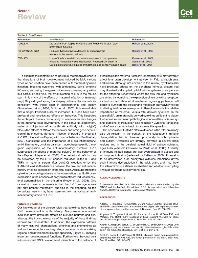
Neuron
Review
Table 1. Continued
Perturbation Key Findings References
TGFb2 KO Die at birth from respiratory failure due to deficits in brain stem
presynaptic function.
Heupel et al., 2008
TGFb2/TGFb3 dKO Reduced tyrosine hydroxylase (TH)+ dopaminergic
neurons in the ventral midbrain.
Roussa et al., 2006
TNFa KO Loss of the homeostatic increase in response to the open eye
following monocular visual deprivation. Reduced MN death in
SC explant cultures. Reduced sympathetic and sensory neuron death.
Kaneko et al., 2008;
Sedel et al., 2004;
Barker et al., 2001
To examine the contribution of individual maternal cytokines to
the alterations of brain development induced by MIA, various
types of perturbation have been carried out: maternal cytokine
injection, blocking cytokines with antibodies, using cytokine
KO mice, and using transgenic mice overexpressing a cytokine
in a particular cell type. Maternal injection of IL-6 in the mouse
can mimic many of the effects of maternal infection or maternal
poly(I:C), yielding offspring that display behavioral abnormalities
consistent with those seen in schizophrenia and autism
(Samuelsson et al., 2006; Smith et al., 2007). It is remarkable
that a single, transient pulse of increased IL-6 can have such
profound and long-lasting effects on behavior. This illustrates
the embryonic brain’s responsivity to relatively subtle changes
in the maternal-fetal environment. In the converse experiment,
maternal coinjection of an anti-IL-6 antibody with poly(I:C)
blocks the effects of MIA on the behavior and brain gene expres-
sion of the offspring. Moreover, injection of poly(I:C) in pregnant
IL-6 KO mice yields offspring with normal behavior (Smith et al.,
2007). Consistent with the importance of a pro-inflammatory/
anti-inflammatory cytokine balance, macrophage-specific trans-
genic expression of the anti-inflammatory cytokine IL-10
suppresses the effects of maternal poly(I:C) on the behavior of
the offspring (Meyer et al., 2008). The behavioral deficits may
be prevented by the IL-10-induced reduction in the IL-6 and
TNFa in maternal serum after poly(I:C) injection, or by the
IL-10-induced shift in balance between the pro- and anti-inflam-
matory cytokine expression in the fetal brain. Also supporting the
cytokine balance hypothesis is the observation that IL-10 over-
expression in the absence of poly(I:C) treatment induces behav-
ioral abnormalities in the offspring (Meyer et al., 2008). One
caveat of these experiments is that the IL-10 transgene was
not only present maternally, but also in the offspring, so the
behavioral results may have stemmed from a postnatal, anti-
inflammatory action of IL-10.
Future DirectionsOur knowledge of the diverse roles that cytokines have during
CNS development is in its infancy. Many well-characterized
cytokines have profound effects on cultured neurons and glia,
although the in vivo relevance of the majority of these findings
remains to demonstrated. In addition, the expression patterns
of many less well-characterized cytokines and chemokines as
well as their receptors and signaling components show striking
regional and developmental stage specificity (Figure 4), implying
important developmental functions. Furthermore, beyond their
roles in normal CNS development, disruption of the balance of
cytokines in the maternal-fetal environment by MIA may severely
affect fetal brain development as seen in PVL, schizophrenia,
and autism. Although not covered in this review, cytokines also
have profound effects on the peripheral nervous system that
may likewise be disrupted by MIA with long-term consequences
for the offspring. Discovering where the MIA-induced cytokines
are acting by localizing the expression of key cytokine receptors
as well as activation of downstream signaling pathways will
begin to illuminate the cellular and molecular pathways involved
in altering fetal neurodevelopment. Also of interest is the relative
importance of maternal- versus fetal-derived cytokines. In the
case of MIA, are maternally derived cytokines sufficient to trigger
the behavioral and neuropathological abnormalities, or is embry-
onic cytokine dysregulation also required? Cytokine transgenic
and KO mice can now begin to address this question.
The observation that MIA alters cytokines in the fetal brain may
also be relevant in the context of the subsequent immune
dysregulation that is observed postnatally in schizophrenia
and autism. Cytokines are strongly elevated in several brain
regions and in the cerebral spinal fluid of autistic subjects,
ages 4–45 years old (reviewed by Pardo et al., 2005). A variety
of immune-related genes are also dysregulated in autistic and
schizophrenic brains (reviewed by Patterson, 2009). It remains
to be determined if an embryonic cytokine imbalance drives
such immune dysregulation in the adult brain, and if so, how
this altered immune state is established and whether interrupting
it would be therapeutically beneficial.
ACKNOWLEDGMENTS
Experiments described from the authors’ laboratory were funded by theNINDS and the McGrath Foundation. B.E.D. is supported by a fellowshipfrom the California Institute for Regenerative Medicine.
REFERENCES
Adachi, T., Takanaga, H., Kunimoto, M., and Asou, H. (2005). Influence of LIFand BMP-2 on differentiation and development of glial cells in primary culturesof embryonic rat cerebral hemisphere. J. Neurosci. Res. 79, 608–615.
Akiyama, H., Tooyama, I., Kondo, H., Ikeda, K., Kimura, H., McGeer, E.G., andMcGeer, P.L. (1994). Early response of brain resident microglia to kainicacid-induced hippocampal lesions. Brain Res. 635, 257–268.
Alfonsi, F., Filippi, P., Salaun, D., deLapeyriere, O., and Durbec, P. (2008). LIFRbeta plays a major role in neuronal identity determination and glial differentia-tion in the mouse facial nucleus. Dev. Biol. 313, 267–278.
Alliot, F., Godin, I., and Pessac, B. (1999). Microglia derive from progenitors,originating from the yolk sac, and which proliferate in the brain. Brain Res.Dev. Brain Res. 117, 145–152.
Neuron 64, October 15, 2009 ª2009 Elsevier Inc. 73

Neuron
Review
Andrews, T., Zhang, P., and Bhat, N.R. (1998). TNFalpha potentiates IFN-gamma-induced cell death in oligodendrocyte progenitors. J. Neurosci. Res.54, 574–583.
Anthony, T.E., and Heintz, N. (2008). Genetic lineage tracing defines distinctneurogenic and gliogenic stages of ventral telencephalic radial glial develop-ment. Neural Dev. 3, 30.
Ashwell, K. (1991). The distribution of microglia and cell death in the fetal ratforebrain. Brain Res. Dev. Brain Res. 58, 1–12.
Bachiller, D., Klingensmith, J., Kemp, C., Belo, J.A., Anderson, R.M., May,S.R., McMahon, J.A., McMahon, A.P., Harland, R.M., Rossant, J., and DeRobertis, E.M. (2000). The organizer factors Chordin and Noggin are requiredfor mouse forebrain development. Nature 403, 658–661.
Baerwald, K.D., and Popko, B. (1998). Developing and mature oligodendro-cytes respond differently to the immune cytokine interferon-gamma. J. Neuro-sci. Res. 52, 230–239.
Bagri, A., Gurney, T., He, X., Zou, Y.R., Littman, D.R., Tessier-Lavigne, M., andPleasure, S.J. (2002). The chemokine SDF1 regulates migration of dentategranule cells. Development 129, 4249–4260.
Barker, V., Middleton, G., Davey, F., and Davies, A.M. (2001). TNFalphacontributes to the death of NGF-dependent neurons during development.Nat. Neurosci. 4, 1194–1198.
Barnabe-Heider, F., Wasylnka, J.A., Fernandes, K.J., Porsche, C., Sendtner,M., Kaplan, D.R., and Miller, F.D. (2005). Evidence that embryonic neuronsregulate the onset of cortical gliogenesis via cardiotrophin-1. Neuron 48,253–265.
Barres, B.A. (2008). The mystery and magic of glia: a perspective on their rolesin health and disease. Neuron 60, 430–440.
Barres, B.A., and Raff, M.C. (1994). Control of oligodendrocyte number in thedeveloping rat optic nerve. Neuron 12, 935–942.
Barres, B.A., Hart, I.K., Coles, H.S., Burne, J.F., Voyvodic, J.T., Richardson,W.D., and Raff, M.C. (1992). Cell death and control of cell survival in the oligo-dendrocyte lineage. Cell 70, 31–46.
Barres, B., Schmid, R., Sendnter, M., and Raff, M. (1993). Multiple extracellularsignals are required for long-term oligodendrocyte survival. Development 118,283.
Barres, B.A., Burne, J.F., Holtmann, B., Thoenen, H., Sendtner, M., and Raff,M.C. (1996). Ciliary neurotrophic factor enhances the rate of oligodendrocytegeneration. Mol. Cell. Neurosci. 8, 146–156.
Bauer, S., and Patterson, P.H. (2006). Leukemia inhibitory factor promotesneural stem cell self-renewal in the adult brain. J. Neurosci. 26, 12089–12099.
Bauer, S., Kerr, B.J., and Patterson, P.H. (2007). The neuropoietic cytokinefamily in development, plasticity, disease and injury. Nat. Rev. Neurosci. 8,221–232.
Beattie, E.C., Stellwagen, D., Morishita, W., Bresnahan, J.C., Ha, B.K., VonZastrow, M., Beattie, M.S., and Malenka, R.C. (2002). Control of synapticstrength by glial TNFalpha. Science 295, 2282–2285.
Blevins, G., and Fedoroff, S. (1995). Microglia in colony-stimulating factor1-deficient op/op mice. J. Neurosci. Res. 40, 535–544.
Bodeutsch, N., and Thanos, S. (2000). Migration of phagocytotic cells anddevelopment of the murine intraretinal microglial network: an in vivo studyusing fluorescent dyes. Glia 32, 91–101.
Bonaguidi, M.A., McGuire, T., Hu, M., Kan, L., Samanta, J., and Kessler, J.A.(2005). LIF and BMP signaling generate separate and discrete types of GFAP-expressing cells. Development 132, 5503–5514.
Bonni, A., Sun, Y., Nadal-Vicens, M., Bhatt, A., Frank, D.A., Rozovsky, I., Stahl,N., Yancopoulos, G.D., and Greenberg, M.E. (1997). Regulation of gliogenesisin the central nervous system by the JAK-STAT signaling pathway. Science278, 477–483.
Borrell, V., and Marın, O. (2006). Meninges control tangential migration of hem-derived Cajal-Retzius cells via CXCL12/CXCR4 signaling. Nat. Neurosci. 9,1284–1293.
74 Neuron 64, October 15, 2009 ª2009 Elsevier Inc.
Boulanger, L.M. (2009). Immune proteins in brain development and synapticplasiticity. Neuron 64, this issue, 93–109.
Boulanger, L.M., and Shatz, C.J. (2004). Immune signalling in neural develop-ment, synaptic plasticity and disease. Nat. Rev. Neurosci. 5, 521–531.
Brown, A.S., Hooton, J., Schaefer, C.A., Zhang, H., Petkova, E., Babulas, V.,Perrin, M., Gorman, J.M., and Susser, E.S. (2004). Elevated maternal inter-leukin-8 levels and risk of schizophrenia in adult offspring. Am. J. Psychiatry161, 889–895.
Bugga, L., Gadient, R.A., Kwan, K., Stewart, C.L., and Patterson, P.H. (1998).Analysis of neuronal and glial phenotypes in brains of mice deficient inleukemia inhibitory factor. J. Neurobiol. 36, 509–524.
Buka, S.L., Tsuang, M.T., Torrey, E.F., Klebanoff, M.A., Wagner, R.L., andYolken, R.H. (2001). Maternal cytokine levels during pregnancy and adultpsychosis. Brain Behav. Immun. 15, 411–420.
Burns, T.M., Clough, J.A., Klein, R.M., Wood, G.W., and Berman, N.E. (1993).Developmental regulation of cytokine expression in the mouse brain. GrowthFactors 9, 253–258.
Campos, L.S., Duarte, A.J., Branco, T., and Henrique, D. (2001). mDll1 andmDll3 expression in the developing mouse brain: role in the establishment ofthe early cortex. J. Neurosci. Res. 64, 590–598.
Carpenter, M.K., Cui, X., Hu, Z.Y., Jackson, J., Sherman, S., Seiger, A., andWahlberg, L.U. (1999). In vitro expansion of a multipotent population of humanneural progenitor cells. Exp. Neurol. 158, 265–278.
Carpentier, P.A., and Palmer, T.D. (2009). Immune influence on adult neuralstem cell regulation and function. Neuron 64, this issue, 79–92.
Cecchini, M., Dominguez, M., Mocci, S., Wetterwald, A., Felix, R., Fleisch, H.,Chisholm, O., Hofstetter, W., Pollard, J., and Stanley, E. (1994). Role of colonystimulating factor-1 in the establishment and regulation of tissue macrophagesduring postnatal development of the mouse. Development 120, 1357.
Chalasani, S.H., Sabelko, K.A., Sunshine, M.J., Littman, D.R., and Raper, J.A.(2003). A chemokine, SDF-1, reduces the effectiveness of multiple axonalrepellents and is required for normal axon pathfinding. J. Neurosci. 23,1360–1371.
Chalasani, S.H., Sabol, A., Xu, H., Gyda, M.A., Rasband, K., Granato, M.,Chien, C.B., and Raper, J.A. (2007). Stromal cell-derived factor-1 antagonizesslit/robo signaling in vivo. J. Neurosci. 27, 973–980.
Chamak, B., Morandi, V., and Mallat, M. (1994). Brain macrophages stimulateneurite growth and regeneration by secreting thrombospondin. J. Neurosci.Res. 38, 221–233.
Chang, Y., Albright, S., and Lee, F. (1994). Cytokines in the central nervoussystem: expression of macrophage colony stimulating factor and its receptorduring development. J. Neuroimmunol. 52, 9–17.
Chang, M.Y., Park, C.H., Son, H., Lee, Y.S., and Lee, S.H. (2004). Develop-mental stage-dependent self-regulation of embryonic cortical precursor cellsurvival and differentiation by leukemia inhibitory factor. Cell Death Differ.11, 985–996.
Checchin, D., Sennlaub, F., Levavasseur, E., Leduc, M., and Chemtob, S.(2006). Potential role of microglia in retinal blood vessel formation. Invest.Ophthalmol. Vis. Sci. 47, 3595.
Chojnacki, A., Shimazaki, T., Gregg, C., Weinmaster, G., and Weiss, S. (2003).Glycoprotein 130 signaling regulates Notch1 expression and activation inthe self-renewal of mammalian forebrain neural stem cells. J. Neurosci. 23,1730–1741.
Cingolani, L.A., Thalhammer, A., Yu, L.M., Catalano, M., Ramos, T., Colicos,M.A., and Goda, Y. (2008). Activity-dependent regulation of synaptic AMPAreceptor composition and abundance by beta3 integrins. Neuron 58, 749–762.
Cockle, J.V., Gopichandran, N., Walker, J.J., Levene, M.I., and Orsi, N.M.(2007). Matrix metalloproteinases and their tissue inhibitors in preterm peri-natal complications. Reprod Sci. 14, 629–645.
Constam, D.B., Schmid, P., Aguzzi, A., Schachner, M., and Fontana, A. (1994).Transient production of TGF-beta 2 by postnatal cerebellar neurons and itseffect on neuroblast proliferation. Eur. J. Neurosci. 6, 766–778.

Neuron
Review
Corbin, J.G., Kelly, D., Rath, E.M., Baerwald, K.D., Suzuki, K., and Popko, B.(1996). Targeted CNS expression of interferon-gamma in transgenic miceleads to hypomyelination, reactive gliosis, and abnormal cerebellar develop-ment. Mol. Cell. Neurosci. 7, 354–370.
de la Mano, A., Gato, A., Alonso, M.I., and Carnicero, E. (2007). Role of inter-leukin-1b in the control of neuroepithelial proliferation and differentiation of thespinal cord during development. Cytokine 37, 128–137.
DeChiara, T.M., Vejsada, R., Poueymirou, W.T., Acheson, A., Suri, C.,Conover, J.C., Friedman, B., McClain, J., Pan, L., Stahl, N., et al. (1995).Mice lacking the CNTF receptor, unlike mice lacking CNTF, exhibit profoundmotor neuron deficits at birth. Cell 83, 313–322.
Deng, W., Pleasure, J., and Pleasure, D. (2008). Progress in periventricularleukomalacia. Arch. Neurol. 65, 1291–1295.
Derouet, D., Rousseau, F., Alfonsi, F., Froger, J., Hermann, J., Barbier, F.,Perret, D., Diveu, C., Guillet, C., Preisser, L., et al. (2004). Neuropoietin,a new IL-6-related cytokine signaling through the ciliary neurotrophic factorreceptor. Proc. Natl. Acad. Sci. USA 101, 4827–4832.
DeVries, M.E., Kelvin, A.A., Xu, L., Ran, L., Robinson, J., and Kelvin, D.J.(2006). Defining the origins and evolution of the chemokine/chemokinereceptor system. J. Immunol. 176, 401–415.
Drogemuller, K., Helmuth, U., Brunn, A., Sakowicz-Burkiewicz, M., Gutmann,D.H., Mueller, W., Deckert, M., and Schluter, D. (2008). Astrocyte gp130expression is critical for the control of Toxoplasma encephalitis. J. Immunol.181, 2683–2693.
Dziembowska, M., Tham, T.N., Lau, P., Vitry, S., Lazarini, F., and Dubois-Dalcq, M. (2005). A role for CXCR4 signaling in survival and migration of neuraland oligodendrocyte precursors. Glia 50, 258–269.
Ellman, L.M., and Susser, E.S. (2009). The promise of epidemiologic studies:neuro-immune mechanisms in the etiologies of brain disorders. Neuron 64,this issue, 25–27.
Elson, G.C., Lelievre, E., Guillet, C., Chevalier, S., Plun-Favreau, H., Froger, J.,Suard, I., de Coignac, A.B., Delneste, Y., Bonnefoy, J.Y., et al. (2000). CLFassociates with CLC to form a functional heteromeric ligand for the CNTFreceptor complex. Nat. Neurosci. 3, 867–872.
Escary, J.L., Perreau, J., Dumenil, D., Ezine, S., and Brulet, P. (1993).Leukaemia inhibitory factor is necessary for maintenance of haematopoieticstem cells and thymocyte stimulation. Nature 363, 361–364.
Falsig, J., van Beek, J., Hermann, C., and Leist, M. (2008). Molecular basis fordetection of invading pathogens in the brain. J. Neurosci. Res. 86, 1434–1447.
Farkas, L.M., Dunker, N., Roussa, E., Unsicker, K., and Krieglstein, K. (2003).Transforming growth factor-beta(s) are essential for the development ofmidbrain dopaminergic neurons in vitro and in vivo. J. Neurosci. 23,5178–5186.
Feng, Z., and Ko, C. (2008). Schwann cells promote synaptogenesis at theneuromuscular junction via transforming growth factor-beta1. J. Neurosci.28, 9599–9609.
Ferrer, I., Serrano, T., and Soriano, E. (1990). Naturally occurring cell death inthe subicular complex and hippocampus in the rat during development.Neurosci. Res. 8, 60–66.
Fontaine, R.H., Cases, O., Lelievre, V., Mesples, B., Renauld, J.C., Loron, G.,Degos, V., Dournaud, P., Baud, O., and Gressens, P. (2008). IL-9/IL-9 receptorsignaling selectively protects cortical neurons against developmentalapoptosis. Cell Death Differ. 15, 1542–1552.
Forger, N.G., Prevette, D., deLapeyriere, O., de Bovis, B., Wang, S., Bartlett,P., and Oppenheim, R.W. (2003). Cardiotrophin-like cytokine/cytokine-likefactor 1 is an essential trophic factor for lumbar and facial motoneuronsin vivo. J. Neurosci. 23, 8854–8858.
Garden, G.A., and Moller, T. (2006). Microglia biology in health and disease.J. Neuroimmune Pharmacol. 1, 127–137.
Gasque, P., Neal, J.W., Singhrao, S.K., McGreal, E.P., Dean, Y.D., Van, B.J.,and Morgan, B.P. (2002). Roles of the complement system in human neurode-generative disorders: pro-inflammatory and tissue remodeling activities. Mol.Neurobiol. 25, 1–17.
Gaulden, J., and Reiter, J.F. (2008). Neur-ons and neur-offs: regulators ofneural induction in vertebrate embryos and embryonic stem cells. Hum. Mol.Genet. 17, R60–R66.
Gauthier, A.S., Furstoss, O., Araki, T., Chan, R., Neel, B.G., Kaplan, D.R., andMiller, F.D. (2007). Control of CNS cell-fate decisions by SHP-2 and its dysre-gulation in Noonan syndrome. Neuron 54, 245–262.
Ge, W., Martinowich, K., Wu, X., He, F., Miyamoto, A., Fan, G., Weinmaster, G.,and Sun, Y.E. (2002). Notch signaling promotes astrogliogenesis via directCSL-mediated glial gene activation. J. Neurosci. Res. 69, 848–860.
Giulian, D., Young, D.G., Woodward, J., Brown, D.C., and Lachman, L.B.(1988). Interleukin-1 is an astroglial growth factor in the developing brain.J. Neurosci. 8, 709–714.
Gregg, C., and Weiss, S. (2005). CNTF/LIF/gp130 receptor complex signalingmaintains a VZ precursor differentiation gradient in the developing ventralforebrain. Development 132, 565–578.
Harris, A.J., and McCaig, C.D. (1984). Motoneuron death and motor unit sizeduring embryonic development of the rat. J. Neurosci. 4, 13–24.
Hatakeyama, J., Bessho, Y., Katoh, K., Ookawara, S., Fujioka, M., Guillemot,F., and Kageyama, R. (2004). Hes genes regulate size, shape and histogenesisof the nervous system by control of the timing of neural stem cell differentia-tion. Development 131, 5539–5550.
Hatta, T., Moriyama, K., Nakashima, K., Taga, T., and Otani, H. (2002). TheRole of gp130 in cerebral cortical development: in vivo functional analysis ina mouse exo utero system. J. Neurosci. 22, 5516–5524.
He, F., Ge, W., Martinowich, K., Becker-Catania, S., Coskun, V., Zhu, W., Wu,H., Castro, D., Guillemot, F., Fan, G., et al. (2005). A positive autoregulatoryloop of Jak-STAT signaling controls the onset of astrogliogenesis. Nat. Neuro-sci. 8, 616–625.
Herbomel, P., Thisse, B., and Thisse, C. (2001). Zebrafish early macrophagescolonize cephalic mesenchyme and developing brain, retina, and epidermisthrough a M-CSF receptor-dependent invasive process. Dev. Biol. 238,274–288.
Herrmann, J.E., Imura, T., Song, B., Qi, J., Ao, Y., Nguyen, T.K., Korsak, R.A.,Takeda, K., Akira, S., and Sofroniew, M.V. (2008). STAT3 is a critical regulatorof astrogliosis and scar formation after spinal cord injury. J. Neurosci. 28,7231–7243.
Heupel, K., Sargsyan, V., Plomp, J., Rickmann, M., Varoqueaux, F., Zhang, W.,and Krieglstein, K. (2008). Loss of transforming growth factor-beta 2 leads toimpairment of central synapse function. Neural Dev. 3, 25.
Holtmann, B., Wiese, S., Samsam, M., Grohmann, K., Pennica, D., Martini, R.,and Sendtner, M. (2005). Triple knock-out of CNTF, LIF, and CT-1 definescooperative and distinct roles of these neurotrophic factors for motoneuronmaintenance and function. J. Neurosci. 25, 1778–1787.
Holzschuh, J., Hauptmann, G., and Driever, W. (2003). Genetic analysis of theroles of Hh, FGF8, and nodal signaling during catecholaminergic systemdevelopment in the zebrafish brain. J. Neurosci. 23, 5507–5519.
Horuk, R., Martin, A.W., Wang, Z., Schweitzer, L., Gerassimides, A., Guo, H.,Lu, Z., Hesselgesser, J., Perez, H.D., Kim, J., et al. (1997). Expression ofchemokine receptors by subsets of neurons in the central nervous system.J. Immunol. 158, 2882–2890.
Huh, G.S., Boulanger, L.M., Du, H., Riquelme, P.A., Brotz, T.M., and Shatz,C.J. (2000). Functional requirement for class I MHC in CNS developmentand plasticity. Science 290, 2155–2159.
Ip, N.Y., McClain, J., Barrezueta, N.X., Aldrich, T.H., Pan, L., Li, Y., Wiegand,S.J., Friedman, B., Davis, S., and Yancopoulos, G.D. (1993). The alpha compo-nent of the CNTF receptor is required for signaling and defines potential CNTFtargets in the adult and during development. Neuron 10, 89–102.
Jung, K.H., Chu, K., Lee, S.T., Kim, S.J., Sinn, D.I., Kim, S.U., Kim, M., andRoh, J.K. (2006). Granulocyte colony-stimulating factor stimulates neurogen-esis via vascular endothelial growth factor with STAT activation. Brain Res.1073-1074, 190–201.
Neuron 64, October 15, 2009 ª2009 Elsevier Inc. 75

Neuron
Review
Kamakura, S., Oishi, K., Yoshimatsu, T., Nakafuku, M., Masuyama, N., andGotoh, Y. (2004). Hes binding to STAT3 mediates crosstalk between Notchand JAK-STAT signalling. Nat. Cell Biol. 6, 547–554.
Kaneko, M., Stellwagen, D., Malenka, R.C., and Stryker, M.P. (2008). Tumornecrosis factor-alpha mediates one component of competitive, experience-dependent plasticity in developing visual cortex. Neuron 58, 673–680.
Kim, S.U., and de Vellis, J. (2005). Microglia in health and disease. J. Neurosci.Res. 81, 302–313.
Kim, I.J., Beck, H.N., Lein, P.J., and Higgins, D. (2002). Interferon gammainduces retrograde dendritic retraction and inhibits synapse formation.J. Neurosci. 22, 4530–4539.
Kirby, B.B., Takada, N., Latimer, A.J., Shin, J., Carney, T.J., Kelsh, R.N., andAppel, B. (2006). In vivo time-lapse imaging shows dynamic oligodendro-cyte progenitor behavior during zebrafish development. Nat. Neurosci. 9,1506–1511.
Kirsch, F., Kruger, C., and Schneider, A. (2008). The receptor for granulocyte-colony stimulating factor (G-CSF) is expressed in radial glia during develop-ment of the nervous system. BMC Dev. Biol. 8, 32.
Klein, R.S., Rubin, J.B., Gibson, H.D., DeHaan, E.N., Alvarez-Hernandez, X.,Segal, R.A., and Luster, A.D. (2001). SDF-1 alpha induces chemotaxis andenhances Sonic hedgehog-induced proliferation of cerebellar granule cells.Development 128, 1971–1981.
Koblar, S.A., Turnley, A.M., Classon, B.J., Reid, K.L., Ware, C.B., Cheema,S.S., Murphy, M., and Bartlett, P.F. (1998). Neural precursor differentiationinto astrocytes requires signaling through the leukemia inhibitory factorreceptor. Proc. Natl. Acad. Sci. USA 95, 3178–3181.
Kondo, Y., Lemere, C.A., and Seabrook, T.J. (2007). Osteopetrotic (op/op)mice have reduced microglia, no Abeta deposition, and no changes in dopa-minergic neurons. J. Neuroinflammation 4, 31.
Kurpius, D., Wilson, N., Fuller, L., Hoffman, A., and Dailey, M.E. (2006). Earlyactivation, motility, and homing of neonatal microglia to injured neuronsdoes not require protein synthesis. Glia 54, 58–70.
LaFerla, F.M., Sugarman, M.C., Lane, T.E., and Leissring, M.A. (2000).Regional hypomyelination and dysplasia in transgenic mice with astrocyte-directed expression of interferon-gamma. J. Mol. Neurosci. 15, 45–59.
Li, M., Sendtner, M., and Smith, A. (1995). Essential function of LIF receptor inmotor neurons. Nature 378, 724–727.
Lieberam, I., Agalliu, D., Nagasawa, T., Ericson, J., and Jessell, T.M. (2005).A Cxcl12-CXCR4 chemokine signaling pathway defines the initial trajectoryof mammalian motor axons. Neuron 47, 667–679.
Lodge, D.J., and Grace, A.A. (2008). Hippocampal dysfunction and disruptionof dopamine system regulation in an animal model of schizophrenia. Neurotox.Res. 14, 97–104.
Louis, J.C., Magal, E., Takayama, S., and Varon, S. (1993). CNTF protection ofoligodendrocytes against natural and tumor necrosis factor-induced death.Science 259, 689–692.
Lu, M., Grove, E.A., and Miller, R.J. (2002). Abnormal development of thehippocampal dentate gyrus in mice lacking the CXCR4 chemokine receptor.Proc. Natl. Acad. Sci. USA 99, 7090–7095.
Ma, Q., Jones, D., Borghesani, P.R., Segal, R.A., Nagasawa, T., Kishimoto, T.,Bronson, R.T., and Springer, T.A. (1998). Impaired B-lymphopoiesis, myelo-poiesis, and derailed cerebellar neuron migration in CXCR4- and SDF-1-defi-cient mice. Proc. Natl. Acad. Sci. USA 95, 9448–9453.
Magdaleno, S., Jensen, P., Brumwell, C.L., Seal, A., Lehman, K., Asbury, A.,Cheung, T., Cornelius, T., Batten, D.M., Eden, C., et al. (2006). BGEM: anin situ hybridization database of gene expression in the embryonic and adultmouse nervous system. PLoS Biol. 4, e86.
Malatesta, P., Hack, M.A., Hartfuss, E., Kettenmann, H., Klinkert, W.,Kirchhoff, F., and Gotz, M. (2003). Neuronal or glial progeny: regional differ-ences in radial glia fate. Neuron 37, 751–764.
76 Neuron 64, October 15, 2009 ª2009 Elsevier Inc.
Marın-Teva, J.L., Dusart, I., Colin, C., Gervais, A., Van Rooijen, N., and Mallat,M. (2004). Microglia promote the death of developing Purkinje cells. Neuron41, 535–547.
Mayer, M., Bhakoo, K., and Noble, M. (1994). Ciliary neurotrophic factor andleukemia inhibitory factor promote the generation, maturation and survival ofoligodendrocytes in vitro. Development 120, 143–153.
Maysami, S., Nguyen, D., Zobel, F., Heine, S., Hopfner, M., and Stangel, M.(2006). Oligodendrocyte precursor cells express a functional chemokinereceptor CCR3: implications for myelination. J. Neuroimmunol. 178, 17–23.
Mehler, M.F., and Kessler, J.A. (1995). Cytokines and neuronal differentiation.Crit. Rev. Neurobiol. 9, 419–446.
Meng, S.Z., Oka, A., and Takashima, S. (1999). Developmental expression ofmonocyte chemoattractant protein-1 in the human cerebellum and brainstem.Brain Dev. 21, 30–35.
Merkle, F.T., Tramontin, A.D., Garcıa-Verdugo, J.M., and Alvarez-Buylla, A.(2004). Radial glia give rise to adult neural stem cells in the subventricularzone. Proc. Natl. Acad. Sci. USA 101, 17528–17532.
Meyer, U., Feldon, J., Schedlowski, M., and Yee, B.K. (2005). Towards animmuno-precipitated neurodevelopmental animal model of schizophrenia.Neurosci. Biobehav. Rev. 29, 913–947.
Meyer, U., Murray, P.J., Urwyler, A., Yee, B.K., Schedlowski, M., and Feldon,J. (2008). Adult behavioral and pharmacological dysfunctions following disrup-tion of the fetal brain balance between pro-inflammatory and IL-10-mediatedanti-inflammatory signaling. Mol. Psychiatry 13, 208–221.
Michaelson, M.D., Bieri, P.L., Mehler, M.F., Xu, H., Arezzo, J.C., Pollard, J.W.,and Kessler, J.A. (1996). CSF-1 deficiency in mice results in abnormal braindevelopment. Development 122, 2661–2672.
Miyasaka, N., Knaut, H., and Yoshihara, Y. (2007). Cxcl12/Cxcr4 chemokinesignaling is required for placode assembly and sensory axon pathfinding inthe zebrafish olfactory system. Development 134, 2459–2468.
Molne, M., Studer, L., Tabar, V., Ting, Y.T., Eiden, M.V., and McKay, R.D.(2000). Early cortical precursors do not undergo LIF-mediated astrocyticdifferentiation. J. Neurosci. Res. 59, 301–311.
Munoz-Fernandez, M.A., and Fresno, M. (1998). The role of tumour necrosisfactor, interleukin 6, interferon-gamma and inducible nitric oxide synthase inthe development and pathology of the nervous system. Prog. Neurobiol. 56,307–340.
Naito, M., Hayashi, S., Yoshida, H., Nishikawa, S., Shultz, L.D., and Takahashi,K. (1991). Abnormal differentiation of tissue macrophage populations in‘osteopetrosis’ (op) mice defective in the production of macrophage colony-stimulating factor. Am. J. Pathol. 139, 657–667.
Nakashima, K., Wiese, S., Yanagisawa, M., Arakawa, H., Kimura, N.,Hisatsune, T., Yoshida, K., Kishimoto, T., Sendtner, M., and Taga, T. (1999a).Developmental requirement of gp130 signaling in neuronal survival and astro-cyte differentiation. J. Neurosci. 19, 5429–5434.
Nakashima, K., Yanagisawa, M., Arakawa, H., Kimura, N., Hisatsune, T.,Kawabata, M., Miyazono, K., and Taga, T. (1999b). Synergistic signaling in fetalbrain by STAT3-Smad1 complex bridged by p300. Science 284, 479–482.
Nimmerjahn, A., Kirchhoff, F., and Helmchen, F. (2005). Resting microglial cellsare highly dynamic surveillants of brain parenchyma in vivo. Science 308,1314–1318.
Nishimune, H., Vasseur, S., Wiese, S., Birling, M.C., Holtmann, B., Sendtner,M., Iovanna, J.L., and Henderson, C.E. (2000). Reg-2 is a motoneuron neuro-trophic factor and a signalling intermediate in the CNTF survival pathway. Nat.Cell Biol. 2, 906–914.
Ohtani, T., Ishihara, K., Atsumi, T., Nishida, K., Kaneko, Y., Miyata, T., Itoh, S.,Narimatsu, M., Maeda, H., Fukada, T., et al. (2000). Dissection of signalingcascades through gp130 in vivo: reciprocal roles for STAT3- and SHP2-medi-ated signals in immune responses. Immunity 12, 95–105.
Oppenheim, R.W., Wiese, S., Prevette, D., Armanini, M., Wang, S., Houenou,L.J., Holtmann, B., Gotz, R., Pennica, D., and Sendtner, M. (2001). Cardiotro-phin-1, a muscle-derived cytokine, is required for the survival of subpopula-tions of developing motoneurons. J. Neurosci. 21, 1283–1291.

Neuron
Review
Pardo, C.A., Vargas, D.L., and Zimmerman, A.W. (2005). Immunity, neurogliaand neuroinflammation in autism. Int. Rev. Psychiatry 17, 485–495.
Paredes, M.F., Li, G., Berger, O., Baraban, S.C., and Pleasure, S.J. (2006).Stromal-derived factor-1 (CXCL12) regulates laminar position of Cajal-Retziuscells in normal and dysplastic brains. J. Neurosci. 26, 9404–9412.
Patterson, P.H. (2009). Immune involvement in schizophrenia and autism:Etiology, pathology and animal models. Behav. Brain Res. 204, 313–321.
Pelton, R., Saxena, B., Jones, M., Moses, H., and Gold, L. (1991). Immunohis-tochemical localization of TGF beta 1, TGF beta 2, and TGF beta 3 in themouse embryo: expression patterns suggest multiple roles during embryonicdevelopment. J. Cell Biol. 115, 1091.
Penner, J.D., and Brown, A.S. (2007). Prenatal infectious and nutritional factorsand risk of adult schizophrenia. Expert Rev. Neurother. 7, 797–805.
Pinto, L., and Gotz, M. (2007). Radial glial cell heterogeneity-The source ofdiverse progeny in the CNS. Prog. Neurobiol. 83, 2–23.
Pitman, M., Emery, B., Binder, M., Wang, S., Butzkueven, H., and Kilpatrick,T.J. (2004). LIF receptor signaling modulates neural stem cell renewal. Mol.Cell. Neurosci. 27, 255–266.
Pollard, J.W. (1997). Role of colony-stimulating factor-1 in reproduction anddevelopment. Mol. Reprod. Dev. 46, 54–60.
Qian, X., Shen, Q., Goderie, S.K., He, W., Capela, A., Davis, A.A., and Temple,S. (2000). Timing of CNS cell generation: a programmed sequence of neuronand glial cell production from isolated murine cortical stem cells. Neuron 28,69–80.
Robinson, S., Tani, M., Strieter, R.M., Ransohoff, R.M., and Miller, R.H. (1998).The chemokine growth-regulated oncogene-alpha promotes spinal cordoligodendrocyte precursor proliferation. J. Neurosci. 18, 10457–10463.
Roussa, E., Wiehle, M., Dunker, N., Becker-Katins, S., Oehlke, O., and Kriegl-stein, K. (2006). Transforming growth factor beta is required for differentiationof mouse mesencephalic progenitors into dopaminergic neurons in vitro andin vivo: ectopic induction in dorsal mesencephalon. Stem Cells 24, 2120–2129.
Samuelsson, A.M., Jennische, E., Hansson, H.A., and Holmang, A. (2006).Prenatal exposure to interleukin-6 results in inflammatory neurodegenerationin hippocampus with NMDA/GABA(A) dysregulation and impaired spatiallearning. Am. J. Physiol. Regul. Integr. Comp. Physiol. 290, R1345–R1356.
Sanyal, S., Kim, S., and Ramaswami, M. (2004). Retrograde Regulation in theCNSNeuron-Specific Interpretations of TGF-b Signaling. Neuron 41, 845–848.
Sasaki, A., Yokoo, H., Naito, M., Kaizu, C., Shultz, L.D., and Nakazato, Y.(2000). Effects of macrophage-colony-stimulating factor deficiency on thematuration of microglia and brain macrophages and on their expression ofscavenger receptor. Neuropathology 20, 134–142.
Schwarting, G.A., Henion, T.R., Nugent, J.D., Caplan, B., and Tobet, S. (2006).Stromal cell-derived factor-1 (chemokine C-X-C motif ligand 12) and chemo-kine C-X-C motif receptor 4 are required for migration of gonadotropin-releasing hormone neurons to the forebrain. J. Neurosci. 26, 6834–6840.
Schweizer, U., Gunnersen, J., Karch, C., Wiese, S., Holtmann, B., Takeda, K.,Akira, S., and Sendtner, M. (2002). Conditional gene ablation of Stat3 revealsdifferential signaling requirements for survival of motoneurons during develop-ment and after nerve injury in the adult. J. Cell Biol. 156, 287.
Sedel, F., Bechade, C., Vyas, S., and Triller, A. (2004). Macrophage-derivedtumor necrosis factor alpha, an early developmental signal for motoneurondeath. J. Neurosci. 24, 2236–2246.
See, J., Mamontov, P., Ahn, K., Wine-Lee, L., Crenshaw, E.B., and Grinspan,J.B. (2007). BMP signaling mutant mice exhibit glial cell maturation defects.Mol. Cell. Neurosci. 35, 171–182.
Sendtner, M., Gotz, R., Holtmann, B., Escary, J.L., Masu, Y., Carroll, P., Wolf,E., Brem, G., Brulet, P., and Thoenen, H. (1996). Cryptic physiological trophicsupport of motoneurons by LIF revealed by double gene targeting of CNTF andLIF. Curr. Biol. 6, 686–694.
Shimazaki, T., Shingo, T., and Weiss, S. (2001). The ciliary neurotrophic factor/leukemia inhibitory factor/gp130 receptor complex operates in the mainte-nance of mammalian forebrain neural stem cells. J. Neurosci. 21, 7642–7653.
Smith, S.E.P., Li, J., Garbett, K., Mirnics, K., and Patterson, P.H. (2007).Maternal immune activation alters fetal brain development through inter-leukin-6. J. Neurosci. 27, 10695.
Song, M.R., and Ghosh, A. (2004). FGF2-induced chromatin remodelingregulates CNTF-mediated gene expression and astrocyte differentiation.Nat. Neurosci. 7, 229–235.
Stellwagen, D., and Malenka, R.C. (2006). Synaptic scaling mediated by glialTNF-alpha. Nature 440, 1054–1059.
Stevens, B., Allen, N.J., Vazquez, L.E., Howell, G.R., Christopherson, K.S.,Nouri, N., Micheva, K.D., Mehalow, A.K., Huberman, A.D., Stafford, B., et al.(2007). The classical complement cascade mediates CNS synapse elimina-tion. Cell 131, 1164–1178.
Stockli, K.A., Lillien, L.E., Naher-Noe, M., Breitfeld, G., Hughes, R.A., Raff,M.C., Thoenen, H., and Sendtner, M. (1991). Regional distribution, develop-mental changes, and cellular localization of CNTF-mRNA and protein in therat brain. J. Cell Biol. 115, 447–459.
Stumm, R., and Hollt, V. (2007). CXC chemokine receptor 4 regulates neuronalmigration and axonal pathfinding in the developing nervous system: implica-tions for neuronal regeneration in the adult brain. J. Mol. Endocrinol. 38,377–382.
Stumm, R.K., Zhou, C., Ara, T., Lazarini, F., Dubois-Dalcq, M., Nagasawa, T.,Hollt, V., and Schulz, S. (2003). CXCR4 regulates interneuron migration in thedeveloping neocortex. J. Neurosci. 23, 5123–5130.
Sun, Y., Nadal-Vicens, M., Misono, S., Lin, M.Z., Zubiaga, A., Hua, X., Fan, G.,and Greenberg, M.E. (2001). Neurogenin promotes neurogenesis and inhibitsglial differentiation by independent mechanisms. Cell 104, 365–376.
Taylor, M.K., Yeager, K., and Morrison, S.J. (2007). Physiological Notchsignaling promotes gliogenesis in the developing peripheral and centralnervous systems. Development 134, 2435–2447.
Tiveron, M., Rossel, M., Moepps, B., Zhang, Y.L., Seidenfaden, R., Favor, J.,Konig, N., and Cremer, H. (2006). Molecular interaction between projectionneuron precursors and invading interneurons via stromal-derived factor 1(CXCL12)/CXCR4 signaling in the cortical subventricular zone/intermediatezone. J. Neurosci. 26, 13273.
Tran, P.B., and Miller, R.J. (2003). Chemokine receptors: signposts to braindevelopment and disease. Nat. Rev. Neurosci. 4, 444–455.
Tropepe, V., Hitoshi, S., Sirard, C., Mak, T.W., Rossant, J., and van der Kooy,D. (2001). Direct neural fate specification from embryonic stem cells: a primitivemammalian neural stem cell stage acquired through a default mechanism.Neuron 30, 65–78.
Tsai, H.H., Frost, E., To, V., Robinson, S., Ffrench-Constant, C., Geertman, R.,Ransohoff, R.M., and Miller, R.H. (2002). The chemokine receptor CXCR2controls positioning of oligodendrocyte precursors in developing spinal cordby arresting their migration. Cell 110, 373–383.
Tseng, C.Y., Ling, E.A., and Wong, W.C. (1983). Light and electron micro-scopic and cytochemical identification of amoeboid microglial cells in the brainof prenatal rats. J. Anat. 136, 837–849.
Turnley, A.M., Morahan, G., Okano, H., Bernard, O., Mikoshiba, K., Allison, J.,Bartlett, P.F., and Miller, J.F. (1991). Dysmyelination in transgenic mice result-ing from expression of class I histocompatibility molecules in oligodendro-cytes. Nature 353, 566–569.
Turrigiano, G.G., and Nelson, S.B. (2004). Homeostatic plasticity in the devel-oping nervous system. Nat. Rev. Neurosci. 5, 97–107.
Ullian, E.M., Sapperstein, S.K., Christopherson, K.S., and Barres, B.A. (2001).Control of synapse number by glia. Science 291, 657–661.
Vashlishan, A.B., Madison, J.M., Dybbs, M., Bai, J., Sieburth, D., Ch’ng, Q.,Tavazoie, M., and Kaplan, J.M. (2008). An RNAi screen identifies genes thatregulate GABA synapses. Neuron 58, 346–361.
Viti, J., Feathers, A., Phillips, J., and Lillien, L. (2003). Epidermal growth factorreceptors control competence to interpret leukemia inhibitory factor as anastrocyte inducer in developing cortex. J. Neurosci. 23, 3385–3393.
Neuron 64, October 15, 2009 ª2009 Elsevier Inc. 77

Neuron
Review
Wake, H., Moorhouse, A.J., Jinno, S., Kohsaka, S., and Nabekura, J. (2009).Resting microglia directly monitor the functional state of synapses in vivoand determine the fate of ischemic terminals. J. Neurosci. 29, 3974.
Wang, C.C., Wu, C.H., Shieh, J.Y., and Wen, C.Y. (2002). Microglial distributionand apoptosis in fetal rat brain. Brain Res. Dev. Brain Res. 139, 337–342.
Wegiel, J., Wisniewski, H.M., Dziewiatkowski, J., Tarnawski, M., Kozielski, R.,Trenkner, E., and Wiktor-Jedrzejczak, W. (1998). Reduced number and alteredmorphology of microglial cells in colony stimulating factor-1-deficient osteo-petrotic op/op mice. Brain Res. 804, 135–139.
Witmer-Pack, M.D., Hughes, D.A., Schuler, G., Lawson, L., McWilliam, A.,Inaba, K., Steinman, R.M., and Gordon, S. (1993). Identification of macro-phages and dendritic cells in the osteopetrotic (op/op) mouse. J. Cell Sci.104, 1021–1029.
Yoshida, K., Taga, T., Saito, M., Suematsu, S., Kumanogoh, A., Tanaka, T.,Fujiwara, H., Hirata, M., Yamagami, T., Nakahata, T., et al. (1996). Targeteddisruption of gp130, a common signal transducer for the interleukin 6 familyof cytokines, leads to myocardial and hematological disorders. Proc. Natl.Acad. Sci. USA 93, 407–411.
Yoshimatsu, T., Kawaguchi, D., Oishi, K., Takeda, K., Akira, S., Masuyama, N.,and Gotoh, Y. (2006). Non-cell-autonomous action of STAT3 in maintenance ofneural precursor cells in the mouse neocortex. Development 133, 2553–2563.
78 Neuron 64, October 15, 2009 ª2009 Elsevier Inc.
Zhang, Y., Taveggia, C., Melendez-Vasquez, C., Einheber, S., Raine, C.S.,Salzer, J.L., Brosnan, C.F., and John, G.R. (2006). Interleukin-11 potentiatesoligodendrocyte survival and maturation, and myelin formation. J. Neurosci.26, 12174–12185.
Zhu, Y., Zhu, Y., Yu, T., Yu, T., Zhang, X., Zhang, X., Nagasawa, T., Nagasawa,T., Wu, J.Y., Wu, J.Y., et al. (2002). Role of the chemokine SDF-1 as the menin-geal attractant for embryonic cerebellar neurons. Nat. Neurosci. 5, 719.
Zhu, Y., Matsumoto, T., Mikami, S., Nagasawa, T., and Murakami, F. (2009).SDF1/CXCR4 signalling regulates two distinct processes of precerebellarneuronal migration and its depletion leads to abnormal pontine nuclei forma-tion. Development 136, 1919–1928.
Zou, Y.R., Kottmann, A.H., Kuroda, M., Taniuchi, I., and Littman, D.R. (1998).Function of the chemokine receptor CXCR4 in haematopoiesis and in cere-bellar development. Nature 393, 595–599.
Zou, X., Bolon, B., Pretorius, J.K., Kurahara, C., McCabe, J., Christiansen,K.A., Sun, N., Duryea, D., Foreman, O., Senaldi, G., et al. (2009). Neonataldeath in mice lacking cardiotrophin-like cytokine is associated with multifocalneuronal hypoplasia. Vet. Pathol. 46, 514–519.