Embed Size (px)
Citation preview
1
Chapter 7
Complement
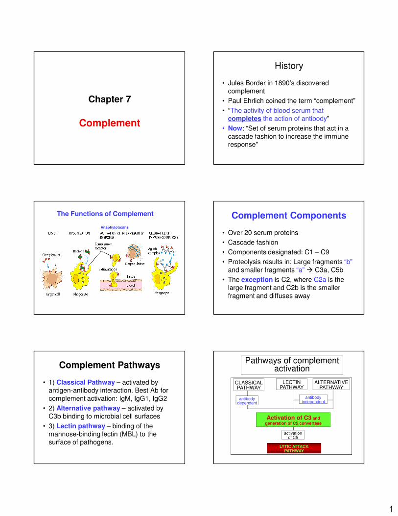
History
• Jules Border in 1890’s discovered complement
• Paul Ehrlich coined the term “complement”
• “The activity of blood serum that completes the action of antibody”
• Now: “Set of serum proteins that act in a
cascade fashion to increase the immune response”
The Functions of Complement
Anaphylotoxins
Complement Components
• Over 20 serum proteins
• Cascade fashion
• Components designated: C1 – C9
• Proteolysis results in: Large fragments “b”
and smaller fragments “a” � C3a, C5b
• The exception is C2, where C2a is the large fragment and C2b is the smaller
fragment and diffuses away
Complement Pathways
• 1) Classical Pathway – activated by antigen-antibody interaction. Best Ab for
complement activation: IgM, IgG1, IgG2
• 2) Alternative pathway – activated by C3b binding to microbial cell surfaces
• 3) Lectin pathway – binding of the mannose-binding lectin (MBL) to the surface of pathogens.
Pathways of complement activation
CLASSICALPATHWAY
ALTERNATIVEPATHWAY
activationof C5
LYTIC ATTACKPATHWAY
antibodydependent
LECTINPATHWAY
antibodyindependent
Activation of C3 andgeneration of C5 convertase
2
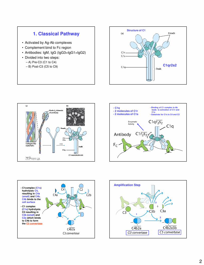
1. Classical Pathway
• Activated by Ag-Ab complexes
• Complement bind to Fc region
• Antibodies: IgM, IgG (IgG3>IgG1>IgG2)
• Divided into two steps:
– A) Pre-C3 (C1 to C4)
– B) Post-C3 (C5 to C9)
Structure of C1
C1qr2s2
- C1q- 2 molecules of C1r- 2 molecules of C1s
- Binding of C1 complex to Ab
leads to activation of C1r and
C1s
- Substrate for C1s is C4 and C2
Enzymatic
Activity
- C1complex (C1s)hydrolysis C4, resulting in C4a (small) and C4b. C4b binds to the cell surface
- C1 complex (C1s) hydrolysisC2 resulting in C2b (small) and
C2a which bindsto C4b to form the C3 convertase
Amplification Step
C3 convertase
1
2
3
3
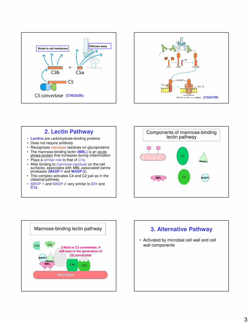
Binds to cell membraneDiffuses away
(C4b2a3b)(C5b6789)
2. Lectin Pathway• Lectins are carbohydrate-binding proteins
• Does not require antibody
• Recognizes mannose residues on glycoproteins
• The mannose-binding lectin (MBL) is an acute phase protein that increases during inflammation
• Plays a similar role to that of C1q
• After binding to mannose-residues on the cell surfaces, associates with MBL-associated serine proteases (MASP-1 and MASP-2).
• This complex activates C4 and C2 just as in the classical pathway
• MASP-1 and MASP-2 very similar to C1r and C1s
Components of mannose-binding lectin pathway
MBL MASP1
Mannose-binding lectin pathway
MBL
C4b2a is C3 convertase; it will lead to the generation of
C5 convertaseMASP1
BACTERIA
3. Alternative Pathway
• Activated by microbial cell wall and cell wall components
4
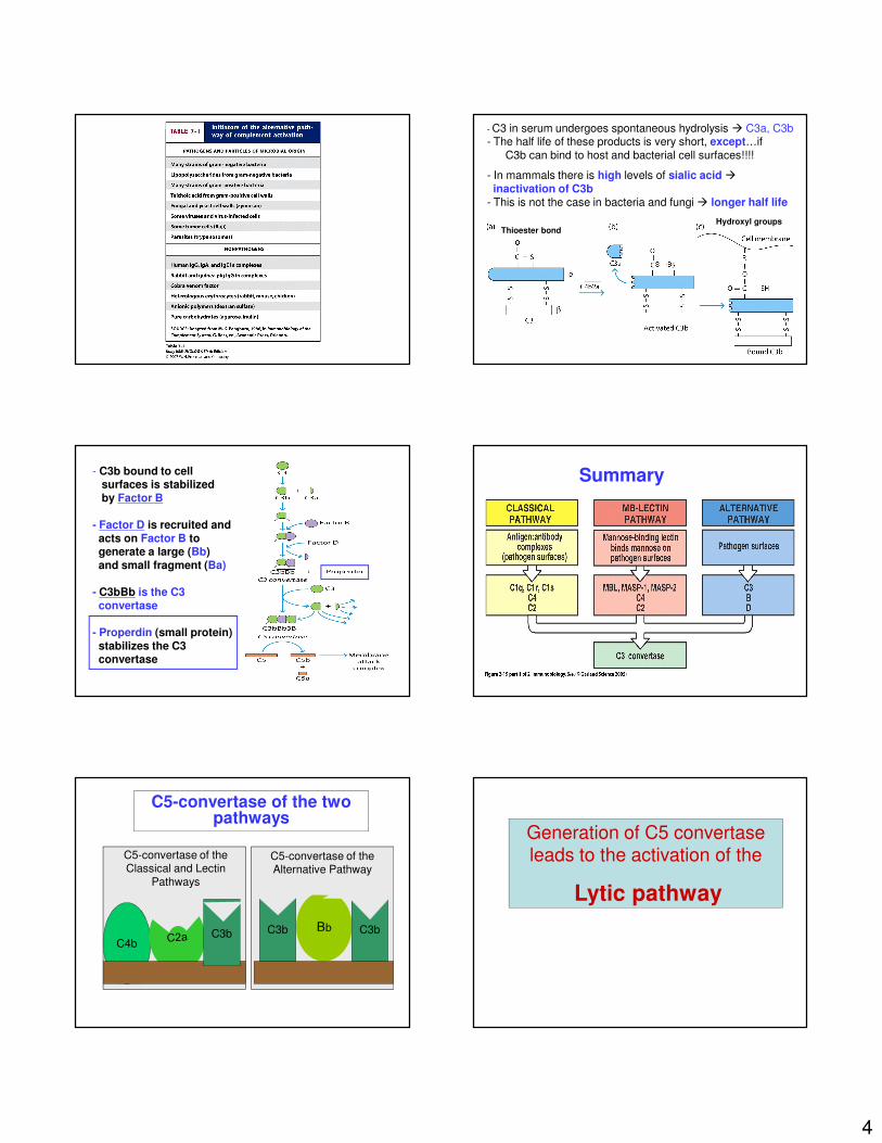
- C3 in serum undergoes spontaneous hydrolysis � C3a, C3b- The half life of these products is very short, except…if
C3b can bind to host and bacterial cell surfaces!!!!
- In mammals there is high levels of sialic acid �inactivation of C3b
- This is not the case in bacteria and fungi � longer half life
Thioester bondHydroxyl groups
- C3b bound to cell surfaces is stabilized by Factor B
- Factor D is recruited andacts on Factor B togenerate a large (Bb) and small fragment (Ba)
- C3bBb is the C3 convertase
- Properdin (small protein)stabilizes the C3 convertase
Figure 2-19 part 1 of 2Summary
C5-convertase of the two pathways
C3b C3b
C5-convertase of the Alternative Pathway
C4bC3b
C5-convertase of the Classical and Lectin
Pathways
Generation of C5 convertase leads to the activation of the
Lytic pathway
5
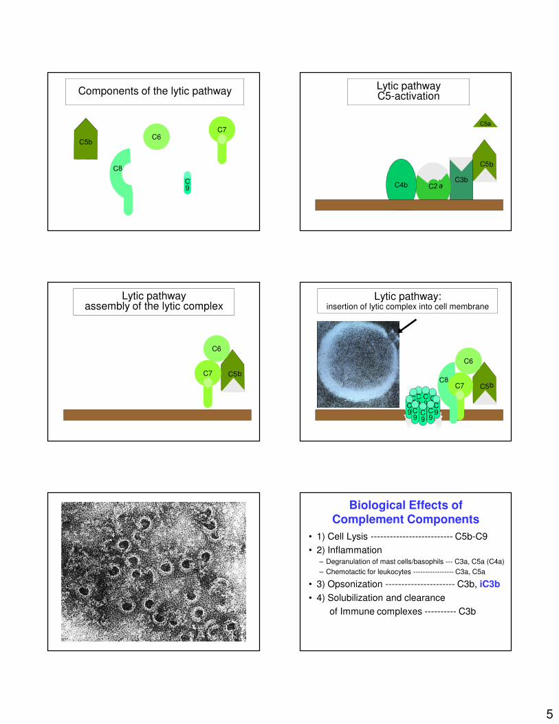
Components of the lytic pathway
C6
C9
C7
Lytic pathwayC5-activation
C3bC4b
b
Lytic pathwayassembly of the lytic complex
b
C6
C7
Lytic pathway:insertion of lytic complex into cell membrane
b
C6
C7
C9
C9
C9
C9C9
C9 C
9C9
C9
Biological Effects of Complement Components
• 1) Cell Lysis -------------------------- C5b-C9
• 2) Inflammation– Degranulation of mast cells/basophils --- C3a, C5a (C4a)
– Chemotactic for leukocytes ----------------- C3a, C5a
• 3) Opsonization ---------------------- C3b, iC3b
• 4) Solubilization and clearance
of Immune complexes ---------- C3b
6
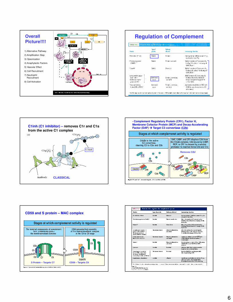
Overall
Picture!!!!
1) Alternative Pathway
2) Amplification Step
3) Opsonization
4) Anaphylactic Factors
5) Vascular Effect
6) Cell Recruitment
7) Neutrophil
Recruitment
8) Cell Activation
Regulation of Complement
CLASSICAL
C1inh (C1 inhibitor) – removes C1r and C1s
from the active C1 complexFigure 2-37 part 2 of 4
- Complement Regulatory Protein (CR1), Factor H, Membrane Cofactor Protein (MCP) and Decay-Accelerating Factor (DAF) ���� Target C3 convertase (C2b)
Removes C2b!
Figure 2-37 part 4 of 4CD59 and S protein – MAC complex
S Protein – Targets C7 CD59 – Targets C9
7
Factor H Factor I C3f C3c
C3b iC3b C3dg
Factor I
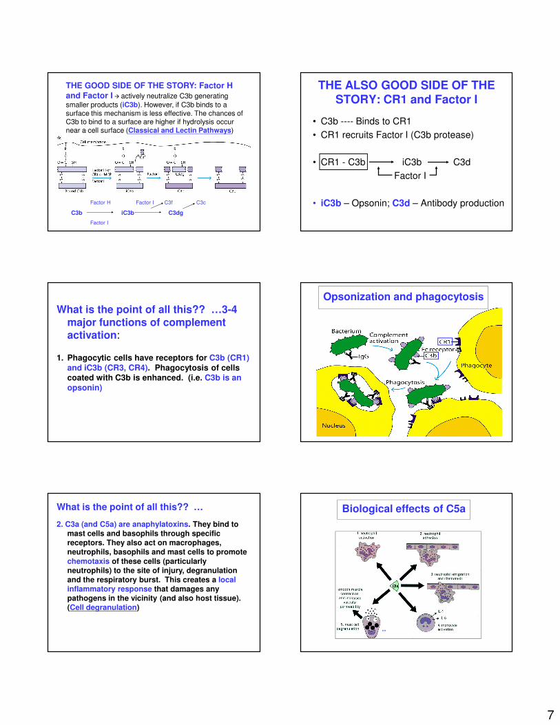
THE GOOD SIDE OF THE STORY: Factor H
and Factor I � actively neutralize C3b generating
smaller products (iC3b). However, if C3b binds to a
surface this mechanism is less effective. The chances of C3b to bind to a surface are higher if hydrolysis occur
near a cell surface (Classical and Lectin Pathways)
THE ALSO GOOD SIDE OF THE STORY: CR1 and Factor I
• C3b ---- Binds to CR1
• CR1 recruits Factor I (C3b protease)
• CR1 - C3b iC3b C3d
Factor I
• iC3b – Opsonin; C3d – Antibody production
What is the point of all this?? …3-4
major functions of complement activation:
1. Phagocytic cells have receptors for C3b (CR1)
and iC3b (CR3, CR4). Phagocytosis of cells coated with C3b is enhanced. (i.e. C3b is an
opsonin)
Opsonization and phagocytosis
What is the point of all this?? …
2. C3a (and C5a) are anaphylatoxins. They bind to mast cells and basophils through specific receptors. They also act on macrophages, neutrophils, basophils and mast cells to promote
chemotaxis of these cells (particularly neutrophils) to the site of injury, degranulation and the respiratory burst. This creates a local inflammatory response that damages any pathogens in the vicinity (and also host tissue). (Cell degranulation)
Biological effects of C5a
**
8
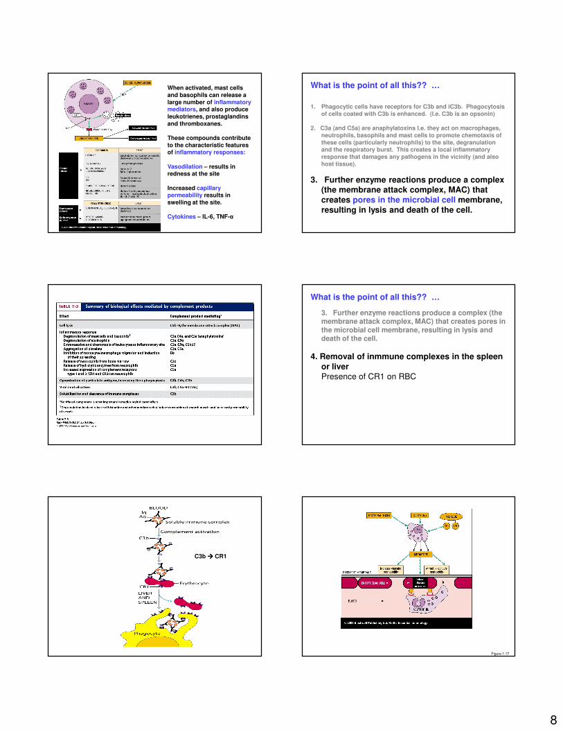
When activated, mast cells and basophils can release a large number of inflammatory mediators, and also produce leukotrienes, prostaglandins and thromboxanes.
These compounds contribute to the characteristic features of inflammatory responses:
Vasodilation – results in redness at the site
Increased capillary permeability results in swelling at the site.
Cytokines – IL-6, TNF-α
What is the point of all this?? …
1. Phagocytic cells have receptors for C3b and iC3b. Phagocytosis of cells coated with C3b is enhanced. (I.e. C3b is an opsonin)
2. C3a (and C5a) are anaphylatoxins I.e. they act on macrophages, neutrophils, basophils and mast cells to promote chemotaxis of these cells (particularly neutrophils) to the site, degranulation and the respiratory burst. This creates a local inflammatory response that damages any pathogens in the vicinity (and also host tissue).
3. Further enzyme reactions produce a complex (the membrane attack complex, MAC) that
creates pores in the microbial cell membrane,
resulting in lysis and death of the cell.
What is the point of all this?? …
3. Further enzyme reactions produce a complex (the membrane attack complex, MAC) that creates pores in the microbial cell membrane, resulting in lysis and death of the cell.
4. Removal of inmmune complexes in the spleen
or liverPresence of CR1 on RBC
C3b ���� CR1
Figure 1.17
9
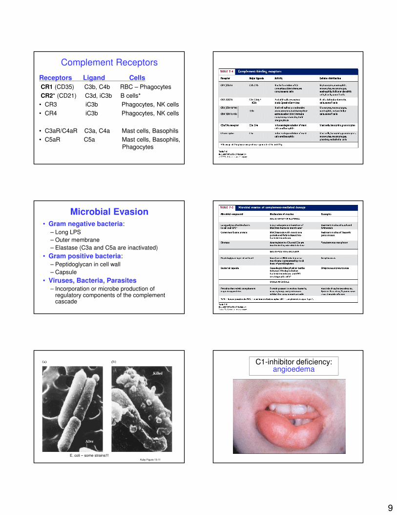
Complement Receptors
Receptors Ligand Cells
CR1 (CD35) C3b, C4b RBC – Phagocytes
CR2* (CD21) C3d, iC3b B cells*
• CR3 iC3b Phagocytes, NK cells
• CR4 iC3b Phagocytes, NK cells
• C3aR/C4aR C3a, C4a Mast cells, Basophils
• C5aR C5a Mast cells, Basophils,
Phagocytes
Microbial Evasion
• Gram negative bacteria:– Long LPS
– Outer membrane
– Elastase (C3a and C5a are inactivated)
• Gram positive bacteria:– Peptidoglycan in cell wall
– Capsule
• Viruses, Bacteria, Parasites– Incorporation or microbe production of
regulatory components of the complement cascade
Kuby Figure 13-11
E. coli – some strains!!!
C1-inhibitor deficiency:angioedema
10
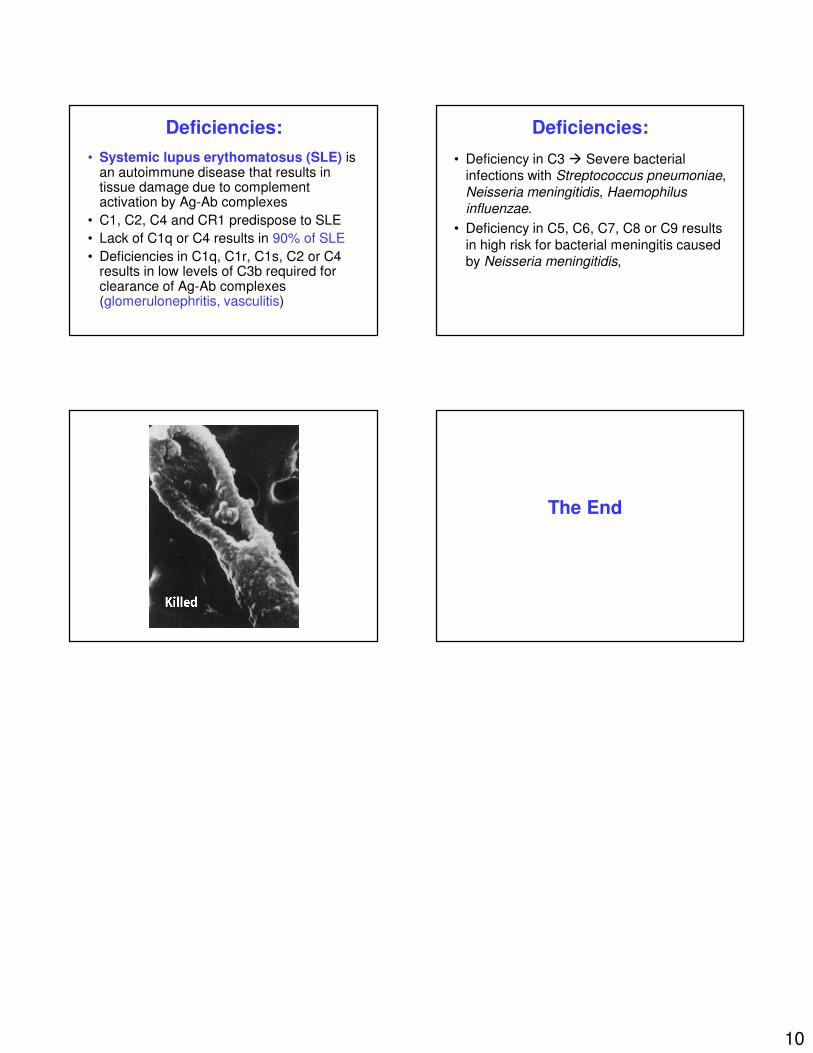
Deficiencies:
• Systemic lupus erythomatosus (SLE) is an autoimmune disease that results in tissue damage due to complement activation by Ag-Ab complexes
• C1, C2, C4 and CR1 predispose to SLE
• Lack of C1q or C4 results in 90% of SLE
• Deficiencies in C1q, C1r, C1s, C2 or C4 results in low levels of C3b required for clearance of Ag-Ab complexes (glomerulonephritis, vasculitis)
Deficiencies:
• Deficiency in C3 � Severe bacterial infections with Streptococcus pneumoniae,
Neisseria meningitidis, Haemophilus influenzae.
• Deficiency in C5, C6, C7, C8 or C9 results
in high risk for bacterial meningitis caused by Neisseria meningitidis,
The End