Embed Size (px)
Citation preview

711
Chapter 20
Nystagmus Basics: Normal Models that Simulate Dysfunction
Louis F. Dell’Osso
Dept. of Neurology, University Hospitals, Cleveland, OH 44106, PH: (216) 421-3224, FX: (216) 231-3461, EM: [email protected]
20.1 INTRODUCTION
Instabilities of the ocular motor system may take either of two forms, nystagmus or saccadic oscillations. Nystagmus (an involuntary oscillation of the eyes) is caused by instabilities in subsystems responsible for slow eye movements, whereas saccadic oscillations stem from subsystems responsible for generating saccadic eye movements. Nystagmus may exhibit either a pendular (sinusoidal) or a jerk waveform. In jerk waveforms, the slow phases displace the eye and fovea away from the target and the fast (saccadic) phases attempt to refoveate it. An important indication of the underlying mechanism for a particular type of nystagmus is the shape of the slow phase. Linear (constant velocity) slow phases of jerk nystagmus stem from tonic imbalances in any of the ocular motor subsystems. Decelerating (decreasing velocity) slow phases of jerk nystagmus are due to failure of gaze holding mechanisms, either central (common neural integrator) or peripheral (extraocular plant). Finally, accelerating (increasing velocity) slow phases of jerk nystagmus indicate a basic instability in a subsystem that causes it to “run away.” Pendular nystagmus indicates a resonant-frequency oscillation of a subsystem.
Identifying the waveform is the first step towards understanding the mechanisms involved in generating each type of nystagmus; one must also identify where in the ocular motor system (i.e., which subsystem, not the anatomical site) the instability arises and how the oscillation affects other

712 Louis Dell’Osso ocular motor responses. Because of the complexity of the system, identifying the cause of the oscillation requires the development of a detailed, quantitative model. In contrast, attempts to do so by studying specific neurophysiological sites has not yielded the insights necessary for a thorough understanding of how the ocular motor system functions and how it deals with specific dysfunction.
In this chapter, I will discuss an approach to modelling the ocular motor system that is primarily based on function, dysfunction, and system-level responses rather than on specific neuroanatomy or neurophysiology. Although the latter two are incorporated into system models as much as possible (based on current knowledge), the absence of specific neuroanatomical or neurophysiological data is not sufficient to preclude a necessary hypothetical function from inclusion in the model. For example, both the common neural integrator and the local, resettable neural integrator were hypothesized and included in models long before they were either located or neural networks were hypothesized to simulate their operation. As we learn more about neurophysiology, it is becoming clear that the original expectations of modelers, namely that neurophysiological signals exist that parallel the functional signals of their models, were probably too optimistic (Robinson, 1994). Rather, it is more likely that most signals carried by neural interconnections are composites of several such functional signals that may defy decomposition into recognizable parts. Indeed, the signals in the hidden layers of even simple neural networks are usually functionally unrecognizable. That being the case, modelling on both the system and the neuronal levels should be pursued, with neither waiting for confirmation from the other. Modelling of specific subpopulations of neurons will continue to elucidate their particular behavior and possibly suggest where they fit into the overall system. However, it is doubtful that prediction of system function will come out of such investigations. The performance of any complex feedback system, including the ocular motor system, is determined by the myriad interconnections of its functional building blocks and not by the performance of any individual block – that is the lesson of negative feedback and its raison d’être.
It will come as no surprise to anyone familiar with modelling that a multitude of solutions exists for each particular input/output relationship to be simulated; simulations are not unique. Thus, within a complex system comprised of many interconnected subsystems, such as the ocular motor system, it is not which simulation of each block that is important to overall behavior but rather, how the blocks interact (i.e., how they are interconnected). It is this fact that has guided our modelling of ocular motor function and dysfunction. The models consist of distributed functional blocks with associated delays. One need only choose the particular model for

Chap. 20. Nystagmus Basics: Normal Models that Simulate Dysfunction 713 each of the subsystems that performs the required tasks of that subsystem and insert it into the total system model. If the system model is designed properly, it will function correctly with any of a number of subsystem realizations. If a particular subsystem choice proves unsatisfactory, another can be substituted without compromising system behavior.
The goals of this type of ocular motor system modelling are: to arrive at a robust model capable of simulating most (and perhaps eventually all) normal behavior; to add to that model the ability to simulate a wide range of specific dysfunctions (e.g., different types of nystagmus or saccadic oscillations); and to use such a model to help understand both normal and abnormal behavior, in a pedagogical setting and in a medical setting. To model is to be forced to quantify the “hand waving” and to learn what works and what does not. The model embodies our thoughts about how a system functions in a mathematical framework and, as it becomes more complex, its emergent properties, which we have neither built in nor anticipated, are what teach us more about system function. Toward that end, we have arrived at a point in this ambitious journey where we are developing a single ocular motor model that exhibits many normal and abnormal behaviors, such as nystagmus. At present (three years from its inception), they include: saccadic responses to step, pulse, and pulse-step target changes; short-latency corrective saccades, pursuit responses to ramp and step-ramp target changes; saccadic hypo- and hypermetria; macro saccadic oscillations; gaze-evoked nystagmus; muscle/gaze-paretic nystagmus in myasthenia gravis or other types of palsies; fixation and saccadic responses during latent/manifest latent nystagmus (LMLN); and fixation, saccadic and pursuit responses during several of the forms of congenital nystagmus (CN).
20.2 HISTORICAL OVERVIEW
There have been many models proposed to simulate specific dysfunction in a limited ocular motor context, using parts of normal models as a foundation (Collins, 1975; Robinson, 1973; Stark, 1968). In some ways, each contributed to our understanding of ocular motor function and dysfunction. Different types of ocular motor abnormalities have one thing in common; they cause unwanted motion of the moving oculocentric coordinate system (i.e., the retina and its fovea-centric base) upon which world images serve as inputs to the ocular motor system. In normal models, retinal image motion is usually presumed to be due to target motion that is unmatched by the eyes. That built-in presumption cannot be made in the presence of nystagmus or saccadic oscillations. Similarly, in normal models, it is presumed that all pulses from the pulse generator are fully integrated by

714 Louis Dell’Osso the common neural integrator; again, as several types of dysfunction demonstrate, that cannot be the case. We make the more reasonable presumption that individuals with either congenital or acquired ocular motor dysfunction have substantially the same ocular motor systems found in normal individuals. Therefore, structures and functions required to simulate the ocular motor system’s responses in the presence of experiments of nature (i.e., the abnormalities causing dysfunction) must also be present in the normal system. Current evidence supports these assumptions, and the models arrived at by simulating dysfunction more closely represent the actual physiological system than do the simplistic and, many times, erroneous models designed to simulate only a limited range of normal behavior (e.g., see the discussion of the need for a resettable neural integrator and for control of the common neural integrator in section 20.2.3 of this chapter).
Most ocular motor system models of version eye movements are of unilateral (containing bidirectional (left and right) signals), yoked control architecture that move the two eyes together in a coordinated manner in both directions. However, the actual architecture of the physiological system is bilateral (each side containing only unidirectional signals), yoked, independent control that allows both conjugate and disjunctive eye movements (Dell'Osso, 1994). There are many reasons for this, including conservation of computing power, simplicity, engineering “elegance”, and the assumption of conjugacy (i.e., a single signal drives both eyes to move similarly). Unfortunately, the latter may not always be the case in normal individuals and certainly is not in those with specific abnormalities (e.g., achiasma or LMLN). Despite the limitations of such models, they are still valuable tools that aid in our understanding of ocular motor control. The models of version discussed in this chapter, with the exception of the Alexander’s law model, have bidirectional, unilateral, and binocular architecture. Their outputs represent either both eyes (presuming conjugacy) or, in some types of dysfunction, the fixating eye. Finally, bidirectional, unilateral, and binocular models can also be made to drive two separate eyes, producing a combination of the two architectures (see section 20.2.4 of this chapter). Obviously, models of vergence do not presume conjugacy (see also Chapter 11, Models of Saccade-Vergence Interactions, by G. Hung and K.J. Ciuffreda in this volume).
20.2.1 Congenital Nystagmus
Congenital nystagmus is the most common type of infantile nystagmus; it may appear at birth or in early infancy (also, see Chapter 21 on the clinical aspects of CN in this volume). CN may exhibit a number of idiosyncratic waveforms that arise from either pendular or jerk nystagmus instabilities (or

Chap. 20. Nystagmus Basics: Normal Models that Simulate Dysfunction 715 both) (Dell'Osso and Daroff, 1975). The first ocular motor system model of dysfunction was that of the saccadic and pursuit responses of individuals with CN (Dell'Osso, 1967; 1968; 1970). That dual-mode model (see Fig. 20.1) was designed and simulated on a Burroughs B5500 computer using Analog Algol software. It incorporated modified, contemporary models of the saccadic and smooth pursuit subsystems (Young and Stark, 1963a; b) and accurately simulated physiological responses both of normals and a CN subject to pulses, steps, pulse-steps, ramps, step-ramps, and sinusoidal target inputs. Because the purpose of the model was to demonstrate how a normal ocular motor system could properly respond to specific target inputs despite an internal nystagmus oscillation, the latter was simulated by an asymmetric triangular waveform with no attempt to suggest either its anatomical site or physiological mechanism. Data from an individual with CN revealed “normal” responses to all of the above stimuli; that is, with the exception of the CN superimposed on each ocular motor response, the underlying responses of this individual were equivalent to those of normal subjects in gains, latencies, and components of the responses.
Once the CN oscillation was introduced into the model, it became clear that none of the observed physiological behavior could be simulated unless the model was able to extract true target position and velocity from the oscillating retinal error signal (i.e., the target signal minus the eye signal). Because individuals with CN perceive a clear and stable world despite the
Figure 20.1. A dual-mode model used to demonstrate how a rudimentary internal monitor of efferent commands allows a normal ocular motor system to respond properly to target inputs in the presence of an internal oscillation (here, congenital nystagmus). In this and the following Figures, T – target, E – eye, e – retinal error, CN – congenital nystagmus, and drop shadows indicate subsystems (their absence indicates a basic Simulink building block).
Dual-Mode CN Model
PerceivedTarget Error
E
Te
Retinal Feedback
Retinal Position Error
INTERNAL MONITOR
E
1
EyePosition
SACCADES
SMOOTH PURSUIT
Gaze-Angle Variation
0Normal
kGain
Delay
1/0CN / Normal
CN
EOM[2-PolePlant]
1Target
Position

716 Louis Dell’Osso continuously oscillating images presented to them by their retinas (i.e., they have no oscillopsia — the illusory motion of the world), the CN must have been canceled out. Thus, in the model some internal copy of the CN motor signal was needed to cancel out the oscillations of the eye to simulate accurately the physiological responses. To relieve the model of its ‘oscillopsia,’ it was necessary to add a delayed copy of the CN motor command to the retinal error signal. That allowed ‘perception’ of true target inputs and simulation of accurate responses, despite the ongoing CN. In effect, the model was using an efference copy signal of motor output to reconstruct true target information. This simple use of a motor command was a radical departure from the existing models of normal ocular motor behavior that functioned without the benefit of efference copy. A later study demonstrated the adequacy of perceived motion in stimulating smooth pursuit. (Yasui and Young, 1975) It was the simple basis for what was to become the internal monitor, an essential component of subsequent system models of ocular motor function and dysfunction. Despite the fact that neither the two subsystems in this model nor its ocular motor plant were physiologically accurate, the broad range of simulations it produced in the presence of an internal oscillation were precise enough that such model robustness would not be exceeded in over thirty years of research.
Later studies into the genesis of CN identified specific waveforms that fell into two categories (Dell'Osso and Daroff, 1975). This suggested that there were at least two sources for the observed instabilities. One was a pair of jω-axis poles for sinusoidal CN (possibly in the smooth pursuit system) (Dell'Osso et al., 1972), and the other, a right-half plane pole for the accelerating slow phases of jerk CN (possibly in the common neural integrator circuitry) (Dell'Osso and Daroff, 1981). The singularities represented by poles on the complex s-plane correspond to roots of the characteristic equation of the differential equation that defines the system under study. Pairs of jw-axis poles define a sinusoidal oscillation whose frequency is given by the value on that axis. A singularity in the right-half plane corresponds to a positive exponential output, causing the system to run away. Beginning about two decades after the above system model, several attempts were made to model CN waveforms (Harris, 1995; Optican and Zee, 1984; Tusa et al., 1992). These models used the suggested right-half plane pole in the neural integrator circuitry to generate CN-like jerk waveforms with accelerating slow phases.
One of the above models (Optican and Zee, 1984) postulated a “reversed” (i.e., opposite sign) velocity pathway associated with the neural integrator. That was based on: the misperception that, despite abundant evidence to the contrary, individuals with CN pursued moving targets in the wrong direction and, similar to albinos (who usually have CN), they also had

Chap. 20. Nystagmus Basics: Normal Models that Simulate Dysfunction 717 some misrouting of their temporal retinal fibers. There is no evidence for such maldevelopment in the great majority of individuals with CN and, given the excellent ability to pursue and use their vestibulo-ocular reflex (Dell'Osso, 1967; 1986; Dell'Osso et al., 1992a; b; c; Kurzan and Büttner, 1989), they appear to have normal ocular motor system structures and interconnections. Although the model simulated some CN waveforms, it also produced behaviors not exhibited by individuals with CN. Integral to the model was the hypothesis that the slow phases were generated from pulse-step mismatches in the “preceding” fast phases. That is, the CN resulting from a large fast phase or voluntary saccade would be predicted to be greater than that from a smaller one. However, this hypothesis is incorrect because first, it is the CN slow phase that is responsible for the genesis of the oscillation; the fast phase follows it and is in fact a response to its generated error. Some CN fast phases simply brake the slow phases, while others also attempt to refoveate the target. Second, the magnitude of CN during fixation of static targets is a function of gaze angle itself, not of the amplitude of the saccade used to acquire a given gaze angle. In fact, for a given individual, the CN at 15° right gaze is the same regardless of how that gaze angle was achieved, including either a slow pursuit or head movement to that gaze angle. The latter would contain no pulse-step mismatch. Thus, the very basis for their model was inconsistent with recorded physiological data taken from individuals with CN.
Despite evidence that the pendular and jerk waveforms arise from different sites and types of instability, Optican and Zee’s 1984 model hypothesized a single source. However, attempts to simulate pendular waveforms using their model failed to match physiological data, and the use of specific non-linearities tailored to produce each waveform greatly weakened that approach. Finally, their model suggested that there could be two distinct version null angles in CN, a condition that had been reported in: individuals with LMLN and adducting-eye fixation that was mistaken for CN, or in CN with a latent component, where alternation in the fixating eye results in a null shift. In many hundreds of recordings made of individuals with CN, I have never observed more than one static version null angle (vergence nulls and asymmetric (a)periodic alternating nulls also exist in some individuals). The absence of a pursuit system in the model precluded testing of the pursuit responses in the presence of CN, and no attempt was made to provide a mechanism for target foveation, the hallmark of CN waveforms. (Dell'Osso and Daroff, 1975) Besides ruling out the putative mechanisms suggested, one of the positive contributions of their model was its demonstration of how the circuitry of the neural integrator might be involved in generating accelerating nystagmus slow phases. It remains to be

718 Louis Dell’Osso demonstrated how that could occur in individuals with gaze-holding failure, due to leaky neural integrators, in addition to CN (Dell'Osso et al., 1993).
A subsequent attempt to model CN (Tusa et al., 1992) was unfortunately based on three patients whose nystagmus was atypical for CN, but instead closely resembled vestibular nystagmus (Raphan et al., 1979). Fixation attempt, which is known to elicit CN, suppressed the nystagmus of these patients, who also had oscillopsia; both of these signs conform to vestibular nystagmus, not CN. Most of the other clinical characteristics of these patients were antithetical to CN. Based on the eye-movement and clinical data, the consensus was that they did not have CN but rather a ‘congenital,’ primary vestibular abnormality with, perhaps, a variable CN-like instability. The model of the primarily linear nystagmus of these unusual patients was an extension of the above CN model but had optokinetic, pursuit, and fixation subsystems. However, the latter included the abnormal, reversed velocity loop around the neural integrator from the previous model with a “switch” to turn it off rather than on with fixation attempt. This is opposite to what is exhibited in CN. Attempts to assess the patients’ pursuit and vestibulo-ocular ‘gains’ failed to take foveation into account and were therefore inaccurate. This led to simulations in the respective subsystems that may have looked like those of the patients but were conceptually flawed. To the student of ocular motor system modelling, parts of the model’s performance were both interesting and instructive, and may have application in a CN model.
A third model of CN (Harris, 1995) used many of the concepts of the preceding two models but tried to avoid the problems described above. One contention was that the neural-integrator time constant in CN is short. That was based on EOG measurements of vestibular nystagmus in the dark and the assumption that velocities just before and after fast phases could provide an estimate of the time constant. Because of the unreliability of EOG and the failure to take into account the slow-phase velocities of the ever-present CN waveforms, this is a questionable method that led to an improbable conclusion. Individuals with CN can maintain foveation of eccentric targets for periods up to 400 msec per CN cycle, which is hardly indicative of a leaky neural integrator. The bottom line is that the waveforms produced by this model also depend on the size of the preceding saccades, a characteristic not representative of CN. Also, the method used to generate pendular waveforms produced only weak, damped pendular oscillations, and not the continuous and varied pendular oscillations of CN. Furthermore, it was suggested that they were more likely to be generated in the dark than in the light. This would come as a surprise to those whose CN is both strong and pendular under both conditions. However, the model does exhibit interesting

Chap. 20. Nystagmus Basics: Normal Models that Simulate Dysfunction 719 characteristics regarding nulls and their shifts with eye velocity, as well as the conditions for ‘oscillopsia.’
20.2.2 Saccadic Pulses
The internal monitor postulated in the dual-mode CN model was to reappear in attempts to explain other types of dysfunction, such as saccadic pulses (previously called macro square-wave jerks) (Dell'Osso et al., 1975). Although the model proposed to account for the generation of saccadic pulses required efference copy for the short-latency return saccade, responses from such a model were not presented. It is presumed that the same logic that produces the short-latency corrective saccades following dysmetric voluntary saccades could be used for the second saccade of a saccadic pulse.
20.2.3 Gaze-Evoked Nystagmus
Gaze-evoked nystagmus (GEN) is an acquired form of jerk nystagmus not present in the primary position but appearing when gaze is directed laterally. It is the most common form of acquired nystagmus, appearing often as a form of drug-induced nystagmus. The first attempt to model an acquired form of nystagmus was a model of GEN; it also required an internal monitor (Abel et al., 1978a). It was simulated on a Systron-Donner SD/80 analog computer containing 95 operational amplifiers, 85 digitally settable potentiometers, and 10 diode function generators, as well as relays and logic circuitry. This saccadic model was also the first to use the local, resettable neural integrator to generate the pulse of activity necessary for saccades (see Fig. 20.2). For several years prior to this use, I had postulated its presence in models developed in our lab. This was based on observations that nature appears to choose redundancy and separation of function over engineering ‘elegance’ and that dysfunction affecting gaze-holding did not affect saccades. The GEN model showed that it simply was not possible to use the previous commonly accepted scheme whereby the output signal from the common neural integrator was used for that purpose. In the presence of partial or total absence of the gaze-holding ability of the common neural integrator, its use to generate saccades was inconsistent with the observed normal saccades generated by individuals with GEN. A separate, local, resettable integrator was needed to turn off the individual burst required for each normal saccade or fast phase of nystagmus. Two non-linearities in the pulse generator determined the pulse height and width respectively for saccades of differing amplitudes. When the local integrator’s output matched the appropriate value of the width non-linearity, it turned off the pulse and was reset to zero.

720 Louis Dell’Osso
Another departure from commonly accepted dogma sprang from modelling GEN. The model predicted that individual populations of neural integrator cells were under neural control, so that they only integrated pulses to that point where their output would match the desired gaze angle. The populations are simulated in the model as two neural integrators whose proportional contributions were adjusted using gains p1 and p2. Integrator control circuitry and provisions for either neural-integrator leakiness or saturation were incorporated into the model (see Fig. 20.2).
In addition to a wide range of normal saccades, the model simulated several varieties of GEN, depending on the degree of neural-integrator leakiness, percentage of neuronal pool that was leaky, or the amount of saturation exhibited by the neural integrator (Abel et al., 1978a). Figure 20.3 shows a simulation of GEN produced using our more recent ocular motor system model (see below). Note that despite the GEN, a normal corrective saccade is made during the 20° refixation, the centripetal slow phases increase in velocity as gaze becomes more lateral, and there is no GEN in the primary position (consistent with clinical observation).
Figure 20.2. A saccadic-system model used to simulate gaze-evoked nystagmus (GEN) employing a more complex internal monitor and a population of neural-integrator cells. In this and the following Figures, Pos – position, NI – neural integrator, NL – non-linearity, and E’ – efference copy of eye position.
SACCADIC PULSEGENERATOR
GEN Model
Saccadic MotorCommand
E
T
E'
Efference Copy[Pos]
Saccadic Inhibition
Leak
NEURAL INTEGRATOR
NI Hold
1
EyePosition
t k
p2
p1
Saturation
Pulse-WidthNL
Pulse-HeightNL
SummerAmp
CompatatorSwitch
ResettableNeural
IntegratorNeural
Integrator
Neural Integrator
Refractory Circuit
INTERNALMONITOR
EOM[2-PolePlant]
2Threshold
1Target
Position

Chap. 20. Nystagmus Basics: Normal Models that Simulate Dysfunction 721
20.2.4 Muscle-Paretic Nystagmus in Myasthenia Gravis
Muscle-paretic (gaze-paretic) nystagmus is caused by a peripheral failure to maintain the tonic signal for eye position. A model of myasthenia gravis, also simulated on the SD/80 analog computer (Abel et al., 1980), included both the stunted saccades of the myasthenic eye plant and the overdriven saccades of the normal eye (behind cover). Either a tonic deficit (leak) or paresis (saturation) in the plant, as shown in Fig. 20.4, simulated different myasthenic responses, including muscle-paretic nystagmus (Schmidt et al., 1980). Muscle-paretic nystagmus is similar to GEN, but the leak is peripheral (in the ocular motor plant) rather than central (in the common neural integrator). Also part of the model was provision for increasing central saccadic gain as an adaptation to the plant deficit and, furthermore, simulating the saccadic hypermetria and macro saccadic oscillations that would result from a Tensilon test that transiently eliminated the plant deficit. Both the internal monitor functions, the local, resettable integrator in the pulse generator, and neural control of the common neural integrators were integral parts of that model. This was a system model that simulated normal and abnormal saccades and muscle-paretic nystagmus; it did not have a pursuit system.
Figure 20.3. A simulation of GEN produced by our ocular motor system model. Shown are fixation at 0° and saccades to 5, 10, 15, and 20°.
0 0.5 1 1.5 20
5
10
15
20
25 Gaze-Evoked Nystagmus
Time (sec)
Eye
Posit
ion (°
)

722 Louis Dell’Osso
Figure 20.4. A saccadic-system model used to simulate myasthenia gravis (MG) built upon the GEN model of Fig. 20.2 and including provisions to lesion the ocular motor plant. In this and the following Figures, RE – right eye and LE – left eye.
Figure 20.5. A simulation of muscle-paretic nystagmus in MG produced by our ocular motor system model. Shown are the saccadic responses of both the covered normal LE and fixating myasthenic RE to a target shift from 0 to 20°.
SACCADIC PULSEGENERATOR
MG Model
Saccadic MotorCommand
E
T
E'
Efference Copy[Pos]
Saccadic Inhibition
Leak
NEURAL INTEGRATOR
e
Retinal Feedback
Retinal Position Error
Leak
RE
LE
NI Hold
E
1
EyePosition
k
t k
p2
p1
Saturation
Saturation
1/0
RE / LE
Pulse-WidthNL
Pulse-HeightNL
SummerAmp
CompatatorSwitch
ResettableNeural
Integrator
Neural Integrator
Neural Integrator
Refractory Circuit
INTERNALMONITOR
EOM[2-PolePlant]
EOM[2-PolePlant]
2Threshold
1Target
Position
0 1 2 3 4 50
10
20
30
40
50
60RE FixationLE Covered
Normal
Myasthenic
Time (sec)
Eye
Posit
ion (°
)
Muscle-Paretic Nystagmus

Chap. 20. Nystagmus Basics: Normal Models that Simulate Dysfunction 723
As Fig. 20.4 shows, one eye (here, the left) could be made weak either by a leak or saturation (similar to those shown for the common neural integrators). Also, either eye could be made the fixating eye by means of a switch. The model simulated the various types of myasthenic responses (including muscle-paretic nystagmus) (Abel et al., 1980). A myasthenic response simulated by our recent ocular motor system model is shown in Fig. 20.5. Here, the reduced gain, hypometric saccades of the fixating myasthenic eye are reflected in the covered, normal eye as high-gain saccades that bring that eye far beyond the target.
20.2.5 Alexander’s Law and Vestibular Nystagmus
Alexander’s law (Alexander, 1912) states that the amplitude of (vestibular) jerk nystagmus increases as gaze is directed towards the fast phase. To simulate this variation in a nystagmus caused by a tonic imbalance, a bilateral, push-pull model was constructed on the SD/80 analog computer (Doslak et al., 1979; 1982). The importance of Alexander’s law stems from the observation that it applies not only to vestibular nystagmus but also to LMLN, and furthermore, that it may play a role in the variation about the null angle of CN. The mechanism proposed in the model was based on steady-state data derived from experiments on normals using caloric stimulation. It predicted linear slow phases whose velocities increased linearly with gaze angle in the direction of the fast phases. This was in agreement with the linear slow phases measured in vestibular nystagmus. However, in another study of normals and patients with vestibular lesions, it suggested that neural-integrator leakiness was responsible for Alexander’s law (Robinson et al., 1984). Although this mechanism was not modeled, it predicts decelerating slow phases due to the leaky common neural integrator. However, as the authors pointed out, the amount of curvature to be expected in the slow-phase waveform, based on the known values of time constants, is so low as to be undetectable in most cases (i.e., they look linear). The second mechanism(Robinson et al., 1984) is the more parsimonious of the two, because it requires only portions of the normal ocular motor system, and without the need for the brain stem circuitry proposed in the Doslak et al. model. (Doslak et al., 1979; 1982) Further study of the linearity of vestibular slow phases is needed. Both mechanisms need to be tested within a robust ocular motor system model.
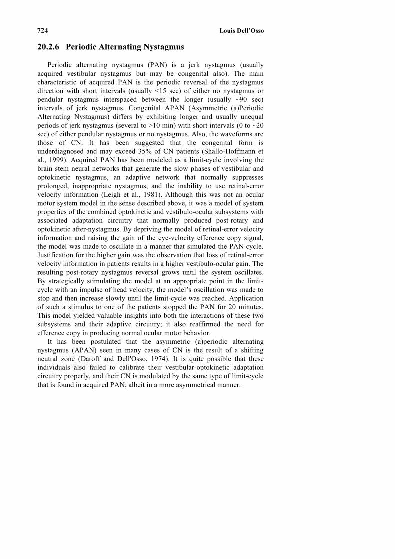
724 Louis Dell’Osso 20.2.6 Periodic Alternating Nystagmus
Periodic alternating nystagmus (PAN) is a jerk nystagmus (usually acquired vestibular nystagmus but may be congenital also). The main characteristic of acquired PAN is the periodic reversal of the nystagmus direction with short intervals (usually <15 sec) of either no nystagmus or pendular nystagmus interspaced between the longer (usually ~90 sec) intervals of jerk nystagmus. Congenital APAN (Asymmetric (a)Periodic Alternating Nystagmus) differs by exhibiting longer and usually unequal periods of jerk nystagmus (several to >10 min) with short intervals (0 to ~20 sec) of either pendular nystagmus or no nystagmus. Also, the waveforms are those of CN. It has been suggested that the congenital form is underdiagnosed and may exceed 35% of CN patients (Shallo-Hoffmann et al., 1999). Acquired PAN has been modeled as a limit-cycle involving the brain stem neural networks that generate the slow phases of vestibular and optokinetic nystagmus, an adaptive network that normally suppresses prolonged, inappropriate nystagmus, and the inability to use retinal-error velocity information (Leigh et al., 1981). Although this was not an ocular motor system model in the sense described above, it was a model of system properties of the combined optokinetic and vestibulo-ocular subsystems with associated adaptation circuitry that normally produced post-rotary and optokinetic after-nystagmus. By depriving the model of retinal-error velocity information and raising the gain of the eye-velocity efference copy signal, the model was made to oscillate in a manner that simulated the PAN cycle. Justification for the higher gain was the observation that loss of retinal-error velocity information in patients results in a higher vestibulo-ocular gain. The resulting post-rotary nystagmus reversal grows until the system oscillates. By strategically stimulating the model at an appropriate point in the limit-cycle with an impulse of head velocity, the model’s oscillation was made to stop and then increase slowly until the limit-cycle was reached. Application of such a stimulus to one of the patients stopped the PAN for 20 minutes. This model yielded valuable insights into both the interactions of these two subsystems and their adaptive circuitry; it also reaffirmed the need for efference copy in producing normal ocular motor behavior.
It has been postulated that the asymmetric (a)periodic alternating nystagmus (APAN) seen in many cases of CN is the result of a shifting neutral zone (Daroff and Dell'Osso, 1974). It is quite possible that these individuals also failed to calibrate their vestibular-optokinetic adaptation circuitry properly, and their CN is modulated by the same type of limit-cycle that is found in acquired PAN, albeit in a more asymmetrical manner.

Chap. 20. Nystagmus Basics: Normal Models that Simulate Dysfunction 725 20.2.7 Endpoint and Rebound Nystagmus
Endpoint nystagmus is a nystagmus present in normals that develops in lateral gaze, usually after prolonged fixation (Abel et al., 1978b). It is a jerk nystagmus with linear, centripetal slow phases and centrifugal fast phases. A system model of endpoint nystagmus (including saccadic, pursuit, and optokinetic subsystems) attributed its development and form to slow, drift velocities in lateral gaze (Eizenman et al., 1990). Reductions in drift velocity during fixation, as opposed to in the dark, were attributed to the pursuit system. However, in the absence of a moving target, it is more probable that the fixation subsystem was responsible.
Rebound nystagmus develops after prolonged lateral gaze followed by a return to primary position; it beats in the opposite direction of the preceding endpoint nystagmus and can be elicited in the light (Shallo-Hoffmann et al., 1990). A combination of a velocity bias and a null shift has been hypothesized as the cause of rebound nystagmus. The velocity bias causes the null to move, and a decrease in the time constant of the common neural integrator determines its exact position (Gordon et al., 1986). This putative mechanism needs to be tested within an ocular motor system model.
Figure 20.6. An ocular motor system block diagram demonstrating the presence or absence of oscillopsia based on the site of an internal oscillation. In this and the following Figures, W – world (target or background), OMS1 – distributed portions of the ocular motor system within efference copy loops, OMS2 – distributed portions of the ocular motor system outside of efference copy loops, LMLN – latent/manifest latent nystagmus, AN – acquired nystagmus, and p or v following a variable’s symbol indicates position or velocity, respectively.
Ocular Motor System Block Diagram
Ev
ep=Wp-Ep
Ep
Wp
Retinal Feedback
RETINA
OMS1W'p
W'v
E'v
E'p
W'v = ev + E'v = Wv - Ev + E'v
ep
ev[AN]
[CN, LMLN, (AN)]
ev=Wv-EvEv
Ep
Wv
W'p = ep + E'p = Wp - Ep + E'p
1Eye
Saccadic
SmoothPursuit
OM2NI
INTERNALMONITOR
[Efference Copy]
1World

726 Louis Dell’Osso 20.2.8 Oscillopsia
The topic of oscillopsia is intimately connected to ocular motor dysfunction, and therefore must be considered in our analysis of models of dysfunction (Dell'Osso et al., 1997). Oscillopsia is the perception of motion of the world when there is no actual physical motion. It may be the perception of a unidirectional ‘spinning’ of the world or a bidirectional oscillation. The first type can be experienced by normal individuals who are spun around in one direction for several minutes and then stopped, and the second type by vibrating one’s finger as it is pressed on the side of one eye. It is an extremely debilitating symptom that interferes with standing, walking, reading. and many other basic functions. Some types of oscillations (usually congenital) are not associated with oscillopsia, while most acquired types are. Ocular motor models of these conditions should demonstrate the same properties. That is, the signals that represent reconstructed target position and velocity (i.e., perceived position and velocity) should either accurately reflect true target parameters (no oscillopsia) or be contaminated with the eye oscillation signal (oscillopsia). An ocular motor system block diagram (Fig. 20.6) demonstrates how lesions in different parts of the ocular motor system (arbitrarily divided into two parts, OM1 and OM2) may or may not produce oscillopsia. OM1 and OM2 are not meant to represent specific anatomical sites; rather, they are distributed functional subsystems. Thus, if a nystagmus position or velocity signal, N, is introduced within OM1 such that both the actual E and efference copy E’ become the desired E + N, the perceived world signal, W’, is given by e + E’ = W – (E + N) + (E + N) = W. That is, there is no oscillopsia. If, on the other hand, N is introduced in OM2, the efference copy signal, E’, would not contain N and W’ = W – N and oscillopsia results. It is hypothesized that oscillations that do not cause oscillopsia lie within the efference copy loops used by the ocular motor system to calculate target parameters, while those that do cause oscillopsia lie outside of those loops.
20.3 CURRENT MODELS
In this chapter, I have concentrated on discussions of system models that fulfill the definition of being robust. These are models that: simulate a wide range of normal responses; simulate the responses to the same stimuli that are exhibited by individuals with specific abnormalities; and, are insensitive to internal changes or errors. Such models may be limited to specific ocular motor subsystems (e.g., saccadic, pursuit, etc.), but do not simply generate waveforms. The latter type of ‘model’ is more an intellectual exercise for

Chap. 20. Nystagmus Basics: Normal Models that Simulate Dysfunction 727 students of control systems than a true simulation of the behavior of an ocular motor system that contains a dysfunction. Furthermore, the specific alterations to the normal system necessary to simulate the desired abnormal behavior should be both physiologically possible and consistent with the known pathophysiology of the abnormality being simulated (e.g., a neural-integrator lesion, leak, or saturation to produce gaze-evoked nystagmus). Experience with system models of dysfunction has shown that, although many proposed subsystem models, functional blocks, or small neural networks might suffice for specific, limited input/output simulations, one cannot easily predict which will not also adversely affect other, even seemingly unrelated, system functions. It is also difficult to prove that one such model is more accurate (i.e., physiological) than the others. The most stringent, and ultimately the best, test of any subsystem model is to place it within a more robust system model and demonstrate that the resulting ocular motor system model can simulate both normal and abnormal behavior when presented with a wide range of stimuli.
We employ this method of “evolving” a model after each subsystem change or addition and, although tedious, it ensures that a particular solution to a desired simulation of dysfunction is compatible with the rest of the system; if it is not, the proposed solution is discarded (i.e., made “extinct”). In addition to being a fail-safe methodology, this “hands-on” evolution provides satisfying support for any incipient feelings of omnipotence the modeler might harbor. However, the transient highs resulting from a successful modification to simulate a desired specific behavior are short lived, only to be dashed by the feelings of despair and incredulity when the model fails to successfully accomplish the next (and there always is a next) supposedly simple simulation. The claim that much more than this (i.e., the ocular motor system) was completed in seven days, is the humbling thought that keeps the modeler in his/her place. The model must always be considered a work in progress, a working hypothesis, and never a finished product. The goal is to change it for the better, not defend any specific part of it.
The remainder of this chapter will concentrate on the development of a robust ocular motor system model that has been used to simulate physiological responses of individuals with either of two disorders, CN or LMLN.
20.3.1 Congenital Nystagmus
Recently, a preliminary simulation of one of the pendular waveforms of CN was incorporated into a normal model of saccades and smooth pursuit (Dell'Osso and Jacobs, 1998). This model was constructed using Simulink (a
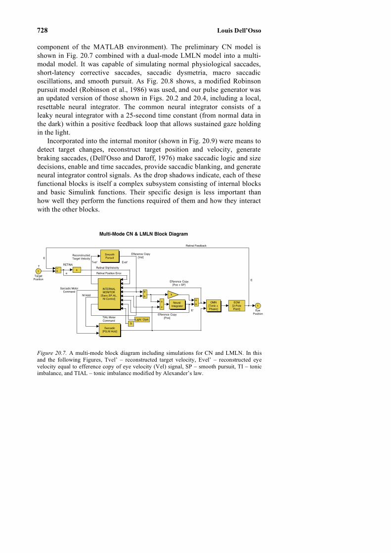
728 Louis Dell’Osso component of the MATLAB environment). The preliminary CN model is shown in Fig. 20.7 combined with a dual-mode LMLN model into a multi-modal model. It was capable of simulating normal physiological saccades, short-latency corrective saccades, saccadic dysmetria, macro saccadic oscillations, and smooth pursuit. As Fig. 20.8 shows, a modified Robinson pursuit model (Robinson et al., 1986) was used, and our pulse generator was an updated version of those shown in Figs. 20.2 and 20.4, including a local, resettable neural integrator. The common neural integrator consists of a leaky neural integrator with a 25-second time constant (from normal data in the dark) within a positive feedback loop that allows sustained gaze holding in the light.
Incorporated into the internal monitor (shown in Fig. 20.9) were means to detect target changes, reconstruct target position and velocity, generate braking saccades, (Dell'Osso and Daroff, 1976) make saccadic logic and size decisions, enable and time saccades, provide saccadic blanking, and generate neural integrator control signals. As the drop shadows indicate, each of these functional blocks is itself a complex subsystem consisting of internal blocks and basic Simulink functions. Their specific design is less important than how well they perform the functions required of them and how they interact with the other blocks.
Figure 20.7. A multi-mode block diagram including simulations for CN and LMLN. In this and the following Figures, Tvel’ – reconstructed target velocity, Evel’ – reconstructed eye velocity equal to efference copy of eye velocity (Vel) signal, SP – smooth pursuit, TI – tonic imbalance, and TIAL – tonic imbalance modified by Alexander’s law.
Multi-Mode CN & LMLN Block Diagram
TIAL MotorCommand
NI Hold
Saccadic MotorCommand
Efference Copy[Vel]Reconstructed
Target Velocity
Efference Copy[Pos]
Efference Copy[Pos + SP]
Retinal Position Error
E
e
E
T
Retinal Feedback
Evel'
E'
Tvel'
Retinal SlipVelocityRETINA
1Eye
Position
k
s
SmoothPursuit
Saccadic[PG,NI Hold]
Neural Integrator
OMN[Tonic +Phasic]
TILight / Dark
INTERNALMONITOR
[Sacc,SP,AL,NI Control]
EOM[2-PolePlant]
1Target
Position

Chap. 20. Nystagmus Basics: Normal Models that Simulate Dysfunction 729
Shown in Fig. 20.10 are the model’s responses to step changes in target position during simulation of orthometria, hypometria, and hypermetria as well as fixation with macro saccadic oscillations. In addition to generating normal pursuit with a damped oscillation (see Fig. 20.11), the smooth pursuit system was capable of oscillating, producing a sinusoidal velocity oscillation that was hypothesized to be the source of pendular CN. When the model was allowed to generate braking saccades, the pendular oscillation of the smooth pursuit system became a pseudopendular oscillation of the eyes, one of the waveforms that are pathognomonic for CN (see Fig. 20.11). Note that although the model programmed 1° braking saccades, the actual braking saccades were truncated by the oppositely directed CN slow phases. The model was able to make normal saccadic and pursuit responses, while oscillating in a pendular manner with two braking saccades per cycle; the latter is also shown in Fig. 20.11. This model was the first step in an attempt to model all of the CN waveforms and their hypothetical sources using a robust normal ocular motor system model as a foundation.
Figure 20.8. A multi-mode model of CN and LMLN containing a modified Robinson pursuit model (capable of oscillating), a saccadic pulse generator containing pulse-height and pulse-width non-linearities and a resettable neural integrator, a leaky common neural integrator with provision to overcome the leak, and distributed delays within and between major subsystems.
TIAL MotorCommand
Tvel'
RETINA
Efference Copy[Pos + SP]
Retinal Position Error
SACCADIC PULSEGENERATOR
NI Hold
(35ms)
Multi-Mode CN & LMLN Model
SMOOTH PURSUITEfference Copy
[Vel]
ReconstructedTarget Velocity
Retinal Feedback
E
Saccadic MotorCommand
Evel'
e
Retinal Slip Velocity
E
T
E'
Efference Copy[Pos]
NEURAL INTEGRATOR1
EyePosition
k
PMC+
VelocitySaturation
50ms
50ms
30ms
15ms1
.007s+1
1/25s+1/25
5
Tonic Gain
1
Phasic Gain
NIHoldPG
.95
OMN[Tonic +Phasic]
TILight / Dark
INTERNALMONITOR
[Sacc,SP,AL,NI Control]
1
25
s
1
CNSGain
EOM[2-PolePlant]
1Target
Position

730 Louis Dell’Osso
Figure 20.9. Functional organization of the Internal Monitor of the multi-mode CN and LMLN model. Target changes are detected and used by the saccadic logic and timing blocks to aid in the generation of saccadic responses. Both target position and velocity are reconstructed for use by the saccadic and pursuit subsystems, respectively; both are also used by the braking saccade logic block that determines the timing and amplitudes of braking saccades. Any existing tonic imbalance produces an Alexander’s law variation that is used in calculating target velocity and sent as an output to the neural integrator. Finally, the reconstructed and efference copy signals are used by the neural integrator control block to determine what portions of saccadic pulses are to be integrated.
e
E'
Tonic Imbalance (AL )
ReconstructedTarget Velocity
Retinal FBEnable
SaccadeEnable
Fast PhaseSize
SampledError
ReconstructedTarget
ReconstructedError
Evel'
Tvel'*
INTERNAL MONITOR [Sacc,SP,AL,NI Control]
4TIAL
MotorCommand
3NI
Hold
2Reconstructed
TargetVelocity
1Saccadic
MotorCommand
loopbreaker
TargetVelocity
Recocnstruction
SaccadeLogic
AlexandersLaw
SaccadeSize
Target ChangeDetection
SaccadeEnable &Timing
NIControl
Saccade& Drift
Blanking
Vel->PosConstant
BrakingSaccade
Logic
PlantModel
8TI
7Light/ Dark
6ECPY[Pos]
5PulseGen[PG]
4ECPY[Vel]
3ECPY[Pos+ SP]
2RetErr
Vel
1RetErrPos

Chap. 20. Nystagmus Basics: Normal Models that Simulate Dysfunction 731
Beginning with our preliminary model of CN and including the functions
required to simulate LMLN (see below), we proceeded to add the ability to produce foveating fast phases to the braking saccades (Jacobs and Dell'Osso, 2000). At present, in addition to the simulations listed above for both the preliminary CN model and the dual-mode LMLN model, this ocular motor system model (still under development) simulates fixation, voluntary saccades, short-latency corrective saccades, pulse-step responses, and smooth pursuit of ramps and step-ramps (Rashbass stimuli) (Rashbass, 1961) of both normals and individuals with either pendular with foveating saccades or pseudopendular with foveating saccades CN waveforms. It also simulates GEN and MG (see Figs. 20.3 and 20.5), since it contains the neural-integrator and plant elements (leaks and saturations) of these earlier models.
Figure 20.9. Functional organization of the Internal Monitor of the multi-mode CN and LMLN model. Target changes are detected and used by the saccadic logic and timing blocks to aid in the generation of saccadic responses. Both target position and velocity are reconstructed for use by the saccadic and pursuit subsystems, respectively; both are also used by the braking saccade logic block that determines the timing and amplitudes of braking saccades. Any existing tonic imbalance produces an Alexander’s law variation that is used in calculating target velocity and sent as an output to the neural integrator. Finally, the reconstructed and efference copy signals are used by the neural integrator control block to determine what portions of saccadic pulses are to be integrated.
Figure 20.10. Simulations of saccadic dysmetria including orthometric, hypometric, and hypermetric saccades and macro saccadic oscillations.

732 Louis Dell’Osso
20.3.2 Latent/Manifest Latent Nystagmus
Latent/manifest latent nystagmus is the second most common type of infantile nystagmus. It may be present in individuals with strabismus (a misalignment of the eyes) and is a jerk nystagmus whose direction is that of the fixating eye. That is, jerk right when the right eye is fixating and jerk left when the left eye is fixating. Pure latent nystagmus (LN) is very rare(Ciuffreda, 1977) and is only present when one eye is occluded. More commonly, manifest latent nystagmus (MLN) is present with both eyes open but only one fixating. (Dell'Osso et al., 1979) The linear slow phases of MLN usually become decelerating when LN is induced by occlusion of one eye. We expanded the capabilities of our preliminary CN model into the multi-modal model of Figs 20.7—9 to model LMLN. We hypothesized that a tonic imbalance was the driving force that produced the linear slow phases of LMLN. They are corrected by foveating fast phases in the direction of the fixating eye and defoveating fast phases when the slow phase velocity exceeds that necessary for good visual acuity (Dell'Osso et al., 1995). The multi-modal model was able to simulate: normal voluntary and corrective
Figure 20.11. Simulations of normal smooth pursuit, fixation with pseudopendular CN, and smooth pursuit with pseudopendular CN.

Chap. 20. Nystagmus Basics: Normal Models that Simulate Dysfunction 733 saccades; fixation; voluntary and corrective saccades during LMLN (of both types); the automatic transition from one type of LMLN to the other as gaze angle changed (based on the change in slow-phase velocity due to Alexander’s law); the eye movements seen during the alternate cover test; and the LMLN changes seen during fixation with the adducting eye (Jacobs and Dell'Osso, 1999). Figure 20.12 shows a simulation of LMLN including saccades to different gaze angles, as well as the automatic transition of the LN from defoveating to foveating fast phases and of the LMLN from foveating to defoveating fast phases. Note that despite the LMLN, larger saccadic refixations include corrective saccades which may be suppressed, allowing the LMLN slow phases to bring the eye onto the target. As the increased size and number of inputs and outputs suggests, the Internal Monitor of Fig. 20.9 has grown considerably from its beginnings as a delay and summing junction. In this model, in addition to the functions listed above for the preliminary CN model, the circuitry provides Alexander’s law variation to nystagmus slow phases. There are extensive interconnections between the different functional networks to ensure that they continue to work synergistically as we add function. We have endeavored to maintain backwards compatibility with our previous models to ensure no loss of function.
20.4 SYSTEM MODELS AND NEUROPHYSIOLOGY
As stated in the beginning of this chapter, these models of dysfunction and normal function are based on properties of the ocular motor system as a whole, as determined from responses to various controlled inputs. They neither depend on specific neurophysiological data nor do they ignore that which exists. However, when a function is deemed necessary for the model’s performance, it suggests that somewhere within the physiological system that function, or a similar one, is being performed and that related signals might be found if one is looking for them. The latter statement comes with the caveat, as discussed in the Introduction, that such signals may be buried within composite signals and be undetectable. At present, these system models consist of functional blocks that are functions of time alone, whereas we know that the physiological system also makes use of spatial maps in its calculations of eye or target parameters (e.g., the retina, superior colliculus, visual cortex, etc.). Thus, some of the model’s functional blocks can be replaced with more physiological spatio-temporal blocks as they are developed. I expect that, as in the past, neurophysiological researchers will continue to search for and elucidate the functions of specific brain stem areas

734 Louis Dell’Osso known to be involved with ocular motor control and we, the modelers, will incorporate those findings into specific functional blocks.
Figure 20.12. Simulations of LMLN showing the variation of the slow-phase velocity with gaze angle and the automatic transition from defoveating to foveating (during LN) and foveating to defoveating (during MLN) fast phases. For LN, the LE was occluded and the RE fixated the target; for MLN, both eyes (BE) were viewing (i.e., neither occluded) and the RE fixated the target. The target either remained at 0° or changed to one of the lateral positions indicated at 1 sec.

Chap. 20. Nystagmus Basics: Normal Models that Simulate Dysfunction 735 20.5 IMPLICATIONS OF THE MODELS
20.5.1 Basic
The most important findings that have come from the modelling of ocular motor dysfunction have been the elucidation of normal ocular motor function and architecture. Models of CN, square-wave pulses in multiple sclerosis, GEN, muscle-paretic nystagmus in myasthenia gravis, PAN, and LMLN revealed the necessity for an internal monitor to reconstruct target position and velocity from efference copy of motor commands and the retinal error signals. Modelling GEN demonstrated the need for a local, resettable neural integrator in the pulse generator and also the need for neural control of the percentage of the pulse that is integrated by the common neural integrator. The latter was also required by models of LMLN when fast phases became defoveating, and by saccadic pulses. Our current model of CN demonstrates how normal fast-phase generating mechanisms may be used to generate both the braking and foveating saccades of many CN waveforms. It also demonstrates how efference copy allows normal function of both the saccadic and smooth pursuit systems despite the presence of complex oscillations. Finally, modelling the uniocular saccades seen in achiasmatic Belgian sheepdogs required bilateral, yoked, independent control architecture of the ocular motor system, suggesting that each eye and muscle can be independently activated under the proper (perhaps, abnormal) circumstances. Although this is unusual in normal humans, its demonstration in abnormal human ocular motor behavior supports the hypothesis that the underlying architecture is uniocular and unimuscular with a strong yoking overlay. Models that do not contain these elements are severely limited in their ability to simulate the variety of ocular motor responses that the physiological system is capable of making.
20.5.2 Clinical
The PAN model described above demonstrated how the normal velocity storage mechanism enters into a limit cycle if not properly calibrated, and the model predicted a method to temporarily stop the PAN. Consideration of efference copy in ocular motor models led to the hypothesis that oscillopsia in acquired nystagmus occurred when the site of the dysfunction was outside efference-copy loops. The LMLN model demonstrated how individuals with LMLN could use their saccadic system to defoveate the target in an effort to utilize the slower tail ends of the slow phases to achieve better visual acuity. The CN model demonstrated how, by means of braking and foveating

736 Louis Dell’Osso saccades, individuals with CN improve their foveation and therefore, their acuity.
20.6 FUTURE DIRECTIONS
20.6.1 Short Term
Because our ocular motor system model is a work in progress, some of the short-term plans listed below may have already been accomplished at the time of publication of this book. With regard to the simulation of LMLN, we are assessing the effects of background on the waveform and will include this in the model. Similarly, the effects of total darkness without a target will be simulated. For the CN simulation, a fixation subsystem will be added to achieve extended foveation of targets for the different waveforms. We will simulate the jerk waveforms and their variation with gaze angle and eye-velocity, and based on our data, the transitions of some individuals from pendular waveforms to jerk waveforms. Also, the model’s emergent property of spontaneous bias shifts (which mimic physiological CN) will be studied to provide for control of that phenomenon. Finally, we plan to expand the types of dysfunction that the model can simulate by adding saccadic oscillations (e.g., flutter, flutter dysmetria, psychogenic nystagmus, and square-wave jerks/oscillations) and other types of nystagmus (e.g., rebound, Brun’s, and acquired pendular nystagmus).
20.6.2 Long Term
As computing power continues to grow, we will be able to increase dramatically the complexity of our models and concurrently demand more from them. Additional specific behaviors will be simulated in more accurate ways. Duplication of unilateral, yoked control architecture into the bilateral, yoked, independent control architecture that defines the physiological system will become both feasible and the accepted norm in well-tested ocular motor models. When specific subsystems become totally specified by neurophysiological studies, their counterparts in the system model can be replaced by neural networks that accomplish and extend the simulation of those subsystems and of the model as a whole. Also, by appropriate time scaling, models will incorporate the plasticity exhibited by the physiological system and be better able to simulate the results of specific dysfunction. This includes both its slow development (in some cases) and the slow adaptations that take place within the ocular motor system to overcome the dysfunction. It will keep us busy, and it will take us much longer than seven days.

Chap. 20. Nystagmus Basics: Normal Models that Simulate Dysfunction 737 20.7 ACKNOWLEDGMENTS
This work emanated from the Ocular Motility Laboratory, Veterans Affairs Medical Center and the Departments of Neurology and Biomedical Engineering, Case Western Reserve University, Cleveland, OH. As the reference list indicates, many of the models that resulted from studies in our laboratory were the result of interdisciplinary collaborative efforts. I would like to specifically acknowledge the modelling contributions of L.A. Abel, M.J. Doslak, and J.B. Jacobs and the medical and physiological contributions of R.B. Daroff, B.T. Troost, D. Schmidt, and R.J. Leigh.
20.8 REFERENCES
Abel L. A., Dell'Osso L. F., Daroff R. B., 1978a, Analog model for gaze-evoked nystagmus, IEEE Trans Biomed Engng. BME-25: 71-75.
Abel L. A., Parker L., Daroff R. B., Dell'Osso L. F., 1978b, Endpoint nystagmus, Invest Ophthalmol Vis Sci. 17: 539-544.
Abel L. A., Dell'Osso L. F., Schmidt D., Daroff R. B., 1980, Myasthenia gravis: Analogue computer model, Exp Neurol. 68: 378-389.
Alexander G., 1912, Die Ohrenkrankhieten im Kindesalter, in: Handbuch der Kinderheilkunde, M. Pfaundler and A. Schlossman, eds., Vlg FCW Vogel, Leipzig, pp. 84-96.
Ciuffreda K. J., 1977, Movements in amblyopia and strabismus. (Ph.D Dissertation), School of Optometry, Univ. of California,, Berkeley.
Collins C. C., 1975, The human oculomotor control system, in: Basic Mechanisms of Ocular Motility and their Clinical Implications, G. Lennerstrand and P. Bach-y-Rita, eds., Pergamon Press, Oxford, pp. 145-180.
Daroff R. B., Dell'Osso L. F., 1974, Periodic alternating nystagmus and the shifting null, Can J Otolaryngol. 3: 367-371.
Dell'Osso L. F., 1967, A model for the horizontal tracking system of a subject with nystagmus, Proc 20th Ann Conf EMB. 9: 24.2.
Dell'Osso L. F., 1968, A Dual-Mode Model for the Normal Eye Tracking System and the System with Nystagmus. (Ph.D. Dissertation), Electrical Engineering (Biomedical), University of Wyoming, Laramie, pp. 1-131.
Dell'Osso L. F., 1970, A dual-mode model for the normal eye tracking system and the system with nystagmus, IEEE Trans Biomed Engng. BME: 87.
Dell'Osso L. F., Gauthier G., Liberman G., Stark L., 1972, Eye movement recordings as a diagnostic tool in a case of congenital nystagmus, Am J Optom Arch Am Acad Optom. 49: 3-13.
Dell'Osso L. F., Daroff R. B., 1975, Congenital nystagmus waveforms and foveation strategy, Doc Ophthalmol. 39: 155-182.
Dell'Osso L. F., Troost B. T., Daroff R. B., 1975, Macro square wave jerks, Neurology. 25: 975-979.

738 Louis Dell’Osso Dell'Osso L. F., Daroff R. B., 1976, Braking saccade--A new fast eye movement, Aviat Space
Environ Med. 47: 435-437. Dell'Osso L. F., Schmidt D., Daroff R. B., 1979, Latent, manifest latent and congenital
nystagmus, Arch Ophthalmol. 97: 1877-1885. Dell'Osso L. F., Daroff R. B., 1981, Clinical disorders of ocular movement, in: Models of
Oculomotor Behavior and Control, B. L. Zuber, ed., CRC Press Inc, West Palm Beach, pp. 233-256.
Dell'Osso L. F., 1986, Evaluation of smooth pursuit in the presence of congenital nystagmus, Neuro ophthalmol. 6: 383-406.
Dell'Osso L. F., Van der Steen J., Steinman R. M., Collewijn H., 1992a, Foveation dynamics in congenital nystagmus I: Fixation, Doc Ophthalmol. 79: 1-23.
Dell'Osso L. F., Van der Steen J., Steinman R. M., Collewijn H., 1992b, Foveation dynamics in congenital nystagmus II: Smooth pursuit, Doc Ophthalmol. 79: 25-49.
Dell'Osso L. F., Van der Steen J., Steinman R. M., Collewijn H., 1992c, Foveation dynamics in congenital nystagmus III: Vestibulo-ocular reflex, Doc Ophthalmol. 79: 51-70.
Dell'Osso L. F., Weissman B. M., Leigh R. J., Abel L. A., Sheth N. V., 1993, Hereditary congenital nystagmus and gaze-holding failure: The role of the neural integrator, Neurology. 43: 1741-1749.
Dell'Osso L. F., 1994, Evidence suggesting individual ocular motor control of each eye (muscle), J Vestib Res. 4: 335-345.
Dell'Osso L. F., Leigh R. J., Sheth N. V., Daroff R. B., 1995, Two types of foveation strategy in 'latent' nystagmus. Fixation, visual acuity and stability, Neuro Ophthalmol. 15: 167-186.
Dell'Osso L. F., Averbuch-Heller L., Leigh R. J., 1997, Oscillopsia suppression and foveation-period variation in congenital, latent, and acquired nystagmus, Neuro Ophthalmol. 18: 163-183.
Dell'Osso L. F., Jacobs J. B., 1998, A preliminary model of congenital nystagmus (CN) incorporating braking saccades, Invest Ophthalmol Vis Sci. 39: S149.
Doslak M. J., Dell'Osso L. F., Daroff R. B., 1979, A model of Alexander's law of vestibular nystagmus, Biol Cyber. 34: 181-186.
Doslak M. J., Dell'Osso L. F., Daroff R. B., 1982, Alexander's law: A model and resulting study, Ann Otol Rhinol Laryngol. 91: 316-322.
Eizenman M., Cheng P., Sharpe J. A., Frecker R. C., 1990, End-point nystagmus and ocular drift: An experimental and theoretical study, Vision Res. 30: 863-877.
Gordon S. E., Hain T. C., Zee D. S., Fetter M., 1986, Rebound nystagmus, Soc Neurosci Abstr. 12: 1091.
Harris C. M., 1995, Problems in modeling congenital nystagmus: Towards a new model, in: Eye Movement Research: Mechanisms, Processes and Applications, J. M. Findlay, R. Walker and R. W. Kentridge, eds., Elsevier, Amsterdam, pp. 239-253.
Jacobs J. B., Dell'Osso L. F., 1999, A dual-mode model of latent nystagmus. ARVO abstracts, Invest Ophthalmol Vis Sci. 40: S962.
Jacobs J. B., Dell'Osso L. F., 2000, A model of congenital nystagmus (CN) incorporating braking and foveating saccades. ARVO abstracts, Invest Ophthalmol Vis Sci. 41: S701.
Kurzan R., Büttner U., 1989, Smooth pursuit mechanisms in congenital nystagmus, Neuro ophthalmol. 9: 313-325.

Chap. 20. Nystagmus Basics: Normal Models that Simulate Dysfunction 739 Leigh R. J., Robinson D. A., Zee D. S., 1981, A hypothetical explanation for periodic
alternating nystagmus: Instability in the optokinetic-vestibular system, Ann NY Acad Sci. 374: 619-635.
Optican L. M., Zee D. S., 1984, A hypothetical explanation of congenital nystagmus, Biol Cyber. 50: 119-134.
Raphan T., Matsuo V., Cohen B., 1979, Velocity storage in the vestibuloocular reflex arc (VOR), Exp Brain Res. 35: 229-248.
Rashbass C., 1961, The relationship between saccadic and smooth tracking eye movements, J Physiol (Lond). 159: 326-338.
Robinson D. A., 1973, Models of saccadic eye movement control systems, Kybernetik. 14: 71-83.
Robinson D. A., Zee D. S., Hain T. C., Holmes A., Rosenberg L. F., 1984, Alexander's law: Its behavior and origin in the human vestibulo-ocular reflex, Ann Neurol. 16: 714-722.
Robinson D. A., Gordon J. L., Gordon S. E., 1986, A model of smooth pursuit eye movements, Biol Cyber. 55: 43-57.
Robinson D. A., 1994, Implications of neural networks for how we think about brain function, in: Movement Control, P. Cordo and S. Harnad, eds., Cambridge University Press, Cambridge, pp. 42-53.
Schmidt D., Dell'Osso L. F., Abel L. A., Daroff R. B., 1980, Myasthenia gravis: Saccadic eye movement waveforms, Exp Neurol. 68: 346-364.
Shallo-Hoffmann J., Schwarze H., Simonsz H. J., Mühlendyck H., 1990, A reexamination of end-point and rebound nystagmus in normals, Invest Ophthalmol Vis Sci. 31: 388-392.
Shallo-Hoffmann J., Faldon M., Tusa R. J., 1999, The incidence and waveform characteristics of periodic alternating nystagmus in congenital nystagmus, Invest Ophthalmol Vis Sci. 40: 2546-2553.
Stark L., 1968, Neurological Control Systems (Studies in Bioengineering), Plenum Press, New York, pp. 1-428.
Tusa R. J., Zee D. S., Hain T. C., Simonsz H. J., 1992, Voluntary control of congenital nystagmus, Clin Vis Sci. 7: 195-210.
Yasui S., Young L. R., 1975, Perceived visual motion as effective stimulus to pursuit eye movement system, Science. 190: 906-908.
Young L. R., Stark L., 1963a, A discrete model for eye tracking movements, IEEE Trans Military Elect MIL 7. 13-115.
Young L. R., Stark L., 1963b, Variable feedback experiments testing a sampled data model for eye tracking movements, IEEE Trans Prof Tech Group Human Factors Electron. HFE: 38-51.