Embed Size (px)
Citation preview

CHAPTER 16THE MOLECULE BASIS OF
INHERITANCE
Copyright © 2002 Pearson Education, Inc., publishing as Benjamin Cummings
Section B: DNA Replication and Repair1. During DNA replication, base pairing enables existing DNA strands to serve
as templates for new complimentary strands2. A large team of enzymes and other proteins carries out DNA replication3. Enzymes proofread DNA during its replication and repair damage to
existing DNA4. The ends of DNA molecules are replicated by a special mechanism

• The specific pairing of nitrogenous bases in DNAwas the flash of inspiration that led Watson andCrick to the correct double helix.
• The possible mechanism for the next step, theaccurate replication of DNA, was clear to Watsonand Crick from their double helix model.
Introduction
Copyright © 2002 Pearson Education, Inc., publishing as Benjamin Cummings

• In a second paper Watson and Crick published theirhypothesis for how DNA replicates.• Essentially, because each strand is complementary to each
other, each can form a template when separated.
• The order of bases on one strand can be used to add incomplementary bases and therefore duplicate the pairs ofbases exactly.
1. During DNA replication, base pairingenables existing DNA strands to serve astemplates for new complimentary strands
Copyright © 2002 Pearson Education, Inc., publishing as Benjamin Cummings
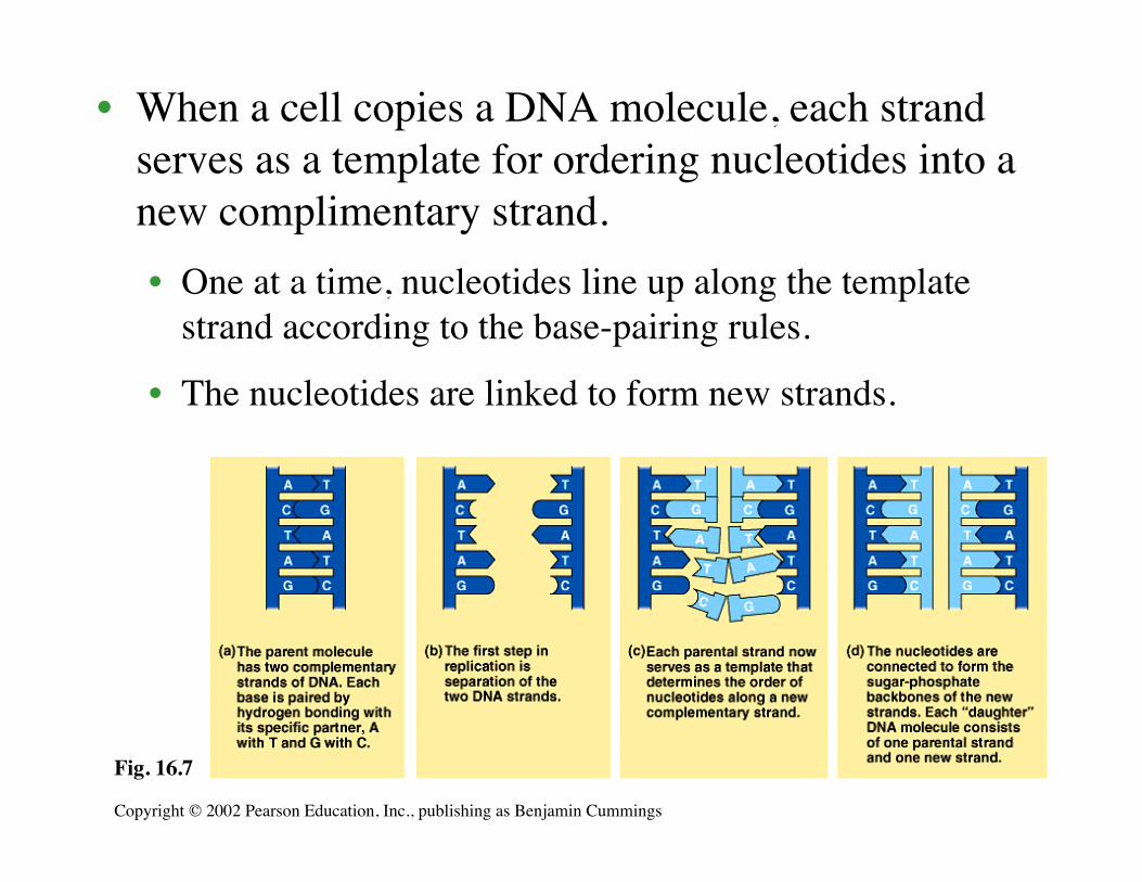
• When a cell copies a DNA molecule, each strandserves as a template for ordering nucleotides into anew complimentary strand.• One at a time, nucleotides line up along the template
strand according to the base-pairing rules.
• The nucleotides are linked to form new strands.
Copyright © 2002 Pearson Education, Inc., publishing as Benjamin Cummings
Fig. 16.7

• Watson and Crick’s model, semiconservativereplication, predicts that when a double helixreplicates each of the daughter molecules will haveone old strand and one newly made strand.
• Other competing models, the conservative modeland the dispersive model, were also proposed.
Copyright © 2002 Pearson Education, Inc., publishing as Benjamin Cummings
Fig. 16.8

• Experiments in the late 1950s by Matthew Meselsonand Franklin Stahl supported the semiconservativemodel, proposed by Watson and Crick, over theother two models.• In their experiments, they labeled the nucleotides of the
old strands with a heavy isotope of nitrogen (15N) whileany new nucleotides would be indicated by a lighterisotope (14N).
• Replicated strands could be separated by density in acentrifuge.
• Each model: the semi-conservative model, theconservative model, and the dispersive model, madespecific predictions on the density of replicated DNAstrands.
Copyright © 2002 Pearson Education, Inc., publishing as Benjamin Cummings

Copyright © 2002 Pearson Education, Inc., publishing as Benjamin Cummings
Fig. 16.9
• The first replication in the 14N medium produced a bandof hybrid (15N-14N) DNA, eliminating the conservativemodel.
• A second replication produced both light and hybridDNA, eliminating the dispersive model and supportingthe semiconservative model.

• It takes E. coli less than an hour to copy each of the 5million base pairs in its single chromosome anddivide to form two identical daughter cells.
• A human cell can copy its 6 billion base pairs anddivide into daughter cells in only a few hours.
• This process is remarkably accurate, with only oneerror per billion nucleotides.
• More than a dozen enzymes and other proteinsparticipate in DNA replication.
2. A large team of enzymes and otherproteins carries out DNA replication
Copyright © 2002 Pearson Education, Inc., publishing as Benjamin Cummings

• The replication of a DNA molecule begins atspecial sites, origins of replication.
• In bacteria, this is a single specific sequence ofnucleotides that is recognized by the replicationenzymes.• These enzymes separate the strands, forming a
replication “bubble”.
• Replication proceeds in both directions until the entiremolecule is copied.
Copyright © 2002 Pearson Education, Inc., publishing as Benjamin Cummings

• In eukaryotes, there may be hundreds or thousandsof origin sites per chromosome.• At the origin sites, the DNA strands separate forming a
replication “bubble” with replication forks at each end.• The replication bubbles elongate as the DNA is replicated
and eventually fuse.
Copyright © 2002 Pearson Education, Inc., publishing as Benjamin Cummings
Fig. 16.10

• DNA polymerases catalyze the elongation of newDNA at a replication fork.
• As nucleotides align with complementary basesalong the template strand, they are added to thegrowing end of the new strand by the polymerase.• The rate of elongation is about 500 nucleotides per
second in bacteria and 50 per second in human cells.The raw nucleotides are nucleoside triphosphates.
• The raw nucleotides are nucleoside triphosphates.• Each has a nitrogen base, deoxyribose, and a
triphosphate tail.
Copyright © 2002 Pearson Education, Inc., publishing as Benjamin Cummings

• As each nucleotide is added, the last two phosphategroups are hydrolyzed to form pyrophosphate.• The exergonic hydrolysis of pyrophosphate to two
inorganic phosphate molecules drives the polymerizationof the nucleotide to the new strand.
Copyright © 2002 Pearson Education, Inc., publishing as Benjamin Cummings
Fig. 16.11
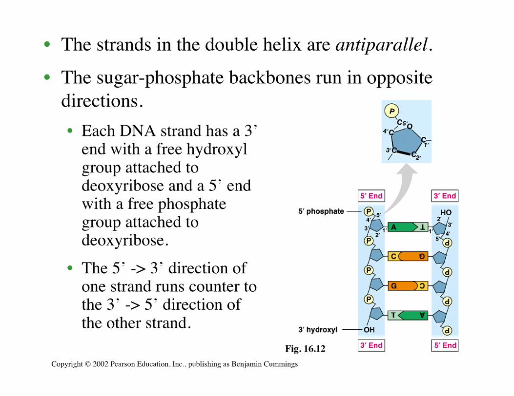
• The strands in the double helix are antiparallel.
• The sugar-phosphate backbones run in oppositedirections.• Each DNA strand has a 3’
end with a free hydroxylgroup attached todeoxyribose and a 5’ endwith a free phosphategroup attached todeoxyribose.
• The 5’ -> 3’ direction ofone strand runs counter tothe 3’ -> 5’ direction ofthe other strand.
Copyright © 2002 Pearson Education, Inc., publishing as Benjamin Cummings
Fig. 16.12

• DNA polymerases can only add nucleotides to thefree 3’ end of a growing DNA strand.
• A new DNA strand can only elongate in the 5’->3’direction.
• This creates a problem at the replication forkbecause one parental strand is oriented 3’->5’ intothe fork, while the other antiparallel parental strandis oriented 5’->3’ into the fork.
• At the replication fork, one parental strand (3’-> 5’into the fork), the leading strand, can be used bypolymerases as a template for a continuouscomplimentary strand.
Copyright © 2002 Pearson Education, Inc., publishing as Benjamin Cummings

• The other parental strand (5’->3’ into the fork), thelagging strand,is copied away fromthe fork in short segments(Okazaki fragments).
• Okazaki fragments,each about 100-200nucleotides, are joinedby DNA ligase to formthe sugar-phosphatebackbone of a singleDNA strand.
Copyright © 2002 Pearson Education, Inc., publishing as Benjamin CummingsFig. 16.13

• DNA polymerases cannot initiate synthesis of apolynucleotide because they can only addnucleotides to the end of an existing chain that isbase-paired with the template strand.
• To start a new chain requires a primer, a shortsegment of RNA.• The primer is about 10 nucleotides long in eukaryotes.
• Primase, an RNA polymerase, linksribonucleotides that are complementary to the DNAtemplate into the primer.• RNA polymerases can start an RNA chain from a single
template strand.Copyright © 2002 Pearson Education, Inc., publishing as Benjamin Cummings

• After formation of the primer,DNA polymerases can adddeoxyribonucleotidesto the 3’ end of theribonucleotide chain.
• Another DNApolymerase laterreplaces the primerribonucleotides withdeoxyribonucleotidescomplimentary tothe template.
Copyright © 2002 Pearson Education, Inc., publishing as Benjamin Cummings
Fig. 16.14

• Returning to the original problem at the replicationfork, the leading strand requires the formation ofonly a single primer as the replication forkcontinues to separate.
• The lagging strand requires formation of a newprimer as the replication fork progresses.
• After the primer is formed, DNA polymerase canadd new nucleotides away from the fork until itruns into the previous Okazaki fragment.
• The primers are converted to DNA before DNAligase joins the fragments together.
Copyright © 2002 Pearson Education, Inc., publishing as Benjamin Cummings

• In addition to primase, DNA polymerases, andDNA ligases, several other proteins haveprominent roles in DNA synthesis.
• A helicase untwists and separates the templateDNA strands at the replication fork.
• Single-strandbinding proteinskeep the unpairedtemplate strandsapart duringreplication.
Copyright © 2002 Pearson Education, Inc., publishing as Benjamin Cummings
Fig. 16.15

Copyright © 2002 Pearson Education, Inc., publishing as Benjamin Cummings
Fig. 16.16
• To summarize, at the replication fork, the leadingstand is copied continuously into the fork from asingle primer.
• The lagging strand is copied awayfrom the fork in short segments,each requiring a new primer.

• It is conventional and convenient to think of theDNA polymerase molecules moving along astationary DNA template.
• In reality, the various proteins involved in DNAreplication form a single large complex that maybe anchored to the nuclear matrix.
• The DNA polymerase molecules “reel in” theparental DNA and “extrude” newly made daughterDNA molecules.
Copyright © 2002 Pearson Education, Inc., publishing as Benjamin Cummings

• Mistakes during the initial pairing of templatenucleotides and complementary nucleotides occurs ata rate of one error per 10,000 base pairs.
• DNA polymerase proofreads each new nucleotideagainst the template nucleotide as soon as it is added.
• If there is an incorrect pairing, the enzyme removesthe wrong nucleotide and then resumes synthesis.
• The final error rate is only one per billion nucleotides.
3. Enzymes proofread DNA during its replicationand repair damage in existing DNA
Copyright © 2002 Pearson Education, Inc., publishing as Benjamin Cummings

• DNA molecules are constantly subject topotentially harmful chemical and physical agents.• Reactive chemicals, radioactive emissions, X-rays, and
ultraviolet light can change nucleotides in ways that canaffect encoded genetic information.
• DNA bases often undergo spontaneous chemicalchanges under normal cellular conditions.
• Mismatched nucleotides that are missed by DNApolymerase or mutations that occur after DNAsynthesis is completed can often be repaired.• Each cell continually monitors and repairs its genetic
material, with over 130 repair enzymes identified inhumans.
Copyright © 2002 Pearson Education, Inc., publishing as Benjamin Cummings

• In mismatch repair, special enzymes fix incorrectlypaired nucleotides.• A hereditary defect in
one of these enzymesis associated with aform of colon cancer.
• In nucleotide excisionrepair, a nuclease cutsout a segment of adamaged strand.• The gap is filled in by
DNA polymerase andligase.
Copyright © 2002 Pearson Education, Inc., publishing as Benjamin Cummings
Fig. 16.17

• The importance of proper function of repairenzymes is clear from the inherited disorderxeroderma pigmentosum.• These individuals are hypersensitive to sunlight.
• In particular, ultraviolet light can produce thyminedimers between adjacent thymine nucleotides.
• This buckles the DNA double helix and interferes withDNA replication.
• In individuals with this disorder, mutations in their skincells are left uncorrected and cause skin cancer.
Copyright © 2002 Pearson Education, Inc., publishing as Benjamin Cummings

• Limitations in the DNA polymerase create problemsfor the linear DNA of eukaryotic chromosomes.
• The usual replication machinery provides no way tocomplete the 5’ ends of daughter DNA strands.• Repeated rounds of replication produce shorter and
shorter DNA molecules.
4. The ends of DNA molecules arereplicated by a special mechanism
Copyright © 2002 Pearson Education, Inc., publishing as Benjamin Cummings

Copyright © 2002 Pearson Education, Inc., publishing as Benjamin Cummings
Fig. 16.18

• The ends of eukaryotic chromosomal DNAmolecules, the telomeres, have special nucleotidesequences.• In human telomeres, this sequence is typically TTAGGG,
repeated between 100 and 1,000 times.
• Telomeres protect genes from being eroded throughmultiple rounds of DNA replication.
Copyright © 2002 Pearson Education, Inc., publishing as Benjamin Cummings
Fig. 16.19a

• Eukaryotic cells have evolved a mechanism torestore shortened telomeres.
• Telomerase uses a short molecule of RNA as atemplate to extend the 3’ end of the telomere.• There is now room for
primase and DNApolymerase to extendthe 5’ end.
• It does not repair the3’-end “overhang,”but it does lengthenthe telomere.
Copyright © 2002 Pearson Education, Inc., publishing as Benjamin Cummings
Fig. 16.19b

• Telomerase is not present in most cells ofmulticellular organisms.
• Therefore, the DNA of dividing somatic cells andcultured cells does tend to become shorter.
• Thus, telomere length may be a limiting factor in thelife span of certain tissues and the organism.
• Telomerase is present in germ-line cells, ensuringthat zygotes have long telomeres.
• Active telomerase is also found in cancerous somaticcells.• This overcomes the progressive shortening that would
eventually lead to self-destruction of the cancer.
Copyright © 2002 Pearson Education, Inc., publishing as Benjamin Cummings