Embed Size (px)
Citation preview

MICROBIOLOGY AND MOLECULAR BIOLOGY REVIEWS, Mar. 2007, p. 48–96 Vol. 71, No. 11092-2172/07/$08.00�0 doi:10.1128/MMBR.00028-06Copyright © 2007, American Society for Microbiology. All Rights Reserved.
Central Roles of Small GTPases in the Development ofCell Polarity in Yeast and Beyond
Hay-Oak Park1*† and Erfei Bi2*†Department of Molecular Genetics, The Ohio State University, Columbus, Ohio,1 and Department of Cell and Developmental Biology,
University of Pennsylvania School of Medicine, Philadelphia, Pennsylvania2
INTRODUCTION .........................................................................................................................................................49ESTABLISHMENT AND MAINTENANCE OF Cdc42 POLARIZATION...........................................................50
The Rho Family GTPase Cdc42 and Its Regulators............................................................................................50Cdc42 GTPase .......................................................................................................................................................50Cdc24, a GEF for Cdc42......................................................................................................................................52Cdc42 GAPs...........................................................................................................................................................53Rho GDI.................................................................................................................................................................53Cycling of Cdc42 between the GDP- and GTP-bound states..........................................................................54
Regulation of Cdc42 Clustering at Sites of Polarized Growth...........................................................................55Establishment of Cdc42 polarization.................................................................................................................55Maintenance of Cdc42 polarization ...................................................................................................................55
(i) The central role of Bem1 in maintaining Cdc42 polarization ..............................................................55(ii) Role of F-actin in maintaining Cdc42 polarization...............................................................................55(iii) Role of exocytosis in maintaining Cdc42 polarization ........................................................................55(iv) A possible mechanism of concentrating Cdc42-GDP at sites of polarized growth ..........................56
DETERMINATION OF THE AXIS FOR CELL POLARIZATION DURING BUDDING ................................56The Rsr1 GTPase Module—Rsr1, Bud2, and Bud5 ............................................................................................57Coupling of the Rsr1 GTPase Module to the Cdc42 GTPase Module..............................................................58
Interaction between Rsr1 and the Cdc42 module ............................................................................................58Model for coupling bud site selection to polarity establishment...................................................................58
Spatial Landmarks That Specify the Site for Polarized Growth .......................................................................58Axial landmarks ....................................................................................................................................................59
(i) Bud3, Bud4, and septins ............................................................................................................................59(ii) Axl1 and Axl2..............................................................................................................................................59
Bipolar landmarks................................................................................................................................................60(i) Bud8 and Bud9............................................................................................................................................60(ii) Rax1 and Rax2 ...........................................................................................................................................61(iii) Other proteins necessary for the bipolar budding pattern.................................................................61
Regulation of Cell Type-Specific Budding Patterns.............................................................................................61Coupling of spatial landmarks to the Rsr1 GTPase module .........................................................................61Model for cell type-specific budding patterns ..................................................................................................62
TEMPORAL CONTROL OF POLARITY ESTABLISHMENT DURING YEAST BUDDING..........................62Cdc42 EFFECTORS AND THEIR ROLES IN POLARITY DEVELOPMENT ...................................................63
Actin Organization ...................................................................................................................................................63Actin patches and endocytosis ............................................................................................................................63Cdc42 effectors and actin patches ......................................................................................................................65Actin cables and exocytosis .................................................................................................................................65Cdc42 effectors and actin cables.........................................................................................................................66
Septin Organization .................................................................................................................................................67General properties and functions of the septins..............................................................................................67Role of Cdc42 in septin organization.................................................................................................................68Is the role of Cdc42 in septin organization universal? ...................................................................................69
Model for the Roles of Cdc42 in Actin and Septin Organization......................................................................70
* Corresponding author. Mailing address for Hay-Oak Park: De-partment of Molecular Genetics, The Ohio State University, 484 West12th Avenue, Columbus, OH 43210-1292. Phone: (614) 688-4575. Fax:(614) 292-4466. E-mail: [email protected]. Mailing address for ErfeiBi: Department of Cell and Developmental Biology, University ofPennsylvania School of Medicine, Biomedical Research Building II/III,Room 1012, 421 Curie Blvd., Philadelphia, PA 19104-6058. Phone:(215) 573-6676. Fax: (215) 898-9871. E-mail: [email protected].
† H.-O.P. and E.B. contributed equally to this work.
48
on January 11, 2019 by guesthttp://m
mbr.asm
.org/D
ownloaded from

The Mitotic Exit Network and Cytokinesis ...........................................................................................................70OTHER TYPES OF CELL POLARIZATION ..........................................................................................................72
Cell Polarization during Mating.............................................................................................................................72Filamentous Growth .................................................................................................................................................74
OTHER SMALL GTPases AND THEIR ROLES IN POLARIZED GROWTH ..................................................76Rho1 Regulators........................................................................................................................................................76
Rho1 GEFs.............................................................................................................................................................76Rho1 GAPs.............................................................................................................................................................77
Rho1 Effectors and Biological Responses..............................................................................................................77Rho1 and cell wall assembly ...............................................................................................................................77Rho1 and actin organization...............................................................................................................................78Rho1 and exocytosis .............................................................................................................................................79
Rho3, Rho4, and Polarized Cell Growth ...............................................................................................................79Rho3, Rho4, and actin organization ..................................................................................................................80Rho3, Rho4, and exocytosis .................................................................................................................................80
What Is the Role of Ras in Polarized Growth?....................................................................................................80COORDINATION OF Cdc42, Rho, AND Rab DURING POLARIZED GROWTH............................................81
Polarisome-Based Signaling Hub ...........................................................................................................................81Exocyst-Based Signaling Hub .................................................................................................................................82
UNIFYING CONCEPTS IN CELL POLARIZATIONS IN DIFFERENT ORGANISMS ..................................82Global and Local Cell Polarity ...............................................................................................................................82Local Activation and Global Inhibition.................................................................................................................83Self-Organization ......................................................................................................................................................83
CONCLUDING REMARKS........................................................................................................................................84ACKNOWLEDGMENTS .............................................................................................................................................84REFERENCES ..............................................................................................................................................................84
INTRODUCTION
Cell polarity is central to the development of most eu-karyotes. It is also critical for the function of many cell typesinvolved in vectored processes such as nutrient transport, neu-ronal signaling, and cell motility. Cell polarization in responseto extracellular or intracellular cues appears to follow a com-mon plan (128). First, a spatial cue marks the site of polarizedgrowth. Signaling molecules relay the spatial information tothe downstream components of polarity establishment, leadingto asymmetric organization of the cytoskeleton. Polarity is thenreinforced with targeted secretion that leads to the depositionof molecules needed for growth at the chosen site. It is appar-ent from a large number of studies of diverse organisms thatthe small-molecular-weight GTPases function as key signalingmolecules in polarity development and that there is a remark-able conservation of these GTPases from yeast to humans atboth structural and functional levels (61, 62, 137, 203).
The budding yeast Saccharomyces cerevisiae is a particularlyattractive model organism because it displays pronounced cellpolarity in response to intracellular and extracellular cues.Cells of budding yeast undergo polarized growth during vari-ous phases of their life cycle, such as budding during vegetativegrowth, mating between haploid cells of opposite mating types,and filamentous growth (FG) upon deprivation of nutrientssuch as nitrogen (Fig. 1). There are three cell types in yeast(here we sometimes refer to S. cerevisiae as “yeast,” althoughwe realize that other yeasts such as Schizosaccharomycespombe are somewhat different from S. cerevisiae): a and � cells(such as normal haploids) and a/� cells (such as normal dip-loids), which are determined by their mating-type loci (220).Both haploid and diploid cells initiate budding when theyreach the critical cell size in the late G1 phase of the cell cycle.Bud growth is initially targeted to the bud tip (apical growth)and then throughout the bud (isotropic growth) until nuclear
division and cytokinesis take place (Fig. 1A). The second formof polarized growth occurs when haploid a and � cells encoun-ter each other and undergo mating to form diploid a/� cells(Fig. 1B). During both budding and mating, the overall cellularorganization is similar, although budding cells have a constric-tion between mother and bud called the bud neck (Fig. 1A). Aspecific site for cell polarization during budding is determinedby the cell type, whereas cell polarization during mating ischemotropic; i.e., a cell of one mating type responds to agradient of a peptide mating pheromone secreted by a cell ofthe opposite mating type (221). The most prominent feature inboth processes is the organization of the polarized actin cy-toskeleton, which guides secretion towards the bud site or thetip of a mating projection, resulting in polarized cell growth(126, 457).
The process of budding is controlled both spatially and tem-porally: bud emergence occurs at a particular site in the cellcortex and at a particular time in the cell cycle. The early stageof budding can be viewed as a number of sequential, coordi-nated events orchestrated by a cascade of small GTPase mod-ules (Fig. 2) (85, 126, 221, 454). First, a specific site for polar-ized growth is determined in the bud site selection step. TheRas family GTPase Rsr1/Bud1 (hereafter called Rsr1) and itsregulators are known to play a key role in this step. Second, theassembly of components required for bud formation takesplace at the chosen site to restrict cell growth to that position.Unlike the bud site selection step, this bud site assembly is anessential step for growth, which requires a group of proteinsincluding the Rho family GTPase Cdc42 and its regulators.Cdc42 interacts with several proteins to trigger downstreamprocesses, including polarization of the actin cytoskeleton andsecretion towards the sites of cell growth. Rho GTPases arealso involved in the polarized organization of the actin cy-toskeleton and cell wall biogenesis. Finally, according to the
VOL. 71, 2007 YEAST SMALL G PROTEINS 49
on January 11, 2019 by guesthttp://m
mbr.asm
.org/D
ownloaded from

polarization cue directed by Cdc42 and Rho GTPases, the Rabfamily GTPase Sec4 regulates secretion or exocytosis from theGolgi apparatus to the plasma membrane, resulting in theemergence and growth of the bud. The purpose of this articleis to review the molecular mechanisms underlying the devel-opment of cell polarity in budding yeast, with a particular focuson the roles of small GTPases. We will discuss GTPase signal-
ing pathways and their coordination mechanisms that are cen-tral to polarized growth. We will also discuss the regulatorymechanisms involved in the spatial and temporal control of cellpolarity. Finally, we will discuss a few key concepts that maydefine unified intellectual frameworks for studying cell polar-izations in diverse organisms.
ESTABLISHMENT AND MAINTENANCEOF Cdc42 POLARIZATION
Yeast cells organize the polarity establishment machinery inresponse to the spatial cues from budding landmarks or matingpheromones. The key player in polarity establishment is Cdc42GTPase, which is involved in actin organization, septin orga-nization, and exocytosis (8, 259, 335) (Fig. 3A). In this section,we will first describe the components of the Cdc42 GTPasemodule. We will then discuss how the establishment and main-tenance of Cdc42 polarization at the sites of polarized growthare regulated. This part includes a discussion of the concept of“symmetry breaking,” which may explain the development ofcell polarity in the absence of spatial cues. In wild-type cells,the establishment of Cdc42 polarization occurs at a specific sitein response to a spatial cue, which is discussed in depth below(see “Coupling of the Rsr1 GTPase Module to the Cdc42GTPase Module”).
The Rho Family GTPase Cdc42 and Its Regulators
Cdc42 GTPase. After being identified first in S. cerevisiae,Cdc42 and its homologs have been found in various other
FIG. 1. Polarized growth in the life cycle of the budding yeast S.cerevisiae. Polarized growth occurs during budding, mating, and nutri-tional starvation. Nutritional starvation triggers filamentous growth,which can be divided into invasive growth (exhibited by haploid cells)and pseudohyphal growth (exhibited by diploid cells). Numbers inpanel C indicate the order of each budding event. The direction ofgrowth is indicated by red arrows.
FIG. 2. Small GTPases and the major events in the early stage ofbudding in S. cerevisiae. Two major structures of actin filaments, actincables and actin patches, are indicated with red lines and red dots,respectively, to highlight the polarized organization of the actin cyto-skeleton.
50 PARK AND BI MICROBIOL. MOL. BIOL. REV.
on January 11, 2019 by guesthttp://m
mbr.asm
.org/D
ownloaded from

eukaryotes including Caenorhabditis elegans, Drosophila mela-nogaster, and humans (for a review, see reference 259). Cdc42,which belongs to the Rho subfamily of the Ras superfamilyGTPases, is highly conserved from yeast to humans at both thesequence (80 to 95% identity in the predicted amino acidsequence) and functional (259) levels. Like all GTPases in theRas superfamily, Cdc42 contains the putative Ras-like effectormotif (also known as “switch I”) near the N terminus (Fig. 3B).The binding partners of Cdc42 through this effector domainare proteins containing the CRIB (for “Cdc42/Rac interactivebinding”) domain (also known as the p21-binding domain or
the GTPase-binding domain [GBD]). The CRIB domain isfound in many Cdc42 downstream effectors, including the p21-activated kinase (PAK) family of protein kinases, which pref-erentially interact with GTP-bound Cdc42 (515). Mutationalstudies of CDC42 in S. cerevisiae indicate that the effectordomain may differentially interact with multiple CRIB domain-containing effectors (see below).
Cdc42 also contains the “Rho insert domain” of �13 aminoacid residues, which is unique to Rho-type GTPases (Fig. 3B).This domain has been implicated in interactions with some ofits downstream effectors such as IQGAPs and guanine nucleo-tide dissociation inhibitors (GDIs) for human Cdc42 (375,621). However, the crystallographic structure of the Cdc42/Rho GDI complex reveals that the switch I and switch IIdomains and the geranylgeranyl moiety of Cdc42 interact withRho GDI (229). Cdc42 also contains the C-terminal CAAX (Ais aliphatic; X is any amino acid) box, CTIL, for prenylationwith the geranylgeranyl isoprene group at the Cys residue(651). Geranylgeranylation has been found in Cdc42s from allother organisms examined. This posttranslational modificationis important for the membrane attachment and function ofCdc42. Cell fractionation experiments indicate that Cdc42 isfound in both soluble and particulate pools, with most in thelatter fraction, which includes the plasma membrane, secretoryvesicles, and other dense materials (651). The particulate poolof Cdc42 is eliminated by mutagenizing the Cys188 in theC-terminal 188CTIL and is decreased in the geranylgeranyl-transferase mutant cdc43 (651). Lipid modification of Cdc42 isthus required for its targeting to the particulate pool. Indeed,Cdc42C188S fused to green fluorescent protein (GFP) fails tolocalize to any internal and plasma membranes (466). A GFPfused to the CAAX motif alone (GFP-188CTIL) localizes to theinternal membranes but not to the plasma membranes, whileGFP-KKSKKCTIL (the polybasic motif plus the CAAX box)is sufficient for targeting to both membranes. However, GFP-KKSKKCTIL still fails to cluster at sites of polarized growth(466), supporting the idea that Cdc42 clustering may requireguanine nucleotide binding (468). Thus, it appears that theCAAX box and its immediate upstream polybasic domain aresufficient for the targeting of Cdc42 to the plasma membrane,while other regions of Cdc42 are likely to be involved in itsclustering at sites of polarized growth.
Cdc42 localizes to the plasma membrane and to sites ofpolarized growth, first to the incipient bud site, the tips ofgrowing buds, and then the mother-bud neck in large-buddedcells (466, 651). It is noteworthy that Cdc42 localization to theincipient bud site is not disrupted by incubation with latrun-culin A, which sequesters G actin and thus causes a rapiddepolymerization of the actin filaments (30). Thus, Cdc42 islikely to arrive to the presumptive bud site independently ofthe localization or integrity of the actin cytoskeleton duringbudding (see below for a further discussion of Cdc42polarization).
The phenotype of a temperature-sensitive (Ts) cdc42 mu-tant, cdc42-1, has provided the first clue for the function ofCdc42 (8, 217). This mutant fails to form a bud at 37°C butcontinues DNA replication, nuclear division, and the increasein cell mass and volume, resulting in an enlarged unbuddedcell. The actin cytoskeleton is haphazardly distributed in themutant cells, indicating that Cdc42 is required for the polar-
FIG. 3. Regulation of Cdc42 in S. cerevisiae. (A) Cdc42 activity isregulated by at least three types of regulators: the GEF Cdc24; theGAPs Bem3, Rga1, Rga2, and Bem2; and the GDI Rdi1. The GEFand perhaps the GAPs are likely to mediate the regulation of Cdc42 bymost internal and external signals. Once activated, Cdc42 regulates theorganization of the actin cytoskeleton and the septins and interactswith components of the exocytic machinery. The polarized actin cy-toskeleton guides exocytosis, leading to polarized cell growth.(B) Structure motifs of S. cerevisiae Cdc42 and their potential bindingpartners based on studies of Cdc42 from yeast to mammals. The switchI region (which is also known as the Ras-like effector region), theswitch II region, the Rho insert domain (which is present only inRho-type GTPases), the polybasic (PB) region (183KKSKK187), theCAAX box (188CAIL191), and their binding partners are indicated. Thecysteine residue in the CAAX box is modified by prenylation (wavyline), and the last three amino acids are cleaved off, followed bycarboxymethylation (see the text for details).
VOL. 71, 2007 YEAST SMALL G PROTEINS 51
on January 11, 2019 by guesthttp://m
mbr.asm
.org/D
ownloaded from

ized organization of the actin cytoskeleton (8). Mutations ofCDC42 or CDC24, which encodes a GDP-GTP exchange fac-tor (GEF) for Cdc42, also prevent targeted secretion so thatnew cell surface growth is not limited to the bud or matingprojection (8, 535). Null mutants of CDC42 or CDC24 are notviable, as expected from their essential role in budding (260).Analyses of many cdc42 mutants have provided much infor-mation about the functional domains of Cdc42 and have un-covered a variety of processes in which Cdc42 is involved (84,183, 259, 293, 394, 465) (see below).
Cdc24, a GEF for Cdc42. As discussed above, CDC24 is alsorequired for bud emergence or the establishment of cell po-larity (215, 216). Temperature-sensitive mutations in CDC24result in the formation of large, round, unbudded cells at therestrictive temperature (215, 216, 534, 535), as is the case withsome of the cdc42-Ts mutants, suggesting that CDC42 andCDC24 are likely to function in the same process, i.e., budemergence (8, 260). These cdc24 mutants exhibit an overalldefect in cell polarity resulting in delocalized chitin and man-nan deposition throughout the cell surface (473, 534, 535).
Cdc24 contains a Dbl homology (DH) domain (388), whichis generally associated with its GEF activity (228). Indeed,Cdc24 displays GEF activity towards Cdc42 (563, 645). Be-cause a hyperactive mutation in CDC42 (cdc42G60D) can by-pass the requirement for Cdc24, it is likely that the GEFactivity towards Cdc42 is the sole essential function for Cdc24in polarized growth (84). Cdc24 also contains other highlyconserved domains including a calponin homology (CH) do-main, which has been implicated in binding to actin in someproteins (551), and the pleckstrin homology (PH) domain,which is thought to bind phosphoinositides (310). A recentgenome-wide characterization of all potential PH domains inyeast indicates that the PH domain of Cdc24, like most of theother yeast PH domains, binds phosphoinositides but with noheadgroup specificity and with low binding affinity (635). Inaddition, the PH domain of Cdc24 alone fails to localize to themembrane (635). Thus, the functional significance of the PHdomain of Cdc24 is not clear. Cdc24 also has two calciumbinding domains (residues 649 to 658 and 820 to 831). It hasbeen reported previously that Bem1, another protein impor-tant for polarity establishment, binds directly to Cdc24 and thatthis interaction is inhibited by Ca2� in vitro (644), which mayexplain why CDC24 was also identified from a screen for mu-tants sensitive to a high level of exogenous calcium (421, 422).
Despite no obvious transmembrane domain or hydrophobicstretches, Cdc24 fractionates to a particulate pool (383, 388,454). It is possible that Cdc24 associates with membranethrough its PH domain and/or through interactions with otherproteins on the plasma membrane. Analyses of deletion andpoint mutations of CDC24 have identified a 56-amino-aciddomain (amino acids 647 to 703) as being necessary and suf-ficient for its localization to sites of polarized growth (572).This domain alone, however, is unable to anchor Cdc24 atthese sites or is unable to support a tight association of Cdc24with the plasma membrane. Anchoring of Cdc24 requires theCdc24 carboxyl-terminal PC (phox and Cdc) domain that in-teracts with Bem1 and also requires Bem1, Rsr1, or the po-tential transmembrane protein Tos2/Ygr221C (572). Not muchis known about Tos2 except that it exhibits a two-hybrid inter-action with Cdc24 and also localizes to sites of polarized
growth (123). Together, these data suggest that Cdc24 local-ization requires both membrane-specific targeting and subse-quent anchoring within a multiprotein complex.
Cdc24 interacts with a number of proteins including Rsr1,Cdc42, Bem1, and Far1 (47, 70, 434, 644). Domains of Cdc24that interact with some of its binding partners have beenmapped. The conserved PB1 (phox and Bem) domain of Bem1interacts with the PC motif-containing region at the C terminusof Cdc24 (47, 69, 249, 565). The mating-specific alleles ofcdc24, which fail to interact with Far1 but are not defective inbudding, have been mapped in the CH domain, suggesting thatFar1 interacts with the CH domain of Cdc24 (70, 408, 409).Which domain of Cdc24 interacts with Rsr1 is less clear. Rsr1in its GTP-bound state can interact with the C-terminal half ofCdc24 in vitro (434), and this interaction is necessary for thelocalization of Cdc24 to the presumptive bud site (436). Con-sistent with these data, the C-terminal half of Cdc24 is impor-tant for its localization to the site of polarized growth, whereasthe N-terminal region is required for its localization to thenucleus (571, 572). On the other hand, it has been shown thatRsr1 exhibits a two-hybrid interaction with the CH domain ofCdc24 (523), which overlaps with the Far1 binding domainlocated near the N terminus. It is possible that more than onedomain of Cdc24 interacts with Rsr1 and that each study mayhave overlooked the other binding site. Several questions re-main. For example, does the CH domain interact directly withRsr1 in a GTP-dependent manner? Does Rsr1 interact withmore than one domain of Cdc24 at the same time in vivo? Isthe Rsr1-Cdc24 interaction important only for guiding Cdc24to the proper bud site, or does it also affect the GEF activityof Cdc24? It has been reported that Cdc24 inhibits boththe intrinsic and the GTPase-activating protein-stimulatedGTPase activity of Rsr1, suggesting that Cdc24 acts as a GTPaseinhibitor protein for Rsr1 (644). However, this notion is some-what surprising given that Rsr1 does not exhibit a high intrinsicGTPase activity in vitro (even in the absence of Cdc24) (435),and thus, the physiological significance of this observation isunclear.
Cdc24 localizes to the presumptive bud site in the late G1
phase and to sites of polarized growth during the cell cycle(408, 522, 571). In wild-type cells, the localization of Cdc24 tothe presumptive bud site is likely to occur through the inter-action with Rsr1-GTP (434, 436, 644). However, Cdc24 stilllocalizes to a single site, although at a random location, in theabsence of Rsr1 in the late G1 phase, indicating that othermechanisms operate in the Cdc24 clustering in cells lackingRsr1. In the late M and early G1 phases, Cdc24 localizes to thenucleus through the interaction with Far1 (411, 522). Theexport of Cdc24 from the nucleus is triggered either by entryinto the cell cycle or by mating pheromones. Activation of thecyclin-dependent kinase (CDK) Cdc28 by G1 cyclin Cln2 trig-gers the degradation of Far1, and as a result, Cdc24 is relo-cated from the nucleus to the presumptive bud site (196). ThisCdc28/Cln2-triggered relocation of Cdc24 defines one step forthe temporal regulation of polarity establishment (see Tempo-ral Control of Polarity Establishment during Yeast Buddingfor further discussion).
Isolation of the mating-specific alleles of CDC24 uncoversimportant roles of Cdc24 in polarity establishment as well ascell fusion during mating (see below [“Cell Polarization during
52 PARK AND BI MICROBIOL. MOL. BIOL. REV.
on January 11, 2019 by guesthttp://m
mbr.asm
.org/D
ownloaded from

Mating”]). Certain alleles of CDC24 also exhibit the bud siteselection defect (534, 535), while others display sensitivity tohigh-calcium growth media (421, 422). In addition, certaincdc24 alleles are also sensitive to high-Na� growth media andexhibit synthetic lethality with a vacuolar ATPase subunit mu-tant, vma5 (611), suggesting that Cdc24 might be involved inNa� tolerance and in vacuole function. However, the role ofCdc24 in calcium-mediated regulation or in vacuole function isnot well established. The genetic interaction between CDC24and the genes involved in vacuole morphology or function islikely to be related to its role as a GEF for Cdc42 sinceCdc42 is implicated in vacuole membrane fusion (402).Cdc42 promotes the assembly of the actin cytoskeleton (seebelow), which is also involved in vacuole inheritance ormovement (224).
Cdc42 GAPs. There are four potential GTPase-activatingproteins (GAPs) for Cdc42: Bem2 (46, 282), Bem3 (645),Rga1/Dbm1 (91, 550), and Rga2 (181, 536). In vitro GAPassays indicate that Bem2 acts on Cdc42 (357) and Rho1 (446);Bem3 acts mainly on Cdc42 and, to a lesser extent, on Rho1(645, 646); and both Rga1 and Rga2 act on Cdc42 (181, 536).These in vitro assays, together with two-hybrid interaction data(536, 550), suggest that Bem3, Rga1, and Rga2 are more spe-cific to Cdc42, whereas Bem2 is a GAP for both Cdc42 andRho1 (Fig. 3A) (see the section on Rho1 GAPs below for morediscussion on Bem2).
Why does Cdc42 need multiple GAPs? Are the GAPs in-volved in distinct functions, or do they share a redundant role?Can they also carry out a specific biological function as a partof the Cdc42 effector? Answers to these questions are not yetclear. In contrast to CDC42, none of the genes encoding theputative Cdc42 GAPs is essential. BEM3 was originally isolatedas a multicopy suppressor of a bem2-Ts mutant, which is de-fective in bud emergence (645). RGA1/DBM1 was identified ina genetic screen designed to isolate mutants that activate thepheromone response pathway in the absence of the Ste4 G�subunit (550) and also as a dominant suppressor of a bem2-Tsmutant (91). Deletion of RGA1 results in increased expressionof a FUS1::lacZ reporter gene. Another potential Cdc42 GAP,Rga2, was identified through its homology to Rga1 (536, 550).The rga1� bem3� mutant in some strain backgrounds producesa high percentage of elongated cells, while the single mutant ofeither rga1� or rga2� does not produce such abnormallyshaped cells (83, 181, 536). A bem3� mutant also produces alow percentage of abnormally shaped cells, such as cells thatare peanut or finger shaped, depending on strain backgrounds.The rga1� bem3� double mutant and the rga1� rga2� bem3�triple mutant show more misshapen cells than the bem3� sin-gle mutant. Deletion of RGA1 also leads to an increase in thebipolar budding pattern in haploids instead of the axial bud-ding pattern (91, 536), suggesting that Rga1 has a distinctfunction in bud site selection that is not shared by Rga2 orBem3. It is not clear why a mutation of a Cdc42 GAP leads toa defect specifically in the axial budding pattern. Distinct phe-notypes of strains lacking RGA1, RGA2, or BEM3 led to asuggestion that each GAP may regulate different functions ofCdc42, although quantitative differences in the GAP activitiesof the mutants may contribute to the overall phenotype (536).Importantly, the strain lacking all three GAPs, rga1� rga2�bem3�, displays a much more elongated bud morphology and
increased induction of FUS1-LacZ than any single or doubleGAP mutants (although the extent of these defects seems tovary depending on strain backgrounds) (83, 181, 536), both ofwhich are consistent with more activation of Cdc42. Rga1,Rga2, and Bem3 may thus play some overlapping roles inregulating morphogenesis and mitogen-activated protein(MAP) kinase (MAPK) activation during the mating response,presumably via their GAP activity towards Cdc42.
The localization of Bem3 and Rga2 is similar to that ofCdc42: both Bem3 and Rga2 were observed at the presumptivebud site, the tips of small buds, and the mother-bud necks ofcells late in the cell cycle, although no distinct signal wasdetected at intermediate stages (83). Rga1 exhibits a distinctlocalization pattern. It localizes to the presumptive bud siteand to the cortex of a tiny bud rather than only at the bud tip.Unlike Bem3 and Rga2, Rga1 localizes as a ring to the bud siteof the neck in cells with a medium or large bud. Later in thecell cycle, Rga1 localizes to the neck as a double ring, withapproximately equal intensities for both rings. Localization ofthese Cdc42 GAPs to the neck, but not to the presumptive budsite or bud tip, depends on septins (83). A large-scale localiza-tion study indicated that Bem2 is found at the bud neck and thecytoplasm (237). It remains to be determined whether thelocalization pattern of each GAP contributes to the functionaldifferences in Cdc42 GAPs. In summary, Cdc42 GAPs arelikely to play differential and overlapping roles in regulatingpolarized cell growth via their GAP activity. An importantquestion is how Cdc42 GAPs are temporally and spatially reg-ulated in the cell cycle so that they can coordinately regulateCdc42 activity.
Rho GDI. Rho GDI (GDP dissociation inhibitor) is knownto display three biochemical activities: inhibiting the dissocia-tion of GDP from Cdc42, Rho, and Rac (95, 165, 312); inhib-iting the intrinsic and GAP-stimulated GTPase activity ofCdc42, Rho, and Rac (95, 208, 214); and extracting Cdc42,Rac, and Rho from cellular membranes into cytosol (232, 416).The first and third activities inhibit GDP/GTP exchange anddecrease the membrane pool of small GTPases, respectively,leading to the perception that Rho GDIs act as negative reg-ulators of these GTPases. In contrast, the second activity main-tains Cdc42, Rho, and Rac in their GTP-bound form, suggest-ing that Rho GDI may also play a positive role in the functionsof these GTPases. The C-terminal lipid modification of RhoGTPases is essential for their binding to the GDIs (208, 229,232) (Fig. 3B). In addition, the switch I and switch II regions ofRho GTPases, which bind to their GEFs, GAPs, and effectors,and the polybasic region, which is required for their targetingto the plasma membrane, are also involved in their interactionswith the GDIs (110, 229, 259) (Fig. 3B). Unlike Rab GDIs,which bind preferentially to the GDP-bound form of RabGTPases (17, 448), Rho GDIs bind equally well to both theGTP- and the GDP-bound forms of Cdc42, Rho, and Rac (208,416). Rab GDIs also play an important role in deliveringand/or loading Rab GTPases to target membranes (177, 447,448), whereas such a role may not exist for Rho GDIs (120), asCdc42 and Rac mutants deficient in their interactions with RhoGDIs appear to target to plasma membranes and elicit normalbiological responses (167, 174, 175).
Rdi1, the only known Rho GDI in S. cerevisiae (365), canefficiently extract Cdc42 and Rho1 from the vacuolar membrane
VOL. 71, 2007 YEAST SMALL G PROTEINS 53
on January 11, 2019 by guesthttp://m
mbr.asm
.org/D
ownloaded from

(133) and can also extract Cdc42 from other membranes includ-ing the plasma membrane (468, 563). Deletion of RDI1 does notproduce any detectable phenotypes (365) and does not affect theclustering of Cdc42 at the sites of polarized growth (468). Rho1and Cdc42 were found in the cytosol of the rdi1� cells to a similarextent as in wild-type cells, suggesting that other mechanisms orother GDI-like activities are responsible for the cycling of theseGTPases between membranes and the cytosol (288). Overexpres-sion of Rdi1 causes a slightly rounder cell morphology in somestrain backgrounds (563) but causes lethality in other strain back-grounds (365) for reasons that are not known. In a cdc24-Tsmutant where the activation of Cdc42 is compromised, overex-pression of Rdi1 causes lethality with cells arrested as large,round, unbudded cells, which is indicative of a loss of cell polarity(563). These overexpression phenotypes are consistent with Rdi1being a negative regulator of Cdc42.
Rdi1 localizes to the sites of polarized growth in the cellcycle, the tip of a small bud and the mother-bud neck duringcytokinesis (468). Unlike Cdc42 (466), Rdi1 does not appear tolocalize to the presumptive bud site and internal membranes(468). The Rdi1 localization pattern is consistent with thepossibility that there might be a pool of Cdc42-GDP at sites ofpolarized growth. This notion is supported by the observationthat the GDP-locked form of Cdc42, Cdc42D57Y, clusters at thepresumptive bud site as efficiently as the wild type and theactive form of Cdc42 (605).
Cycling of Cdc42 between the GDP- and GTP-bound states.CDC24, which encodes the only known GEF for Cdc42 (563,645), is an essential gene (215, 388, 534), suggesting that theexchange of GTP for GDP is essential for Cdc42 function.Three cdc42 alleles, cdc42G12V, cdc42Q61L, and cdc42D118A,which contain mutations in the putative GTP-binding and hy-drolysis domains, cause dominant dosage-dependent lethality,suggesting that GTP hydrolysis by Cdc42 is essential for itsnormal function (650). Consistent with this interpretation, thetriple mutant of Cdc42 GAPs, rga1� rga2� bem3�, is defectivein bud morphogenesis and septin organization (see below fordetails) (83, 181, 536). Cdc42 still possesses GTPase activity tosome extent in the rga1� rga2� bem3� mutant (645), consistentwith the fact that the phenotype of the GAP triple mutant isless severe than that of the mutants expressing the GTP-lockedform of Cdc42. It is possible that there is an additional GAP(such as Bem2) for Cdc42 and/or that the intrinsic GTPaseactivity of Cdc42 is sufficient for cell survival.
Cdc42 GTPase appears to function in two different modes:one, like Ras, to turn on signaling pathways in its GTP-boundstate and another, like the translation elongation factor EF-Tu(EF-1 in eukaryotes), to assemble a macromolecular structuresuch as the septin ring (181). The cycle of nucleotide bindingand GTP hydrolysis is critical for the latter mode of action, aswas also proposed for Sec4 (601) and Rsr1 (434, 435, 484). Thecycling feature of Cdc42 can best explain the paradoxical phe-notypes of the cdc42G60D mutation, which makes Cdc42 hyper-active and causes multiple buddings per cell cycle yet is com-pletely recessive in terms of the budding phenotype (84).Perhaps the Cdc42G60D mutant protein may cycle more slowlythan wild-type Cdc42. If rapid cycling of Cdc42 is required forestablishing and maintaining a single site for budding in eachcell, wild-type Cdc42 should outcompete the mutant Cdc42 forrecruiting effectors and other downstream components to a
single site, thus suppressing the mutant phenotype (84). Thishypothesis may explain, at least in part, the recessiveness of thecdc42G60D allele. This hypothesis is also supported by a recentobservation: fluorescence recovery after photobleaching(FRAP) analysis indicates that wild-type Cdc42 at the pre-sumptive bud site recovers much more quickly after photo-bleaching than Cdc42Q61L and Cdc42D57Y, which are expectedto be the GTP- or GDP-locked Cdc42, respectively (605).However, it is likely that there are some intrinsic differences
FIG. 4. Model for the establishment and maintenance of Cdc42polarization in the absence of a spatial cue. (Top panel) It is thoughtthat activated Cdc42 clusters spontaneously and transiently and be-comes stabilized by interactions among its effectors and/or regulators.To enhance and/or maintain the initial Cdc42 polarization at a growthsite, at least two positive feedback mechanisms are operating: anactomyosin-based delivery of Cdc42 and a Bem1-based activation ofCdc42. These positive actions are counteracted by endocytosis-medi-ated dispersal and GDI-mediated extraction of Cdc42 away from theplasma membrane. (Bottom panel) A molecular model for Cdc42polarization at a growth site. Cdc42 in its GDP-bound state is carriedon secretory vesicles, which are transported along actin cables to thegrowth site by the type V myosin Myo2. The “Cdc42-GDP” cargo iscaptured by Msb3 and Msb4 or other factors capable of binding toCdc42-GDP, resulting in the local accumulation of Cdc42-GDP,poised for activation by the GEF Cdc24. The Bem1-based complexfunctions to bring Cdc42, Cdc24, and the PAK Cla4 together, resultingin the phosphorylation of Cdc24 by Cla4 and in the further accumu-lation of Cdc42-GTP at the growth site. How endocytosis mediates thedispersal of Cdc42 from the polarization site remains unknown.
54 PARK AND BI MICROBIOL. MOL. BIOL. REV.
on January 11, 2019 by guesthttp://m
mbr.asm
.org/D
ownloaded from

between the cdc42G60D and cdc42Q61L mutants, since the formeris recessive, whereas the latter is dominant.
Regulation of Cdc42 Clustering at Sites of Polarized Growth
One of the key issues concerning the role of Cdc42 in po-larity development is to understand how Cdc42 itself becomespolarized and how its polarization state is maintained duringthe cell cycle (Fig. 4). Cdc42 localizes to the presumptive budsite, which is determined by the bud site selection machinery.However, Cdc42 localizes to sites of polarized growth even inthe absence of spatial cues (245, 466, 604, 605, 651). AlthoughCdc42 polarization at a random bud site occurs under non-physiological conditions, it has allowed us to decipher themechanisms of cell polarization and appreciate the concept of“symmetry breaking” in budding yeast. In this section, we willdiscuss how Cdc42 polarization is established and maintained.Conceptually, it is useful to distinguish the establishment ofCdc42 polarization from the maintenance of its polarization. Itis, however, difficult to separate these two processes, becausethey are occurring almost simultaneously at the same site. Forthe purpose of discussion, we will separate the two issues here.
Establishment of Cdc42 polarization. Establishment ofCdc42 polarization in wild-type cells occurs at a spatially de-fined site, which requires the coupling of the Cdc42 module tothe Rsr1 GTPase module (see below [“Coupling of the Rsr1GTPase to the Cdc42 GTPase Module”]). Activation of Cdc42by its GEF Cdc24 is required for its polarization. When thespatial regulatory mechanism of cell polarity is absent, such asin rsr1� mutants, the budding process does not seem to becompromised, except that the mutant cells bud at a randomsite (45, 87). This spontaneous cell polarization process in theapparent absence of spatial cues is called “symmetry break-ing.” Cdc42 polarization also occurs normally once per cellcycle at a random location in rsr1� cells (245, 466, 604, 605,651). Thus, there is a default pathway leading to Cdc42 polar-ization in the absence of putative upstream events such as budsite selection. Other mechanisms must operate in rsr1� (andpresumably also in wild-type) cells to establish and maintainCdc42 polarization. Cdc42 polarization can occur in the rsr1�and bem1 single mutants but not in the rsr1� bem1 doublemutant (245) or in bem1� cells in which filamentous actin(F-actin) has been disrupted by latrunculin A (LatA) (605).Cdc42 still polarizes to a single random site when bud siteselection, F-actin, and microtubules are simultaneously dis-rupted (245). Thus, the predetermined spatial cue, F-actin, andthe microtubule are not essential for Cdc42 polarization per se,although it does not rule out the possibility that these compo-nents act cooperatively to enhance Cdc42 polarization. Takentogether, these results indicate that bud site selection proteins,the signaling protein Bem1, which binds to Cdc42-GTP (58),and F-actin all share an essential role in achieving a polariza-tion state of Cdc42 (245, 605). One possibility is that thespontaneous clustering of activated Cdc42 in the absence of aspatial cue initiates Cdc42 polarization, but Bem1 or F-actinplays a crucial role in stabilizing, amplifying, and/or maintain-ing this initial polarization state.
Various active forms of Cdc42, Cdc42G12V (196, 466) andCdc42Q61L (604), are able to polarize in G1-arrested cells inthe presence of endogenous Cdc42. Cdc42G60D, as the sole
source of Cdc42 in the cell, can also cluster on the plasmamembrane randomly at more than one site, resulting in mul-tiple budding events per nuclear cycle (84). Thus, it has beenproposed that the activated Cdc42 becomes clustered throughstochastic movement on the plasma membrane (84, 604) andthat this initial clustering of Cdc42 could be stabilized by theinteractions with Cdc42 effectors in the cell cortex (84).
Maintenance of Cdc42 polarization. Cdc42 is highly dynamicat the sites of polarized growth, as indicated by FRAP analysis(605), suggesting that the Cdc42 concentration at these siteshas to be dynamically maintained. Because GTP- or GDP-locked Cdc42 recovers more slowly than wild-type Cdc42 afterphotobleaching, cycling between the two nucleotide-boundstates of Cdc42 is likely to be important for its dynamic accu-mulation at sites of polarized growth (605).
(i) The central role of Bem1 in maintaining Cdc42 polar-ization. Bem1 binds to Cdc24, Cdc42-GTP, and Cla4, a PAKknown to be an effector of Cdc42 (58, 196, 446, 644). It hasbeen suggested that this protein complex enhances Cdc42 po-larization and is thus involved in both the establishment andthe maintenance of Cdc42 polarization. Cdc28/G1 cyclin com-plexes trigger Cdc42 activation indirectly through Cdc24, aGEF for Cdc42. Activated Cdc42 binds to Bem1, which, inturn, binds to Cdc24 and Cla4, and Cla4 phosphorylates Cdc24(58, 69, 196, 245). This cascade of events may result in theaccumulation of more Cdc24 and Cdc42 at sites of polarizedgrowth. Thus, it has been proposed that the components of theBem1-mediated protein complex constitute a positive feedbackloop to establish and maintain Cdc42 polarization (69, 245)(see below [Temporal Control of Polarity Establishment dur-ing Yeast Budding] for further discussion).
(ii) Role of F-actin in maintaining Cdc42 polarization. Twomajor filamentous actin structures found in yeast are actincables and actin patches, which are required for polarizedexocytosis and endocytosis, respectively (457). Cdc42 polarizesnormally in cells treated with latrunculin A (30, 246), whichdisrupts all F-actin structures, suggesting that Cdc42 polariza-tion can be established and maintained in the absence of F-actin. However, when cells are treated with a less potent drug,latrunculin B (which disrupts all actin cables but not all actinpatches), Cdc42 fails to cluster at the sites of polarized growth,suggesting that endocytosis may be involved in Cdc42 dispersalfrom its polarization site. In addition, in mutants conditionallydefective in actin cable formation or in post-Golgi vesicletransport, Cdc42 polarization can occur initially but cannot bemaintained (246). Thus, it appears that once Cdc42 polariza-tion is established in an F-actin-independent manner, actincable-mediated delivery of Cdc42 is required to counteract theactin patch-mediated dispersal of Cdc42 such that a dynamicpool of Cdc42 can be maintained at the sites of polarizedgrowth.
(iii) Role of exocytosis in maintaining Cdc42 polarization.Cdc42 polarization is not maintained in a number of mutantsthat are defective at various stages of exocytosis. For example,Cdc42 fails to maintain polarization in the tropomyosin mutanttpm2� tpm1-2 (246, 460, 604, 637), the type V myosin mutantsmyo2-16 (246) and myo2-66 (604), and the late sec mutantssec4-8 and sec5-24 (637). These data indicate that Cdc42 isdelivered to sites of polarized growth through exocytosis andthat actomyosin-based vesicle transport and the subsequent
VOL. 71, 2007 YEAST SMALL G PROTEINS 55
on January 11, 2019 by guesthttp://m
mbr.asm
.org/D
ownloaded from

vesicle tethering and/or fusion are required for maintainingCdc42 polarization. It is noteworthy that the establishmentof Cdc42 polarization is independent of polarized exocytosis,as Cdc42 polarization can occur in cells treated with LatA (30,246) as well as in cells carrying tropomyosin and myo2 muta-tions (246, 460, 604, 637), all of which block polarized secretionbut not secretion per se. In summary, current data support theview that Cdc42 polarization initiates polarized growth, whilepolarized exocytosis reinforces or maintains polarized growthby delivering more polarity factors, including Cdc42, to sites ofpolarized growth.
(iv) A possible mechanism of concentrating Cdc42-GDP atsites of polarized growth. The Rsr1 GTPase module is likely tobe involved in the recruitment of Cdc42 to the proper bud site(292). In rsr1� cells, however, a mechanism involving Msb3and Msb4 is likely to operate. Msb3 and Msb4 may also carryout a similar function in wild-type cells as well, but it becomesmore apparent in rsr1� cells. Msb3 and Msb4, a pair of ho-mologous proteins each containing a Rab GAP domain (12, 13,168), function as dosage-dependent suppressors of cdc24 andcdc42 mutants (51, 168, 563). Although a deletion of eitherMSB3 or MSB4 does not produce any obvious defect, thedeletion of both genes results in a large, round mother withsmall buds, and a significant fraction of the double mutant cellshas a disorganized actin cytoskeleton (32, 51). Thus, Msb3 andMsb4 may function in the same pathway as Cdc42. Msb3 andMsb4 interact with Spa2, a scaffold protein of the “polarisome”(563) (see below). Spa2 localizes to the presumptive bud siteprior to Start in the late G1 phase, and this localization isdependent upon Cdc42 but not its GEF Cdc24, suggesting thatCdc42-GDP may play some role in bud site assembly (470).Interestingly, Msb3 and Msb4 bind specifically to Cdc42-GDPand Rho1-GDP but not Rho3 and Rho4 (563). Like Cdc42,Msb3 and Msb4 localize to sites of polarized growth (51, 168).Together, these results have led to a hypothesis that Msb3 andMsb4 are involved in recruiting Cdc42 from the cytosol and/orcapturing Cdc42 from the secretory pathway, increasing a localpool of Cdc42-GDP at the sites of polarized growth, which ispoised for activation by the GEF Cdc24 (563). Other functionsof Msb3 and Msb4, including their GAP activity towards RabGTPases and their functions in the Rho1 pathway, will bediscussed below.
DETERMINATION OF THE AXIS FOR CELLPOLARIZATION DURING BUDDING
Different cell types in S. cerevisiae display distinct patterns inthe selection of a cortical site (bud site) for polarized growth.Haploid a and � cells bud in the axial pattern in which bothmother and daughter cells select a bud site immediately adja-cent to their previous division site. In contrast, diploid a/� cellsbud in the bipolar pattern: mother cells select a bud site adja-cent to their daughter or on the opposite end of the cell,whereas daughter cells almost exclusively choose a bud sitedirected away from their mother (89, 158, 222) (Fig. 5A).These two distinct patterns of budding reflect genetic program-ming of cell polarization. The choice of a bud site determinesthe axis of cell polarity and ultimately the cell division plane,which is perpendicular to the axis of cell polarity. These pat-terns of cell division result in characteristic shapes of micro-
colonies on a solid surface (Fig. 5A) and distinct patterns ofbud scars, which mark the sites of cell division on the mothercell surface (Fig. 5B). In cells undergoing axial budding, thedivision site is likely marked by a spatial signal(s) that specifiesthe location of the new bud site (89). Since the starvation andrefeeding of axially budding cells result in the formation of anew bud at a nonaxial site, the spatial signal for the axialbudding pattern appears to be transient in that it lasts onlyfrom one budding event to the next (89). Despite the transientnature of the axial spatial cues, haploid a or � cells exhibit theaxial pattern with remarkably high fidelity during continuouslogarithmic growth. Unlike the transient nature of the axialspatial cue, the bipolar spatial cue(s) appears to consist ofpersistent cortical markers that are present at both poles ofdiploid a/� cells (89).
The different budding patterns are likely to occur in re-sponse to cell type-specific cortical markers, which are associ-ated with the plasma membrane. A large number of genes arerequired for producing these specific budding patterns (45, 87,163, 207, 476, 636). These genes can be divided into threegroups based on their requirement for each budding pattern.The first group of genes, which includes RSR1/BUD1, BUD2,and BUD5, is required for both budding patterns and thusencodes proteins that constitute the “general site selection
FIG. 5. Cell type-specific budding patterns in S. cerevisiae. (A) Ax-ial and bipolar patterns of cell division as observed in cells growing ona solid surface. The axes of cell polarity are indicated with red arrows.(B) Patterns of bud scars on the yeast cell surface resulting from thetwo modes of budding. On each cell, a single birth scar marks the poleat which the cell was attached to its mother. A bud scar shown as a bluering marks a division site on the mother cell surface. Bud scars can bevisualized by staining with calcofluor dye or by scanning electron mi-croscopy. In the axial pattern, scars form a continuous chain as shownin the two cells on the left. In the bipolar pattern, scars cluster aroundthe poles: the birth pole (proximal pole) and the pole opposite thebirth end (distal pole). (Modified from reference 85 with permission.© 1999 by Annual Reviews.)
56 PARK AND BI MICROBIOL. MOL. BIOL. REV.
on January 11, 2019 by guesthttp://m
mbr.asm
.org/D
ownloaded from

machinery” (89). The second group, which includes BUD3,BUD4, AXL1, and AXL2/BUD10, is required only for the axialpattern. The third group, which includes BUD7 to BUD9, isrequired only for bipolar budding. The deletion of any of thesegenes, collectively called “BUD genes,” results in a bud siteselection defect but no obvious growth defect. Thus, the BUDgene products are involved in marking the site for polarizedgrowth or directing growth at a specific location. In this sec-tion, we will first discuss the Ras-like GTPase Rsr1 and itsregulators Bud2 and Bud5, which play a key role in linking thespatial cues to the downstream polarity machinery. We willthen discuss how the Rsr1 GTPase module may be coupled tothe Cdc42 module. Finally, we will discuss the molecular na-ture of the spatial landmarks and models for the cell type-specific budding patterns.
The Rsr1 GTPase Module—Rsr1, Bud2, and Bud5
Each component of the Rsr1 GTPase module, Rsr1, Bud2,and Bud5, belongs to a highly conserved family of proteins (45,86, 87, 435). Rsr1 belongs to the Ras family of GTPases (45).The putative effector region of Rsr1 is identical to that of Ras(45) and is necessary for the interaction with its downstreamtarget, Cdc24 (434). Bud2 is a large polypeptide of 1,104 aminoacid resides containing a domain similar to that of the RasGAP family of proteins such as NF1 (435). Bud2 activates GTP
hydrolysis by Rsr1, and thus Bud2 acts as a GAP for Rsr1 (44,435, 438). Bud5 is likely to encode a polypeptide of 608amino acid residues (272) containing a domain similar tothat of the GEFs for the Ras family of GTPases (86, 453).Bud5 acts as a GEF for Rsr1 (44, 644). Thus, Rsr1 GTPase,the GAP Bud2, and the GEF Bud5 constitute a functionalGTPase module involved in the selection of a proper site forgrowth (435) (Fig. 6).
Phenotypes of the rsr1 mutants provided the first clue for themechanism by which the Rsr1 GTPase module functions. Expres-sion of rsr1G12V or rsr1K16N, which is expected to encode Rsr1constitutively in the GTP-bound state or in the GDP-bound (ornucleotide-empty) state, respectively, leads to random budding(484). Consistent with this observation, the deletion of BUD2 orBUD5 results in random budding (86, 435). The cycling of Rsr1between the GTP- and GDP-bound states is therefore critical forits function in bud site selection. Rsr1 interacts with specific bind-ing partners depending on its GTP- or GDP-bound state (292,434, 644), as discussed below (see Coupling of the Rsr1 GTPaseModule to the Cdc42 GTPase Module).
The localization of Rsr1, Bud2, and Bud5 is consistent withtheir functions at the presumptive bud site in each cell type.Rsr1 fused to GFP localizes to the plasma membrane and thenbecomes concentrated at sites of polarized growth, first to thepresumptive bud site, the bud tip, and then the mother-budneck at a later stage of the cell cycle (436). GFP-Rsr1 is alsopresent at the internal organelle membranes, particularly thevacuolar membrane (436). Subcellular fractionation of Rsr1 isalso consistent with its localization pattern (434). Localizationof Rsr1 to the plasma membrane and to the sites of polarizedgrowth requires both the CAAX box and the polylysine resi-dues near the C terminus (436). The replacement of Cys269with Ser in the CAAX box of Rsr1 abolishes all membraneassociation of Rsr1, whereas the replacement of all Lys resi-dues at positions 260 to 264 with Ser disrupts the localizationof Rsr1 to the plasma membrane and to the sites of polarizedgrowth but not to the internal organelle membrane (436).Since both mutations cause random budding (436), localizationof Rsr1 to the plasma membrane and to the sites of polarizedgrowth is necessary for its function in bud site selection,whereas its localization to the internal organelle membrane isnot sufficient for its function. It is not known whether thelocalization of Rsr1 to the internal membrane indicates otherunknown functions of Rsr1 or reflects intermediate locationsduring its delivery to the plasma membrane.
Bud2 localizes in a patch at the incipient bud site in the lateG1 phase: an axial bud site in haploid a or � cells or either theproximal or distal pole of diploid a/� cells (437). Bud2 localizesto the mother-bud neck after bud emergence and then delo-calizes around the G2/M phase in all cell types (358, 437). Inhaploid a or � cells, Bud5 localizes to the presumptive bud sitein G1, to the tip of growing buds after bud emergence, and thento the mother-bud neck around the G2/M phase. In the late Mphase, Bud5-GFP appears as a double ring at the neck, whichsplits into two single rings upon cell separation, and bothmother and daughter cells inherit a single ring. Thus, most ofthe newly born G1 cells have the Bud5 ring at the division site(273, 358). This localization pattern of Bud5 is similar to thatof Axl2, which is required for the axial budding pattern (seebelow), throughout the cell cycle. Bud5-GFP exhibits distinct
FIG. 6. Model for the molecular pathway governing axial and bi-polar budding in haploid a or � cells and in diploid a/� cells. Althoughphysical interactions have been demonstrated for some proteins, manyof them are postulated based on genetic and localization data. Thefigure also does not imply that all the proteins shown necessarilyinteract at the same time (see the text for details and Fig. 11 for Cdc42effectors and their downstream components). (Modified from the sup-porting online material from reference 273 with permission of thepublisher.)
VOL. 71, 2007 YEAST SMALL G PROTEINS 57
on January 11, 2019 by guesthttp://m
mbr.asm
.org/D
ownloaded from

localization patterns in diploid a/� cells, particularly during G1
and M phases. Before bud emergence, Bud5-GFP is present atboth poles: as a ring at one pole, which is the previous divisionsite, and in a patch at the opposite pole, which becomes a newbud site. After bud emergence, Bud5-GFP localizes through-out the periphery of the bud, as seen in haploid cells. At a laterstage of the cell cycle, Bud5-GFP localizes to the neck and onepole of the mother cell (and/or bud tip), while a small percent-age of cells shows a Bud5-GFP signal only at the neck (273,358). Taken together, these specific patterns of localization ofBud2 and Bud5 are probably important for proper bud siteselection, since the overexpression of Bud2 or Bud5 results inthe mislocalization of each protein and causes random budding(273). Consistent with this notion, some alleles of BUD5 thatdisrupt the proper localization of Bud5 only in a/� cells arespecifically defective in the bipolar budding pattern (273). Mis-localization of Bud5 in other bud mutants suggests that Bud5localizes to specific sites in each cell type through the interac-tion with the cell type-specific landmark (272, 273) (see below).
Coupling of the Rsr1 GTPase Module to theCdc42 GTPase Module
Interaction between Rsr1 and the Cdc42 module. Numerousgenetic interactions between the BUD genes and genes in-volved in bud site assembly have suggested a functional inter-action between the Rsr1 GTPase module and the Cdc42GTPase module (for reviews, see references 126 and 221) (Fig. 6).RSR1 was isolated as a multicopy suppressor of a cdc24-Ts(cdc24-4) mutant (45). Certain alleles of CDC24, includingcdc24-4, exhibit a bud site selection defect (534, 535). RSR1also interacts with CDC42 (292) (see below). A bud5 mutationexacerbates the phenotype of a bem1 mutant (bem1-2): thebud5 bem1-2 double mutant fails to undergo bud emergence atthe nonpermissive temperature, while the single mutants can(86). Rsr1 also physically interacts with proteins involved inbud site assembly in a guanine nucleotide-dependent manner.Rsr1-GTP interacts with Cdc24 (434, 644) through its putative“Ras-like effector domain” (434) and also with Cdc42 (292),whereas Rsr1-GDP interacts with Bem1 (434).
The interaction between Rsr1 GTPase and Cdc42 GTPase isparticularly interesting. RSR1 was identified as an allele-spe-cific dosage suppressor of a cdc42 allele (cdc42-118) that isdefective in polarity establishment (292). This suppression re-quires the “Ras-like effector domain” of Rsr1 and the cyclingof Rsr1 between its GDP- and GTP-bound states (292). Inaddition, an rsr1 deletion mutant was found to be syntheticlethal at 30°C with cdc42-118 (292). Rsr1 also physically inter-acts with Cdc42, and this interaction appears to be enhanced inthe presence of Cdc24 (292). Interestingly, the rsr1-7 mutant,which carries mutations in the polylysine repeat near the Cterminus of Rsr1, suppresses a cdc24 mutant but fails to sup-press a cdc42 mutant. The mutation may thus disrupt theinteraction between Rsr1 and Cdc42 but not the interactionbetween Rsr1 and Cdc24. These data are consistent with theidea that the interaction between Rsr1 and Cdc42 is likely to bedirect, rather than being bridged by Cdc24, a GEF for Cdc42(292). These findings provide a novel mechanism of actionof GTPases controlling polarity establishment. Interestingly,
GTPase heterodimerization as a mechanism for activatingGTPase signaling in bacteria (518) and plants (607) has re-cently been reported.
What would the physiological significance of the interactionbetween Rsr1 and Cdc42 be? The interaction between Rsr1and Cdc42 may contribute to the localization of Cdc42 to theproper bud site, although it has not been directly addressed.The genetic interaction between RSR1 and CDC42 suggeststhat Rsr1 functions not only in selection of a growth site butalso in polarity establishment. The latter role of Rsr1 becomesphenotypically apparent when polarity establishment is com-promised as in a cdc42-Ts mutant (292). In addition, RSR1 isessential in the absence of GIC1 and GIC2, which encode tworelated targets of Cdc42 that are involved in polarity establish-ment (278). Cells lacking all three genes, RSR1, GIC1, andGIC2, fail to undergo bud emergence. A detailed analysis oflive cells by high-resolution microscopy also indicates that Rsr1is required for selecting and stabilizing the polarity axis in theG1 phase of the cell cycle (426). Thus, it is likely that Rsr1 hasa role in polarity establishment, although the exact role of Rsr1in polarity establishment is yet to be determined.
Model for coupling bud site selection to polarity establish-ment. By analogy to Sec4 guiding vesicle targeting to theplasma membrane (61, 601), a scheme in which the Rsr1GTPase cycle orchestrates bud site assembly to the proper bud sitehas been proposed (Fig. 7) (292, 434). The model hypothesizesthat the cycle between Rsr1-GTP and Rsr1-GDP coupled withthe differential affinities of each of these species for bindingpartners may trigger the ordered assembly of a complex at thebud site. First, Bud5 catalyzes the conversion of Rsr1 from theGDP-bound state to the GTP-bound state at the site where acortical cue is located (step 1). Rsr1-GTP associates with Cdc24and Cdc42 and guides these proteins to the presumptive bud sitewhere Bud2 (and Bud5) localizes (step 2). Bud2 activates GTPhydrolysis by Rsr1, resulting in the dissociation of Cdc24 fromRsr1 (step 3). Cdc24 then catalyzes the conversion of GDP-Cdc42to GTP-Cdc42 (step 4), which then triggers actin cytoskeletonassembly and targeted secretion. Finally, the action of Bud5 mayrecycle Rsr1, thus allowing further shuttling of the essential com-ponents for bud site assembly. Bem1 might join the complexthrough an interaction with Cdc24, Cdc42, or Rsr1-GDP (58, 69,434, 446, 644). Several rounds of this cycle may be necessary toassemble critical levels of Cdc42-GTP and its associated proteinsat the proper bud site. The model hypothesizes that the orderedassembly of a complex through multiple protein-protein interac-tions might ensure the establishment of polarity at a correct lo-cation. This model provides a simplified view for linking the budsite selection machinery to the Cdc42 module, but the detailsremain to be tested.
In the absence of the Rsr1 GTPase module, localizationof Cdc24 and Cdc42 to a random bud site may occur througha distinct default pathway yet to be identified or by a “sym-metry-breaking” mechanism (245, 604, 605) as discussedabove.
Spatial Landmarks That Specify the Sitefor Polarized Growth
The Rsr1 GTPase module is coupled not only to the keypolarity machinery but also to the spatial landmark that spec-
58 PARK AND BI MICROBIOL. MOL. BIOL. REV.
on January 11, 2019 by guesthttp://m
mbr.asm
.org/D
ownloaded from

ifies the site for polarized growth in each cell type. In thissection, we first will describe proteins that are involved in thedetermination of the axis for cell polarization in each cell type.We will then discuss how the spatial landmarks are linked tothe Rsr1 GTPase module and how the cell type-specific bud-ding patterns might be determined.
Axial landmarks. The axial budding pattern depends on atransient cortical marker that involves a group of proteins suchas Bud3, Bud4, Axl1, and Axl2/Bud10 (hereafter called Axl2)(4, 87, 88, 163, 207, 476, 491). Mutations in BUD3, BUD4,AXL1, and AXL2 result in bipolar budding in haploid a or �cells but do not affect normal bipolar budding of diploid a/�cells. Genetic and localization data support the view that thecycle of assembly and disassembly of a protein complex at themother-bud neck provides a spatial memory of the positionfrom one cell cycle to the next, acting as an inherited landmarkfor axial budding.
(i) Bud3, Bud4, and septins. Bud3 and Bud4 localize to themother-bud neck at or after the G2 phase (88, 491). Prior tocytokinesis, Bud3 and Bud4 appear as a double ring encirclingthe mother-bud neck, which splits into two single rings, one oneach progeny, after cell division (88, 491). Septins, a family ofrelated proteins including Cdc3, Cdc10, Cdc11, Cdc12, and
Shs1/Sep7 (335), also play an important role in axial budding.Some alleles of septin genes, such as cdc10-10 and cdc11-6, aredefective in axial budding (88, 152). Extra copies of BUD4suppress the temperature-sensitive growth of a cdc12 mutant(491). Localization of Bud3 and Bud4 depends on the integrityof septins (88, 491), which localize as a ring to the incipient budsite and as a collar to the neck of budded cells (153, 202, 279,336) (see below). Thus, Bud3 and Bud4 are likely to assembleat the mother-bud neck through a direct interaction with sep-tins during the G2 and M phases, whereas the assembly ofseptins and the bud site complex at the axial bud site in thesubsequent division cycle is likely to occur through the actionof the Rsr1 and Cdc42 GTPase modules during the G1 phase(88, 181, 491). However, molecular details of the assembly ofthe axial landmark and the mechanisms by which the septinsdetermine the localization of Bud3 and/or Bud4 are notknown. Understanding the biochemical properties of Bud3 andBud4 will shed light on their structural and/or regulatory rolein axial budding.
(ii) Axl1 and Axl2. Axl1 and Axl2 are also required for theaxial pattern but not for the bipolar pattern (163, 207, 476).Interestingly, Axl1 is expressed in a and � cells but not in a/�cells, and ectopic expression of AXL1 increases axial budding
FIG. 7. Model of how the Rsr1/Bud1 GTPase cycle directs polarity establishment to a specific site. In step 1, Bud5 exchanges GDP for GTPon Rsr1. In step 2, Rsr1-GTP associates with Cdc24 and Cdc42 and guides them to the bud site. In step 3, Bud2 activates Rsr1 to hydrolyze theGTP bound to Rsr1. In step 4, Dissociation of GDP-bound Rsr1 from Cdc24 may activate Cdc24, which catalyzes the exchange of GDP for GTPon Cdc42. Cdc42-GTP then triggers actin assembly and exocyst localization to establish an axis of polarity. Bud5 at the bud site may convert Rsr1to a GTP-bound state (dashed line), allowing for another cycle of signal transduction (see the text for details). (Adapted from reference 292 withpermission of the publisher.)
VOL. 71, 2007 YEAST SMALL G PROTEINS 59
on January 11, 2019 by guesthttp://m
mbr.asm
.org/D
ownloaded from

in a/� cells (163). Although Axl1 is likely to be a key compo-nent for the cell type-specific budding patterns, how Axl1 func-tions in axial budding is yet to be established. Axl1 shareshomology with the insulin-degrading enzyme family of endo-proteases and is also required for processing of the matingpheromone a-factor precursor (4). Amino acid substitutionswithin the presumptive active site of Axl1 cause defects in theprocessing of the a-factor precursor but do not perturb bud siteselection (4), suggesting that the protease activity of Axl1 is notimportant for bud site selection.
Axl2 is a transmembrane glycoprotein with an N-terminalsignal sequence and a transmembrane domain in the middle.Axl2 is thus predicted to have the type I membrane topology,similar to that of integrin (476). As expected from the pre-dicted structure, Axl2 is heavily glycosylated at the N-terminalhalf of the protein. Axl2 exhibits no similarity to the ligand-binding or catalytic domains of known transmembrane recep-tors. It is possible that Axl2 functions in a manner analogous tothat of noncatalytic receptors, such as integrins, for whichclustering appears to be important for sending a signal to thedownstream components (207). However, it remains a possi-bility that a specific extracellular ligand for Axl2 exists, such asa component of the cell wall. It is also not known whether Axl2undergoes clustering and the formation of oligomeric complexlike the integrin or other extracellular matrix receptors andwhether such clustering is required for the axial pattern.
Unlike Bud3 and Bud4, Axl1 and Axl2 are detectable beforethe G2 phase: the Axl2 signal is most intense in cells withemerging buds and appears at the periphery of small buds(207, 476). The localized Axl1 signal is absent in late G1 phaseand weak in the S phase but is prominent in newly divided cellsin early G1 phase and in medium- and large-budded cells (341).Both Axl1 and Axl2 localize to the mother-bud neck as a ringin cells with medium- or large-sized buds. Axl1 and Axl2 ap-pear as a double ring encircling the mother-bud neck prior tocytokinesis, and this double ring splits into two single ringsafter cytokinesis (207, 476). Axl1 localizes normally in theabsence of a component of the Rsr1 GTPase module, which ispredicted to function downstream of the axial landmark. Incontrast, the Axl1-GFP signal is completely lost or diminishedin the absence of Bud4 or Bud3, respectively (340). NeitherBud3 nor Axl2 requires the other for localization to the mother-bud neck, although Bud3 seems to be necessary for the efficientassembly of tight double rings of Axl2 at the neck (207). Bud4seems to localize to the mother-bud neck normally in theabsence of Axl2 (476).
Axl2 is delivered to the cell surface via the secretory pathway(452, 490). Since secretion is directed to the incipient bud site,this would suggest that the spatial cue directing targeted se-cretion is already present at the site before delivery of Axl2 inlate G1 phase. In this view, localization of Axl2 to the mother-bud neck during M phase and its subsequent inheritance at thedivision site after cytokinesis are likely to be more importantfor the axial pattern than its localization to the incipient budsite in late G1 phase. It is noteworthy that Axl2 fails to localizespecifically to the bud side of the mother-bud neck in the pmt4mutants, which are defective in O-linked glycosylation of somesecretory and cell surface proteins, and daughter cells of thepmt4 mutants exhibit a specific defect in the axial pattern
(490). These data support the notion that the localization ofAxl2 to the mother-bud neck is important for proper bud siteselection in the subsequent cell division cycle. On the otherhand, a series of experiments addressing the significance of cellcycle-dependent expression of AXL2 indicates that a G1 pulseof AXL2 expression plays a direct role in the localization andfunction of Axl2 (341), suggesting that the localization of Axl2depends on the timing of its passage through the secretorypathway. It would be important to understand how the axiallandmark is assembled and when/how the axial landmark islinked to the downstream component, i.e., the Rsr1 GTPasemodule (see below).
Bipolar landmarks. The bipolar budding pattern appears todepend on the persistent cortical markers that are present atboth poles of diploid a/� cells (89). A group of genes includingBUD7 to BUD9, RAX1, and RAX2 is specifically required forbipolar budding (93, 162, 271, 636). A mutation in any of thesegenes disrupts the bipolar budding pattern of a/� cells but doesnot affect the axial budding pattern of a or � cells.
(i) Bud8 and Bud9. BUD8 and BUD9 appear to have highlyspecific roles in the bipolar pattern. The bud8 mutants budalmost exclusively at the proximal pole (the birth pole) (Fig.5B), whereas bud9 mutants bud predominantly at the distalpole (the pole opposite the birth end) (636). These unipolarpatterns are different from the axial pattern, since bud sites donot appear in a sequential chain as viewed by the stainingof bud scars. Rather, they occur as a cluster in the vicinity ofeither pole in no particular order. These distinct phenotypes ofbud8 and bud9 mutants indicate that BUD8 and BUD9 encodethe key components that mark the poles distal and proximal tothe birth pole of the daughter cell, respectively (636). Indeed,GFP fusions of Bud8 and Bud9 localize to the distal pole andthe proximal pole of daughter cells, respectively (93, 210, 271,495). Although one report suggests that both Bud8 and Bud9localize to the distal pole of daughter cells (553), this discrep-ancy is likely due to the overexpression of the proteins ordifferences in strain backgrounds.
Bud8 is delivered to the presumptive bud site (i.e., the distalpole of a newly born cell) just before bud emergence, whileBud9 is delivered to the bud side of the mother-bud neck(which becomes the proximal pole of the daughter cell) justbefore cytokinesis (495). However, the levels of BUD8 mRNAand BUD9 mRNA peak in late G2/M and G1 phase, respec-tively, suggesting that their translation and/or delivery to thecell surface are delayed and presumably regulated in a cellcycle-dependent manner (495). This timing of transcription ofBud8 and Bud9 is important for their localization, as shown bya promoter swap experiment (495). The delivery of Bud8 andBud9 is dependent on actin, and the delivery of Bud9, but notthat of Bud8, is also dependent on septins (210, 495). Bud8fails to localize in cells lacking the formin Bni1, which nucle-ates the formation of actin cables (458), indicating that the budtip-directed actin cables are necessary for Bud8 localization(210, 415, 563). Bud8 also fails to localize to the bud tip in theabsence of RAX1, whose localization is also dependent on Bni1(271) (see below). It is puzzling, however, that daughter a/�cells of actin mutants are not defective in distal-pole buddingdespite the fact that the localization of Bud8 is dependent on
60 PARK AND BI MICROBIOL. MOL. BIOL. REV.
on January 11, 2019 by guesthttp://m
mbr.asm
.org/D
ownloaded from

actin. It is possible that these actin mutants still have enoughactin cables and thus Bud8 directed towards the bud tip (seealso below [Other proteins necessary for the bipolar buddingpattern]).
Both Bud8 and Bud9 contain a predicted N-terminal extra-cellular domain, which appears to be heavily glycosylated, anda short cytoplasmic domain that is sandwiched by two trans-membrane domains. The cytoplasmic domains of Bud8 andBud9 are very similar to each other, unlike their extracellulardomains. It has been postulated that the similar cytoplasmicdomains may provide essentially the same signal at both polesof a diploid cell, thus being recognized by a common down-stream target such as a component of the Rsr1 GTPase module(Fig. 6) (210) (see below).
(ii) Rax1 and Rax2. RAX1 and RAX2 were originally iden-tified as extragenic suppressors of the axl1 mutant (163). Al-though a functional linkage between Axl1 and Rax1 (or Rax2)still remains to be examined, recent studies indicate that Rax1and Rax2 are important for bipolar budding (93, 162, 271, 415).The budding patterns of the rax1 and rax2 mutants suggest thatboth proteins are involved in selecting a bud site at the distalpole of daughter cells as well as at the distal or proximal poleof mother cells (271). Both Rax1 and Rax2 appear to be inte-gral membrane proteins. Rax2 has a type I orientation, with itslong N-terminal domain in the extracytoplasmic space (271).GFP-tagged Rax1 and Rax2 localize to the distal pole as wellas to the division site on both mother and daughter cells, andtheir localization to the division sites is persistent throughmultiple cell division cycles (93, 162, 271). In fact, Rax1 andRax2 appear to be very stable proteins, unlike Axl2, whichundergoes a rapid turnover (93, 271). These properties of Rax1and Rax2 fit well with the persistent nature of the bipolarlandmark, which has been postulated based on physiologicalobservations (89). Localization and biochemical studies sug-gest that Rax1 and Rax2 interact closely with each other andwith Bud8 and Bud9 in the establishment and/or maintenanceof the cortical landmarks for bipolar budding (271). Rax1 maybe necessary for the efficient delivery of Bud8 and Bud9 to theproper sites.
Several questions regarding the bipolar budding pattern re-main unanswered. In particular, what provides the spatial sig-nal for bipolar budding? Although Bud9 at the proximal poleof daughter cells was detectable in their first budding cycleafter cell division, and GFP-Bud8 was sometimes detected onthe mother side of the mother-bud neck, Bud8 or Bud9 israrely present at the division sites on mother cells (210, 495). IfRax1 and Rax2 by themselves can provide the spatial signal atthese sites as persistent bipolar markers, it is puzzling that theyseem unable to do so at the proximal pole of daughter cells.
(iii) Other proteins necessary for the bipolar budding pat-tern. The actin cytoskeleton is likely to play a direct role in theplacement of bipolar budding cues, since specific actin muta-tions that have little or no effect on cell growth, such as act1-116 and act1-117, nevertheless perturb the bipolar buddingpattern (629). Actin is required for the proper localization ofthe potential bipolar landmarks Bud8 and Bud9 (210, 495).Interestingly, in a/� cells carrying specific mutations in theACT1 gene, daughter cells correctly position their first bud atthe distal pole of the cell, unlike the mother cells, which budrandomly, supporting the notion that different rules govern
bud site selection of mother and daughter cells in a/� diploids(629). Mutations in several other genes, including those thatencode components of the “polarisome,” such as BNI1, SPA2,PEA2, and BUD6/AIP3, are defective in the bipolar buddingpattern, but the wild-type gene products of these mutants arealso involved in other cellular processes (415, 521, 538, 588,636). Mutations in RVS161, RVS167, AIP3/BUD6, SEC3,SEC4, SEC9, END3, VRP1, PHO85, MSB3, or MSB4 also con-vert the bipolar pattern to a random pattern without affectingthe axial pattern (51, 150, 533, 538, 563, 564, 584, 588, 629,636). In addition, a genome-wide screen for mutants defectivein bipolar budding has identified many other genes, includinggenes involved in protein modification, lipid metabolism, genetranscription, and translation (415). It is unlikely that all ofthese gene products have a direct role in bipolar budding.Some of these genes are likely to control the actin cytoskeletonor secretory pathway and thus affect the delivery of bipolarlandmarks such as Bud8, Bud9, Rax1, or Rax2. Others maybe involved in the expression of a component of the bipolarlandmark.
Regulation of Cell Type-Specific Budding Patterns
Coupling of spatial landmarks to the Rsr1 GTPase module.Bud2 and Bud5 are likely linked to the cell type-specific land-marks. Bud5 mislocalizes in haploid a cells carrying a deletionof AXL2 (axl2�) but not in diploid a/� cells homozygous foraxl2�. Similarly, Bud5 mislocalizes in haploid cells lackingBud3 and, to a lesser extent, in cells lacking Bud4 (273, 358).Bud5 also fails to localize properly in a/� cells homozygous forbud8� but not in a bud8� cells (273). These localization studiessuggest that the interaction of Bud5 with the axial or bipolarlandmark determines its localization in haploid or diploid cells,respectively (Fig. 6). Consistent with this notion, Bud5 copu-rifes with Axl2 and Bud8 (272, 273). This idea is further sup-ported by the isolation of bipolar-specific alleles of BUD5,which are defective in the bipolar budding pattern but not inthe axial pattern (272, 273, 636). These bipolar-specific bud5mutant proteins fail to localize in diploid a/� cells but not inhaploid a cells (272, 273).
The distinct localization of Bud2, a GAP for Rsr1, and theisolation of the bipolar-specific alleles of BUD2 suggest thatBud2 localizes to the presumptive bud site independently ofBud5. Although Bud2 and Bud5 appear to localize to thepresumptive bud site independently, localization of each pro-tein to the proper bud site cannot be maintained in the absenceof the other protein (273, 437). Mutations in the conservedresidues of the putative GAP domain of Bud2 lead to randombudding in all cell types (A. Sanson and H.-O. Park, unpub-lished data), whereas specific bud2 mutants that are defectivein the bipolar pattern but not in the axial pattern have beenisolated (636). Thus, the Bud2 GAP activity is necessary forboth budding patterns, whereas a distinct domain of Bud2 islikely to be involved in the interaction with the bipolar land-mark. It is not known yet whether Bud2 directly interacts withany spatial landmark. Localization studies suggest that Bud2 islikely to arrive at the presumptive bud site after Bud5 duringthe G1 phase (437). Thus, Bud5 may be important for initiatingthe Rsr1 GTPase cycle by activating Rsr1 to the GTP-boundstate, whereas Bud2 may be more important for targeting the
VOL. 71, 2007 YEAST SMALL G PROTEINS 61
on January 11, 2019 by guesthttp://m
mbr.asm
.org/D
ownloaded from

bud site assembly proteins, which are brought to the presump-tive bud site through the interaction with Rsr1-GTP, to theproper bud site (see below).
Model for cell type-specific budding patterns. Budding pat-terns depend on the cell types rather than ploidy (158, 222).Cell types are controlled by transcriptional regulators encodedby the mating loci MATa and MAT� (220). How is the spatialinformation inherited from one cell division cycle to the next,thus allowing cells to exhibit the specific budding patterns withsuch high fidelity? Despite our current knowledge of severalproteins that control or constitute the spatial landmarks, themechanism by which the cell type-specific budding pattern isdetermined is mostly unknown. Here, we consider the twosimplest models to explain the budding patterns, althoughmany other models are also possible.
It has been hypothesized that the cell type-specific buddingpattern is produced by the transcriptional repression of somegenes critical to axial budding by the corepressor a1-�2 presentin diploid a/� cells (87). AXL1 may be such a gene, becauseAxl1, which is necessary only for axial budding, is expressed ina or � cells but not in a/� cells (163). Furthermore, ectopicexpression of AXL1 in a/� cells reverts bipolar budding to axialbudding to some extent (163). Thus, Axl1 may be a key com-ponent of the axial landmark or may facilitate the usage of theaxial landmark in a or � cells. According to this model, an axial
spatial landmark such as Axl2 would be active only in thepresence of Axl1. In a/� cells where AXL1 is not expressed, theaxial landmark is no longer active. This model predicts thatbipolar budding may occur by default (Fig. 8A). However,some recent data suggest that the determination of the celltype-specific budding pattern is more complex than this modelwould predict. In the absence of both putative bipolar land-marks, Bud8 and Bud9, diploid a/� cells exhibit a partiallyrandomized budding pattern with an increased tendency tobud at the proximal pole rather than at random sites, suggest-ing that a/� cells have some ability to use the axial cues in theabsence of both putative bipolar landmarks. In fact, when apotential axial landmark is also absent in the a/� bud8 bud9mutant, a more fully randomized budding pattern is observed(210). These observations suggest that bipolar budding doesnot occur simply by default. The second model hypothesizesthat the bipolar budding pattern is established by the concertedaction of multiple proteins including Bud8, Bud9, Rax1, andRax2, which are specifically required for bipolar budding. Ac-cording to this model, Axl1 may block the function of Rax1and/or Rax2, thus inhibiting bipolar budding in haploid a or �cells. In diploid a/� cells where AXL1 is not expressed, Rax1and Rax2 would be active for the establishment of the bipolarlandmark (Fig. 8B). Several aspects of these models remain tobe tested. Many factors other than those considered in thesemodels are also likely to contribute to cell type-specific bud-ding patterns.
TEMPORAL CONTROL OF POLARITYESTABLISHMENT DURING YEAST BUDDING
Control of the cell division cycle and several cell cycle-regulated events such as DNA replication, mitosis, and cellularmorphogenesis in budding yeast have been well described else-where previously (317). Here, we will limit our discussion topotential regulatory mechanisms for the control of bud emer-gence. An important, but still poorly understood, aspect ofbudding is how bud emergence is triggered at the right time inthe cell cycle and also only once per cell cycle. Activation of thecyclin-dependent protein kinase Cdc28 complexed with a G1
cyclin, Cln1 or Cln2 (G1 CDK), is crucial for the polarizationof the actin cytoskeleton in the late G1 phase (316). Since budsite selection and bud site assembly are likely to precede po-larized actin organization (Fig. 2) (126), a protein functioningin these steps may be regulated by G1 CDKs. Consistent withthis notion, recent studies suggest that the localization ofCdc24, a GEF for Cdc42, to the incipient bud site requires theG1 CDK (196, 390). In addition, Cdc24 is phosphorylated byCdc28/Cln kinase in vitro (390). However, mutations in sixputative CDK phosphorylation sites of Cdc24 do not seem toaffect its function in vivo (196). It is thus unlikely that G1 CDKsdirectly regulate the GEF activity or localization of Cdc24 byphosphorylating Cdc24 in vivo. Analyses of some of cdc42mutants such as cdc42G60D and cdc42D38E have also suggestedthat Cdc42 plays a critical role in ensuring that bud emergenceoccurs once per cell cycle (84, 465). Regulation of Cdc24 GEFactivity is thus likely to be crucial for polarity establishment atthe right time in the cell cycle. However, how Cdc24 is regu-lated is still not clear. It has been proposed that Cdc24 isautoinhibited and that the inhibition is released by its interac-
FIG. 8. Models for cell type control of budding patterns. (A) Axl1may act positively on the axial landmark proteins, and bipolar buddingmay occur as a default in the absence of Axl1 in a/� cells. (B) Axl1 mayact indirectly by blocking the function of Rax1 and Rax2, which play apositive role in promoting bipolar budding in the absence of Axl1 ina/� cells. For simplicity, only Axl2, which interacts with Bud5, is shownas an axial landmark (see the text for details).
62 PARK AND BI MICROBIOL. MOL. BIOL. REV.
on January 11, 2019 by guesthttp://m
mbr.asm
.org/D
ownloaded from

tion with Rsr1 and Bem1 during vegetative growth and withFar1 during mating (523, 614). Although these are intriguingpossibilities, whether Rsr1 and/or Far1 activates Cdc24 has notbeen directly tested.
Although Cdc24 is unlikely to be a direct target of G1 CDKs,it is phosphorylated by the PAK Cla4, which is a target ofCdc42 (58, 69). There are opposing views about the functionalsignificance of Cdc24 phosphorylation. One study suggests thatthe phosphorylation of Cdc24 by Cla4 leads to the dissociationof Cdc24 from Bem1, ending polarized bud growth (196). Onthe other hand, another group found no difference in theassociation of phosphorylated Cdc24 and Bem1. In addition,overexpression of a B-type cyclin mutant, clb1�152, which isknown to trigger the depolarizing switch effectively, did notalter the association of Cdc24 with Bem1 (58). Instead, thatstudy argued that Cdc24 phosphorylation by Cla4 is likely to bea part of the positive feedback loop to promote polarizedgrowth. Thus, it is not clear whether phosphorylation is in-volved in the initial activation of Cdc24 at bud emergence ortermination of polarized growth.
Bem1 may also play an important role in the temporal reg-ulation of polarity establishment. Bem1 might be a direct tar-get of CDK since recent proteomic data using an analog-sensitive cdc28 mutant (cdc28-as) identified Bem1 as one ofmany proteins phosphorylated by Cdc28 (580). A positive feed-back loop has been proposed in which Cdc42-GTP interactswith Bem1, which localizes to the incipient bud site and stabi-lizes Cdc24 at the site, and Cdc24 in turn activates Cdc42,leading to apical growth (58, 69). However, the temporal orderof these events at the bud site is not completely understood.Several questions need to be addressed in order to assess therole of Bem1 in polarized growth. For example, is Bem1 phos-phorylated by CDK in vivo? If so, when does it occur in the cellcycle? Is Bem1 phosphorylation important for bud emergenceor is it involved in the termination of polarized growth?
Some of the potential landmark proteins for bud site selec-tion may be regulated in a cell cycle-dependent manner. Aproteomics study using cdc28-as indicates that Bud3, Bud4,and Axl2 are phosphorylated by CDK (580). Although thefunctional significance of the phosphorylation of these Budproteins is not known, it is tempting to speculate that a land-mark protein whose activity is regulated by CDK triggers theactivation of the Rsr1 GTPase module at a specific time. TheRsr1 GTPase module may then promote polarity establish-ment at the right time and at the right place. Another potentialtarget of cell cycle-dependent regulation is the Rsr1 GAPBud2, since a bud2 mutant is lethal in the absence of two G1
cyclins, CLN1 and CLN2 (48, 103). Recent genome-wide two-hybrid screens also identified interactions between BUD2 andCLN2 (123, 581). However, whether Bud2 is a target of the G1
CDK is not known. In summary, despite the existence of manypieces of thought-provoking data, how polarity establishmentis regulated in a cell cycle-dependent manner is not well un-derstood.
Cdc42 EFFECTORS AND THEIR ROLESIN POLARITY DEVELOPMENT
Cdc42 is required for the independent polarization of theactin and septin cytoskeletons. Cdc42 is also required for po-
larized exocytosis, mainly through its role in actin organization(66, 247, 259, 454, 456) but also through its direct interactionswith the secretory components (5, 641). There are three classesof Cdc42 effectors: the formin Bni1 (138); the PAKs Ste20(532, 643), Cla4 (102), and Skm1 (361); and the pair of CRIBmotif-containing, yeast-specific proteins Gic1 and Gic2 (67, 68,90). In the following section, we will describe the roles of theactin and septin cytoskeletons in polarized cell growth and howCdc42 regulates the polarized organization of these cytoskel-etons through its effectors. We will also discuss exocytosis andother biological processes in which Cdc42 is involved.
Actin Organization
There are three filamentous actin structures in S. cerevisiae:actin patches, actin cables, and actin rings (398) (Fig. 9B).Actin patches are concentrated at sites of polarized growth,and actin cables are polarized towards these sites. The acto-myosin-based ring assembles at the mother-bud neck in lateanaphase (9, 274). Mutations in ACT1, the only gene encodingactin in S. cerevisiae, or the disruption of F-actin by latrunculinA causes depolarized growth but does not block secretion perse (30, 418). Together, these data firmly establish that thepolarized actin cytoskeleton is required for polarized exocyto-sis. The key question is how Cdc42 regulates the polarizedorganization of these actin structures.
Actin patches and endocytosis. Signaling pathways criticalfor cellular processes such as cell growth, cell polarity, cellmotility, stress responses to environmental stimuli, nutrientuptake, and ion homeostasis are all initiated at the plasmamembrane. Many proteins and lipids involved in these pro-cesses are recycled back to the plasma membrane or are tar-geted for degradation through endocytosis (131, 135). In S.cerevisiae, there is receptor-mediated endocytosis as well asfluid-phase endocytosis, both of which require the actin cy-toskeleton for the internalization step (135, 403). Recent stud-ies have led to a much clearer picture of the molecular path-ways underlying receptor-mediated endocytosis (263, 267, 268,413), whereas the mechanisms underlying fluid-phase endocy-tosis remain unclear. The best-studied receptor-mediated en-docytosis in S. cerevisiae involves the internalization and deg-radation of Ste2, the receptor for the mating pheromone�-factor (135, 403). Endocytosis also regulates cell wall assem-bly by controlling the levels of the plasma membrane-associ-ated, cell wall synthetic enzymes such as chitin synthase III(Chs3) (585, 649) and 1,3-�-glucan synthase (Fks1) (135, 583).
Actin patches are motile, short-lived, cortical foci with adiameter of �200 nm (475) and a life span of �10 s (76, 121,274, 537, 599). The patches are made of short, branched actinfilaments (475, 634). These filaments are nucleated by theevolutionarily conserved Arp2/3 complex (451, 615, 616),which is activated by Las17 (also known as Bee1), a yeasthomolog of Wiskott-Aldrich syndrome protein (WASP) (319,350, 615), Pan1 (129, 560), the type I myosins Myo3 and Myo5(139, 171, 172, 190, 303), Abp1 (127, 189), and F-actin (for areview, see reference 398). The Arp2/3 complex is also re-quired for actin patch motility (267, 616). Accumulating evi-dence indicates that actin patches are the sites of endocytosis.First, there are approximately 50 genes involved in receptorinternalization, and most of these gene products colocalize
VOL. 71, 2007 YEAST SMALL G PROTEINS 63
on January 11, 2019 by guesthttp://m
mbr.asm
.org/D
ownloaded from

with the actin patches. Mutations in these genes cause variousdefects in endocytosis (135, 457). Approximately one-third ofthese endocytic proteins regulate actin dynamics, i.e., the as-sembly and the disassembly of the actin patches (135). Second,actin patches are associated with the invaginated plasma mem-brane (401, 475), consistent with the role of actin at the inter-nalization step of endocytosis (299). Finally, vesicles (endo-somes) labeled with the lipophilic membrane dye FM4-64colocalize with actin patch components (236, 268).
Various cell biological approaches involving live-cell imag-ing of fluorophore-tagged proteins in wild-type and endocyticmutant strains have led to a working model of how the modularendocytic machinery is assembled and functions at the inter-nalization step of clathrin- and actin-mediated endocytosis(267, 268, 398). The first module, called the coat module,consists of clathrin, Sla1 (contains four SH3 domains), Pan1(contains two Eps15-homologous domains), End3 (containstwo Eps15-homologous domains), and Sla2 (contains one epsinN-terminal homology [ENTH] domain, which may bind to phos-phoinositides, and one talin-like domain) (131, 188, 384). Thismodule may also include other ENTH domain-containing pro-teins such as Ent1/Ent2 and Yap1801/Yap1802 (putative clath-rin adaptor proteins in yeast) (131, 384). These adaptor pro-
teins, which bind to PIP2 (phosphatidylinositol bisphosphate)via their ENTH domains (11, 250, 251), are required for thecortical association of clathrin (413). Sla1 and End3 may reg-ulate the initiation of actin polymerization by binding to theactivators of the Arp2/3 complex, such as Pan1 (560) and Las17(413). Sla2 interacts with PIP2, clathrin, and F-actin via itsN-terminal ENTH domain, the central coiled-coil region, andthe C-terminal talin-like domain and thus potentially links theendocytic machinery to the actin cytoskeleton (267). Overall,the coat module likely recruits cargoes and regulates actindynamics.
The second module, called the actin module (268), isthought to regulate coat internalization. The actin moduleincludes actin, the capping proteins Cap1 and Cap2, whichfunction as an obligatory heterodimer (18, 19, 281), the fimbrinSac6 (7), the actin-binding protein Abp1 (127), and the Arp2/3complex (617). These components target to the actin patchesin an F-actin-dependent manner (268). The third module,called the WASP/Myo module, includes the yeast WASP Las17(319, 350, 615), the type I myosins Myo3/Myo5 (139, 171, 172,190, 303), and Bbc1/Mti1 (389, 541). This module remainsimmotile at the plasma membrane and is thought to regulateactin filament nucleation via the Arp2/3 complex. The last
FIG. 9. Localization of Cdc42 and Cdc42-controlled actin and septin organization. (A) Cdc42 localization (green) and the directionality of cellgrowth (blue arrows) in the cell cycle are indicated. Components of the exocytic machinery such as the exocyst display a similar localization patternas Cdc42. A cell cycle-triggered switch from apical growth to isotropic growth occurs around the G2/M transition. (B) Actin organization in thecell cycle. Actin patches (red spots), actin cables (red lines), and the actin ring (red circle) are indicated. The short, branched actin filaments inthe patches are nucleated by the Arp2/3 complex and its activators, whereas the linear actin filaments in the cables and the rings are nucleated bythe formins Bni1 (brown) and Bnr1 (yellow), whose localization patterns are also indicated. Presumably, Bni1 exists in the bud cortex during theisotropic growth phase at G2/M phase, but its concentration is too low to be detected by fluorescence microscopy. Thus, Bni1 is drawn as a dashedline around the bud cortex at the G2/M phase. It is not clear whether Bnr1 still localizes at the bud neck after the actomyosin ring contraction andis thus indicated as a dashed line in telophase cells. (C) Septin organization in the cell cycle. Septin collar or hourglass (purple) formation involvesat least three steps: septin recruitment, which requires Cdc42-GTP, its effectors Gic1 and Gic2, and possibly the polarisome; septin ring assembly,which requires Cdc42 cycling between the GDP- and GTP-bound states, its GAPs, and possibly its effector Cla4; and maturation of the septin ringinto a collar, which requires Cla4 and GTP binding of the septins.
64 PARK AND BI MICROBIOL. MOL. BIOL. REV.
on January 11, 2019 by guesthttp://m
mbr.asm
.org/D
ownloaded from

module, called the amphiphysin module, consists of Rvs161and Rvs167 (57, 100, 160, 334), both of which contain BARdomains that are known to tubulate membranes in vitro (442).The timing of the recruitment of the Rvs161-Rvs167 het-erodimer (160) to the actin patches is consistent with a role inendocytic vesicle scission (382). Type I myosins and phospho-inositides are also thought to be involved in the scission process(263, 548). These four modules function in a highly coordi-nated manner to carry out the internalization step of endocy-tosis.
Cdc42 effectors and actin patches. Actin patches are con-centrated at sites of polarized growth in the cell cycle (9, 457).The major question is how does Cdc42 trigger the polarizationof the patches at these sites? Cdc42 is required for the dockingand/or the fusion of vacuolar membranes in S. cerevisiae (133,402) but does not seem to play an important role at the inter-nalization stage of endocytosis, although this issue has notbeen comprehensively examined. Most of the known cdc42-Tsand cdc24-Ts mutants still form numerous actin patches at thenonpermissive temperature, but these patches are randomlydistributed in the cell cortex in unbudded and budded cells (8,9, 260), suggesting that Cdc42 is required for the polarizedorganization, but not the assembly, of the actin patches. How-ever, it is still possible that Cdc42 plays a minor role in theregulation of actin patch assembly and/or dynamics in wild-type cells. This conclusion is supported by the following facts.First, permeabilized cdc42-Ts cells are defective in actin patchassembly in vitro, and this defect can be corrected by purifiedrecombinant GTP-locked Cdc42 (320) or the recombinantPAK Ste20 (132). Second, the PAKs Ste20 and Cla4 are knownto phosphorylate the type I myosins Myo3 and Myo5 (618,619), which are thought to function downstream of Cdc42 andact together with Las17 and Vrp1 (yeast WIP, WASP interact-ing protein) to regulate actin patch assembly (139, 171, 303).These results led to a hierarchal pathway starting from Cdc42-GTP to PAKs, to type I myosins plus Las17 and Vrp1, and tothe Arp2/3 complex, leading to actin patch assembly (302).This pathway explains why the yeast WASP Las17 does nothave a Cdc42-GTP-binding motif like its mammalian counter-parts. However, this PAK-mediated pathway does not play amajor role in either actin patch assembly or the polarizedorganization of the patches, because conditional lethal alleles(Ts or ATP analog sensitive) of CLA4 in an ste20� backgrounddo not affect the overall assembly or the polarized organizationof actin patches (102, 186, 608). It is not clear whether thePAK-activated, myosin I-mediated pathway regulates the dy-namics of the actin patches. In shmoos of myo3� myo5� cells,the life span of actin patches is increased threefold, and thenumber of motile patches is decreased significantly, althoughthe velocity of the moving patches is unchanged (537). Theseresults are consistent with the suggestion that type I myosinsmay be involved in the scission process of the endocytic vesicles(263).
What mediates the Cdc42-dependent polarization of theactin patches? There are two general possibilities. First, theearliest localized endocytic components, for example, those ofthe coat module, are good candidates for receiving the Cdc42-initiated polarization signal. Indeed, a recent study indicatesthat the ENTH domain of Ent1 and Ent2, redundant epsin-likeproteins that are required for endocytosis and that are thought
to be components of the coat module, interacts with Cdc42GAPs Rga1, Rga2, and Bem3 (10). These interactions areindependent of the endocytic role of Ent1 and Ent2 and arebelieved to temporally and spatially link polarity machinery toendocytosis machinery. Ent2 also interacts with Cdc24 in atwo-hybrid assay (123), although the physiological significanceof this interaction is not clear. Second, a putative factor in-volved in actin patch clustering may be delivered via post-Golgivesicles and actin cables to the sites of polarized growth (168).This hypothesis explains why actin patches are always clusteredat the ends of actin cables (9, 274) and why a defect in secre-tion or actin cable assembly can cause defects in actin patchorganization (168, 461). This hypothesis also implies thatCdc42 does not have to interact directly with actin patch com-ponents. Formins (Bni1 and Bnr1), which are required for theformation of actin cables (140, 458, 486, 487) and actin rings(573) (see below), are also required for the targeting of theyeast WASP Las17 to the sites of polarized growth (302). Inaddition, the yeast WIP Vrp1 is required for the targeting ofLas17 to the actin patches (302, 537). However, it is not clearwhether the formins interact directly with Vrp1 or other patchcomponents or whether formins affect actin patch polarizationindirectly through their function in actin cable formation.
Actin cables and exocytosis. Actin cables are dynamic struc-tures that disappear within �15 s in cells treated with the actinmonomer-sequestering drug latrunculin A (LatA) (30, 274,628). Actin cables are aligned along the cortex of the cell fromthe mother cell compartment to the presumptive bud site or tothe daughter cell compartment. Actin cables are polarizedtowards sites of active cell growth, where actin patches areclustered (9, 274, 418) (Fig. 9B). For this reason, actin cablesand patches are thought to mediate polarized exocytosis (9).There are two arrays of actin cables in budding yeast: onenucleated by the formin Bni1, which localizes to the bud tip,and the other nucleated by the formin Bnr1, which localizes tothe bud neck (138, 140, 242, 289, 398, 399, 458, 459, 486, 487,628, 636) (Fig. 9B). Deletion of either BNI1 or BNR1 is toler-ated by the cell, but double deletion is lethal (138, 242, 270,289, 587), suggesting that cells can survive with one array ofactin cables directed towards either the bud tip or the budneck. Cells carrying bni1-Ts bnr1� alleles lose actin cablesrapidly at the nonpermissive temperature without affecting ac-tin patch assembly, although the polarization of the patches islost over time. As a result of the cable loss, most cells becomelarge, round, and unbudded (140, 487). The current thought isthat the formins Bni1 and Bnr1 nucleate actin filaments inde-pendently of the Arp2/3 complex. These filaments are stabi-lized by tropomyosins and other actin-binding proteins such asSac6 (yeast fimbrin) (7) and ABP140 (23, 628), forming actincables.
Exocytosis is a multistep process in which an intracellularvesicle moves to the plasma membrane, and the fusion of thevesicular membrane and plasma membrane ensues, resultingin the release of vesicular content into the extracellular space(Fig. 10). Each step of exocytosis is regulated by distinct RabGTPases and their effector pathways (199, 419, 544). Whenexocytosis is instructed by a polarization signal, polarized exo-cytosis occurs, suggesting intimate interactions between thepolarity machinery and exocytosis during polarized cell growth.The final step of exocytosis, i.e., the transport, tethering, and
VOL. 71, 2007 YEAST SMALL G PROTEINS 65
on January 11, 2019 by guesthttp://m
mbr.asm
.org/D
ownloaded from

fusion of the post-Golgi vesicles with the plasma membrane,should provide ample targets for such interactions. The post-Golgi vesicles are transported by the type V myosin Myo2using actin cables as tracks (66, 460), and the transport alsodepends on the Rab GTPase Sec4 and its GEF, Sec2, both ofwhich are associated with the vesicles (192, 600). Vesicle teth-ering is carried out by an evolutionarily conserved complexcalled the exocyst, which consists of eight subunits (Sec3, Sec5,Sec6, Sec8, Sec10, Sec15, Exo70, and Exo84) in S. cerevisiae(566). The exocyst is an effector of Sec4 (198) and is thought toform dynamically during each round of exocytosis (63). Sec4and seven subunits of the exocyst, including Exo70, are asso-ciated with the post-Golgi vesicles, whereas Sec3 and a fractionof Exo70 are associated with the plasma membrane at sites ofpolarized growth (63). Upon the arrival of a vesicle at theplasma membrane, the entire exocyst is formed and tethers thevesicle to the plasma membrane for the subsequent membranefusion event that is mediated by the SNARE proteins (63, 480).The target SNAREs for the post-Golgi fusion, including Sec9(65, 309) and Sso1 (1, 591), localize uniformly to the plasmamembrane. The vesicle SNARE Snc1 is carried on the vesicles(455) and localizes to sites of polarized growth (318). Sec1, aprotein required for exocytosis (420, 508), localizes to sites ofpolarized growth (77) and interacts with both the exocyst and
the SNARE complexes (612). These components of the vesicletethering and fusion machinery represent excellent targets forregulation by polarity machinery.
In the last decade, compelling evidence indicates that actincables direct polarized secretion (66, 275, 461), whereas actinpatches are involved in endocytosis (see above) (135, 398, 457).Tropomyosins are the only proteins known to decorate actincables specifically (457). There are two isoforms of tropomyo-sins, the major isoform Tpm1 and the minor isoform Tpm2,both of which together define an essential role in maintainingthe actin cable structures (122, 330, 331, 461). Shifting tpm1-2tpm2� mutant cells to the nonpermissive temperature for �1min results in the loss of actin cables and the depolarization ofexocytosis and cell surface growth, without affecting the polar-ization of actin patches and endocytosis, suggesting that actincables are responsible for exocytosis (461). In contrast, thelas17� mutant is defective in actin patch polarization andendocytosis (404, 540) but not exocytosis (275). Actin cablesare thought to function as tracks along which the type Vmyosin Myo2 moves secretory vesicles (193, 261, 503, 505)and various organelles, including vacuoles (80–82, 224, 248),peroxisomes (227), late compartments of the Golgi appara-tus (478, 479), nucleus (40, 305, 567, 630), and, perhaps,mitochondria (251, 252) from the mother cell to the daugh-ter cell. Another type V myosin, Myo4, also uses actin cablesto transport specific mRNAs from the mother cell to thedaughter cells (56, 254, 530, 555). More detailed discussionsof the roles of actin cables and myosin V’s in exocytosis andorganelle segregation can be found in several excellent re-views (66, 247, 398, 460, 504).
Cdc42 effectors and actin cables. Formins are a family ofproteins that nucleate actin cables in yeast and stress fibers inmammalian cells (15, 141, 211, 223, 398, 423, 647). This familyof proteins, in general, has a small GBD at the N terminus anda conserved formin homology 2 (FH2) domain at the C termi-nus. The FH2 domain is necessary and sufficient for the nu-cleation activity of the formins (458, 486), and FH2 forms adimer, which associates with the barbed ends of actin filaments(399, 458, 626). Formins can function as a processive “leaky”cap that allows the elongation of the filament while capping thebarbed end, as in the case of Bni1 in S. cerevisiae (647, 648). Inother cases, such as the formin Cdc12 in the fission yeast S.pombe, it functions as a “tight” cap that blocks elongation butcan be opened by binding of profilin (291). The nucleationactivity of FH2 is thought to be negatively regulated by anautoinhibitory loop formed between the N-terminal GBD andthe diaphanous-autoregulatory domain (DAD), which is lo-cated within the C-terminal end of the FH2 domain. Binding ofan active Rho GTPase to the GBD opens up the loop, leadingto the activation of formins (15, 16, 141, 211, 223, 423, 647).FH2 activity is also regulated positively by profilin, which bindsto the upstream FH1 domain (291, 458, 486). In the case ofBni1, but not Bnr1, the actin-binding protein Bud6 binds to aregion of Bni1 immediately downstream of the DAD and reg-ulates FH2 activity (397, 399). Bni1 also binds to the polarityprotein Spa2 (164) and to translation elongation factor 1�(EF1�), which binds and bundles F-actin (582). The functionalsignificance of these interactions is not known.
The regulation of formins by Rho GTPases has been bestillustrated with RhoA and the mammalian formin mDia2
FIG. 10. Role of Cdc42 in polarization of actin cables and exocy-tosis. Cdc42 regulates the polarization of actin cables by directly in-teracting with the formin Bni1 and regulating its localization and/oractivation. Cdc42 controls polarized secretion mainly via its role inactin cable organization, but it also interacts directly with the exocystsubunit Sec3, endowing the exocyst with one polarization signal. Rho1and Rho3 also interact with Sec3 and Exo70, respectively, providingthe exocyst additional polarization signals. Sec4 (brown) and its GEF,Sec2 (light blue), associate with the secretory vesicles to guaranteeSec4 in its GTP-bound form, which is required for vesicle transportfrom the mother cell to the bud and also for the interaction with thevesicle-associated partial exocyst through a Sec4-Sec15 interaction (allexocyst subunits are shown in light gray). Upon vesicle arrival at thebud tip, the exocyst components on the vesicle interact with Sec3 andthe fraction of Exo70 already localized at the bud tip, resulting in theassembly of a complete exocyst, which is required for vesicle tethering.Thus, an exocyst is formed dynamically for each round of vesicletransport. After fulfilling its function at the plasma membrane, Sec4-GTP is hydrolyzed with the help of its GAPs Msb3 and Msb4, andSec4-GDP returns to the cytosol. By segregating its GEF and GAPs atdistinct locations, the Sec4 GTPase cycle is employed to carry out itsexocytic functions efficiently. Msb3, Msb4, and Bni1 are brought toclose proximity by interacting with distinct domains of Spa2. SHD,Spa2 homology domain.
66 PARK AND BI MICROBIOL. MOL. BIOL. REV.
on January 11, 2019 by guesthttp://m
mbr.asm
.org/D
ownloaded from

(mammalian diaphanous) (16). In S. cerevisiae, the GBD ofBni1 has been shown to interact with Cdc42 (138) and Rho1(289), while the GBD of Bnr1 has been shown to interact withRho4 (242, 270). As discussed in previous sections, Cdc42 isinvolved mainly in the polarized organization, but not the as-sembly, of the actin cables and patches. Cdc42 may play a rolein actin cable polarization, at least in part, by regulating Bni1localization. The localization of Bni1 at sites of active cellgrowth is completely lost in a cdc42-Ts mutant grown at thenonpermissive temperature (425). Interestingly, multicopyCDC42 suppresses both the lethality and the actin cable as-sembly defect in rho3 rho4 mutant cells, suggesting that Cdc42also plays a minor, but important, role in the local activation ofBni1 in the cell (116, 370). In addition to the direct interactionbetween Cdc42 and Bni1, Cdc42 also regulates the phosphor-ylation of Bni1 through its effector, the PAK Ste20 (187),although in vitro studies indicate that the phosphorylation ofthe C-terminal half of Bni1 (from FH1 to COOH) is notrequired for its nucleation activity (397). The functional sig-nificance of this phosphorylation is not known. It also has yetto be established whether Cdc42 affects the assembly and/ororganization of the actin cables directly or indirectly via otherpathways.
How does Cdc42 direct polarized exocytosis? Cdc42 is likelyto contribute to this process by directing the polarized organi-zation of the actin cables (see above) (66, 460). Accumulatingevidence, however, suggests that Cdc42 may play a more directrole in exocytosis that is independent of its role in actin orga-nization. A temperature-sensitive mutation in CDC42, cdc42-6,causes post-Golgi vesicle accumulation at 33°C, without dis-playing apparent defects in actin organization. Thus, Cdc42may regulate the vesicle docking and fusion apparatus to pro-vide maximal function at sites of polarized growth (5). Inaddition, Cdc42 interacts with the N-terminal domain of Sec3,a component of the exocyst, which localizes to the sites ofpolarized growth independently of F-actin (149), and this in-teraction is required for the polarized localization of Sec3(641). Sec3 is unique among the exocyst components in that adeletion of SEC3 does not cause cell lethality or a significantdefect in secretion at 25°C (150, 613). Overexpression of Sec1or Sec4 can suppress the lethality and secretion defect of sec3�cells at 37°C (612), suggesting that other components of theexocytic machinery, such as Sec4, Sec1, and perhaps the vesicleSNARE Snc1, may also interact with Cdc42. Msb3 and Msb4,which display GAP activity towards Sec4 (12, 13, 168), localizeto sites of polarized growth, and their localization depends onCdc42. In addition, multicopy MSB3 and MSB4 suppress cdc24and cdc42 mutants, and Msb3 and Msb4 also interact specifi-cally with Cdc42-GDP (51, 168, 563). These results suggest thatMsb3 and Msb4 define another linkage between Cdc42 andexocytosis.
Cdc42 is likely involved in exocytosis in mammalian cells aswell. Mammalian Cdc42 controls protein sorting to the baso-lateral plasma membrane of epithelial cells (297), and Cdc42localizes predominantly to a brefeldin A-sensitive compart-ment of the Golgi apparatus (136). Mammalian Cdc42 inter-acts with the coatomer at the Golgi apparatus (622) and re-cruits the Arp2/3 complex to stimulate actin assembly, whichthus promotes Golgi-derived vesicle formation (161, 547).Coatomer-bound Cdc42 also inhibits dynein recruitment to the
COPI vesicles (92). Although both yeast and mammalianCdc42s function in exocytosis, their predominant locations ofaction and mechanisms of action may be different in thesesystems.
Septin Organization
General properties and functions of the septins. Septins area family of GTP-binding, filament-forming proteins that areconserved from yeast to humans but are noticeably absent inplants and Dictyostelium (99, 144, 148, 180, 184, 205, 276, 283,335, 336, 347, 391, 414, 485, 543, 575, 594). The number ofseptin genes varies among organisms, with 7 in S. cerevisiae andS. pombe, 2 in Caenorhabditis elegans, 5 in Drosophila, and 13in mammals (594). All septins share a similar structure, includ-ing a central GTP-binding domain, a polybasic region, whichbinds to phosphatidylinositol polyphosphates (PIPs), and aseptin-unique domain. Most septins have a coiled-coil regionat their C termini and have variable-length N termini (485,594). Septins purified from yeast, Drosophila, and mammals orrecombinant septins form heteromeric complexes, which poly-merize into filaments in vitro (143, 147, 157, 234, 284, 381,519). The role of GTP binding and hydrolysis in septin func-tions is not well established. Some data indicate that GTPbinding is essential for septin filament formation (381), whileother data indicate that GTP binding and hydrolysis are notrequired for this process (143, 387, 519). GTP binding mayregulate septin organization in S. cerevisiae (593), septin-septininteractions (78, 143), and septin interactions with nonseptinproteins, such as the interactions with PIPs for membraneassociation (78, 640).
In S. cerevisiae, five vegetatively expressed septins, Cdc3,Cdc10, Cdc11, Cdc12, and Shs1 (also known as Sep7), formheteromeric complexes with a stoichiometry of Cdc3:Cdc10:Cdc11:Cdc12 of 2:2:2:2 (143, 157, 392, 592, 598). The interac-tions among individual septins in the complexes have beendetermined by a variety of in vitro and in vivo approaches(592). The septin-septin interaction patterns within the com-plexes are remarkably conserved between S. cerevisiae and thefission yeast S. pombe (21, 594). In S. cerevisiae, CDC3 andCDC12 are essential for cell viability and are thus consideredthe core subunits, whereas SHS1, CDC10, and CDC11 can bedeleted under certain growth conditions. The growth defect ofthe deletion mutants varies from mild (in the case of the shs1�mutants) to very severe (in the case of the cdc11� mutants).CDC11 is essential for cell viability in some strain backgrounds(592). When one of the essential septins is inactivated by atemperature-sensitive mutation, the entire septin structure islost at the bud neck (153, 202, 279). This property makes thecharacterization of certain mutations in septin and in septin-associated proteins feasible.
Recent studies on live cells with the GFP-tagged septinsindicate that septin collar formation involves at least threesteps: septin recruitment, ring assembly, and ring maturation(Fig. 9C) (83, 253, 335). Septins are first recruited to thepresumptive bud site as unorganized septin clouds or patches,which are then transformed into the first organized structure,the septin ring, within minutes (253). Indirect immunofluores-cence with antibodies against native septins showed that acortical septin ring forms in the late G1 phase, approximately
VOL. 71, 2007 YEAST SMALL G PROTEINS 67
on January 11, 2019 by guesthttp://m
mbr.asm
.org/D
ownloaded from

15 min before bud emergence. Around bud emergence, theseptin ring expands into a collar spanning the entire bud neck.During cytokinesis, the collar is split into two distinct ringsmarking the division sites at both the mother and daughtersides (153, 202, 279, 327). FRAP studies indicate that theseptin structures prior to bud emergence are highly dynamic(83, 113). Upon bud emergence, the septin collar at the budneck is stabilized. During cytokinesis, the split septin rings aredynamic again. This cell cycle-regulated septin dynamic mayexplain why septin-containing “neck filaments” are found onlyin small- to medium-budded cells but not in unbudded cells orcells undergoing cytokinesis by electron microscopy (71–73).Presumably, the labile septin structures in the latter cells arenot maintained under the experimental conditions for electronmicroscopy but can be detected by GFP-tagged septins orindirect immunofluorescence.
Septins are best known for their role in cytokinesis. How-ever, their requirement in this process varies tremendouslyfrom one organism to another. For example, septins play anessential role in cytokinesis in S. cerevisiae (215), presumablyby functioning as a scaffold that tethers cytokinetic compo-nents, such as those involved in the formation of the actomy-osin contractile ring and of the septum, at the bud neck (52,325, 326, 587) and/or as a diffusion barrier to restrict the lateralmovement of the cytokinetic components (112). In contrast,deletion of all four vegetatively expressed septins in the fissionyeast S. pombe causes only a mild defect in cell separation (49,561), perhaps by affecting the precise positioning of the septumdegradation enzymes at the division site (364). Septins alsoplay important roles in cytokinesis in other organisms such asDrosophila (3, 412), C. elegans (414), and mammals (285). Inaddition to cytokinesis, septins are involved in exocytosis (41,42, 234), mitosis (542, 543, 552), integrity and motility of sper-matozoa (240, 286), and apoptosis (191, 300). Recently, in-creased expression of septins in diverse tumor cells has beenreported (205, 485, 509).
How are the septin heteromeric complexes involved in di-verse cellular functions? Studies of yeast suggest that the septincollar (or hourglass) at the bud neck may serve as a scaffold(184, 335, 336) for many proteins involved in diverse cellularprocesses such as bud site selection (88, 491), chitin deposition(108), cytokinesis (52, 325, 326, 477, 500, 587), and the mor-phogenetic checkpoint (39, 338, 528). Mammalian septins arealso thought to form diverse cellular scaffolds at discrete loca-tions to perform specific functions (283, 543). For example,mammalian septin Sept2 is associated with cleavage furrowduring cytokinesis (285) and with the kinetochore during mi-tosis (542). The Sept2/6/7 complexes regulate microtubule dy-namics through an interaction of Sept2 with MAP4, a micro-tubule binding and bundling protein (295). The putativescaffold role of the mammalian septins in the cellular functionsdescribed above has not been extensively examined. In S. cer-evisiae, accumulating evidence also suggests that septins form adiffusion barrier at the bud neck, which restricts the movementof membrane and membrane-associated proteins between themother and daughter cell compartments (38, 554). These pro-teins include the putative factors involved in actin patch sta-bility (38), the membrane protein Ist2, which may be involvedin ion homeostasis (554), and the bud cortex-localized Lte1,the putative GEF for the small GTPase Tem1, which is essen-
tial for mitotic exit (37, 79, 439). It is worth emphasizing thatthe role of septins in cytokinesis is dependent on the cell cycle.Prior to the actomyosin ring contraction (from late G1 phase totelophase), the septin collar functions as a scaffold to recruitand maintain cytokinesis components such as the type II my-osin Myo1, Iqg1 (IQGAP), the S. pombe Cdc15 homologyprotein Hof1, and the formin Bnr1 at the bud neck. In telo-phase, the split septin rings are thought to function as diffusionbarriers that sandwich the actomyosin ring and the membranetrafficking components at the bud neck (112). The septin bar-rier is also thought to restrict endoplasmic reticulum mem-brane proteins from diffusing across the bud neck (344).
Many important questions regarding septin functions re-main unanswered. Although many proteins in S. cerevisiae areknown to localize to the bud neck in a septin-dependent man-ner, it is not clear whether any of these proteins directly inter-act with the septin scaffold. In addition, the high-order struc-tures of septins are poorly understood. Which structuralorganization of the septins, such as septin heteromeric com-plexes or the highly stable septin collar, functions as a scaffold?Several septin-associated proteins are asymmetrically localizedat the mother-bud neck, while some proteins such as Bud3 andBud4 are localized to the entire septin collar (88, 491). Theproteins involved in the synthesis of bud scar chitin, such asBni4, Chs3, and Chs4, are localized to the mother side of thebud neck (108), whereas the proteins involved in the morpho-genetic checkpoint are localized to the daughter side of thebud neck (39, 338, 528). What is the structural basis for theasymmetric localization of these septin-associated proteins?Are septin filaments intrinsically polarized like actin filamentsand microtubules? None of the septins possesses a transmem-brane domain unlike the transmembrane domain-containingproteins in the tight junction of epithelial cells, which is aproven diffusion barrier. How do the septins then form a phys-ical barrier at the inner surface of the plasma membrane with-out tightly zipping the septin structure to the plasma mem-brane?
Role of Cdc42 in septin organization. Because the septincollar plays important roles in numerous cellular processes asdescribed above, it is crucial to understand how this collarstructure is initially formed. The Cdc42 GTPase module ap-pears to regulate all three stages of the formation of the septincollar (Fig. 11). Temperature-sensitive mutations in CDC42and CDC24 block the recruitment of the septins to the pre-sumptive bud site at the nonpermissive temperature, suggest-ing that Cdc42-GTP is required for the recruitment step (253).This role of Cdc42 is mediated at least in part by Gic1 andGic2, a pair of Cdc42 effectors (67, 90) that interact directlywith the septin Cdc12 (253). The gic1� gic2� mutant is tem-perature sensitive for growth (67, 90) and fails to recruit sep-tins at 37°C but appears to do so normally at lower tempera-tures (253). It is thus likely that another pathway is involved inseptin recruitment at lower temperatures (253). This parallelpathway appears to consist of the “polarisome” (521), a poorlydefined network of interacting proteins, which includes thescaffold protein Spa2 (520, 521, 590), the formin Bni1 (anothereffector of Cdc42) (138), the actin-binding protein Bud6 (20,399), and Pea2, a protein whose function is unknown (588).Thus, Cdc42 appears to regulate septin recruitment through
68 PARK AND BI MICROBIOL. MOL. BIOL. REV.
on January 11, 2019 by guesthttp://m
mbr.asm
.org/D
ownloaded from

the Gic proteins and the polarisome (Fig. 11). How the polari-some functions in this process is yet to be established at themolecular level.
After septin recruitment, Cdc42, its GAPs (Rga1, Rga2, andBem3), and the PAK Cla4, an effector of Cdc42, are thought toplay a role in septin ring formation (Fig. 11). Mutations affect-ing the cycling of Cdc42 between its GDP- and GTP-boundstates confer profound defects in septin ring assembly. Thesemutations include cdc42Q61L and cdc42V36T,K94E, both of whichpossess decreased intrinsic GTPase activity, and cdc42K186R,which increases the intrinsic GTPase activity (181). Deletion ofall Cdc42 GAPs, rga1� rga2� bem3�, delays septin ring for-mation without affecting septin recruitment (83, 181, 536). Therole of Cdc42 GAPs in promoting septin ring assembly mayexplain the surprising observation that multicopy RGA1 and, toa lesser extent, BEM3 and RGA2 suppress the growth andseptin ring assembly defects of the cdc12-6 mutant at the min-imal restrictive temperature (30 to 32°C), in which septins areotherwise recruited to the bud tip (83, 454). It is, however, notclear why overexpression of the genes that are required forCdc42 cycling suppresses defects in septin ring assembly. Thecycling of Cdc42 may be required for efficient septin recruit-ment to the presumptive bud site (181). GTP hydrolysis byCdc42 may control the quantity of localized “free septins”(released from Cdc42 effectors or other downstream pathways)that are competent for ring assembly (83, 253, 335). Alterna-tively, Cdc42 GAP may function as a part of the effector for
septin assembly (83, 253, 335). In addition to the cdc42 mu-tants, cla4� cells fail to assemble a normal septin ring, in somecases (102, 182, 187, 265, 593, 608), suggesting that Cla4 playsa role in the initial septin ring assembly. Two other mutationsin the effector domain of CDC42, cdc42V44A and cdc42D38E,also cause defects in septin ring formation (465, 467). Becausethese Cdc42 mutant proteins are defective in their interactionswith Cdc42 effectors such as Cla4, Gic1, and Gic2 and also withCdc42 regulators such as the GAP Bem3 (465, 467), it isdifficult to attribute the septin defects associated with thesecdc42 mutants to a particular biochemical defect. Nevertheless,the studies described above indicate that Cdc42, its threeGAPs, and its effector, Cla4, are involved in septin ringassembly.
Upon bud emergence, the septin ring expands into a collarstructure and becomes stabilized, as indicated by FRAP (83,113). This ring-to-collar transition in septin organization ap-pears to be regulated by Cla4 (113, 593) and by GTP bindingof the septins (593). Cla4 directly phosphorylates certain sep-tins and efficiently suppresses the septin collar assembly de-fects of a cdc10 cdc12 double mutant that is defective in GTPbinding and in septin filament assembly of the septins (593).Thus, Cdc42 appears to regulate septin collar formation viaCla4. Several other kinases such as Gin4 (337) and Elm1 (60)as well as septin-interacting proteins such as Bni5 (306) local-ize to the bud neck in a septin-dependent manner and are alsoinvolved in septin collar formation. Cdc42 and Cla4 are re-quired for the activation of the Gin4 kinase (568), indicatinganother route through which Cla4 regulates septin collarassembly.
It is worth emphasizing that the septin collar assembly is acontinuous and efficient process. It is thus sometimes difficultto assign a protein function at a particular stage of septin collarassembly. However, the three-step model (Fig. 9C) helps todefine the structure-function correlations for different high-order septin structures and also helps to define the functions ofspecific septin regulators. For example, when septins are notorganized into a ring structure, such as those at the bud tip ofcertain cdc42 mutants and the triple mutant lacking Cdc42GAPs, the septins are strictly associated with elongated budmorphology (83, 182). In contrast, a highly organized septinstructure, such as the septin collar in wild-type cells, is associ-ated with normal, oval-shaped bud morphology (83, 182).Cdc42 appears to be involved in all stages of septin collarassembly. A number of other proteins described above areinvolved in later stages of this process. However, several ques-tions regarding the molecular pathways involved in regulationof the high-order septin structures remain. What is the role ofGTP binding and hydrolysis in septin filament formation?When are the septin filaments formed: at the recruitmentstage, the ring formation stage, or the collar formation stage?What are the in vivo septin structures, at least at the electronmicroscopy level, corresponding to the three stages in septincollar assembly?
Is the role of Cdc42 in septin organization universal? Cdc42regulates septin recruitment partly via its effectors, Gic1 andGic2 (253), both of which contain a conserved CRIB domain,which specifically interacts with Cdc42-GTP (67, 90). Gic1 andGic2 also interact directly with the septin Cdc12 (253). How-ever, Gic1 and Gic2 homologs are not found anywhere else,
FIG. 11. Model for the role of Cdc42 in the polarized organizationof the actin cytoskeleton and septins. Cdc42 controls the polarizedorganization of the actin cables by directly interacting and possiblyactivating the formin Bni1 and also by indirectly affecting the bud necklocalization of the other formin Bnr1 via its role in septin recruitmentand/or ring/collar assembly. Cdc42 controls polarized septin organiza-tion in late G1 via its effectors Gic1/Gic2, Bni1, and Cla4. Ste20,another effector of Cdc42, could participate in both the actin and theseptin organization by phosphorylating Bni1. Actin cables may mediatethe role of Bni1 and perhaps the polarisome in septin organization byguiding the delivery of a putative factor, “X,” which may be directlyinvolved in septin recruitment as indicated by the question mark.Cdc42 effectors could also be involved in enhancing Cdc42 polarizationat growth sites by interacting with each other. For simplicity, this typeof potential positive feedback mechanism is not depicted here (see thetext for a detailed description of the model).
VOL. 71, 2007 YEAST SMALL G PROTEINS 69
on January 11, 2019 by guesthttp://m
mbr.asm
.org/D
ownloaded from

except in a very few yeast species belonging to Ascomycetes(http://db.yeastgenome.org/homolog/images2/chr8/YHR061C.tree.gif). Mammalian Cdc42 regulates septin organization, atleast in part, via its effector, Borg3 (258). Borg3 contains aCRIB domain and interacts directly with septin complexescontaining Sept6 and Sept7 (519). Cdc42 appears to inhibit theinteraction between Borg3 and the septins in vitro (258, 519).Ectopic expression of Borg3 disrupts septin organization inMDCK cells (258), but the precise role of Borg3 in this processremains unknown. Interestingly, Borg3 homologs are limitedto metazoans (258). Why do the Gic proteins in S. cerevisiaeand the Borg proteins in mammals, which are capable of in-teracting with the highly conserved proteins Cdc42 and septins,bear strong species-specific features? The answer is likely toreflect the need to organize septins into distinct structures indifferent organisms, so that other organism-specific processescan be coordinated.
Model for the Roles of Cdc42 in Actinand Septin Organization
Here, we propose a model to explain how Cdc42 controlspolarized organization of the actin cytoskeleton and of theseptins at the beginning of the cell cycle (Fig. 11). This modelsynthesizes the vast and fragmented pool of information col-lected on individual polarity proteins into the molecular path-ways that govern Cdc42-mediated cytoskeletal polarization.According to the model, Cdc42 controls the polarized organi-zation of the actin cytoskeleton and the septins through twogenetically separable, biochemically cross talking pathways:one involving the polarisome with the scaffold Spa2 and theformin Bni1 as the centerpiece and the other involving Gic1and Gic2. The polarisome pathway mainly leads to the polar-ized organization of actin cables through the regulation ofcellular location and perhaps the activity of Bni1. This pathwayalso fine-tunes septin recruitment, possibly through its role inactin organization and/or polarized exocytosis. The Gic-medi-ated pathway mainly regulates septin recruitment via a directinteraction with the septin complex. This pathway also affectsactin cable organization by regulating the initial localization ofBnr1, which depends on the septins. In addition, the polari-some components Spa2 and Bud6 interact with the Gic pro-teins, allowing cross talk between these two pathways (255)(S. E. Tcheperegine and E. Bi, unpublished data) (Fig. 11).
This model is consistent with and/or can explain the follow-ing observations. (i) Polarized actin and septin organizationscan occur independently (30, 153, 279, 454), but both requireCdc42 function (96, 183, 253, 454). (ii) The deletion of indi-vidual polarisome components is synthetically lethal with gic1�gic2� (51, 255). (iii) Mutants in the genes encoding polarisomecomponents and the cla4 mutant exhibit a synthetic lethalinteraction (574). The deletion of any polarisome componentsin combination with a conditional lethal allele of CLA4 arrestscells with septins concentrated at the bud tip under restrictiveconditions (187, 265). Given that the polarisome and Cla4affect septin recruitment and ring/collar assembly, respectively,it is not surprising to see that the cells carrying mutations forpolarisome components and CLA4 display severe defects inseptin organization. Similarly, gic1� gic2� is synthetically sickwith cla4� (90), and the triple mutants display severe defects in
septin organization (M. Iwase and E. Bi, unpublished results).(iv) Ste20 phosphorylates Bni1 and may activate the polari-some (187). This result is also consistent with the observationsthat ste20� and cla4� are synthetically lethal and that theconditional inactivation of CLA4 in the ste20� backgroundresults in severe defects in septin organization (102, 187, 265,608). (v) Recent studies indicate that F-actin may play a fine-tuning role in septin organization (253, 265, 294). Septin re-cruitment and ring assembly appear to be delayed in cellstreated with LatA (253). This model predicts that the simulta-neous loss of F-actin and the Gic1/Gic2 effectors might com-pletely block septin recruitment. (vi) Septins are required forthe targeting of Bnr1 to the bud neck (459), suggesting thatBnr1 may function downstream of the septins. Consistent withthe model, bni1� and bnr1� are synthetically lethal (587), andbni1 mutations are synthetically lethal with the septin mutationcdc12-5 (H. Fares and J. R. Pringle, personal communication)(459).
This model also raises several questions. Are the interac-tions between the Gic proteins and the polarisome (Gic-Spa2and Gic-Bud6) essential at 37°C but dispensable at lower tem-peratures, given that the gic1� gic2� mutant is temperaturesensitive for growth (67, 90)? How are these interactions reg-ulated at the molecular level? How does F-actin affect septinrecruitment? How do the septins interact with Bnr1? Do theGic proteins affect actin organization independently of theseptins and Bnr1?
The Mitotic Exit Network and Cytokinesis
The exit from mitosis, the contraction of the actomyosin ringand septum formation, and the entry into the next G1 phase inS. cerevisiae are all controlled by a signaling cascade called themitotic exit network (MEN) (Fig. 12) (for recent reviews, seereferences 59, 376, 531, 549, and 556). The counterpart of thisnetwork in the fission yeast S. pombe is called the septuminitiation network (376, 531), and several key components ofthe MEN are also conserved in mammalian systems (34, 531,549). The MEN starts with a small GTPase module consistingof the GTPase Tem1 (526), its putative GEF, Lte1 (524, 525),and its bipartite GAP, Bub2-Bfa1 (Fig. 12A) (173). In normalanaphase cells, Tem1 and the Bub2-Bfa1 complex localize tothe spindle pole body (SPB) destined to a daughter cell (alsothe old SPB) (37, 156, 441), whereas Lte1 localizes to thedaughter cell cortex (37, 439). This spatial segregation of theGAP and the GEF for Tem1 ensures that the MEN is notactivated until the daughter-destined SPB passes through thebud neck (Fig. 12B) (37, 439). The spatially restricted activa-tion of the MEN is further safeguarded by the protein kinaseKin4, which localizes to the SPB and the cortex of the mothercell in anaphase (104, 440). Kin4 is thought to inhibit MENactivation by counteracting the activity of the polo kinaseCdc5, which activates Tem1 by inhibiting its GAP (235). Whena spindle is misaligned in the mother cell, both Kin4 and theBub2-Bfa1 complex localize to both SPBs, inactivating Tem1and preventing mitotic exit (104, 440). Thus, factors involved inthe spatial regulation of MEN activation link spindle positionto mitotic exit, which defines the spindle position checkpoint(SPOC) (54). Once Tem1 is activated, it binds to Cdc15 kinase(37, 526), which in turn activates a protein kinase complex
70 PARK AND BI MICROBIOL. MOL. BIOL. REV.
on January 11, 2019 by guesthttp://m
mbr.asm
.org/D
ownloaded from

consisting of the kinases Dbf2 and Dbf20 and their regulatorysubunit, Mob1 (290, 343, 353). This cascade of kinase activa-tion leads to the release of the protein phosphatase Cdc14(194) from the nucleolus into the nucleus (31, 527, 597), whichpromotes mitotic exit by inactivating Cdc28/cyclin B (Cdk1)activity (256, 602) and also by reversing Cdk1-dependent phos-phorylation (596). Cdc15 (625), Dbf2/20-Mob1 (159, 343, 633),and Cdc14 (43) also localize to the bud neck in late anaphaseand play an essential role in septum formation independent oftheir role in mitotic exit (43, 324, 327). The lack of septumformation in MEN mutants or in mutants lacking the septumsynthetic enzyme Chs2 does not affect the assembly of theactomyosin ring (50, 324, 327, 500, 587) but affects the con-traction of the ring (50, 324, 500). Thus, the MEN is requiredfor both mitotic exit and cytokinesis, and these two functionsbifurcate at the level of Cdc14 (Fig. 12A). While the mecha-nisms underlying the role of the MEN in mitotic exit are wellestablished, its targets in cytokinesis remain unknown.
What is the role of Cdc42 in regulating the MEN? Cdc42affects MEN activation at multiple levels (Fig. 12C). Cdc42 andits GEF, Cdc24, are required for the asymmetric localization ofLte1 (230, 257, 517). Ras and the Cdc42 effector Cla4 acttogether to target Lte1 to the bud cortex (516, 632). The PAKCla4 phosphorylates Lte1 either directly or indirectly (230,517), thus promoting the interaction between Lte1 and Ras(516). This interaction is thought to stabilize the anchoring ofLte1 at the bud cortex. Ste20, another Cdc42 effector and aPAK, may function in parallel to Lte1 to promote mitotic exit(230), although the molecular basis for the role of Ste20 in thisprocess remains unknown. Gic1 and Gic2 also promote MENactivation by interfering with the binding of the Bub2-Bfa1GAP to Tem1 (231). Gic1 and Gic2 are also required for thebud cortex localization of Lte1 at 37°C (257). Septins arethought to function as a diffusion barrier at the bud neck (38,554), which is required for restricting the localization of Lte1to the bud cortex (79, 230, 257). Because Cdc42 is required forthe initial formation of the septin ring through multiple effec-tors including the PAKs and the Gics (see previous sections),Cdc42 could affect Lte1 localization indirectly via septin ringassembly. Because LTE1 is essential only at lower tempera-tures (524), other factors that display an Lte1-like localizationand/or activity may be required for MEN activation at highertemperatures. It is not known whether Cdc42 plays any role inrestricting Kin4 to the mother cell cortex. The spatial regula-tion of Tem1 activation represents an excellent example of amodified version of the global inhibition and local activationmechanism discussed below.
Although Cdc42 localizes to the mother-bud neck in large-budded cells (466, 651), the function of Cdc42 at the neck oflarge-budded cells remains unknown, as Cdc42 is not requiredfor actin ring formation during cytokinesis (573). It has beenassumed that Cdc42 may be involved in targeted membranedeposition at the bud neck to promote septum formationand/or degradation. Such a hypothesis remains to be tested.Cdc42 is indirectly involved in cytokinesis through its role inthe formation of the septin ring, which is essential for thetethering of cytokinesis proteins such as the type II myosinMyo1 to the bud neck prior to late anaphase or telophase (52,326). Septins also function as a two-ring diffusion barrier thatsandwiches and restricts the lateral movement of proteins in-
FIG. 12. Role of Cdc42 in mitotic exit. (A) The MEN in S. cerevi-siae. The activation of Tem1 leads to the activation of a kinase cascade,resulting in the release of the phosphatase Cdc14 from the nucleolus tothe nucleus, which regulates mitotic exit and cytokinesis indepen-dently. The regulators for Tem1 GTPase cycling and the localization ofthe key components of the MEN are indicated. (B) In normal ana-phase cells, Tem1 and its GAP localize to the daughter-bound SPB(old SPB) (SPB is indicated by a solid black circle). This localizationpattern ensures that Tem1 is activated only after the spindle penetratesinto the bud, where Lte1 (�), the putative GEF for Tem1, is localizedat the cortex. The septins and the mother cell cortex-localized inhibitorKin4 (�) provide additional layers of regulation to further restrict theactivation of Tem1 in the bud. (C) Cdc42 regulates the MEN bymultiple means through its role in septin ring assembly (see the sectionon Cdc42 and septin organization) and its effectors Cla4, Gic1, andGic2 to affect the localization of Lte1, through Ste20 to activate theMEN in parallel to Lte1 by an undefined mechanism, and throughblocking the binding of the GAP Bub2 to Tem1 (see the text forfurther details).
VOL. 71, 2007 YEAST SMALL G PROTEINS 71
on January 11, 2019 by guesthttp://m
mbr.asm
.org/D
ownloaded from

volved in cytokinesis and cell separation at the bud neck duringtelophase (112).
Cdc42 interacts with Boi1 (47), which has been implicated incell polarity and actin cytoskeleton reorganization. Boi1 and itsfunctional homolog, Boi2, also interact with Bem1 and otherRho GTPases (47, 368). Boi1 and Boi2 localize to the bud neckin late anaphase (206), and their localizations depend on spin-dle midzone-localized Aurora kinase Ipl1 (417). Based on thisand other functional analyses, Boi1 and Boi2 are proposed tofunction as “abscission inhibitors” in a “NoCut pathway” thatdelays the completion of cytokinesis in response to spindlemidzone defects to prevent chromosome breakage (417).Whether this novel role of Boi1 and Boi2 is linked to theirfunction in polarity and how exactly they inhibit cytokinesisremain to be determined.
OTHER TYPES OF CELL POLARIZATION
Cell Polarization during Mating
Haploid yeast cells are able to redirect their polarization tomate with a cell of the opposite mating type. During mating,cell polarity is determined by a pheromone gradient that isestablished by the position of the appropriate mating partner(Fig. 1B). Cell polarization can be induced toward an artifi-cially produced gradient of the peptide pheromone, demon-strating that the pheromone is a sufficient chemotropic signal(510). Mating pheromones (a and � factors) are sensed byseven transmembrane receptors, �-factor receptor (encodedby STE2) of a cells and a-factor receptor (encoded by STE3) of� cells. Both receptors activate a G-protein heterotrimer con-sisting of an � subunit (Gpa1) and a �� subunit dimer (Ste4and Ste18). Binding of a pheromone to the appropriate recep-tor promotes the exchange of GDP for GTP on the G� sub-unit, leading to the dissociation of the G�� heterodimer. Uponactivation, G�� orchestrates several well-characterized cellularresponses, including signal transduction pathways, leading totranscriptional induction, cell cycle arrest, and morphologicalchanges, leading to the formation of a mating projection calledthe shmoo (Fig. 13). A major target of the G�� is a MAPkinase cascade, which includes Ste20 (a PAK-like kinase, aMAPK kinase kinase kinase), Ste5 (a scaffold protein for theMAP kinase cascade), Ste11 (a MAPK kinase kinase), Ste7(a MAPK kinase), Fus3 (a MAP kinase), and Ste12 (a tran-scription factor). Much is known about the signal transductionpathways governing these responses, and it has been exten-sively reviewed elsewhere (114, 545). Here, we will first discusshow cells change the direction of cell polarization from the budsite towards the mating partner and then discuss how Cdc42 isinvolved in MAPK signaling and polarized cell growth, leadingto shmoo formation.
The Cdc42 GTPase module, which is coupled to the spatiallandmark for budding by the Rsr1 GTPase module duringvegetative growth, also plays a critical role in linking G��activation to cell polarization (see below). The adaptor Far1 isnecessary for linking G�� activation to the Cdc42 GEF Cdc24(70, 408). Although the external pheromone signal normallyoverrides the internal signal from the bud site, the bud site isused for shmooing in the absence of a chemotropic system asin mating-specific mutants of CDC24 or FAR1 or in the pres-
ence of uniform pheromone concentrations. These cdc24(cdc24-m1, cdc24-m2, and cdc24-m3) and far1 (far1-s) mutantsstill form the mating projections at wild-type efficiencies but inrandom directions with respect to the pheromone gradient,because these cells are defective in orienting their axes che-motropically (409, 589). When both systems that affect budpositioning and chemotropism are lacking, as in the case of thedouble mutant cdc24-m1 rsr1�, cells are defective in shmooformation. It is therefore likely that the stabilization of thegrowth axis during mating requires positional signaling fromeither the pheromone receptor or the specific bud site selec-tion proteins in the absence of chemotropism.
During mating, cells ignore the preexisting intrinsic spatialcues and establish a new landmark in response to receptoractivation. How is the spatial information of the bud site over-ridden in the presence of a mating partner? It is noteworthythat Axl2 and Bud4 are down-regulated in response to phero-mones (476, 491). This decreased expression of the potentialaxial landmarks may allow cells to ignore the spatial cue foraxial budding and polarize towards a mating partner, but it isnot clear how cells also override the bipolar signal, which isapparently used for bipolar budding of axl2 or bud4 mutants.Thus, down-regulation of Axl2 and Bud4 would not be suffi-cient for cells to override the spatial information of the budsite. Although the underlying mechanism is not completelyunderstood, several recent findings provide some insights intothe problem. Far1 appears to play a dual role during mating:Far1 triggers cell cycle arrest as an inhibitor of the G1 cyclin-Cdc28 protein kinase (443, 444) and promotes cell polarizationas an adaptor protein linking G�� and Cdc24 for polarityestablishment (70, 408, 589). In haploid G1 cells, Cdc24 is inthe nucleus through an association with Far1 (411, 522, 571).In the absence of the mating pheromone, activation of the cellcycle kinase Cdc28 complexed with a G1 cyclin (Cln1 or Cln2)triggers the degradation of Far1 at bud emergence (522), thusallowing Cdc24 to interact with Rsr1 for polarity establishmenttoward the proper bud site (434, 644). It has been proposedthat Rsr1 also activates Cdc24 during budding (523). Uponpheromone treatment, the inactivation of Cdc28-Cln kinaseleads to the stabilization of Far1 and the relocation of theFar1-Cdc24 complex into the cytoplasm by the exportin Msn5(55). Far1 may recruit Cdc24 to the site of activation of theheterotrimeric G protein, thus leading to the activation ofCdc24 at the site. Cdc24 exhibits a two-hybrid interaction withRsr1, and the interaction is diminished by a mutation in theregion that is also involved in the interaction with Far1, thussuggesting that Far1 and Rsr1 bind to the overlapping domainsof Cdc24 (523). This overlapping binding site may account forthe competitive binding of Far1 and Rsr1 to Cdc24. Thus, ithas been proposed that Cdc24, when complexed with Far1, isunable to interact with Rsr1, thereby allowing the targeting ofCdc24 to the mating site instead of the bud site (197). Al-though this is an interesting possibility, some available dataconflict with this notion, and some issues are controversial (see“Cdc24, a GEF for Cdc42” and Temporal Control of PolarityEstablishment during Yeast Budding for further discussion).Cdc24 also interacts with Bem1 (47) and activates Cdc42 at thelocation where pheromone receptors are clustered (29). Acti-vated Cdc42 presumably directs polarized actin organization,exocytosis, endocytosis, and cell wall assembly as it does during
72 PARK AND BI MICROBIOL. MOL. BIOL. REV.
on January 11, 2019 by guesthttp://m
mbr.asm
.org/D
ownloaded from

FIG. 13. Role of Cdc42 in cell polarization during mating. (A) In response to a gradient of mating pheromones secreted by cells of the oppositemating type, haploid a or � cells polarize to form the mating projection (stage 1). Cdc42 and other polarity proteins localize to the tip of the matingprojection. Actin cables and patches are also polarized towards the tip. Septins, which may be involved in defining membrane domains, localizeas patches at the base of the mating projection. The signaling events at the shmoo tip, indicated by the black box in the a cell at stage 1, areillustrated in panel B. During cell fusion (stage 2), the membrane protein Fus1 localizes to the cell-cell contact zone. Together with Cdc42, Bni1,and the polarisome component Spa2, Fus1 is involved in the localized delivery of secretory vesicles (black circles) to the cell-cell contact zone,leading to the eventual cell fusion. (B) Upon pheromone binding, the receptor Ste2 clusters and activates the heterotrimeric G protein, whichanchors to the plasma membrane via the lipid-modified tails (indicated by the wavy line) of its � (Gpa1) and � (Ste18) subunits. The GTP-bound� subunit dissociates from the � (Ste4)-� complex. The �� complex then triggers the activation of the MAPK cascade, resulting in transcriptionalactivation and G1 arrest. The activation of G�� also triggers polarized growth towards the pheromone gradient. The blue lines indicateprotein-protein interactions (see the text for details). Rho1 has been shown to interact with the � subunit and is thus recruited to the shmoo tip.See Fig. 15 for the Rho1 pathway. For simplicity, extensive cross talks between the MAPK cascade and actin organization are not indicated here.
VOL. 71, 2007 YEAST SMALL G PROTEINS 73
on January 11, 2019 by guesthttp://m
mbr.asm
.org/D
ownloaded from

budding. Thus, it appears that the same Cdc42-based machin-ery functions in polarized morphogenesis during mating andbudding in response to distinct signals, an external signal dur-ing mating and an internal signal (or cell cycle clock) duringbudding.
Cdc42 regulates the mating response via at least two distinctpathways: the activation of the MAPK pathway and the for-mation of the mating projection (Fig. 13). It is not clearwhether and/or to what extent there is cross talk between thesetwo pathways at the molecular level. Cdc42 and its GEF,Cdc24, are required for MAPK activation (532, 643) and mor-phogenesis, leading to the formation of the mating projection(see below). Cdc42 is likely to activate the PAK Ste20, asspecific pheromone-resistant cdc42 alleles that do not affectbudding but that affect MAPK signaling and Ste20 localizationwere isolated (24, 400). It has also been shown that Cdc42-GTP interacts with Ste20 in vitro and in vivo (445). The po-larity protein Bem1 also interacts with Ste5 (307, 345), a scaf-fold protein that interacts with multiple components of theMAPK pathway and endows the shared MAPK core compo-nents with signaling specificity in the mating response (64, 134,507). This Bem1-Ste5 interaction and an interaction betweenCdc42 and Bem1 are thought to enhance MAPK signal trans-duction (307, 345, 400). Once activated by Cdc42, Ste20 in turnphosphorylates an autoinhibitory domain at the N-terminalregion of Ste11, leading to its activation (125) and conse-quently turning on the MAPK pathway. Ste20 interacts directlywith the G-protein �-subunit Ste4, thus linking the MAPKcascade to the receptor-coupled G protein. This interactionbetween Ste20 and Ste4 is essential for MAPK activation(308).
In addition to shmoo formation, Cdc24 and Cdc42 are alsorequired for the subsequent cell fusion by restricting and/ormaintaining cell fusion proteins such as Fus1 to the cell-cellcontact zone during mating. The cell fusion zone during matingcan be viewed as two shmoo tips facing each other (stage 2)(Fig. 13A) (35, 36). Fus1, an O-glycosylated membrane protein(577), is required for the normal localization of vesicles to thecell fusion zone (166). These vesicles presumably carry en-zymes for the formation and subsequent degradation of theapposing cell wall (or septum) of a prezygote, leading to cellfusion. The cytoplasmic domain of Fus1 interacts with Chs5, alate-Golgi protein required for delivering chitin synthase Chs3-containing vesicles to the plasma membrane during budding(494). The chs5 mutants are defective in targeting Fus1 to theshmoo tip (406, 493) and fail to mate at the cell fusion step(492). This fusion defect can be partially suppressed by increas-ing the dosage of Fus1 in the cell (492). Fus1 also interacts withCdc42-GTP and the formin Bni1 in two-hybrid assays (406).Mutants lacking Bni1 or other polarisome components includ-ing Spa2 and Pea2 are also defective in cell fusion (117). Spa2is required for clustering vesicles at the cell fusion zone duringmating (166). Cells lacking the tropomyosin Tpm1 are severelydefective in maintaining actin cables and also fail at the cellfusion step during mating (328). Cdc42 and Bni1 are respon-sible for the localized formation of actin cables, which arestabilized by Tpm1. The polarized actin cables guide the Myo2-powered delivery of the Chs5-escorted, Chs3-containing vesi-cles to the cell fusion zone. Fus1 may be delivered to theplasma membrane via the Chs5-marked vesicles. Cdc42 then
maintains Fus1 at the plasma membrane via a direct or anindirect interaction.
Two mating-specific mutants of CDC24, cdc24-m5 andcdc24-m6, were isolated and were found to have mutated res-idues in the conserved catalytic domain of Cdc24 GEF (36).Interestingly, the cdc24-m6 mutant responds normally to phero-mones and orients its growth towards a mating partner, yet itaccumulates prezygotes during mating. Despite normal exocy-tosis, cdc24-m6 mutant cells fail to localize proteins requiredfor cell fusion. Thus, Cdc24 GEF activity is likely to be re-quired for maintaining or restricting specific proteins essentialfor cell fusion to the cell-cell contact region. However, it is notclear why certain residues of the GEF domain are importantonly for mating, since Cdc24 GEF activity is also essential forbudding. It remains to be seen whether these Cdc24 mutantproteins are partially defective in their GEF activity andwhether higher GEF activity is required during mating in com-parison to budding.
Despite the involvement of common players such as Cdc42and its downstream targets in the polarization of the actincytoskeleton during both budding and mating, it appears thatthere are some intrinsic differences between these processes.In contrast to budding in which localization of Cdc42 does notdepend on actin (30), an intact actin cytoskeleton is required tomaintain the polarized localization of Cdc42 and Bem1 at theshmoo tip (29). This is consistent with the finding that evenafter an axis of polarization is established, constant reinforce-ment is required to stabilize and/or maintain the polarity dur-ing mating (410). When F-actin is disrupted by LatA, Cdc42,Bem1, Spa2, and septin Cdc10 are all dispersed from shmootips, and pheromone receptors are no longer clustered even inthe presence of their ligands (29). Receptor clustering is pre-sumably required for the localization of many downstreamcomponents including the polarity proteins mentioned above.This result also implies that it is nearly impossible to separatethe role of Cdc42 in polarized actin organization from its rolein MAPK signaling. Consistent with this notion, the MAPKFus3 phosphorylates Bni1 and is required for Bni1 localizationand polarized actin organization during mating (367).
Filamentous Growth
Haploid cells penetrate agar in response to nutritional lim-itation. This mode of growth is termed invasive growth. Uponnitrogen starvation, diploid cells undergo a dimorphic transi-tion known as pseudohyphal growth, which results in the for-mation of chains of cells with separated cytoplasms and a cellwall linkage between different cellular compartments (178,296). Collectively, these modes of cell growth are called FG(Fig. 1C) (64), although all three terms and additional oneshave been used interchangeably in many papers. The physio-logical function of FG is thought to enable yeast cells to foragefor nutrients (178, 296). The regulatory networks involved infilamentous growth are complex and have been extensivelydiscussed in several excellent reviews (64, 428, 483, 507). Here,we will briefly review the major molecular events involved inpseudohyphal formation.
Upon nitrogen starvation, several important cellular changesoccur in a coordinated fashion: a change in the budding pat-tern from bipolar to unipolar (178), a G2 delay coupled with
74 PARK AND BI MICROBIOL. MOL. BIOL. REV.
on January 11, 2019 by guesthttp://m
mbr.asm
.org/D
ownloaded from

cell elongation (296), the adhesion between cellular compart-ments as the result of the expression of new adhesive mole-cules on the cell surface (332, 333), and an inhibition of theexpression of enzymes that degrade the cell wall and septa(428) (Fig. 14). The nutritional signal is sensed by two mem-brane proteins, the osmosensor Sho1 and the mucin-relatedglycoprotein Msb2, both of which interact with each other toregulate filamentous growth (101). In addition, Msb2 interactswith Cdc42-GTP preferentially, and this interaction is thoughtto trigger the activation of the FG-specific MAPK pathway(101), which consists of the same core components involved inthe mating response (329). The scaffold protein Ste50 associ-ates with Ste11, and this interaction is required for optimalinvasive growth and hyperosmotic stress (high-osmolarity glyc-erol) signaling but has a lesser role in the pheromone response(576). Ste50 also interacts with Cdc42 via its C-terminal
“RA” (Ras association) domain (but this RA domain doesnot bind to Ras2) (576) and thus serves as an adaptor totether Ste11 to the plasma membrane where Cdc42 andactivated Ste20 are located, leading to the activation ofSte11 by Ste20 (576). The activation of the FG-specificMAPK pathway leads to the expression of Flo11, a glyco-sylphosphatidylinositol-linked cell surface protein, whichmediates cell-cell adhesion (332, 333). The nutritional signalfrom the membrane sensors Sho1 and Msb2 is somehowsensed by Ras2 (178), which also activates the MAPK cas-cade (178, 396). At the same time, Ras2 activates the cyclicAMP-dependent protein kinase A (PKA), which directlyregulates two functionally antagonistic transcriptional fac-tors, Flo8 and Sfl1, to control Flo11 expression (430). Thus,Ras2 regulates Flo11 expression through both MAPK andPKA pathways. It is thought that the MAPK Fus3 is mainly
FIG. 14. Signaling pathways leading to pseudohyphal growth. Nitrogen starvation is sensed by Ras2, which controls the expression of the cellsurface flocculin Flo11 via two distinct pathways, the cAMP-dependent PKA pathway (in blue) (AC, adenylyl cyclase) and the MAPK pathway (ingreen). PKA activates the transcriptional activator Flo8 and inactivates the transcriptional repressor Sfl1, both of which contribute to the increasedexpression of Flo11. Ras2 somehow activates Cdc42, which activates the MAPK cascade via its effector, Ste20. The MAP kinase Kss1 inactivatesthe repressors Dig1 and Dig2, thereby allowing Ste12 and Tec1 to activate the expression of Flo11 as well as of other proteins involved in cellelongation. As in budding and mating, Cdc42 regulates polarized actin organization via its effectors (in red), thereby contributing to the elongatedcell morphology. In response to the nitrogen starvation signal, Sok2 negatively regulates the expression of the transcription factors Phd1 and Swi5,which activate Flo11 expression directly or via the daughter cell-specific transcription factor Ash1. The two transcription factors Swi5 and Ace2activate the expression of the endoglucanase Egt2 and the endochitinase Cts1. Decreased expression of these key enzymes that are required forcell separation after cytokinesis causes cell attachment during filamentous growth.
VOL. 71, 2007 YEAST SMALL G PROTEINS 75
on January 11, 2019 by guesthttp://m
mbr.asm
.org/D
ownloaded from

involved in mating by activating the transcriptional het-erodimer Ste12-Mcm1, whereas the MAPK Kss1 is mainlyinvolved in filamentous growth by activating the transcrip-tional heterodimer Ste12-Tec1 (98, 352). However, genome-wide studies indicate that the expression profiles specifiedby Fus3 and Kss1 overlap more than previously thought (64,472, 639). The PKA pathway also affects the budding pat-tern. Both PKA and MAPK pathways may contribute to theG2 delay, which is responsible for cell elongation (428, 483).
Epistasis analyses indicate that Cdc42 functions downstreamof Ras2 and activates the MAPK cascade via Ste20 (396).Specific mutations in CDC42 that affect filamentous growth,but not budding, were isolated. Analysis of these mutationsindicate that Cdc42 effectors, including the CRIB motif-con-taining proteins Gic1 and Gic2 and the PAK Skm1, may beinvolved in filamentous growth (394). Cdc42 interacts withSho1 and also with Msb2 to promote differential activation ofthe FG-specific MAPK Kss1 (101). Screens for mutations thataffect pseudohyphal growth indicate that the formin Bni1, thescaffold Spa2, and the tropomyosin Tpm1 are also required forpseudohyphal growth (393). Thus, the same Cdc42-controlledpolarity machinery is operating in filamentous growth as well.However, it is not clear how Ras2 activates Cdc42 and whetherthe role of Cdc42 in MAPK signaling can be separated from itsrole in polarized actin organization during filamentous growth.
OTHER SMALL GTPases AND THEIR ROLESIN POLARIZED GROWTH
Besides Cdc42, there are five other Rho proteins, Rho1 toRho5, in S. cerevisiae. Among them, Rho1 has been the mostextensively studied Rho protein, and its role in cell wall assem-bly has been discussed in detail in several excellent reviews(218, 313). Here, we will briefly discuss the functions of Rho1in polarized cell growth. We will also discuss Rho3 and Rho4,whose functions in actin organization and exocytosis have beenstudied to some extent. Very little is known about Rho2 (351)and Rho5 (481), two nonessential Rho GTPases in S. cerevi-siae. Multicopy RHO1 or RHO2 suppresses the temperature-sensitive growth of cells carrying a deletion of ROM2, whichencodes a GEF for Rho1 (424) (see below). In addition, mul-ticopy RHO2 suppresses the growth defects of profilin (356)and tor2 deletion strains (497), both of which are defective inactin organization (201, 498). The tor2 deletion strain is alsosuppressed by multicopy RHO1 or ROM2 (497). Furthermore,multicopy RHO2 plus RHO1 suppresses the temperature-sen-sitive growth of a mutant allele of BEM2 (282), which encodesa GAP for Rho1 (446, 646). Together, these genetic datasuggest that Rho2 may share some functions with Rho1 andmay regulate actin organization by an unknown mechanism.Rho5, which possesses an effector domain very similar to thatof Cdc42/Rac1 (481), has been reported to regulate transientactin depolarization during a temperature shift to 37°C andalso to down-regulate the Pkc1-dependent cell wall integrityCWI pathway (501). However, the physiological significance ofthese observations is not clear, as the phenotypes of rho5mutants appear to vary depending on the strain backgrounds(K. Singh and H.-O. Park, unpublished observations).
Rho1 Regulators
Rho1 GEFs. Like Cdc42 and other small GTPases, the in-terconversion of Rho1 between its GDP- and GTP-boundstates is regulated by at least three types of regulators: GEFs,GAPs, and GDI. There are at least three known GEFs, whichinclude Rom1, Rom2, and Tus1 (Fig. 15). Multicopy ROM1 orROM2 suppresses the cold-sensitive growth phenotype causedby a dominant negative allele of RHO1, rho1G22S,D125N (424).
FIG. 15. Signaling pathways mediating the roles of Rho1 in cellwall assembly and actin organization. Cell wall stresses are sensed bythe five type I membrane proteins Wsc1, Wsc2, Wsc3, Mid2, and Mtl1,with the major players indicated by the thick lines and boldface type.These sensors then activate the GEFs Rom1 and Rom2, which in turnactivate Rho1. Activated Rho1 controls cell wall assembly in two dis-tinct ways: as the regulatory subunit of the glucan synthases Fks1 andFks2 and by activating the Pkc1-MAPK-mediated CWI pathway, whichincreases the expression of cell wall synthetic enzymes. Rho1-GTP alsocontrols polarized exocytosis as indicated in Fig. 10. Rho1-GTP alsoregulates actin ring formation via Bni1 to effect cytokinesis. Mss4, thePI4P-5K, synthesizes the plasma membrane pool of PI4,5P2, whichrecruits the Rho1 GEFs to the plasma membrane, thus contributing toRho1 activation. PI4,5P2 and Tor2 are involved in recruiting and/ormaintaining the pair of PH domain-containing proteins, Slm1 andSlm2, at the plasma membrane. These Slm proteins are thought tofunction in actin organization via Pkc1 but not the MAPK pathway. Inresponse to heat stress, Rho1-Pkc1 is thought to function in transientactin depolarization, whereas the MAPK pathway is thought to func-tion in subsequent actin repolarization during the recovery phase (seethe text for further details). SBF, Swi4/Swi6 cell cycle box bindingfactors.
76 PARK AND BI MICROBIOL. MOL. BIOL. REV.
on January 11, 2019 by guesthttp://m
mbr.asm
.org/D
ownloaded from

Like most Rho GEFs, Rom1 and Rom2 contain the DH do-main, which is responsible for GEF activity for Rho GTPases,and the PH domain, which is implicated in the membraneassociation of the proteins and is also commonly found in RhoGEFs (424). Like Rho1 (627), Rom2 localizes to sites of po-larized growth during budding and mating (355). A deletion ofROM1 does not produce any obvious phenotype, whereas adeletion of ROM2 causes temperature-sensitive growth at 37°Cin most strain backgrounds (355, 424). The deletion of ROM1and ROM2 together causes cell lethality. Like cells depleted forRho1, cells depleted for both Rom1 and Rom2 (424) androm2� cells grown at 37°C (355) arrest as small-budded cells.The temperature sensitivity of a rom2� strain is suppressedstrongly by multicopy RHO1 or RHO2 and weakly by RHO3 orRHO4, but not by CDC42, suggesting that Rom2 functions asa GEF mainly for Rho1 and Rho2. Indeed, Rom2 displaysGEF activity towards Rho1 (424, 497). As described below,Rom1 and Rom2 are mainly involved in cell wall integrity byactivating Rho1. In addition, multicopy ROM2 or RHO2 sup-presses the temperature sensitivity (355) caused by the dele-tion of KAR3 (380) and CIK1 (94, 354, 427, 546), which encodea microtubule minus-end-directed kinesin and its targetingsubunit, respectively. It is not clear how Rom2 and Rho2 reg-ulate microtubule-related functions. Besides Rom1 and Rom2,Tus1 functions as a GEF for Rho1. Tus1 has a DH and a PHdomain and has GEF activity towards Rho1 in vitro (496).Diploid strains homozygous for tus1� are temperature sensi-tive for growth at 39°C, and this temperature sensitivity issuppressed by 1 M sorbitol, by multicopy ROM2 or RHO2, orby activated versions of genes encoding kinases (Pkc1, Bck1,and Mkk1) constituting the CWI pathway (496) (see below).Tus1 thus appears to regulate cell wall assembly by activatingRho1 and Rho2.
Rho1 GAPs. There are four known GAPs for Rho1: Bem2,Lrg1, Sac7, and Bag7 (Fig. 15). Bem2 was identified from agenetic screen for mutations (46) that display synthetic lethal-ity with the deletion of MSB1, a common multicopy suppressorfor several cdc24-Ts and cdc42-Ts mutants (45, 169). Bem2 hasa Rho GAP domain, which is preceded by a PH domain, anddisplays GAP activity towards both Rho1 and Cdc42 in vitro(357, 446, 646). The deletion of BEM2 causes temperature-sensitive growth at 37°C, and the mutant cells arrest mostly aslarge, round, unbudded cells and sometimes as large, roundcells with a small bud (46, 282, 446). This phenotype resemblesthat caused by a loss of Cdc42 function, which is intuitivelyopposite to what is expected for the deletion of a Cdc42 GAP(91). The bem2� mutation is synthetically lethal with rga1�.The temperature sensitivity of bem2� cells is suppressed byincreasing the dosage of the Cdc42 effector Gic1 (90) and alsoby simultaneously increasing the dosage of Rho1 and Rho2(282). Like rho1 mutants, bem2 mutants display cell wall de-fects (97), and the overexpression of Bem2 down-regulates thePkc1-Mpk1-mediated cell wall integrity pathway downstreamof Rho1 (499). Together, these results suggest that Bem2 func-tions in both Cdc42- and Rho1-mediated processes, but theunderlying mechanisms remain obscure. Lrg1, which contains aRho GAP domain in its C-terminal region and three LIMdomains in its N-terminal region, interacts with Rho1 (603)and has GAP activity towards Rho1 in vitro but not for anyother Rhos and Cdc42 (151). Among other Rho GAPs tested,
Lrg1 is uniquely required for the negative regulation of Rho1-mediated glucan synthase activity (603). Lrg1 localizes to thetip of the mating projection and is required for efficient cellfusion by regulating Rho1-mediated localized glucan synthesisat the shmoo tip (151). Sac7 was originally identified from agenetic screen for mutations that suppress the temperature-sensitive growth of the actin allele act1-4 (130). The deletion ofSAC7 causes a cold-sensitive defect in growth and actin orga-nization (130, 497). Sac7 contains a Rho GAP domain anddisplays GAP activity specifically for Rho1 but not for Rho2and Cdc42. Null mutations of SAC7 and ROM2 suppress eachother, and the deletion of SAC7 or overexpression of ROM2suppresses the temperature-sensitive mutant of TOR2, which isknown to regulate the Rho1-Pkc1-Mpk1 cell wall integritypathway and actin organization (497). Together, these resultssuggest that Sac7 is a Rho1-specific GAP. Bag7 contains a RhoGAP domain but no other obvious motifs. Bag7 interacts withRho1, but not Rho2-Rho5 and Cdc42, and has GAP activitytowards Rho1 (481, 499). A deletion of BAG7 does not exhibitany obvious phenotype. However, the overexpression of BAG7suppresses the cold sensitivity of sac7� cells, suggesting thatBag7 and Sac7 may share some functions, perhaps in actinorganization (497, 499). The deletion of individual Rho1 GAPsproduces different growth defects, suggesting that these GAPshave differentiated roles in regulating Rho1-mediated pro-cesses. Indeed, Bem2 and Sac7 are known to regulate theRho1-Pkc1-Mkp1 signaling pathway (362, 499), whereas Bag7and Sac7 are thought to regulate some aspect of Rho1-medi-ated actin organization (499), and Lrg1 is required for Rho1-mediated regulation of glucan synthase activity (603).
Rho1 is involved in cell wall synthesis, stress responses, actinorganization, and exocytosis (see below). The challenge is tofigure out how the activity of Rho1 is regulated to control thesedistinct processes. Specifically, where and how are differentRho1 regulators such as the GEFs and GAPs localized in thecell cycle? How are the activities of the GEFs and GAPs ofRho1 regulated? Are the GEFs and GAPs regulated in re-sponses to different environmental stimuli? Knowledge on theshared or unique role for each GEF and GAP and their reg-ulatory mechanisms would be crucial for understanding thediverse roles of Rho1.
Rho1 Effectors and Biological Responses
Rho1 and cell wall assembly. The rigidity of the cell wall andthe internal turgor pressure against the plasma membraneoffer yeast cells excellent physical and chemical protectionagainst a sudden change in osmolarity in their natural livingenvironment (313). The cell wall is also elastic, ideal for main-taining cell shape and allowing localized surface expansion.Like the polarity proteins and the actin cytoskeleton discussedabove, cell wall remodeling, including its synthesis and break-down, must occur dynamically at sites of polarized cell growthso that plasma membrane and cell wall expansion are coordi-nated. The cell wall of S. cerevisiae consists of an electron-transparent inner layer and an electron-dense outer layer. Theinner layer is composed of 1,3-�-glucan and chitin (N-acetyl-glucosamine polymers), accounting for �50 to 60% of the walldry weight, and this layer is mainly responsible for the mechan-ical strength of the wall. The outer layer is composed of heavily
VOL. 71, 2007 YEAST SMALL G PROTEINS 77
on January 11, 2019 by guesthttp://m
mbr.asm
.org/D
ownloaded from

glycosylated mannoproteins, accounting for 35 to 40% of thewall dry weight, and this layer mediates cell-cell interactionsduring mating and controls the accessibility of extracellularsubstances such as wall-degrading enzymes in plant tissues tothe inner layer and the plasma membrane (287). The wall alsoconsists of 1,6-�-glucan, accounting for 5 to 10% of the dryweight, whose main function is to link glycosylphosphatidyl-inositol-dependent cell wall proteins to �-1,3-glucan and pos-sibly chitin to form a branched cell wall network (74, 287). The�-1,3-glucan is synthesized at the plasma membrane, and�-1,6-glucan synthesis may also occur at the plasma mem-brane, but it depends on the function of an intact secretorypathway (74, 287). Chitin, accounting for 1 to 2% of the walldry weight, is synthesized at localized areas of the plasmamembrane, mainly at the base of the bud from late G1 to G2/Mphases and at the division septum in late anaphase and telo-phase (74). Here, we focus on how Rho1 controls �-1,3-glucansynthesis during bud growth by regulating the level and theactivity of the �-1,3-glucan synthase under normal and stressedconditions.
Two related genes, FKS1 and FKS2, encode the catalyticsubunits of �-1,3-glucan synthase. Fks1 is the predominantlyexpressed form under normal growth conditions, while Fks2 isinduced mainly under stress conditions such as high tempera-ture, nutritional starvation, and mating pheromone treatment(313). Both Fks1 and Fks2 contain multiple membrane-span-ning domains (119, 244, 372). The deletion of FKS1 or FKS2does not cause cell lethality, but the deletion of both genes islethal (244, 372). Rho1 regulates �-1,3-glucan synthesis via atleast two distinct pathways, as a direct regulatory subunit ofglucan synthase and as an upstream activator of the CWIpathway (Fig. 15) (313). These two pathways can be geneticallyseparated from each other by specific mutations in RHO1(488). Temperature-sensitive mutants of rho1 are defective inGTP-stimulated glucan synthase activity (124, 462). Both Rho1and Fks1 localize to sites of polarized growth (462, 627), Rho1copurifies with Fks1, and GTP-bound Rho1 stimulates its en-zymatic activity in vitro (124, 371, 462). Rho1-GDP and Fks1are transported via secretory vesicles to the sites of polarizedgrowth (2, 374). The activation of Fks1 at the plasma mem-brane is thought to be controlled by the membrane-associatedGEFs Rom1 and Rom2 (2). Rho1 and Fks1, particularly thelatter, appear to colocalize with actin patches (462, 583, 627).The actin patch-associated movement of Fks1 may be involvedin localized cell wall remodeling (583) as well as endocytosis(106). Thus, Rho1 directly regulates �-1,3-glucan synthase ac-tivity under normal growth conditions. An in vitro assay basedon crude membrane preparations from S. cerevisiae indicatesthat Rho1, but not other Rhos and Cdc42, may also regulate�-1,6-glucan synthase activity (595).
Rho1 also regulates the expression of cell wall-related genessuch as FKS1, FKS2, and CHS3, which encodes the catalyticsubunit of chitin synthase III via the CWI pathway (74, 218,313) (Fig. 15). The major components of the CWI pathwayfunction as follows. Rho1-GTP activates its effector Pkc1 (thesole protein kinase C in S. cerevisiae), which activates a MAPKcascade from Bck1 (MAPK kinase kinase), Mkk1, and Mkk2(two functionally redundant MAPK kinases) to Mpk1 (MAPK;also called Slt2). Mpk1 then activates at least two transcrip-tional factors, the Swi4/Swi6 cell cycle box binding factors and
Rlm1, to regulate the expression of genes required for the G1/Stransition, such as G1 cyclins (Cln1, Cln2, Pcl1, and Pcl2), andgenes required for cell wall biogenesis, such as the enzymesmentioned above. Like Rho1, Pkc1 (109), Mkk1/2, and Mpk1(590) localize to sites of polarized growth. In addition, Pkc1localizes to the nucleus and the mitotic spindle (109), andMpk1 also localizes to the nucleus (590). This evidence andother evidence suggest that Pkc1 may play an Mpk1-dependentand an Mpk1-independent role in regulating microtubule-re-lated functions (313). PKC1 is an essential gene, and the con-ditional depletion of Pkc1 results in cell cycle-specific cell lysisat all growth temperatures, which is suppressed by high osmo-larity, such as the presence of 1 M sorbitol (314, 432). Incontrast, cells lacking each component of the MAPK cascade(or Mkk1 and Mkk2 together) exhibit a temperature-sensitivecell lysis defect, which is suppressed by osmotic stabilizingagents (304), indicating that Pkc1 has additional targets be-sides the MAPK pathway (313) (see below).
The CWI pathway responds to various extracellular stimuli.For example, cell wall stresses caused by calcofluor, Congo red,zymolyase, or caffeine are sensed by five known type I mem-brane proteins, Wsc1 (also called Hcs77 and Slg1), Wsc2,Wsc3, Mid2, and Mtl1, with the first three and last two definingtwo groups based mainly on sequence homology (313). Amongthem, Wsc1 and Mid2 play a dominant role, as the doublemutant is inviable but can be rescued by high osmolarity (449,463). The intracellular domains of Wsc1 and Mid2 interactwith and activate Rom2 (449), triggering the activation of theCWI pathway. The activation of CWI is also regulated byphospholipids (218, 313). There are two essential phospha-tidylinositol-4 kinases (PI4Ks) in S. cerevisiae, the plasmamembrane-localized Stt4 and the Golgi-localized Pik1, whichsynthesize phosphatidylinositol-4-phosphate (PI4P) at both lo-cations. PI4P is further converted to phosphatidylinositol-4,5-bisphosphate (PI4,5P2) by the single essential PI4P-5 kinase(PI4P-5K), Mss4. The plasma membrane pool of PI4,5P2 isrequired for the localization and the activation of the Rho1GEF Rom2 via an interaction between the PIP2 and the PHdomain of Rom2. This interaction is essential for the activationof the CWI pathway (25–27).
Rho1 and actin organization. Rho1 regulates actin organi-zation via two distinct pathways: the CWI pathway and theformin Bni1 (Fig. 15). Specific rho1-Ts alleles display actinorganization defects at the nonpermissive temperature (200,219). Cells lacking Mpk1 also cannot polarize their actin cy-toskeleton at 37°C (373). Thus, the Rho1-mediated CWI path-way is required for actin organization, at least, at 37°C. Upona shift to 37°C, wild-type cells depolarize actin patches andcables transiently, reaching the peak at approximately 30 to 45min after the shift, and cells repolarize their actin cytoskeletonat between 60 and 120 min (111, 322). The heat stress is thoughtto weaken the cell wall and thus activate the CWI pathway (269).Further analysis indicates that the CWI pathway bifurcates atPkc1 to regulate actin depolarization and repolarization. Thedepolarization phase does not involve the MAPK cascade, but therepolarization phase does (107) (Fig. 15). Disruption of the actincytoskeleton by latrunculin B also activates the CWI pathway(213). It is yet to be established how the CWI pathway regulatesactin organization at the molecular level.
The CWI pathway also functions in Tor (target of rapa-
78 PARK AND BI MICROBIOL. MOL. BIOL. REV.
on January 11, 2019 by guesthttp://m
mbr.asm
.org/D
ownloaded from

mycin)-mediated actin organization. Tor1 and Tor2, a pair ofhighly conserved Ser/Thr kinases, control cell growth in re-sponse to nutrients. Both Tor1 and Tor2 possess multiple do-mains that mediate protein-protein interactions to form Torcomplexes, TORC1 and TORC2, respectively (339, 360, 623).These two complexes share essential functions in regulatingtranslation and G1 progression. They also perform distinctfunctions. TORC1 controls cell growth via a rapamycin-sensi-tive pathway regulating transcription, translation, and ribo-some biogenesis. In contrast, TORC2 signals through a rapa-mycin-insensitive pathway to regulate the actin cytoskeleton(498). In S. cerevisiae, the CWI pathway is thought to mediatesignaling from TORC2 to the actin cytoskeleton, because theoverexpression of Pkc1, Bck1, Mkk1, or Mpk1 suppresses thegrowth and actin organization defects of the tor2-Ts cells (219).Because Mpk1 activation is not impaired in the tor2-Ts cells, itis equally possible that Tor2 and the CWI pathway signal inparallel to the actin cytoskeleton (313).
The formins Bni1 and Bnr1 are required for actin cableformation during polarized growth (140, 486) and are requiredfor actin ring formation during cytokinesis (573, 587). Rho1binds to Bni1 (138, 289) and is required for the activation offormin at 37°C (116). This activation requires Pkc1 but not thedownstream MAPK cascade of the CWI pathway (116). Thus,it appears that Rho1-Pkc1 controls transient actin depolariza-tion during heat stress (107) and also controls formin-mediatedactin cable formation at 37°C (116). It is not clear how thesetwo functions of Pkc1 are coordinated temporally and spatiallyduring the temperature shift.
Phospholipids and TORC2 also act in concert to regulateactin organization. Mss4, the PI4P-5K, is required for actinorganization at 37°C, suggesting that PI4,5P2 plays an essentialrole in this process (111). Slm1 and Slm2, a pair of functionallyredundant proteins carrying the PH domains, are targeted tothe plasma membrane in a PIP2-dependent manner. Condi-tional inactivation of Slm1 and Slm2 (e.g., slm1-Ts slm2�)results in a depolarized actin cytoskeleton, secretion, andCdc42 and Rho1 localization (28, 142). The growth and actinorganization defects of slm1-Ts slm2� cells are suppressed bya deletion of the Rho1 GAP Sac7 or overexpression of Pkc1but not by its downstream components of the CWI pathway(28). In addition, both Slm1 and Slm2 physically associate withthe components of the TORC2 complex and are directly phos-phorylated by the Tor2 kinase (28, 142). Furthermore, temper-ature-sensitive mutations in TOR2 (tor1� tor2-Ts) are sup-pressed by the overexpression of MSS4, ROM2, or PKC1 (219).Together, these data suggest that PIP2 and TORC2 coordi-nately regulate the membrane association of Slm1 and Slm2,which regulates actin organization via Pkc1, but not the MAPKcascade.
Rho1 and exocytosis. As discussed above, Rho1 regulatespolarized exocytosis by regulating actin cable assembly via theformin Bni1. In addition, Rho1 binds to Sec3, a component ofthe exocyst that is essential for the tethering of the post-Golgivesicles to the plasma membrane. The Rho1-Sec3 interactionis thought to play a role in the spatial regulation of exocytosis(200). In addition, during heat stress, Rho1 and Pkc1, but notthe MAPK components of the CWI pathway, regulate the exitof Chs3, the catalytic subunit of chitin synthase III, from the
trans-Golgi network/early endosomes, which is essential for itstargeting to the plasma membrane (585, 586).
Rho3, Rho4, and Polarized Cell Growth
Rho3 and Rho4 are another two small GTPases involved inpolarized growth in S. cerevisiae. Among the nine predictedRho GAPs (506), only Rgd1 is shown to display in vitro GAPactivity towards Rho3 and Rho4 (115) (Fig. 16). Rgd1 containsa C-terminal Rho GAP domain and N-terminal FCH (FER/CIP4 homology) (238, 578) and DEP (disheveled, EGL-10,and pleckstrin) (359) domains, both of which are involved inmembrane association. No GEFs for Rho3 and Rho4 havebeen identified. It is also not known whether Rdi1, the onlyRho GDI in S. cerevisiae, acts on Rho3 and Rho4. Thus, theregulation of Rho3 and Rho4 is far from clear. The deletion ofRHO3 causes severe defects in cell growth, whereas the dele-tion of RHO4 does not produce any obvious phenotype. How-ever, the deletion of both RHO3 and RHO4 causes lethality at30°C, and the overexpression of RHO4 suppresses the growthdefects of rho3�, suggesting that Rho3 and Rho4 have someoverlapping functions, even though the two GTPases shareonly �35% identity in their amino acid sequences (369).Depletion of Rho3 and Rho4 together results in large,round cells with a small bud and with a disorganized actincytoskeleton (369, 370). Thus, Rho3 and Rho4 are thoughtto be required for the maintenance, but not the initiation, of
FIG. 16. Model for roles of Rho3 in exocytosis and actin organiza-tion. Activated Rho3 interacts with Myo2 and Exo70 to effect vesicletransport and vesicle tethering during exocytosis. Activated Rho3 andRho4 (not depicted here) may activate the formins directly via theirbinding to the RBD of the formins (dashed line with a question mark).No physical interaction between these Rho proteins and the formins,except for the Rho4-Bnr1 interaction, has been reported. Alterna-tively, Rho3 and Rho4 could activate the formins indirectly and alsoaffect actin patch polarization via their role in exocytosis (see thesection on Cdc42 polarization). The specific GEFs and the GAPs,except Rgd1, of Rho3 and Rho4 are not known.
VOL. 71, 2007 YEAST SMALL G PROTEINS 79
on January 11, 2019 by guesthttp://m
mbr.asm
.org/D
ownloaded from

polarized cell growth. Because the growth defects of cellsdepleted for Rho3 and Rho4 are suppressed by an increaseddosage of CDC42, BEM1, or SEC4, Rho3 and Rho4 arethought to play a role in polarized actin organization andexocytosis (241, 370).
Rho3, Rho4, and actin organization. The depletion of bothRho3 and Rho4 or specific mutations in the effector domainsof Rho3 (rho3Y40C and rho3E46G) causes the depolarization ofactin patches and the loss of actin cables (6, 370). The deletionof the Rho-binding domain (RBD) of the formin Bni1 or Bnr1suppresses the growth and actin cable formation defects ofrho3� rho4� cells, suggesting that Rho3 and Rho4 may acti-vate the formins by binding to the RBDs and thus relieving aputative autoinhibitory loop formed between the RBD regionat the N terminus and the DAD at the C terminus of formins(116). This idea remains to be tested. It has been shown thatRho4 interacts with Bnr1, but not with Bni1, by two-hybrid andin vitro protein-binding assays (242), but no data are availablefor the interaction of Rho3 with either Bni1 or Bnr1.
Rho3, Rho4, and exocytosis. Overexpression of the Rab Sec4suppresses the growth defects of rho3� rho4� cells (241), sug-gesting that Rho3 and Rho4 are involved in exocytosis. Indeed,Rho3 interacts with both the type V myosin Myo2 and theexocyst subunit Exo70 (474). A specific mutation in the Rho3effector domain (rho3E51V) causes the accumulation of post-Golgi vesicles preferentially in the daughter cells at the per-missive temperature and in the entire cells at the nonpermis-sive temperature (6). This mutant Rho3 no longer interactswith either Myo2 or Exo70. These data suggest that Rho3 playsa role in vesicle transport and tethering (6) (Fig. 16).
Polarized actin organization and exocytosis are intimatelylinked in S. cerevisiae. Depolarized actin organization such asthat in cdc24 and cdc42 mutants results in depolarized exocy-tosis. Mutants defective in the late secretory pathway alsoexhibit a depolarized actin cytoskeleton (22, 168). Even cdc42and rho3 mutants that were initially thought to display specificdefects in exocytosis (5, 6) were found to have defects in actinorganization (22). One intriguing possibility is that Rho3 reg-ulates the delivery of other small GTPases that are required forBni1 activation and/or localization via a secretory pathway andthus regulates actin organization indirectly. Although it re-mains to be tested, this hypothesis is consistent with the fol-lowing observations. First, no direct interaction between Rho3and Bni1 has been observed. Second, the growth defects ofrho3� rho4� cells are suppressed by the overexpression ofCdc42 (370), which reinforces itself at the sites of polarizedgrowth via the actomyosin-based delivery or secretory pathway(604). Rho1 is also delivered via the secretory pathway (2, 374).Finally, Cdc42 and Rho1 bind to Bni1, resulting in the local-ization and/or activation of Bni1.
There are many questions concerning the mechanisms bywhich Rho3 and Rho4 function. For example, where doesRho3 or Rho4 localize in the cell cycle? What are the GEFsand GAPs, in addition to Rgd1, for Rho3 and/or Rho4? Whatis the underlying mechanism for the cross talk between Rho3-and Rho4-mediated functions and the Rho1-Pkc1-mediatedCWI pathway, as suggested by a number of genetic studies?For example, the deletion of RGD1 is synthetically lethal withthe deletion of WSC1, which encodes one of the membranesensors for the CWI pathway (105). The lethality of rgd1�
wsc1� cells requires the presence of RHO3 or RHO4 (146) andis suppressed by multicopy RHO1, RHO2, MPK1, and MTL1,encoding a membrane sensor (105). As hypothesized for thecoupling of the functions of Rho3 in actin organization andexocytosis described above, the genetic interactions betweenRho3/Rho4 and the CWI pathway might also indicate theinvolvement of Rho3/Rho4 in the delivery of membrane sen-sors of the CWI pathway via the secretory pathway.
What Is the Role of Ras in Polarized Growth?
Ras1 and Ras2 (hereafter referred to as Ras for simplicity),two closely related Ras proteins in S. cerevisiae (170, 277, 562),are involved in choosing a developmental program or life stylesuch as budding, filamentous growth, or sporulation by sensingand responding to nutrients such as carbon and nitrogensources (502). During budding, Ras is required for cell growthand for the passage through the G1/S transition, two intimatelylinked processes (311, 502, 606). Ras is regulated by the GEFCdc25 (209, 262) and the GAPs Ira1 and Ira2 (557–559). Inresponse to nutrients, Ras activates adenylyl cyclase to increasethe cyclic AMP (cAMP) concentration, which in turn activatescAMP-dependent PKA by releasing the three catalytic sub-units Tpk1 to Tpk3 from the single regulatory subunit Bcy1(75, 569, 570), and Tpks then activate several transcriptionalfactors, thus increasing the expression of genes involved instress responses, ribosome biogenesis, and filamentous growth(311, 502, 606). One of the major outputs of the Ras-cAMPpathway is to increase the cell size critical for passage throughStart (264). During this period, the G1 cyclin Cln3 is increasedat the protein level, perhaps due to the increased rate oftranslation as a result of Ras-cAMP pathway activation (204).Ras triggers filamentous growth via two pathways, the Cdc42-MAPK pathway and the cAMP-PKA pathway (see above)(Fig. 14) (311, 395). The cellular cAMP concentration is down-regulated by two cAMP phospodiesterases: the low-affinityPde1 and the high-affinity Pde2 (311). Pde1 functions mainlyduring glucose readdition or intracellular acidification (346),whereas Pde2 functions mainly during filamentous growth(298, 342, 429).
The Ras-cAMP pathway and the Rho1-Pkc1 CWI pathwayappear to have an antagonistic relationship, which is mediatedin part by a putative transcriptional factor, Rpi1 (280, 539).There are also cross talks between the Ras-cAMP pathway andother signaling pathways involved in growth controls (311, 502,606). In the fission yeast S. pombe, Ras1 regulates cell mor-phogenesis during vegetative growth and mating by regulatingCdc42 and the mating MAPK pathway, respectively (431).These Ras1-mediated pathways are initiated by two distinctRas1 GEFs (431). Similarly, Ras in Dictyostelium is also in-volved in cytoskeleton regulation and chemotaxis (323). DoesRas play a major role in polarized growth during budding in S.cerevisiae? A deletion of RAS2 is synthetically lethal with adeletion of TPM1 at 35°C, which encodes the major isoform oftropomyosin in S. cerevisiae (225). In addition, Ras2 and theCdc42 effector Cla4 function together to target Lte1 appropri-ately to the bud cortex to regulate the mitotic exit network(516) (see the discussion on Cdc42 and MEN). Thus, Ras mayplay a more direct role in the regulation of polarized actinorganization and/or growth during budding.
80 PARK AND BI MICROBIOL. MOL. BIOL. REV.
on January 11, 2019 by guesthttp://m
mbr.asm
.org/D
ownloaded from
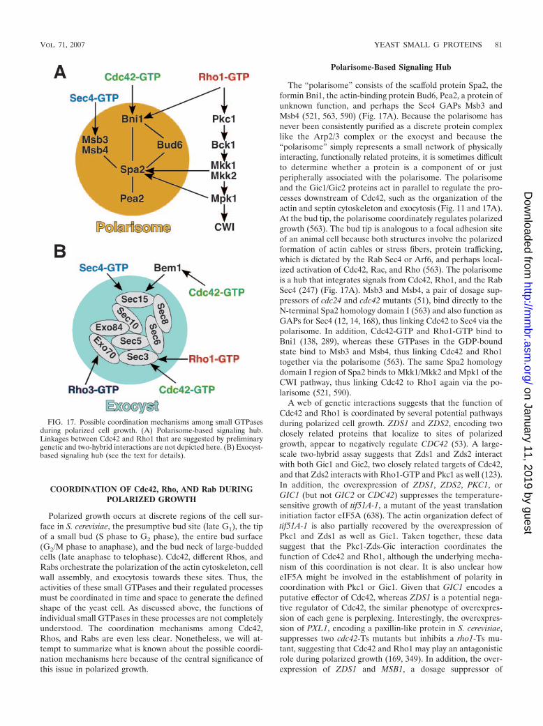
COORDINATION OF Cdc42, Rho, AND Rab DURINGPOLARIZED GROWTH
Polarized growth occurs at discrete regions of the cell sur-face in S. cerevisiae, the presumptive bud site (late G1), the tipof a small bud (S phase to G2 phase), the entire bud surface(G2/M phase to anaphase), and the bud neck of large-buddedcells (late anaphase to telophase). Cdc42, different Rhos, andRabs orchestrate the polarization of the actin cytoskeleton, cellwall assembly, and exocytosis towards these sites. Thus, theactivities of these small GTPases and their regulated processesmust be coordinated in time and space to generate the definedshape of the yeast cell. As discussed above, the functions ofindividual small GTPases in these processes are not completelyunderstood. The coordination mechanisms among Cdc42,Rhos, and Rabs are even less clear. Nonetheless, we will at-tempt to summarize what is known about the possible coordi-nation mechanisms here because of the central significance ofthis issue in polarized growth.
Polarisome-Based Signaling Hub
The “polarisome” consists of the scaffold protein Spa2, theformin Bni1, the actin-binding protein Bud6, Pea2, a protein ofunknown function, and perhaps the Sec4 GAPs Msb3 andMsb4 (521, 563, 590) (Fig. 17A). Because the polarisome hasnever been consistently purified as a discrete protein complexlike the Arp2/3 complex or the exocyst and because the“polarisome” simply represents a small network of physicallyinteracting, functionally related proteins, it is sometimes difficultto determine whether a protein is a component of or justperipherally associated with the polarisome. The polarisomeand the Gic1/Gic2 proteins act in parallel to regulate the pro-cesses downstream of Cdc42, such as the organization of theactin and septin cytoskeleton and exocytosis (Fig. 11 and 17A).At the bud tip, the polarisome coordinately regulates polarizedgrowth (563). The bud tip is analogous to a focal adhesion siteof an animal cell because both structures involve the polarizedformation of actin cables or stress fibers, protein trafficking,which is dictated by the Rab Sec4 or Arf6, and perhaps local-ized activation of Cdc42, Rac, and Rho (563). The polarisomeis a hub that integrates signals from Cdc42, Rho1, and the RabSec4 (247) (Fig. 17A). Msb3 and Msb4, a pair of dosage sup-pressors of cdc24 and cdc42 mutants (51), bind directly to theN-terminal Spa2 homology domain I (563) and also function asGAPs for Sec4 (12, 14, 168), thus linking Cdc42 to Sec4 via thepolarisome. In addition, Cdc42-GTP and Rho1-GTP bind toBni1 (138, 289), whereas these GTPases in the GDP-boundstate bind to Msb3 and Msb4, thus linking Cdc42 and Rho1together via the polarisome (563). The same Spa2 homologydomain I region of Spa2 binds to Mkk1/Mkk2 and Mpk1 of theCWI pathway, thus linking Cdc42 to Rho1 again via the po-larisome (521, 590).
A web of genetic interactions suggests that the function ofCdc42 and Rho1 is coordinated by several potential pathwaysduring polarized cell growth. ZDS1 and ZDS2, encoding twoclosely related proteins that localize to sites of polarizedgrowth, appear to negatively regulate CDC42 (53). A large-scale two-hybrid assay suggests that Zds1 and Zds2 interactwith both Gic1 and Gic2, two closely related targets of Cdc42,and that Zds2 interacts with Rho1-GTP and Pkc1 as well (123).In addition, the overexpression of ZDS1, ZDS2, PKC1, orGIC1 (but not GIC2 or CDC42) suppresses the temperature-sensitive growth of tif51A-1, a mutant of the yeast translationinitiation factor eIF5A (638). The actin organization defect oftif51A-1 is also partially recovered by the overexpression ofPkc1 and Zds1 as well as Gic1. Taken together, these datasuggest that the Pkc1-Zds-Gic interaction coordinates thefunction of Cdc42 and Rho1, although the underlying mecha-nism of this coordination is not clear. It is also unclear howeIF5A might be involved in the establishment of polarity incoordination with Pkc1 or Gic1. Given that GIC1 encodes aputative effector of Cdc42, whereas ZDS1 is a potential nega-tive regulator of Cdc42, the similar phenotype of overexpres-sion of each gene is perplexing. Interestingly, the overexpres-sion of PXL1, encoding a paxillin-like protein in S. cerevisiae,suppresses two cdc42-Ts mutants but inhibits a rho1-Ts mu-tant, suggesting that Cdc42 and Rho1 may play an antagonisticrole during polarized growth (169, 349). In addition, the over-expression of ZDS1 and MSB1, a dosage suppressor of
FIG. 17. Possible coordination mechanisms among small GTPasesduring polarized cell growth. (A) Polarisome-based signaling hub.Linkages between Cdc42 and Rho1 that are suggested by preliminarygenetic and two-hybrid interactions are not depicted here. (B) Exocyst-based signaling hub (see the text for details).
VOL. 71, 2007 YEAST SMALL G PROTEINS 81
on January 11, 2019 by guesthttp://m
mbr.asm
.org/D
ownloaded from

cdc42-Ts mutants (45, 169), suppresses the temperature-sensi-tive growth of a glucan synthase mutant, fks2� fks1-Ts (514).Several genetic screens for genes that functionally interact withCDC42 and CDC24 led to the identification of BEM4 (348),whose gene product interacts with multiple small GTPasesincluding Rsr1, Cdc42, Rho1, Rho2, and Rho4 by two-hybridassays (123, 348). Again, these genetic data suggest multiplecross talks between Cdc42 and Rho1, but the mechanistic de-tails require further investigation.
Exocyst-Based Signaling Hub
The development of cell polarity is ultimately to establishdistinct cellular domains or cellular asymmetry, which requirespolarized secretion. The exocyst, an effector of Sec4 that con-sists of eight subunits (Sec3, Sec5, Sec6, Sec8, Sec10, Sec15,Exo70, and Exo84), plays an essential role in the tethering ofthe post-Golgi vesicles (199, 419, 544). The current idea is thatSec4, the Rab GTPase that is essential for protein traffickingfrom the Golgi apparatus to the plasma membrane, is requiredfor vesicle transport and tethering and, indirectly, for the as-sembly of SNARE complexes leading to membrane fusion(199, 419). It has been proposed that Sec4 regulates exocytosisvia two coordinated pathways (195). In one pathway, Sec4,along with its GEF, Sec2, and an incomplete version of theexocyst, is associated with the secretory vesicles and is trans-ported towards sites of polarized growth via the actin cablesand the type V myosin Myo2. Upon the arrival of the vesiclesat their destination at the cell cortex, a complete exocyst com-plex is formed by joining the vesicle-associated and the plasmamembrane-associated subunits of the exocyst (63), leading tovesicle tethering. Sec1 then mediates the binding of the exocystto the assembled SNAREs by interacting with both of them(77, 612), leading to membrane fusion. In a parallel pathway,Sec4 interacts with another effector, Sro7 (and perhaps with itsparalog, Sro77), which belongs to the lethal giant larva (lgl)tumor suppressor family (266, 301, 309), which in turn interactswith the target SNARE Sec9 (309) to somehow promote thefunction of SNARE complexes. These two pathways also talkto each other via direct interactions between Exo84 and Sro7/Sro77 (642). This hypothesis may explain complex geneticinteractions observed among the components depicted inFig. 17B.
The exocyst defines another hub that integrates the signalsfrom many small GTPases during polarized growth (Fig. 17B).The Sec4-Sec15 interaction is thought to promote the forma-tion and/or function of the exocyst (63, 198). Sec3 interactswith both Rho1 (200) and Cdc42 (641) in a competitive man-ner, and these interactions provide temporally regulated po-larization signals to the exocyst. Another component of theexocyst, Exo70, interacts with Rho3 (6, 474, 482). This inter-action is thought to positively regulate exocytosis, but the un-derlying mechanism remains unclear. In addition, Sec15 inter-acts with Bem1 (123, 155, 196), which interacts with Cdc42 andseveral other proteins involved in polarity establishment (58).The Sec15-Bem1 interaction is required for the efficient tar-geting of Sec15, particularly at an early stage of budding (155).Thus, the exocyst coordinates the signaling from Cdc42, Rho1,Rho3, and Sec4 during polarized growth. As in yeast, Rab andRho family proteins have also been found to interact with the
exocyst in mammalian cells (419), suggesting that the regula-tion of the secretion machinery by small GTPases is evolution-arily conserved. The challenge now is to figure out how allthese small GTPases regulate the exocyst at the biochemicallevel.
Cdc42 and Rho3 are also linked by two structurally relatedproteins, Boi1 and Boi2, both of which carry an SH3 domain attheir N termini and a PH domain at their C termini (47, 368).It is not clear whether this Cdc42-Rho3 communication occursthrough the exocyst directly, a point worthy of further investi-gation. Both Boi1 and Boi2 interact with the second SH3domain of Bem1 (47), and the N-terminal region containingthe first SH3 domain of Bem1 interacts with Sec15 (155),suggesting that the Boi proteins are linked to the exocyst viathe Sec15-Bem1 interaction. Like other polarity proteins, Boi1and Boi2 localize to the presumptive bud site and to the budcortex early in the cell cycle and to the bud neck in lateanaphase (206, 417). The PH domain of Boi1 is responsible forthe bud cortex localization, whereas the SH3 domain is respon-sible for the bud neck localization (206). The deletion of eitherBOI1 or BOI2 does not cause any detectable phenotype, butthe deletion of both genes causes temperature-sensitivegrowth, with many cells arrested as large, round mother cellswith or without a bud, and the double mutant cells are proneto lysis even at the permissive temperature. Overexpression ofthe PH domain-containing region of Boi1 dramatically de-creases cell growth, which is suppressed by the overexpressionof Cdc42, and Cdc42 interacts with this region of Boi1 by atwo-hybrid assay. In addition, multicopy RHO3 effectively sup-presses the temperature-sensitive growth of boi1� boi2� cells(47, 368). Together, these data suggest that Cdc42 and Rho3are functionally linked via Boi1 and Boi2, although the under-lying mechanism remains unknown.
UNIFYING CONCEPTS IN CELL POLARIZATIONSIN DIFFERENT ORGANISMS
So far, we have discussed the key players in polarity devel-opment in S. cerevisiae. Comparative studies of cell polariza-tion in multiple organisms have indicated that the coremachinery involved in the establishment and maintenance ofcell polarity is conserved from yeast to humans (128, 407).Here, we outline a few key concepts for understanding cellpolarization and the central roles of small GTPases in polaritydevelopment in diverse organisms.
Global and Local Cell Polarity
Global cell polarity (the mother-bud polarity) in S. cerevisiaeis determined by intrinsic signals that direct polarized actinorganization, which leads to polarized secretion for budgrowth. Such polarity is maintained until late anaphase ortelophase, when the growth machinery is redirected to the budneck to promote cytokinesis and cell separation (Fig. 9) (315,457). Local polarity (the polarity within the bud) is a result ofthe fine-tuning of the global polarity by several factors, such asthe polarisome in response to cell cycle signals, and is respon-sible for the concrete bud shape (563).
In filamentous fungi, polarized growth is established duringgerm tube initiation from a spore and also during hyphal
82 PARK AND BI MICROBIOL. MOL. BIOL. REV.
on January 11, 2019 by guesthttp://m
mbr.asm
.org/D
ownloaded from

branching (212, 610). Sustained polarized growth at these sitesresults in the elongation of the germ tube or the growinghyphae. The major elements involved in polarized growth infilamentous fungi are similar to those in S. cerevisiae (212, 450,609, 610): the conserved, small GTPase-based signaling net-works required for polarity establishment (212, 609); the po-larized actin cytoskeleton (33, 226, 471, 579), which presum-ably guides secretory vesicles carrying cell wall synthetic anddigestive enzymes to the growth sites; and the “Spitzenkorper”(apical body in German) (179), which may function as a vesiclesupply center near the hyphal tip to control the fine shape ofthe tip (176, 464). The conserved polarity establishment pro-teins are centered on the Cdc42 GTPase module (212, 609).The polarized actin cables (sometimes called actin fibers orarrays in filamentous fungi) are thought to direct vesicle trans-port from the cell body to the apical region of the hypha (33)and are thus responsible for the global polarity of the hypha.As in S. cerevisiae, vesicles carrying cell wall synthetic anddigestive enzymes are delivered to the apex of a hypha, makingthe hyphal tip the most active area in cell wall remodeling(529). Local cell polarity, which shapes the hyphal tip, is de-termined by the mode of cell wall deposition and cross-linkingat the apical region, which creates a favorite site for turgorpressure to push the plasma membrane against an elastic wallat the hyphal tip. The turgor pressure may also drive thevesicles from the Spitzenkorper to the cell surface.
Despite the similarities of the major elements involved inpolarity development in yeast and filamentous fungi, there areseveral key differences between bud and hyphal morphogene-sis. First, cell cycle control of polarized growth is important inS. cerevisiae (315, 316) but not in filamentous fungi such asAshbya gossypii (a plant pathogen whose genome is closelyrelated to that of S. cerevisiae) (450, 609, 610). Second, the roleof microtubules is not apparent in polarized growth in S. cer-evisiae (9) and even in hyphal elongation in C. albicans (631).In contrast, microtubules and/or motors such as dynein andkinesin seem to be important in the morphogenesis of filamen-tous fungi (118, 243, 511–513). Microtubules modulate polar-ized growth in terms of the hyphal elongation rate and growthdirection either by directly participating in vesicle transport(511) or by transporting some polarity factors to the hyphal tipto influence actin assembly, as was also shown for the fissionyeast Schizosaccharomyces pombe (145, 185, 363, 366). Finally,most of the cortical markers for bud site selection in S. cerevi-siae are either missing or poorly conserved in most filamentousfungi (212), suggesting that the mechanisms for selecting theaxis of cell polarization differ significantly between S. cerevisiaeand filamentous fungi.
Local Activation and Global Inhibition
All polarized cells, including budding yeast and human neu-trophils, must develop mechanisms by which cell polarity isrestricted to a single cortical site, while the rest of the cellsurface is prevented from becoming additional sites of cellpolarization. Although there are several possible explanations,the singularity of a polarity axis can be best explained by thehypothesis of “local activation and global inhibition,” a conceptthat was extensively used to explain the chemotactic behaviorsof human neutrophils and the social amoeba Dictyostelium
(239, 379, 433). The essence of the hypothesis is that localactivation at the leading edge is coupled with delivery-basedpositive feedback to amplify the activating signal. At the sametime, a global inhibitor in the cell prevents other portions ofthe cell surface from becoming another leading edge. Thecombined actions of the localized, amplified positive signal andof the global negative signal lead to a highly polarized cellalong a single anterior-posterior axis. At the molecular level,the local activators at the leading edge in both mammalianneutrophils and Dictyostelium are likely to be phosphoinositi-des such as PIP3 and small GTPases such as Cdc42 (154, 469,620), whereas the global inhibitor is represented, at least inpart, by PTEN (phosphatase and tensin homolog), a phos-phatase that dephosphorylates protein and phosphoinositidesincluding PIP3 (321, 377, 378, 624). According to the sameidea, the local activators for polarized growth in S. cerevisiaeare Cdc42 and the proteins involved in its polarization, whilethe global inhibitor remains to be identified. It is interestingthat compartmentalized activators and inhibitors, which can beviewed as a modified version of the local activation and globalinhibition model, are involved in the Cdc42-regulated mitoticexit network (Fig. 12).
Self-Organization
Self-organization has emerged as a central theme in cellbiology. It can be defined simply as the capacity of a macro-molecular complex or organelle to organize its own structureusing a limited number of interacting components (385). Self-organized structures are in a constant state of nonequilibriumand exhibit oscillatory behaviors at the expense of energy.Thus, self-organization is intrinsically dynamic and endows bi-ological systems with great flexibility and robustness (233, 385).Self-organization has been analyzed extensively in cytoskeletonand motor proteins. Microtubules and actin filaments, togetherwith their motor proteins such as kinesins and myosins, canself-organize into dynamic macrostructures such as the mitoticspindle and the actomyosin contractile apparatus, which drivenuclear division, cell migration, and cytokinesis (386, 405, 489).Self-organization is also a key feature in the selection of celldivision sites in Escherichia coli (233) and in the establishmentof cell polarity in mammalian chemotactic cells (624).
The establishment and maintenance of cell polarity in S.cerevisiae likely involve self-organization. As discussed in thesections above, activated Cdc42 can spontaneously polarize atthe cell cortex in the absence of temporal and spatial cues (84,196, 604). The major role of Cdc42 in polarized cell growth isto direct the formation of a polarized actomyosin system,which has the features of a self-organized structure and isinvolved in positive feedback to reinforce Cdc42 polarization(460, 604, 605). Thus, the minimal polarity machinery in S.cerevisiae could include two self-organizing structures, a local-ized Cdc42-based signaling cap at the bud tip and a globalactomyosin-based transport system that interprets and ampli-fies the polarization signal from the Cdc42-based cap. Thevalidity of this hypothesis requires extensive experimental in-vestigation and theoretical modeling.
VOL. 71, 2007 YEAST SMALL G PROTEINS 83
on January 11, 2019 by guesthttp://m
mbr.asm
.org/D
ownloaded from

CONCLUDING REMARKS
Substantial progress in deciphering the molecular basis ofcell polarity in budding yeast has been made. Key signalingpathways that regulate polarization during the mitotic cell cy-cle and during mating have been identified and extensivelycharacterized. These studies have uncovered the central rolesof small GTPases in the development of cell polarity. GTPaseshave numerous molecular interactions with effectors and reg-ulators and can be involved in multiple distinct signaling path-ways. Some of the machinery is specific to yeast, but thegeneral principles underlying cell polarity and the core com-ponents in the signaling pathways appear to be highly con-served throughout evolution. An understanding of the spatialand temporal control of the GTPases and their regulators inyeast is thus undoubtedly relevant to other eukaryotes. Thereare, however, many unanswered questions, as we tried to high-light in each section. One important, but still poorly under-stood, aspect is the regulation of each component, particularlyregulators of GTPases such as GEFs and GAPs, involved inpolarity development. Another important aspect is to under-stand how different GTPases are coordinated at the molecularlevel during polarized cell growth. The recent development ofnew technologies in genomics and proteomics and of fluores-cence-based technology has provided a better understanding ofoverall as well as detailed pictures of cellular networks con-trolling polarity development. The major challenges are tointegrate all the fragmented information into cohesive molec-ular pathways that can be easily understood in terms of cellphysiology and function and to determine how these pathwaysare wired into an elegant machinery that operates with suchtremendous fidelity and flexibility during the development ofcell polarity.
ACKNOWLEDGMENTS
We acknowledge many contributions from the members of the Parkand Bi laboratories. We thank Wei Guo for valuable discussions re-garding exocytosis and E. Oakley for careful editing. We are alsograteful to the anonymous reviewers for many insightful comments.We apologize for not citing all relevant work because of the broadscope and the space limitation of this review.
Our research has been supported by National Institutes of Healthgrants (GM56997 and GM76375) and an American Heart Associationgrant (0555303B) to H.-O.P. and by a National Institutes of Healthgrant (GM59216) and an American Cancer Society grant (RSG-02-039-05-CSM) to E.B.
REFERENCES
1. Aalto, M. K., H. Ronne, and S. Keranen. 1993. Yeast syntaxins Sso1p andSso2p belong to a family of related membrane proteins that function invesicular transport. EMBO J. 12:4095–4104.
2. Abe, M., H. Qadota, A. Hirata, and Y. Ohya. 2003. Lack of GTP-boundRho1p in secretory vesicles of Saccharomyces cerevisiae. J. Cell Biol. 162:85–97.
3. Adam, J. C., J. R. Pringle, and M. Peifer. 2000. Evidence for functionaldifferentiation among Drosophila septins in cytokinesis and cellularization.Mol. Biol. Cell 11:3123–3135.
4. Adames, N., K. Blundell, M. N. Ashby, and C. Boone. 1995. Role of yeastinsulin-degrading enzyme homologs in propheromone processing and budsite selection. Science 270:464–467.
5. Adamo, J. E., J. J. Moskow, A. S. Gladfelter, D. Viterbo, D. J. Lew, and P. J.Brennwald. 2001. Yeast Cdc42 functions at a late step in exocytosis, spe-cifically during polarized growth of the emerging bud. J. Cell Biol. 155:581–592.
6. Adamo, J. E., G. Rossi, and P. Brennwald. 1999. The Rho GTPase Rho3has a direct role in exocytosis that is distinct from its role in actin polarity.Mol. Biol. Cell 10:4121–4133.
7. Adams, A. E., D. Botstein, and D. G. Drubin. 1991. Requirement of yeastfimbrin for actin organization and morphogenesis in vivo. Nature 354:404–408.
8. Adams, A. E., D. I. Johnson, R. M. Longnecker, B. F. Sloat, and J. R.Pringle. 1990. CDC42 and CDC43, two additional genes involved in bud-ding and the establishment of cell polarity in the yeast Saccharomycescerevisiae. J. Cell Biol. 111:131–142.
9. Adams, A. E. M., and J. R. Pringle. 1984. Relationship of actin and tubulindistribution to bud growth in wild-type and morphogenetic-mutant Saccha-romyces cerevisiae. J. Cell Biol. 98:934–945.
10. Aguilar, R. C., S. A. Longhi, J. D. Shaw, L.-Y. Yeh, S. Kim, A. Schon, E.Freire, A. Hsu, W. K. McCormick, H. A. Watson, and B. Wendland. 2006.Epsin N-terminal homology domains perform an essential function regu-lating Cdc42 through binding Cdc42 GTPase-activating proteins. Proc.Natl. Acad. Sci. USA 103:4116–4121.
11. Aguilar, R. C., H. A. Watson, and B. Wendland. 2003. The yeast epsin Ent1is recruited to membranes through multiple independent interactions.J. Biol. Chem. 278:10737–10743.
12. Albert, S., and D. Gallwitz. 2000. Msb4p, a protein involved in Cdc42p-dependent organization of the actin cytoskeleton, is a Ypt/Rab-specificGAP. Biol. Chem. 381:453–456.
13. Albert, S., and D. Gallwitz. 1999. Two new members of a family of Ypt/RabGTPase activating proteins. Promiscuity of substrate recognition. J. Biol.Chem. 274:33186–33189.
14. Albert, S., E. Will, and D. Gallwitz. 1999. Identification of the catalyticdomains and their functionally critical arginine residues of two yeastGTPase-activating proteins specific for Ypt/Rab transport GTPases.EMBO J. 18:5216–5225.
15. Alberts, A. S. 2002. Diaphanous-related formin homology proteins. Curr.Biol. 12:R796.
16. Alberts, A. S. 2001. Identification of a carboxyl-terminal diaphanous-relatedformin homology protein autoregulatory domain. J. Biol. Chem. 276:2824–2830.
17. Alory, C., and W. E. Balch. 2001. Organization of the Rab-GDI/CHMsuperfamily: the functional basis for choroideremia disease. Traffic 2:532–543.
18. Amatruda, J. F., J. F. Cannon, K. Tatchell, C. Hug, and J. A. Cooper. 1990.Disruption of the actin cytoskeleton in yeast capping protein mutants.Nature 344:352–354.
19. Amatruda, J. F., and J. A. Cooper. 1992. Purification, characterization, andimmunofluorescence localization of Saccharomyces cerevisiae capping pro-tein. J. Cell Biol. 117:1067–1076.
20. Amberg, D. C., J. E. Zahner, J. W. Mulholland, J. R. Pringle, and D.Botstein. 1997. Aip3p/Bud6p, a yeast actin-interacting protein that is in-volved in morphogenesis and the selection of bipolar budding sites. Mol.Biol. Cell 8:729–753.
21. An, H., J. L. Morrell, J. L. Jennings, A. J. Link, and K. L. Gould. 2004.Requirements of fission yeast septins for complex formation, localization,and function. Mol. Biol. Cell 15:5551–5564.
22. Aronov, S., and J. E. Gerst. 2004. Involvement of the late secretory pathwayin actin regulation and mRNA transport in yeast. J. Biol. Chem. 279:36962–36971.
23. Asakura, T., T. Sasaki, F. Nagano, A. Satoh, H. Obaishi, H. Nishioka, H.Imamura, K. Hotta, K. Tanaka, H. Nakanishi, and Y. Takai. 1998. Isolationand characterization of a novel actin filament-binding protein from Sac-charomyces cerevisiae. Oncogene 16:121–130.
24. Ash, J., C. Wu, R. Larocque, M. Jamal, W. Stevens, M. Osborne, D. Y.Thomas, and M. Whiteway. 2003. Genetic analysis of the interface betweenCdc42p and the CRIB domain of Ste20p in Saccharomyces cerevisiae. Ge-netics 163:9–20.
25. Audhya, A., and S. D. Emr. 2003. Regulation of PI4,5P2 synthesis bynuclear-cytoplasmic shuttling of the Mss4 lipid kinase. EMBO J. 22:4223–4236.
26. Audhya, A., and S. D. Emr. 2002. Stt4 PI 4-kinase localizes to the plasmamembrane and functions in the Pkc1-mediated MAP kinase cascade. Dev.Cell 2:593–605.
27. Audhya, A., M. Foti, and S. D. Emr. 2000. Distinct roles for the yeastphosphatidylinositol 4-kinases, Stt4p and Pik1p, in secretion, cell growth,and organelle membrane dynamics. Mol. Biol. Cell 11:2673–2689.
28. Audhya, A., R. Loewith, A. B. Parsons, L. Gao, M. Tabuchi, H. Zhou, C.Boone, M. N. Hall, and S. D. Emr. 2004. Genome-wide lethality screenidentifies new PI4,5P2 effectors that regulate the actin cytoskeleton. EMBOJ. 23:3747–3757.
29. Ayscough, K. R., and D. G. Drubin. 1998. A role for the yeast actin cy-toskeleton in pheromone receptor clustering and signalling. Curr. Biol.8:927–930.
30. Ayscough, K. R., J. Stryker, N. Pokala, M. Sanders, P. Crews, and D. G.Drubin. 1997. High rates of actin filament turnover in budding yeast androles for actin in establishment and maintenance of cell polarity revealedusing the actin inhibitor latrunculin-A. J. Cell Biol. 137:399–416.
31. Azzam, R., S. L. Chen, W. Shou, A. S. Mah, G. Alexandru, K. Nasmyth,R. S. Annan, S. A. Carr, and R. J. Deshaies. 2004. Phosphorylation by cyclin
84 PARK AND BI MICROBIOL. MOL. BIOL. REV.
on January 11, 2019 by guesthttp://m
mbr.asm
.org/D
ownloaded from

B-Cdk underlies release of mitotic exit activator Cdc14 from the nucleolus.Science 305:516–519.
32. Bach, S., O. Bouchat, D. Portetelle, and M. Vandenbol. 2000. Co-deletionof the MSB3 and MSB4 coding regions affects bipolar budding and perturbsthe organization of the actin cytoskeleton. Yeast 16:1015–1023.
33. Bachewich, C., and I. B. Heath. 1998. Radial F-actin arrays precede newhypha formation in Saprolegnia: implications for establishing polar growthand regulating tip morphogenesis. J. Cell Sci. 111:2005–2016.
34. Balasubramanian, M. K., E. Bi, and M. Glotzer. 2004. Comparative anal-ysis of cytokinesis in budding yeast, fission yeast and animal cells. Curr. Biol.14:R806–R818.
35. Barale, S., D. McCusker, and R. A. Arkowitz. 2006. Cdc42p GDP/GTPcycling is necessary for efficient cell fusion during yeast mating. Mol. Biol.Cell 17:2824–2838.
36. Barale, S., D. McCusker, and R. A. Arkowitz. 2004. The exchange factorCdc24 is required for cell fusion during yeast mating. Eukaryot. Cell3:1049–1061.
37. Bardin, A. J., R. Visintin, and A. Amon. 2000. A mechanism for couplingexit from mitosis to partitioning of the nucleus. Cell 102:21–31.
38. Barral, Y., V. Mermall, M. S. Mooseker, and M. Snyder. 2000. Compart-mentalization of the cell cortex by septins is required for maintenance ofcell polarity in yeast. Mol. Cell 5:841–851.
39. Barral, Y., M. Parra, S. Bidlingmaier, and M. Snyder. 1999. Nim1-relatedkinases coordinate cell cycle progression with the organization of the pe-ripheral cytoskeleton in yeast. Genes Dev. 13:176–187.
40. Beach, D. L., J. Thibodeaux, P. Maddox, E. Yeh, and K. Bloom. 2000. Therole of the proteins Kar9 and Myo2 in orienting the mitotic spindle ofbudding yeast. Curr. Biol. 10:1497–1506.
41. Beites, C. L., K. A. Campbell, and W. S. Trimble. 2005. The septin Sept5/CDCrel-1 competes with alpha-SNAP for binding to the SNARE complex.Biochem. J. 385:347–353.
42. Beites, C. L., H. Xie, R. Bowser, and W. S. Trimble. 1999. The septinCDCrel-1 binds syntaxin and inhibits exocytosis. Nat. Neurosci. 2:434–439.
43. Bembenek, J., J. Kang, C. Kurischko, B. Li, J. R. Raab, K. D. Belanger,F. C. Luca, and H. Yu. 2005. Crm1-mediated nuclear export of Cdc14 isrequired for the completion of cytokinesis in budding yeast. Cell Cycle4:961–971.
44. Bender, A. 1993. Genetic evidence for the roles of the bud-site-selectiongenes BUD5 and BUD2 in control of the Rsr1p (Bud1p) GTPase in yeast.Proc. Natl. Acad. Sci. USA 90:9926–9929.
45. Bender, A., and J. R. Pringle. 1989. Multicopy suppression of the cdc24budding defect in yeast by CDC42 and three newly identified genes includ-ing the ras-related gene RSR1. Proc. Natl. Acad. Sci. USA 86:9976–9980.
46. Bender, A., and J. R. Pringle. 1991. Use of a screen for synthetic lethal andmulticopy suppressee mutants to identify two new genes involved in mor-phogenesis in Saccharomyces cerevisiae. Mol. Cell. Biol. 11:1295–1305.
47. Bender, L., H. S. Lo, H. Lee, V. Kokojan, V. Peterson, and A. Bender. 1996.Associations among PH and SH3 domain-containing proteins and Rho-typeGTPases in yeast. J. Cell Biol. 133:879–894.
48. Benton, B., A. H. Tinkelenberg, D. Jean, S. D. Plump, and F. R. Cross. 1993.Genetic analysis of Cln/Cdc28 regulation of cell morphogenesis in buddingyeast. EMBO J. 12:5267–5275.
49. Berlin, A., A. Paoletti, and F. Chang. 2003. Mid2p stabilizes septin ringsduring cytokinesis in fission yeast. J. Cell Biol. 160:1083–1092.
50. Bi, E. 2001. Cytokinesis in budding yeast: the relationship between acto-myosin ring function and septum formation. Cell Struct. Funct. 26:529–537.
51. Bi, E., J. B. Chiavetta, H. Chen, G. C. Chen, C. S. Chan, and J. R. Pringle.2000. Identification of novel, evolutionarily conserved Cdc42p-interactingproteins and of redundant pathways linking Cdc24p and Cdc42p to actinpolarization in yeast. Mol. Biol. Cell 11:773–793.
52. Bi, E., P. Maddox, D. J. Lew, E. D. Salmon, J. N. McMillan, E. Yeh, andJ. R. Pringle. 1998. Involvement of an actomyosin contractile ring in Sac-charomyces cerevisiae cytokinesis. J. Cell Biol. 142:1301–1312.
53. Bi, E., and J. R. Pringle. 1996. ZDS1 and ZDS2, genes whose products mayregulate Cdc42p in Saccharomyces cerevisiae. Mol. Cell. Biol. 16:5264–5275.
54. Bloecher, A., G. M. Venturi, and K. Tatchell. 2000. Anaphase spindleposition is monitored by the BUB2 checkpoint. Nat. Cell Biol. 2:556–558.
55. Blondel, M., P. M. Alepuz, L. S. Huang, S. Shaham, G. Ammerer, and M.Peter. 1999. Nuclear export of Far1p in response to pheromones requiresthe export receptor Msn5p/Ste21p. Genes Dev. 13:2284–2300.
56. Bobola, N., R. P. Jansen, T. H. Shin, and K. Nasmyth. 1996. Asymmetricaccumulation of Ash1p in postanaphase nuclei depends on a myosin andrestricts yeast mating-type switching to mother cells. Cell 84:699–709.
57. Bon, E., P. Recordon-Navarro, P. Durrens, M. Iwase, E. A. Toh, and M.Aigle. 2000. A network of proteins around Rvs167p and Rvs161p, twoproteins related to the yeast actin cytoskeleton. Yeast 16:1229–1241.
58. Bose, I., J. E. Irazoqui, J. J. Moskow, E. S. Bardes, T. R. Zyla, and D. J.Lew. 2001. Assembly of scaffold-mediated complexes containing Cdc42p,the exchange factor Cdc24p, and the effector Cla4p required for cell cycle-regulated phosphorylation of Cdc24p. J. Biol. Chem. 276:7176–7186.
59. Bosl, W. J., and R. Li. 2005. Mitotic-exit control as an evolved complexsystem. Cell 121:325–333.
60. Bouquin, N., Y. Barral, R. Courbeyrette, M. Blondel, M. Snyder, and C.Mann. 2000. Regulation of cytokinesis by the Elm1 protein kinase in Sac-charomyces cerevisiae. J. Cell Sci. 113:1435–1445.
61. Bourne, H. R., D. A. Sanders, and F. McCormick. 1990. The GTPasesuperfamily: a conserved switch for diverse cell functions. Nature 348:125–132.
62. Bourne, H. R., D. A. Sanders, and F. McCormick. 1991. The GTPasesuperfamily: conserved structure and molecular mechanism. Nature 349:117–126.
63. Boyd, C., T. Hughes, M. Pypaert, and P. Novick. 2004. Vesicles carry mostexocyst subunits to exocytic sites marked by the remaining two subunits,Sec3p and Exo70p. J. Cell Biol. 167:889–901.
64. Breitkreutz, A., and M. Tyers. 2002. MAPK signaling specificity: it takestwo to tango. Trends Cell Biol. 12:254–257.
65. Brennwald, P., B. Kearns, K. Champion, S. Keranen, V. Bankaitis, and P.Novick. 1994. Sec9 is a SNAP-25-like component of a yeast SNARE com-plex that may be the effector of Sec4 function in exocytosis. Cell 79:245–258.
66. Bretscher, A. 2003. Polarized growth and organelle segregation in yeast: thetracks, motors, and receptors. J. Cell Biol. 160:811–816.
67. Brown, J. L., M. Jaquenoud, M. P. Gulli, J. Chant, and M. Peter. 1997.Novel Cdc42-binding proteins Gic1 and Gic2 control cell polarity in yeast.Genes Dev. 11:2972–2982.
68. Burbelo, P. D., D. Drechsel, and A. Hall. 1995. A conserved binding motifdefines numerous candidate target proteins for both Cdc42 and RacGTPases. J. Biol. Chem. 270:29071–29074.
69. Butty, A.-C., N. Perrinjaquet, A. Petit, M. Jaquenoud, J. E. Segall, K.Hofmann, C. Zwahlen, and M. Peter. 2002. A positive feedback loop sta-bilizes the guanine-nucleotide exchange factor Cdc24 at sites of polariza-tion. EMBO J. 21:1565–1576.
70. Butty, A.-C., P. M. Pryciak, L. S. Huang, I. Herskowitz, and M. Peter. 1998.The role of Far1p in linking the heterotrimeric G protein to polarity es-tablishment proteins during yeast mating. Science 282:1511–1516.
71. Byers, B. 1981. Cytology of the yeast life cycle, p. 59–96. In J. R. Broach(ed.), The molecular biology of the yeast Saccharomyces: life cycle andinheritance. Cold Spring Harbor Laboratory Press, Cold Spring Harbor,NY.
72. Byers, B., and L. Goetsch. 1976. A highly ordered ring of membrane-associated filaments in budding yeast. J. Cell Biol. 69:717–721.
73. Byers, B., and L. Goetsch. 1976. Loss of the filamentous ring in cytokinesis-defective mutants of budding yeast. J. Cell Biol. 70:35a.
74. Cabib, E., D. H. Roh, M. Schmidt, L. B. Crotti, and A. Varma. 2001. Theyeast cell wall and septum as paradigms of cell growth and morphogenesis.J. Biol. Chem. 276:19679–19682.
75. Cannon, J. F., and K. Tatchell. 1987. Characterization of Saccharomycescerevisiae genes encoding subunits of cyclic AMP-dependent protein kinase.Mol. Cell. Biol. 7:2653–2663.
76. Carlsson, A. E., A. D. Shah, D. Elking, T. S. Karpova, and J. A. Cooper.2002. Quantitative analysis of actin patch movement in yeast. Biophys. J.82:2333–2343.
77. Carr, C. M., E. Grote, M. Munson, F. M. Hughson, and P. J. Novick. 1999.Sec1p binds to SNARE complexes and concentrates at sites of secretion.J. Cell Biol. 146:333–344.
78. Casamayor, A., and M. Snyder. 2003. Molecular dissection of a yeast septin:distinct domains are required for septin interaction, localization, and func-tion. Mol. Cell. Biol. 23:2762–2777.
79. Castillon, G. A., N. R. Adames, C. H. Rosello, H. S. Seidel, M. S. Longtine,J. A. Cooper, and R. A. Heil-Chapdelaine. 2003. Septins have a dual role incontrolling mitotic exit in budding yeast. Curr. Biol. 13:654–658.
80. Catlett, N. L., J. E. Duex, F. Tang, and L. S. Weisman. 2000. Two distinctregions in a yeast myosin-V tail domain are required for the movement ofdifferent cargoes. J. Cell Biol. 150:513–526.
81. Catlett, N. L., and L. S. Weisman. 2000. Divide and multiply: organellepartitioning in yeast. Curr. Opin. Cell Biol. 12:509–516.
82. Catlett, N. L., and L. S. Weisman. 1998. The terminal tail region of a yeastmyosin-V mediates its attachment to vacuole membranes and sites of po-larized growth. Proc. Natl. Acad. Sci. USA 95:14799–14804.
83. Caviston, J. P., M. Longtine, J. R. Pringle, and E. Bi. 2003. The role ofCdc42p GTPase-activating proteins in assembly of the septin ring in yeast.Mol. Biol. Cell 14:4051–4066.
84. Caviston, J. P., S. E. Tcheperegine, and E. Bi. 2002. Singularity in budding:a role for the evolutionarily conserved small GTPase Cdc42p. Proc. Natl.Acad. Sci. USA 99:12185–12190.
85. Chant, J. 1999. Cell polarity in yeast. Annu. Rev. Cell Dev. Biol. 15:365–391.
86. Chant, J., K. Corrado, J. R. Pringle, and I. Herskowitz. 1991. Yeast BUD5,encoding a putative GDP-GTP exchange factor, is necessary for bud siteselection and interacts with bud formation gene BEM1. Cell 65:1213–1224.
87. Chant, J., and I. Herskowitz. 1991. Genetic control of bud site selection inyeast by a set of gene products that constitute a morphogenetic pathway.Cell 65:1203–1212.
88. Chant, J., M. Mischke, E. Mitchell, I. Herskowitz, and J. R. Pringle. 1995.
VOL. 71, 2007 YEAST SMALL G PROTEINS 85
on January 11, 2019 by guesthttp://m
mbr.asm
.org/D
ownloaded from

Role of Bud3p in producing the axial budding pattern of yeast. J. Cell Biol.129:767–778.
89. Chant, J., and J. R. Pringle. 1995. Patterns of bud-site selection in the yeastSaccharomyces cerevisiae. J. Cell Biol. 129:751–765.
90. Chen, G. C., Y. J. Kim, and C. S. Chan. 1997. The Cdc42 GTPase-associ-ated proteins Gic1 and Gic2 are required for polarized cell growth inSaccharomyces cerevisiae. Genes Dev. 11:2958–2971.
91. Chen, G. C., L. Zheng, and C. S. Chan. 1996. The LIM domain-containingDbm1 GTPase-activating protein is required for normal cellular morpho-genesis in Saccharomyces cerevisiae. Mol. Cell. Biol. 16:1376–1390.
92. Chen, J. L., R. V. Fucini, L. Lacomis, H. Erdjument-Bromage, P. Tempst,and M. Stamnes. 2005. Coatomer-bound Cdc42 regulates dynein recruit-ment to COPI vesicles. J. Cell Biol. 169:383–389.
93. Chen, T., T. Hiroko, A. Chaudhuri, F. Inose, M. Lord, S. Tanaka, J. Chant,and A. Fujita. 2000. Multigenerational cortical inheritance of the Rax2protein in orienting polarity and division in yeast. Science 290:1975–1978.
94. Chu, H. M., M. Yun, D. E. Anderson, H. Sage, H. W. Park, and S. A. Endow.2005. Kar3 interaction with Cik1 alters motor structure and function.EMBO J. 24:3214–3223.
95. Chuang, T. H., X. Xu, U. G. Knaus, M. J. Hart, and G. M. Bokoch. 1993.GDP dissociation inhibitor prevents intrinsic and GTPase activating pro-tein-stimulated GTP hydrolysis by the Rac GTP-binding protein. J. Biol.Chem. 268:775–778.
96. Cid, V. J., L. Adamikova, M. Sanchez, M. Molina, and C. Nombela. 2001.Cell cycle control of septin ring dynamics in the budding yeast. Microbiol-ogy 147:1437–1450.
97. Cid, V. J., R. Cenamor, M. Sanchez, and C. Nombela. 1998. A mutation inthe Rho1-GAP-encoding gene BEM2 of Saccharomyces cerevisiae affectsmorphogenesis and cell wall functionality. Microbiology 144:25–36.
98. Cook, J. G., L. Bardwell, and J. Thorner. 1997. Inhibitory and activatingfunctions for MAPK Kss1 in the S. cerevisiae filamentous-growth signallingpathway. Nature 390:85–88.
99. Cooper, J. A., and D. P. Kiehart. 1996. Septins may form a ubiquitousfamily of cytoskeletal filaments. J. Cell Biol. 134:1345–1348.
100. Crouzet, M., M. Urdaci, L. Dulau, and M. Aigle. 1991. Yeast mutantaffected for viability upon nutrient starvation: characterization and cloningof the RVS161 gene. Yeast 7:727–743.
101. Cullen, P. J., W. Sabbagh, Jr., E. Graham, M. M. Irick, E. K. van Olden, C.Neal, J. Delrow, L. Bardwell, and G. F. Sprague, Jr. 2004. A signaling mucinat the head of the Cdc42- and MAPK-dependent filamentous growth path-way in yeast. Genes Dev. 18:1695–1708.
102. Cvrckova, F., C. De Virgilio, E. Manser, J. R. Pringle, and K. Nasmyth.1995. Ste20-like protein kinases are required for normal localization of cellgrowth and for cytokinesis in budding yeast. Genes Dev. 9:1817–1830.
103. Cvrckova, F., and K. Nasmyth. 1993. Yeast G1 cyclins CLN1 and CLN2 anda GAP-like protein have a role in bud formation. EMBO J. 12:5277–5286.
104. D’Aquino, K. E., F. Monje-Casas, J. Paulson, V. Reiser, G. M. Charles, L.Lai, K. M. Shokat, and A. Amon. 2005. The protein kinase Kin4 inhibits exitfrom mitosis in response to spindle position defects. Mol. Cell 19:223–234.
105. de Bettignies, G., D. Thoraval, C. Morel, M. F. Peypouquet, and M.Crouzet. 2001. Overactivation of the protein kinase C-signaling pathwaysuppresses the defects of cells lacking the Rho3/Rho4-GAP Rgd1p in Sac-charomyces cerevisiae. Genetics 159:1435–1448.
106. deHart, A. K., J. D. Schnell, D. A. Allen, J. Y. Tsai, and L. Hicke. 2003.Receptor internalization in yeast requires the Tor2-Rho1 signaling path-way. Mol. Biol. Cell 14:4676–4684.
107. Delley, P. A., and M. N. Hall. 1999. Cell wall stress depolarizes cell growthvia hyperactivation of RHO1. J. Cell Biol. 147:163–174.
108. DeMarini, D. J., A. E. M. Adams, H. Fares, C. De Virgilio, G. Valle, J. S.Chuang, and J. R. Pringle. 1997. A septin-based hierarchy of proteinsrequired for localized deposition of chitin in the Saccharomyces cerevisiaecell wall. J. Cell Biol. 139:75–93.
109. Denis, V., and M. S. Cyert. 2005. Molecular analysis reveals localization ofSaccharomyces cerevisiae protein kinase C to sites of polarized growth andPkc1p targeting to the nucleus and mitotic spindle. Eukaryot. Cell 4:36–45.
110. DerMardirossian, C., and G. M. Bokoch. 2005. GDIs: central regulatorymolecules in Rho GTPase activation. Trends Cell Biol. 15:356–363.
111. Desrivieres, S., F. T. Cooke, P. J. Parker, and M. N. Hall. 1998. MSS4, aphosphatidylinositol-4-phosphate 5-kinase required for organization of theactin cytoskeleton in Saccharomyces cerevisiae. J. Biol. Chem. 273:15787–15793.
112. Dobbelaere, J., and Y. Barral. 2004. Spatial coordination of cytokineticevents by compartmentalization of the cell cortex. Science 305:393–396.
113. Dobbelaere, J., M. S. Gentry, R. L. Hallberg, and Y. Barral. 2003. Phos-phorylation-dependent regulation of septin dynamics during the cell cycle.Dev. Cell 4:345–357.
114. Dohlman, H. G., and J. W. Thorner. 2001. Regulation of G protein-initiatedsignal transduction in yeast: paradigms and principles. Annu. Rev. Bio-chem. 70:703–754.
115. Doignon, F., C. Weinachter, O. Roumanie, and M. Crouzet. 1999. The yeastRgd1p is a GTPase activating protein of the Rho3 and Rho4 proteins.FEBS Lett. 459:458–462.
116. Dong, Y., D. Pruyne, and A. Bretscher. 2003. Formin-dependent actinassembly is regulated by distinct modes of Rho signaling in yeast. J. CellBiol. 161:1081–1092.
117. Dorer, R., C. Boone, T. Kimbrough, J. Kim, and L. H. Hartwell. 1997.Genetic analysis of default mating behavior in Saccharomyces cerevisiae.Genetics 146:39–55.
118. Doshi, P., C. A. Bossie, J. H. Doonan, G. S. May, and N. R. Morris. 1991.Two alpha-tubulin genes of Aspergillus nidulans encode divergent proteins.Mol. Gen. Genet. 225:129–141.
119. Douglas, C. M., F. Foor, J. A. Marrinan, N. Morin, J. B. Nielsen, A. M.Dahl, P. Mazur, W. Baginsky, W. Li, M. El-Sherbeini, J. A. Clemas, S. M.Mandala, B. R. Frommer, and M. B. Kurtz. 1994. The Saccharomycescerevisiae FKS1 (ETG1) gene encodes an integral membrane protein whichis a subunit of 1,3-�-D-glucan synthase. Proc. Natl. Acad. Sci. USA 91:12907–12911.
120. Dovas, A., and J. R. Couchman. 2005. RhoGDI: multiple functions in theregulation of Rho family GTPase activities. Biochem. J. 390:1–9.
121. Doyle, T., and D. Botstein. 1996. Movement of yeast cortical actin cytoskel-eton visualized in vivo. Proc. Natl. Acad. Sci. USA 93:3886–3891.
122. Drees, B., C. Brown, B. G. Barrell, and A. Bretscher. 1995. Tropomyosin isessential in yeast, yet the TPM1 and TPM2 products perform distinctfunctions. J. Cell Biol. 128:383–392.
123. Drees, B. L., B. Sundin, E. Brazeau, J. P. Caviston, G.-C. Chen, W. Guo,K. G. Kozminski, M. W. Lau, J. J. Moskow, A. Tong, L. R. Schenkman, A.McKenzie III, P. Brennwald, M. Longtine, E. Bi, C. Chan, P. Novick, C.Boone, J. R. Pringle, T. N. Davis, S. Fields, and D. G. Drubin. 2001. Aprotein interaction map for cell polarity development. J. Cell Biol. 154:549–576.
124. Drgonova, J., T. Drgon, K. Tanaka, R. Kollar, G. C. Chen, R. A. Ford, C. S.Chan, Y. Takai, and E. Cabib. 1996. Rho1p, a yeast protein at the interfacebetween cell polarization and morphogenesis. Science 272:277–279.
125. Drogen, F., S. M. O’Rourke, V. M. Stucke, M. Jaquenoud, A. M. Neiman,and M. Peter. 2000. Phosphorylation of the MEKK Ste11p by the PAK-likekinase Ste20p is required for MAP kinase signaling in vivo. Curr. Biol.10:630–639.
126. Drubin, D. 1991. Development of cell polarity in budding yeast. Cell 65:1093–1096.
127. Drubin, D. G., K. G. Miller, and D. Botstein. 1988. Yeast actin-bindingproteins: evidence for a role in morphogenesis. J. Cell Biol. 107:2551–2561.
128. Drubin, D. G., and W. J. Nelson. 1996. Origins of cell polarity. Cell 84:335–344.
129. Duncan, M. C., M. J. Cope, B. L. Goode, B. Wendland, and D. G. Drubin.2001. Yeast Eps15-like endocytic protein, Pan1p, activates the Arp2/3 com-plex. Nat. Cell Biol. 3:687–690.
130. Dunn, T. M., and D. Shortle. 1990. Null alleles of SAC7 suppress temper-ature-sensitive actin mutations in Saccharomyces cerevisiae. Mol. Cell. Biol.10:2308–2314.
131. Dupre, S., D. Urban-Grimal, and R. Haguenauer-Tsapis. 2004. Ubiquitinand endocytic internalization in yeast and animal cells. Biochim. Biophys.Acta 1695:89–111.
132. Eby, J. J., S. P. Holly, F. van Drogen, A. V. Grishin, M. Peter, D. G. Drubin,and K. J. Blumer. 1998. Actin cytoskeleton organization regulated by thePAK family of protein kinases. Curr. Biol. 8:967–970.
133. Eitzen, G., N. Thorngren, and W. Wickner. 2001. Rho1p and Cdc42p actafter Ypt7p to regulate vacuole docking. EMBO J. 20:5650–5656.
134. Elion, E. A. 2000. Pheromone response, mating and cell biology. Curr.Opin. Microbiol. 3:573–581.
135. Engqvist-Goldstein, A. E., and D. G. Drubin. 2003. Actin assembly andendocytosis: from yeast to mammals. Annu. Rev. Cell Dev. Biol. 19:287–332.
136. Erickson, J. W., C. Zhang, R. A. Kahn, T. Evans, and R. A. Cerione. 1996.Mammalian Cdc42 is a brefeldin A-sensitive component of the Golgi ap-paratus. J. Biol. Chem. 271:26850–26854.
137. Etienne-Manneville, S., and A. Hall. 2002. Rho GTPases in cell biology.Nature 420:629–635.
138. Evangelista, M., K. Blundell, M. S. Longtine, C. J. Chow, N. Adames, J. R.Pringle, M. Peter, and C. Boone. 1997. Bni1p, a yeast formin linking Cdc42pand the actin cytoskeleton during polarized morphogenesis. Science 276:118–122.
139. Evangelista, M., B. M. Klebl, A. H. Tong, B. A. Webb, T. Leeuw, E. Leberer,M. Whiteway, D. Y. Thomas, and C. Boone. 2000. A role for myosin-I inactin assembly through interactions with Vrp1p, Bee1p, and the Arp2/3complex. J. Cell Biol. 148:353–362.
140. Evangelista, M., D. Pruyne, D. C. Amberg, C. Boone, and A. Bretscher.2002. Formins direct Arp2/3-independent actin filament assembly to polar-ize cell growth in yeast. Nat. Cell Biol. 4:32–41.
141. Evangelista, M., S. Zigmond, and C. Boone. 2003. Formins: signaling ef-fectors for assembly and polarization of actin filaments. J. Cell Sci. 116:2603–2611.
142. Fadri, M., A. Daquinag, S. Wang, T. Xue, and J. Kunz. 2005. The pleckstrinhomology domain proteins Slm1 and Slm2 are required for actin cytoskel-
86 PARK AND BI MICROBIOL. MOL. BIOL. REV.
on January 11, 2019 by guesthttp://m
mbr.asm
.org/D
ownloaded from

eton organization in yeast and bind phosphatidylinositol-4,5-bisphosphateand TORC2. Mol. Biol. Cell 16:1883–1900.
143. Farkasovsky, M., P. Herter, B. Voss, and A. Wittinghofer. 2005. Nucleotidebinding and filament assembly of recombinant yeast septin complexes. Biol.Chem. 386:643–656.
144. Faty, M., M. Fink, and Y. Barral. 2002. Septins: a ring to part mother anddaughter. Curr. Genet. 41:123–131.
145. Feierbach, B., F. Verde, and F. Chang. 2004. Regulation of a formin com-plex by the microtubule plus end protein tea1p. J. Cell Biol. 165:697–707.
146. Fernandes, H., O. Roumanie, S. Claret, X. Gatti, D. Thoraval, F. Doignon,and M. Crouzet. 2006. The Rho3 and Rho4 small GTPases interact func-tionally with Wsc1p, a cell surface sensor of the protein kinase C cell-integrity pathway in Saccharomyces cerevisiae. Microbiology 152:695–708.
147. Field, C. M., O. Al-Awar, J. Rosenblatt, M. L. Wong, B. Alberts, and T. J.Mitchison. 1996. A purified Drosophila septin complex forms filaments andexhibits GTPase activity. J. Cell Biol. 133:605–616.
148. Field, C. M., and D. Kellogg. 1999. Septins: cytoskeletal polymers or sig-nalling GTPases? Trends Cell Biol. 9:387–394.
149. Finger, F. P., T. E. Hughes, and P. Novick. 1998. Sec3p is a spatial landmarkfor polarized secretion in budding yeast. Cell 92:559–571.
150. Finger, F. P., and P. Novick. 1997. Sec3p is involved in secretion andmorphogenesis in Saccharomyces cerevisiae. Mol. Biol. Cell 8:647–662.
151. Fitch, P. G., A. E. Gammie, D. J. Lee, V. B. de Candal, and M. D. Rose.2004. Lrg1p is a Rho1 GTPase-activating protein required for efficient cellfusion in yeast. Genetics 168:733–746.
152. Flescher, E. G., K. Madden, and M. Snyder. 1993. Components requiredfor cytokinesis are important for bud site selection in yeast. J. Cell Biol.122:373–386.
153. Ford, S. K., and J. R. Pringle. 1991. Cellular morphogenesis in the Sac-charomyces cerevisiae cell cycle: localization of the CDC11 gene product andthe timing of events at the budding site. Dev. Genet. 12:281–292.
154. Franca-Koh, J., and P. N. Devreotes. 2004. Moving forward: mechanisms ofchemoattractant gradient sensing. Physiology 19:300–308.
155. France, Y. E., C. Boyd, J. Coleman, and P. J. Novick. 2006. The polarity-establishment component Bem1p interacts with the exocyst complexthrough the Sec15p subunit. J. Cell Sci. 119:876–888.
156. Fraschini, R., C. D’Ambrosio, M. Venturetti, G. Lucchini, and S. Piatti.2006. Disappearance of the budding yeast Bub2-Bfa1 complex from themother-bound spindle pole contributes to mitotic exit. J. Cell Biol. 172:335–346.
157. Frazier, J. A., M. L. Wong, M. S. Longtine, J. R. Pringle, M. Mann, T. J.Mitchison, and C. Field. 1998. Polymerization of purified yeast septins:evidence that organized filament arrays may not be required for septinfunction. J. Cell Biol. 143:737–749.
158. Freifelder, D. 1960. Bud position in Saccharomyces cerevisiae. J. Bacteriol.80:567–568.
159. Frenz, L. M., S. E. Lee, D. Fesquet, and L. H. Johnston. 2000. The buddingyeast Dbf2 protein kinase localises to the centrosome and moves to the budneck in late mitosis. J. Cell Sci. 113:3399–3408.
160. Friesen, H., C. Humphries, Y. Ho, O. Schub, K. Colwill, and B. Andrews.2006. Characterization of the yeast amphiphysins Rvs161 and Rvs167 re-veals roles for the Rvs heterodimer in vivo. Mol. Biol. Cell 17:1306–1321.
161. Fucini, R. V., J. L. Chen, C. Sharma, M. M. Kessels, and M. Stamnes. 2002.Golgi vesicle proteins are linked to the assembly of an actin complexdefined by mAbp1. Mol. Biol. Cell 13:621–631.
162. Fujita, A., M. Lord, T. Hiroko, F. Hiroko, T. Chen, C. Oka, Y. Misumi, andJ. Chant. 2004. Rax1, a protein required for the establishment of thebipolar budding pattern in yeast. Gene 327:161–169.
163. Fujita, A., C. Oka, Y. Arikawa, T. Katagai, A. Tonouchi, S. Kuhara, and Y.Misumi. 1994. A yeast gene necessary for bud-site selection encodes aprotein similar to insulin-degrading enzymes. Nature 372:567–570.
164. Fujiwara, T., K. Tanaka, A. Mino, M. Kikyo, K. Takahashi, K. Shimizu,and Y. Takai. 1998. Rho1p-Bni1p-Spa2p interactions: implication in local-ization of Bni1p at the bud site and regulation of the actin cytoskeleton inSaccharomyces cerevisiae. Mol. Biol. Cell 9:1221–1233.
165. Fukumoto, Y., K. Kaibuchi, Y. Hori, H. Fujioka, S. Araki, T. Ueda, A.Kikuchi, and Y. Takai. 1990. Molecular cloning and characterization of anovel type of regulatory protein (GDI) for the rho proteins, ras p21-likesmall GTP-binding proteins. Oncogene 5:1321–1328.
166. Gammie, A. E., V. Brizzio, and M. D. Rose. 1998. Distinct morphologicalphenotypes of cell fusion mutants. Mol. Biol. Cell 9:1395–1410.
167. Gandhi, P. N., R. M. Gibson, X. Tong, J. Miyoshi, Y. Takai, M. Koniecz-kowski, J. R. Sedor, and A. L. Wilson-Delfosse. 2004. An activating mutantof Rac1 that fails to interact with Rho GDP-dissociation inhibitor stimu-lates membrane ruffling in mammalian cells. Biochem. J. 378:409–419.
168. Gao, X. D., S. Albert, S. E. Tcheperegine, C. G. Burd, D. Gallwitz, and E.Bi. 2003. The GAP activity of Msb3p and Msb4p for the Rab GTPase Sec4pis required for efficient exocytosis and actin organization. J. Cell Biol.162:635–646.
169. Gao, X. D., J. P. Caviston, S. E. Tcheperegine, and E. Bi. 2004. Pxl1p, apaxillin-like protein in Saccharomyces cerevisiae, may coordinate Cdc42p
and Rho1p functions during polarized growth. Mol. Biol. Cell 15:3977–3985.
170. Garcia-Ranea, J. A., and A. Valencia. 1998. Distribution and functionaldiversification of the ras superfamily in Saccharomyces cerevisiae. FEBSLett. 434:219–225.
171. Geli, M. I., R. Lombardi, B. Schmelzl, and H. Riezman. 2000. An intactSH3 domain is required for myosin I-induced actin polymerization. EMBOJ. 19:4281–4291.
172. Geli, M. I., and H. Riezman. 1996. Role of type I myosins in receptor-mediated endocytosis in yeast. Science 272:533–535.
173. Geymonat, M., A. Spanos, S. J. Smith, E. Wheatley, K. Rittinger, L. H.Johnston, and S. G. Sedgwick. 2002. Control of mitotic exit in buddingyeast. In vitro regulation of Tem1 GTPase by Bub2 and Bfa1. J. Biol. Chem.277:28439–28445.
174. Gibson, R. M., P. N. Gandhi, X. Tong, J. Miyoshi, Y. Takai, M. Koniecz-kowski, J. R. Sedor, and A. L. Wilson-Delfosse. 2004. An activating mutantof Cdc42 that fails to interact with Rho GDP-dissociation inhibitor localizesto the plasma membrane and mediates actin reorganization. Exp. Cell Res.301:211–222.
175. Gibson, R. M., and A. L. Wilson-Delfosse. 2001. RhoGDI-binding-defectivemutant of Cdc42Hs targets to membranes and activates filopodia formationbut does not cycle with the cytosol of mammalian cells. Biochem. J. 359:285–294.
176. Gierz, G., and S. Bartnicki-Garcia. 2001. A three-dimensional model offungal morphogenesis based on the vesicle supply center concept. J. Theor.Biol. 208:151–164.
177. Gilbert, P. M., and C. G. Burd. 2001. GDP dissociation inhibitor domain IIrequired for Rab GTPase recycling. J. Biol. Chem. 276:8014–8020.
178. Gimeno, C. J., P. O. Ljungdahl, C. A. Styles, and G. R. Fink. 1992. Unipolarcell divisions in the yeast S. cerevisiae lead to filamentous growth: regulationby starvation and RAS. Cell 68:1077–1090.
179. Girbardt, M. 1957. Der Spitzenkorper von Polystictus versicolor (L). Planta50:47–59.
180. Gladfelter, A. S. 2006. Control of filamentous fungal cell shape by septinsand formins. Nat. Rev. Microbiol. 4:223–229.
181. Gladfelter, A. S., I. Bose, T. R. Zyla, E. S. G. Bardes, and D. J. Lew. 2002.Septin ring assembly involves cycles of GTP loading and hydrolysis byCdc42p. J. Cell Biol. 156:315–326.
182. Gladfelter, A. S., L. Kozubowski, T. R. Zyla, and D. J. Lew. 2005. Interplaybetween septin organization, cell cycle and cell shape in yeast. J. Cell Sci.118:1617–1628.
183. Gladfelter, A. S., J. J. Moskow, T. R. Zyla, and D. J. Lew. 2001. Isolationand characterization of effector-loop mutants of CDC42 in yeast. Mol. Biol.Cell 12:1239–1255.
184. Gladfelter, A. S., J. R. Pringle, and D. J. Lew. 2001. The septin cortex at theyeast mother-bud neck. Curr. Opin. Microbiol. 4:681–689.
185. Glynn, J. M., R. J. Lustig, A. Berlin, and F. Chang. 2001. Role of bud6p andtea1p in the interaction between actin and microtubules for the establish-ment of cell polarity in fission yeast. Curr. Biol. 11:836–845.
186. Goeckeler, Z. M., and R. B. Wysolmerski. 1995. Myosin light chain kinase-regulated endothelial cell contraction: the relationship between isometrictension, actin polymerization, and myosin phosphorylation. J. Cell Biol.130:613–627.
187. Goehring, A. S., D. A. Mitchell, A. H. Tong, M. E. Keniry, C. Boone, andG. F. Sprague, Jr. 2003. Synthetic lethal analysis implicates Ste20p, a p21-activated protein kinase, in polarisome activation. Mol. Biol. Cell 14:1501–1516.
188. Goode, B. L., and A. A. Rodal. 2001. Modular complexes that regulate actinassembly in budding yeast. Curr. Opin. Microbiol. 4:703–712.
189. Goode, B. L., A. A. Rodal, G. Barnes, and D. G. Drubin. 2001. Activation ofthe Arp2/3 complex by the actin filament binding protein Abp1p. J. CellBiol. 153:627–634.
190. Goodson, H. V., B. L. Anderson, H. M. Warrick, L. A. Pon, and J. A.Spudich. 1996. Synthetic lethality screen identifies a novel yeast myosin Igene (MYO5): myosin I proteins are required for polarization of the actincytoskeleton. J. Cell Biol. 133:1277–1291.
191. Gottfried, Y., A. Rotem, R. Lotan, H. Steller, and S. Larisch. 2004. Themitochondrial ARTS protein promotes apoptosis through targeting XIAP.EMBO J. 23:1627–1635.
192. Goud, B., A. Salminen, N. C. Walworth, and P. J. Novick. 1988. A GTP-binding protein required for secretion rapidly associates with secretoryvesicles and the plasma membrane in yeast. Cell 53:753–768.
193. Govindan, B., R. Bowser, and P. Novick. 1995. The role of Myo2, a yeastclass V myosin, in vesicular transport. J. Cell Biol. 128:1055–1068.
194. Gray, C. H., V. M. Good, N. K. Tonks, and D. Barford. 2003. The structureof the cell cycle protein Cdc14 reveals a proline-directed protein phos-phatase. EMBO J. 22:3524–3535.
195. Grosshans, B. L., A. Andreeva, A. Gangar, S. Niessen, J. R. Yates III, P.Brennwald, and P. Novick. 2006. The yeast lgl family member Sro7p is aneffector of the secretory Rab GTPase Sec4p. J. Cell Biol. 172:55–66.
196. Gulli, M. P., M. Jaquenoud, Y. Shimada, G. Niederhauser, P. Wiget, andM. Peter. 2000. Phosphorylation of the Cdc42 exchange factor Cdc24 by the
VOL. 71, 2007 YEAST SMALL G PROTEINS 87
on January 11, 2019 by guesthttp://m
mbr.asm
.org/D
ownloaded from

PAK-like kinase Cla4 may regulate polarized growth in yeast. Mol. Cell6:1155–1167.
197. Gulli, M. P., and M. Peter. 2001. Temporal and spatial regulation ofRho-type guanine-nucleotide exchange factors: the yeast perspective.Genes Dev. 15:365–379.
198. Guo, W., D. Roth, C. Walch-Solimena, and P. Novick. 1999. The exocyst isan effector for Sec4p, targeting secretory vesicles to sites of exocytosis.EMBO J. 18:1071–1080.
199. Guo, W., M. Sacher, J. Barrowman, S. Ferro-Novick, and P. Novick. 2000.Protein complexes in transport vesicle targeting. Trends Cell Biol. 10:251–255.
200. Guo, W., F. Tamanoi, and P. Novick. 2001. Spatial regulation of the exocystcomplex by Rho1 GTPase. Nat. Cell Biol. 3:353–360.
201. Haarer, B. K., S. H. Lillie, A. E. Adams, V. Magdolen, W. Bandlow, andS. S. Brown. 1990. Purification of profilin from Saccharomyces cerevisiae andanalysis of profilin-deficient cells. J. Cell Biol. 110:105–114.
202. Haarer, B. K., and J. R. Pringle. 1987. Immunofluorescence localization ofthe Saccharomyces cerevisiae CDC12 gene product to the vicinity of the 10nm filaments in the mother-bud neck. Mol. Cell. Biol. 7:3678–3687.
203. Hall, A. 1998. Rho GTPases and the actin cytoskeleton. Science 279:509–514.
204. Hall, D. D., D. D. Markwardt, F. Parviz, and W. Heideman. 1998. Regu-lation of the Cln3-Cdc28 kinase by cAMP in Saccharomyces cerevisiae.EMBO J. 17:4370–4378.
205. Hall, P. A., and S. E. Russell. 2004. The pathobiology of the septin genefamily. J. Pathol. 204:489–505.
206. Hallett, M. A., H. S. Lo, and A. Bender. 2002. Probing the importance andpotential roles of the binding of the PH-domain protein Boi1 to acidicphospholipids. BMC Cell Biol. 3:16–29.
207. Halme, A., M. Michelitch, E. L. Mitchell, and J. Chant. 1996. Bud10pdirects axial cell polarization in budding yeast and resembles a transmem-brane receptor. Curr. Biol. 6:570–579.
208. Hancock, J. F., and A. Hall. 1993. A novel role for RhoGDI as an inhibitorof GAP proteins. EMBO J. 12:1915–1921.
209. Haney, S. A., and J. R. Broach. 1994. Cdc25p, the guanine nucleotideexchange factor for the ras proteins of Saccharomyces cerevisiae, promotesexchange by stabilizing Ras in a nucleotide-free state. J. Biol. Chem. 269:16541–16548.
210. Harkins, H. A., N. Page, L. R. Schenkman, C. De Virgilio, S. Shaw, H.Bussey, and J. R. Pringle. 2001. Bud8p and Bud9p, proteins that may markthe sites for bipolar budding in yeast. Mol. Biol. Cell 12:2497–2518.
211. Harris, E. S., and H. N. Higgs. 2004. Actin cytoskeleton: formins lead theway. Curr. Biol. 14:R520–R522.
212. Harris, S. D., and M. Momany. 2004. Polarity in filamentous fungi: movingbeyond the yeast paradigm. Fungal Genet. Biol. 41:391–400.
213. Harrison, J. C., E. S. Bardes, Y. Ohya, and D. J. Lew. 2001. A role for thePkc1p/Mpk1p kinase cascade in the morphogenesis checkpoint. Nat. CellBiol. 3:417–420.
214. Hart, M. J., Y. Maru, D. Leonard, O. N. Witte, T. Evans, and R. A. Cerione.1992. A GDP dissociation inhibitor that serves as a GTPase inhibitor for theRas-like protein CDC42Hs. Science 258:812–815.
215. Hartwell, L. H. 1971. Genetic control of the cell division cycle in yeast. IV.Genes controlling bud emergence and cytokinesis. Exp. Cell Res. 69:265–276.
216. Hartwell, L. H., J. Culotti, J. R. Pringle, and B. J. Reid. 1974. Geneticcontrol of the cell division cycle in yeast. Science 183:46–51.
217. Hartwell, L. H., R. K. Mortimer, J. Culotti, and M. Culotti. 1973. Geneticcontrol of the cell division cycle in yeast. V. Genetic analysis of cdc mutants.Genetics 74:267–286.
218. Heinisch, J. J., A. Lorberg, H. P. Schmitz, and J. J. Jacoby. 1999. Theprotein kinase C-mediated MAP kinase pathway involved in the mainte-nance of cellular integrity in Saccharomyces cerevisiae. Mol. Microbiol.32:671–680.
219. Helliwell, S. B., A. Schmidt, Y. Ohya, and M. N. Hall. 1998. The Rho1effector Pkc1, but not Bni1, mediates signalling from Tor2 to the actincytoskeleton. Curr. Biol. 8:1211–1214.
220. Herskowitz, I. 1988. Life cycle of the budding yeast Saccharomyces cerevi-siae. Microbiol. Rev. 52:536–553.
221. Herskowitz, I., H.-O. Park, S. Sanders, N. Valtz, and M. Peter. 1995.Programming of cell polarity in budding yeast by endogenous and exoge-nous signals. Cold Spring Harbor Symp. Quant. Biol. 60:717–727.
222. Hicks, J. B., J. N. Strathern, and I. Herskowitz. 1977. Interconversion ofyeast mating types. III. Action of the homothallism (HO) gene in cellshomozygous for the mating type locus. Genetics 85:395–405.
223. Higgs, H. N. 2005. Formin proteins: a domain-based approach. TrendsBiochem. Sci. 30:342–353.
224. Hill, K. L., N. L. Catlett, and L. S. Weisman. 1996. Actin and myosinfunction in directed vacuole movement during cell division in Saccharomy-ces cerevisiae. J. Cell Biol. 135:1535–1549.
225. Ho, J., and A. Bretscher. 2001. Ras regulates the polarity of the yeast actincytoskeleton through the stress response pathway. Mol. Biol. Cell 12:1541–1555.
226. Hoch, H. C., and R. J. Howard. 1980. Ultrastructure of freeze-substitutedhyphae of the basidiomycete Laetisaria arvalis. Protoplasma 103:281–297.
227. Hoepfner, D., M. van den Berg, P. Philippsen, H. F. Tabak, and E. H.Hettema. 2001. A role for Vps1p, actin, and the Myo2p motor in peroxi-some abundance and inheritance in Saccharomyces cerevisiae. J. Cell Biol.155:979–990.
228. Hoffman, G. R., and R. A. Cerione. 2002. Signaling to the Rho GTPases:networking with the DH domain. FEBS Lett. 513:85–91.
229. Hoffman, G. R., N. Nassar, and R. A. Cerione. 2000. Structure of the Rhofamily GTP-binding protein Cdc42 in complex with the multifunctionalregulator RhoGDI. Cell 100:345–356.
230. Hofken, T., and E. Schiebel. 2002. A role for cell polarity proteins in mitoticexit. EMBO J. 21:4851–4862.
231. Hofken, T., and E. Schiebel. 2004. Novel regulation of mitotic exit by theCdc42 effectors Gic1 and Gic2. J. Cell Biol. 164:219–231.
232. Hori, Y., A. Kikuchi, M. Isomura, M. Katayama, Y. Miura, H. Fujioka, K.Kaibuchi, and Y. Takai. 1991. Post-translational modifications of the C-terminal region of the rho protein are important for its interaction withmembranes and the stimulatory and inhibitory GDP/GTP exchange pro-teins. Oncogene 6:515–522.
233. Howard, M., and K. Kruse. 2005. Cellular organization by self-organiza-tion: mechanisms and models for Min protein dynamics. J. Cell Biol. 168:533–536.
234. Hsu, S.-C., C. D. Hazuka, R. Roth, D. L. Foletti, J. Heuser, and R. H.Scheller. 1998. Subunit composition, protein interactions, and structures ofthe mammalian brain sec6/8 complex and septin filaments. Neuron 20:1111–1122.
235. Hu, F., Y. Wang, D. Liu, Y. Li, J. Qin, and S. J. Elledge. 2001. Regulationof the Bub2/Bfa1 GAP complex by Cdc5 and cell cycle checkpoints. Cell107:655–665.
236. Huckaba, T. M., A. C. Gay, L. F. Pantalena, H. C. Yang, and L. A. Pon.2004. Live cell imaging of the assembly, disassembly, and actin cable-dependent movement of endosomes and actin patches in the budding yeast,Saccharomyces cerevisiae. J. Cell Biol. 167:519–530.
237. Huh, W. K., J. V. Falvo, L. C. Gerke, A. S. Carroll, R. W. Howson, J. S.Weissman, and E. K. O’Shea. 2003. Global analysis of protein localizationin budding yeast. Nature 425:686–691.
238. Icking, A., K. Schilling, A. Wiesenthal, N. Opitz, and W. Muller-Esterl.2006. FCH/Cdc15 domain determines distinct subcellular localization ofNOSTRIN. FEBS Lett. 580:223–228.
239. Iglesias, P. A., and A. Levchenko. 2002. Modeling the cell’s guidance sys-tem. Sci. STKE 2002:RE12.
240. Ihara, M., A. Kinoshita, S. Yamada, H. Tanaka, A. Tanigaki, A. Kitano, M.Goto, K. Okubo, H. Nishiyama, O. Ogawa, C. Takahashi, S. Itohara, Y.Nishimune, M. Noda, and M. Kinoshita. 2005. Cortical organization by theseptin cytoskeleton is essential for structural and mechanical integrity ofmammalian spermatozoa. Dev. Cell 8:343–352.
241. Imai, J., A. Toh-e, and Y. Matsui. 1996. Genetic analysis of the Saccharo-myces cerevisiae RHO3 gene, encoding a rho-type small GTPase, providesevidence for a role in bud formation. Genetics 142:359–369.
242. Imamura, H., K. Tanaka, T. Hihara, M. Umikawa, T. Kamei, K. Takahashi,T. Sasaki, and Y. Takai. 1997. Bni1p and Bnr1p: downstream targets of theRho family small G-proteins which interact with profilin and regulate actincytoskeleton in Saccharomyces cerevisiae. EMBO J. 16:2745–2755.
243. Inoue, S., B. G. Turgeon, O. C. Yoder, and J. R. Aist. 1998. Role of fungaldynein in hyphal growth, microtubule organization, spindle pole body mo-tility and nuclear migration. J. Cell Sci. 111:1555–1566.
244. Inoue, S. B., N. Takewaki, T. Takasuka, T. Mio, M. Adachi, Y. Fujii, C.Miyamoto, M. Arisawa, Y. Furuichi, and T. Watanabe. 1995. Characteriza-tion and gene cloning of 1,3-beta-D-glucan synthase from Saccharomycescerevisiae. Eur. J. Biochem. 231:845–854.
245. Irazoqui, J. E., A. S. Gladfelter, and D. J. Lew. 2003. Scaffold-mediatedsymmetry breaking by Cdc42p. Nat. Cell Biol. 5:1062–1070.
246. Irazoqui, J. E., A. S. Howell, C. L. Theesfeld, and D. J. Lew. 2005. Opposingroles for actin in Cdc42p polarization. Mol. Biol. Cell 16:1296–1304.
247. Irazoqui, J. E., and D. J. Lew. 2004. Polarity establishment in yeast. J. CellSci. 117:2169–2171.
248. Ishikawa, K., N. L. Catlett, J. L. Novak, F. Tang, J. J. Nau, and L. S.Weisman. 2003. Identification of an organelle-specific myosin V receptor.J. Cell Biol. 160:887–897.
249. Ito, T., Y. Matsui, T. Ago, K. Ota, and H. Sumimoto. 2001. Novel modulardomain PB1 recognizes PC motif to mediate functional protein-proteininteractions. EMBO J. 20:3938–3946.
250. Itoh, T., S. Koshiba, T. Kigawa, A. Kikuchi, S. Yokoyama, and T. Tak-enawa. 2001. Role of the ENTH domain in phosphatidylinositol-4,5-bisphosphate binding and endocytosis. Science 291:1047–1051.
251. Itoh, T., and T. Takenawa. 2004. Regulation of endocytosis by phospha-tidylinositol 4,5-bisphosphate and ENTH proteins. Curr. Top. Microbiol.Immunol. 282:31–47.
252. Itoh, T., A. Watabe, E. A. Toh, and Y. Matsui. 2002. Complex formationwith Ypt11p, a rab-type small GTPase, is essential to facilitate the function
88 PARK AND BI MICROBIOL. MOL. BIOL. REV.
on January 11, 2019 by guesthttp://m
mbr.asm
.org/D
ownloaded from

of Myo2p, a class V myosin, in mitochondrial distribution in Saccharomycescerevisiae. Mol. Cell. Biol. 22:7744–7757.
253. Iwase, M., J. Luo, S. Nagaraj, M. Longtine, H. B. Kim, B. K. Haarer, C.Caruso, Z. Tong, J. R. Pringle, and E. Bi. 2006. Role of a Cdc42p effectorpathway in recruitment of the yeast septins to the presumptive bud site.Mol. Biol. Cell 17:1110–1125.
254. Jansen, R. P., C. Dowzer, C. Michaelis, M. Galova, and K. Nasmyth. 1996.Mother cell-specific HO expression in budding yeast depends on the un-conventional myosin Myo4p and other cytoplasmic proteins. Cell 84:687–697.
255. Jaquenoud, M., and M. Peter. 2000. Gic2p may link activated Cdc42p tocomponents involved in actin polarization, including Bni1p and Bud6p(Aip3p). Mol. Cell. Biol. 20:6244–6258.
256. Jaspersen, S. L., J. F. Charles, and D. O. Morgan. 1999. Inhibitory phos-phorylation of the APC regulator Hct1 is controlled by the kinase Cdc28and the phosphatase Cdc14. Curr. Biol. 9:227–236.
257. Jensen, S., M. Geymonat, A. L. Johnson, M. Segal, and L. H. Johnston.2002. Spatial regulation of the guanine nucleotide exchange factor Lte1 inSaccharomyces cerevisiae. J. Cell Sci. 115:4977–4991.
258. Joberty, G., R. R. Perlungher, P. J. Sheffield, M. Kinoshita, M. Noda, T.Haystead, and I. G. Macara. 2001. Borg proteins control septin organiza-tion and are negatively regulated by Cdc42. Nat. Cell Biol. 3:861–866.
259. Johnson, D. I. 1999. Cdc42: an essential Rho-type GTPase controllingeukaryotic cell polarity. Microbiol. Mol. Biol. Rev. 63:54–105.
260. Johnson, D. I., and J. R. Pringle. 1990. Molecular characterization ofCDC42, a Saccharomyces cerevisiae gene involved in the development ofcell polarity. J. Cell Biol. 111:143–152.
261. Johnston, G. C., J. A. Prendergast, and R. A. Singer. 1991. The Saccharo-myces cerevisiae MYO2 gene encodes an essential myosin for vectorialtransport of vesicles. J. Cell Biol. 113:539–551.
262. Jones, S., M. L. Vignais, and J. R. Broach. 1991. The CDC25 protein ofSaccharomyces cerevisiae promotes exchange of guanine nucleotides boundto Ras. Mol. Cell. Biol. 11:2641–2646.
263. Jonsdottir, G. A., and R. Li. 2004. Dynamics of yeast myosin I: evidence fora possible role in scission of endocytic vesicles. Curr. Biol. 14:1604–1609.
264. Jorgensen, P., I. Rupes, J. R. Sharom, L. Schneper, J. R. Broach, and M.Tyers. 2004. A dynamic transcriptional network communicates growth po-tential to ribosome synthesis and critical cell size. Genes Dev. 18:2491–2505.
265. Kadota, J., T. Yamamoto, S. Yoshiuchi, E. Bi, and K. Tanaka. 2004. Septinring assembly requires concerted action of polarisome components, a PAKkinase Cla4p, and the actin cytoskeleton in Saccharomyces cerevisiae. Mol.Biol. Cell 15:5329–5345.
266. Kagami, M., A. Toh-e, and Y. Matsui. 1998. Sro7p, a Saccharomyces cer-evisiae counterpart of the tumor suppressor l(2)gl protein, is related tomyosins in function. Genetics 149:1717–1727.
267. Kaksonen, M., Y. Sun, and D. G. Drubin. 2003. A pathway for associationof receptors, adaptors, and actin during endocytic internalization. Cell115:475–487.
268. Kaksonen, M., C. P. Toret, and D. G. Drubin. 2005. A modular design forthe clathrin- and actin-mediated endocytosis machinery. Cell 123:305–320.
269. Kamada, Y., U. S. Jung, J. Piotrowski, and D. E. Levin. 1995. The proteinkinase C-activated MAP kinase pathway of Saccharomyces cerevisiae medi-ates a novel aspect of the heat shock response. Genes Dev. 9:1559–1571.
270. Kamei, T., K. Tanaka, T. Hihara, M. Umikawa, H. Imamura, M. Kikyo, K.Ozaki, and Y. Takai. 1998. Interaction of Bnr1p with a novel Src homology3 domain-containing Hof1p. Implication in cytokinesis in Saccharomycescerevisiae. J. Biol. Chem. 273:28341–28345.
271. Kang, P. J., E. Angerman, K. Nakashima, J. R. Pringle, and H.-O. Park.2004. Interactions among Rax1p, Rax2p, Bud8p, and Bud9p in markingcortical sites for bipolar bud-site selection in yeast. Mol. Biol. Cell 15:5145–5157.
272. Kang, P. J., B. Lee, and H.-O. Park. 2004. Specific residues of the GDP/GTP exchange factor Bud5p are involved in establishment of the celltype-specific budding pattern in yeast. J. Biol. Chem. 279:27980–27985.
273. Kang, P. J., A. Sanson, B. Lee, and H.-O. Park. 2001. A GDP/GTP ex-change factor involved in linking a spatial landmark to cell polarity. Science292:1376–1378.
274. Karpova, T. S., J. G. McNally, S. L. Moltz, and J. A. Cooper. 1998. Assem-bly and function of the actin cytoskeleton of yeast: relationships betweencables and patches. J. Cell Biol. 142:1501–1517.
275. Karpova, T. S., S. L. Reck-Peterson, N. B. Elkind, M. S. Mooseker, P. J.Novick, and J. A. Cooper. 2000. Role of actin and Myo2p in polarizedsecretion and growth of Saccharomyces cerevisiae. Mol. Biol. Cell 11:1727–1737.
276. Kartmann, B., and D. Roth. 2001. Novel roles for mammalian septins: fromvesicle trafficking to oncogenesis. J. Cell Sci. 114:839–844.
277. Kataoka, T., S. Powers, C. McGill, O. Fasano, J. Strathern, J. Broach, andM. Wigler. 1984. Genetic analysis of yeast RAS1 and RAS2 genes. Cell37:437–445.
278. Kawasaki, R., K. Fujimura-Kamada, H. Toi, H. Kato, and K. Tanaka. 2003.The upstream regulator, Rsr1p, and downstream effector, Gic1p and Gic2p,
of the Cdc42p small GTPase coordinately regulate initiation of budding inSaccharomyces cerevisiae. Genes Cells 8:235–250.
279. Kim, H. B., B. K. Haarer, and J. R. Pringle. 1991. Cellular morphogenesisin the Saccharomyces cerevisiae cell cycle: localization of the CDC3 geneproduct and the timing of events at the budding site. J. Cell Biol. 112:535–544.
280. Kim, J. H., and S. Powers. 1991. Overexpression of RPI1, a novel inhibitorof the yeast Ras-cyclic AMP pathway, down-regulates normal but not mu-tationally activated ras function. Mol. Cell. Biol. 11:3894–3904.
281. Kim, K., A. Yamashita, M. A. Wear, Y. Maeda, and J. A. Cooper. 2004.Capping protein binding to actin in yeast: biochemical mechanism andphysiological relevance. J. Cell Biol. 164:567–580.
282. Kim, Y.-J., L. Francisco, G.-C. Chen, E. Marcotte, and C. S. M. Chan. 1994.Control of cellular morphogenesis by the Ipl2/Bem2 GTPase-activatingprotein: possible role of protein phosphorylation. J. Cell Biol. 127:1381–1394.
283. Kinoshita, M. 2006. Diversity of septin scaffolds. Curr. Opin. Cell Biol.18:54–60.
284. Kinoshita, M., C. M. Field, M. L. Coughlin, A. F. Straight, and T. M.Mitchison. 2002. Self- and actin-templated assembly of mammalian septins.Dev. Cell 3:791–802.
285. Kinoshita, M., S. Kumar, A. Mizoguchi, C. Ide, A. Kinoshita, T. Haraguchi,Y. Hiraoka, and M. Noda. 1997. Nedd5, a mammalian septin, is a novelcytoskeletal component interacting with actin-based structures. Genes Dev.11:1535–1547.
286. Kissel, H., M. M. Georgescu, S. Larisch, K. Manova, G. R. Hunnicutt, andH. Steller. 2005. The Sept4 septin locus is required for sperm terminaldifferentiation in mice. Dev. Cell 8:353–364.
287. Klis, F. M., P. Mol, K. Hellingwerf, and S. Brul. 2002. Dynamics of cell wallstructure in Saccharomyces cerevisiae. FEMS Microbiol. Rev. 26:239–256.
288. Koch, G., K. Tanaka, T. Masuda, W. Yamochi, H. Nonaka, and Y. Takai.1997. Association of the Rho family small GTP-binding proteins with RhoGDP dissociation inhibitor (Rho GDI) in Saccharomyces cerevisiae. Onco-gene 15:417–422.
289. Kohno, H., K. Tanaka, A. Mino, M. Umikawa, H. Imamura, T. Fujiwara, Y.Fujita, K. Hotta, H. Qadota, T. Watanabe, Y. Ohya, and Y. Takai. 1996.Bni1p implicated in cytoskeletal control is a putative target of Rho1p smallGTP binding protein in Saccharomyces cerevisiae. EMBO J. 15:6060–6068.
290. Komarnitsky, S. I., Y. C. Chiang, F. C. Luca, J. Chen, J. H. Toyn, M. Winey,L. H. Johnston, and C. L. Denis. 1998. DBF2 protein kinase binds to andacts through the cell cycle-regulated MOB1 protein. Mol. Cell. Biol. 18:2100–2107.
291. Kovar, D. R., J. R. Kuhn, A. L. Tichy, and T. D. Pollard. 2003. The fissionyeast cytokinesis formin Cdc12p is a barbed end actin filament cappingprotein gated by profilin. J. Cell Biol. 161:875–887.
292. Kozminski, K. G., L. Beven, E. Angerman, A. H. Y. Tong, C. Boone, andH.-O. Park. 2003. Interaction between a Ras and a Rho GTPase couplesselection of a growth site to the development of cell polarity in yeast. Mol.Biol. Cell 14:4958–4970.
293. Kozminski, K. G., A. J. Chen, A. A. Rodal, and D. G. Drubin. 2000.Functions and functional domains of the GTPase Cdc42p. Mol. Biol. Cell11:339–354.
294. Kozubowski, L., J. R. Larson, and K. Tatchell. 2005. Role of the septin ringin the asymmetric localization of proteins at the mother-bud neck in Sac-charomyces cerevisiae. Mol. Biol. Cell 16:3455–3466.
295. Kremer, B. E., T. Haystead, and I. G. Macara. 2005. Mammalian septinsregulate microtubule stability through interaction with the microtubule-binding protein MAP4. Mol. Biol. Cell 16:4648–4659.
296. Kron, S. J., C. A. Styles, and G. R. Fink. 1994. Symmetric cell division inpseudohyphae of the yeast Saccharomyces cerevisiae. Mol. Biol. Cell 5:1003–1022.
297. Kroschewski, R., A. Hall, and I. Mellman. 1999. Cdc42 controls secretoryand endocytic transport to the basolateral plasma membrane of MDCKcells. Nat. Cell Biol. 1:8–13.
298. Kubler, E., H. U. Mosch, S. Rupp, and M. P. Lisanti. 1997. Gpa2p, aG-protein alpha-subunit, regulates growth and pseudohyphal developmentin Saccharomyces cerevisiae via a cAMP-dependent mechanism. J. Biol.Chem. 272:20321–20323.
299. Kubler, E., and H. Riezman. 1993. Actin and fimbrin are required for theinternalization step of endocytosis in yeast. EMBO J. 12:2855–2862.
300. Larisch, S., Y. Yi, R. Lotan, H. Kerner, S. Eimerl, W. T. Parks, Y. Gottfried,S. B. Reffey, M. P. de Caestecker, D. Danielpour, N. Book-Melamed, R.Timberg, C. S. Duckett, R. J. Lechleider, H. Steller, J. Orly, S. J. Kim, andA. B. Roberts. 2000. A novel mitochondrial septin-like protein, ARTS,mediates apoptosis dependent on its P-loop motif. Nat. Cell Biol. 2:915–921.
301. Larsson, K., F. Bohl, I. Sjostrom, N. Akhtar, D. Strand, B. M. Mechler, R.Grabowski, and L. Adler. 1998. The Saccharomyces cerevisiae SOP1 andSOP2 genes, which act in cation homeostasis, can be functionally substi-tuted by the Drosophila lethal(2)giant larvae tumor suppressor gene. J. Biol.Chem. 273:33610–33618.
302. Lechler, T., G. A. Jonsdottir, S. K. Klee, D. Pellman, and R. Li. 2001. A
VOL. 71, 2007 YEAST SMALL G PROTEINS 89
on January 11, 2019 by guesthttp://m
mbr.asm
.org/D
ownloaded from

two-tiered mechanism by which Cdc42 controls the localization and activa-tion of an Arp2/3-activating motor complex in yeast. J. Cell Biol. 155:261–270.
303. Lechler, T., A. Shevchenko, and R. Li. 2000. Direct involvement of yeasttype I myosins in Cdc42-dependent actin polymerization. J. Cell Biol. 148:363–373.
304. Lee, K. S., and D. E. Levin. 1992. Dominant mutations in a gene encodinga putative protein kinase (BCK1) bypass the requirement for a Saccharo-myces cerevisiae protein kinase C homolog. Mol. Cell. Biol. 12:172–182.
305. Lee, L., S. K. Klee, M. Evangelista, C. Boone, and D. Pellman. 1999.Control of mitotic spindle position by the Saccharomyces cerevisiae forminBni1p. J. Cell Biol. 144:947–961.
306. Lee, P. R., S. Song, H. S. Ro, C. J. Park, J. Lippincott, R. Li, J. R. Pringle,C. De Virgilio, M. S. Longtine, and K. S. Lee. 2002. Bni5p, a septin-interacting protein, is required for normal septin function and cytokinesis inSaccharomyces cerevisiae. Mol. Cell. Biol. 22:6906–6920.
307. Leeuw, T., A. Fourest-Lieuvin, C. Wu, J. Chenevert, K. Clark, M. Whiteway,D. Y. Thomas, and E. Leberer. 1995. Pheromone response in yeast: asso-ciation of Bem1p with proteins of the MAP kinase cascade and actin.Science 270:1210–1213.
308. Leeuw, T., C. Wu, J. D. Schrag, M. Whiteway, D. Y. Thomas, and E.Leberer. 1998. Interaction of a G-protein �-subunit with a conserved se-quence in Ste20/PAK family protein kinases. Nature 391:191–195.
309. Lehman, K., G. Rossi, J. E. Adamo, and P. Brennwald. 1999. Yeast homo-logues of tomosyn and lethal giant larvae function in exocytosis and areassociated with the plasma membrane SNARE, Sec9. J. Cell Biol. 146:125–140.
310. Lemmon, M. A., and K. M. Ferguson. 2000. Signal-dependent membranetargeting by pleckstrin homology (PH) domains. Biochem. J. 350:1–18.
311. Lengeler, K. B., R. C. Davidson, C. D’Souza, T. Harashima, W. C. Shen, P.Wang, X. Pan, M. Waugh, and J. Heitman. 2000. Signal transductioncascades regulating fungal development and virulence. Microbiol. Mol.Biol. Rev. 64:746–785.
312. Leonard, D., M. J. Hart, J. V. Platko, A. Eva, W. Henzel, T. Evans, and R. A.Cerione. 1992. The identification and characterization of a GDP-dissocia-tion inhibitor (GDI) for the CDC42Hs protein. J. Biol. Chem. 267:22860–22868.
313. Levin, D. E. 2005. Cell wall integrity signaling in Saccharomyces cerevisiae.Microbiol. Mol. Biol. Rev. 69:262–291.
314. Levin, D. E., and E. Bartlett-Heubusch. 1992. Mutants in the S. cerevisiaePKC1 gene display a cell cycle-specific osmotic stability defect. J. Cell Biol.116:1221–1229.
315. Lew, D., and S. Reed. 1995. Cell cycle control of morphogenesis in buddingyeast. Curr. Opin. Genet. Dev. 5:17–23.
316. Lew, D. J., and S. I. Reed. 1993. Morphogenesis in the yeast cell cycle:regulation by Cdc28 and cyclins. J. Cell Biol. 120:1305–1320.
317. Lew, D. J., T. Weinert, and J. R. Pringle. 1997. Cell cycle control inSaccharomyces cerevisiae, p. 607–695. In J. R. Pringle, J. R. Broach, andE. W. Jones (ed.), The molecular and cellular biology of the yeast Saccha-romyces, vol. 3. Cell cycle and cell biology. Cold Spring Harbor LaboratoryPress, Plainview, NY.
318. Lewis, M. J., B. J. Nichols, C. Prescianotto-Baschong, H. Riezman, andH. R. Pelham. 2000. Specific retrieval of the exocytic SNARE Snc1p fromearly yeast endosomes. Mol. Biol. Cell 11:23–38.
319. Li, R. 1997. Bee1, a yeast protein with homology to Wiscott-Aldrich syn-drome protein, is critical for the assembly of cortical actin cytoskeleton.J. Cell Biol. 136:649–658.
320. Li, R., Y. Zheng, and D. G. Drubin. 1995. Regulation of cortical actincytoskeleton assembly during polarized cell growth in budding yeast. J. CellBiol. 128:599–615.
321. Li, Z., X. Dong, Z. Wang, W. Liu, N. Deng, Y. Ding, L. Tang, T. Hla, R.Zeng, L. Li, and D. Wu. 2005. Regulation of PTEN by Rho small GTPases.Nat. Cell Biol. 7:399–404.
322. Lillie, S. H., and S. S. Brown. 1994. Immunofluorescence localization of theunconventional myosin, Myo2p, and the putative kinesin-related protein,Smy1p, to the same regions of polarized growth in Saccharomyces cerevisiae.J. Cell Biol. 125:825–842.
323. Lim, C. J., G. B. Spiegelman, and G. Weeks. 2002. Cytoskeletal regulationby Dictyostelium Ras subfamily proteins. J. Muscle Res. Cell Motil. 23:729–736.
324. Lim, H. H., F. M. Yeong, and U. Surana. 2003. Inactivation of mitotickinase triggers translocation of MEN components to mother-daughter neckin yeast. Mol. Biol. Cell 14:4734–4743.
325. Lippincott, J., and R. Li. 1998. Dual function of Cyk2, a cdc15/PSTPIPfamily protein, in regulating actomyosin ring dynamics and septin distribu-tion. J. Cell Biol. 143:1947–1960.
326. Lippincott, J., and R. Li. 1998. Sequential assembly of myosin II, anIQGAP-like protein, and filamentous actin to a ring structure involved inbudding yeast cytokinesis. J. Cell Biol. 140:355–366.
327. Lippincott, J., K. B. Shannon, W. Shou, R. J. Deshaies, and R. Li. 2001. TheTem1 small GTPase controls actomyosin and septin dynamics during cyto-kinesis. J. Cell Sci. 114:1379–1386.
328. Liu, H., and A. Bretscher. 1992. Characterization of TPM1 disrupted yeastcells indicates an involvement of tropomyosin in directed vesicular trans-port. J. Cell Biol. 118:285–299.
329. Liu, H., C. A. Styles, and G. R. Fink. 1993. Elements of the yeast phero-mone response pathway required for filamentous growth of diploids. Sci-ence 262:1741–1744.
330. Liu, H. P., and A. Bretscher. 1989. Disruption of the single tropomyosingene in yeast results in the disappearance of actin cables from the cytoskel-eton. Cell 57:233–242.
331. Liu, H. P., and A. Bretscher. 1989. Purification of tropomyosin from Sac-charomyces cerevisiae and identification of related proteins in Schizosaccha-romyces and Physarum. Proc. Natl. Acad. Sci. USA 86:90–93.
332. Lo, W. S., and A. M. Dranginis. 1996. FLO11, a yeast gene related to theSTA genes, encodes a novel cell surface flocculin. J. Bacteriol. 178:7144–7151.
333. Lo, W. S., and A. M. Dranginis. 1998. The cell surface flocculin Flo11 isrequired for pseudohyphae formation and invasion by Saccharomyces cer-evisiae. Mol. Biol. Cell 9:161–171.
334. Lombardi, R., and H. Riezman. 2001. Rvs161p and Rvs167p, the two yeastamphiphysin homologs, function together in vivo. J. Biol. Chem. 276:6016–6022.
335. Longtine, M., and E. Bi. 2003. Regulation of septin organization and func-tion in yeast. Trends Cell Biol. 13:403–409.
336. Longtine, M. S., D. J. DeMarini, M. L. Valencik, O. S. Al-Awar, H. Fares,C. De Virgilio, and J. R. Pringle. 1996. The septins: roles in cytokinesis andother processes. Curr. Opin. Cell Biol. 8:106–119.
337. Longtine, M. S., H. Fares, and J. R. Pringle. 1998. Role of the yeast Gin4pprotein kinase in septin assembly and the relationship between septin as-sembly and septin function. J. Cell Biol. 143:719–736.
338. Longtine, M. S., C. L. Theesfeld, J. N. McMillan, E. Weaver, J. R. Pringle,and D. J. Lew. 2000. Septin-dependent assembly of a cell cycle-regulatorymodule in Saccharomyces cerevisiae. Mol. Cell. Biol. 20:4049–4061.
339. Lorberg, A., and M. N. Hall. 2004. TOR: the first 10 years. Curr. Top.Microbiol. Immunol. 279:1–18.
340. Lord, M., F. Inose, T. Hiroko, T. Hata, A. Fujita, and J. Chant. 2002.Subcellular localization of Axl1, the cell type-specific regulator of polarity.Curr. Biol. 12:1347–1352.
341. Lord, M., M. C. Yang, M. Mischke, and J. Chant. 2000. Cell cycle programsof gene expression control morphogenetic protein localization. J. Cell Biol.151:1501–1512.
342. Lorenz, M. C., and J. Heitman. 1997. Yeast pseudohyphal growth is regu-lated by GPA2, a G protein alpha homolog. EMBO J. 16:7008–7018.
343. Luca, F. C., M. Mody, C. Kurischko, D. M. Roof, T. H. Giddings, and M.Winey. 2001. Saccharomyces cerevisiae Mob1p is required for cytokinesisand mitotic exit. Mol. Cell. Biol. 21:6972–6983.
344. Luedeke, C., S. B. Frei, I. Sbalzarini, H. Schwarz, A. Spang, and Y. Barral.2005. Septin-dependent compartmentalization of the endoplasmic reticu-lum during yeast polarized growth. J. Cell Biol. 169:897–908.
345. Lyons, D. M., S. K. Mahanty, K.-Y. Choi, M. Manandhar, and E. A. Elion.1996. The SH3 domain protein Bem1 coordinates mitogen-activated pro-tein kinase cascade activation with cell cycle control in Saccharomycescerevisiae. Mol. Cell. Biol. 16:4095–4106.
346. Ma, P., S. Wera, P. Van Dijck, and J. M. Thevelein. 1999. The PDE1-encoded low-affinity phosphodiesterase in the yeast Saccharomyces cerevi-siae has a specific function in controlling agonist-induced cAMP signaling.Mol. Biol. Cell 10:91–104.
347. Macara, I. G., R. Baldarelli, C. M. Field, M. Glotzer, Y. Hayashi, S.-C. Hsu,M. B. Kennedy, M. Kinoshita, M. Longtine, C. Low, L. J. Maltais, L.McKenzie, T. J. Mitchison, T. Nishikawa, M. Noda, E. M. Petty, M. Peifer,J. R. Pringle, P. J. Robinson, D. Roth, S. E. H. Russell, H. Stuhlmann, M.Tanaka, T. Tanaka, W. S. Trimble, J. Ware, N. J. Zeleznik-Le, and B.Zieger. 2002. Mammalian septins nomenclature. Mol. Biol. Cell 13:4111–4113.
348. Mack, D., K. Nishimura, B. K. Dennehey, T. Arbogast, J. Parkinson, A.Toh-e, J. R. Pringle, A. Bender, and Y. Matsui. 1996. Identification of thebud emergence gene BEM4 and its interactions with rho-type GTPases inSaccharomyces cerevisiae. Mol. Cell. Biol. 16:4387–4395.
349. Mackin, N. A., T. J. Sousou, and S. E. Erdman. 2004. The PXL1 gene ofSaccharomyces cerevisiae encodes a paxillin-like protein functioning in po-larized cell growth. Mol. Biol. Cell 15:1904–1917.
350. Madania, A., P. Dumoulin, S. Grava, H. Kitamoto, C. Scharer-Brodbeck, A.Soulard, V. Moreau, and B. Winsor. 1999. The Saccharomyces cerevisiaehomologue of human Wiskott-Aldrich syndrome protein Las17p interactswith the Arp2/3 complex. Mol. Biol. Cell 10:3521–3538.
351. Madaule, P., R. Axel, and A. M. Myers. 1987. Characterization of twomembers of the rho gene family from the yeast Saccharomyces cerevisiae.Proc. Natl. Acad. Sci. USA 84:779–783.
352. Madhani, H. D., C. A. Styles, and G. R. Fink. 1997. MAP kinases withdistinct inhibitory functions impart signaling specificity during yeast differ-entiation. Cell 91:673–684.
353. Mah, A. S., J. Jang, and R. J. Deshaies. 2001. Protein kinase Cdc15 acti-
90 PARK AND BI MICROBIOL. MOL. BIOL. REV.
on January 11, 2019 by guesthttp://m
mbr.asm
.org/D
ownloaded from

vates the Dbf2-Mob1 kinase complex. Proc. Natl. Acad. Sci. USA 98:7325–7330.
354. Manning, B. D., J. G. Barrett, J. A. Wallace, H. Granok, and M. Snyder.1999. Differential regulation of the Kar3p kinesin-related protein by twoassociated proteins, Cik1p and Vik1p. J. Cell Biol. 144:1219–1233.
355. Manning, B. D., R. Padmanabha, and M. Snyder. 1997. The Rho-GEFRom2p localizes to sites of polarized cell growth and participates in cy-toskeletal functions in Saccharomyces cerevisiae. Mol. Biol. Cell 8:1829–1844.
356. Marcoux, N., S. Cloutier, E. Zakrzewska, P. M. Charest, Y. Bourbonnais,and D. Pallotta. 2000. Suppression of the profilin-deficient phenotype bythe RHO2 signaling pathway in Saccharomyces cerevisiae. Genetics 156:579–592.
357. Marquitz, A. R., J. C. Harrison, I. Bose, T. R. Zyla, J. N. McMillan, andD. J. Lew. 2002. The Rho-GAP Bem2p plays a GAP-independent role inthe morphogenesis checkpoint. EMBO J. 21:4012–4025.
358. Marston, A. L., T. Chen, M. C. Yang, P. Belhumeur, and J. Chant. 2001. Alocalized GTPase exchange factor, Bud5, determines the orientation ofdivision axes in yeast. Curr. Biol. 11:803–807.
359. Martemyanov, K. A., P. V. Lishko, N. Calero, G. Keresztes, M. Sokolov,K. J. Strissel, I. B. Leskov, J. A. Hopp, A. V. Kolesnikov, C.-K. Chen, J.Lem, S. Heller, M. E. Burns, and V. Y. Arshavsky. 2003. The DEP domaindetermines subcellular targeting of the GTPase activating protein RGS9 invivo. J. Neurosci. 23:10175–10181.
360. Martin, D. E., and M. N. Hall. 2005. The expanding TOR signaling net-work. Curr. Opin. Cell Biol. 17:158–166.
361. Martin, H., A. Mendoza, J. M. Rodriguez-Pachon, M. Molina, and C.Nombela. 1997. Characterization of SKM1, a Saccharomyces cerevisiae geneencoding a novel Ste20/PAK-like protein kinase. Mol. Microbiol. 23:431–444.
362. Martin, H., J. M. Rodriguez-Pachon, C. Ruiz, C. Nombela, and M. Molina.2000. Regulatory mechanisms for modulation of signaling through the cellintegrity Slt2-mediated pathway in Saccharomyces cerevisiae. J. Biol. Chem.275:1511–1519.
363. Martin, S. G., and F. Chang. 2005. New end take off: regulating cell polarityduring the fission yeast cell cycle. Cell Cycle 4:1046–1049.
364. Martin-Cuadrado, A. B., J. L. Morrell, M. Konomi, H. An, C. Petit, M.Osumi, M. Balasubramanian, K. L. Gould, F. Del Rey, and C. R. de Aldana.2005. Role of septins and the exocyst complex in the function of hydrolyticenzymes responsible for fission yeast cell separation. Mol. Biol. Cell 16:4867–4881.
365. Masuda, T., K. Tanaka, H. Nonaka, W. Yamochi, A. Maeda, and Y. Takai.1994. Molecular cloning and characterization of yeast rho GDP dissociationinhibitor. J. Biol. Chem. 269:19713–19718.
366. Mata, J., and P. Nurse. 1997. tea1 and the microtubular cytoskeleton areimportant for generating global spatial order within the fission yeast cell.Cell 89:939–949.
367. Matheos, D., M. Metodiev, E. Muller, D. Stone, and M. D. Rose. 2004.Pheromone-induced polarization is dependent on the Fus3p MAPK actingthrough the formin Bni1p. J. Cell Biol. 165:99–109.
368. Matsui, Y., R. Matsui, R. Akada, and A. Toh-e. 1996. Yeast src homologyregion 3 domain-binding proteins involved in bud formation. J. Cell Biol.133:865–878.
369. Matsui, Y., and A. Toh-e. 1992. Isolation and characterization of two novelras superfamily genes in Saccharomyces cerevisiae. Gene 114:43–49.
370. Matsui, Y., and A. Toh-e. 1992. Yeast RHO3 and RHO4 ras superfamilygenes are necessary for bud growth, and their defect is suppressed by a highdose of bud formation genes CDC42 and BEM1. Mol. Cell. Biol. 12:5690–5699.
371. Mazur, P., and W. Baginsky. 1996. In vitro activity of 1,3-beta-D-glucansynthase requires the GTP-binding protein Rho1. J. Biol. Chem. 271:14604–14609.
372. Mazur, P., N. Morin, W. Baginsky, M. el-Sherbeini, J. A. Clemas, J. B.Nielsen, and F. Foor. 1995. Differential expression and function of twohomologous subunits of yeast 1,3-beta-D-glucan synthase. Mol. Cell. Biol.15:5671–5681.
373. Mazzoni, C., P. Zarzov, A. Rambourg, and C. Mann. 1993. The SLT2(MPK1) MAP kinase homolog is involved in polarized cell growth in Sac-charomyces cerevisiae. J. Cell Biol. 123:1821–1833.
374. McCaffrey, M., J. S. Johnson, B. Goud, A. M. Myers, J. Rossier, M. R.Popoff, P. Madaule, and P. Boquet. 1991. The small GTP-binding proteinRho1p is localized on the Golgi apparatus and post-Golgi vesicles in Sac-charomyces cerevisiae. J. Cell Biol. 115:309–319.
375. McCallum, S. J., W. J. Wu, and R. A. Cerione. 1996. Identification of aputative effector for Cdc42Hs with high sequence similarity to the RasGAP-related protein IQGAP1 and a Cdc42Hs binding partner with similarity toIQGAP2. J. Biol. Chem. 271:21732–21737.
376. McCollum, D., and K. L. Gould. 2001. Timing is everything: regulation ofmitotic exit and cytokinesis by the MEN and SIN. Trends Cell Biol. 11:89–95.
377. Meili, R., and R. A. Firtel. 2003. Two poles and a compass. Cell 114:153–156.
378. Meili, R., A. T. Sasaki, and R. A. Firtel. 2005. Rho rocks PTEN. Nat. CellBiol. 7:334–335.
379. Meinhardt, H. 1999. Orientation of chemotactic cells and growth cones:models and mechanisms. J. Cell Sci. 112:2867–2874.
380. Meluh, P. B., and M. D. Rose. 1990. KAR3, a kinesin-related gene requiredfor yeast nuclear fusion. Cell 60:1029–1041.
381. Mendoza, M., A. A. Hyman, and M. Glotzer. 2002. GTP binding inducesfilament assembly of a recombinant septin. Curr. Biol. 12:1858–1863.
382. Merrifield, C. J., D. Perrais, and D. Zenisek. 2005. Coupling betweenclathrin-coated-pit invagination, cortactin recruitment, and membrane scis-sion observed in live cells. Cell 121:593–606.
383. Michelitch, M., and J. Chant. 1996. A mechanism of Bud1p GTPase actionsuggested by mutational analysis and immunolocalization. Curr. Biol.6:446–454.
384. Miliaras, N. B., and B. Wendland. 2004. EH proteins: multivalent regula-tors of endocytosis (and other pathways). Cell Biochem. Biophys. 41:295–318.
385. Misteli, T. 2001. The concept of self-organization in cellular architecture.J. Cell Biol. 155:181–185.
386. Mitchison, T. J. 1992. Self-organization of polymer-motor systems in thecytoskeleton. Philos. Trans. R. Soc. Lond. B Biol. Sci. 336:99–106.
387. Mitchison, T. J., and C. M. Field. 2002. Cytoskeleton: what does GTP dofor septins? Curr. Biol. 12:R788–R790.
388. Miyamoto, S., Y. Ohya, Y. Sano, S. Sakaguchi, H. Iida, and Y. Anraku.1991. A DBL-homologous region of the yeast CLS4/CDC24 gene product isimportant for Ca2�-modulated bud assembly. Biochem. Biophys. Res.Commun. 181:604–610.
389. Mochida, J., T. Yamamoto, K. Fujimura-Kamada, and K. Tanaka. 2002.The novel adaptor protein, Mti1p, and Vrp1p, a homolog of Wiskott-Aldrich syndrome protein-interacting protein (WIP), may antagonisticallyregulate type I myosins in Saccharomyces cerevisiae. Genetics 160:923–934.
390. Moffat, J., and B. Andrews. 2004. Late-G1 cyclin-CDK activity is essentialfor control of cell morphogenesis in budding yeast. Nat. Cell Biol. 6:59–66.
391. Momany, M., J. Zhao, R. Lindsey, and P. J. Westfall. 2001. Characteriza-tion of the Aspergillus nidulans septin (asp) gene family. Genetics 157:969–977.
392. Mortensen, E. M., H. McDonald, J. Yates III, and D. R. Kellogg. 2002. Cellcycle-dependent assembly of a Gin4-septin complex. Mol. Biol. Cell 13:2091–2105.
393. Mosch, H.-U., and G. R. Fink. 1997. Dissection of filamentous growth bytransposon mutagenesis in Saccharomyces cerevisiae. Genetics 145:671–684.
394. Mosch, H.-U., T. Kohler, and G. H. Braus. 2001. Different domains of theessential GTPase Cdc42p required for growth and development of Saccha-romyces cerevisiae. Mol. Cell. Biol. 21:235–248.
395. Mosch, H.-U., E. Kubler, S. Krappmann, G. R. Fink, and G. H. Braus.1999. Crosstalk between the Ras2p-controlled mitogen-activated proteinkinase and cAMP pathways during invasive growth of Saccharomyces cer-evisiae. Mol. Biol. Cell 10:1325–1335.
396. Mosch, H.-U., R. L. Roberts, and G. R. Fink. 1996. Ras2 signals via theCdc42/Ste20/mitogen-activated protein kinase module to induce filamen-tous growth in Saccharomyces cerevisiae. Proc. Natl. Acad. Sci. USA 93:5352–5356.
397. Moseley, J. B., and B. L. Goode. 2005. Differential activities and regulationof Saccharomyces cerevisiae formin proteins Bni1 and Bnr1 by Bud6. J. Biol.Chem. 280:28023–28033.
398. Moseley, J. B., and B. L. Goode. 2006. The yeast actin cytoskeleton: fromcellular function to biochemical mechanism. Microbiol. Mol. Biol. Rev.70:605–645.
399. Moseley, J. B., I. Sagot, A. L. Manning, Y. Xu, M. J. Eck, D. Pellman, andB. L. Goode. 2004. A conserved mechanism for Bni1- and mDia1-inducedactin assembly and dual regulation of Bni1 by Bud6 and profilin. Mol. Biol.Cell 15:896–907.
400. Moskow, J. J., A. S. Gladfelter, R. E. Lamson, P. M. Pryciak, and D. J. Lew.2000. Role of Cdc42p in pheromone-stimulated signal transduction in Sac-charomyces cerevisiae. Mol. Cell. Biol. 20:7559–7571.
401. Mulholland, J., D. Preuss, A. Moon, A. Wong, D. Drubin, and D. Botstein.1994. Ultrastructure of the yeast actin cytoskeleton and its association withthe plasma membrane. J. Cell Biol. 125:381–391.
402. Muller, O., D. I. Johnson, and A. Mayer. 2001. Cdc42p functions at thedocking stage of yeast vacuole membrane fusion. EMBO J. 20:5657–5665.
403. Munn, A. L. 2001. Molecular requirements for the internalisation step ofendocytosis: insights from yeast. Biochim. Biophys. Acta 1535:236–257.
404. Naqvi, S. N., R. Zahn, D. A. Mitchell, B. J. Stevenson, and A. L. Munn.1998. The WASp homologue Las17p functions with the WIP homologueEnd5p/verprolin and is essential for endocytosis in yeast. Curr. Biol. 8:959–962.
405. Nedelec, F. J., T. Surrey, A. C. Maggs, and S. Leibler. 1997. Self-organiza-tion of microtubules and motors. Nature 389:305–308.
406. Nelson, B., A. B. Parsons, M. Evangelista, K. Schaefer, K. Kennedy, S.Ritchie, T. L. Petryshen, and C. Boone. 2004. Fus1p interacts with compo-nents of the Hog1p mitogen-activated protein kinase and Cdc42p morpho-
VOL. 71, 2007 YEAST SMALL G PROTEINS 91
on January 11, 2019 by guesthttp://m
mbr.asm
.org/D
ownloaded from

genesis signaling pathways to control cell fusion during yeast mating.Genetics 166:67–77.
407. Nelson, W. J. 2003. Adaptation of core mechanisms to generate cell polar-ity. Nature 422:766–774.
408. Nern, A., and R. A. Arkowitz. 1999. A Cdc24p-Far1p-G�� protein complexrequired for yeast orientation during mating. J. Cell Biol. 144:1187–1202.
409. Nern, A., and R. A. Arkowitz. 1998. A GTP-exchange factor required forcell orientation. Nature 391:195–198.
410. Nern, A., and R. A. Arkowitz. 2000. G proteins mediate changes in cellshape by stabilizing the axis of polarity. Mol. Cell 5:853–864.
411. Nern, A., and R. A. Arkowitz. 2000. Nucleocytoplasmic shuttling of theCdc42p exchange factor Cdc24p. J. Cell Biol. 148:1115–1122.
412. Neufeld, T. P., and G. M. Rubin. 1994. The Drosophila peanut gene isrequired for cytokinesis and encodes a protein similar to yeast putative budneck filament proteins. Cell 77:371–379.
413. Newpher, T. M., R. P. Smith, V. Lemmon, and S. K. Lemmon. 2005. In vivodynamics of clathrin and its adaptor-dependent recruitment to the actin-based endocytic machinery in yeast. Dev. Cell 9:87–98.
414. Nguyen, T. Q., H. Sawa, H. Okano, and J. G. White. 2000. The C. elegansseptin genes, unc-59 and unc-61, are required for normal postembryoniccytokineses and morphogenesis but have no essential function in embryo-genesis. J. Cell Sci. 113:3825–3837.
415. Ni, L., and M. Snyder. 2001. A genomic study of the bipolar bud siteselection pattern in Saccharomyces cerevisiae. Mol. Biol. Cell 12:2147–2170.
416. Nomanbhoy, T. K., and R. Cerione. 1996. Characterization of the interac-tion between RhoGDI and Cdc42Hs using fluorescence spectroscopy.J. Biol. Chem. 271:10004–10009.
417. Norden, C., M. Mendoza, J. Dobbelaere, C. V. Kotwaliwale, S. Biggins, andY. Barral. 2006. The NoCut pathway links completion of cytokinesis tospindle midzone function to prevent chromosome breakage. Cell 125:85–98.
418. Novick, P., and D. Botstein. 1985. Phenotypic analysis of temperature-sensitive yeast actin mutants. Cell 40:405–416.
419. Novick, P., and W. Guo. 2002. Ras family therapy: Rab, Rho and Ral talkto the exocyst. Trends Cell Biol. 12:247–249.
420. Novick, P., and R. Schekman. 1979. Secretion and cell-surface growth areblocked in a temperature-sensitive mutant of Saccharomyces cerevisiae.Proc. Natl. Acad. Sci. USA 76:1858–1862.
421. Ohya, Y., S. Miyamoto, Y. Ohsumi, and Y. Anraku. 1986. Calcium-sensitivecls4 mutant of Saccharomyces cerevisiae with a defect in bud formation. J.Bacteriol. 165:28–33.
422. Ohya, Y., Y. Ohsumi, and Y. Anraku. 1986. Isolation and characterizationof Ca2�-sensitive mutants of Saccharomyces cerevisiae. J. Gen. Microbiol.132:979–988.
423. Olson, M. F. 2003. GTPase signalling: new functions for diaphanous-relatedformins. Curr. Biol. 13:R360–R362.
424. Ozaki, K., K. Tanaka, H. Imamura, T. Hihara, T. Kameyama, H. Nonaka,H. Hirano, Y. Matsuura, and Y. Takai. 1996. Rom1p and Rom2p areGDP/GTP exchange proteins (GEPs) for the Rho1p small GTP bindingprotein in Saccharomyces cerevisiae. EMBO J. 15:2196–2207.
425. Ozaki-Kuroda, K., Y. Yamamoto, H. Nohara, M. Kinoshita, T. Fujiwara, K.Irie, and Y. Takai. 2001. Dynamic localization and function of Bni1p at thesites of directed growth in Saccharomyces cerevisiae. Mol. Cell. Biol. 21:827–839.
426. Ozbudak, E. M., A. Becskei, and A. van Oudenaarden. 2005. A system ofcounteracting feedback loops regulates Cdc42p activity during spontaneouscell polarization. Dev. Cell 9:565–571.
427. Page, B. D., L. L. Satterwhite, M. D. Rose, and M. Snyder. 1994. Localiza-tion of the Kar3 kinesin heavy chain-related protein requires the Cik1interacting protein. J. Cell Biol. 124:507–519.
428. Pan, X., T. Harashima, and J. Heitman. 2000. Signal transduction cascadesregulating pseudohyphal differentiation of Saccharomyces cerevisiae. Curr.Opin. Microbiol. 3:567–572.
429. Pan, X., and J. Heitman. 1999. Cyclic AMP-dependent protein kinaseregulates pseudohyphal differentiation in Saccharomyces cerevisiae. Mol.Cell. Biol. 19:4874–4887.
430. Pan, X., and J. Heitman. 2002. Protein kinase A operates a molecularswitch that governs yeast pseudohyphal differentiation. Mol. Cell. Biol.22:3981–3993.
431. Papadaki, P., V. Pizon, B. Onken, and E. C. Chang. 2002. Two Ras path-ways in fission yeast are differentially regulated by two Ras guanine nucleo-tide exchange factors. Mol. Cell. Biol. 22:4598–4606.
432. Paravicini, G., M. Cooper, L. Friedli, D. J. Smith, J. L. Carpentier, L. S.Klig, and M. A. Payton. 1992. The osmotic integrity of the yeast cellrequires a functional PKC1 gene product. Mol. Cell. Biol. 12:4896–4905.
433. Parent, C. A., and P. N. Devreotes. 1999. A cell’s sense of direction. Science284:765–770.
434. Park, H.-O., E. Bi, J. R. Pringle, and I. Herskowitz. 1997. Two active statesof the Ras-related Bud1/Rsr1 protein bind to different effectors to deter-mine yeast cell polarity. Proc. Natl. Acad. Sci. USA 94:4463–4468.
435. Park, H.-O., J. Chant, and I. Herskowitz. 1993. BUD2 encodes a GTPase-
activating protein for Bud1/Rsr1 necessary for proper bud-site selection inyeast. Nature 365:269–274.
436. Park, H.-O., P. J. Kang, and A. W. Rachfal. 2002. Localization of theRsr1/Bud1 GTPase involved in selection of a proper growth site in yeast.J. Biol. Chem. 277:26721–26724.
437. Park, H.-O., A. Sanson, and I. Herskowitz. 1999. Localization of Bud2p, aGTPase-activating protein necessary for programming cell polarity in yeastto the presumptive bud site. Genes Dev. 13:1912–1917.
438. Park, H. O., and J. Chant. 1996. Bud2 protein, p. 200–203. In M. Zerial andL. Huber (ed.), The guidebook to the small GTPases. Oxford UniversityPress, New York, NY.
439. Pereira, G., T. Hofken, J. Grindlay, C. Manson, and E. Schiebel. 2000. TheBub2p spindle checkpoint links nuclear migration with mitotic exit. Mol.Cell 6:1–10.
440. Pereira, G., and E. Schiebel. 2005. Kin4 kinase delays mitotic exit in re-sponse to spindle alignment defects. Mol. Cell 19:209–221.
441. Pereira, G., T. U. Tanaka, K. Nasmyth, and E. Schiebel. 2001. Modes ofspindle pole body inheritance and segregation of the Bfa1p-Bub2p check-point protein complex. EMBO J. 20:6359–6370.
442. Peter, B. J., H. M. Kent, I. G. Mills, Y. Vallis, P. J. Butler, P. R. Evans, andH. T. McMahon. 2004. BAR domains as sensors of membrane curvature:the amphiphysin BAR structure. Science 303:495–499.
443. Peter, M., A. Gartner, J. Horecka, G. Ammerer, and I. Herskowitz. 1993.FAR1 links the signal transduction pathway to the cell cycle machinery inyeast. Cell 73:747–760.
444. Peter, M., and I. Herskowitz. 1994. Direct inhibition of the yeast cyclin-dependent kinase Cdc28-Cln by Far1. Science 265:1228–1231.
445. Peter, M., A. Neiman, H.-O. Park, M. van Lohuizen, and I. Herskowitz.1996. Interaction between the small GTP-binding protein CDC42 and theprotein kinase STE20 is necessary for signal transduction in the yeastpheromone pathway. EMBO J. 15:7046–7059.
446. Peterson, J., Y. Zheng, L. Bender, A. Myers, R. Cerione, and A. Bender.1994. Interactions between the bud emergence proteins Bem1p and Bem2pand Rho-type GTPases in yeast. J. Cell Biol. 127:1395–1406.
447. Pfeffer, S. 2005. A model for Rab GTPase localization. Biochem. Soc.Trans. 33:627–630.
448. Pfeffer, S., and D. Aivazian. 2004. Targeting Rab GTPases to distinctmembrane compartments. Nat. Rev. Mol. Cell Biol. 5:886–896.
449. Philip, B., and D. E. Levin. 2001. Wsc1 and Mid2 are cell surface sensors forcell wall integrity signaling that act through Rom2, a guanine nucleotideexchange factor for Rho1. Mol. Cell. Biol. 21:271–280.
450. Philippsen, P., A. Kaufmann, and H. P. Schmitz. 2005. Homologues ofyeast polarity genes control the development of multinucleated hyphae inAshbya gossypii. Curr. Opin. Microbiol. 8:370–377.
451. Pollard, T. D., and G. G. Borisy. 2003. Cellular motility driven by assemblyand disassembly of actin filaments. Cell 113:453–465.
452. Powers, J., and C. Barlowe. 2002. Erv14p directs a transmembrane secre-tory protein into COPII-coated transport vesicles. Mol. Biol. Cell 142:1209–1222.
453. Powers, S., E. Gonzales, T. Christensen, J. Cubert, and D. Broek. 1991.Functional cloning of BUD5, a CDC25-related gene from S. cerevisiae thatcan suppress a dominant-negative RAS2 mutant. Cell 65:1225–1231.
454. Pringle, J. R., E. Bi, H. A. Harkins, J. E. Zahner, C. De Virgilio, J. Chant,K. Corrado, and H. Fares. 1995. Establishment of cell polarity in yeast.Cold Spring Harbor Symp. Quant. Biol. 60:729–744.
455. Protopopov, V., B. Govindan, P. Novick, and J. E. Gerst. 1993. Homologs ofthe synaptobrevin/VAMP family of synaptic vesicle proteins function on thelate secretory pathway in S. cerevisiae. Cell 74:855–861.
456. Pruyne, D., and A. Bretscher. 2000. Polarization of cell growth in yeast. I.Establishment and maintenance of polarity states. J. Cell Sci. 113:365–375.
457. Pruyne, D., and A. Bretscher. 2000. Polarization of cell growth in yeast. II.The role of the cortical actin cytoskeleton. J. Cell Sci. 113:571–585.
458. Pruyne, D., M. Evangelista, C. Yang, E. Bi, S. Zigmond, A. Bretscher, andC. Boone. 2002. Role of formins in actin assembly: nucleation and barbed-end association. Science 297:612–615.
459. Pruyne, D., L. Gao, E. Bi, and A. Bretscher. 2004. Stable and dynamic axesof polarity use distinct formin isoforms in budding yeast. Mol. Biol. Cell15:4971–4989.
460. Pruyne, D., A. Legesse-Miller, L. Gao, Y. Dong, and A. Bretscher. 2004.Mechanisms of polarized growth and organelle segregation in yeast. Annu.Rev. Cell Dev. Biol. 20:559–591.
461. Pruyne, D. W., D. H. Schott, and A. Bretscher. 1998. Tropomyosin-contain-ing actin cables direct the Myo2p-dependent polarized delivery of secretoryvesicles in budding yeast. J. Cell Biol. 143:1931–1945.
462. Qadota, H., C. P. Python, S. B. Inoue, M. Arisawa, Y. Anraku, Y. Zheng, T.Watanabe, D. E. Levin, and Y. Ohya. 1996. Identification of yeast Rho1pGTPase as a regulatory subunit of 1,3-beta-glucan synthase. Science 272:279–281.
463. Rajavel, M., B. Philip, B. M. Buehrer, B. Errede, and D. E. Levin. 1999.Mid2 is a putative sensor for cell integrity signaling in Saccharomycescerevisiae. Mol. Cell. Biol. 19:3969–3976.
464. Reynaga-Pena, C. G., G. Gierz, and S. Bartnicki-Garcia. 1997. Analysis of
92 PARK AND BI MICROBIOL. MOL. BIOL. REV.
on January 11, 2019 by guesthttp://m
mbr.asm
.org/D
ownloaded from

the role of the Spitzenkorper in fungal morphogenesis by computer simu-lation of apical branching in Aspergillus niger. Proc. Natl. Acad. Sci. USA94:9096–9101.
465. Richman, T. J., and D. I. Johnson. 2000. Saccharomyces cerevisiae Cdc42pGTPase is involved in preventing the recurrence of bud emergence duringthe cell cycle. Mol. Cell. Biol. 20:8548–8559.
466. Richman, T. J., M. M. Sawyer, and D. I. Johnson. 2002. Saccharomycescerevisiae Cdc42p localizes to cellular membranes and clusters at sites ofpolarized growth. Eukaryot. Cell 1:458–468.
467. Richman, T. J., M. M. Sawyer, and D. I. Johnson. 1999. The Cdc42pGTPase is involved in a G2/M morphogenetic checkpoint regulating theapical-isotropic switch and nuclear division in yeast. J. Biol. Chem. 274:16861–16870.
468. Richman, T. J., K. A. Toenjes, S. E. Morales, K. C. Cole, B. T. Wasserman,C. M. Taylor, J. A. Koster, M. F. Whelihan, and D. I. Johnson. 2004.Analysis of cell-cycle specific localization of the Rdi1p RhoGDI and thestructural determinants required for Cdc42p membrane localization andclustering at sites of polarized growth. Curr. Genet. 45:339–349.
469. Rickert, P., O. D. Weiner, F. Wang, H. R. Bourne, and G. Servant. 2000.Leukocytes navigate by compass: roles of PI3Kgamma and its lipid prod-ucts. Trends Cell Biol. 10:466–473.
470. Rida, C. G., and U. Surana. 2005. Cdc42-dependent localization of polari-some component Spa2 to the incipient bud site is independent of theGDP/GTP exchange factor Cdc24. Eur. J. Cell Biol. 84:939–949.
471. Roberson, R. W. 1992. The actin cytoskeleton in hyphal cells of Sclerotiumrolfsii. Mycologia 84:41–51.
472. Roberts, C. J., B. Nelson, M. J. Marton, R. Stoughton, M. R. Meyer, H. A.Bennett, Y. D. He, H. Dai, W. L. Walker, T. R. Hughes, M. Tyers, C. Boone,and S. H. Friend. 2000. Signaling and circuitry of multiple MAPK pathwaysrevealed by a matrix of global gene expression profiles. Science 287:873–880.
473. Roberts, R. L., B. Bowers, M. L. Slater, and E. Cabib. 1983. Chitin synthesisand localization in cell division cycle mutants of Saccharomyces cerevisiae.Mol. Cell. Biol. 3:922–930.
474. Robinson, N. G., L. Guo, J. Imai, E. A. Toh, Y. Matsui, and F. Tamanoi.1999. Rho3 of Saccharomyces cerevisiae, which regulates the actin cytoskel-eton and exocytosis, is a GTPase which interacts with Myo2 and Exo70.Mol. Cell. Biol. 19:3580–3587.
475. Rodal, A. A., L. Kozubowski, B. L. Goode, D. G. Drubin, and J. H. Hartwig.2005. Actin and septin ultrastructures at the budding yeast cell cortex. Mol.Biol. Cell 16:372–384.
476. Roemer, T., K. Madden, J. Chang, and M. Snyder. 1996. Selection of axialgrowth sites in yeast requires Axl2p, a novel plasma membrane glycopro-tein. Genes Dev. 10:777–793.
477. Roh, D. H., B. Bowers, M. Schmidt, and E. Cabib. 2002. The septationapparatus, an autonomous system in budding yeast. Mol. Biol. Cell 13:2747–2759.
478. Rossanese, O. W., and B. S. Glick. 2001. Deconstructing Golgi inheritance.Traffic 2:589–596.
479. Rossanese, O. W., C. A. Reinke, B. J. Bevis, A. T. Hammond, I. B. Sears, J.O’Connor, and B. S. Glick. 2001. A role for actin, Cdc1p, and Myo2p in theinheritance of late Golgi elements in Saccharomyces cerevisiae. J. Cell Biol.153:47–61.
480. Rothman, J. E. 1996. The protein machinery of vesicle budding and fusion.Protein Sci. 5:185–194.
481. Roumanie, O., C. Weinachter, I. Larrieu, M. Crouzet, and F. Doignon.2001. Functional characterization of the Bag7, Lrg1 and Rgd2 RhoGAPproteins from Saccharomyces cerevisiae. FEBS Lett. 506:149–156.
482. Roumanie, O., H. Wu, J. N. Molk, G. Rossi, K. Bloom, and P. Brennwald.2005. Rho GTPase regulation of exocytosis in yeast is independent of GTPhydrolysis and polarization of the exocyst complex. J. Cell Biol. 170:583–594.
483. Rua, D., B. T. Tobe, and S. J. Kron. 2001. Cell cycle control of yeastfilamentous growth. Curr. Opin. Microbiol. 4:720–727.
484. Ruggieri, R., A. Bender, Y. Matsui, S. Powers, Y. Takai, J. R. Pringle, andK. Matsumoto. 1992. RSR1, a ras-like gene homologous to Krev-1 (smg21A/rap1A): role in the development of cell polarity and interactions with theRas pathway in Saccharomyces cerevisiae. Mol. Cell. Biol. 12:758–766.
485. Russell, S. E., and P. A. Hall. 2005. Do septins have a role in cancer? Br. J.Cancer 93:499–503.
486. Sagot, I., S. K. Klee, and D. Pellman. 2002. Yeast formins regulate cellpolarity by controlling the assembly of actin cables. Nat. Cell Biol. 4:42–50.
487. Sagot, I., A. A. Rodal, J. Moseley, B. L. Goode, and D. Pellman. 2002. Anactin nucleation mechanism mediated by Bni1 and profilin. Nat. Cell Biol.4:626–631.
488. Saka, A., M. Abe, H. Okano, M. Minemura, H. Qadota, T. Utsugi, A. Mino,K. Tanaka, Y. Takai, and Y. Ohya. 2001. Complementing yeast rho1 mu-tation groups with distinct functional defects. J. Biol. Chem. 276:46165–46171.
489. Sambeth, R., and A. Baumgaertner. 2001. Autocatalytic polymerizationgenerates persistent random walk of crawling cells. Phys. Rev. Lett. 86:5196–5199.
490. Sanders, S. L., M. Gentzsch, W. Tanner, and I. Herskowitz. 1999. O-glycosylation of Axl2/Bud10p by Pmt4p is required for its stability, local-ization, and function in daughter cells. J. Cell Biol. 145:1177–1188.
491. Sanders, S. L., and I. Herskowitz. 1996. The Bud4 protein of yeast, requiredfor axial budding, is localized to the mother/bud neck in a cell cycle-dependent manner. J. Cell Biol. 134:413–427.
492. Santos, B., A. Duran, and M. H. Valdivieso. 1997. CHS5, a gene involved inchitin synthesis and mating in Saccharomyces cerevisiae. Mol. Cell. Biol.17:2485–2496.
493. Santos, B., and M. Snyder. 2003. Specific protein targeting during celldifferentiation: polarized localization of Fus1p during mating depends onChs5p in Saccharomyces cerevisiae. Eukaryot. Cell 2:821–825.
494. Santos, B., and M. Snyder. 1997. Targeting of chitin synthase 3 to polarizedgrowth sites in yeast requires Chs5p and Myo2p. J. Cell Biol. 136:95–110.
495. Schenkman, L. R., C. Caruso, N. Page, and J. R. Pringle. 2002. The role ofcell cycle-regulated expression in the localization of spatial landmark pro-teins in yeast. J. Cell Biol. 156:829–841.
496. Schmelzle, T., S. B. Helliwell, and M. N. Hall. 2002. Yeast protein kinasesand the RHO1 exchange factor TUS1 are novel components of the cellintegrity pathway in yeast. Mol. Cell. Biol. 22:1329–1339.
497. Schmidt, A., M. Bickle, T. Beck, and M. N. Hall. 1997. The yeast phospha-tidylinositol kinase homolog TOR2 activates RHO1 and RHO2 via theexchange factor ROM2. Cell 88:531–542.
498. Schmidt, A., J. Kunz, and M. N. Hall. 1996. TOR2 is required for organi-zation of the actin cytoskeleton in yeast. Proc. Natl. Acad. Sci. USA 93:13780–13785.
499. Schmidt, A., T. Schmelzle, and M. N. Hall. 2002. The RHO1-GAPs SAC7,BEM2 and BAG7 control distinct RHO1 functions in Saccharomyces cer-evisiae. Mol. Microbiol. 45:1433–1441.
500. Schmidt, M., B. Bowers, A. Varma, D.-H. Roh, and E. Cabib. 2002. Inbudding yeast, contraction of the actomyosin ring and formation of theprimary septum at cytokinesis depend on each other. J. Cell Sci. 115:293–302.
501. Schmitz, H. P., S. Huppert, A. Lorberg, and J. J. Heinisch. 2002. Rho5pdownregulates the yeast cell integrity pathway. J. Cell Sci. 115:3139–3148.
502. Schneper, L., K. Duvel, and J. R. Broach. 2004. Sense and sensibility:nutritional response and signal integration in yeast. Curr. Opin. Microbiol.7:624–630.
503. Schott, D., J. Ho, D. Pruyne, and A. Bretscher. 1999. The COOH-terminaldomain of Myo2p, a yeast myosin V, has a direct role in secretory vesicletargeting. J. Cell Biol. 147:791–808.
504. Schott, D., T. Huffaker, and A. Bretscher. 2002. Microfilaments and micro-tubules: the news from yeast. Curr. Opin. Microbiol. 5:564–574.
505. Schott, D. H., R. N. Collins, and A. Bretscher. 2002. Secretory vesicletransport velocity in living cells depends on the myosin-V lever arm length.J. Cell Biol. 156:35–39.
506. Schultz, J., F. Milpetz, P. Bork, and C. P. Ponting. 1998. SMART, a simplemodular architecture research tool: identification of signaling domains.Proc. Natl. Acad. Sci. USA 95:5857–5864.
507. Schwartz, M. A., and H. D. Madhani. 2004. Principles of MAP kinasesignaling specificity in Saccharomyces cerevisiae. Annu. Rev. Genet. 38:725–748.
508. Scott, B. L., J. S. Van Komen, H. Irshad, S. Liu, K. A. Wilson, and J. A.McNew. 2004. Sec1p directly stimulates SNARE-mediated membrane fu-sion in vitro. J. Cell Biol. 167:75–85.
509. Scott, M., P. L. Hyland, G. McGregor, K. J. Hillan, S. E. Russell, and P. A.Hall. 2005. Multimodality expression profiling shows SEPT9 to be overex-pressed in a wide range of human tumours. Oncogene 24:4688–4700.
510. Segall, J. E. 1993. Polarization of yeast cells in spatial gradients of alphamating factor. Proc. Natl. Acad. Sci. USA 90:8332–8336.
511. Seiler, S., F. E. Nargang, G. Steinberg, and M. Schliwa. 1997. Kinesin isessential for cell morphogenesis and polarized secretion in Neurosporacrassa. EMBO J. 16:3025–3034.
512. Seiler, S., and M. Plamann. 2003. The genetic basis of cellular morpho-genesis in the filamentous fungus Neurospora crassa. Mol. Biol. Cell 14:4352–4364.
513. Seiler, S., M. Plamann, and M. Schliwa. 1999. Kinesin and dynein mutantsprovide novel insights into the roles of vesicle traffic during cell morpho-genesis in Neurospora. Curr. Biol. 9:779–785.
514. Sekiya-Kawasaki, M., M. Abe, A. Saka, D. Watanabe, K. Kono, M. Mine-mura-Asakawa, S. Ishihara, T. Watanabe, and Y. Ohya. 2002. Dissection ofupstream regulatory components of the Rho1p effector, 1,3-beta-glucansynthase, in Saccharomyces cerevisiae. Genetics 162:663–676.
515. Sells, M. A., and J. Chernoff. 1997. Emerging from the Pak: the p21-activated protein kinase family. Trends Cell Biol. 7:162–167.
516. Seshan, A., and A. Amon. 2005. Ras and the Rho effector Cla4 collaborateto target and anchor Lte1 at the bud cortex. Cell Cycle 4:940–946.
517. Seshan, A., A. J. Bardin, and A. Amon. 2002. Control of Ite1 localization bycell polarity determinants and Cdc14. Curr. Biol. 12:2098–2110.
518. Shan, S., R. M. Stroud, and P. Walter. 2004. Mechanism of association andreciprocal activation of two GTPases. PLoS Biol. 2:e320.
519. Sheffield, P. J., C. J. Oliver, B. E. Kremer, S. Sheng, Z. Shao, and I. G.
VOL. 71, 2007 YEAST SMALL G PROTEINS 93
on January 11, 2019 by guesthttp://m
mbr.asm
.org/D
ownloaded from

Macara. 2003. Borg/Septin interactions and the assembly of mammalianseptin heterodimers, trimers, and filaments. J. Biol. Chem. 278:3483–3488.
520. Sheu, Y. J., Y. Barral, and M. Snyder. 2000. Polarized growth controls cellshape and bipolar bud site selection in Saccharomyces cerevisiae. Mol. Cell.Biol. 20:5235–5247.
521. Sheu, Y. J., B. Santos, N. Fortin, C. Costigan, and M. Snyder. 1998. Spa2pinteracts with cell polarity proteins and signaling components involved inyeast cell morphogenesis. Mol. Cell. Biol. 18:4053–4069.
522. Shimada, Y., M.-P. Gulli, and M. Peter. 2000. Nuclear sequestration of theexchange factor Cdc24p by Far1 regulates cell polarity during mating. Nat.Cell Biol. 2:117–124.
523. Shimada, Y., P. Wiget, M. P. Gulli, E. Bi, and M. Peter. 2004. The nucle-otide exchange factor Cdc24p may be regulated by auto-inhibition. EMBOJ. 23:1051–1062.
524. Shirayama, M., Y. Matsui, K. Tanaka, and A. Toh-e. 1994. Isolation of aCDC25 family gene, MSI2/LTE1, as a multicopy suppressor of ira1. Yeast10:451–461.
525. Shirayama, M., Y. Matsui, and A. Toh-e. 1996. Dominant mutant alleles ofyeast protein kinase gene CDC15 suppress the lte1 defect in termination ofM phase and genetically interact with CDC14. Mol. Gen. Genet. 251:176–185.
526. Shirayama, M., Y. Matsui, and A. Toh-e. 1994. The yeast TEM1 gene, whichencodes a GTP-binding protein, is involved in termination of M phase. Mol.Cell. Biol. 14:7476–7482.
527. Shou, W., J. H. Seol, A. Shevchenko, C. Baskerville, D. Moazed, Z. W.Chen, J. Jang, H. Charbonneau, and R. J. Deshaies. 1999. Exit from mitosisis triggered by Tem1-dependent release of the protein phosphatase Cdc14from nucleolar RENT complex. Cell 97:233–244.
528. Shulewitz, M. J., C. J. Inouye, and J. Thorner. 1999. Hsl7 localizes to aseptin ring and serves as an adaptor in a regulatory pathway that relievestyrosine phosphorylation of Cdc28 protein kinase in Saccharomyces cerevi-siae. Mol. Cell. Biol. 19:7123–7137.
529. Sietsma, J. H., H. A. B. Wosten, and J. G. H. Wessels. 1995. Cell wall growthand protein secretion in fungi. Can. J. Bot. 73(Suppl. 1):S388–S395.
530. Sil, A., and I. Herskowitz. 1996. Identification of an asymmetrically local-ized determinant, Ash1, required for lineage-specific transcription of theyeast HO gene. Cell 84:711–722.
531. Simanis, V. 2003. Events at the end of mitosis in the budding and fissionyeasts. J. Cell Sci. 116:4263–4275.
532. Simon, M. N., C. De Virgilio, B. Souza, J. R. Pringle, A. Abo, and S. I. Reed.1995. Role for the Rho-family GTPase Cdc42 in yeast mating-pheromonesignal pathway. Nature 376:702–705.
533. Sivadon, P., F. Bauer, M. Aigle, and M. Crouzet. 1995. Actin cytoskeletonand budding pattern are altered in the yeast rvs161 mutant: the Rvs161protein shares common domains with the brain protein amphiphysin. Mol.Gen. Genet. 246:485–495.
534. Sloat, B., A. Adams, and J. R. Pringle. 1981. Roles of the CDC24 geneproduct in cellular morphogenesis during the Saccharomyces cerevisiae cellcycle. J. Cell Biol. 89:395–405.
535. Sloat, B. F., and J. R. Pringle. 1978. A mutant of yeast defective in cellularmorphogenesis. Science 200:1171–1173.
536. Smith, G. R., S. A. Givan, P. Cullen, and G. F. Sprague, Jr. 2002. GTPase-activating proteins for Cdc42. Eukaryot. Cell 1:469–480.
537. Smith, M. G., S. R. Swamy, and L. A. Pon. 2001. The life cycle of actinpatches in mating yeast. J. Cell Sci. 114:1505–1513.
538. Snyder, M. 1989. The SPA2 protein of yeast localizes to sites of cell growth.J. Cell Biol. 108:1419–1429.
539. Sobering, A. K., U. S. Jung, K. S. Lee, and D. E. Levin. 2002. Yeast Rpi1 isa putative transcriptional regulator that contributes to preparation for sta-tionary phase. Eukaryot. Cell 1:56–65.
540. Soulard, A., S. Friant, C. Fitterer, C. Orange, G. Kaneva, G. Mirey, and B.Winsor. 2005. The WASP/Las17p-interacting protein Bzz1p functions withMyo5p in an early stage of endocytosis. Protoplasma 226:89–101.
541. Soulard, A., T. Lechler, V. Spiridonov, A. Shevchenko, R. Li, and B. Winsor.2002. Saccharomyces cerevisiae Bzz1p is implicated with type I myosins inactin patch polarization and is able to recruit actin-polymerizing machineryin vitro. Mol. Cell. Biol. 22:7889–7906.
542. Spiliotis, E. T., M. Kinoshita, and W. J. Nelson. 2005. A mitotic septinscaffold required for mammalian chromosome congression and segregation.Science 307:1781–1785.
543. Spiliotis, E. T., and W. J. Nelson. 2006. Here come the septins: novelpolymers that coordinate intracellular functions and organization. J. CellSci. 119:4–10.
544. Spiliotis, E. T., and W. J. Nelson. 2003. Spatial control of exocytosis. Curr.Opin. Cell Biol. 15:430–437.
545. Sprague, G. F., Jr., and J. W. Thorner. 1992. Pheromone response andsignal transduction during the mating process of Saccharomyces cerevisiae,p. 657–744. In E. W. Jones, J. R. Pringle, and J. R. Broach (ed.), Themolecular and cellular biology of the yeast Saccharomyces, vol. 2. Geneexpression. Cold Spring Harbor Laboratory Press, Plainview, NY.
546. Sproul, L. R., D. J. Anderson, A. T. Mackey, W. S. Saunders, and S. P.
Gilbert. 2005. Cik1 targets the minus-end kinesin depolymerase Kar3 tomicrotubule plus ends. Curr. Biol. 15:1420–1427.
547. Stamnes, M. 2002. Regulating the actin cytoskeleton during vesicular trans-port. Curr. Opin. Cell Biol. 14:428–433.
548. Stefan, C. J., S. M. Padilla, A. Audhya, and S. D. Emr. 2005. The phos-phoinositide phosphatase Sjl2 is recruited to cortical actin patches in thecontrol of vesicle formation and fission during endocytosis. Mol. Cell. Biol.25:2910–2923.
549. Stegmeier, F., and A. Amon. 2004. Closing mitosis: the functions of theCdc14 phosphatase and its regulation. Annu. Rev. Genet. 38:203–232.
550. Stevenson, B. J., B. Ferguson, C. De Virgilio, E. Bi, J. R. Pringle, G.Ammerer, and G. F. J. Sprague. 1995. Mutation of RGA1, which encodesa putative GTPase-activating protein for the polarity-establishment proteinCdc42p, activates the pheromone-response pathway in the yeast Saccharo-myces cerevisiae. Genes Dev. 9:2949–2963.
551. Stradal, T., W. Kranewitter, S. J. Winder, and M. Gimona. 1998. CHdomains revisited. FEBS Lett. 431:134–137.
552. Surka, M. C., C. W. Tsang, and W. S. Trimble. 2002. The mammalian septinMSF localizes with microtubules and is required for the completion ofcytokinesis. Mol. Biol. Cell 13:3532–3545.
553. Taheri, N., T. Kohler, G. H. Braus, and H.-U. Mosch. 2000. Asymmetricallylocalized Bud8p and Bud9p proteins control yeast cell polarity and devel-opment. EMBO J. 19:6686–6696.
554. Takizawa, P. A., J. L. DeRisi, J. E. Wilhelm, and R. D. Vale. 2000. Plasmamembrane compartmentalization in yeast by messenger RNA transport anda septin diffusion barrier. Science 290:341–344.
555. Takizawa, P. A., A. Sil, J. R. Swedlow, I. Herskowitz, and R. D. Vale. 1997.Actin-dependent localization of an RNA encoding a cell-fate determinantin yeast. Nature 389:90–93.
556. Tan, A. L., P. C. Rida, and U. Surana. 2005. Essential tension and con-structive destruction: the spindle checkpoint and its regulatory links withmitotic exit. Biochem. J. 386:1–13.
557. Tanaka, K., B. K. Lin, D. R. Wood, and F. Tamanoi. 1991. IRA2, anupstream negative regulator of RAS in yeast, is a RAS GTPase-activatingprotein. Proc. Natl. Acad. Sci. USA 88:468–472.
558. Tanaka, K., M. Nakafuku, T. Satoh, M. S. Marshall, J. B. Gibbs, K.Matsumoto, Y. Kaziro, and A. Toh-e. 1990. S. cerevisiae genes IRA1 andIRA2 encode proteins that may be functionally equivalent to mammalianras GTPase activating protein. Cell 60:803–807.
559. Tanaka, K., M. Nakafuku, F. Tamanoi, Y. Kaziro, K. Matsumoto, and A.Toh-e. 1990. IRA2, a second gene of Saccharomyces cerevisiae that encodesa protein with a domain homologous to mammalian ras GTPase-activatingprotein. Mol. Cell. Biol. 10:4303–4313.
560. Tang, H. Y., J. Xu, and M. Cai. 2000. Pan1p, End3p, and S1a1p, three yeastproteins required for normal cortical actin cytoskeleton organization, asso-ciate with each other and play essential roles in cell wall morphogenesis.Mol. Cell. Biol. 20:12–25.
561. Tasto, J. J., J. L. Morrell, and K. L. Gould. 2003. An anillin homologue,Mid2p, acts during fission yeast cytokinesis to organize the septin ring andpromote cell separation. J. Cell Biol. 160:1093–1103.
562. Tatchell, K., D. T. Chaleff, D. DeFeo-Jones, and E. M. Scolnick. 1984.Requirement of either of a pair of ras-related genes of Saccharomycescerevisiae for spore viability. Nature 309:523–527.
563. Tcheperegine, S. E., X.-D. Gao, and E. Bi. 2005. Regulation of cell polarityby interactions of Msb3 and Msb4 with Cdc42 and polarisome components.Mol. Cell. Biol. 25:8567–8580.
564. Tennyson, C. N., J. Lee, and B. Andrews. 1998. A role for the Pcl9-Pho85cyclin-cdk complex at the M/G1 boundary in Saccharomyces cerevisiae. Mol.Microbiol. 28:69–79.
565. Terasawa, H., Y. Noda, T. Ito, H. Hatanaka, S. Ichikawa, K. Ogura, H.Sumimoto, and F. Inagaki. 2001. Structure and ligand recognition of thePB1 domain: a novel protein module binding to the PC motif. EMBO J.20:3947–3956.
566. TerBush, D. R., T. Maurice, D. Roth, and P. Novick. 1996. The exocyst is amultiprotein complex required for exocytosis in Saccharomyces cerevisiae.EMBO J. 15:6483–6494.
567. Theesfeld, C. L., J. E. Irazoqui, K. Bloom, and D. J. Lew. 1999. The role ofactin in spindle orientation changes during the Saccharomyces cerevisiae cellcycle. J. Cell Biol. 146:1019–1032.
568. Tjandra, H., J. Compton, and D. Kellogg. 1998. Control of mitotic events bythe Cdc42 GTPase, the Clb2 cyclin and a member of the PAK kinase family.Curr. Biol. 8:991–1000.
569. Toda, T., S. Cameron, P. Sass, M. Zoller, J. D. Scott, B. McMullen, M.Hurwitz, E. G. Krebs, and M. Wigler. 1987. Cloning and characterization ofBCY1, a locus encoding a regulatory subunit of the cyclic AMP-dependentprotein kinase in Saccharomyces cerevisiae. Mol. Cell. Biol. 7:1371–1377.
570. Toda, T., S. Cameron, P. Sass, M. Zoller, and M. Wigler. 1987. Threedifferent genes in S. cerevisiae encode the catalytic subunits of the cAMP-dependent protein kinase. Cell 50:277–287.
571. Toenjes, K. A., M. M. Sawyer, and D. I. Johnson. 1999. The guanine-nucleotide-exchange factor Cdc24p is targeted to the nucleus and polarizedgrowth sites. Curr. Biol. 9:1183–1186.
94 PARK AND BI MICROBIOL. MOL. BIOL. REV.
on January 11, 2019 by guesthttp://m
mbr.asm
.org/D
ownloaded from

572. Toenjes, K. A., D. Simpson, and D. I. Johnson. 2004. Separate membranetargeting and anchoring domains function in the localization of the S.cerevisiae Cdc24p guanine nucleotide exchange factor. Curr. Genet. 45:257–264.
573. Tolliday, N., L. VerPlank, and R. Li. 2002. Rho1 directs formin-mediatedactin ring assembly during budding yeast cytokinesis. Curr. Biol. 12:1864–1870.
574. Tong, A. H. Y., M. Evangelista, A. B. Parsons, H. Xu, G. D. Bader, N. Page,M. Robinson, S. Raghibizadeh, C. W. V. Hogue, H. Bussey, B. Andrews, M.Tyers, and C. Boone. 2001. Systematic genetic analysis with ordered arraysof yeast deletion mutants. Science 294:2364–2368.
575. Trimble, W. S. 1999. Septins: a highly conserved family of membrane-associated GTPases with functions in cell division and beyond. J. Membr.Biol. 169:75–81.
576. Truckses, D. M., J. E. Bloomekatz, and J. Thorner. 2006. The RA domainof Ste50 adaptor protein is required for delivery of Ste11 to the plasmamembrane in the filamentous growth signaling pathway of the yeast Sac-charomyces cerevisiae. Mol. Cell. Biol. 26:912–928.
577. Trueheart, J., and G. R. Fink. 1989. The yeast cell fusion protein FUS1 isO-glycosylated and spans the plasma membrane. Proc. Natl. Acad. Sci.USA 86:9916–9920.
578. Tsujita, K., S. Suetsugu, N. Sasaki, M. Furutani, T. Oikawa, and T.Takenawa. 2006. Coordination between the actin cytoskeleton and mem-brane deformation by a novel membrane tubulation domain of PCH pro-teins is involved in endocytosis. J. Cell Biol. 172:269–279.
579. Tucker, B. E., H. C. Hoch, and R. C. Staples. 1984. The involvement ofF-actin in Uromyces cell differentiation: the effects of cytochalasin E andphalloidin. Protoplasma 135:88–101.
580. Ubersax, J. A., E. L. Woodbury, P. N. Quang, M. Paraz, J. D. Blethrow, K.Shah, K. M. Shokat, and D. O. Morgan. 2003. Targets of the cyclin-dependent kinase Cdk1. Nature 425:859–864.
581. Uetz, P., L. Giot, G. Cagney, T. A. Mansfield, R. S. Judson, J. R. Knight, D.Lockshon, V. Narayan, M. Srinivasan, P. Pochart, A. Qureshi-Emili, Y. Li,B. Godwin, D. Conover, T. Kalbfleisch, G. Vijayadamodar, M. Yang, M.Johnston, S. Fields, and J. M. Rothberg. 2000. A comprehensive analysis ofprotein-protein interactions in Saccharomyces cerevisiae. Nature 403:623–627.
582. Umikawa, M., K. Tanaka, T. Kamei, K. Shimizu, H. Imamura, T. Sasaki,and Y. Takai. 1998. Interaction of Rho1p target Bni1p with F-actin-bindingelongation factor 1�: implication in Rho1p-regulated reorganization of theactin cytoskeleton in Saccharomyces cerevisiae. Oncogene 16:2011–2016.
583. Utsugi, T., M. Minemura, A. Hirata, M. Abe, D. Watanabe, and Y. Ohya.2002. Movement of yeast 1,3-beta-glucan synthase is essential for uniformcell wall synthesis. Genes Cells 7:1–9.
584. Vaduva, G., N. C. Martin, and A. K. Hopper. 1997. Actin-binding verprolinis a polarity development protein required for the morphogenesis andfunction of the yeast actin cytoskeleton. J. Cell Biol. 139:1821–1833.
585. Valdivia, R. H., D. Baggott, J. S. Chuang, and R. W. Schekman. 2002. Theyeast clathrin adaptor protein complex 1 is required for the efficient reten-tion of a subset of late Golgi membrane proteins. Dev. Cell 2:283–294.
586. Valdivia, R. H., and R. Schekman. 2003. The yeasts Rho1p and Pkc1pregulate the transport of chitin synthase III (Chs3p) from internal stores tothe plasma membrane. Proc. Natl. Acad. Sci. USA 100:10287–10292.
587. Vallen, E. A., J. Caviston, and E. Bi. 2000. Roles of Hof1p, Bni1p, Bnr1p,and Myo1p in cytokinesis in Saccharomyces cerevisiae. Mol. Biol. Cell 11:593–611.
588. Valtz, N., and I. Herskowitz. 1996. Pea2 protein of yeast is localized to sitesof polarized growth and is required for efficient mating and bipolar bud-ding. J. Cell Biol. 135:725–739.
589. Valtz, N., M. Peter, and I. Herskowitz. 1995. FAR1 is required for orientedpolarization of yeast cells in response to mating pheromones. J. Cell Biol.131:863–873.
590. van Drogen, F., and M. Peter. 2002. Spa2p functions as a scaffold-likeprotein to recruit the Mpk1p MAP kinase module to sites of polarizedgrowth. Curr. Biol. 12:1698–1703.
591. Van Komen, J. S., X. Bai, T. L. Rodkey, J. Schaub, and J. A. McNew. 2005.The polybasic juxtamembrane region of Sso1p is required for SNAREfunction in vivo. Eukaryot. Cell 4:2017–2028.
592. Versele, M., B. Gullbrand, M. J. Shulewitz, V. J. Cid, S. Bahmanyar, R. E.Chen, P. Barth, T. Alber, and J. Thorner. 2004. Protein-protein interactionsgoverning septin heteropentamer assembly and septin filament organiza-tion in Saccharomyces cerevisiae. Mol. Biol. Cell 15:4568–4583.
593. Versele, M., and J. Thorner. 2004. Septin collar formation in budding yeastrequires GTP binding and direct phosphorylation by the PAK, Cla4. J. CellBiol. 164:701–715.
594. Versele, M., and J. Thorner. 2005. Some assembly required: yeast septinsprovide the instruction manual. Trends Cell Biol. 15:414–424.
595. Vink, E., R. J. Rodriguez-Suarez, M. Gerard-Vincent, J. C. Ribas, H. deNobel, H. van den Ende, A. Duran, F. M. Klis, and H. Bussey. 2004. An invitro assay for (136)-beta-D-glucan synthesis in Saccharomyces cerevisiae.Yeast 21:1121–1131.
596. Visintin, R., K. Craig, E. S. Hwang, S. Prinz, M. Tyers, and A. Amon. 1998.
The phosphatase Cdc14 triggers mitotic exit by reversal of Cdk-dependentphosphorylation. Mol. Cell 2:709–718.
597. Visintin, R., E. S. Hwang, and A. Amon. 1999. Cfi1 prevents premature exitfrom mitosis by anchoring Cdc14 phosphatase in the nucleolus. Nature398:818–823.
598. Vrabioiu, A. M., S. A. Gerber, S. P. Gygi, C. M. Field, and T. J. Mitchison.2004. The majority of the Saccharomyces cerevisiae septin complexes do notexchange guanine nucleotides. J. Biol. Chem. 279:3111–3118.
599. Waddle, J. A., T. S. Karpova, R. H. Waterston, and J. A. Cooper. 1996.Movement of cortical actin patches in yeast. J. Cell Biol. 132:861–870.
600. Walch-Solimena, C., R. N. Collins, and P. J. Novick. 1997. Sec2p mediatesnucleotide exchange on Sec4p and is involved in polarized delivery ofpost-Golgi vesicles. J. Cell Biol. 137:1495–1509.
601. Walworth, N. C., B. Goud, A. K. Kabcenell, and P. J. Novick. 1989. Muta-tional analysis of SEC4 suggests a cyclical mechanism for the regulation ofvesicular traffic. EMBO J. 8:1685–1693.
602. Wasch, R., and F. R. Cross. 2002. APC-dependent proteolysis of the mitoticcyclin Clb2 is essential for mitotic exit. Nature 418:556–562.
603. Watanabe, D., M. Abe, and Y. Ohya. 2001. Yeast Lrg1p acts as a specializedRhoGAP regulating 1,3-beta-glucan synthesis. Yeast 18:943–951.
604. Wedlich-Soldner, R., S. Altschler, L. Wu, and R. Li. 2003. Spontaneous cellpolarization through actomyosin-based delivery of the Cdc42 GTPase. Sci-ence 299:1231–1235.
605. Wedlich-Soldner, R., S. C. Wai, T. Schmidt, and R. Li. 2004. Robust cellpolarity is a dynamic state established by coupling transport and GTPasesignaling. J. Cell Biol. 166:889–900.
606. Weeks, G., and G. B. Spiegelman. 2003. Roles played by Ras subfamilyproteins in the cell and developmental biology of microorganisms. Cell.Signal. 15:901–909.
607. Weibel, P., A. Hiltbrunner, L. Brand, and F. Kessler. 2003. Dimerization ofToc-GTPases at the chloroplast protein import machinery. J. Biol. Chem.278:37321–37329.
608. Weiss, E. L., A. C. Bishop, K. M. Shokat, and D. G. Drubin. 2000. Chemicalgenetic analysis of the budding-yeast p21-activated kinase Cla4p. Nat. CellBiol. 2:677–685.
609. Wendland, J. 2001. Comparison of morphogenetic networks of filamentousfungi and yeast. Fungal Genet. Biol. 34:63–82.
610. Wendland, J., and A. Walther. 2005. Ashbya gossypii: a model for fungaldevelopmental biology. Nat. Rev. Microbiol. 3:421–429.
611. White, W. H., and D. I. Johnson. 1997. Characterization of synthetic-lethalmutants reveals a role for the Saccharomyces cerevisiae guanine-nucleotideexchange factor Cdc24p in vacuole function and Na� tolerance. Genetics147:43–55.
612. Wiederkehr, A., J. O. De Craene, S. Ferro-Novick, and P. Novick. 2004.Functional specialization within a vesicle tethering complex: bypass of asubset of exocyst deletion mutants by Sec1p or Sec4p. J. Cell Biol. 167:875–887.
613. Wiederkehr, A., Y. Du, M. Pypaert, S. Ferro-Novick, and P. Novick. 2003.Sec3p is needed for the spatial regulation of secretion and for the inheri-tance of the cortical endoplasmic reticulum. Mol. Biol. Cell 14:4770–4782.
614. Wiget, P., Y. Shimada, A. C. Butty, E. Bi, and M. Peter. 2004. Site-specificregulation of the GEF Cdc24p by the scaffold protein Far1p during yeastmating. EMBO J. 23:1063–1074.
615. Winter, D., T. Lechler, and R. Li. 1999. Activation of the yeast Arp2/3complex by Bee1p, a WASP-family protein. Curr. Biol. 9:501–504.
616. Winter, D., A. V. Podtelejnikov, M. Mann, and R. Li. 1997. The complexcontaining actin-related proteins Arp2 and Arp3 is required for the motilityand integrity of yeast actin patches. Curr. Biol. 7:519–529.
617. Winter, D. C., E. Y. Choe, and R. Li. 1999. Genetic dissection of thebudding yeast Arp2/3 complex: a comparison of the in vivo and structuralroles of individual subunits. Proc. Natl. Acad. Sci. USA 96:7288–7293.
618. Wu, C., S. F. Lee, E. Furmaniak-Kazmierczak, G. P. Cote, D. Y. Thomas,and E. Leberer. 1996. Activation of myosin-I by members of the Ste20pprotein kinase family. J. Biol. Chem. 271:31787–31790.
619. Wu, C., V. Lytvyn, D. Y. Thomas, and E. Leberer. 1997. The phosphoryla-tion site for Ste20p-like protein kinases is essential for the function ofmyosin-I in yeast. J. Biol. Chem. 272:30623–30626.
620. Wu, D. 2005. Signaling mechanisms for regulation of chemotaxis. Cell Res.15:52–56.
621. Wu, W.-J., D. A. Leonard, R. A. Cerione, and D. Manor. 1997. Interactionbetween Cdc42Hs and RhoGDI is mediated through the Rho insert region.J. Biol. Chem. 272:26153–26158.
622. Wu, W. J., J. W. Erickson, R. Lin, and R. A. Cerione. 2000. The gamma-subunit of the coatomer complex binds Cdc42 to mediate transformation.Nature 405:800–804.
623. Wullschleger, S., R. Loewith, and M. N. Hall. 2006. TOR signaling ingrowth and metabolism. Cell 124:471–484.
624. Xu, J., F. Wang, A. Van Keymeulen, P. Herzmark, A. F. Straight, K. Kelly,Y. Takuwa, N. Sugimoto, T. J. Mitchison, and H. R. Bourne. 2003. Diver-gent signals and cytoskeletal assemblies regulate self-organizing polarity inneutrophils. Cell 114:201–214.
625. Xu, S., H. K. Huang, P. Kaiser, M. Latterich, and T. Hunter. 2000. Phos-
VOL. 71, 2007 YEAST SMALL G PROTEINS 95
on January 11, 2019 by guesthttp://m
mbr.asm
.org/D
ownloaded from

phorylation and spindle pole body localization of the Cdc15p mitotic reg-ulatory protein kinase in budding yeast. Curr. Biol. 10:329–332.
626. Xu, Y., J. B. Moseley, I. Sagot, F. Poy, D. Pellman, B. L. Goode, and M. J.Eck. 2004. Crystal structures of a formin homology-2 domain reveal atethered dimer architecture. Cell 116:711–723.
627. Yamochi, W., K. Tanaka, H. Nonaka, A. Maeda, T. Musha, and Y. Takai.1994. Growth site localization of Rho1 small GTP-binding protein and itsinvolvement in bud formation in Saccharomyces cerevisiae. J. Cell Biol.125:1077–1093.
628. Yang, H. C., and L. A. Pon. 2002. Actin cable dynamics in budding yeast.Proc. Natl. Acad. Sci. USA 99:751–756.
629. Yang, S., K. R. Ayscough, and D. G. Drubin. 1997. A role for the actincytoskeleton of Saccharomyces cerevisiae in bipolar bud-site selection.J. Cell Biol. 136:111–123.
630. Yin, H., D. Pruyne, T. C. Huffaker, and A. Bretscher. 2000. Myosin Vorientates the mitotic spindle in yeast. Nature 406:1013–1015.
631. Yokoyama, K., H. Kaji, K. Nishimura, and M. Miyaji. 1990. The role ofmicrofilaments and microtubules in apical growth and dimorphism of Can-dida albicans. J. Gen. Microbiol. 136:1067–1075.
632. Yoshida, S., R. Ichihashi, and A. Toh-e. 2003. Ras recruits mitotic exitregulator Lte1 to the bud cortex in budding yeast. J. Cell Biol. 161:889–897.
633. Yoshida, S., and A. Toh-e. 2001. Regulation of the localization of Dbf2 andMob1 during cell division of Saccharomyces cerevisiae. Genes Genet. Syst.76:141–147.
634. Young, M. E., J. A. Cooper, and P. C. Bridgman. 2004. Yeast actin patchesare networks of branched actin filaments. J. Cell Biol. 166:629–635.
635. Yu, J. W., J. M. Mendrola, A. Audhya, S. Singh, D. Keleti, D. B. DeWald,D. Murray, S. D. Emr, and M. A. Lemmon. 2004. Genome-wide analysis ofmembrane targeting by S. cerevisiae pleckstrin homology domains. Mol.Cell 13:677–688.
636. Zahner, J. E., H. A. Harkins, and J. R. Pringle. 1996. Genetic analysis of thebipolar pattern of bud-site selection in the yeast Saccharomyces cerevisiae.Mol. Cell. Biol. 16:1857–1870.
637. Zajac, A., X. Sun, J. Zhang, and W. Guo. 2005. Cyclical regulation of theexocyst and cell polarity determinants for polarized cell growth. Mol. Biol.Cell 16:1500–1512.
638. Zanelli, C. F., and S. R. Valentini. 2005. Pkc1 acts through Zds1 and Gic1to suppress growth and cell polarity defects of a yeast eIF5A mutant.Genetics 171:1571–1581.
639. Zeitlinger, J., I. Simon, C. T. Harbison, N. M. Hannett, T. L. Volkert, G. R.Fink, and R. A. Young. 2003. Program-specific distribution of a transcrip-
tion factor dependent on partner transcription factor and MAPK signaling.Cell 113:395–404.
640. Zhang, J., C. Kong, H. Xie, P. S. McPherson, S. Grinstein, and W. S.Trimble. 1999. Phosphatidylinositol polyphosphate binding to the mamma-lian septin H5 is modulated by GTP. Curr. Biol. 9:1458–1467.
641. Zhang, X., E. Bi, P. Novick, L. Du, K. G. Kozminski, J. H. Lipschutz, andW. Guo. 2001. Cdc42 interacts with the exocyst and regulates polarizedsecretion. J. Biol. Chem. 276:46745–46750.
642. Zhang, X., P. Wang, A. Gangar, J. Zhang, P. Brennwald, D. TerBush, andW. Guo. 2005. Lethal giant larvae proteins interact with the exocyst complexand are involved in polarized exocytosis. J. Cell Biol. 170:273–283.
643. Zhao, Z. S., T. Leung, E. Manser, and L. Lim. 1995. Pheromone signallingin Saccharomyces cerevisiae requires the small GTP-binding protein Cdc42pand its activator CDC24. Mol. Cell. Biol. 15:5246–5257.
644. Zheng, Y., A. Bender, and R. A. Cerione. 1995. Interactions among proteinsinvolved in bud-site selection and bud-site assembly in Saccharomyces cer-evisiae. J. Biol. Chem. 270:626–630.
645. Zheng, Y., R. Cerione, and A. Bender. 1994. Control of the yeast bud-siteassembly GTPase Cdc42: catalysis of guanine nucleotide exchange byCdc24 and stimulation of GTPase activity by Bem3. J. Biol. Chem. 269:2369–2372.
646. Zheng, Y., M. J. Hart, K. Shinjo, T. Evans, A. Bender, and R. A. Cerione.1993. Biochemical comparisons of the Saccharomyces cerevisiae Bem2 andBem3 proteins. Delineation of a limit Cdc42 GTPase-activating proteindomain. J. Biol. Chem. 268:24629–24634.
647. Zigmond, S. H. 2004. Formin-induced nucleation of actin filaments. Curr.Opin. Cell Biol. 16:99–105.
648. Zigmond, S. H., M. Evangelista, C. Boone, C. Yang, A. C. Dar, F. Sicheri,J. Forkey, and M. Pring. 2003. Formin leaky cap allows elongation in thepresence of tight capping proteins. Curr. Biol. 13:1820–1823.
649. Ziman, M., J. S. Chuang, and R. W. Schekman. 1996. Chs1p and Chs3p,two proteins involved in chitin synthesis, populate a compartment of theSaccharomyces cerevisiae endocytic pathway. Mol. Biol. Cell 7:1909–1919.
650. Ziman, M., J. M. O’Brien, L. A. Ouellette, W. R. Church, and D. I. Johnson.1991. Mutational analysis of CDC42Sc, a Saccharomyces cerevisiae gene thatencodes a putative GTP-binding protein involved in the control of cellpolarity. Mol. Cell. Biol. 11:3537–3544.
651. Ziman, M., D. Preuss, J. Mulholland, J. M. O’Brien, D. Botstein, and D. I.Johnson. 1993. Subcellular localization of Cdc42p, a Saccharomyces cerevi-siae GTP-binding protein involved in the control of cell polarity. Mol. Biol.Cell 4:1307–1316.
96 PARK AND BI MICROBIOL. MOL. BIOL. REV.
on January 11, 2019 by guesthttp://m
mbr.asm
.org/D
ownloaded from