Embed Size (px)
Citation preview

INFECTION AND IMMUNITY, Oct. 2003, p. 5855–5870 Vol. 71, No. 100019-9567/03/$08.00�0 DOI: 10.1128/IAI.71.10.5855–5870.2003Copyright © 2003, American Society for Microbiology. All Rights Reserved.
Rab GTPases Are Recruited to Chlamydial Inclusions in Both aSpecies-Dependent and Species-Independent Manner
Kimberly A. Rzomp, Luella D. Scholtes, Benjamin J. Briggs,Gary R. Whittaker, and Marci A. Scidmore*
Department of Microbiology and Immunology, College of Veterinary Medicine,Cornell University, Ithaca, New York 14853
Received 7 March 2003/Returned for modification 14 May 2003/Accepted 23 June 2003
Chlamydiae are obligate intracellular bacteria that replicate within an inclusion that is trafficked to the peri-Golgi region where it fuses with exocytic vesicles. The host and chlamydial proteins that regulate the traffickingof the inclusion have not been identified. Since Rab GTPases are key regulators of membrane trafficking, weexamined the intracellular localization of several green fluorescent protein (GFP)-tagged Rab GTPases inchlamydia-infected HeLa cells. GFP-Rab4 and GFP-Rab11, which function in receptor recycling, and GFP-Rab1, which functions in endoplasmic reticulum (ER)-to-Golgi trafficking, are recruited to Chlamydia tracho-matis, Chlamydia muridarum, and Chlamydia pneumoniae inclusions, whereas GFP-Rab5, GFP-Rab7, and GFP-Rab9, markers of early and late endosomes, are not. In contrast, GFP-Rab6, which functions in Golgi-to-ERand endosome-to-Golgi trafficking, is associated with C. trachomatis inclusions but not with C. pneumoniae orC. muridarum inclusions, while the opposite was observed for the Golgi-localized GFP-Rab10. Colocalizationstudies between transferrin and GFP-Rab11 demonstrate that a portion of GFP-Rab11 that localizes to in-clusions does not colocalize with transferrin, which suggests that GFP-Rab11’s association with the inclusionis not mediated solely through Rab11’s association with transferrin-containing recycling endosomes. Finally,GFP-Rab GTPases remain associated with the inclusion even after disassembly of microtubules, which dis-perses recycling endosomes and the Golgi apparatus within the cytoplasm, suggesting a specific interactionwith the inclusion membrane. Consistent with this, GFP-Rab11 colocalizes with C. trachomatis IncG at theinclusion membrane. Therefore, chlamydiae recruit key regulators of membrane trafficking to the inclusion,which may function to regulate the trafficking or fusogenic properties of the inclusion.
Chlamydiae are major bacterial pathogens of ocular, uro-genital, and pulmonary mucosal surfaces (51). Infectionscaused by Chlamydia trachomatis are the leading cause of bac-terially acquired sexually transmitted disease (10), as well as ofpreventable blindness worldwide (64). In addition, Chlamydiapneumoniae infections are major causes of upper respiratorytract infections and have recently been linked to chronic heartdisease (24, 25). Chlamydiae are obligate intracellular bacteriathat replicate within a nonacidified vacuole termed an inclu-sion (26). Within the inclusion, chlamydiae undergo a biphasicdevelopmental cycle that alternates between the infectiousmetabolically inactive elementary body (EB) and the nonin-fectious metabolically active reticulate body (40). Althoughchlamydiae enter nonprofessional phagocytes by multiplemechanisms (reviewed in reference 26), once the chlamydiaeare internalized, they actively modify the properties of thenascent vacuole during the first 2 h postinfection, resulting intrafficking of the inclusion to the peri-Golgi region, fusion ofthe inclusion with a subset of Golgi-derived exocytic vesicles,and avoidance of lysosomal fusion (57). The molecular mech-anisms that chlamydiae utilize to control the biogenesis of thevacuole are not known. However, chlamydial gene expressionis required (57), suggesting that chlamydial proteins that are
secreted into the host cell cytoplasm or incorporated into theinclusion membrane are likely to be important mediators ofthese properties (49). Although chlamydiae do not appear totraffic through early or late endocytic organelles, transferrin(Tfn)-containing tubular endosomes are intimately associatedwith both nascent and mature chlamydial inclusions (2, 54, 55,70).
To facilitate the intracellular trafficking of chlamydiae, chla-mydiae are likely to exploit host trafficking pathways via re-cruitment of components of the host’s membrane traffickingmachinery to the inclusion membrane. Intracellular membranetrafficking and organelle biogenesis are tightly controlled by avariety of highly conserved membrane and soluble cellularfactors, including N-ethylmalemide-sensitive factor (NSF) at-tachment receptor proteins (v-SNARE and t-SNARE), NSF,soluble NSF attachment proteins, the small GTP binding pro-teins, ADP-ribosylating factors, and Rab GTPases (reviewed inreference 50). To date, the association of these host factorswith the inclusion has not been investigated. However, traf-ficking of the inclusion is dependent on host microfilamentsand microtubules, although differences in dependence on thehost cytoskeleton have been observed between different chla-mydial species (14, 53). In addition, the intracellular traffickingand the perinuclear localization of the C. trachomatis inclusionare also dependent on Ca2� (35) and the minus-ended micro-tubule motor dynein (14). In contrast, the intracellular traf-ficking of the C. pneumoniae inclusion appears to be dyneinindependent (14).
* Corresponding author. Mailing address: Department of Microbi-ology and Immunology, College of Veterinary Medicine, Cornell Uni-versity, Ithaca, NY 14853. Phone: (607) 253-4059. Fax: (607) 253-3384.E-mail: [email protected].
5855
on April 18, 2019 by guest
http://iai.asm.org/
Dow
nloaded from

Rab GTPases are the largest family of Ras-like smallGTPases (45). More than 50 mammalian Rab GTPases havebeen identified, each localizing to a distinct organelle or or-ganellar domain and function at every known transport stepwithin the cell, including both constitutive and regulated path-ways (reviewed in references 16, 46, and 66). Rab GTPasescycle between a cytoplasmic, GDP-bound, inactive state and amembrane-associated, GTP-bound, active state and regulatemembrane traffic at multiple steps, including formation oftransport vesicles at donor membranes, transport and dockingof vesicles, and fusion of vesicles at target membranes. RabGTPases regulate central roles, such as SNARE recruitmentand vesicle tethering, as well as other functions specific todistinct membrane trafficking steps, primarily through specificrecruitment of effector molecules (66).
Recently, the selective inclusion or retention of RabGTPases on vacuolar membranes has been shown to regulatethe biogenesis of phagosomes inhabited by several bacteriaincluding Mycobacterium species (71) and Salmonella entericaserovar Typhimurium (39). Because of the importance andspecificity imparted by Rab GTPases in membrane traffickingand their role in the maturation of bacterium-containing vacu-oles, we sought to determine whether chlamydiae exploit hostvesicular trafficking machinery through selective recruitmentor exclusion of Rab GTPases to the inclusion membrane. Sinceinclusions interact with both Tfn-containing recycling endo-somes and the Golgi apparatus, we chose to examine the in-tracellular localization of a collection of Rab GTPases thatfunction in both endocytic and biosynthetic membrane traffick-ing in chlamydia-infected HeLa cells. In this report, we exam-ined the subcellular localization of Rab GTPases that func-tion in endosomal trafficking (Rab4, Rab5, Rab7, Rab9, andRab11) (7, 8, 23, 34, 37, 38, 68), Tfn receptor recycling (Rab4and Rab11) (37, 47, 67), endosome-to-trans-Golgi network(TGN) trafficking (Rab6 and Rab11) (18, 36, 73), endoplasmic
reticulum (ER)-to-Golgi and intra-Golgi trafficking (Rab1)(65), and Golgi-to-ER retrograde trafficking (Rab6) (22, 72)and demonstrate that chlamydiae recruit a subset of GFP-RabGTPases to the inclusion membrane in both a species-depen-dent and species-independent manner.
MATERIALS AND METHODS
Cell culture and organisms. Monolayer cultures of HeLa 229 epithelial cells(CCL 1.2; American Type Tissue Culture) were grown in RPMI 1640 (Media-tech, Inc., Herndon, Va.) supplemented with 10% fetal bovine serum (Media-tech) and 10 �g of gentamicin (Invitrogen, Carlsbad, Calif.) per ml at 37°C in anatmosphere of 5% CO2 and 95% humidified air. C. trachomatis LGV 443 (se-rotype L2), UW-3/CX (serotype D), Chlamydia muridarum (previously referredto as Chlamydia trachomatis mouse pneumonitis biovar), and C. pneumoniae(AR-39) were propagated in HeLa cells and purified by renografin densitycentrifugation (9). Purified C. pneumoniae was provided by Ted Hackstadt andKate Wolf (Rocky Mountain Laboratories [RML], National Institute of Allergyand Infectious Diseases [NIAID], National Institutes of Health [NIH], Hamil-ton, Mont.), while stock cultures of C. trachomatis and C. muridarum wereprovided by Harlan Caldwell (RML, NIAID, NIH).
Reagents and antibodies. Mouse antichlamydial lipopolysaccharide (LPS) wasgenerously provided by Harlan Caldwell (RML, NIAID, NIH), and rabbit anti-IncG was prepared as described previously (58). Goat anti-mouse immunoglob-ulin G (IgG) conjugated to Alexa 568 and Tfn conjugated to Alexa 568 werepurchased from Molecular Probes (Eugene, Oreg.), goat anti-mouse IgG con-jugated to Cy5 was purchased from Zymed Laboratories, Inc. (South San Fran-cisco, Calif.), and nocodazole was purchased from CalBiochem (San Diego,Calif.).
Plasmid constructions. Mammalian expression plasmids containing humangreen fluorescent protein (GFP)-tagged Rab5 (GFP-Rab5) and GFP-Rab7 weregenerously provided by Craig Roy (Yale University, New Haven, Conn.) andpEGFP-Rab9 was generously provided by Suzanne Pfeffer (Department of Bio-chemistry, Stanford University, Stanford, Calif.). pEGFP-Rab11A was con-structed by PCR amplification using a 5� gene-specific primer designed with a 5�EcoRI site (GAATTCATGGGCACCCGCGACGACGAG) and a 3� gene-spe-cific primer designed with a 5� XhoI site (CTCGAGTTAGATGTTCTGACAGCACTGC). The PCR product was digested with EcoRI and XhoI and clonedinto the EcoRI and SalI sites of pEGFPC2. pGreenLantern-Rab11A was used asthe template and obtained from Craig Roy (Yale University). pEGFP-Rab11Bwas constructed by PCR amplification using a 5� gene-specific primer designedwith an EcoRI site (GAATTCATGGGGACCCGGGACGACGAG) and a 3�gene-specific primer designed with a XhoI site (CTCGAGTCACAGGTTCTGGCAGCAGTGC) using a HeLa cDNA library (Clontech, Palo Alto, Calif.) asthe template DNA. The PCR product was digested with EcoRI and XhoI andcloned into the EcoRI and SalI sites of pEGFPC2 (Clontech, Palo Alto, Calif.).pEGFP-Rab4A and pEGFP-Rab6A/B were constructed by cloning the codingregions of the respective human gene contained on BamHI/XhoI fragments intothe BglII and SalI sites of pEGFPC1 (Clontech) using standard molecular biol-ogy techniques. cDNA clones containing each human Rab gene were generouslyprovided by Guthrie cDNA Resource Center, Guthrie Research Institute, Sayre,Pa. (www.cDNA.org). pEGFP-Rab4B was constructed by cloning the codingregion of human Rab4B contained within an EcoRI/XhoI fragment (GuthriecDNA Resource Center) into the EcoRI and SalI sites of pEGFPC2. All PCRamplifications were performed using HiFidelity Platinum Taq polymerase (In-vitrogen), and each fusion construct was confirmed by DNA sequencing (BioRe-source Center, Cornell University, Ithaca, N.Y.).
Eukaryotic transfection. HeLa 229 cells grown on 12-mm-diameter coverslips(no. 1 thickness) in 24-well plates were washed once in serum-free RPMI 1640(Mediatech) and transfected with Lipofectamine using a total of 0.4 �g of DNAper well according to the manufacturer’s protocol (Invitrogen). For C. tracho-
TABLE 1. Rab GTPases examined and sites of action
Rab GTPase Site(s) of actiona Reference(s)
Rab1 ER to Golgi, intra-Golgi 65Rab4 EE to PM 37, 69Rab5 PM to EE 7, 23Rab6 Golgi to ER, intra-Golgi, EE to TGN 22, 36, 72Rab7 EE to LE, LE to lysosome 38Rab9 LE to TGN 34Rab10 Golgi-associated 13Rab11 RE to PM, EE to TGN, TGN to PM 11, 47, 52,
67, 73
a Abbreviations: EE, early endosome; PM, plasma membrane; LE, late endo-some; RE, recycling endosome.
FIG. 1. Localization of GFP-Rab GTPases in C. trachomatis serovar L2-infected HeLa cells. HeLa cells were transiently transfected with DNAcontaining the indicated GFP-tagged Rab GTPases (B to L) or with pEGFPC1 (A). At 24 h posttransfection, the cells were infected with C.trachomatis serovar L2, and at 18 h postinfection, the cells were fixed and viewed by LSCM. Arrowheads indicate chlamydial inclusions, asdetermined by indirect immunofluorescence staining with antichlamydial LPS (data not shown). Specific associations between GFP-Rab1A,GFP-Rab4 (GFP-Rab4A and GFP-Rab4B), GFP-Rab6 (GFP-Rab6A and GFP-Rab6B), and GFP-Rab11 (GFP-Rab11A and GFP-Rab11B) andthe C. trachomatis serovar L2 inclusion were detected, as shown by rim-like fluorescence staining around the entire periphery of the inclusion. Bar,10 �m.
5856 RZOMP ET AL. INFECT. IMMUN.
on April 18, 2019 by guest
http://iai.asm.org/
Dow
nloaded from

VOL. 71, 2003 Rab GTPase RECRUITMENT TO CHLAMYDIAL INCLUSIONS 5857
on April 18, 2019 by guest
http://iai.asm.org/
Dow
nloaded from

matis serovar L2 infections, 24 h posttransfection, the cells were infected at amultiplicity of infection of approximately 1 and incubated for an additional 18 hat 37°C in RPMI 1640 containing 10% fetal bovine serum at 37°C unless other-wise noted (9). For C. trachomatis serovar D, C. muridarum, and C. pneumoniae,the chlamydial inoculum was centrifuged onto transfected HeLa cell mono-layers for 1 h at room temperature (RT) at 900 � g, and cycloheximide wasadded to growth media at a final concentration of 1 �g/ml. Cells were infectedfor 18 h, except for C. pneumoniae infections, which were allowed to proceedfor 44 h.
LSCM. Cells were fixed in 4% formaldehyde in phosphate-buffered saline(PBS) for 60 min at RT. For antibody labeling, fixed cells were permeabilizedin PBS containing 0.05% saponin and 0.2% bovine serum albumin for 10 minat RT, and primary and secondary antibodies were incubated in permeabili-zation buffer sequentially for 60 min each at RT. Coverslips were mountedonto glass slides using Prolong Antifade (Molecular Probes) and viewed bylaser-scanning confocal microscopy (LSCM). An Olympus Fluoview 500 con-focal laser-scanning imaging system equipped with krypton, argon, andHe-Ne lasers on an Olympus IX70 inverted microscope with a PLAPO 60�objective was used (Olympus America, Inc., Melville, N.Y.). Confocal imageswere processed using Adobe Photoshop 6.0 (Adobe Systems, Inc., MountainView, Calif.).
Nocodazole treatment of cells. Cells were transfected and infected as describedabove. Prior to fixation, cells were treated with 20 �M nocodazole or 0.1%dimethyl sulfoxide for the indicated times at 37°C. After the indicated treatment,cells were fixed in 4% formaldehyde in PBS and labeled with antibodies asdescribed above.
Tfn uptake. Cells were serum starved for 2 h in RPMI 1640 (Mediatech) at37°C before the addition of Alexa 568-conjugated Tfn (Molecular Probes) at afinal concentration of 10 �g/ml. Cells were incubated with Alexa 568-conjugatedTfn for 60 min, washed once in Hank’s balanced salt solution, and fixed for 60min at RT in PBS containing 4% formaldehyde. Cells were stained with anti-chlamydial LPS as described above and viewed by LSCM.
RESULTS
GFP-Rab1, GFP-Rab4, GFP-Rab6, and GFP-Rab11 are as-sociated with the C. trachomatis inclusion. If Rab GTPasesfunction in the maturation, biogenesis, or fusogenicity of thechlamydial inclusion, we reasoned that we would expect to seerecruitment of specific Rab proteins to the inclusion mem-brane in infected cells. Since GFP-tagged Rab fusion proteinsexpressed in tissue culture cells have been shown to localize tosubcellular compartments similar to those of their respectiveendogenous counterparts (5, 11, 12, 18, 20, 30, 32, 42, 59, 60,72), we chose to examine the intracellular localization of GFPor enhanced GFP (EGFP) fusion proteins in infected cells.We analyzed the following set of GFP-tagged Rab GTPases:Rab1A, Rab4 (Rab4A and Rab4B), Rab5, Rab6 (Rab6Aand Rab6B), Rab7, Rab9, Rab10, and Rab11 (Rab11A andRab11B) (Table 1). All human Rab genes were fused in framewith EGFP except for Rab5 and Rab7, which were fused inframe with GFP, and all are referred to as GFP-Rab proteins.The collection consisted of Rab proteins that act at distinctsteps in the endocytic and exocytic pathways as summarized inthe introduction and in Table 1.
To identify the subcellular localization of each GFP-Rab
GTPase, HeLa cells were transiently transfected with DNAcontaining each respective GFP-Rab protein. Cells were alsotransfected with a plasmid expressing only GFP (Fig. 1A).Twenty-four hours posttransfection, HeLa cells were infectedwith C. trachomatis serovar L2 at a multiplicity of infection ofapproximately 1, and at 18 h postinfection, the intracellularlocalization of each GFP-Rab fusion protein was analyzed byLSCM. Chlamydial inclusions were identified by labeling withantichlamydial LPS (data not shown). Colocalization experi-ments with well-characterized cellular markers of endocyticorganelles and the Golgi apparatus demonstrated that eachGFP-Rab protein localized to its appropriate subcellular local-ization (data not shown). No differences in localization pat-terns were observed between isoforms of the same Rab pro-tein. As shown in Fig. 1, in C. trachomatis serovar L2-infectedcells, a subset of GFP-Rab proteins localize specifically to thechlamydial inclusion. Although adjacent to the inclusion, GFP-Rab5 (Fig. 1E), GFP-Rab7 (Fig. 1H), GFP-Rab9 (Fig. 1I), andGFP-Rab10 (13) (Fig. 1J) do not associate specifically with theinclusion. In contrast, GFP-Rab1A (Fig. 1B), GFP-Rab4A andGFP-Rab4B (Fig. 1C and D), GFP-Rab6A and GFP-Rab6B(Fig. 1F and G), and GFP-Rab11A and GFP-Rab11B (Fig. 1Kand L) are enriched at the periphery of the inclusion in adiscrete rim-like staining pattern indistinguishable from theintracellular staining patterns of chlamydial inclusion mem-brane proteins (49) and Fig. 8. Because not all GFP-Rab pro-teins analyzed localize circumferentially to the inclusion, theinclusion association that we detect between GFP-Rab1, GFP-Rab4, GFP-Rab6, and GFP-Rab11 is unlikely to be the resultof an interaction with GFP. The failure to detect localization ofGFP-Rab5, GFP-Rab7, and GFP-Rab9 is consistent with theabsence of classical endocytic markers within the inclusion (29,55, 70). Of the GFP-Rab proteins that are associated with theinclusion, GFP-Rab4 and GFP-Rab11 localize primarily topericentriolar recycling endosomes (52, 60, 67, 69), and GFP-Rab1 and GFP-Rab6 localize to early Golgi compartments (18,44, 65).
GFP-Rab proteins are associated with chlamydial inclu-sions in both a species-independent and species-dependentfashion. To date, only one host protein, 14-3-3�, a phospho-serine binding protein that regulates cell cycle control, mito-genic signal transduction, and apoptotic cell death, has beenshown to interact with the chlamydial inclusion membrane (21,56). 14-3-3� directly interacts with C. trachomatis IncG, and itsassociation with the chlamydial inclusion is species specific,since it is associated only with C. trachomatis and C. muridarum(mouse pneumonitis biovar) inclusions and not with Chlamydiapsittaci or C. pneumoniae inclusions (56).
To determine the specificity of interaction for each GFP-Rab fusion protein, we examined their intracellular localiza-
FIG. 2. Localization of GFP-Rab GTPases in C. trachomatis serovar D-infected HeLa cells. HeLa cells were transiently transfected with DNAcontaining the indicated GFP-tagged Rab GTPases (B to L) or pEGFPC1 (A). At 24 h posttransfection, the cells were infected with C.trachomatis serovar D, and at 18 h postinfection, the cells were fixed and viewed by LSCM. Arrowheads indicate chlamydial inclusions,as determined by indirect immunofluorescence staining with antichlamydial LPS (data not shown). Specific associations between GFP-Rab1A, GFP-Rab4 (GFP-Rab4A and GFP-Rab4B), GFP-Rab6 (GFP-Rab6A and GFP-Rab6B), and GFP-Rab11 (GFP-Rab11A and GFP-Rab11B) and C. trachomatis serovar D inclusion were detected, as shown by rim-like fluorescence staining around the entire periphery ofthe inclusion. Bar, 10 �m.
5858 RZOMP ET AL. INFECT. IMMUN.
on April 18, 2019 by guest
http://iai.asm.org/
Dow
nloaded from

VOL. 71, 2003 Rab GTPase RECRUITMENT TO CHLAMYDIAL INCLUSIONS 5859
on April 18, 2019 by guest
http://iai.asm.org/
Dow
nloaded from

5860 RZOMP ET AL. INFECT. IMMUN.
on April 18, 2019 by guest
http://iai.asm.org/
Dow
nloaded from
tion in HeLa cells infected with the following representativechlamydial strains: C. trachomatis serovar D (Fig. 2), C. muri-darum (Fig. 3), and C. pneumoniae AR39 (Fig. 4). As in theprevious experiment, HeLa cells transiently expressing eachGFP-Rab protein were infected with the indicated chlamydialserovars and grown in the presence of cycloheximide to permitgrowth of C. trachomatis serovar D and C. pneumoniae. Thepresence of cycloheximide in the growth media did not alterthe localization of any GFP-Rab protein in C. trachomatisserovar L2-infected HeLa cells (data not shown). Due to theslower growth of C. pneumoniae, C. pneumoniae infectionswere allowed to proceed for 44 h. Similar to the results ob-tained with serovar L2, no colocalization was observed be-tween GFP-Rab5 (Fig. 2E, 3E, and 4E), GFP-Rab7 (Fig. 2H,3H, and 4H), or GFP-Rab9 (Fig. 2I, 3I, and 4I) and anychlamydial inclusion, while GFP-Rab1A (Fig. 2B, 3B, and 4B),GFP-Rab4A and GFP-Rab4B (Fig. 2C and D, 3C and D, and4C and D) and GFP-Rab11A and GFP-Rab11B (Fig. 2K andL, 3K and L, and 4K and L) localized specifically to all chla-mydial inclusions. In contrast, GFP-Rab6A and GFP-Rab6Blocalized only to C. trachomatis serovar L2 (Fig. 1F and G)and serovar D inclusions (Fig. 2F and G) but not to C.muridarum (Fig. 3F and G) or C. pneumoniae (Fig. 4F andG) inclusions. The opposite was observed for GPF-Rab10(Fig. 1J, 2J, 3J, and 4J). GFP-Rab recruitment to inclusionsmay be temporally regulated with respect to the chlamydialdevelopmental cycle. Therefore, because the C. pneumoniaedevelopmental cycle differs dramatically in duration fromthat of C. trachomatis species, we examined the intracellularlocalization of GFP-Rab6 in C. pneumoniae-infected HeLacells at different times postinfection (18 to 96 h postinfec-tion). Consistent with what was observed at 44 h postinfection,GFP-Rab6 was not recruited to the inclusion at any time pointexamined (data not shown). Therefore, a subset of GFP-RabGTPases is recruited to chlamydial inclusions in both a species-dependent and species-independent fashion (summarized inTable 2).
The association of GFP-Rab GTPases with chlamydial in-clusions is microtubule independent. The pericentriolar local-ization and morphology of recycling endosomes and the Golgiapparatus are dependent on an intact microtubule network(15, 63). Since chlamydiae also traffic to and replicate in thepericentriolar region of the host cell, the association of peri-centriolar Rab proteins (Rab4 and Rab11) and Golgi-localizedRab proteins (Rab1, Rab6, and Rab10) with inclusions maysimply reflect their presence in the same region of the cell.Therefore, if GFP-Rab GTPases are associated with inclusionsdue to the physical proximity of each Rab protein to the in-clusion, then dispersal of pericentriolar recycling endosomesand the Golgi apparatus should redistribute the inclusion-as-sociated GFP-Rabs to the cytoplasm. On the other hand, ifthere is a specific interaction between any of the GFP-Rab
proteins and the inclusion, then those GFP-Rabs should re-main associated with the inclusion even in the absence of anintact microtubule network. To test this, we analyzed the in-tracellular localization of each GFP-Rab protein after treat-ment with the microtubule-destabilizing drug nocodazole inHeLa cells infected with C. trachomatis serovar L2 (GFP-Rab1,GFP-Rab4, GFP-Rab6, and GFP-Rab11) or C. pneumoniae(GFP-Rab10) (Fig. 5). HeLa cells were transiently transfectedwith DNA containing each fusion protein, and 24 h posttrans-fection, cells were mock infected or infected with the indicatedchlamydial strain. After confirming by fluorescence microscopythat each GFP-Rab protein was specifically localized to theinclusion, cells were treated for 3 h with nocodazole. Micro-tubules were completely dissembled during this incubationperiod as determined by indirect immunofluorescence micros-copy using a monoclonal antitubulin antibody (data not shown).In uninfected nocodazole-treated cells, the intracellular localiza-tion of each of the GFP-Rab proteins was dramatically altered incomparison to untreated cells (Fig. 5). Each of the endosome-localized GFP-Rab proteins, GFP-Rab4 (Fig. 5D and E) andGFP-Rab11 (Fig. 5J and K), and the Golgi-localized Rabs, GFP-Rab1 (Fig. 5A and B), GFP-Rab6 (Fig. 5G and H), and GFP-Rab10 (Fig. 5M and N) were dispersed throughout the cytoplasm.In infected cells, the majority of each GFP-Rab protein re-mained associated with the inclusion despite the absence ofintact microtubules as indicated by the distinct rim-like stain-ing pattern surrounding the chlamydial inclusion (Fig. 5C, F, I,L, and O). Identical results were obtained in C. muridarum-infected cells expressing GFP-Rab10 (data not shown). There-fore, intact microtubules are not required to maintain the as-sociation of GFP-Rab1, GFP-Rab4, GFP-Rab6, or GFP-Rab11with C. trachomatis serovar L2 inclusions or of GFP-Rab10with C. pneumoniae or C. muridarum inclusions. These datasuggest that a stable interaction exists between each GFP-Rabfusion protein and the inclusion membrane and that mainte-nance of each interaction is not dependent on the pericentrio-lar localization of either recycling endosomes or the Golgiapparatus.
Next, we examined whether microtubules were required forthe initial association with the chlamydial inclusion. For thisexperiment, we analyzed only GFP-Rab11A. To determinewhether GFP-Rab11A’s initial association with the inclusionwas dependent on microtubules, microtubules were depoly-merized prior to chlamydial infection. Transfected cells weretreated with nocodazole for 3 h prior to chlamydial infectionand during the subsequent 8-h infection period. Cells weresubsequently fixed and labeled with antichlamydial LPS. Aspreviously reported, in the absence of microtubules, chlamyd-iae remain dispersed throughout the cytoplasm in nonfusedvacuoles (53) (Fig. 6). However, even under these conditions,GFP-Rab11A is still associated with individual EB-containingvacuoles, demonstrating that GFP-Rab11A’s initial association
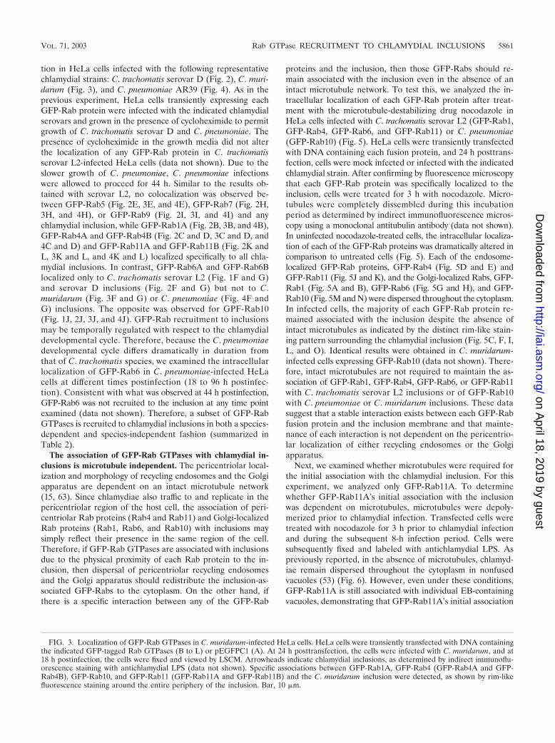
FIG. 3. Localization of GFP-Rab GTPases in C. muridarum-infected HeLa cells. HeLa cells were transiently transfected with DNA containingthe indicated GFP-tagged Rab GTPases (B to L) or pEGFPC1 (A). At 24 h posttransfection, the cells were infected with C. muridarum, and at18 h postinfection, the cells were fixed and viewed by LSCM. Arrowheads indicate chlamydial inclusions, as determined by indirect immunoflu-orescence staining with antichlamydial LPS (data not shown). Specific associations between GFP-Rab1A, GFP-Rab4 (GFP-Rab4A and GFP-Rab4B), GFP-Rab10, and GFP-Rab11 (GFP-Rab11A and GFP-Rab11B) and the C. muridarum inclusion were detected, as shown by rim-likefluorescence staining around the entire periphery of the inclusion. Bar, 10 �m.
VOL. 71, 2003 Rab GTPase RECRUITMENT TO CHLAMYDIAL INCLUSIONS 5861
on April 18, 2019 by guest
http://iai.asm.org/
Dow
nloaded from

5862 RZOMP ET AL. INFECT. IMMUN.
on April 18, 2019 by guest
http://iai.asm.org/
Dow
nloaded from

with the inclusion is also microtubule independent and thatlocalization of the chlamydial vacuole at the pericentriolarregion of the host cell is not required.
The association of GFP-Rab11A with the inclusion is in-dependent of the Tfn-containing endosome association withthe inclusion. Although Rab11 preferentially localizes toTfn-containing pericentriolar recycling endosomes in non-polarized tissue culture cells, Rab11-positive Tfn-negativeendosomes have been reported (67). Because Tfn-containingtubular endosomes are closely associated but do not fuse witheither early or late chlamydial inclusions (2, 54, 55, 70), wewanted to determine whether GFP-Rab11A is associated withthe chlamydial inclusion via its association with Tfn-containingrecycling endosomes. To address this question, we comparedthe intracellular localization of fluorescently labeled Tfn (Al-exa 568-labeled Tfn) with that of GFP-Rab11A in C. tracho-matis serovar L2-infected HeLa cells. Transiently transfectedinfected cells were serum starved for 2 h, incubated for 60 minat 37°C in the presence of Tfn, fixed, and labeled with anti-chlamydial LPS. Labeled cells were then viewed by LSCM(Fig. 7). A steady-state distribution of endosomes containingfluorescent Tfn is obtained under these labeling conditions. Ininfected cells, in addition to the Tfn localized in the peri-centriolar region, Tfn also localized to discrete sites adja-cent to the inclusion membrane (55) (Fig. 7). Although Tfnand EGFP-Rab11A partially colocalize at the inclusion mem-brane, a portion of the inclusion-associated GFP-Rab11A doesnot colocalize with Tfn (Fig. 7). These observations demon-strate that not all inclusion-associated GFP-Rab11A is associ-ated with Tfn-containing endosomes.
GFP-Rab11A partially colocalizes with C. trachomatis IncGat the inclusion membrane. The chlamydial inclusion mem-brane comprises a large family of highly variable chlamydialinclusion membrane proteins, termed Inc proteins (3, 49). Thecarboxy-terminal domains of many Inc proteins are exposed tothe host cell cytoplasm, which may permit interaction with hostcytoplasmic proteins (49, 56). Therefore, Rab GTPases may berecruited to the inclusion through direct interactions with chla-mydial inclusion membrane proteins. To investigate this, wecompared the intracellular localization of GFP-Rab11A to thelocalization of a well-characterized C. trachomatis inclusionmembrane protein, IncG (58), by LSCM. As shown in Fig. 8,some colocalization between the inclusion-associated GFP-Rab11A and IncG was observed, as indicated by the yellowimmunofluorescence staining suggesting a close associationof GFP-Rab11A with the inclusion membrane. However,GFP-Rab11A and IncG did not colocalize completely. Inaddition, in many infected cells, chlamydial Inc-laden fibersextend from the inclusion membrane (6). As shown in Fig. 8,GPF-Rab11A colocalizes to some of these IncG-laden fi-bers. A greater degree of colocalization with these fibers isseen in cells expressing GFP-Rab11AQ70L, a constitutively
active GTPase-deficient Rab11 allele (data not shown). Thefunction of these Inc-laden fibers is not known, but it hasbeen suggested that chlamydial antigens may be trafficked tothe plasma membrane along these fibers, since several in-tracellular chlamydial antigens have been shown to colocalizewith these fibers under certain growth conditions (6, 76). Thisis the first report of a host protein that localizes to Inc-ladenfibers.
GFP-Rab11A is associated with C. trachomatis inclusionsearly during the chlamydial developmental cycle. As men-tioned above, the recruitment of Rab proteins to chlamydialinclusions may be regulated temporally with respect to thechlamydial developmental cycle, and this in turn may influencethe role that each Rab protein may play during chlamydialinfection. As the first step in determining the stage during thechlamydial developmental cycle that GPF-Rab11A is recruitedto the chlamydial inclusions, we examined the intracellularlocalization of GFP-Rab11A in C. trachomatis-infected HeLacells during the initial 8 h postinfection. HeLa cells expressingGFP-Rab11A were infected at a multiplicity of infection ofapproximately 25, and at different times postinfection, the in-tracellular localization of GFP-Rab11A was determined byLSCM. Beginning as early as 1 h postinfection, GFP-Rab11Acolocalizes with individual chlamydial vacuoles (Fig. 9A, B,and C), and by 4 h postinfection, a distinct rim of GFP-Rab11A is observed surrounding each chlamydial vacuole(Fig. 9G, H, and I). GFP-Rab11A remains associated withthe chlamydial vacuoles throughout the time course of theexperiment and can be seen associated with chlamydial vacu-oles that have not yet been trafficked to the pericentriolarregion of the cell.
TABLE 2. Summary of GFP-Rab interactions withchlamydial inclusions
GFP-RabGFP-Rab interaction with chlamydial inclusiona
CT L2 CT D MoPn Cpn
GFP-Rab5 � � � �GFP-Rab7 � � � �GFP-Rab9 � � � �
GFP-Rab1 � � � �GFP-Rab4 � � � �GFP-Rab11 � � � �
GFP-Rab6 � � � �GFP-Rab10 � � � �
a Interaction with inclusions (�) or no association with chlamydial inclusions(�) of C. trachomatis serovar L2 (CT L2), C. trachomatis serovar D (CT D),C. muridarum (formerly C. trachomatis mouse pneumonitis biovar) (MoPn), andC. pneumoniae (Cpn). Interactions were determined by LSCM.
FIG. 4. Localization of GFP-Rab GTPases in C. pneumoniae AR39-infected HeLa cells. HeLa cells were transiently transfected with DNAcontaining the indicated GFP-tagged Rab GTPases (B to L) or pEGFPC1 (A). At 24 h posttransfection, the cells were infected with C. pneumoniaestrain AR39, and at 44 h postinfection, the cells were fixed and viewed by LSCM. Arrowheads indicate chlamydial inclusions, as determined byindirect immunofluorescence staining with antichlamydial LPS (data not shown). Specific associations between GFP-Rab1A, GFP-Rab4 (GFP-Rab4A and GFP-Rab4B), GFP-Rab10, and GFP-Rab11 (GFP-Rab11A and GFP-Rab11B) and the C. pneumoniae inclusion were detected, asshown by rim-like fluorescence staining around the entire periphery of the inclusion. Bar, 10 �m.
VOL. 71, 2003 Rab GTPase RECRUITMENT TO CHLAMYDIAL INCLUSIONS 5863
on April 18, 2019 by guest
http://iai.asm.org/
Dow
nloaded from

5864 RZOMP ET AL. INFECT. IMMUN.
on April 18, 2019 by guest
http://iai.asm.org/
Dow
nloaded from

DISCUSSION
The specific host and chlamydial factors that mediate thebiogenesis or intracellular trafficking of the chlamydial in-clusion remain largely undefined. Here, we report a specificand intimate association of a subset of eukaryotic GFP-RabGTPases with mature chlamydial inclusions. Specific associa-tions between GFP-Rab4 and GFP-Rab11 isoforms (whichfunction sequentially in Tfn receptor recycling) and GFP-Rab1(which functions in ER-to-Golgi and intra-Golgi trafficking)were detected with all chlamydial inclusions tested. In contrast,association of the Golgi-localized Rab GFP-Rab6 was ob-served only with C. trachomatis inclusions, while association ofanother Golgi-localized Rab, GFP-Rab10, was observed onlywith C. pneumoniae and C. muridarum inclusions. Since not allGFP-Rab proteins tested associated with the inclusion, theinteractions that we observed are specific and not likely due toGFP itself. These data suggest that chlamydiae selectively re-cruit critical components of the host Rab membrane traffickingmachinery to the inclusion in both a species-dependent andspecies-independent fashion.
We examined the intracellular distribution of Rab GTPasesin infected HeLa cells that overexpressed amino-terminalGFP-tagged Rab GTPases. In this transient-transfection sys-tem, no toxic effects were observed, and each GFP-Rab local-ized to subcellular compartments similar to those of their en-dogenous counterparts (5, 11, 12, 18, 20, 30, 32, 42, 59, 60, 72)(data not shown). This approach will aid in future functionalstudies by allowing the use of activated, GTP-restricted, anddominant interfering inactive mutants and will also facilitatethe examination of Rab-inclusion interactions in viable cellsthrough the use of time-lapse video microscopy. These obser-vations demonstrate that GFP-Rab GTPases, when overex-pressed, are able to interact with the chlamydial inclusion, butthey do not demonstrate that endogenous Rab GTPases be-have in a similar fashion. Attempts to localize endogenous RabGTPases in infected HeLa cells by indirect immunofluores-cence microscopy using antibodies specific to Rab4, Rab6, andRab11 were unsuccessful. The inability to detect endogenousRab proteins in infected cells may be due to the fact that thelevels of endogenous Rab proteins recruited to the inclusionare too low to detect by indirect immunofluorescence micros-copy using the available antibodies. Therefore, although over-expressed GFP-tagged Rab proteins localize to subcellularcompartments similar to those of their endogenous counter-parts and are thought to function similarly, the role of endog-enous Rab GTPases in infected cells should be examined fur-ther in the future. However, consistent with a possible role forRab6A during chlamydial infection, Rab6A is transcriptionallyinduced in C. trachomatis-infected HeLa cells (77). In addition,Rab4, Rab6, and Rab11 isoforms are ubiquitously expressed,
which suggests that they are expressed in chlamydia-infectedcells in vivo.
It has been shown previously that the chlamydial inclusiondoes not interact with the host’s degradative endosomal orlysosomal pathway as demonstrated by the absence of classicalendocytic markers, such as Tfn, Tfn receptor, early endosomalantigen 1 (EEA1), cation-independent M6PR, and lysosomalglycoprotein Lamp1 (29, 54, 55, 61, 70). The failure to detectspecific associations between the endosomal Rabs, GFP-Rab5,GFP-Rab7, and GFP-Rab9, and the inclusion is consistentwith these previous data. However, since we examined onlycells infected for 18 h, we cannot discount transient interac-tions with the inclusion. This is unlikely, given that even asearly as 5 min postinfection, EEA1 does not associate with theinclusion membrane (54). Thus, chlamydiae appear to bypassthe early endocytic pathway completely.
Even though no fusion between endosomes and the inclu-sion has been detected, an intimate association of Tfn-contain-ing tubular endosomes with inclusions has been described (2,54, 55, 70). Tfn endosomal association with the inclusion isdependent on early chlamydial gene expression and occurs by2 h postinfection. Both GFP-Rab4 and GFP-Rab11, which arerecruited to all inclusions, are primarily associated with recy-cling endosomes in nonpolarized epithelial cells (60, 67). How-ever, not all inclusion-associated GFP-Rab11A colocalizeswith Tfn or its receptor. Because recycling endosomes areheterogeneous in nature (62), these data suggest that inclu-sion-associated GFP-Rab11 or GFP-Rab4 may be associatedwith different populations of recycling endosomes, includingthose that are Tfn positive and those that are Tfn negative (67).Alternatively, GFP-Rab4 or GFP-Rab11, as well as the otherinclusion-associated GFP-Rab proteins, may be anchored with-in the inclusion membrane. We cannot distinguish betweenthese two possibilities by LSCM. Therefore, immunoelectronmicroscopy should be done to clarify whether each GFP-Rabprotein is associated with the inclusion via an intimate as-sociation of nonfused closely juxtaposed vesicles, as wasdemonstrated for Tfn-containing vesicles (51), or anchoredwithin the inclusion membrane. Rab5, Rab4, and Rab11regulate sequential steps along the recycling pathway andare localized to distinct domains on early and recycling endo-somes (60). Therefore, the absence of GFP-Rab5 and theassociation of GFP-Rab4 and GFP-Rab11 demonstrate thatinclusions are associated with markers or endosomal do-mains that are characteristic of late steps in the recycling path-way.
Recruitment of Rab proteins to the inclusion may regulatethe transport of the nascent inclusion to the peri-Golgi region,since isoforms of both Rab6 (36) and Rab11 (73) regulatetransport from early endosomes to the TGN. Consistent with
FIG. 5. Association of GFP-Rab proteins with chlamydial inclusions is maintained in the absence of microtubules. Transiently transfected HeLacells were mock infected (A, B, D, E, G, H, J, K, M, and N) or infected with C. trachomatis serovar L2 for 18 h (C, F, I, and L) or with C.pneumoniae for 44 h (O). Cells were treated with 20 �M nocodazole for 3 h to disassemble microtubules, fixed, permeabilized, and stained withantichlamydial LPS (data not shown). Although treatment with nocodazole disrupts the microtubule network and causes dispersal of recyclingendosomes and the Golgi apparatus, GFP-Rab1, GFP-Rab4, GFP-Rab6, GFP-Rab11, and GFP-Rab10 remain associated with the inclusion.Arrowheads indicate inclusions. Bar, 5 �m.
VOL. 71, 2003 Rab GTPase RECRUITMENT TO CHLAMYDIAL INCLUSIONS 5865
on April 18, 2019 by guest
http://iai.asm.org/
Dow
nloaded from

this, Rab proteins interact with several different molecularmotors, such as myosin Vb (32), cytoplasmic dynein light chain(5), and rabkinesin 6 (17). However, to function in this man-ner, Rab proteins would have to be recruited to the inclusionearly during development. At least for GFP-Rab11A, we de-tect association with chlamydia-containing vacuoles as early as1 h postinfection, a point during chlamydial development whenchlamydia-containing vacuoles are in the process of being traf-ficked to the peri-Golgi region (57).
One of the unique characteristics of all chlamydial inclusionsis their ability to fuse with Golgi-derived vesicles containingNBD-sphingomyelin, endogenously synthesized from 6{N-[(7-nitrobenzo-2-oxa-1,3-diazol-4-yl)amino]caproylsphingosine}(C6-NBD-ceramide) (27, 48, 74) in a process that is dependenton early chlamydial gene expression (57). The recruitment ofGolgi-localized Rab GTPases to the inclusion may provide amechanism for delivery and fusion of exocytic vesicles with theinclusion. Since fusion with exocytic vesicles is a propertyshared by all chlamydial inclusions, Rab1, which is associatedwith all inclusions, may regulate this trafficking step. Alterna-
tively, chlamydiae may exploit functionally redundant path-ways regulated by either Rab6 or Rab10. On the other hand,the roles of Rab1 and Rab6 during chlamydial infection mightnot be related to their Golgi trafficking function but mightinstead be related to Rab6’s role in endosome-to-TGN traf-ficking or Rab1’s proposed role in transcytosis in polarizedepithelial cells (31). Chlamydiae also obtain other moleculesfrom the host, including eukaryotic glycerophospholipids andcholesterol (75). Therefore, Rab GTPases may also be in-volved in regulating acquisition of these compounds. Con-sistent with this, Rab11 has recently been shown to be in-volved in regulating cholesterol trafficking and homeostasis(30). Finally, because of Rab11’s association with the Inc-laden fibers, Rab11 may play a role in trafficking chlamydialantigens along these fibers, thus providing a mechanism forthe appearance of chlamydial antigens at the plasma mem-brane (6, 76).
The distinct rim-like intracellular localization patterns of theGFP-Rab GTPases, the colocalization of Rab11A with IncG,and the fact that each GFP-Rab protein remains associated
FIG. 6. Association of GFP-Rab11A with the chlamydial inclusion is independent of microtubules. Transiently transfected HeLa cells express-ing GFP-Rab11A were treated with nocodazole (D to F) or dimethyl sulfoxide (A to C) for 3 h prior to chlamydial infection to depolymerizemicrotubules. Cells were then infected with C. trachomatis serovar L2 at a multiplicity of infection of approximately 20. The cells wereinfected for 8 h in the presence of nocodazole, fixed in formaldehyde, permeabilized, labeled with antichlamydial LPS, and incubated withgoat anti-mouse IgG conjugated to Alexa 594 (red). (A and D) GFP-Rab11A (green), (B and E) antichlamydial LPS (red), (C) panels Aand B merged, (F) panels D and E merged. In the complete absence of microtubules, GFP-Rab11A is still recruited to individual chlamydia-containing vacuoles (D to F). The arrowhead indicates Golgi-localized chlamydial vacuoles, and arrows indicate examples of dispersed chlamydialvacuoles. Bar, 5 �m.
5866 RZOMP ET AL. INFECT. IMMUN.
on April 18, 2019 by guest
http://iai.asm.org/
Dow
nloaded from

with the inclusion even after microtubules are disassembledand both pericentriolar recycling endosomes and the Golgiapparatus are redistributed strongly support a specific interac-tion with the inclusion membrane. These interactions may bemediated directly through an interaction between each Rab
protein and an inclusion membrane protein, as was recentlydemonstrated between 14-3-3 and IncG (56). Alternatively,Rab recruitment to the inclusion may be mediated throughRab effector interactions, since Rab1 (1, 41), Rab4 (5, 33, 43),Rab6 (17), and Rab11 (28, 78) interact with a variety of effec-tor molecules.
The differential association of GFP-Rab GTPases to inclu-sions containing different chlamydial species highlightsthe fact that individual inclusions interact differently withtheir hosts. On the basis of comparative genomic analysis,the protein compositions of chlamydial inclusion membranesare thought to differ dramatically for different chlamydial spe-cies (3). The exposure of a different complement of inclu-sion membrane proteins to the host cytosol creates the poten-tial for extreme diversity in host-pathogen interactions. Forinstance, GFP-Rab6 may associate only with C. trachomatisinclusions, because it interacts with an inclusion membraneprotein that is present and exposed only in C. trachomatisinclusions. Although recent data have suggested that differ-ences in tissue tropism and disease expression for differentchlamydial species may be due in part to genetic differencesfound within a specific region of the chlamydial genometermed the plasticity zone (4, 19), the differential associa-tion of Rab GTPases and 14-3-3 species-specific interac-tions with chlamydial inclusions suggest that different hostinteractions with the inclusion may also play significantroles.
Although we have not yet determined the biological conse-quences of Rab recruitment to the inclusion, the association ofGFP-tagged Rab GTPases involved in both recycling and bio-synthetic pathways to the inclusion demonstrates for the firsttime that chlamydiae recruit key regulators of membrane traf-ficking to the inclusion in both a species-specific and species-independent fashion. These data lend support to the idea thatchlamydiae, by modulating the activity of host factors thatfunction in vesicle trafficking, control the biogenesis of the
FIG. 7. GFP-Rab11A partially colocalizes with Tfn-containing en-dosomes at the periphery of the chlamydial inclusion. Transiently trans-fected HeLa cells expressing GFP-Rab11A were infected with C. tra-chomatis serovar L2 for 18 h. Recycling endosomes were labeled withAlexa 568-labeled Tfn by serum starving cells for 2 h and incubatedwith Alexa 568-labeled Tfn for 60 min at 37°C. Cells were fixed, per-meabilized, stained with antichlamydial LPS, and incubated with goatanti-mouse IgG conjugated to Cy5 (blue). In the inset in panel D, thearrowhead indicates Rab11A-positive Tfn-negative regions aroundthe inclusion and the arrow indicates Rab11A-positive Tfn-positiveregions. (A) GFP-Rab11A (green), (B) Alexa 568-labeled Tfn (red),(C) antichlamydial LPS (blue), (D) panels A, B, and C merged. Bar,5 �m.
FIG. 8. GFP-Rab11A partially colocalizes with IncG at the inclusion membrane. Transiently transfected HeLa cells expressing GFP-Rab11Awere infected with C. trachomatis serovar L2 for 18 h. Cells were fixed, permeabilized, stained with polyclonal anti-C. trachomatis IncG, andincubated with goat anti-rabbit IgG conjugated to Alexa 594. GFP-Rab11A colocalizes with IncG at many regions along the inclusion membrane,as indicated by the yellow fluorescence, but there are regions that are IncG positive but Rab11A negative. In addition, Rab11A partially colocalizesto the IncG-laden fibers that stain and that extend from the inclusion membrane. The arrowheads indicate representative areas where GFP-Rab11A and IncG colocalize along the fibers. Two inclusions, at different focal planes, are visible within the cell, and each is indicated with anasterisk. (A) GFP-Rab11A (green), (B) IncG (red), and (C) panels A and B merged. Bar, 5 �m.
VOL. 71, 2003 Rab GTPase RECRUITMENT TO CHLAMYDIAL INCLUSIONS 5867
on April 18, 2019 by guest
http://iai.asm.org/
Dow
nloaded from

FIG. 9. Temporal analysis of GFP-Rab11A recruitment to C. trachomatis inclusions. HeLa cells transiently expressing GFP-Rab11A wereinfected at a multiplicity of infection of approximately 20. At different times postinfection (shown to the left of the figure), the cells were fixed andstained with antichlamydial LPS. The cells were then incubated with goat anti-mouse IgG conjugated to Alexa 594 (red) and viewed by LSCM.Beginning as early as 1 h postinfection, colocalization of EBs and GFP-Rab11A is observed. At each time point, insets show representative vacuolescolocalizing with GFP-Rab11A. (A, D, G, J, and M) GFP-Rab11A, (B, E, H, K, and N) antichlamydial LPS, (C, F, I, L, and O) GFP-Rab11A(green) and antichlamydial LPS (red) merged. Bars, 10 �m (O) and 2 �m (inset in panel O).
5868
on April 18, 2019 by guest
http://iai.asm.org/
Dow
nloaded from
inclusion. Further characterization through the expression ofdominant interfering and constitutively active Rab mutantsshould help to elucidate the roles that Rab GTPases may playduring chlamydial infection and determine whether recruit-ment of Rab GTPases is essential for the intracellular survivalof chlamydia.
ACKNOWLEDGMENTS
We thank Craig Roy for providing GFP-Rab5, GFP-Rab7, andGFP-Rab11 constructs, Suzanne Pfeffer for providing the GFP-Rab9construct, Ted Hackstadt and Kate Wolf for providing purified C.pneumoniae, and Harlan Caldwell for providing C. trachomatis and C.muridarum stocks and antichlamydial LPS antiserum. We also thankBarbara Butcher, Helene Marquis, Ruth Collins, and David Russellfor critical reading of the manuscript.
REFERENCES
1. Allan, B. B., B. D. Moyer, and W. E. Balch. 2000. Rab1 recruitment of p115into a cis-SNARE complex: programming budding COPII vesicles for fusion.Science 289:444–448.
2. Al-Younes, H. M., T. Rudel, and T. F. Meyer. 1999. Characterization andintracellular trafficking pattern of vacuoles containing Chlamydia pneu-moniae in human epithelial cells. Cell. Microbiol. 1:237–247.
3. Bannantine, J. P., R. S. Griffiths, V. Viratyosin, W. J. Brown, and D. D.Rockey. 2000. A secondary structural motif predictor of protein localizationto the chlamydial inclusion membrane. Cell. Microbiol. 2:35–48.
4. Belland, R. J., M. A. Scidmore, D. D. Crane, D. M. Hogan, W. Whitmire, G.McClarty, and H. D. Caldwell. 2001. Chlamydia trachomatis cytotoxicityassociated with complete and partial cytotoxin genes. Proc. Natl. Acad. Sci.USA 98:13984–13989.
5. Bielli, A., P. O. Thornquist, A. G. Hendrick, R. Finn, K. Fitzgerald, andM. W. McCaffrey. 2001. The small GTPase Rab4A interacts with the centralregion of cytoplasmic dynein light intermediate chain-1. Biochem. Biophys.Res. Commun. 281:1141–1153.
6. Brown, W. J., Y. A. Skeiky, P. Probst, and D. D. Rockey. 2002. Chlamydialantigens colocalize within IncA-laden fibers extending from the inclusionmembrane into the host cytosol. Infect. Immun. 70:5860–5864.
7. Bucci, C., R. G. Parton, I. H. Mather, H. Stunnenber, K. Simons, B. Hoflack,and M. Zerial. 1992. The small GTPase rab5 functions as a regulatory factorin the early endocytic pathway. Cell 70:715–728.
8. Bucci, C., P. Thomsen, P. Nicoziani, J. McCarthy, and B. van Deurs. 2000.Rab7: a key to lysosome biogenesis. Mol. Biol. Cell 11:467–480.
9. Caldwell, H. D., J. Kromhout, and J. Schachter. 1981. Purification andpartial characterization of the major outer membrane protein of Chlamydiatrachomatis. Infect. Immun. 31:1161–1176.
10. Centers for Disease Control and Prevention. 1997. Chlamydia trachomatisgenital tract infections–United States, 1995. Morb. Mortal. Wkly. Rep. 46:193–198.
11. Chen, W., Y. Feng, D. Chen, and A. Wandinger-Ness. 1998. Rab11 is re-quired for trans-Golgi network-to-plasma membrane transport and a pref-erential target for GDP dissociation inhibitor. Mol. Biol. Cell 9:3241–3257.
12. Chen, W., and A. Wandinger-Ness. 2001. Expression and functional analysesof Rab8 and Rab11a in exocytic transport from trans-Golgi network. Meth-ods Enzymol. 329:165–175.
13. Chen, Y. T., C. Holcomb, and H. P. Moore. 1993. Expression and localizationof two low molecular weight GTP-binding proteins, Rab8 and Rab10, byepitope tag. Proc. Natl. Acad. Sci. USA 90:6508–6512.
14. Clausen, J. D., G. Christiansen, H. U. Holst, and S. Birklund. 1997. Chla-mydia trachomatis utilizes the host cell microtubule network during earlyevents of infection. Mol. Microbiol. 25:441–449.
15. Cole, N. B., and J. Lippincott-Schwartz. 1995. Organization of organellesand membrane traffic by microtubules. Curr. Opin. Cell Biol. 7:55–64.
16. Collins, R. N., and P. Brennwald. 2000. Rab proteins. GTPases. Front. Mol.Biol. 24:137–175.
17. Echard, A., F. Jollivet, O. Martinez, J. J. Lacapere, A. Rousselet, I. Janoueix-Lerosey, and B. Goud. 1998. Interaction of a Golgi-associated kinesin-likeprotein with Rab6. Science 279:580–585.
18. Echard, A., F. J. M. Opdam, H. J. P. C. de Leeuw, F. Jollivet, P. Savelkoul,W. Hendriks, J. Voorberg, B. Goud, and J. A. M. Fransen. 2000. Alternativesplicing of the human Rab6A gene generates two close but functionallydifferent isoforms. Mol. Biol. Cell 11:3819–3833.
19. Fehlner-Gardiner, C., C. Roshick, J. H. Carlson, S. Hughes, R. J. Belland,H. D. Caldwell, and G. McClarty. 2002. Molecular basis defining humanChlamydia trachomatis tissue tropism. A possible role for tryptophan syn-thase. J. Biol. Chem. 277:26893–26903.
20. Feng, Y., B. Press, W. Chen, J. Zimmerman, and A. Wandinger-Ness. 2001.Expression and properties of Rab7 in endosome function. Methods Enzy-mol. 329:175–187.
21. Fu, H., R. R. Subramanian, and S. C. Masters. 2000. 14–3–3 proteins:structure, function and regulation. Annu. Rev. Pharmacol. Toxicol. 40:617–647.
22. Girod, A., B. Storrie, J. C. Simpson, L. Johannes, B. Goud, L. M. Roberts,J. M. Lord, T. Nilsson, and R. Pepperkok. 1999. Evidence for a COP-I-independent transport route from the Golgi complex to the endoplasmicreticulum. Nat. Cell Biol. 1:423–430.
23. Gorvel, J. P., P. Chavrier, M. Zerial, and J. Gruenberg. 1991. rab5 controlsearly endosome fusion in vitro. Cell 64:915–925.
24. Grayston, J. T., M. B. Aldous, A. Easton, S. P. Wang, C. C. Kuo, L. A.Campbell, and J. Altman. 1993. Evidence that Chlamydia pneumoniae causespneumonia and bronchitis. J. Infect. Dis. 168:1231–1235.
25. Grayston, J. T., and L. A. Campbell. 1999. The role of Chlamydia pneu-moniae in atherosclerosis. Clin. Infect. Dis. 28:993–994.
26. Hackstadt, T. 1999. Cell biology, p. 101–138. In R. S. Stephens (ed.), Chla-mydia: intracellular biology, pathogenesis, and immunity. American Societyfor Microbiology, Washington, D.C.
27. Hackstadt, T., M. A. Scidmore, and D. D. Rockey. 1995. Lipid metabolism inChlamydia trachomatis-infected cells: directed trafficking of Golgi-derivedsphingolipids to the chlamydial inclusion. Proc. Natl. Acad. Sci. USA 92:4877–4881.
28. Hales, C. M., R. Griner, K. C. Hobdy-Henderson, M. C. Dorn, D. Hardy, R.Kumar, J. Navarre, E. K. L. Chan, L. A. Lapierre, and J. R. Goldenring.2001. Identification and characterization of a family of Rab11-interactingproteins. J. Biol. Chem. 276:39067–39075.
29. Heinzen, R. A., M. A. Scidmore, D. D. Rockey, and T. Hackstadt. 1996.Differential interaction with endocytic and exocytic pathways distinguishparasitophorous vacuoles of Coxiella burnetii and Chlamydia trachomatis.Infect. Immun. 64:796–809.
30. Holtta-Vuori, M., K. Tanhuanpaa, W. Mobius, P. Somerharju, and E.Ikonen. 2002. Modulation of cellular cholesterol transport and homeostasisby Rab11. Mol. Biol. Cell 13:3107–3122.
31. Jin, M., L. Saucan, M. G. Farquhar, and G. E. Palade. 1996. Rab1a andmultiple other Rab proteins are associated with the transcytotic pathway inrat liver. J. Biol. Chem. 271:30105–30113.
32. Lapierre, L. A., R. Kumar, C. M. Hales, J. Navarre, S. G. Bhartur, J. O.Burnette, J. D. W. Provance, J. A. Mercer, M. Bahler, and J. R. Goldenring.2001. Myosin Vb is associated with plasma membrane recycling systems.Mol. Biol. Cell 12:1843–1857.
33. Lindsay, A. J., A. G. Hendrick, G. Cantalupo, F. Senic-Matuglia, B. Goud, C.Bucci, and M. W. McCaffrey. 2002. Rab coupling protein (RCP), a novelRab4 and Rab11 effector protein. J. Biol. Chem. 277:12190–12199.
34. Lombardi, D., T. Soldati, M. A. Riederer, Y. Goda, M. Zerial, and S. R.Pfeffer. 1993. Rab9 functions in transport between late endosomes and thetrans Golgi network. EMBO J. 12:677–682.
35. Majeed, M., M. Gustafsson, E. Kihlstrom, and O. Stendalh. 1993. Roles ofCa2� and F-actin in intracellular aggregation of Chlamydia trachomatis ineukaryotic cells. Infect. Immun. 61:1406–1414.
36. Mallard, F., B. L. Tang, T. Galli, D. Tenza, A. Saint-Pol, X. Yue, C. Antony,W. Hong, B. Goud, and L. Johannes. 2002. Early/recycling endosomes-to-TGN transport involves two SNARE complexes and a Rab6 isoform. J. CellBiol. 156:653–664.
37. McCaffrey, M. W., A. Bielli, G. Cantalupo, S. Mora, V. Roberti, M. Santillo,F. Drummond, and C. Bucci. 2001. Rab4 affects both recycling and degra-dative endosomal trafficking. FEBS Lett. 495:21–30.
38. Meresse, S., J. P. Gorvel, and P. Chavrier. 1995. The rab7 GTPase resides ona vesicular compartment connected to lysosomes. J. Cell Sci. 108:3349–3358.
39. Meresse, S., O. Steele-Mortimer, B. B. Finlay, and J. P. Gorvel. 1999. Therab7 GTPase controls the maturation of Salmonella typhimurium-containingvacuoles in HeLa cells. EMBO J. 18:4394–4403.
40. Moulder, J. W. 1985. Comparative biology of intracellular parasitism. Mi-crobiol. Rev. 49:298–337.
41. Moyer, B. D., B. B. Allan, and W. E. Balch. 2001. Rab1 interaction with aGM130 effector complex regulates COPII vesicle cis-Golgi tethering. Traffic2:268–276.
42. Moyer, B. D., J. Matteson, and W. E. Balch. 2001. Expression of wild-typeand mutant green fluorescent protein-Rab1 for fluorescence microscopyanalysis. Methods Enzymol. 329:6–14.
43. Nagelkerken, B., E. Van Anken, M. Van Raak, L. Gerez, K. Mohrmann, N.Van Uden, J. Holthuizen, L. Pelkmans, and P. Van Der Sluijs. 2000. Rabap-tin4, a novel effector of the small GTPase rab4a, is recruited to perinuclearrecycling vesicles. Biochem. J. 346:593–601.
44. Opdam, F. J., A. Echard, H. J. Croes, J. A. van den Hurk, R. A. van deVorstenbosch, L. A. Ginsel, B. Goud, and J. A. Fransen. 2000. The smallGTPase Rab6B, a novel Rab6 subfamily member, is cell-type specificallyexpressed and localised to the Golgi apparatus. J. Cell Sci. 113:2725–2735.
45. Pereira-Leal, J. B., and M. C. Seabra. 2000. The mammalian Rab family ofsmall GTPases: definition of family and subfamily sequence motifs suggestsa mechanism for functional specificity in the Ras superfamily. J. Mol. Biol.301:1077–1087.
46. Pfeffer, S. R. 2001. Rab GTPases: specifying and deciphering organelleidentity and function. Trends Cell Biol. 11:487–491.
VOL. 71, 2003 Rab GTPase RECRUITMENT TO CHLAMYDIAL INCLUSIONS 5869
on April 18, 2019 by guest
http://iai.asm.org/
Dow
nloaded from
47. Ren, M., G. Xu, J. Zeng, C. De Lemos-Charandini, M. Adesnik, and D.Sabatini. 1998. Hydrolysis of GTP on Rab11 is required for the directdelivery of transferrin from the pericentriolar recycling compartment to thecell surface but not from sorting endosomes. Proc. Natl. Acad. Sci. USA95:6187–6192.
48. Rockey, D. D., E. R. Fischer, and T. Hackstadt. 1996. Temporal analysis ofthe developing Chlamydia psittaci inclusion by use of fluorescence and elec-tron microscopy. Infect. Immun. 64:4269–4278.
49. Rockey, D. D., M. A. Scidmore, J. P. Bannantine, and W. J. Brown. 2002.Proteins in the chlamydial inclusion membrane. Microbes Infect. 4:333–340.
50. Rothman, J. E., and F. T. Wieland. 1996. Protein sorting by transport vesi-cles. Science 272:227–234.
51. Schachter, J. 1999. Infection and disease epidemiology, p. 139–169. In R. S.Stephens (ed.), Chlamydia: intracellular biology, pathogenesis, and immu-nity. American Society for Microbiology, Washington, D.C.
52. Schlierf, B., G. H. Fey, J. Hauber, G. M. Hocke, and O. Rosorius. 2000.Rab11b is essential for recycling of transferrin to the plasma membrane. Exp.Cell Res. 259:257–265.
53. Schramm, N., and P. B. Wyrick. 1995. Cytoskeletal requirements in Chla-mydia trachomatis infection in host cells. Infect. Immun. 63:324–332.
54. Scidmore, M. A., E. R. Fischer, and T. Hackstadt. 2003. Restricted fusion ofChlamydia trachomatis vesicles with endocytic compartments during the ini-tial stages of infection. Infect. Immun. 71:973–984.
55. Scidmore, M. A., E. R. Fischer, and T. Hackstadt. 1996. Sphingolipids andglycoproteins are differentially trafficked to the Chlamydia trachomatis inclu-sion. J. Cell Biol. 134:363–374.
56. Scidmore, M. A., and T. Hackstadt. 2001. Mammalian 14–3–3� associateswith the Chlamydia trachomatis inclusion membrane via its interaction withIncG. Mol. Microbiol. 39:1638–1650.
57. Scidmore, M. A., D. D. Rockey, E. R. Fischer, R. A. Heinzen, and T. Hack-stadt. 1996. Vesicular interactions of the Chlamydia trachomatis inclusionare determined by chlamydial early protein synthesis rather than route ofentry. Infect. Immun. 64:5366–5372.
58. Scidmore-Carlson, M. A., E. I. Shaw, C. A. Dooley, E. R. Fischer, and T.Hackstadt. 1999. Identification and characterization of a Chlamydia tracho-matis early operon encoding four novel inclusion membrane proteins. Mol.Microbiol. 33:753–765.
59. Short, B., C. Preisinger, J. Schaletzky, R. Kopajtich, and F. A. Barr. 2002.The Rab6 GTPase regulates recruitment of the dynactin complex to Golgimembranes. Curr. Biol. 12:1792–1795.
60. Sonnichsen, B., S. De Renzis, E. Nielsen, J. Rietdorf, and M. Zerial. 2000.Distinct membrane domains on endosomes in the recycling pathway visual-ized by multicolor imaging of Rab4, Rab5, and Rab11. J. Cell Biol. 149:901–913.
61. Taraska, T., D. M. Ward, R. S. Ajioka, P. B. Wyrick, S. R. Davis-Kaplan,C. H. Davis, and J. Kaplan. 1996. The late chlamydial inclusion membraneis not derived from the endocytic pathway and is relatively deficient in hostproteins. Infect. Immun. 64:3713–3727.
62. Teter, K., G. Chandy, B. Quinones, K. Pereyra, T. Machen, and H.-P. H.Moore. 1998. Cellubrevin-targeted fluorescence uncovers heterogeneity inthe recycling endosomes. J. Biol. Chem. 273:19625–19633.
63. Thyberg, J., and S. Moskalewski. 1999. Role of microtubules in the organi-zation of the Golgi complex. Exp. Cell Res. 246:263–279.
64. Thylefors, B., A. D. Negrel, R. Pararajasegaram, and K. Y. Dadzie. 1995.Global data on blindness. Bull. W. H. O. 73:115–121.
65. Tisdale, E. J., J. R. Bourne, R. Khosravi-Far, C. J. Der, and W. E. Balch.1992. GTP-binding mutants of rab1 and rab2 are potent inhibitors of vesic-ular transport from the endoplasmic reticulum to the Golgi complex. J. CellBiol. 119:749–761.
66. Tuvim, M. J., R. Adachi, S. Hoffenberg, and B. F. Dickey. 2001. Trafficcontrol: Rab GTPases and the regulation of interorganellar transport. NewsPhysiol. Sci. 16:56–61.
67. Ullrich, O., S. Reinsch, S. Urbe, M. Zerial, and R. G. Parton. 1996. Rab11regulates recycling through the pericentriolar recycling endosome. J. CellBiol. 135:913–924.
68. van der Sluijs, P., M. Hull, P. Webster, P. Male, B. Goud, and I. Mellman.1992. The small GTP-binding protein rab4 controls an early sorting event onthe endocytic pathway. Cell 70:729–740.
69. van der Sluijs, P., M. Hull, A. Zahraoui, A. Tavitian, B. Goud, and I.Mellman. 1991. The small GTP-binding protein rab4 is associated with earlyendosomes. Proc. Natl. Acad. Sci. USA 88:6313–6317.
70. van Ooij, C., G. Apodaca, and J. Engel. 1997. Characterization of the Chla-mydia trachomatis vacuole and its interaction with the host endocytic path-way in HeLa cells. Infect. Immun. 65:758–766.
71. Via, L. E., D. Deretic, R. J. Ulmer, N. S. Hibler, L. A. Huber, and V. Deretic.1997. Arrest of mycobacterial phagosome maturation is caused by a block invesicle fusion between stages controlled by rab5 and rab7. J. Biol. Chem.272:13326–13331.
72. White, J., L. Johannes, F. Mallard, A. Girod, S. Grill, S. Reinsch, P. Keller,B. Tzschaschel, A. Echard, B. Goud, and E. H. Stelzer. 1999. Rab6 coordi-nates a novel Golgi to ER retrograde transport pathway in live cells. J. CellBiol. 147:743–760.
73. Wilcke, M., L. Johannes, T. Galli, V. Mayau, B. Goud, and J. Salamero.2000. Rab11 regulates the compartmentalization of early endosomes re-quired for efficient transport from early endosomes to the trans-Golgi net-work. J. Cell Biol. 151:1207–1220.
74. Wolf, K., and T. Hackstadt. 2001. Sphingomyelin trafficking in Chlamydiapneumoniae-infected cells. Cell. Microbiol. 3:145–152.
75. Wylie, J. L., G. M. Hatch, and G. McClarty. 1997. Host cell phospholipidsare trafficked to and then modified by Chlamydia trachomatis. J. Bacteriol.179:7233–7242.
76. Wyrick, P. B., J. Choon, S. T. Knight, D. Goyeau, E. S. Stuart, and A. B.MacDonald. 1994. Chlamydia trachomatis antigens on the surface of infectedhuman endometrial epithelial cells. Immunol. Infect. Dis. 4:131–141.
77. Xia, M., R. E. Bumgarner, M. F. Lampe, and W. E. Stamm. 2003. Chlamydiatrachomatis infection alters host cell transcription in diverse cellular path-ways. J. Infect. Dis. 187:424–434.
78. Zeng, J., M. Ren, D. Gravotta, C. De Lemos-Charandini, M. Lui, H. Erd-jument-Bromage, P. Tempst, G. Xu, T. H. Shen, T. Morimoto, M. Adesnik,and D. Sabatini. 1999. Identification of a putative effector protein for Rab11that participates in transferrin recycling. Proc. Natl. Acad. Sci. USA 96:2840–2845.
Editor: B. B. Finlay
5870 RZOMP ET AL. INFECT. IMMUN.
on April 18, 2019 by guest
http://iai.asm.org/
Dow
nloaded from