Embed Size (px)
Citation preview
Plant Physiol. (1974) 53, 732-737
Cellulase and Abscission in the Red Kidney Bean (Phcaseolusvulgaris)1
Received for publication August 6, 1973 and in revised form January 10, 1974
PHILIP D. REID AND HEATHER G. STRONGDevartment of Biological Sciences, Smith College, Northampton, Massachusetts 01060
FRANCIS LEW AND LOWELL N. LEWISDeoartment of Plant Sciences, University of California Riverside, California 92502
ABSTRACT
Cellulase (8-1 ,4-glucan-glucanohydrolase EC 3.2.1.4) ac-tivity in the abscission zone of red kidney bean (Phaseolus vul-garis) was previously shown to exist in at least two differentmolecular forms. The form of the enzyme which has an isoelec-tric point of 4.5 is present in both abscising and nonabscisingtissue and requires grinding for extraction. Another form ofthe enzyme which has an isoelectric point of 9.5 is present onlyin tissue in which the abscission process has been induced. Fur-ther, much of this form of cellulase can be removed from thetissue by vacuum infiltration with buffer. Time course studiesindicate that while the increase in measureable cellulase ac-tivity in tissue which is actively undergoing abscission was dueprimarily to the appearance of cellulase 9.5, this form of theenzyme cannot be removed by vacuum infiltration until afterthe breakstrength of the abscission zone has decreased nearlyto zero. The intracellular localization of these two forms ofcellulase is discussed.
The role of cellulase in mediating the process of abscission hasbeen difficult to determine even though several laboratories havebeen able to show a correlation between the activity of the en-zyme and the event of abscission (1, 5, 9, 11, 13). This problemhas been compounded by the recent report that cellulase existsin more than one molecular form (10, 11). Lewis and Varner(11) were able to demonstrate a correlation between cellulaseactivity and decrease in breakstrength of abscission zones ofPhaseolus vulgaris, and further that the increase in activity asabscission proceeded was due to the de novo synthesis of a formof the enzyme which was more soluble in a high salt buffer thanin the same buffer without added salt.
Lewis et al. (10) have reported that, in freshly harvested ab-scission zones, only an acidic form of cellulase could be ex-tracted and that this cellulase was relatively unstable. After in-duction of abscission, the acidic form could still be detected aswell as at least one other form of the enzyme which was a basicprotein, as determined by isoelectric focusing. Further charac-terization of these two cellulases indicated differences in sensi-tivity to p-chloromercuribenzoate, mol wt, and stability at 50 C.
'This work was supported by National Science FoundationGrant H6B-17850 to L. N. L.
This study reports our attempts to follow the activities of thesetwo forms of cellulase during the process of abscission as mea-sured by decreasing breakstrengths of abscission zones.
MATERIALS AND METHODS
Plant Material. Abscission zones were taken from the petiolesegment just below the primary leaf blades of 10- to 12-day-oldred kidney beans, Phaseolus vulgaris. For large experiments in-volving 1000 or more abscission zones, the plants were grown inthe greenhouse at 27 + 2 C in a peat-sponge rock mix andwatered daily with 0.5 Hoagland's solution. Smaller amounts oftissue were taken from plants grown in a growth chamber at 25C with 8 hr of light from both incandescent and fluorescentlamps at an intensity of 1000 ft-c, followed by 2 hr of incandes-cent light of 100 ft-c and 14 hr of dark. Chamber grown plantswere grown in a vermiculite-gravel medium and watered dailywith 0.5 Hoagland's solution.
Treatment of Explants. Explants were prepared by removingthe primary leaf blades and the cotyledons, and the plant was cutoff just above the soil leaving the main axis plus the petioles in-cluding the distal abscission zone of the primary leaves as a unit.Zero time (untreated abscission zones) were prepared by re-moving the distal abscission zone including the pulvinus plus 3to 5 mm of petiole. Ethylene treatment was carried out by al-lowing the explants to sit in a beaker of water for 24 hr followedby exposure to 50 ,ul/l of ethylene gas in an airtight chamber.
Breakstrength. The breakstrength across the abscission zonewas measured as the force necessary to separate pulvinus frompetiole. Petioles were cut from the explant at the node. A stripof cheesecloth, 2.5 X 0.5 cm, was twisted around the petioleabout 0.5 cm below the separation layer and clamped firmlywith a hemostat. Secured petioles were suspended from a Chatil-lion-pull gauge. Tension force was applied manually by pullingthe pulvinus until the tissue broke apart. This technique gave arepeatable indication of the abscission process between about200 to 10 g. Forces greater than 200 g caused rupture of thetissue in places other than the separation layer and forces lessthan 10 g were difficult to measure.
Cellulase Extraction. Cellulase (,/-1,4-glucan-glucanohydro-lase EC 3 .2. 1 .4) was routinely extracted by grinding the tissuein 20 mm sodium phosphate buffer fortified with 1 M NaCI, pH6.1 (5-10 zones/ml) with a Waring Blendor or by first sub-jecting the tissue to vacuum infiltration and then grinding.Vacuum-infiltrated extracts were prepared by placing the ab-scission zones in high salt buffer in a 50-ml suction flask (5-10abscission zones/ml). A vacuum was applied to the flask withan aspirator and then released two or three times until bubbles
732 www.plantphysiol.orgon January 15, 2019 - Published by Downloaded from
Copyright © 1974 American Society of Plant Biologists. All rights reserved.

CELLULASE AND ABSCISSION IN P. VULGARIS
15 20 25FRACTION NUMBER
30
Table I. Extraction of Cellulase from Red Kidntey Bean AbscissionZones
Cellulase was extracted by grinding in 20 mm sodium phosphate10 buffer, pH 6.1, with or without addition of 1 M NaCl. Estimation
of per cent cellulase 9.5 is based on recovery of cellulase fromisoelectric focusing.
8
6 pH
4
2
FRACTION NUMBER
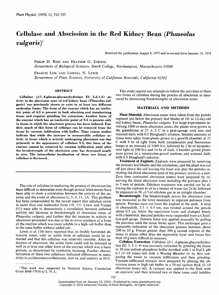
FIG. 1. Isoelectric focusing pattern of cellulase activity fromfreshly harvested and ethylene-treated abscission zones. Cellulaseactivity was extracted by grinding abscission zones in 20 mm phos-phate buffer, pH 6.1, fortified with 1 M NaCl. Enzyme activity wasfocused in ampholine buffers (pH 3-10) at 600 v for 24 hr. Three-ml fractions were collected and adjusted to pH 6.1 before assay.A: cellulase activity extracted from zero time tissue. B: Cellulaseactivity extracted from abscission zones which had been treatedwith 50 julll of ethylene gas for 24 hr beginning 24 hr after prepa-ration of explants.
were no longer emitted from the tissue (1-2 min). While cellu-lase could be directly assayed from the clear supernatant at thistime, the flask was routinely placed on ice and gently shakenfor 15 min after which time the tissue was removed, blotteddry, and then subjected to grinding as above. The extracts pre-pared by grinding were filtered through nylon cloth, and theseextracts as well as those prepared by vacuum infiltration werecentrifuged for 10 min at 10,000g. Extracts prepared by vac-uum infiltration did not have a pellet either with zero timetissue or that which had been ethylene-treated. The extractswere then assayed and prepared for isoelectric focusing.
No.of ~~~~~~~~~~~~~Esti-Tissue No. of Fresh Buffer Uis matedTissue Abscission wt Additions Units Cellulase
Zones 9.5
g total per zone %Zero time 1228 33 No NaCl 583 0.48 0
1 M NaCl 690 0.56 0Ethylene- 996 27 No NaCi 1668 1.62 40-50
treated I M NaCl 6143 6.15 80-90
Enzyme Assay. Cellulase activity was assayed by measuringthe reduction in viscosity of a solution of CM2-cellulose, type7HF, (Hercules Powder Co., Wilmington, Del.). The assaymixture contained two parts of CMC-7HF, 1.2% (w/v) in 20mM sodium phosphate buffer, pH 6.1, and one part of the en-zyme solution. Drainage time through a calibrated portion of a100-ul pipette was used as a measure of viscosity. Viscosity wasusually measured after 1 to 2 hr reaction time. Enzyme reac-tions and viscosity measurements were done at room tempera-ture of about 23 C. Viscosity data were converted to relativeunits of activity B/g-hr as described by Almin et al. (5) and ex-tended to Phaseolus cellulase by Lewis and Varner (1 1).
Isoelectric Focusing. Isoelectric focusing was carried out asdescribed by Haglund (8). The enzyme extracts were dialyzedagainst two changes of 1% glycine for 3 hr, combined withampholine buffers (pH 3.0-10.0), and placed on an LKB Model1801 isoelectric focusing apparatus. The proteins were focusedfor 36 hr at 600 v. Fractions collected from the column werethen adjusted to pH 6.1 using 0.2 M Na,HPO4 or NaH2PO4.Each fraction was then assayed for cellulase activity. Percent-age of recovery was calculated as the ratio of the total unitsrecovered from the column to the total units put on the columnafter dialysis.
RESULTS
Isoelectric focusing of cellulase extracted from freshlyharvested (zero time) abscission zones indicated that the en-zyme had an isoelectric point of about pH 4.5 (Fig. IA). Afterinduction of abscission with ethylene, there was an increase incellulase activity, and when this activity was subjected to isoe-lectric focusing, the majority of cellulase activity was recoveredin two separate regions of the gradient (Fig. 1B). Lewis et al.(11) have referred to that form of cellulase which focuses be-tween pH 4.2 and 4.6 as cellulase 4.5 and that which focusesbetween pH 9.2 and 9.6 as cellulase 9.5, and have partiallycharacterized these two cellulases. Other minor peaks were con-sistently observed; however, they represent a small percentageof the total activity, and we have not made an attempt to studythe properties of the other forms of cellulase at this time.
Attempts to determine whether cellulase 4.5 and cellulase9.5 were differentially extractable indicated that a small butrepeatable increase in cellulase activity (about 3-fold) couldbe extracted with the low salt buffer after abscission had beeninduced (Table I). A much greater increase (about 11-fold) in
SAbbreviations: CM: carboxymethyl; B: estimated arbitrary en-zyme activity.
A
733
5
a
10
9-
z
aa8J
" a-
I-
> 4-
Ua
-92Ji
hi
a
2I-
> 2
9--AhiJ
I-
>I9-
04
-J
hi
Plant Physiol. Vol. 53, 1974
_L 1_
www.plantphysiol.orgon January 15, 2019 - Published by Downloaded from Copyright © 1974 American Society of Plant Biologists. All rights reserved.

REID, STRONG, LEW, AND LEWIS
cellulase activity was observed in that cellulase which requiredhigh salt buffer for extraction. At zero time there was no de-tectable cellulase 9.5 by isoelectric focusing in either extract.
Table lI. Exti'action of Cel/blase from Kidniey Beani AbscissiontZones by Vacuuiim Inifiltrationz
Infiltration was followed by grinding in 20 mm phosphatebuffer, pH 6.1, fortified with 1 M NaCl.
Vacuum Infiltrated Ground Combined total
Units U/g U/g Total No. of(U) U/zone fresh Units U/zone fresh u zones U/zone
wt wt
Zero time 91 0.06 2. 8 3514 2.4 106.0 3605 1488 2.4Untreated48 hr 260 0.16 7.5 3234 2.0 93. 7 3494 1634 2.1
Ethylene48 hr 5624 3.2 131.0 7176 4.0 139.0 12818 1784 7.2
VACUUM INFILTRATED
Since about half of the activity previously referred to as solublecellulase (11) from ethylene-treated tissue was cellulase 9.5 andhalf cellulase 4.5, it does not appear that differential solubilityresults in a clear separation of these two molecular forms ofcellulase.
Table II and Figure 2 present evidence which suggests thatcellulase 4.5 may be primarily endocellular and that cellulase9.5 is the form found outside of the membrane. Cellulase wasextracted either before abscission was induced (zero time) orafter an ethylene induction treatment by first placing the abscis-sion zones in 20 mm sodium phosphate buffer fortified with 1M NaCl and subjecting the tissue to vacuum infiltration. Aftershaking, the tissue was removed, and the clear supernatant wasassayed for cellulase activity. The tissue was then blotted dryand ground in more of the high salt fortified buffer, centrifuged,and this supernatant was also assayed for cellulase activity.Cellulase extracted by these techniques resulted in recovery ofmore activity per abscission zone (2.4 units/zone) from zerotime tissue than was recovered by the combination of grinding
GROUND
UNTREATED (zero hrs)
5 10 15 20 25
0
:)200 20- v
pH I I
a 1X1z
C2H4 (48hr.)
5 0 15 20 25FRACTION NO.
FIG. 2. Isoelectric focusing patterns of cellulases extracted by vacuum infiltration and by grinding nonabscising and ethylene-treated abscissionzones. Cellulase was extracted from 200 abscission zones by first placing the segments in 20 mm phosphate buffer, pH 6.1, in a vacuum flask anddecreasing the pressure with a faucet aspirator. Segments were then removed and ground in more of the same buffer. a: Isoelectric focusing pat-tern of cellulase activity extracted by vacuum infiltration technique from freshly harvested abscission zones. Ten per cent of the units put on thecolumn were recovered. b: Isoelectric focusing pattern of cellulase activity extracted by grinding freshly harvested abscission zones after vacuuminfiltration. Twenty per cent of the units put on the column were recovered. c: Isoelectric focusing pattem of cellulase extracted from ethylenetreated abscission zones by vacuum infiltration. Thirty-five per cent of the units put on the column were recovered. d: Isoelectric focusing patternof cellulase activity extracted by grinding ethylene treated abscission zones after vacuum infiltration. One hundred per cent of the units put onthe column were recovered.
aJ
-J-J
wQIJ0
U) I2
I0
a
30 35
10
-J8 D-J
w0
4 I-z
2
U)
w0*
Io
d
FRACTION NO.
a
4
2
30 35_~~~~~~ Is
__ __
734 Plant Physiol. Vol. 53, 1974
20~
-1
www.plantphysiol.orgon January 15, 2019 - Published by Downloaded from Copyright © 1974 American Society of Plant Biologists. All rights reserved.

CELLULASE AND ABSCISSION IN. P. VILGARIS
in low salt buffer followed by grinding in a high salt buffer asreported in Table I (1.04 units/zone). The amount of activityper abscission zone extracted from ethylene-treated tissue, how-ever, was similar in both experiments.
Total cellulase activity (that extracted by vacuum infiltrationplus that extracted by grinding) again showed about a 3-foldincrease from ethylene-treated tissue over that extracted fromzero time tissue (Table II). Only about 3% of the total activityfound in zero time tissue could be extracted by the vacuum in-filtration technique, whereas 44% of the total activity found inethylene-treated tissue could be extracted by this method.
Extracts prepared by the vacuum infiltration grinding tech-nique were placed on an isoelectric focusing column, and theresults are indicated in Figure 2. Vacuum infiltration of thezero time abscission zones led to a release of a small amount ofcellulase 4.5 (Fig. 2a). A greater amount of this form of theenzyme was obtained by then grinding this tissue (Fig. 2b).Again, cellulase 9.5 was not detected in the freshly harvestedabscission zones. The increased cellulase activity which was ex-tracted by vacuum infiltration of ethylene-treated abscissionzones was primarily cellulase 9.5 (Fig. 2c). Grinding this tissuereleased even more cellulase 9.5 as well as the cellulase 4.5activity (Fig. 2d).
Since the above results suggested not only that the increase incellulase activity which correlates with the abscission processwas due to the appearance of cellulase 9.5, but further that thisform of the enzyme could be washed out of the abscission zoneswithout disrupting the tissue by grinding, we then attempted tocorrelate the decrease in breakstrength of abscission zones withthe appearance of cellulase 9.5.
Figure 3 shows the results of such an experiment. In this ex-periment, abscission was allowed to proceed slowly by standingthe explants in a beaker of water on the laboratory bench. Atintervals, samples were taken to test breakstrength of the abscis-sion zones. Samples were also removed and cellulase extractedby both the vacuum infiltration and grinding techniques. Thegraph shows that a slight increase in cellulase activity did occuras the breakstrength decreased and continued to rise even afterthe breakstrength became essentially zero. However, cellulase9.5 did not begin to appear until the abscission process wasnearly completed (about 50 hr after excision). This activity also
A VACUUM-INF0 GROUND EXT
' TOTAL CEL> 80_*REAKSTRE
>L)- z60-
w K
L) 20_ *:'. 1.
continued to increase after the abscission process was com-pleted. The cellulase extracted in this experiment by vacuuminfiltration was shown to be cellulase 9.5 both by isoelectricfocusing and ion exchange chromatography using CM Sepha-dex (10).
Exposure to ethylene after the explants have been agedcauses a much more rapid decrease in breakstrength (Fig. 4).The breakstrength decreased from about 200 g to zero in lessthan 10 hr. In tissue which has not been exposed to ethylene, acomparable decrease in breakstrength takes more than 40 hr(Fig. 3).
Figure 4 shows that even in tissue treated with ethylene,cellulase 9.5 does not appear in the fraction which can bewashed out of the tissue without grinding until after the processof abscission has been completed.
DISCUSSION
The hypothesis that cell wall hydrolyzing enzymes, particu-larly cellulase, are involved in bringing about abscission hasbeen suggested by several workers. Craker and Abeles (6)showed that cellulase increased prior to the morphologicalevent of abscission in beans. Ratner et al. (13) found similarresults in citrus. More recently, Lewis and Varner (11) wereable to show only an inverse correlation between cellulase ac-tivity and breakstrength in bean abscission zones. The latterworkers reported their data in relative units of enzyme activityrather than percentage of change in viscosity. The incubationtime for the reaction was much shorter (1 hr versus 16 hr), andbreakstrength was measured by pulling the zones apart.One of the difficulties with this hypothesis has been that
cellulase could be isolated from tissues which were not abscis-ing. Since multiple forms of cellulase have been reported infungi as well as bacteria (7), it is reasonable to speculate thatmultiple forms may also exist in higher plants.
Sheldrake (14) showed that cellulase activity extracted fromdifferent tissues of Acer pseudoplatanus have different pHoptima and certain of these cellulases required the presence ofdetergent in the extraction medium to be solubilized. Lewis andVarner (11) presented evidence which suggested that there weretwo forms of cellulase in abscission zones of Phaseolus vulgaris,
.TRATED EXTRACT CELLULASEACT CELLULASE
ULASE
OTH - 200 ,
/ 150
zw
cn
50
HOURS AFTER EXCISION
FIG. 3. Time course measurement of cellulase activity and breakstrength measurements from kidney bean abscission zones. Red kidney beanseedlings, 11 to 13 days old, were explanted and aged. Breakstrength (---) data points represent the average breakstrength of 20 abscission zones.Vertical lines represent 95% confidence intervals. Cellulase activity was extracted from 20 abscission zones by vacuum infiltration in 10 ml ofcold 20 mm sodium phosphate buffer with 1 M NaCl, pH 6.1. The abscission zones were then removed and ground in 10 ml of the same bufferwith a mortar and pestle with a pinch of purified sand. Both extracts were filtered through nylon cloth and centrifuged at 10,OOOg for 15 mibefore assaying cellulase activity.
Plant Physiol. Vol. 53, 1974 735
www.plantphysiol.orgon January 15, 2019 - Published by Downloaded from Copyright © 1974 American Society of Plant Biologists. All rights reserved.

REID, STRONG, LEW, AND LEWIS
C A VACUUM-INFILTRATED FRACTION CELLULASE
zD40 - --- BREAKSTRENOTH 200
E
> z: 0 30 -i150
U. 50 ppm ETHYLENE APPLIED ffi }2 .rn~~~~~~~~~~~~~~~~~~~rCL 20 10034
w~~~~~~~~~~~~~~~
10 Ii50
50 ppm ETHYLENE APPLIED4
10 20 30 40
HOURS AFTER EXCISION
FIG. 4. Time course measurement of cellulase activity and breakstrength from ethylene-treated kidney bean abscission zones. Red kidney beanseedlings, 11 to 13 days old, were explanted and aged for 24 hr and then exposed to 50 Al/l of ethylene gas in a closed chamber. Breakstrength(----) data points represent the average breakstrength of 20 abscission zones. Vertical lines represent 95% confidence intervals. Cellulase activitywas extracted from 20 abscission zones by vacuum infiltration in 10 ml of cold 20 mM sodium phosphate buffer with 1 M NaCl, pH 6.1. Cellu-lase extracted by this technique was shown to be almost exclusively (greater than 957c) cellulase 9.5 by isoelectric focusing.
one of which could be solubilized only with a high salt (1 MNaCl) fortified buffer. These authors further showed that theincrease in cellulase activity following ethylene treatment wasdue to the appearance of the latter form and that it was synthe-sized de novo during this process.
Ahlgren et al. (3, 4) demonstrated that certain fungal cellu-lases had distinguishable isoelectric points. Using the techniqueof isoelectric focusing, we have been able to show that at leasttwo forms of cellulase are present in abscission zones of redkidney beans which have been exposed to ethylene. These twoforms have now been isolated and partially characterized (10).One form of the enzyme (cellulase 9.5) is not present in freshlyharvested nonabscising tissue. The other form of the enzyme(cellulase 4.5) which can be extracted at zero time has the sameisoelectric point as a form which can be extracted from ethyl-ene treated tissue by grinding. However, it has not been estab-lished that the protein from zero time tissue is the same as thatfrom ethylene treated tissue. In fact the cellulase activity iso-lated from zero time tissue has so far not been characterizedsince it appears to be less stable than the form which can beisolated after ethylene treatment.The existence of intracellular, cell bound and extracellular
forms of cellulase has been demonstrated for bacteria (15).Such localization of cellulase in higher plants has been moredifficult to achieve. Recently, Abeles and Leather (2) wereable to isolate cellulase activity from kidney bean abscissionzones by first vacuum infiltrating the tissue with water followedby centrifugation which removed the liquid from the apparentfree space of the tissue. Cellulase activity was demonstrated inthis liquid and it was the activity of cellulase isolated by thisprocedure which was correlated with the decrease in break-strength during abscission. This cellulase activity was calledexocellular cellulase by these workers and its activity was com-pared to endocellular cellulase which they released by grindingthe tissue. More recently, Rasmussen (12) has extracted cellu-lase from citrus by the same procedure.A similar approach to isolating cellulase was attempted in
our laboratory, incorporating modifications based on both theLewis and Varner (11) suggestion that two separate cellulasescould be distinguished by their different solubilities in low salt
versus high salt buffers and the evidence of Abeles and Leather(2) regarding cellulase activity which could be washed out ofthe cell walls versus that which required grinding to be released.The data presented in Table II extend this idea by illustratingthat in nonabscising tissue, very little cellulase could be washedout of the cell walls even if high salt buffer was used. Subse-quent grinding of this same tissue released much more cellulaseactivity all of which appeared to be cellulase 4.5 (Fig. 2).
After abscission was stimulated by treatment with ethylene,a large amount of cellulase activity could be washed out byvacuum infiltration with the high salt buffer. It can further beseen that when this cellulase was placed on an isoelectric focus-ing column 99% of the activity recovered was cellulase 9.5.Subsequent grinding of this tissue released much more cellulaseactivity and both cellulase 4.5 and cellulase 9.5 were recoveredfrom the isoelectric focusing column.
Since most of the cellulase 4.5 could be removed only bygrinding of either zero time or ethylene-treated tissue, it istempting to speculate that this molecular form may be inter-cellular (endocellular by the terminology of Abeles andLeather) and that the activity of this form does not change ap-preciably as abscission proceeds. However, since the recoveryfrom the isoelectric focusing run was much less for the vacuuminfiltrated extracts than for the extracts prepared by grinding.such a conclusion would disregard the possibility that losses inactivity during isoelectric focusing could result in misinterpret-ing the data in Figure 2. For example, the losses in activityfrom freshly harvested tissue could be due to the disappearanceof an unstable basic form of the enzyme. Such a result couldnot be detected by this technique.
Webster (16) has shown that as abscission occurs in thistissue there is both a breakdown of the cell walls in the abscis-sion layer as well as extrusion of cell contents into the separa-tion cavity. The cellulase activity which was removed by vac-uum infiltration in abscising tissue could therefore be fromthe cytoplasm rather than from the extracellular region. Inorder to prevent this type of artifact, the abscission zones werethoroughly rinsed with distilled water prior to the enzyme ex-traction. Even after prior rinsing with distilled water there wasa 50-fold increase in the amount of activity extracted by
736 Plant Physiol. Vol. 53, 1974
www.plantphysiol.orgon January 15, 2019 - Published by Downloaded from Copyright © 1974 American Society of Plant Biologists. All rights reserved.

CELLULASE AND ABSCIl
vacuum infiltration from ethylene-treated versus zero timeabscission zones.
It has also been suggested that one of the effects of ethylenemay be to alter membrane permeability and that treatment withethylene may result in the release of cellulase which was presentwithin the plasmalemma prior to this treatment (2). Our resultsdo not preclude this possibility; however, since cellulase 9.5which appeared only after the onset of abscission is a smallermolecule than the cellulase 4.5 (10), it is possible that this formcould more readily penetrate the plasmalemma and subse-quently be removed from this tissue without grinding.
Since cellulase 9.5 could be removed by vacuum infiltrationof abscission zone tissue and did not appear in any extract untilabscission had been induced, it was tempting to speculate thatthis form of the enzyme may play a more direct role in mediat-ing the abscission process than cellulase 4.5. Time coursestudies indicated, however, that the breakstrength had de-creased to nearly zero before cellulase 9.5 appeared in the ex-tracts prepared by vacuum infiltration (Figs. 3 and 4).Our data suggest the hypothesis that the process of abscission
as measured by decreasing breakstrength is not initiated bycellulase. It is possible that cellulase does play an importantrole in the final separation process since the sensitivity of ourbreakstrength measurements are not reliable below about 10 g.We would further suggest that the form of the enzyme we callcellulase 9.5 may be more directly involved in this final separa-tion process than is cellulase 4.5.
L ATURE CrIED
1. ABELES, F. B. 1969. Abscission: role of cellulase. Plant Physiol. 44: 447452.2. ABELES, F. B. AND G. R. LEATHER. 1971. Abscission: control of cellulase
secretion by ethylene. Planta 97: 87-91.
SSION IN P. VULGARIS 737
3. AHLGREN, E., K-E. ERICKSSON, AND 0. ESTERBERG. 1967. Characterizationof cellulases and related enzymes by isoelectric focusing, gel filtration andzone electrophoresis. I. Studies on Aspergillus enzymes. Acta Chem. Scand.21: 937-944.
4. AHLGREN, E., K-E. ERICESSON, AND 0. VESTERBERG. 1967. Characterizationof cellulases and related enzymes by isoelectric focusing, gel filtration andzone electrophoresis. II. Studies on Stereum sanguinolentum, Fomes annosutsand Chrysosporium lignorum. Acta Chem. Scand. 21: 1193-1200.
5. ALMIN, K. E., K-E. ERIESSON, AND C. JANSSON. 1967. Enzymatic degradationof polymers. II. Viscometric determination of cellulase in absolute terms.Biochim. Biophys. Acta 139: 248-253.
6. CRAKER, L. E. AND F. B. ABELES. 1969. Abscission: quantitative measure-
ment with a recording abscissor. Plant Physiol. 44: 1139-1143.7. GOULD, ROBERT F., (ed.). 1969. Cellulases and Their Applications. Advances
in Chemistry Series, Vol. 95. American Chemical Society, Washington, D.C.8. HAGLUND, H. 1967. Isoelectric focusing in natural pH gradients-a technique
of growing importance for fractionation and characterization of proteins.Sci. Tools 14: 17-23.
9. HORTON, R. F. AND D. J. OSBORNE. 1967. Senescence, abscission and cellulaseactivity in Phaseolus vulgaris. Nature 214: 1086-1088.
10. LEwis, L. N., F. T. LEW, P. D. REID, AND J. E. BARNES. 1972. Isozymes ofcellulase in the abscission zone of Phaseolus vulgaris. In: D. J. Carr, ed.,Plant Growth Substances 1970, Springer-Verlag, Berlin, pp. 234-239.
11. LEWIS, L. N. A-ND J. E. VARNER. 1970. Synthesis of cellulase during abscis-sion of Phaseolus vulgaris leaf explants. Plant Physiol. 46: 194-199.
12. RASMUSSEN, G. K. 1973. Changes in cellulase and pectinase activities infruit tissues and separation zones of citrus treated with cycloheximide.Plant Physiol. 51: 626-628.
13. RATN-ER, A., R. GOREN, AND S. P. MONSELISE. 1969. Activity of pectin esteraseand cellulase in the abscission zone of citrus leaf explants. Plant Physiol.44: 1717-1723.
14. SHELDRAKE, A. R. 1970. Cellulase and cell differentiation in Acer pseudo-platanus. Planta 95: 167-178.
15. SUZuxE, H., K. YAMAN-E, A-ND K. NiSIZAWA. 1969. Extracellular and cell boundcellulase components of bacteria. In: Robert F. Gould, ed., Cellulases andTheir Applications. Advances in Chemistry Series, Vol. 95. AmericanChemical Society, Washington, D.C. pp. 60-82.
16. WEBSTER, B. D. 1968. Anatomical aspects of abscission. Plant Physiol. 43:1545-1559.
Plant Physiol. Vol. 53, 1974
www.plantphysiol.orgon January 15, 2019 - Published by Downloaded from Copyright © 1974 American Society of Plant Biologists. All rights reserved.