Embed Size (px)
Citation preview
Hindawi Publishing CorporationCase Reports in SurgeryVolume 2013, Article ID 413462, 4 pageshttp://dx.doi.org/10.1155/2013/413462
Case ReportPenetrating Neck Injury to the Superior Thoracic ArteryManaged by Video-Assisted Thoracoscopic Surgery
Victor W. Wong,1 Stephanie D. Gordy,1 Martin Schreiber,1 and Brandon H. Tieu2
1 Department of Surgery, Oregon Health and Science University, 3181 SW Sam Jackson Park Road, Portland, OR 97239, USA2Division of Cardiothoracic Surgery, Department of Surgery, Oregon Health and Science University,3181 SW Sam Jackson Park Road, L353, Portland, OR 97239, USA
Correspondence should be addressed to Brandon H. Tieu; [email protected]
Received 22 December 2012; Accepted 9 January 2013
Academic Editors: S. P. Saha and F. Turegano
Copyright © 2013 Victor W. Wong et al.This is an open access article distributed under the Creative CommonsAttribution License,which permits unrestricted use, distribution, and reproduction in any medium, provided the original work is properly cited.
Penetrating trauma to the axillary artery and its branches is uncommon and associated with high morbidity and mortality. Openexploration is mandated in hemodynamically unstable patients, but surgical exposure can be difficult due to the concentrationof vital structures and complex anatomy in this region. Computed tomographic angiography is a potential diagnostic modality inhemodynamically stable patients. In these patients, endovascular therapies may provide a feasible means of controlling hemorrhagewhile minimizing surgical complications. A high incidence of concomitant intrathoracic injury has resulted in an expanding rolefor video-assisted thoracoscopic surgery. In this paper, we present a case of penetrating injury to the superior thoracic artery thatwas not amenable to endovascular therapy and was ultimately managed with thoracoscopic surgery.
1. Introduction
The subclavian and axillary arteries, involved in about 3% ofpenetrating neck and chest trauma and <5% of all vasculartrauma, are relatively protected by overlying musculoskeletalstructures [1, 2]. However, when injury does occur, morbidityand mortality can be substantial due to the density of vitalsurrounding structures and the difficulty in both detectingand controlling hemorrhage [3, 4]. Interventional radiologyand endovascular strategies may be a viable option for selectupper extremity arterial injuries in stable patients [5, 6].Open surgical approaches to the subclavian or axillary arteryare indicated in the hemodynamically unstable patient butassociated with significant morbidity. More recently, video-assisted thoracoscopic surgery (VATS) has been increasinglyutilized for the diagnosis and treatment of penetratinginjuries to the torso in the acute trauma setting [7, 8].
2. Case Report
A 28-year-old healthy man was stabbed in the neck abovethe left clavicle with a long knife. He was brought to
the hospital in stable condition with an oxygen saturationof 100%. He was mildly tachypneic (22 breaths/min) andcomplained of left-sided chest pain. Exam revealed a 2 cmsupraclavicular wound without evidence of external bleedingor expanding hematoma, a strong left radial pulse, and nomotor sensory deficits. Aportable chest radiograph revealed amoderate-sized left pneumothorax. A tube thoracostomywasperformed in the emergency department and immediatelyyielded 200mL of blood. Chest computed tomographicangiography and neck computed tomographic angiography(CTA) revealed a hematoma posterior to the clavicle and asmall contrast blush from a superior thoracic artery branchinto the thoracic cavity (Figures 1(a) and 1(b)). Chest tubeoutput at that time remained minimal, and the patient washemodynamically stable and was transferred to the surgicalward.
Chest CT also identified an intrafissural chest tube thatwas subsequently pulled back two centimeters, resulting inmore than one liter of blood over the next hour. The patientwas transiently tachycardic (130 beats/min) and hypotensive(90mm Hg systolic BP) but responded to a bolus of intra-venous crystalloid en route to transfer to the intensive care
2 Case Reports in Surgery
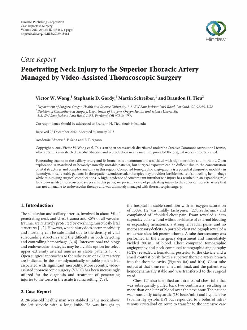
(a) Axial chest CT with intravenous contrast demon-strating active intrathoracic bleeding from a branch ofthe left superior thoracic artery. Red arrow points tothe area of contrast extravasation
(b) 3D reconstruction of the images from (a). Yellowarrow points to the area of active contrast extravasa-tion. White arrowhead points to thoracostomy tube
(c) Arteriogram of left axillary artery demonstratingcontrast extravasation from the superior thoracic artery(yellow arrow). The small size of the bleeding vessel(∼1mm) precluded any endovascular attempts to con-trol bleeding
∗
(d) Schematic of the major branches of the subclavianand axillary artery. Clavicle has been partially resected.Asterisk indicates superior thoracic artery
Figure 1: Radiographic images and schematic of vascular injury.
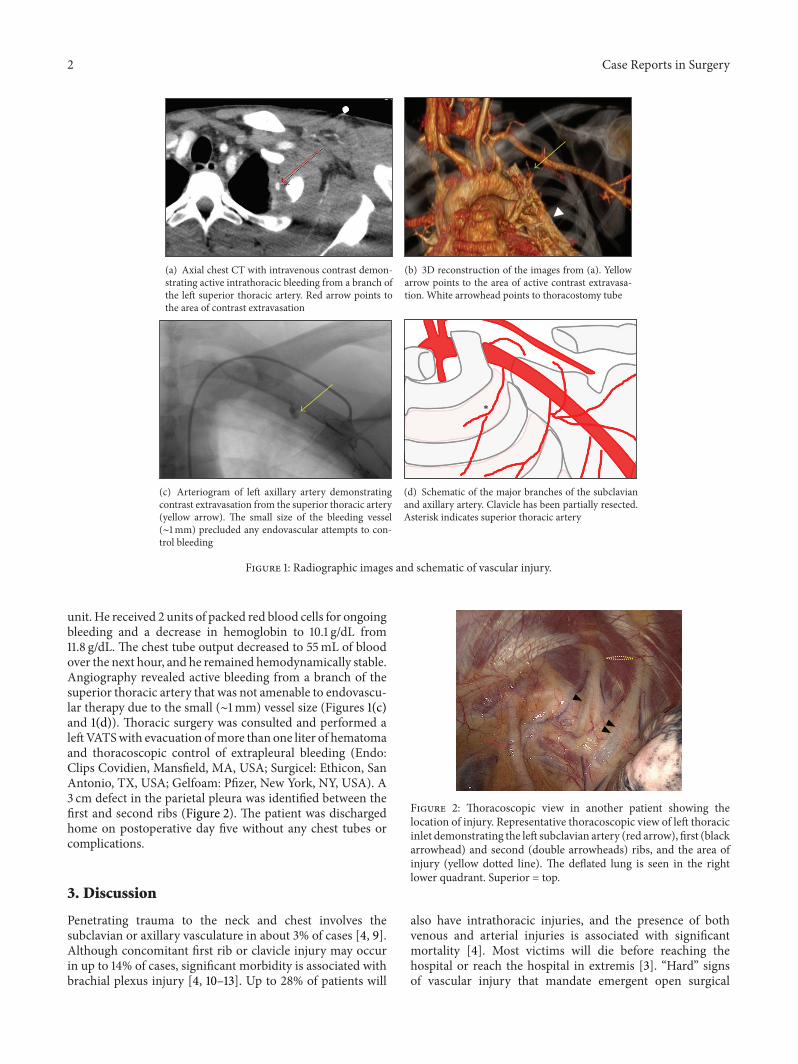
unit. He received 2 units of packed red blood cells for ongoingbleeding and a decrease in hemoglobin to 10.1 g/dL from11.8 g/dL. The chest tube output decreased to 55mL of bloodover the next hour, and he remained hemodynamically stable.Angiography revealed active bleeding from a branch of thesuperior thoracic artery that was not amenable to endovascu-lar therapy due to the small (∼1mm) vessel size (Figures 1(c)and 1(d)). Thoracic surgery was consulted and performed aleftVATSwith evacuation ofmore than one liter of hematomaand thoracoscopic control of extrapleural bleeding (Endo:Clips Covidien, Mansfield, MA, USA; Surgicel: Ethicon, SanAntonio, TX, USA; Gelfoam: Pfizer, New York, NY, USA). A3 cm defect in the parietal pleura was identified between thefirst and second ribs (Figure 2). The patient was dischargedhome on postoperative day five without any chest tubes orcomplications.
3. Discussion
Penetrating trauma to the neck and chest involves thesubclavian or axillary vasculature in about 3% of cases [4, 9].Although concomitant first rib or clavicle injury may occurin up to 14% of cases, significant morbidity is associated withbrachial plexus injury [4, 10–13]. Up to 28% of patients will
Figure 2: Thoracoscopic view in another patient showing thelocation of injury. Representative thoracoscopic view of left thoracicinlet demonstrating the left subclavian artery (red arrow), first (blackarrowhead) and second (double arrowheads) ribs, and the area ofinjury (yellow dotted line). The deflated lung is seen in the rightlower quadrant. Superior = top.
also have intrathoracic injuries, and the presence of bothvenous and arterial injuries is associated with significantmortality [4]. Most victims will die before reaching thehospital or reach the hospital in extremis [3]. “Hard” signsof vascular injury that mandate emergent open surgical
Case Reports in Surgery 3
treatment include severe bleeding, unexplained hypotension,expanding hematoma, or absent peripheral pulses. “Soft”signs of vascular injury include stable small hematomas,minor continuous bleeding, or mild hypotension.
In situations where immediate surgical exploration is notindicated, diagnostic imaging may be warranted to optimizethe treatment approach. Plain radiographs may be used todiagnose the presence of pneumothorax or hemothorax andcan identify foreign bodies. Doppler ultrasound has beenutilized in some centers as a noninvasive tool to effectivelydetect vascular injury but is user dependent, and expertisemay not be widely available [1, 4]. Routine arteriographywas traditionally advocated for all penetrating injuries tozone 1 of the neck (between clavicles and cricoid cartilage),but recent studies have questioned the low yield and theinvasive nature of this approach [14, 15]. Multidetector CTangiography has been shown to be highly sensitive andspecific for evaluating penetrating neck and chest trauma andhas been recommended as the first-line approach for stablepatients with suspected vascular trauma [16, 17].
Up to 50% of penetrating injuries to the subclavian oraxillary artery may be amenable to endovascular therapy[5]. Endovascular repair has been associated with shorteroperative times, less blood loss, and comparable short-term patency compared to open repair [6, 18, 19]. However,longterm outcome studies after endovascular interventionare lacking. Open surgical repair is indicated for unstablepatients, but exposure of injured vessels is often challenging.In general, infraclavicular incisions with either retraction orresection of the medial clavicle can provide adequate expo-sure to the subclavian artery. Division of the pectoral musclescan provide exposure to the axillary artery. The addition ofa median sternotomy and anterolateral thoracotomy exposesthe proximal subclavian vessels via a “trapdoor” approach.
In the acute trauma setting, VATS has traditionally beenused for retained hemothorax or to diagnose diaphrag-matic injury [20]. A recent prospective multicenter studyfound VATS to be the most commonly performed interven-tion following tube thoracostomy for retained hemothorax(33.5%) with a success rate of 70% [21]. Other studies havedemonstrated its utility for chest wall bleeding, diagnosingtransmediastinal or cardiac injury, persistent pneumothorax,and blunt chest injury [22, 23]. Despite its increasing rangeof indications, VATS should only be considered in thehemodynamically stable trauma patient. In summary, wepresent a case of penetrating injury to the thoracic inletwith ongoing intrathoracic bleeding from a superior thoracicartery branch that was not amenable to endovascular repairbut successfully managed with thoracoscopic surgery.
Conflict of Interests
The authors have no financial conflict of interests to disclose.
Acknowledgments
The authors would like to thank the Radiology Departmentat Oregon Health and Science University (Drs. Fatma Berk,Nicolas R. Cahanding, and Robert E. Barton).
References
[1] D. Demetriades and J. A. Asensio, “Subclavian and axillaryvascular injuries,” Surgical Clinics of North America, vol. 81, no.6, pp. 1357–1373, 2001.
[2] C. E. Hyre, D. F. Cikrit, S. G. Lalka, A. P. Sawchuk, and M.C. Dalsing, “Aggressive management of vascular injuries of thethoracic outlet,” Journal of Vascular Surgery, vol. 27, no. 5, pp.880–885, 1998.
[3] D. Demetriades, A. Salim, C. Brown, M. Martin, and P. Rhee,“Neck injuries,” Current Problems in Surgery, vol. 44, no. 1, pp.13–85, 2007.
[4] D. Demetriades, S. Chahwan, H. Gomez et al., “Penetratinginjuries to the subclavian and axillary vessels,” Journal of theAmerican College of Surgeons, vol. 188, no. 3, pp. 290–295, 1999.
[5] J. S. Danetz, A. D. Cassano, M. C. Stoner, R. R. Ivatury, andM. M. Levy, “Feasibility of endovascular repair in penetratingaxillosubclavian injuries: a retrospective review,” Journal ofVascular Surgery, vol. 41, no. 2, pp. 246–254, 2005.
[6] P. Castelli, R. Caronno, G. Piffaretti et al., “Endovascular repairof traumatic injuries of the subclavian and axillary arteries,”Injury, vol. 36, no. 6, pp. 778–782, 2005.
[7] S. Milanchi, I. Makey, R. McKenna, and D. R. Margulies,“Video-assisted thoracoscopic surgery in the management ofpenetrating and blunt thoracic trauma,” Journal of MinimalAccess Surgery, vol. 5, no. 3, pp. 63–66, 2009.
[8] P. H. Navsaria and A. J. Nicol, “Video-assisted thoracoscopicpericardial window for penetrating cardiac trauma,” SouthAfrican Journal of Surgery, vol. 44, no. 1, pp. 18–20, 2006.
[9] P. H. Lin, A. J. Koffron, P. J. Guske et al., “Penetrating injuries ofthe subclavian artery,” American Journal of Surgery, vol. 185, no.6, pp. 580–584, 2003.
[10] H. Gill, W. Jenkins, S. Edu, W. Bekker, A. J. Nicol, and P. H.Navsaria, “Civilian penetrating axillary artery injuries,” WorldJournal of Surgery, vol. 35, no. 5, pp. 962–966, 2011.
[11] E. Degiannis, R. D. Levy, T. Potokar, and R. Saadia, “Penetratinginjuries of the axillary artery,” Australian and New ZealandJournal of Surgery, vol. 65, no. 5, pp. 327–330, 1995.
[12] J. D. Richardson, R. B. McElvein, and J. K. Trinkle, “First ribfracture: a hallmark of severe trauma,” Annals of Surgery, vol.181, no. 3, pp. 251–254, 1975.
[13] E. H. Phillips, W. F. Rogers, and M. R. Gaspar, “First ribfractures: incidence of vascular injury and indications forangiography,” Surgery, vol. 89, no. 1, pp. 42–47, 1981.
[14] F. Munera, S. Cohn, and L. A. Rivas, “Penetrating injuries ofthe neck: use of helical computed tomographic angiography,”Journal of Trauma—Injury, Infection and Critical Care, vol. 58,no. 2, pp. 413–418, 2005.
[15] B. O. Patterson, P. J. Holt, M. Cleanthis, N. Tai, T. Carrell, and T.M. Loosemore, “Imaging vascular trauma,” The British Journalof Surgery, vol. 99, no. 4, pp. 494–505, 2012.
[16] K. Inaba, B. C. Branco, J. Menaker et al., “Evaluation of mul-tidetector computed tomography for penetrating neck injury: aprospectivemulticenter study,”The Journal of Trauma andAcuteCare Surgery, vol. 72, no. 3, pp. 576–583, 2012.
[17] K. Shanmuganathan and J.Matsumoto, “Imaging of penetratingchest trauma,” Radiologic Clinics of North America, vol. 44, no.2, pp. 225–238, 2006.
[18] E. S. Xenos, M. Freeman, S. Stevens, D. Cassada, J. Pacanowski,andM. Goldman, “Covered stents for injuries of subclavian andaxillary arteries,” Journal of Vascular Surgery, vol. 38, no. 3, pp.451–454, 2003.
4 Case Reports in Surgery
[19] A. V. Patel, M. L. Marin, F. J. Veith, A. Kerr, and L. A. Sanchez,“Endovascular graft repair of penetrating subclavian arteryinjuries,” Journal of Endovascular Surgery, vol. 3, no. 4, pp. 382–388, 1996.
[20] N. Ahmed and D. Jones, “Video-assisted thoracic surgery: stateof the art in trauma care,” Injury, vol. 35, no. 5, pp. 479–489,2004.
[21] J. DuBose, K. Inaba, D. Demetriades et al., “Management ofpost-traumatic retained hemothorax: a prospective, observa-tional, multicenter AAST study,” The Journal of Trauma andAcute Care Surgery, vol. 72, no. 1, pp. 11–22, 2012.
[22] S. R. Casos and J. D. Richardson, “Role of thoracoscopy in acutemanagement of chest injury,” Current Opinion in Critical Care,vol. 12, no. 6, pp. 584–589, 2006.
[23] J. W. Smith, G. A. Franklin, B. G. Harbrecht, and J. D.Richardson, “Early VATS for blunt chest trauma: amanagementtechnique underutilized by acute care surgeons,” Journal ofTrauma—Injury, Infection and Critical Care, vol. 71, no. 1, pp.102–105, 2011.

Submit your manuscripts athttp://www.hindawi.com
Stem CellsInternational
Hindawi Publishing Corporationhttp://www.hindawi.com Volume 2014
Hindawi Publishing Corporationhttp://www.hindawi.com Volume 2014
MEDIATORSINFLAMMATION
of
Hindawi Publishing Corporationhttp://www.hindawi.com Volume 2014
Behavioural Neurology
EndocrinologyInternational Journal of
Hindawi Publishing Corporationhttp://www.hindawi.com Volume 2014
Hindawi Publishing Corporationhttp://www.hindawi.com Volume 2014
Disease Markers
Hindawi Publishing Corporationhttp://www.hindawi.com Volume 2014
BioMed Research International
OncologyJournal of
Hindawi Publishing Corporationhttp://www.hindawi.com Volume 2014
Hindawi Publishing Corporationhttp://www.hindawi.com Volume 2014
Oxidative Medicine and Cellular Longevity
Hindawi Publishing Corporationhttp://www.hindawi.com Volume 2014
PPAR Research
The Scientific World JournalHindawi Publishing Corporation http://www.hindawi.com Volume 2014
Immunology ResearchHindawi Publishing Corporationhttp://www.hindawi.com Volume 2014
Journal of
ObesityJournal of
Hindawi Publishing Corporationhttp://www.hindawi.com Volume 2014
Hindawi Publishing Corporationhttp://www.hindawi.com Volume 2014
Computational and Mathematical Methods in Medicine
OphthalmologyJournal of
Hindawi Publishing Corporationhttp://www.hindawi.com Volume 2014
Diabetes ResearchJournal of
Hindawi Publishing Corporationhttp://www.hindawi.com Volume 2014
Hindawi Publishing Corporationhttp://www.hindawi.com Volume 2014
Research and TreatmentAIDS
Hindawi Publishing Corporationhttp://www.hindawi.com Volume 2014
Gastroenterology Research and Practice
Hindawi Publishing Corporationhttp://www.hindawi.com Volume 2014
Parkinson’s Disease
Evidence-Based Complementary and Alternative Medicine
Volume 2014Hindawi Publishing Corporationhttp://www.hindawi.com


![Case Report Chylous Fistula following Axillary ...downloads.hindawi.com/journals/cris/2016/6098019.pdf · er neck dissection, which has an incidence of % [ ]. ... e thoracic duct](https://img.dokumen.tips/doc/110x75/5fa84ebd5dbab2650952d201/case-report-chylous-fistula-following-axillary-er-neck-dissection-which-has.jpg)