Embed Size (px)
Citation preview

Penetrating Neck Trauma
Diane M. Birnbaumer, M.D., FACEP Professor of Clinical Medicine David Geffen School of Medicine at UCLA Senior Physician Department of Emergency Medicine Harbor-UCLA Medical Center
What IS Penetrating Neck Trauma?
Injuries from GSW, stabbing, penetrating debris
Violates the platysma Often injures multiple structures Mortality highly variable: 2-65%

Prehospital Management
Cervical spine immobilization? Airway management? Venous access?

Prehospital Management
Cervical spine immobilization? Of course! Or maybe not…
Less than 0.5% of patients with penetrating wounds to the neck have an unstable cervical spine injury
All of them tend to have signs or symptoms of neurologic impairment or altered mental status, so…
If altered, immobilize If neuro findings, immobilize

Prehospital Management
Airway management? Oxygen; suction as needed Position of comfort if possible Assist, intubate as needed
Venous access? On opposite side of injury, if possible

Assessment of the Patient
Where is the injury? Anterior or posterior? Deep to the platysma? Which zone(s) is (are) involved?

Anterior or Posterior?
Most important structures are in the anterior triangle
Posterior: Spinal cord

Anatomy of the Neck: Triangles
Anterior Triangle Midline anteriorly Sternocleidomastoid
posteriorly Clavicle inferiorly Mandible superiorly
Posterior Triangle Sternocleidomastoid
anteriorly Trapezius posteriorly Clavicle inferiorly

The Platysma
1
Is the injury deep to the platysma?
If not, just close and go
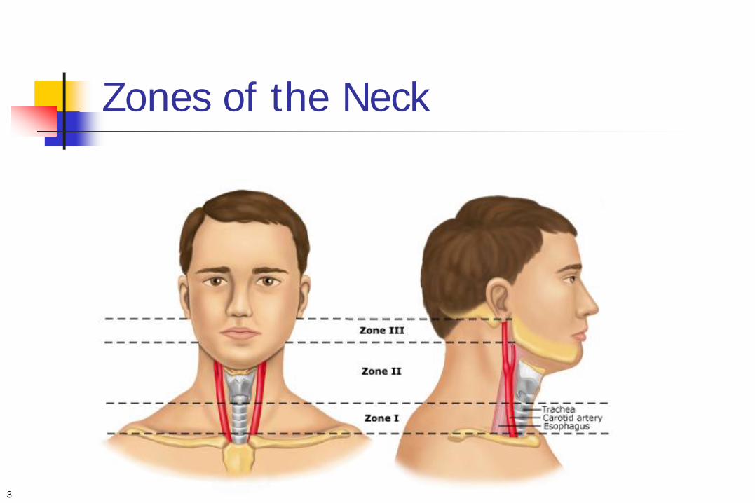
Zones of the Neck
3
Anatomy of the Neck
Zone I Between the sternal notch and clavicles inferiorly and the
cricoid cartilage superiorly
Zone II Between the cricoid cartilage inferiorly and the angle of the
mandible superiorly
Zone III Between the angle of the mandible inferiorly and the base of
the skull superiorly


Zone I Contains:
Common carotid artery Vertebral artery Subclavian artery Major vessels of the
upper mediastinum Apices of lungs
Esophagus Trachea Thyroid Thoracic duct Spinal cord

Zone II Contains:
Common carotid artery Vertebral artery Larynx Trachea Esophagus Pharynx
Internal jugular vein Vagus nerve Recurrent laryngeal
nerve Sympathetic trunk Spinal cord

Zone III contains:
Internal and external carotid arteries
Vertebral arteries Jugular veins Salivary and parotid glands Cranial nerves IX-XII Spinal cord Floor of mouth / skull

Penetrating injuries broken down by zone
Gracias 2001
Osborn 2008
Thoma 2008
Bagheri 2008
Brennen 2010
Zone I 20% 14% 16% 15%
Zone II 77% 36% 64% 85%
Zone III 39% 25% 18% 20% 8%
Post Triangle 42%

What gets injured?
Location Study 1 (1275) Study 2 Thoma
Arterial 320 (25%) 12.8% 13.3%
Venous 281 (22%) 11.3%
Tracheolaryngeal 253 (20%) 10.1% 3.9%
Pharyngoesophageal 240 (19%) 9.6% 8.8%
Spinal cord 76 (6%) 3% 0.5%
Neurologic, other 85 (7%) 3.4%
Thoracic duct 20 (2%) 0%

Where is the injury? Anterior or posterior? Deep to the platysma? Which zone(s) is (are) involved?
Is the patient stable or unstable? Are there any “hard findings” on exam?
Assessment of the Patient

Signs of injury: Examine for These
Hard Signs
Expanding Hematoma Severe active bleeding Shock not responsive to IVF Decreased/absent radial
pulse Vascular bruit or thrill Cerebral ischemia Airway obstruction
Soft Signs Hemoptysis/hematemesis Oropharyngeal blood Dyspnea Dysphonia/dysphagia Subcutaneous/mediastinal air Chest tube air leak Non-expanding hematoma Focal neurologic deficit

Initial Management
Venous access; opposite side of injury Oxygen; airway management as needed* Bleeding profusely? Pressure; do not clamp
structures

Airway Management
What are the issues? Potential to lose the airway if injury worsens Potential to worsen the injury by patient
coughing, worsening bleeding, direct trauma

Penetrating Neck Injury Airway Management
Immediate airway management Decreasing mental status Expanding hematoma Direct laryngotracheal trauma Hypoventilation Hypoxia
Selective intubation Ah… here’s where the rubber meets the road

Selective airway management Consider in any patient leaving the ED for a study or
transportation Consider in any patient who may develop airway
compromise
Okay, that sounds good, but… How to do it????
Penetrating Neck Injury Airway Management

Penetrating Neck Injury Airway Management
What are your options? RSI with orotracheal intubation Awake intubation Fiberoptic Videoscopic intubation Extraglottic devices Nasotracheal Cricothyrotomy

Penetrating Neck Injury Airway Management
Airway Management RSI with direct laryngoscopy
Most data available; usually a safe practice; have backup airway available
Fiberoptic intubation Awake fiberoptic an option May be difficult / impossible in a bloody airway
Videoscopic devices Little data published; success rates high
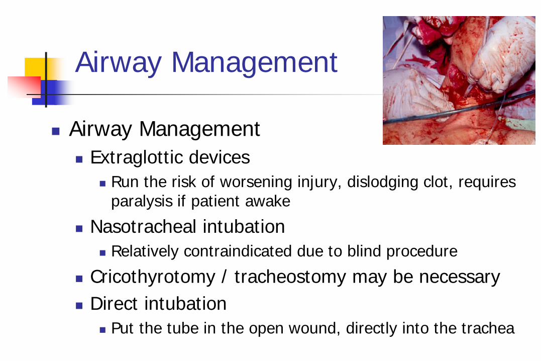
Airway Management
Airway Management Extraglottic devices
Run the risk of worsening injury, dislodging clot, requires paralysis if patient awake
Nasotracheal intubation Relatively contraindicated due to blind procedure
Cricothyrotomy / tracheostomy may be necessary Direct intubation
Put the tube in the open wound, directly into the trachea

Penetrating Neck Trauma Airway Management
Mandavia, et al USC, 1993-1996 78% GSW, 21% SW 748 patients
In 11%, airway emergently managed in ED RSI – 39 Cricothyrotomy / tracheostomy - 2 No drugs – 5 Fiberoptic - 12

Platysma Violation?
Wound Care, Discharge
Patient Stable?
Emergent Airway, then
OR
Symptomatic?
Symptomatic
Angiography, Esophagoscopy, Bronchoscopy Zone I
Zone II
Zone III
OR
Angiography
Observe
Observe
Yes
Yes
No
No
No
Yes
No
Yes
Penetrating Neck Trauma The Traditional Approach

Imaging?
What does the current literature say?

2001 Gracias et al.
Retrospective case series of 68 patients, 23 of which were included: 13 patients were determined to have trajectories
with low likelihood of vascular or aerodigestive injuries and managed non-operatively. 4 were discharged from the ED 7 were discharged within 24 hours

2008 Osborn et al.
Retrospective case series of 120 patients, 65 of which were included: 24 received a CTA, 6 got explored (25%)
25% determined to have injury 0 negative explorations
41 received no CTA, 27 got explored (66%) 34% determined to have injury 13 had negative explorations (48%) and 4 minor
superficial bleeding vessels

2008 Thoma et al.
A prospective observational study of 203 patients with 159 stab wounds, 42 GSW’s, one automobile part and one explosive shrapnel: 25 were managed operatively 8 were managed endovascularly 158 received what the surgeon believed to be
appropriate imaging and work-up and were managed expectantly
No clinically relevant missed injuries

Vick & Islam 2008
Retrospective case series of 19,363 pediatric trauma patients, including 39 children with 42 injuries violating the platysma: Six patients underwent mandatory exploration, four
were nontherapeutic; Eighteen patients underwent imaging (68% CTA),
15 were observed and avoided surgical exploration.
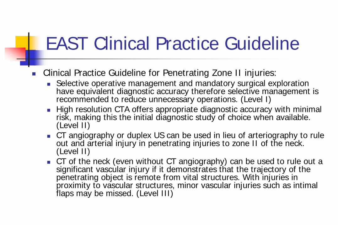
EAST Clinical Practice Guideline Clinical Practice Guideline for Penetrating Zone II injuries:
Selective operative management and mandatory surgical exploration have equivalent diagnostic accuracy therefore selective management is recommended to reduce unnecessary operations. (Level I)
High resolution CTA offers appropriate diagnostic accuracy with minimal risk, making this the initial diagnostic study of choice when available. (Level II)
CT angiography or duplex US can be used in lieu of arteriography to rule out and arterial injury in penetrating injuries to zone II of the neck. (Level II)
CT of the neck (even without CT angiography) can be used to rule out a significant vascular injury if it demonstrates that the trajectory of the penetrating object is remote from vital structures. With injuries in proximity to vascular structures, minor vascular injuries such as intimal flaps may be missed. (Level III)

Kesser 2009 et al.
Invited Commentary “The standard of care is no longer surgical
exploration for all penetrating neck trauma as numerous studies have clearly demonstrated that stable patients can be imaged and/or observed.”

Imaging?
Stable patients can be imaged and observed Unstable patients need to go to the OR Hard findings are critical in the diagnostic
algorithm

Platysma Violation?
Wound Care, Discharge
Patient Unstable or
Hard signs of injury?
OR
CTA Neck Appropriate imaging or intervention
Observe/ Discharge
No
Yes
Yes
No
OR
Obvious Injury
No obvious injury but trajectory suggests possible injury
No obvious injury and trajectory away from vital structures

References/Images Bagheri SC, Khan A, Bell RB. Penetrating Neck Injuries. Oral and Maxillofacial Surgery Clinics of North America. August 2008;20(3):393-414. Brennan J, Lopez M, Gibbons MD, et. al. Penetrating Neck Trauma in Operation Iraqi Freedom. Otolaryngology – Head and Neck Surgery. December
2011;144:180-185 Brywczynski JJ, Barrett TW, Lyon JA, Cotton BA. Management of Penetrating Neck Injury in the Emergency Department: A Structued Literature Review.
Emergency Medicine Journal. 2008;25:711-715 Demetriades et al. “Complex Problems in Penetrating Neck Trauma.” Surgical Clinics of North America Vol 76;4. August 1996; 661-683 Gracias VH, Reilly PM, Philpott J, et. al. Computed Tomography in the Evaluation of Penetrating Neck Trauma: A Preliminary Study. Archives of Surgery.
November 2001;136:1231-1235 Jarvik et al. “Penetrating Neck Trauma: Sensitivity of Clinical Examination and Cost-effectiveness of Angiography.” AJNR Am J Neuroradiol 16;647-654.
April 1995 Kendall et al. “Penetrating Neck Trauma.” Emergency Medicine Clinics of North America. Vol 16;1. Feb 1998. 86-105 Kesser BW, Chance E, Kleiner D, Young JS. Contemporary Management of Penetrating Neck Trauma. The American Surgeon. January 2009;75:1-10 Levy, David and Brian Gruber. “Neck Trauma” emedicine. Dec 10,2009. Lustenberger T, Talving P, Lam L. et. al. Unstable Cervical Spine Fracture After Penetrating Neck Injury: A Rare Entity in an Analysis of 1,069 Patients.
The Journal of Trauma, Injury, Infection, and Critical Care. April 2001;70:870-872 Mazolewski PJ, Curry JD, Browder T, Fildes J. Computed Tomographic Scan Can Be Used for Surgical Decision Making in Zone II Penetrating Neck
Injuries. The Journal of Trauma, Injury, Infection, and Critical Care. August 2001;51:315-319 Munera F, Soto JA, Palacio A, et. al. Diagnosis of Arterial Injuries Casued by Penetrating Trauma to the Neck: Comparison of Helical CT Angiography and
Conventional Angiography. Journal of Radiology. August 2000;216:356-362 Newton, K. Chapter 41: Neck. In: Walls, Ron M, editor. Rosen’s Emergency Medicine: Concepts and Clinical Practice. Mosby Elsevier 2010. p. 377-
386 Osborn TM, Bell RB, Qaisi W, Long WB. Computed Tomographic Angiography as an Aid to Clinical Decision Making in the Selective Management of
Penetrating Injuries to the Neck: A Reduction in the Need for Operative Explorations. The Journal of Trauma, Injury, Infection, and Critical Care. June 2008;64:1466-1471
Steenburg SD, Sliker CW, Shanmuganathan K, Siegel EL. Imaging Evaluation of Penetrating Neck Injuries. July 2010;30:869-886 Tisherman SA, Bokhari F, Collier B. Clinical Practice Guideline: Penetrating Zone II Neck Trauma. The Journal of Trauma, Injury, Infection, and Critical
Care. May 2008;64:1392-1405 Thoma M, Navsaria PH, Edu S, Nicol AJ. Analysis of 203 Patients with Penetrating Neck Injuries. World Journal of Surgery. 2008;32:2716-2723 Vick LR, Islam S. Adding Insult to Injury: Neck Exploration for Penetrating Pediatric Neck Trauma. The American Surgeon. November 2008;74:1104-1106

Any Questions?