Embed Size (px)
Citation preview

Cummings Ch 115:Penetrating and Blunt Trauma
to the Neck
Kimanh NguyenMay 29, 2013
Vital Structures
• Air passages– Trachea, larynx, pharynx, lungs
• Vascular– Carotid, jugular, subclavian, innominate, aortic arch
• Gastrointestinal– Pharynx, esophagus
• Neurologic– Spinal cord, brachial plexus, peripheral nerves,
cranial nerves

Kinetic Energy
• Kinetic energy affects magnitude of injury:• KE = ½ M (V1 – V2)2
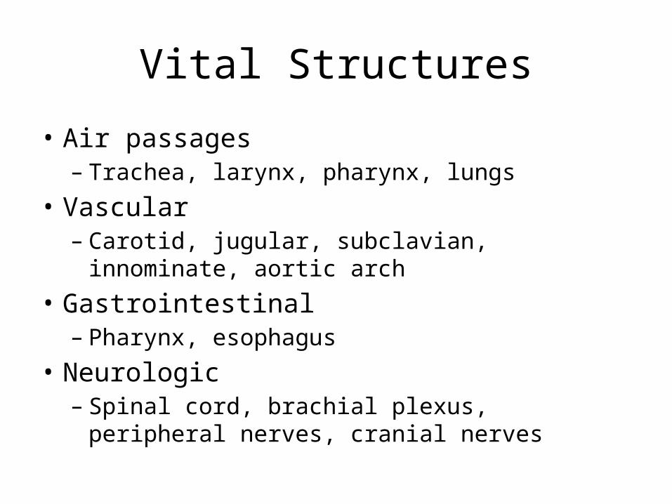
Handguns
• Projectile type• Speed– Handguns/pistols are low velocity (90-600 m/s)
• Caliber– .44-caliber magnum is comparable to a rifle
• Yaw– Tumbling bullet causes injury in a wider path

Rifles
• Military bullets– Jacket creates smoother flight, clean hole,
through-and-through wound– High velocity (760 m/s) transmits energy waves to
surrounding tissue• Hunting rifles with expanding bullets– Soft-tips expand, create large wound cavity, may
not exit, may fragment• High mortality

Different Missiles
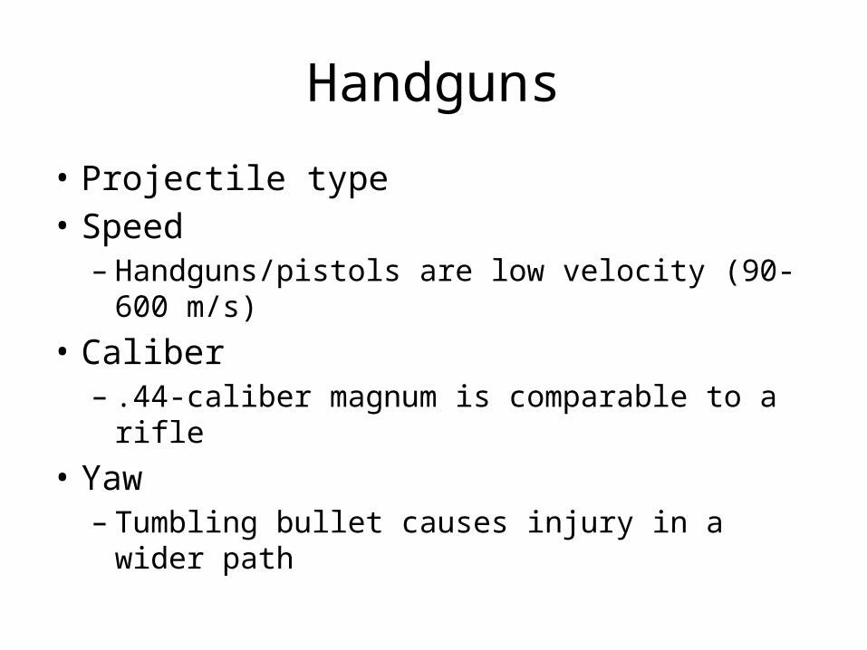
Shotguns
• Velocity ~ 300 m/s• Distance– Pellets scatter at longer distances
• Type of weapon– Sawed-off shotgun sprays the shot earlier
• Size of projectile (shot)– Birdshots (< 3.5 mm, 12m range)– Buckshots (> 3.5 mm, 150m range). Comparable to
handgun bullet wounds• Wadding

Stab Injuries
• Single-entry vs multiple stab wounds• Higher incidence of subclavian vessel
laceration due to downward direction• Lower incidence of spinal injuries
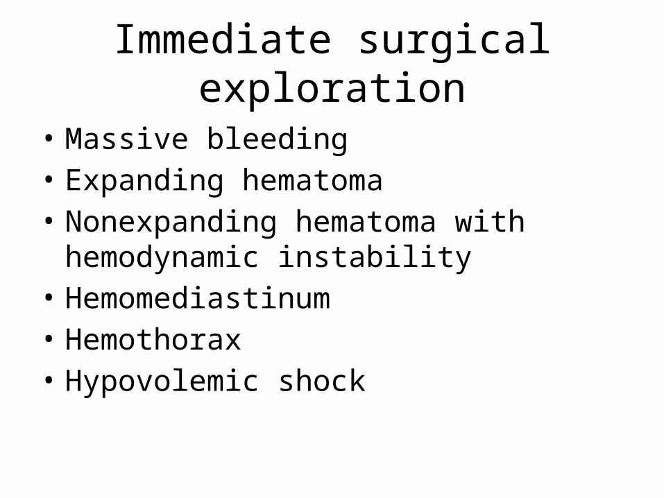
Immediate surgical exploration
• Massive bleeding• Expanding hematoma• Nonexpanding hematoma with hemodynamic
instability• Hemomediastinum• Hemothorax• Hypovolemic shock
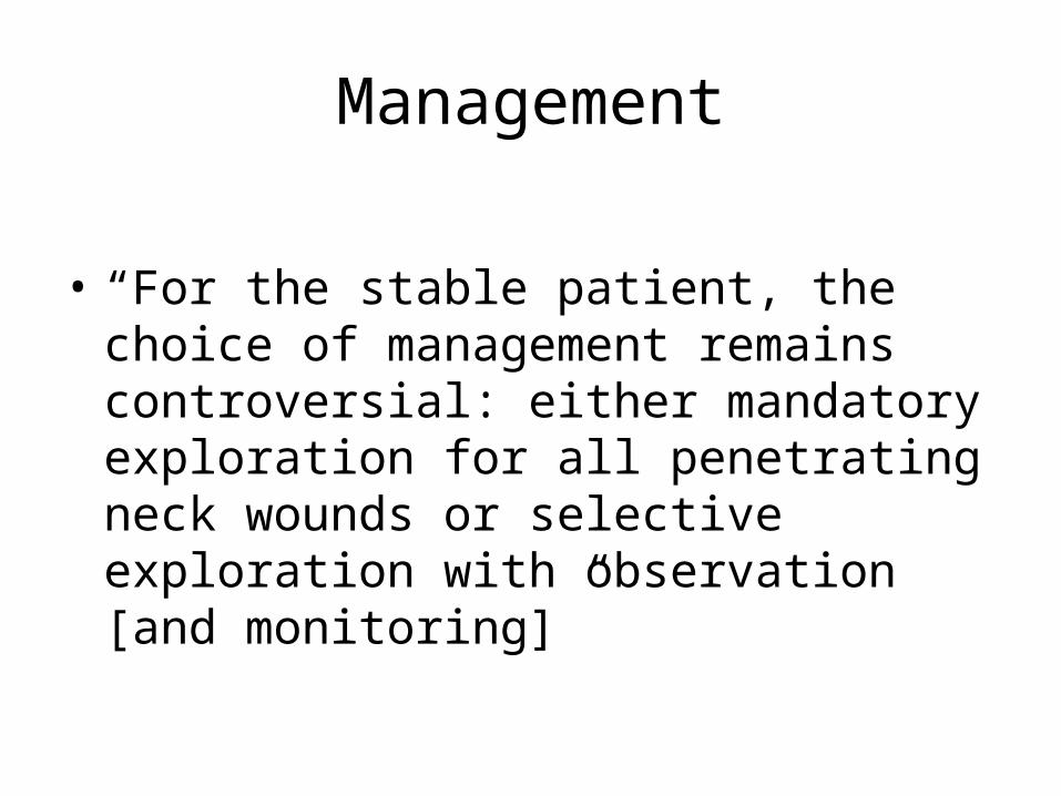
Management
• “For the stable patient, the choice of management remains controversial: either mandatory exploration for all penetrating neck wounds or selective exploration with observation [and monitoring]”
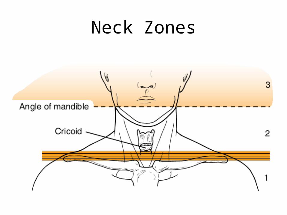
Neck Zones
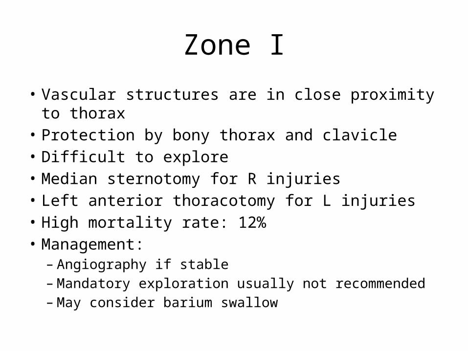
Zone I
• Vascular structures are in close proximity to thorax• Protection by bony thorax and clavicle• Difficult to explore• Median sternotomy for R injuries• Left anterior thoracotomy for L injuries• High mortality rate: 12%• Management:
– Angiography if stable – Mandatory exploration usually not recommended– May consider barium swallow
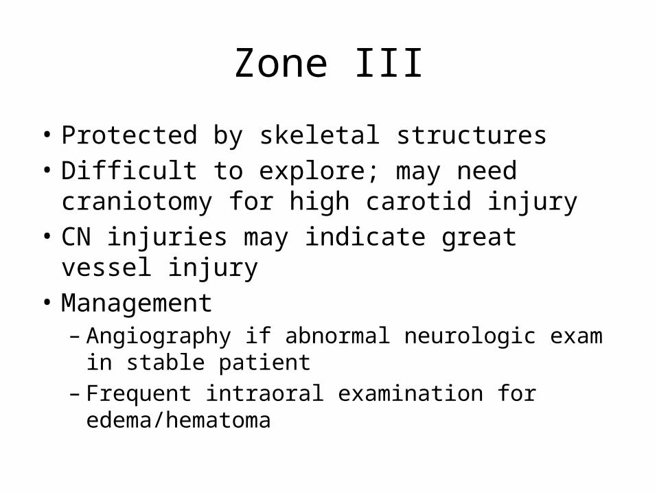
Zone III
• Protected by skeletal structures• Difficult to explore; may need craniotomy for
high carotid injury• CN injuries may indicate great vessel injury• Management– Angiography if abnormal neurologic exam in stable
patient– Frequent intraoral examination for
edema/hematoma
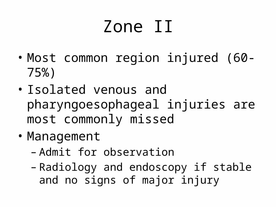
Zone II
• Most common region injured (60-75%)• Isolated venous and pharyngoesophageal
injuries are most commonly missed • Management – Admit for observation– Radiology and endoscopy if stable and no signs of
major injury
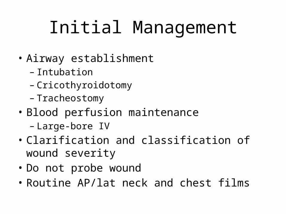
Initial Management
• Airway establishment– Intubation– Cricothyroidotomy– Tracheostomy
• Blood perfusion maintenance– Large-bore IV
• Clarification and classification of wound severity• Do not probe wound• Routine AP/lat neck and chest films

Management of Penetrating Neck Injury

Management of Penetrating Zone II Injury
Vascular Penetration
• Zone I– Thoracic surgery
• Zone III– Temporary pressure or carotid arterial bypass– No. 4 Fogarty catheter
• Jugular– Ligation
• Carotid– Ligation of ECA– Lateral arteriorhaphy, end-to-end anastomosis, autogenous grafting– IR transcatheter arterial embolization

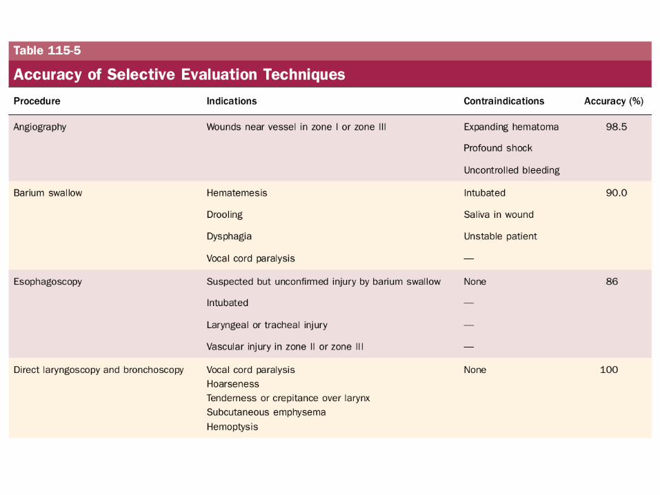
Digestive Tract Injury
• Gastrograffin swallow• Barium swallow• Flexible esophagoscopy (risk of missing perforations near
CP and hypopharynx)• Rigid esophagoscopy• Neck exploration for subQ emphysema or mediastinitis;
localization with methylene blue• Management of esophageal injury
– 2-layer closure with wound irrigation, debridement, drainage, possible muscle flap
– Lateral cervical esophagostomy, later definitive repair

Laryngotracheal Injury
• Repair mucosal lacerations within 24 hours• Soft laryngeal stent for badly macerated
mucosa• 6-week trach below or through the injury for
significant injuries that detach a tracheal ring or encroach on the airway
Blunt Neck Injury
• Occult cervical spine injury• Delayed onset of signs and symptoms• Careful observation• Thrombosis, intimal tears, dissection,
pseudoaneurysm
Cummings Ch 116:Differential Diagnosis
of Neck Masses
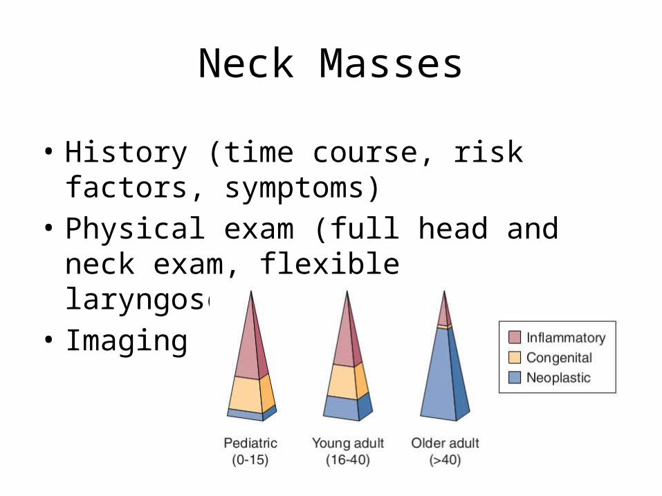
Neck Masses
• History (time course, risk factors, symptoms)• Physical exam (full head and neck exam,
flexible laryngoscopy)• Imaging
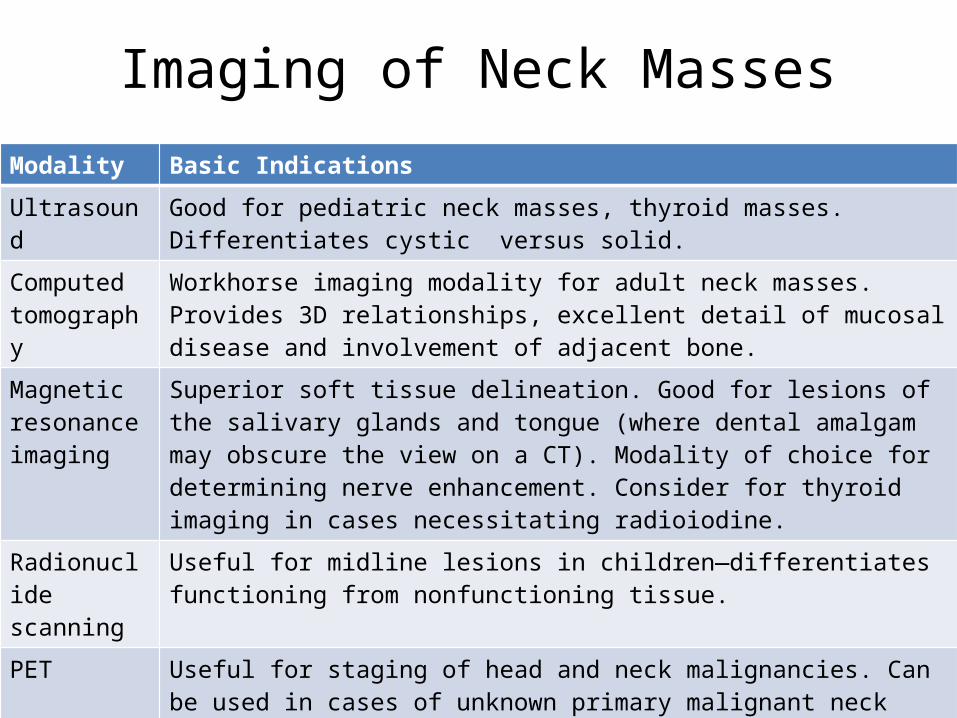
Imaging of Neck MassesModality Basic Indications
Ultrasound Good for pediatric neck masses, thyroid masses. Differentiates cystic versus solid.
Computed tomography
Workhorse imaging modality for adult neck masses. Provides 3D relationships, excellent detail of mucosal disease and involvement of adjacent bone.
Magnetic resonance imaging
Superior soft tissue delineation. Good for lesions of the salivary glands and tongue (where dental amalgam may obscure the view on a CT). Modality of choice for determining nerve enhancement. Consider for thyroid imaging in cases necessitating radioiodine.
Radionuclide scanning
Useful for midline lesions in children—differentiates functioning from nonfunctioning tissue.
PET Useful for staging of head and neck malignancies. Can be used in cases of unknown primary malignant neck masses or treated neck disease.
Angiography Useful for lesions encasing the carotid and vascular lesions. Conventional angiography should be considered for preoperative assessment in cases of potential carotid artery sacrifice or where embolization is required.

Initial workup
• Antibiotic trial• Further investigation for concerning
signs/symptoms– Unilateral, enlarging, asymmetric, supraclavicular
fossae, not associated with infections• Imaging• Biopsy– FNA (gold standard), repeat FNA, core needle
biopsy, open biopsy, neck dissection (SCCA)
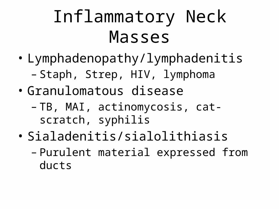
Inflammatory Neck Masses
• Lymphadenopathy/lymphadenitis– Staph, Strep, HIV, lymphoma
• Granulomatous disease– TB, MAI, actinomycosis, cat-scratch, syphilis
• Sialadenitis/sialolithiasis– Purulent material expressed from ducts

Congenital Neck Masses
• Rule out malignancy in adults• Thyroglossal duct cyst– Midline neck mass that elevates with tongue protrusion
or swallowing– Rule out median ectopic thyroid– Sistrunk procedure
• Branchial cleft anomalies– Cyst, sinus, or fistula– 1st arch (1%), 2nd arch (95%), 3rd and 4th arch (rare)– Complete excision of the tract
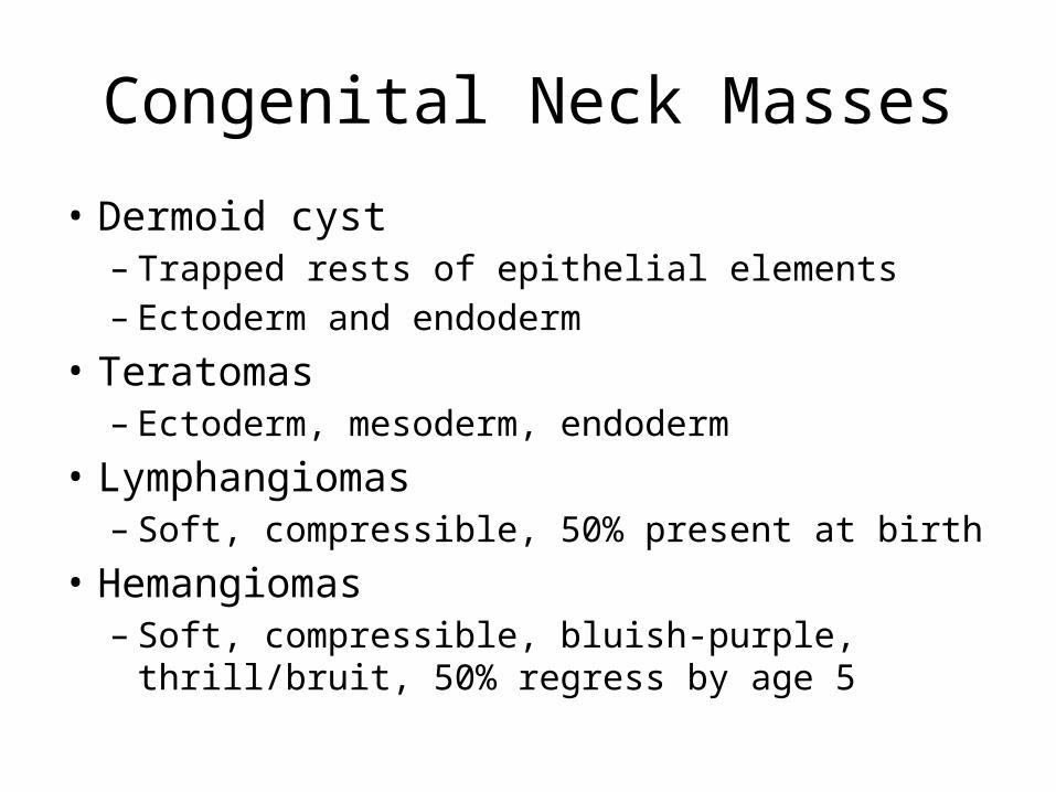
Congenital Neck Masses
• Dermoid cyst– Trapped rests of epithelial elements– Ectoderm and endoderm
• Teratomas– Ectoderm, mesoderm, endoderm
• Lymphangiomas– Soft, compressible, 50% present at birth
• Hemangiomas– Soft, compressible, bluish-purple, thrill/bruit, 50%
regress by age 5

Primary Neoplasms of the Neck
• Lymphoma– Most common H&N malignancy in children– 80% of HL have cervical disease– 33% of NHL have cervical disease (90% B-cell)
• Thyroid neoplasms– 90% of thyroid nodules are benign
• Salivary gland neoplasms– 80% parotid, 15% SMG

Primary Neoplasms of the Neck
• Neurogenic neoplasms– Schwannoma (most common), neurofibromas, malignant
peripheral nerve sheath tumors, neuromas• Paragangliomas– Neuroectoderm origin, secrete catecholamines
• Carotid body, jugulotympanic region, vagus nerve– 10% autosomal dominant/syndromic, 10% multicentric, <10%
malignant– Salt and pepper appearance on T1-MRI
• Lipomas– Mostly in posterior neck

Unknown Primary SCCA
• Thorough physical exam• Imaging of the head, neck, and chest• Panendoscopy and biopsies (BOT, tonsils, NP,
HP)