Embed Size (px)
Citation preview
CASE REPORT
Penetrating neck injury from a screwdriver: can theNo Zone approach be applied to Zone I injuries?Nikita R Bhatt,1,2 Morgan McMonagle1
1Department of Surgery,University Hospital Waterford,Waterford, Ireland2Department of Surgery,University Hospital Limerick,Limerick, Ireland
Correspondence toDr Nikita R Bhatt,[email protected]
Accepted 16 November 2015
To cite: Bhatt NR,McMonagle M. BMJ CaseRep Published online:[please include Day MonthYear] doi:10.1136/bcr-2015-212666
SUMMARYThe newer approach to management of penetrating neckinjuries (PNI) involves the No Zone approach:comprehensive physical examination combined with CTangiography for triage to effectively identify or excludevascular and aerodigestive injury. This approach,however, has a low negative exploration rate; there isrisk of missing occult injuries especially Zone I and IIIPNI. We report a case of a patient with PNI to Zone I ofthe neck who was haemodynamically stable atpresentation; CT scan revealed complete occlusion of thecommon carotid artery. Immediate surgical explorationrevealed an occult hypopharyngeal injury in addition tothe arterial trauma, which was missed on the CT scan.Hence the No Zone approach needs cautious clinicalapplication, especially in Zone I injuries.
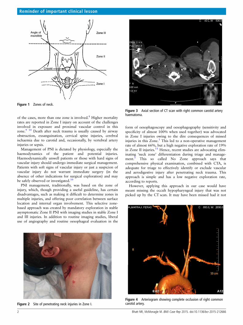
BACKGROUNDPenetrating neck injuries (PNI) often involve mul-tiple concomitant structures, which makes surgicalexposure and management challenging.1 PNI aredivided into three anatomical zones (figure 1),which helps in the approach and management ofwounds.2 Zone I injuries are common in stabbingwounds and almost one-third are asymptomatic atpresentation.3 Multidetector CT angiography(CTA) has a high sensitivity and specificity ofalmost 90–100% and 93.5–100%, respectively, forPNI.4 Hence, according to the new No Zoneapproach for managing PNI, comprehensive phys-ical examination combined with CTA is adequatefor triage to effectively identify or exclude vascularand aerodigestive injury.5 This approach, however,may not be adequate to diagnose all injuries in aZone I trauma.
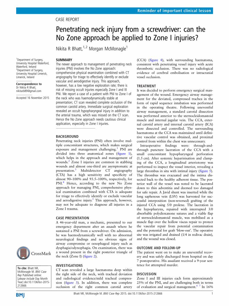
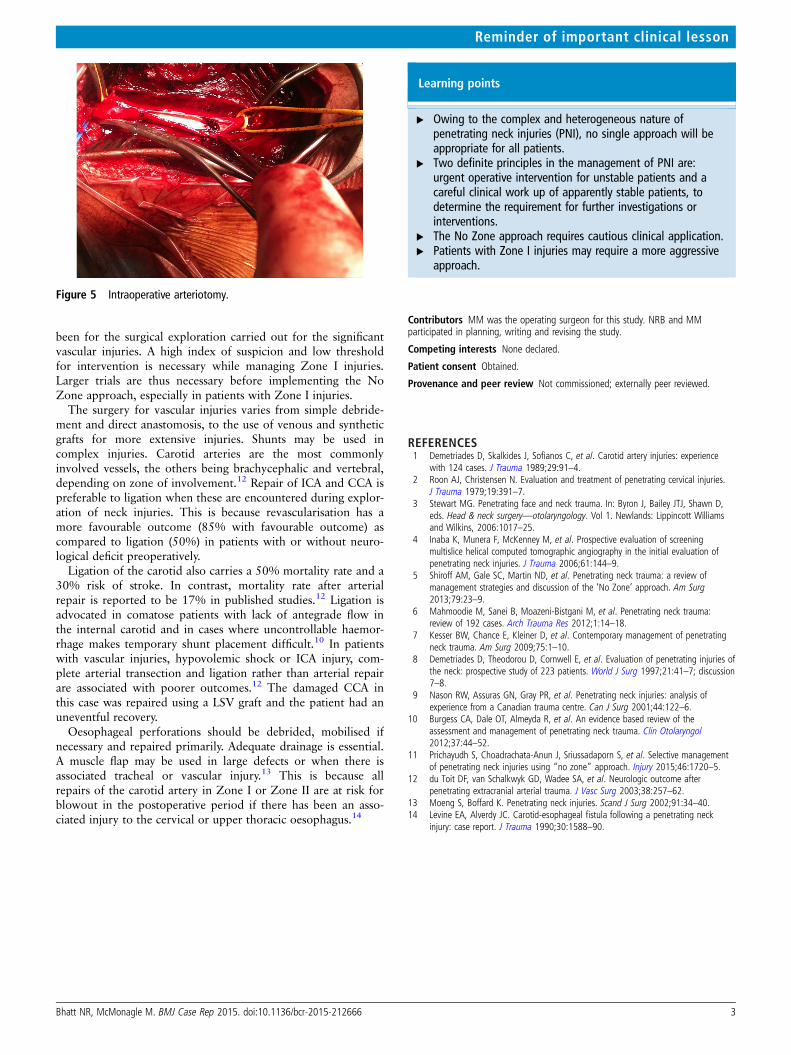
CASE PRESENTATIONA 46-year-old man, a mechanic, presented to ouremergency department after an assault where hesustained a PNI from a screwdriver. On admission,he was haemodynamically well with no abnormalneurological findings and no obvious signs ofairway compromise or oesophageal injury such asdysphagia/odynophagia. On examination, there wasa wound present in the right posterior triangle ofthe neck (Zone I) (figure 2).
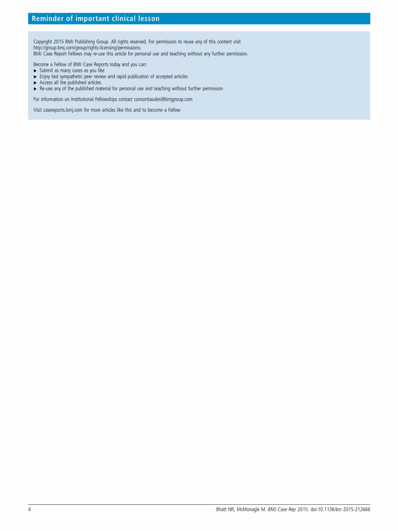
INVESTIGATIONSCT scan revealed a large haematoma deep withinthe right side of the neck, with tracheal deviationand radiological evidence/mild tracheal compres-sion (figure 3). In addition, there was completeocclusion of the right common carotid artery
(CCA) (figure 4), with surrounding haematoma,consistent with penetrating vessel injury with acutethrombotic occlusion. There was no radiologicalevidence of cerebral embolisation or intracranialvessel occlusion.
TREATMENTIt was decided to perform emergency surgical man-agement of the wound. Emergency airway manage-ment for the deviated, compressed trachea in theform of rapid sequence intubation was performedin the operating theatre. Following uneventfulairway management, a standard carotid dissectionwas performed anterior to the sternocleidomastoidmuscle and internal jugular vein. The CCA, exter-nal carotid artery and internal carotid artery (ICA)were dissected and controlled. The surroundinghaematoma at the CCAwas maintained until defini-tive vascular control was obtained, and proximalcontrol from within the chest was unnecessary.Intraoperative findings were: through-and-
through puncture laceration of the CCA with asmall concomitant hypopharyngeal perforation(1.5 cm). After systemic heparinisation and clamp-ing of the CCA, a longitudinal arteriotomy wasperformed to inspect the vessel interior, revealing alarge thrombus in situ with intimal injury (figure 5).The thrombus was evacuated and the intima dis-sected back to the healthy adherent tissue. The pos-terior wall of the vessel was noted to be lacerateddown to thin adventitia and deemed too damagedfor safe repair. A Javid shunt was inserted while thelong saphenous vein (LSV) was harvested for suc-cessful interposition (non-reversed) grafting of theinjured CCA using 5/0 prolene. The laceration inthe hypopharynx, repaired with interrupted 3/0absorbable polydioxanone sutures and a viable flapof sternocleidomastoid muscle, was mobilised as amuscle flap over the hollow viscus repair to protectthe vascular repair from potential contaminationand the potential for graft ‘blow-out’. The operativesite was irrigated and drained (1/4 in redivac drain),and the wound was closed.
OUTCOME AND FOLLOW-UPThe patient went on to make an uneventful recov-ery and was safely discharged from hospital on day7 postoperative. His assailant received a 9-year sen-tence for attempted murder.
DISCUSSIONZone I and III injuries each form approximately25% of the PNI, and are challenging both in termsof evaluation and surgical management.6 7 In 16%
Bhatt NR, McMonagle M. BMJ Case Rep 2015. doi:10.1136/bcr-2015-212666 1
Reminder of important clinical lesson
of the cases, more than one zone is involved.8 Higher mortalityrates are reported in Zone I injury on account of the challengesinvolved in exposure and proximal vascular control in thiszone.9 10 Death after neck trauma is usually caused by airwayobstruction, exsanguination, cervical spine injuries, cerebralischaemia due to carotid and, occasionally, by vertebral arteryinjuries or sepsis.
Management of PNI is dictated by physiology, especially thehaemodynamics of the patient and potential injuries.Haemodynamically unwell patients or those with hard signs ofvascular injury should undergo immediate surgical management.Patients with soft signs of vascular injury or just a suspicion ofvascular injury do not warrant immediate surgery (in theabsence of other indications for surgical exploration) and maybe safely observed or investigated.10
PNI management, traditionally, was based on the zone ofinjury, which, though providing a useful guideline, has certaindisadvantages, such as making it difficult to determine zones inmultiple injuries, and offering poor correlation between surfacelocation and internal organ involvement. This selective zone-based approach was created by mandatory exploration in stableasymptomatic Zone II PNI with imaging studies in stable Zone Iand III injuries. In addition to routine imaging studies, liberaluse of angiography and routine oesophageal evaluation in the
form of oesophagoscopy and oesophagography (sensitivity andspecificity of almost 100% when used together) was advocatedin Zone I injuries owing to the dire consequences of missedinjuries in this Zone.3 This led to a non-operative managementrate of almost 66%, but a high negative exploration rate of 19%in Zone II injuries.11 Hence, recent studies are advocating elim-inating ‘neck zone’ differentiation during triage and manage-ment.5 This so called No Zone approach says thatcomprehensive physical examination, combined with CTA, isadequate for triage to effectively identify or exclude vascularand aerodigestive injury after penetrating neck trauma. Thisapproach is simple and has a low negative exploration rate,according to reports.
However, applying this approach in our case would havemeant missing the occult hypopharyngeal injury that was notpicked up by the CT scan. It may have been missed had it not
Figure 1 Zones of neck.
Figure 2 Site of penetrating neck injuries in Zone I.
Figure 3 Axial section of CT scan with right common carotid arteryhaematoma.
Figure 4 Arteriogram showing complete occlusion of right commoncarotid artery.
2 Bhatt NR, McMonagle M. BMJ Case Rep 2015. doi:10.1136/bcr-2015-212666
Reminder of important clinical lesson
been for the surgical exploration carried out for the significantvascular injuries. A high index of suspicion and low thresholdfor intervention is necessary while managing Zone I injuries.Larger trials are thus necessary before implementing the NoZone approach, especially in patients with Zone I injuries.
The surgery for vascular injuries varies from simple debride-ment and direct anastomosis, to the use of venous and syntheticgrafts for more extensive injuries. Shunts may be used incomplex injuries. Carotid arteries are the most commonlyinvolved vessels, the others being brachycephalic and vertebral,depending on zone of involvement.12 Repair of ICA and CCA ispreferable to ligation when these are encountered during explor-ation of neck injuries. This is because revascularisation has amore favourable outcome (85% with favourable outcome) ascompared to ligation (50%) in patients with or without neuro-logical deficit preoperatively.
Ligation of the carotid also carries a 50% mortality rate and a30% risk of stroke. In contrast, mortality rate after arterialrepair is reported to be 17% in published studies.12 Ligation isadvocated in comatose patients with lack of antegrade flow inthe internal carotid and in cases where uncontrollable haemor-rhage makes temporary shunt placement difficult.10 In patientswith vascular injuries, hypovolemic shock or ICA injury, com-plete arterial transection and ligation rather than arterial repairare associated with poorer outcomes.12 The damaged CCA inthis case was repaired using a LSV graft and the patient had anuneventful recovery.
Oesophageal perforations should be debrided, mobilised ifnecessary and repaired primarily. Adequate drainage is essential.A muscle flap may be used in large defects or when there isassociated tracheal or vascular injury.13 This is because allrepairs of the carotid artery in Zone I or Zone II are at risk forblowout in the postoperative period if there has been an asso-ciated injury to the cervical or upper thoracic oesophagus.14
Learning points
▸ Owing to the complex and heterogeneous nature ofpenetrating neck injuries (PNI), no single approach will beappropriate for all patients.
▸ Two definite principles in the management of PNI are:urgent operative intervention for unstable patients and acareful clinical work up of apparently stable patients, todetermine the requirement for further investigations orinterventions.
▸ The No Zone approach requires cautious clinical application.▸ Patients with Zone I injuries may require a more aggressive
approach.
Contributors MM was the operating surgeon for this study. NRB and MMparticipated in planning, writing and revising the study.
Competing interests None declared.
Patient consent Obtained.
Provenance and peer review Not commissioned; externally peer reviewed.
REFERENCES1 Demetriades D, Skalkides J, Sofianos C, et al. Carotid artery injuries: experience
with 124 cases. J Trauma 1989;29:91–4.2 Roon AJ, Christensen N. Evaluation and treatment of penetrating cervical injuries.
J Trauma 1979;19:391–7.3 Stewart MG. Penetrating face and neck trauma. In: Byron J, Bailey JTJ, Shawn D,
eds. Head & neck surgery—otolaryngology. Vol 1. Newlands: Lippincott Williamsand Wilkins, 2006:1017–25.
4 Inaba K, Munera F, McKenney M, et al. Prospective evaluation of screeningmultislice helical computed tomographic angiography in the initial evaluation ofpenetrating neck injuries. J Trauma 2006;61:144–9.
5 Shiroff AM, Gale SC, Martin ND, et al. Penetrating neck trauma: a review ofmanagement strategies and discussion of the ‘No Zone’ approach. Am Surg2013;79:23–9.
6 Mahmoodie M, Sanei B, Moazeni-Bistgani M, et al. Penetrating neck trauma:review of 192 cases. Arch Trauma Res 2012;1:14–18.
7 Kesser BW, Chance E, Kleiner D, et al. Contemporary management of penetratingneck trauma. Am Surg 2009;75:1–10.
8 Demetriades D, Theodorou D, Cornwell E, et al. Evaluation of penetrating injuries ofthe neck: prospective study of 223 patients. World J Surg 1997;21:41–7; discussion7–8.
9 Nason RW, Assuras GN, Gray PR, et al. Penetrating neck injuries: analysis ofexperience from a Canadian trauma centre. Can J Surg 2001;44:122–6.
10 Burgess CA, Dale OT, Almeyda R, et al. An evidence based review of theassessment and management of penetrating neck trauma. Clin Otolaryngol2012;37:44–52.
11 Prichayudh S, Choadrachata-Anun J, Sriussadaporn S, et al. Selective managementof penetrating neck injuries using “no zone” approach. Injury 2015;46:1720–5.
12 du Toit DF, van Schalkwyk GD, Wadee SA, et al. Neurologic outcome afterpenetrating extracranial arterial trauma. J Vasc Surg 2003;38:257–62.
13 Moeng S, Boffard K. Penetrating neck injuries. Scand J Surg 2002;91:34–40.14 Levine EA, Alverdy JC. Carotid-esophageal fistula following a penetrating neck
injury: case report. J Trauma 1990;30:1588–90.
Figure 5 Intraoperative arteriotomy.
Bhatt NR, McMonagle M. BMJ Case Rep 2015. doi:10.1136/bcr-2015-212666 3
Reminder of important clinical lesson
Copyright 2015 BMJ Publishing Group. All rights reserved. For permission to reuse any of this content visithttp://group.bmj.com/group/rights-licensing/permissions.BMJ Case Report Fellows may re-use this article for personal use and teaching without any further permission.
Become a Fellow of BMJ Case Reports today and you can:▸ Submit as many cases as you like▸ Enjoy fast sympathetic peer review and rapid publication of accepted articles▸ Access all the published articles▸ Re-use any of the published material for personal use and teaching without further permission
For information on Institutional Fellowships contact [email protected]
Visit casereports.bmj.com for more articles like this and to become a Fellow
4 Bhatt NR, McMonagle M. BMJ Case Rep 2015. doi:10.1136/bcr-2015-212666
Reminder of important clinical lesson