Embed Size (px)
Citation preview

CARDIOLOGY CASE DISCUSSIONCARDIOLOGY CASE DISCUSSION
VICTOR TSENG, MDANDREW MCCUE, M4
NOVEMBER 22, 2013
A 35-Year-Old Man with A 35-Year-Old Man with DyspneaDyspnea

Chief Complaint/HPI
35 YEAR OLD MAN
COUGH, CORYZA, FEVERstarted 9/5/2013 minimal pinkish sputum, slowly progressive, mild wheeze, no chest painhome T 39◦C with occasional rigors ROS: (-) pharyngitis, (-) adenopathy, (-) reflux, (-) sinus congestion, (+) rhinoconjunctivitis, (+) malaise, (+) myalgias
DYSPNEAbecame noticeable on 10/15/2013 new 4-pillow orthopnea, + PND, slight pedal edema to thighs, now chairbound with SOBROS: (-) palpitations, (-) pleurisy, (-) presyncope, (-) unilateral calf swelling

MEDICAL HISTORY
Ph+ ALL (August 2012) s/p URD PB SCT (May 2013) good engraftment molecular remission
Grade 2 GVHD Colitis and Dermatitis
E. faecalis bacteremia and severe sepsis (Feb 2013)
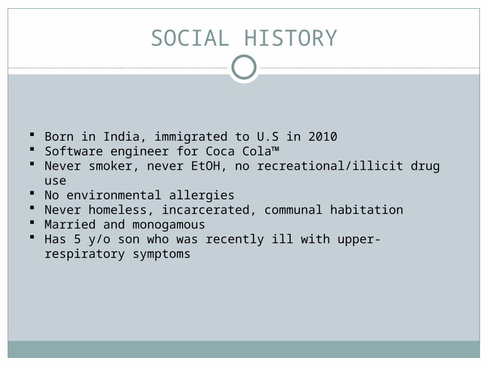
SOCIAL HISTORY
Born in India, immigrated to U.S in 2010 Software engineer for Coca Cola™ Never smoker, never EtOH, no recreational/illicit drug use No environmental allergies Never homeless, incarcerated, communal habitation Married and monogamous Has 5 y/o son who was recently ill with upper-respiratory
symptoms

MEDICATIONS
acyclovir 400 mg, PO, q12hr
albuterol 90 mcg/inh MDI QID, PRN
benzonatate 100 mg, PO, TID, PRN
dasatinib 40 mg, PO, qDay
lorazepam 0.5 mg, PO, q6hr, PRN
oxycodone 10 mg, PO, q4hr, PRN
pentamidine Inhale, qMonth

VITAL SIGNS
T 36.4
BP 85/40
HR 160 (regular)
RR 32
SpO2 100%
WT 152 lbs
HT 67 inches

PHYSICAL EXAM
GEN: diaphoretic, severe distress HEENT: sclera with suffusion, posterior pharynx with erythema, oral
mucosa moist, no thyromegaly or bruit, no sinus tenderness NEURO: AO x 4, normal sensorium, no lateralizing motor or sensory
deficits COR: very rapid regular, summation gallop, no rubs, nondisplaced apical
impulse VASCULAR:
v: engorged IJV,+ HJR, + Kussmaul’s a: pulsus pressure = 20, pulsus alternans (2+/1+), warm extremities
RESPIRATORY: speaking in clipped sentences, clear lung fields, good air entry
ABDOMEN: soft, flat, non-tender, non-distended, MSK: normal ROM, no joint effusions SKIN: no rashes LYMPH: no cervical adenopathy

LABORATORY DATA (PART 1)
135
4.3
100
25
17
0.7164 16.0
13.5
40.5161
9.42.0
5.6 2.9
0.9
438 172
104
Tn-I/CK-MB Pending

CXR
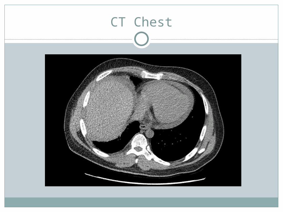
CT Chest

CORONARY ANGIOGRAPHY (LHC)
RAO Left System LAO Right System

ECHOCARDIOGRAM (TTE) I

ECHOCARDIOGRAM (TTE) II

LABORATORY DATA (PART 2)
PCR (Nasopharyngeal Swab)
AdenoV NEG
RSV-A/B POS (A)
PIV (1 – 3) NEG
Influenza A/B NEG
MPnV NEG
RhinoV NEG
Serology (Peripheral Blood)
EchoV NEG
EnteroV NEG
EBV NEG
HSV -1/2 NEG
HTLV – 1/2 NEG
PCR (Peripheral Blood)
AdenoV NEG
CoxV-A/B1 – B6
NEG
EnteroV NEG
CMB NEG
HIV NEG
Troponin-I 30.22
ESR 17
CRP 22.8
BNP 73

HEMODYNAMICS
18 (≤8)
20/11 (25/5)
11 (≤12)
PA SpO2 = 58%
Art SpO2 = 99%
CO (Fick) = 3.4 LPM
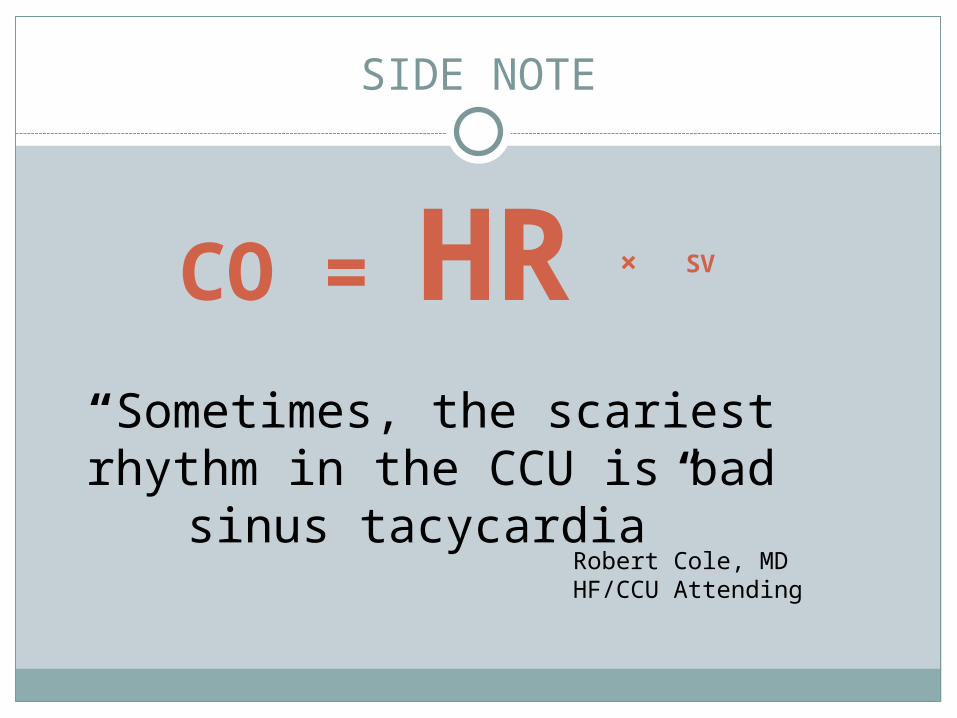
SIDE NOTE
CO = HR × SV
“Sometimes, the scariest rhythm in the CCU is bad sinus
tacycardia”Robert Cole, MDHF/CCU Attending

HISTOPATHOLOGY I
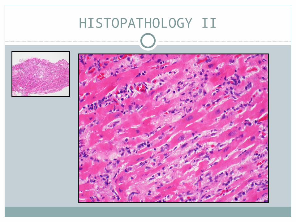
HISTOPATHOLOGY II

DIAGNOSIS
DIAGNOSISFulminant Lymphocytic MyocarditisRSV Bronchiolitis
COMPLICATIONSCardiogenic ShockAcute RV FailureVentricular Tachycardia Storm
HOSPITAL COURSE
Transferred to CCU Swan-Ganz (PA) Catheter for tailored IVF and inotrope
therapy High-dose IV Steroids x 3d then PO taper ST elevations resolved by day 4, Tn-I peaked at 34.7 ng/mL Shock/Tachycardia resolved by 1 week Ribavirin + Palivizumab (Synagis™), aggressive pulmonary
toilet LifeVest for protection of VT RVEF with recovery to normal by 2 weeks Good rehabilitation and discharged home Doing well
FRANK CORRIGAN III, MD
MYOCARDITIS
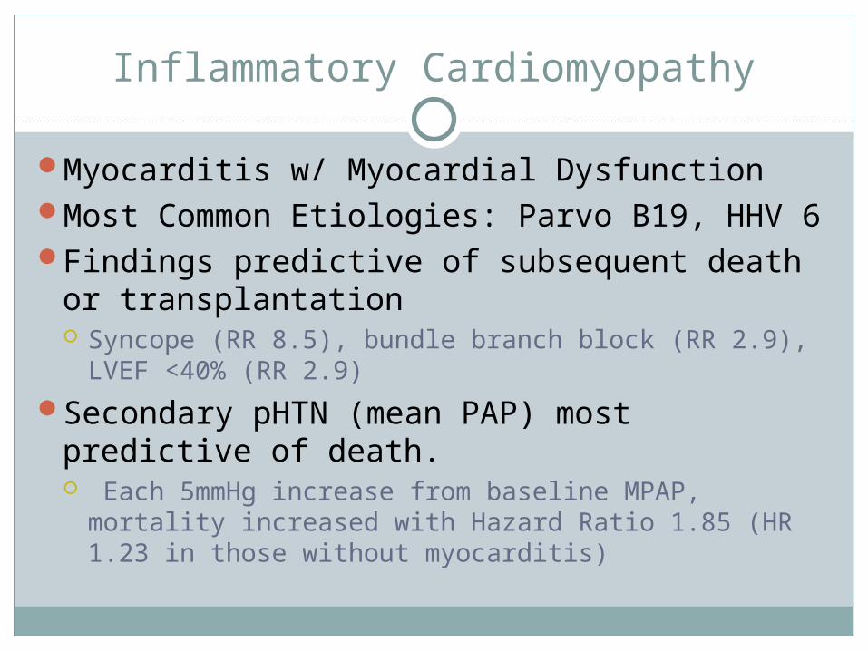
Inflammatory Cardiomyopathy
Myocarditis w/ Myocardial DysfunctionMost Common Etiologies: Parvo B19, HHV 6 Findings predictive of subsequent death or
transplantation Syncope (RR 8.5), bundle branch block (RR 2.9), LVEF
<40% (RR 2.9)
Secondary pHTN (mean PAP) most predictive of death. Each 5mmHg increase from baseline MPAP, mortality
increased with Hazard Ratio 1.85 (HR 1.23 in those without myocarditis)

Classification
Giant Cell etiology unknown. Most severe symptoms. Mediated
by T cells and giant cells
Fulminant Most commonly a viral etiology p/w with acute HF up to 2 weeks after distinct viral
prodrome.
Acute p/w less distinct symptom onset. Have established
ventricular systolic dysfunction.

Outcomes
Patients with fulminant myocarditis, although more severely ill, are more likely to recover than those with acute myocarditis.
At 11 year follow-up, transplant-free survival was 93% fulminant vs. 45% acute.
Giant Cell Myocarditis: rate of death or cardiac transplant 89%. Median survival from symptom onset 5.5 months

Who to Biopsy?
New onset HF of ≤ 2 weeks duration associated with a normal LV chamber size and hemodynamic compromise
New onset HF of 2 weeks – 3 months duration associated with a dilated LV
New ventricular arrhythmias2nd or 3rd degree AV blockFailure to respond to usual care within 1-2
weeks

EMB vs. Cardiac MR
CMR shows most common site of focal involvement was epicardial surface of LV free wall.
Study showed EMB + myocarditis in 19/21 patients directed by CMR imaging (suggestive of myocarditis)
*Most EMBs obtained from RV side of IVS

Dallas Criteria
Active myocarditis- an inflammatory infiltrate of the myocardium with necrosis &/or degeneration of adjacent myocytes not typical of ischemic damage associated with CAD
Borderline myocarditis- the inflammatory infiltrate is too sparse or myocyte injury is not demonstated

References
Cooper, LT Jr. 2009. Myocarditis. NEJM 360(15):1526 Goldberg, LR, Suk, J, et at. 1999. Predictors of adverse outcome of biopsy-proven
myocarditis. J Am Coll Cardiology. 33:A850. McCarthy RE, Boehmer JP, et al. 2000. Long-term outcome of fulminant myocarditis as
compared to acute. NEJM 342(10):690 Mahgholdt H, Goedecke C, et al. 2004. Cardiovascular magnetic resonance assessment
of human myocarditis. Circulation. 109(10):1250. Aretz HT, Billingham ME, et al. 1987. Myocarditis: a histopathologic definition and
classification. Am J Cardiovasc Pathol. 1(1):3.