Embed Size (px)
Citation preview
BioMed CentralBMC Genomics
ss
Open AcceResearch articleGenetic interaction network of the Saccharomyces cerevisiae type 1 phosphatase Glc7Michael R Logan1,2, Thao Nguyen1, Nicolas Szapiel1, James Knockleby1, Hanting Por1, Megan Zadworny1, Michael Neszt2, Paul Harrison1,3, Howard Bussey1,3, Craig A Mandato*1,2, Jackie Vogel*1,3,4 and Guillaume Lesage*4Address: 1Department of Biology, McGill University, Montreal (QC), Canada, 2Anatomy and Cell Biology, McGill University Montreal (QC), Canada, 3Developmental Biology Research Initiative, McGill University, Montreal (QC), Canada and 4Cell Imaging and Analysis Network (CIAN), McGill University, Montreal (QC), Canada
Email: Michael R Logan - [email protected]; Thao Nguyen - [email protected]; Nicolas Szapiel - [email protected]; James Knockleby - [email protected]; Hanting Por - [email protected]; Megan Zadworny - [email protected]; Michael Neszt - [email protected]; Paul Harrison - [email protected]; Howard Bussey - [email protected]; Craig A Mandato* - [email protected]; Jackie Vogel* - [email protected]; Guillaume Lesage* - [email protected]
* Corresponding authors
AbstractBackground: Protein kinases and phosphatases regulate protein phosphorylation, a critical means ofmodulating protein function, stability and localization. The identification of functional networks for proteinphosphatases has been slow due to their redundant nature and the lack of large-scale analyses. Wehypothesized that a genome-scale analysis of genetic interactions using the Synthetic Genetic Array couldreveal protein phosphatase functional networks. We apply this approach to the conserved type 1 proteinphosphatase Glc7, which regulates numerous cellular processes in budding yeast.
Results: We created a novel glc7 catalytic mutant (glc7-E101Q). Phenotypic analysis indicates that thisnovel allele exhibits slow growth and defects in glucose metabolism but normal cell cycle progression andchromosome segregation. This suggests that glc7-E101Q is a hypomorphic glc7 mutant. Synthetic GeneticArray analysis of glc7-E101Q revealed a broad network of 245 synthetic sick/lethal interactions reflectingthat many processes are required when Glc7 function is compromised such as histone modification,chromosome segregation and cytokinesis, nutrient sensing and DNA damage. In addition, mitochondrialactivity and inheritance and lipid metabolism were identified as new processes involved in buffering Glc7function. An interaction network among 95 genes genetically interacting with GLC7 was constructed byintegration of genetic and physical interaction data. The obtained network has a modular architecture, andthe interconnection among the modules reflects the cooperation of the processes buffering Glc7 function.
Conclusion: We found 245 genes required for the normal growth of the glc7-E101Q mutant. Functionalgrouping of these genes and analysis of their physical and genetic interaction patterns bring newinformation on Glc7-regulated processes.
Published: 15 July 2008
BMC Genomics 2008, 9:336 doi:10.1186/1471-2164-9-336
Received: 1 May 2008Accepted: 15 July 2008
This article is available from: http://www.biomedcentral.com/1471-2164/9/336
© 2008 Logan et al; licensee BioMed Central Ltd. This is an Open Access article distributed under the terms of the Creative Commons Attribution License (http://creativecommons.org/licenses/by/2.0), which permits unrestricted use, distribution, and reproduction in any medium, provided the original work is properly cited.
Page 1 of 18(page number not for citation purposes)
BMC Genomics 2008, 9:336 http://www.biomedcentral.com/1471-2164/9/336
BackgroundRegulation of phosphorylation state is a mechanism forcontrolling the function, localization and stability of pro-teins in vivo and is critical for the regulation of essentialprocesses such as polarity and morphogenesis, chromo-some segregation, cytokinesis, and cell cycle control [1-4].Together, protein kinases (which mediate phosphoryla-tion) and protein phosphatases (PPases) (which mediatede-phosphorylation) provide precise temporal and spatialregulation of their target substrates. In the budding yeastSaccharomyces cerevisiae, the dynamic localization ofPPases suggests that an extensive cross talk between theseprocesses is critical for the proper execution of cell divi-sion [2,5]. However, many of the substrates and regula-tory proteins that participate in this cross talk remainunidentified. Insight into PPase function has lagged sig-nificantly in comparison to kinases since PPases: (1) fre-quently require regulatory proteins that determine theirtargeting to particular substrates, cellular location or proc-ess and (2) often exhibit considerable functional redun-dancy. In budding yeast, for example, ~90% of the known32 PPases are non-essential [6].
Study of genetic interaction networks is a powerful meansto get insight into gene function, and several highthroughput methods were developed during the last dec-ade to generate genome-wide maps of genetic interactions[7-13]. However, such maps are still missing for yeastPPases. To date investigations of PPase function in bud-ding yeast have utilized mutations outside the catalyticsite [5,14-16] or probed genetic interactions using singleor double knockout PPase mutations [6]. Theseapproaches present constraints for large-scale analysis ofPPase function since: (1) mutations outside the catalyticdomain generally affect the formation of a specific class ofholoenzyme or the ability to interact with co-factor(s)required for the proper targeting of the PPase to a subsetof substrates [17-19], and (2) the use of knockout alleles,in addition to precluding the analysis of the essentialPPases, may lead to promiscuous and/or low-affinityinteraction of regulatory subunits with PPases of the sameclass. For these reasons, we designed a strategy based on ahypomorphic catalytic PPase mutation that uses the Syn-thetic Genetic Array (SGA). This method generates a largenumber of double mutant combinations, and has beensuccessfully used to identify genetic interaction networksin yeast in a wide range of cellular processes[12,13,20,21].
In the budding yeast Saccharomyces cerevisiae, 32 genesencode predicted or demonstrated catalytic subunits ofPPases. Only two of these PPases are clearly essential: thePP1-type PPase, Glc7 (glycogen deficient) [1,22], andCdc14 (cell division cycle), a dual-specificity phosphatasethat regulates mitotic/meiotic exit [23]. We selected Glc7
for our analyses since a framework of genetic and proteininteractions exists from previous studies of conditionalglc7 mutants, but a comprehensive large-scale geneticanalysis had not yet been performed. Glc7 critically regu-lates numerous processes such as glucose and glycogenmetabolism, sporulation, chromosome segregation, mei-osis, mRNA transport, transcription, and amino acid bio-synthesis [1,3,15,17,22,24,25]. A search in the BioGRIDdatabase (version 2.0.41, June 1, 2008 [26]) returned a listof 114 proteins reported to physically interact with Glc7.Some of these proteins are known substrates (Cbf2, Fin1,Red1, Gsy2), while others are thought to be regulatorysubunits (Gac1, Reg1, Reg2, Ref2, Sip5, Glc8, Bud14,Bni4, Sds22). However, the functional significance of themajority of these interactions remains unclear. In con-trast, only 30 genes have been reported to show geneticinteractions with various glc7 mutants. The vast majorityof these genetic interactions are dosage dependent andonly 7 genes (BUD14, DAM1, DOC1, GLC8, RHR2,SET1and SLT2) have been reported to show synthetic lethal/sick interaction with different conditional glc7 alleles. Thefunctional clustering of genetic interactions of conditionalglc7 alleles suggests the majority of these mutations com-promise specific aspects of Glc7 function. As such, thesealleles are highly suited for cell biology. However an unbi-ased large-scale analysis revealing a comprehensive pic-ture of Glc7 signaling in vivo requires a novel glc7 alleleaffecting many functions of Glc7 at a time.
In an effort to further define the signaling network forGlc7, we created a novel glc7 catalytic mutant (glc7-E101Q) suitable for SGA. The glc7-E101Q mutant exhib-ited slow growth and impaired glycogen accumulation onglucose, but no specific delay in the cell cycle. Theimpaired growth and defects in glycogen synthesis of theglc7-E101Q mutant were rescued by introduction of a sin-gle copy of wild type GLC7, indicating that glc7-E101Q isa recessive, hypomorphic allele. SGA analysis revealed abroad network of 245 synthetic sick/lethal (SSL) interac-tions that encompassed all known Glc7-regulated proc-esses, and suggested additional previously unknownfunctions for Glc7.
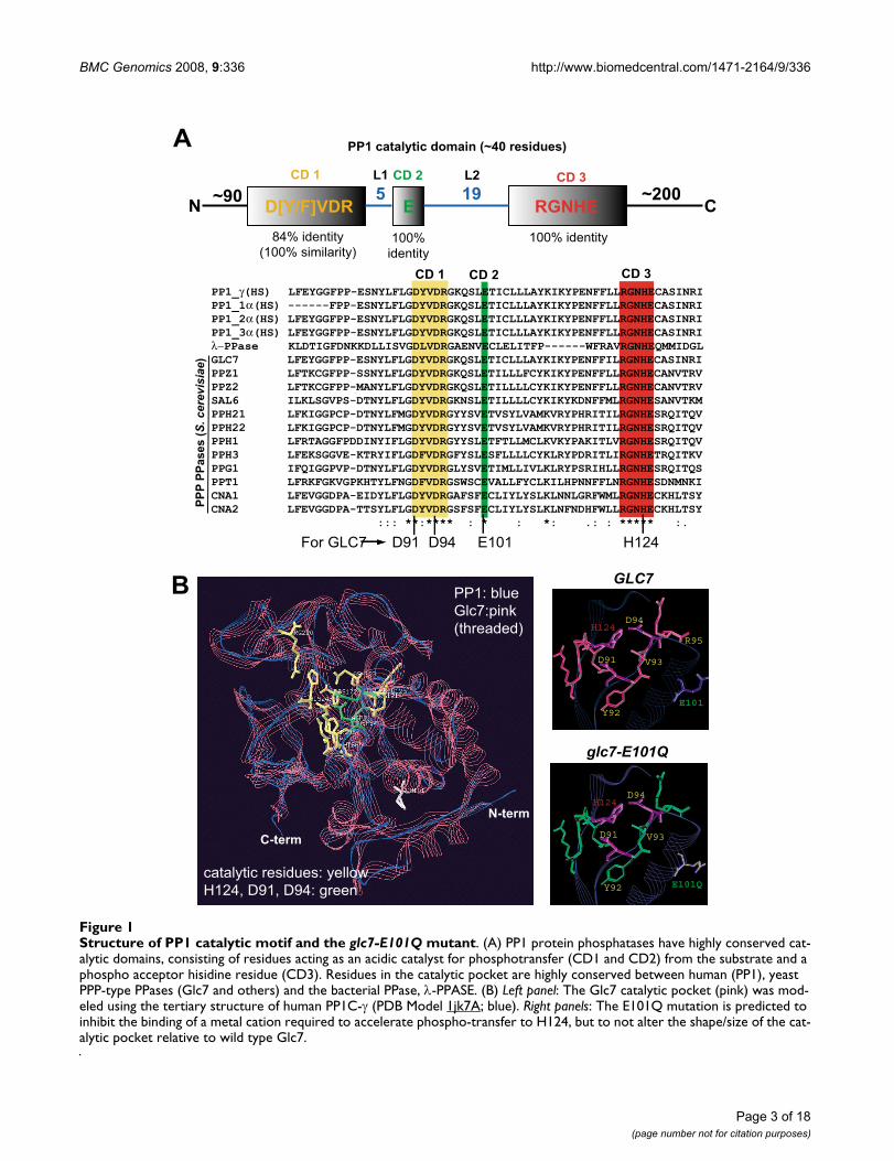
ResultsCreation of a catalytic-deficient GLC7 mutant, glc7-E101QGlc7 is the sole essential member of the PPP family ofPPases in budding yeast and members of this family sharea conserved catalytic motif. Residues in the Glc7 catalyticdomain (CD1-3) are evolutionarily conserved withhuman PP1 orthologs and the PP1-like bacterial λ-PPASE(Figure 1A). Given the high degree of conservation in thecatalytic residues, Glc7 is expected to have catalytic char-acteristics similar to those of previously characterizedPP1-type PPases. Phosphate hydrolysis by PP1 occurs via
Page 2 of 18(page number not for citation purposes)
BMC Genomics 2008, 9:336 http://www.biomedcentral.com/1471-2164/9/336
Page 3 of 18(page number not for citation purposes)
Structure of PP1 catalytic motif and the glc7-E101Q mutantFigure 1Structure of PP1 catalytic motif and the glc7-E101Q mutant. (A) PP1 protein phosphatases have highly conserved cat-alytic domains, consisting of residues acting as an acidic catalyst for phosphotransfer (CD1 and CD2) from the substrate and a phospho acceptor hisidine residue (CD3). Residues in the catalytic pocket are highly conserved between human (PP1), yeast PPP-type PPases (Glc7 and others) and the bacterial PPase, λ-PPASE. (B) Left panel: The Glc7 catalytic pocket (pink) was mod-eled using the tertiary structure of human PP1C-γ (PDB Model 1jk7A; blue). Right panels: The E101Q mutation is predicted to inhibit the binding of a metal cation required to accelerate phospho-transfer to H124, but to not alter the shape/size of the cat-alytic pocket relative to wild type Glc7.

BMC Genomics 2008, 9:336 http://www.biomedcentral.com/1471-2164/9/336
a onestep reaction and is dependent on metal ion catalysts(Mn2+ and Fe2+). This hydrolysis reaction (Additional File1) is dependent on a phosphoesterase motifDXH(X)nGDXXD(X)nGNHD/E (where n = ~25 aminoacids) whose active residues are a general acid catalyst(H124 in Glc7) and the carboxyl oxygen of two invariantaspartic acids (D91 and D94 in Glc7) [27]. Similar tohuman PP1 and Glc7, λ-PPASE (a bacteriophage orthologof PP1) shares this conserved catalytic domain (Figure 1A)and has been previously examined in structure-functionanalyses of the active residues in the catalytic pocket.Mutation of either D49 or D52 (D92 and D95 in PP1C-γ;D91 and D94 in Glc7, respectively) to a non-reactiveasparagine residue (D>N) resulted in a catalytically deadenzyme in in vitro PPase assays. However, mutation of E59(E>Q; E102 in PP1C and E101 in Glc7) reduced the cata-lytic activity of λ-PPASE ~8 fold and impaired Mn2+
recruitment, but did not significantly alter its substratebinding [28]. An E>Q substitution in the equivalent resi-due of Glc7 (E101Q) is therefore predicted to reduce cat-alytic activity by perturbing the recruitment of the metalco-factor that enhances phospho-transfer. Using humanPP1C-γ as a template, we found that the conserved resi-dues of Glc7 in the CD1-3 regions form a tertiary structurevery similar to human PP1. Furthermore, analysis of the 3-D model of the E101Q mutation in Glc7 indicated thatthe overall shape and size of the catalytic pocket was notsignificantly changed (Figure 1B). We predicted thatreduction of Glc7 catalytic activity in vivo, as a result of theE101Q mutation, would result in a recessive, hypomor-phic glc7 allele suitable for an unbiased SGA analysis.
glc7-E101Q mutant exhibits slow growth but no appreciable cell cycle delay or defect in chromosome segregationThe introduction of a glc7-D91N mutation into diploidcells resulted in inviable haploid progeny by tetrad dissec-tion (data not shown). This finding is consistent with theprevious observation that this mutation results in a cata-lytically dead form of the enzyme in vivo [28]. In contrast,haploid glc7-E101Q colonies isolated by tetrad dissectionwere viable but consistently small compared to wild type(GLC7) when grown on rich medium (YPAD) (data notshown). Analysis of the growth curve for three haploidisolates of glc7-E101Q indicated impaired kinetics com-pared to wild type cells (Figure 2A). To determine whetherslow growth was due to a delay in cell cycle progression,mutant and wild type cells from log phase cultures wereharvested, stained with propidium iodide and DNA con-tent measured by FACS analysis. As shown in Figure 2B,FACS analysis revealed that proportion of G1, S, or M-phase cells in glc7-E101Q cultures was not statistically dif-ferent from that of wild type cells. This result correlatedwith the observation that proportion of unbudded, small-
budded and large-budded cells was similar between wildtype and glc7-E101Q strains (Figure 2C).
Studies using conditional glc7 mutants have demon-strated a role for Glc7 in the attachment of spindle micro-tubules to kinetochores during metaphase andchromosome segregation [1,29-33]. Therefore, we nextexamined glc7-E101Q cells for chromosome segregationdefects and sensitivity to the microtubule-destabilizingagent, benomyl. To examine bi-orientation of sister chro-matids, anaphase cells (mitotic spindle>2.5 μm) express-ing GFP-LacI and centromeric repeats (CEN15) of thelactose operon (lacO) [34] were examined by fluorescencemicroscopy. As shown in Figure 2D, GFP-LacI segregatedto both spindle pole bodies (marked by the spindle polemarker, Spc29-CFP) in a similar manner to wild type cells.We also examined for chromosome mis-segregation inglc7-E101Q cells following several growth cycles using asectoring assay [35]. Wild type, glc7-E101Q and ame1-6(positive control) strains lacking a functional ADE2 gene(ade2Δ-101) were transformed with a centromeric frag-ment of chromosome III containing the SUP11 gene (Cir-cle III/SUP11) and analyzed for loss of the fragment after4-day incubation on YPD medium lacking adenine. Theame1-6 strain exhibited frequent chromosome loss asindicated by the appearance of red sectors in the colonies(Figure 2E). In contrast, glc7-E101Q and wild type colo-nies induced negligible loss of the Circle III/SUP11 frag-ment, and the vast majority of colonies remained white incoloration (Figure 2E). We also tested glc7-E101Q cells forsensitivity to the microtubule-disrupting agent, benomyl.When compared to the wild type, glc7-E101Q cells hadconsistently slower growth on both DMSO control andbenomyl plates. However, the slow growth of glc7-E101Qwas not exacerbated by benomyl treatment (Figure 2F).
To investigate the possibility that the slow growth pheno-type of glc7-E101Q cells might be the result of either pro-tein instability or changes in protein modifications toGlc7, whole cell extracts were examined by Western blotanalysis. Protein extracts from strains expressing Glc7-ProA or glc7-E101Q-ProA fusion proteins were preparedfrom asynchronous cells (1D immunoblot) or from G1, Sand G2/M-arrested cells (2D immunoblot) and examinedusing anti-ProA antibodies. The abundance of the glc7-E101Q-ProA mutant was not reduced relative to wild typeGlc7-ProA by 1D immunoblot analysis (Additional File2A). Furthermore, in each case, a single isoform was iden-tified for Glc7-ProA and glc7-E101Q-ProA in extractsderived from G1, S and G2/M arrested cells (AdditionalFile 2B). These findings indicate that the slow growth ofglc7-E101Q strains was not the result of protein instabilityor the accumulation of aberrant modifications to the Glc7protein.
Page 4 of 18(page number not for citation purposes)

BMC Genomics 2008, 9:336 http://www.biomedcentral.com/1471-2164/9/336
Page 5 of 18(page number not for citation purposes)
glc7-E101Q mutant exhibits slow growth but no appreciable delay in the cell cycle or chromosome segregation defectFigure 2glc7-E101Q mutant exhibits slow growth but no appreciable delay in the cell cycle or chromosome segregation defect. (A) The growth of wild type (GLC7) vs glc7-E101Q strains in rich medium (YPAD) was determined from early log-phase cultures. (B) FACs analysis for DNA content (propidium iodide staining) of log-phase wild type and glc7-E101Q cells. Cell counts are shown plotted against fluorescence intensity (PI). (C) Budding index of wild type and glc7-E101Q mutant strains. (D) Fluorescence microscopy of anaphase cells (mitotic spindle >2.5 μm) wild type and glc7-E101Q cells expressing a chromosome [GFP-LacI (green) and centromeric (CEN15) repeats of the lactose operon (lacO)] and spindle pole body (SPB) [Spc29-CFP (red)] markers. The percentage of cells with chromosomes localized to both SPBs is indicated at the bottom (n = 3, 300 cells/experiment). (E) Test for chromosome segregation using a sectoring assay. Wild type GLC7, glc7-E101Q and ame1-6 strains lacking a functional ADE2 gene (ade2Δ-101) and transformed with a Circle III/SUP11 were plated on to medium lacking adenine (YPD) and scored for appearance of red sectors after 4-day incubation. (F) Spotting assay of glc7-E101Q vs. wild type on DMSO control or benomyl plates (5-fold serial dilutions of 1.0 × 10⁶ cells/ml; 5 μl/spot). Benomyl-sensitive (tub1-1) and resist-ant (tub2-104) strains were plated as controls.
BMC Genomics 2008, 9:336 http://www.biomedcentral.com/1471-2164/9/336
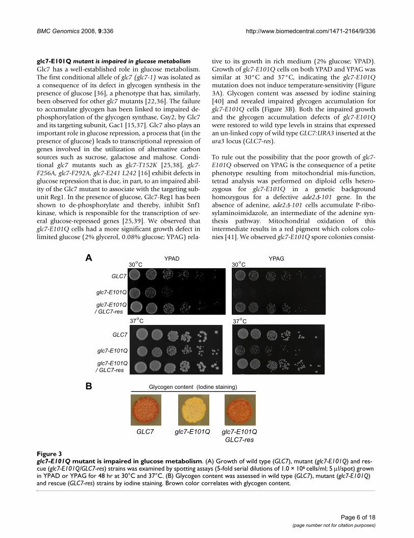
glc7-E101Q mutant is impaired in glucose metabolismGlc7 has a well-established role in glucose metabolism.The first conditional allele of glc7 (glc7-1) was isolated asa consequence of its defect in glycogen synthesis in thepresence of glucose [36], a phenotype that has, similarly,been observed for other glc7 mutants [22,36]. The failureto accumulate glycogen has been linked to impaired de-phosphorylation of the glycogen synthase, Gsy2, by Glc7and its targeting subunit, Gac1 [15,37]. Glc7 also plays animportant role in glucose repression, a process that (in thepresence of glucose) leads to transcriptional repression ofgenes involved in the utilization of alternative carbonsources such as sucrose, galactose and maltose. Condi-tional glc7 mutants such as glc7-T152K [25,38], glc7-F256A, glc7-F292A, glc7-E241 L242 [16] exhibit defects inglucose repression that is due, in part, to an impaired abil-ity of the Glc7 mutant to associate with the targeting sub-unit Reg1. In the presence of glucose, Glc7-Reg1 has beenshown to de-phosphorylate and thereby, inhibit Snf1kinase, which is responsible for the transcription of sev-eral glucose-repressed genes [25,39]. We observed thatglc7-E101Q cells had a more significant growth defect inlimited glucose (2% glycerol, 0.08% glucose; YPAG) rela-
tive to its growth in rich medium (2% glucose; YPAD).Growth of glc7-E101Q cells on both YPAD and YPAG wassimilar at 30°C and 37°C, indicating the glc7-E101Qmutation does not induce temperature-sensitivity (Figure3A). Glycogen content was assessed by iodine staining[40] and revealed impaired glycogen accumulation forglc7-E101Q cells (Figure 3B). Both the impaired growthand the glycogen accumulation defects of glc7-E101Qwere restored to wild type levels in strains that expressedan un-linked copy of wild type GLC7:URA3 inserted at theura3 locus (GLC7-res).
To rule out the possibility that the poor growth of glc7-E101Q observed on YPAG is the consequence of a petitephenotype resulting from mitochondrial mis-function,tetrad analysis was performed on diploid cells hetero-zygous for glc7-E101Q in a genetic backgroundhomozygous for a defective ade2Δ-101 gene. In theabsence of adenine, ade2Δ-101 cells accumulate P-ribo-sylaminoimidazole, an intermediate of the adenine syn-thesis pathway. Mitochondrial oxidation of thisintermediate results in a red pigment which colors colo-nies [41]. We observed glc7-E101Q spore colonies consist-
glc7-E101Q mutant is impaired in glucose metabolismFigure 3glc7-E101Q mutant is impaired in glucose metabolism. (A) Growth of wild type (GLC7), mutant (glc7-E101Q) and res-cue (glc7-E101Q/GLC7-res) strains was examined by spotting assays (5-fold serial dilutions of 1.0 × 10⁶ cells/ml; 5 μl/spot) grown in YPAD or YPAG for 48 hr at 30°C and 37°C. (B) Glycogen content was assessed in wild type (GLC7), mutant (glc7-E101Q) and rescue (GLC7-res) strains by iodine staining. Brown color correlates with glycogen content.
Page 6 of 18(page number not for citation purposes)
BMC Genomics 2008, 9:336 http://www.biomedcentral.com/1471-2164/9/336
ently accumulated this red pigment on medium lackingadenine demonstrating the presence of functional mito-chondria (data not shown). Taken together, these resultssupport that the glc7-E101Q mutation is a stable recessiveallele and exhibits impaired catalytic activity in vivo.
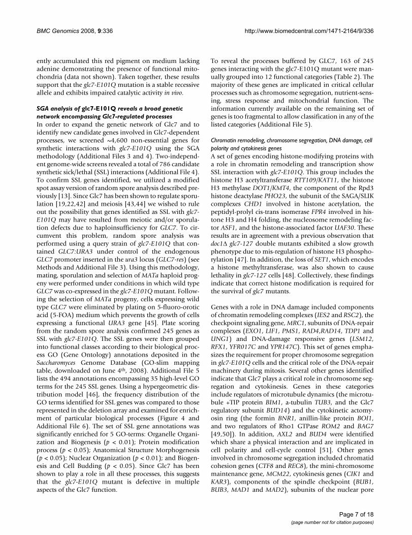
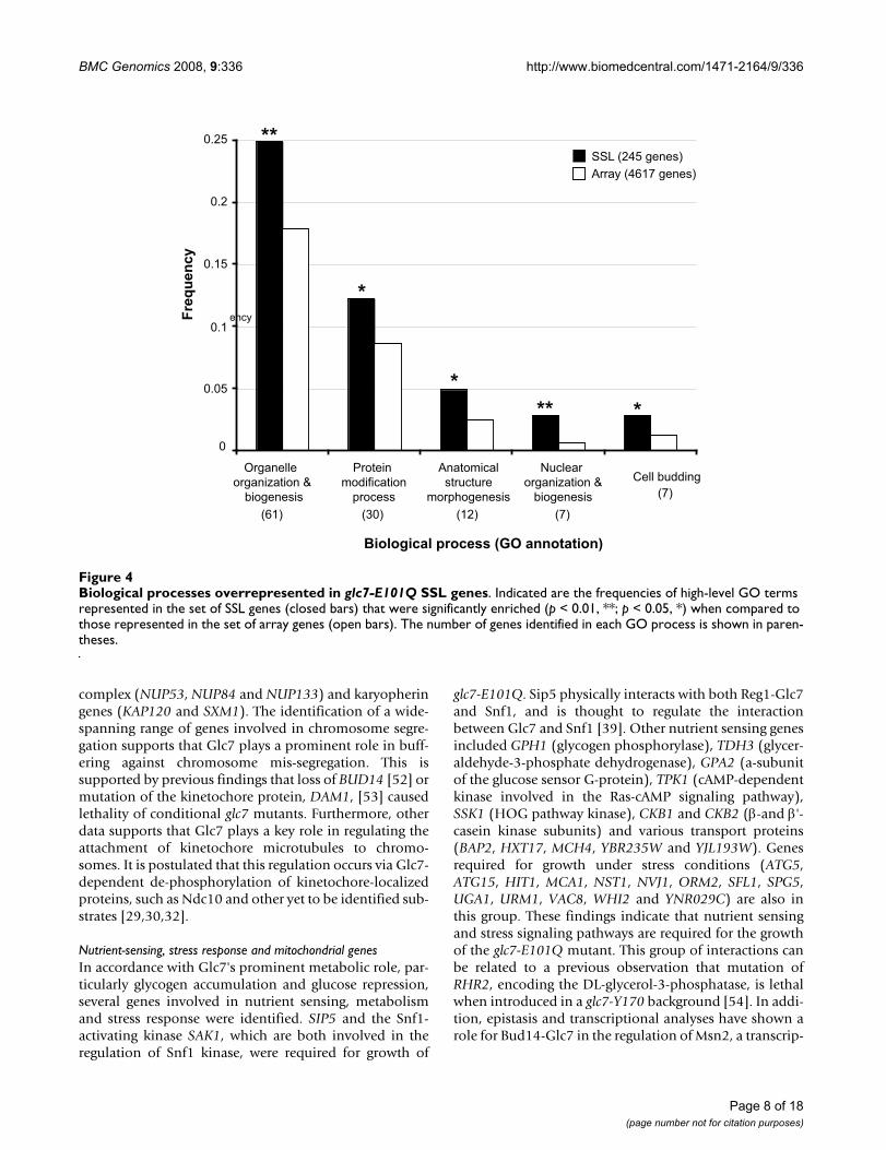
SGA analysis of glc7-E101Q reveals a broad genetic network encompassing Glc7-regulated processesIn order to expand the genetic network of Glc7 and toidentify new candidate genes involved in Glc7-dependentprocesses, we screened ~4,600 non-essential genes forsynthetic interactions with glc7-E101Q using the SGAmethodology (Additional Files 3 and 4). Two-independ-ent genome-wide screens revealed a total of 786 candidatesynthetic sick/lethal (SSL) interactions (Additional File 4).To confirm SSL genes identified, we utilized a modifiedspot assay version of random spore analysis described pre-viously [13]. Since Glc7 has been shown to regulate sporu-lation [19,22,42] and meiosis [43,44] we wished to ruleout the possibility that genes identified as SSL with glc7-E101Q may have resulted from meiotic and/or sporula-tion defects due to haploinsufficiency for GLC7. To cir-cumvent this problem, random spore analysis wasperformed using a query strain of glc7-E101Q that con-tained GLC7:URA3 under control of the endogenousGLC7 promoter inserted in the ura3 locus (GLC7-res) (seeMethods and Additional File 3). Using this methodology,mating, sporulation and selection of MATa haploid prog-eny were performed under conditions in which wild typeGLC7 was co-expressed in the glc7-E101Q mutant. Follow-ing the selection of MATa progeny, cells expressing wildtype GLC7 were eliminated by plating on 5-fluoro-oroticacid (5-FOA) medium which prevents the growth of cellsexpressing a functional URA3 gene [45]. Plate scoringfrom the random spore analysis confirmed 245 genes asSSL with glc7-E101Q. The SSL genes were then groupedinto functional classes according to their biological proc-ess GO (Gene Ontology) annotations deposited in theSaccharomyces Genome Database (GO-slim mappingtable, downloaded on June 4th, 2008). Additional File 5lists the 494 annotations encompassing 35 high-level GOterms for the 245 SSL genes. Using a hypergeometric dis-tribution model [46], the frequency distribution of theGO terms identified for SSL genes was compared to thoserepresented in the deletion array and examined for enrich-ment of particular biological processes (Figure 4 andAdditional File 6). The set of SSL gene annotations wassignificantly enriched for 5 GO-terms: Organelle Organi-zation and Biogenesis (p < 0.01); Protein modificationprocess (p < 0.05); Anatomical Structure Morphogenesis(p < 0.05); Nuclear Organization (p < 0.01); and Biogen-esis and Cell Budding (p < 0.05). Since Glc7 has beenshown to play a role in all these processes, this suggeststhat the glc7-E101Q mutant is defective in multipleaspects of the Glc7 function.
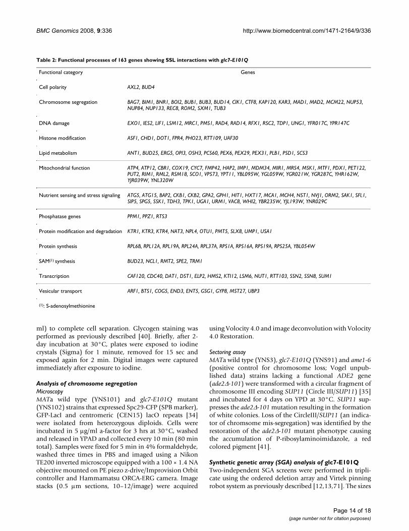
To reveal the processes buffered by GLC7, 163 of 245genes interacting with the glc7-E101Q mutant were man-ually grouped into 12 functional categories (Table 2). Themajority of these genes are implicated in critical cellularprocesses such as chromosome segregation, nutrient-sens-ing, stress response and mitochondrial function. Theinformation currently available on the remaining set ofgenes is too fragmental to allow classification in any of thelisted categories (Additional File 5).
Chromatin remodeling, chromosome segregation, DNA damage, cell polarity and cytokinesis genesA set of genes encoding histone-modifying proteins witha role in chromatin remodeling and transcription showSSL interaction with glc7-E101Q. This group includes thehistone H3 acetyltransferase RTT109/KAT11, the histoneH3 methylase DOT1/KMT4, the component of the Rpd3histone deactylase PHO23, the subunit of the SAGA/SLIKcomplexes CHD1 involved in histone acetylation, thepeptidyl-prolyl cis-trans isomerase FPR4 involved in his-tone H3 and H4 folding, the nucleosome remodeling fac-tor ASF1, and the histone-associated factor UAF30. Theseresults are in agreement with a previous observation thatdoc1Δ glc7-127 double mutants exhibited a slow growthphenotype due to mis-regulation of histone H3 phospho-rylation [47]. In addition, the loss of SET1, which encodesa histone methyltransferase, was also shown to causelethality in glc7-127 cells [48]. Collectively, these findingsindicate that correct histone modification is required forthe survival of glc7 mutants.
Genes with a role in DNA damage included componentsof chromatin remodeling complexes (IES2 and RSC2), thecheckpoint signaling gene, MRC1, subunits of DNA-repaircomplexes (EXO1, LIF1, PMS1, RAD4,RAD14, TDP1 andUNG1) and DNA-damage responsive genes (LSM12,RFX1, YFR017C and YPR147C). This set of genes empha-sizes the requirement for proper chromosome segregationin glc7-E101Q cells and the critical role of the DNA-repairmachinery during mitosis. Several other genes identifiedindicate that Glc7 plays a critical role in chromosome seg-regation and cytokinesis. Genes in these categoriesinclude regulators of microtubule dynamics (the microtu-bule +TIP protein BIM1, a-tubulin TUB3, and the Glc7regulatory subunit BUD14) and the cytokinetic actomy-osin ring (the formin BNR1, anillin-like protein BOI1,and two regulators of Rho1 GTPase ROM2 and BAG7[49,50]). In addition, AXL2 and BUD4 were identifiedwhich share a physical interaction and are implicated incell polarity and cell-cycle control [51]. Other genesinvolved in chromosome segregation included chromatidcohesion genes (CTF8 and REC8), the mini-chromosomemaintenance gene, MCM22, cytokinesis genes (CIK1 andKAR3), components of the spindle checkpoint (BUB1,BUB3, MAD1 and MAD2), subunits of the nuclear pore
Page 7 of 18(page number not for citation purposes)
BMC Genomics 2008, 9:336 http://www.biomedcentral.com/1471-2164/9/336
complex (NUP53, NUP84 and NUP133) and karyopheringenes (KAP120 and SXM1). The identification of a wide-spanning range of genes involved in chromosome segre-gation supports that Glc7 plays a prominent role in buff-ering against chromosome mis-segregation. This issupported by previous findings that loss of BUD14 [52] ormutation of the kinetochore protein, DAM1, [53] causedlethality of conditional glc7 mutants. Furthermore, otherdata supports that Glc7 plays a key role in regulating theattachment of kinetochore microtubules to chromo-somes. It is postulated that this regulation occurs via Glc7-dependent de-phosphorylation of kinetochore-localizedproteins, such as Ndc10 and other yet to be identified sub-strates [29,30,32].
Nutrient-sensing, stress response and mitochondrial genesIn accordance with Glc7's prominent metabolic role, par-ticularly glycogen accumulation and glucose repression,several genes involved in nutrient sensing, metabolismand stress response were identified. SIP5 and the Snf1-activating kinase SAK1, which are both involved in theregulation of Snf1 kinase, were required for growth of
glc7-E101Q. Sip5 physically interacts with both Reg1-Glc7and Snf1, and is thought to regulate the interactionbetween Glc7 and Snf1 [39]. Other nutrient sensing genesincluded GPH1 (glycogen phosphorylase), TDH3 (glycer-aldehyde-3-phosphate dehydrogenase), GPA2 (a-subunitof the glucose sensor G-protein), TPK1 (cAMP-dependentkinase involved in the Ras-cAMP signaling pathway),SSK1 (HOG pathway kinase), CKB1 and CKB2 (β-and β'-casein kinase subunits) and various transport proteins(BAP2, HXT17, MCH4, YBR235W and YJL193W). Genesrequired for growth under stress conditions (ATG5,ATG15, HIT1, MCA1, NST1, NVJ1, ORM2, SFL1, SPG5,UGA1, URM1, VAC8, WHI2 and YNR029C) are also inthis group. These findings indicate that nutrient sensingand stress signaling pathways are required for the growthof the glc7-E101Q mutant. This group of interactions canbe related to a previous observation that mutation ofRHR2, encoding the DL-glycerol-3-phosphatase, is lethalwhen introduced in a glc7-Y170 background [54]. In addi-tion, epistasis and transcriptional analyses have shown arole for Bud14-Glc7 in the regulation of Msn2, a transcrip-
Biological processes overrepresented in glc7-E101Q SSL genesFigure 4Biological processes overrepresented in glc7-E101Q SSL genes. Indicated are the frequencies of high-level GO terms represented in the set of SSL genes (closed bars) that were significantly enriched (p < 0.01, **; p < 0.05, *) when compared to those represented in the set of array genes (open bars). The number of genes identified in each GO process is shown in paren-theses.
Page 8 of 18(page number not for citation purposes)
BMC Genomics 2008, 9:336 http://www.biomedcentral.com/1471-2164/9/336
tion factor controlling the expression of stress-responseelement (STRE)-genes [55].
Genes with a role in mitochondrial activity and inherit-ance included subunits of the ATP synthase (ATP4 andATP12), genes required for cytochrome-c oxidase function(COX19, CYC7 and SCO1), genes involved in mitochon-drial replication (RIM1)-transcription (HAP2, MTF1 andPET122) and -translation (MSK1, RML2 and RSM18),genes required for mitochondrial homeostasis/metabo-lism (CBR1, MIR1, MRS4 and PDX1) and genes with arole in mitochondrial morphology and distribution(IMP1, MDM34 and YPT11). This group also containsgenes that localize to the mitochondria (FMP42, PUT2,VPS73, YBL095W, YGL059W, YGR021W, YGR287C,YHR161W, YJR039W and YNL320W) and may berequired for optimal mitochondrial function. Thus, mito-chondrial function is essential when PP1 function is com-promised.
Transcription, protein synthesis and protein regulation genesGenes involved in transcription included transcriptionfactors (CAF120, DAT1,DST1, HMS2, and SUM1), elonga-tor complex components (ELP2 and KTI12), mediatorcomplex components (NUT1, SSN2 and SSN8), a tran-scription termination factor (RTT103) and RNA process-ing factors (CDC40 and LSM6). Protein synthesis genesincluded components of the large ribosomal subunit(RPL6B, RPL12A, RPL19A, RPL24A and RPL37), compo-nents of the small ribosomal subunit (RPS1A, RPS16A,RPS19A and RPS25A) and YBL054W, which is putativelyinvolved in rRNA synthesis. Other genes were identifiedwith various roles in protein modification such as proteinglycosylation (KTR1, KTR3, KTR4 and PMT5), ubiquitin-regulated protein degradation (NPL4, OTU1, SLX8, UMP1and USA1) and protein phosphorylation (PPZ1, PPM1and RTS3). All these genes likely play a role in the expres-sion of and/or the modification/turnover gene productsessential for processes buffered by Glc7.
The synthetic lethal interaction of glc7-E101Q with thePPZ1 phosphatase and two PP2A subunits (PPM1 andRTS3) suggests an interplay between these phosphatases.Few synthetic lethal interactions are currently available forPPZ1, PPM1 and RTS3. However, it was interesting to notethat glc7-E101Q and PPZ1 share synthetic lethal interac-tions with both BIM1 and BUB3. In addition, it has beenpreviously suggested that an exchange of regulatory subu-nits between Glc7 and PPZ phosphatase may be the basisfor the observed synthetic lethal interaction of glc8 withglc7 mutants and ppz1 ppz2 [56]. These observations sup-port that signaling cross-talk occurs between these phos-phatases.
Vesicle transport, lipid metabolism and S-adenosylmethionine synthesis genesGenes with a role in vesicle transport included ARF1 (coatformation small GTPase), BTS1 (geranygeranyl diphos-phate synthase required for the interaction of smallGTPases with vesicle/organelle membranes), regulators ofGolgi vesicle targeting and fusion (COG complex subunitCOG5, the TRAPP subunit GSG1, and the Ypt1 GTPaseActivating Protein, GYP8) and regulatory proteinsinvolved in endocytosis (actin cytoskeleton regulator,END3, phosphoinositide-binding protein, ENT5, and thecoat regulators, MST27 and UBP3). These interactionsmay indicate a requirement for the transport of specificcargo(s) in the glc7-E101Q mutant that are required forviability. Alternatively, they may reflect a more direct rolefor Glc7 in the regulation of vesicle transport. This is sup-ported by previous studies that indicate a regulatory func-tion for Glc7 on components of vesicle trafficking. Forexample, Glc7 and its targeting subunit, Scd5, were shownto regulate Pan1, an actin regulatory protein involved inendocytosis. Interestingly, both Pan1 and End3 (identi-fied in our SGA analysis) exhibited physical interactionswith Scd5, implicating them in Glc7 recruitment to sitesof endocytosis [57]. In addition, a separate study demon-strated a role for Glc7 in the regulation of SNARE proteincomplexes that are required for vesicle-membrane fusion[58].
Lipid metabolism genes identified included those func-tioning in ergosterol metabolism (ERG5 and OSH3), fattyacid metabolism (ANT1 and PCS60), phospholipidmetabolism (OPI3, PLB1, PSD1 and SCS3) and a set ofgenes for peroxisomal proteins (PEX6, PEX29 and PEX31).The intimate synthetic lethal relationships between lipidmetabolism, mitochondrial function and vesicle transportgenes is probably the basis for this set of SSL interactions.
Genes involved in S-adenosylmethionine (SAM) -depend-ent processes included methyltransferases (BUD23,NCL1, RMT2 and TRM1) and the SAM decarboxylaseSPE2. The deletion of these genes likely impacts on SAMpool and this, in turn, affects Glc7-dependent processesthat utilize this co-factor such as histone methylation,ergosterol synthesis and phospholipid synthesis.
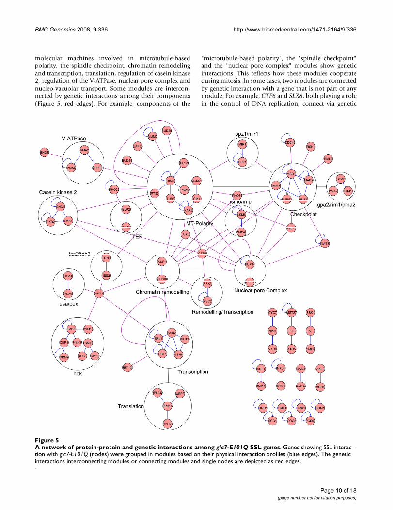
Network analysis of the Glc7 interacting genesWe performed a network analysis of the glc7-E101Q SSLgenes by examining protein-protein and genetic interac-tions available from the BioGRID database (version2.0.41, released on June 1, 2008 [26]). This analysisrevealed 95 genes that are interconnected through 99 and212 protein-protein and genetic interactions, respectively.Based on protein-protein interactions, we identified 23distinct molecular modules involving 72 GLC7 geneticinteractors (Figure 5). These modules are components of
Page 9 of 18(page number not for citation purposes)
BMC Genomics 2008, 9:336 http://www.biomedcentral.com/1471-2164/9/336
molecular machines involved in microtubule-basedpolarity, the spindle checkpoint, chromatin remodelingand transcription, translation, regulation of casein kinase2, regulation of the V-ATPase, nuclear pore complex andnucleo-vacuolar transport. Some modules are intercon-nected by genetic interactions among their components(Figure 5, red edges). For example, components of the
"microtubule-based polarity", the "spindle checkpoint"and the "nuclear pore complex" modules show geneticinteractions. This reflects how these modules cooperateduring mitosis. In some cases, two modules are connectedby genetic interaction with a gene that is not part of anymodule. For example, CTF8 and SLX8, both playing a rolein the control of DNA replication, connect via genetic
A network of protein-protein and genetic interactions among glc7-E101Q SSL genesFigure 5A network of protein-protein and genetic interactions among glc7-E101Q SSL genes. Genes showing SSL interac-tion with glc7-E101Q (nodes) were grouped in modules based on their physical interaction profiles (blue edges). The genetic interactions interconnecting modules or connecting modules and single nodes are depicted as red edges.
Page 10 of 18(page number not for citation purposes)
BMC Genomics 2008, 9:336 http://www.biomedcentral.com/1471-2164/9/336
interactions to the "microtubule-base polarity", "nuclearpore complex" and "remodeling/transcription" modules.Finally, some modules, such as "NPL4-OTU1" (whoseproducts are involved in polyubquitin-mediated proteindegradation) and "AXL2-BUD4", are not connected to anyother module. This probably reflects that the map forgenetic interactions is still incomplete and that somegenes showing SSL interaction with glc7-E101Q have notyet been screened by the SGA. Additional SGA screens willbe required to obtain a comprehensive interaction profilefor these genes.
DiscussionA number of studies support that Glc7 plays a critical rolein the regulation of several cellular processes such as cellpolarity, chromosome segregation, cytokinesis, and cellcycle control [1-4]. However, the precise role of Glc7 inthe majority of these processes and the identity of Glc7substrates involved in the regulation of these eventsremains poorly understood. The lack of informationregarding Glc7's signaling network is due, in part, to alarge disconnect between genetic and proteomic dataavailable. Currently, the number of protein-protein inter-actions reported greatly exceeds genetic interactions[26,59,60]. The genetic interaction space, however, ismuch more dense than the physical interaction network,and the number of SSL combinations for a given gene isestimated to be 4-times that of protein-protein interac-tions [13]. The high number of physical interactions anddiverse phenotypes of conditional glc7 mutants suggestGlc7 has a large and complex SSL interaction network.
In order to expand the genetic interaction data set forGlc7, we examined a novel catalytic mutant, glc7-E101Q,for synthetic lethal interactions using SGA. Existing condi-tional glc7 alleles contain mutations outside of the Glc7catalytic domain that can alter the binding of regulatorysubunits [1,18,19,22,25], potentially resulting in allele-specific genetic interaction patterns. To achieve our goalto globally map the GLC7 genetic interactions, a hypo-morphic glc7 allele was constructed. Based on 3-dimen-sional modeling and previous structure-function studies,the SGA query mutant glc7-E101Q was predicted toexhibit reduced catalytic efficiency while maintainingstructural similarity to wild type GLC7. That two irontransporter genes (FET3 and MRS4), were found in ourSGA analysis suggested that a normal iron supply isimportant for the normal growth of the glc7-E101Qmutant, and is consistent with the idea that this allele iscatalytically compromised due to altered metal cofactorbinding. Furthermore, phenotypic analyses indicated glc7-E101Q is a stable recessive hypomorphic allele. Similar tosome conditional glc7 mutants, glc7-E101Q cells exhib-ited impaired growth in glucose-limited medium anddefects in glycogen accumulation [16,22,36]. However, in
contrast to other glc7 mutants that have been shown tohave chromosome mis-segregation and cell cycle progres-sion defects (e.g.: glc7-10 [1,32], glc7-129 [29] and glc7-Y170 [31]), glc7-E101Q cells had no appreciable cell cycledelay or chromosome loss. These observations suggest athreshold effect may occur in glc7-E101Q cells, such thatweakened catalytic efficiency of Glc7 is tolerated and/orcompensated at differing levels depending on the cellularprocess and the demand for Glc7 activity.
SGA analysis of glc7-E101Q resulted in a large number(786) of SSL hits in two separate screens. We expect thatthe large number of genes identified be due, at least inpart, to the haploinsufficiency observed for glc7-E101Qmutants, which resulted in reduced spore viability andplaced constraints on colony scoring. To solve this prob-lem and generate a high confidence dataset, we modifiedthe confirmation procedure by creating a second querystrain that expressed a selectable copy of wild type GLC7(GLC7-res). Sporulation, meiosis and growth of haploidprogeny were performed in glc7-E101Q cells expressingwild type GLC7 inserted at the URA3 locus. Pinning oncounter-selective medium then eliminated haploid cellsexpressing the wild type GLC7 allele. Using this strategy,false positive hits were rejected and 245 hits were con-firmed as true SSL interactions. We anticipate this strategymay be applicable to confirm SGA screens of other querygenes that interfere with sporulation or spore germina-tion.
Genes showing synthetic sick/lethal interactions withglc7-E101Q were grouped in 12 functional classes.Although only one (BUD14) of 7 previously reported SSLinteractions (BUD14, DAM1, DOC1, GLC8, RHR2, SET1and SLT2) for GLC7 was identified in our analysis, anumber of established Glc7-mediated processes were cap-tured in our screen. In particular, sets of genes fall intofunctional classes encompassing the 7 previously reportedGLC7 SSL interactions, which are chromatin remodeling,chromosome segregation/cytokinesis, and nutrient sens-ing/metabolism. In addition, certain SSL genes identifiedhighlight events that appear to be directly regulated byGlc7. These include genes involved in glucose repression(SIP5) [39], chromosome segregation (BUD14) [18] andendocytosis (END3) [57]. Lastly, our SGA analysis of glc7-E101Q provides a rich data set of genetic interactions thatexpands on previously known Glc7-regulated processes,and the glc7-E101Q synthetic interaction pattern revealsnew facets of Glc7 function. For example, the requirementfor mitochondrial and phospholipid metabolism geneswhen Glc7 function is compromised suggests a role forGlc7 in these processes.
Page 11 of 18(page number not for citation purposes)

BMC Genomics 2008, 9:336 http://www.biomedcentral.com/1471-2164/9/336
ConclusionTo uncover the genetic interaction network of the essentialPP1 gene GLC7, a novel catalytic mutant allele, glc7-E101Q, was used in an unbiased genome-wide screen forSSL interactions. We found 245 genes whose deletion wasdetrimental for the growth of glc7-E101Q. Functionalgrouping of these genes and analysis of their physical andgenetic interaction expand the genetic network of estab-lished Glc7-regulated processes and suggest novel regula-tory roles for Glc7.
MethodsMedia, growth conditions and strain manipulationsYeast strains (Table 1) were created through PCR-basedtransformation [61,62]. Media (rich media: YPAD andYPD (2% glucose), YPAG (3% glycerol with 0.08% glu-cose); synthetic media: SC and SD) were prepared as pre-viously described [63]. Standard methods for culture ofyeast strains and integrative transformation, mating,sporulation and tetrad analysis was performed as previ-ously described [63]. SGA analysis was performed instrains derived from the S288C diploid BY4743 [64]. Allother analyses were performed in strains derived from theS288C diploid Y270 [65].
Mutagenesis of Glc7 and strain constructionA 1.2 kb PCR product containing 3' sequence of the GLC7ORF was cloned into pBSK using KpnI and NotI to createpGLC7-1. The KANMX4 selection cassette was amplifiedby PCR from pFA6A-KANMX4, digested with NotI andSacI and cloned into pGLC7-1, creating pGLC7-KANMX4.This construct was used as a template for PCR-basedmutagenesis of the catalytic domain at codon 273 (D91N:GAT to AAT) and codon 303 (E101Q: GAG to CAG). Asilent restriction site (AvrII in D91N and HindIII inE101Q) was introduced into the mutagenic 5' primer tomark the mutation. Mutagenic PCR products were trans-formed into Y270 as previously described [65], and G418-resistant transformants selected for on YPAD containing200 μg/ml G418 (GIBCO). Integration into the GLC7locus was confirmed by amplification of a PCR productthat spanned the ORF and the KANMX4 cassette and byrestriction digest with AvrII or HindIII. Finally, the pres-ence of a single point mutation (D91N or E101Q) in theORF and no others was confirmed by sequencing. Diploidstrains heterozygous for D91N (YNS19) and E101Q(YNS53) mutations were sporulated, and haploid glc7-E101Q:KANMX4 MATa and MATa segregants (YNS91,YNS90) isolated for phenotypic analyses. A similar clon-ing strategy was used to create ProA fusions of both wildtype GLC7 and glc7-E101Q. A Glc7 PCR product lackingthe endogenous stop codon was cloned into pBSK(pGLC7-2) and the ProA and KANMX4 selectable markersfrom pFA6A-ProA-KANMX4 [66] integrated using NotIand SacI, creating pGLC7-ProA. This construct was used
for PCR directed mutagenesis and transformation intoY270 as described above, sporulated and haploid segre-gants (YNS26, YNS27) isolated for the analysis of proteinstability. Wild type GLC7-proA strains were derived in asimilar manner (YNS23, YNS44).
For glc7-E101Q rescue experiments, a GLC7 was clonedinto the integrative plasmid pRS306 by first amplifying aPCR product from genomic DNA that contained the entireGLC7 ORF and 0.5 kb 5' promoter sequence and 0.3 kb 3'UTR, digested with KpnI and SacI and ligated into pRS306[67], resulting in pGLC7-3. pGLC7-3 was linearized withStuI, and transformed into the ura3 locus of the hetero-zygous diploid strain YNS53. Integration at the ura3 locusresulted in wild type copy of GLC7 marked with URA3(GLC7-res; strains YNS138, YNS141) on the opposite arm(116167–116970) of chromosome V relative to the GLC7locus (432491–433954). In ~40 meiotic events GLC7-ressegregated as an un-linked gene (28TT : 3NPD : 9PD)independently of GLC7 and glc7-E101Q.
To create the SGA query strain, YNS98, a NATMX4 PCRproduct with homology to the GLC7 3' UTR sequenceswas amplified by PCR from p4339 [12], and used toreplace the KANMX4 cassette in YNS90. Genomic DNA ofthe resulting strain was then used to amplify a PCR prod-uct containing the E101Q mutation and the NATMX4 cas-sette. This PCR product was transformed into Y5563according to previously described methods [13]. To createthe query strain for random spore analysis (YNS100), aMATa glc7-E101Q:NATMX4/GLC7-res strain was first iso-lated by tetrad dissection from a heterozygous diploid(YNS99 × YNS98) and then backcrossed to Y5663.
Growth assays, FACS and glycogen stainingTo assess cell growth, wild type (YNS3) and glc7-E101Q(YNS91) and GLC7-res (YNS138) strains were grown tomid-log phase in YPAD, diluted to 5.5 × 10⁵ cells/ml(~0.05 OD units) and incubated at 30°C for a total of 7hours. Cells were collected at 1-hour intervals, and celldensity determined using a hemocytometer. For spottingassays, log phase cells grown in liquid YPAD were dilutedto 1.0 × 10⁶ cells/ml (~0.1 OD units) and 5-fold serialdilutions spotted (5 μl/spot) to YPAD or YPAG plates andincubated for 2–3 days at 18°C, 25°C, 30°C and 37°C.To determine benomyl-sensitivity, cells were examined inparallel with benomyl-sensitive (tub1-1) and resistant(tub2-104) strains [68,69] on 10 μg/ml benomyl orDMSO plates incubated for 2–3 days at 30°C. Cell cycleprogression of wild type and glc7-E101Q strains was deter-mined by FACS analysis as previously described [70].Fixed cells from the FACS analysis were also used for budindex scoring, and the proportion of un-budded, small-budded (bud <50% mother cell) and large-budded cellsdetermined. Cells were pre-treated with Zymolase (20 μg/
Page 12 of 18(page number not for citation purposes)
BMC Genomics 2008, 9:336 http://www.biomedcentral.com/1471-2164/9/336
Page 13 of 18(page number not for citation purposes)
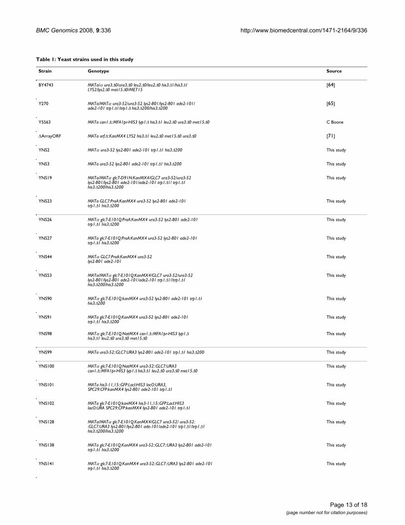
Table 1: Yeast strains used in this study
Strain Genotype Source
BY4743 MATa/α ura3Δ0/ura3Δ0 leu2Δ0/leu2Δ0 his3Δ1/his3Δ1 LYS2/lys2Δ0 met15Δ0/MET15
[64]
Y270 MATa/MATα ura3-52/ura3-52 lys2-801/lys2-801 ade2-101/ade2-101 trp1Δ1/trp1Δ his3Δ200/his3Δ200
[65]
Y5563 MATa can1Δ::MFA1pr-HIS3 lyp1Δ his3Δ1 leu2Δ0 ura3Δ0 met15Δ0 C Boone
ΔArrayORF MATa orfΔ::KanMX4 LYS2 his3Δ1 leu2Δ0 met15Δ0 ura3Δ0 [71]
YNS2 MATα ura3-52 lys2-801 ade2-101 trp1Δ1 his3Δ200 This study
YNS3 MATa ura3-52 lys2-801 ade2-101 trp1Δ1 his3Δ200 This study
YNS19 MATa/MATα glc7-D91N:KanMX4/GLC7 ura3-52/ura3-52 lys2-801/lys2-801 ade2-101/ade2-101 trp1Δ1/ trp1Δ1 his3Δ200/his3Δ200
This study
YNS23 MATa GLC7:ProA:KanMX4 ura3-52 lys2-801 ade2-101 trp1Δ1 his3Δ200
This study
YNS26 MATα glc7-E101Q:ProA:KanMX4 ura3-52 lys2-801 ade2-101 trp1Δ1 his3Δ200
This study
YNS27 MATa glc7-E101Q:ProA:KanMX4 ura3-52 lys2-801 ade2-101 trp1Δ1 his3Δ200
This study
YNS44 MATα GLC7:ProA:KanMX4 ura3-52 lys2-801 ade2-101
This study
YNS53 MATa/MATα glc7-E101Q:KanMX4/GLC7 ura3-52/ura3-52 lys2-801/lys2-801 ade2-101/ade2-101 trp1Δ1//trp1Δ1 his3Δ200/his3Δ200
This study
YNS90 MATα glc7-E101Q:kanMX4 ura3-52 lys2-801 ade2-101 trp1Δ1 his3Δ200
This study
YNS91 MATa glc7-E101Q:KanMX4 ura3-52 lys2-801 ade2-101 trp1Δ1 his3Δ200
This study
YNS98 MATα glc7-E101Q:NatMX4 can1Δ::MFA1pr-HIS3 lyp1Δ his3Δ1 leu2Δ0 ura3Δ0 met15Δ0
This study
YNS99 MATa ura3-52::GLC7:URA3 lys2-801 ade2-101 trp1Δ1 his3Δ200 This study
YNS100 MATα glc7-E101Q:NatMX4 ura3-52::GLC7:URA3 can1Δ::MFA1pr-HIS3 lyp1Δ his3Δ1 leu2Δ0 ura3Δ0 met15Δ0
This study
YNS101 MATa his3-11,15::GFP:LacI:HIS3 lacO:URA3, SPC29:CFP:kanMX4 lys2-801 ade2-101 trp1Δ1
This study
YNS102 MATa glc7-E101Q:kanMX4 his3-11,15::GFP:LacI:HIS3 lacO:URA SPC29:CFP:kanMX4 lys2-801 ade2-101 trp1Δ1
This study
YNS128 MATa/MATα glc7-E101Q:KanMX4/GLC7 ura3-52/ ura3-52::GLC7:URA3 lys2-801/lys2-801 ade-101/ade2-101 trp1Δ1/trp1Δ1 his3Δ200/his3Δ200
This study
YNS138 MATa glc7-E101Q:KanMX4 ura3-52::GLC7::URA3 lys2-801 ade2-101 trp1Δ1 his3Δ200
This study
YNS141 MATα glc7-E101Q:KanMX4 ura3-52::GLC7::URA3 lys2-801 ade2-101 trp1Δ1 his3Δ200
This study
BMC Genomics 2008, 9:336 http://www.biomedcentral.com/1471-2164/9/336
ml) to complete cell separation. Glycogen staining wasperformed as previously described [40]. Briefly, after 2-day incubation at 30°C, plates were exposed to iodinecrystals (Sigma) for 1 minute, removed for 15 sec andexposed again for 2 min. Digital images were capturedimmediately after exposure to iodine.
Analysis of chromosome segregationMicroscopyMATa wild type (YNS101) and glc7-E101Q mutant(YNS102) strains that expressed Spc29-CFP (SPB marker),GFP-LacI and centromeric (CEN15) lacO repeats [34]were isolated from heterozygous diploids. Cells wereincubated in 5 μg/ml a-factor for 3 hrs at 30°C, washedand released in YPAD and collected every 10 min (80 mintotal). Samples were fixed for 5 min in 4% formaldehyde,washed three times in PBS and imaged using a NikonTE200 inverted microscope equipped with a 100 × 1.4 NAobjective mounted on PE piezo z-drive/Improvision Orbitcontroller and Hammamatsu ORCA-ERG camera. Imagestacks (0.5 μm sections, 10–12/image) were acquired
using Volocity 4.0 and image deconvolution with Volocity4.0 Restoration.
Sectoring assayMATa wild type (YNS3), glc7-E101Q (YNS91) and ame1-6(positive control for chromosome loss; Vogel unpub-lished data) strains lacking a functional ADE2 gene(ade2Δ-101) were transformed with a circular fragment ofchromosome III encoding SUP11 (Circle III/SUP11) [35]and incubated for 4 days on YPD at 30°C. SUP11 sup-presses the ade2Δ-101 mutation resulting in the formationof white colonies. Loss of the CircleIII/SUP11 (an indica-tor of chromsome mis-segregation) was identified by therestoration of the ade2Δ-101 mutant phenotype causingthe accumulation of P-ribosylaminoimidazole, a redcolored pigment [41].
Synthetic genetic array (SGA) analysis of glc7-E101QTwo-independent SGA screens were performed in tripli-cate using the ordered deletion array and Virtek pinningrobot system as previously described [12,13,71]. The sizes
Table 2: Functional processes of 163 genes showing SSL interactions with glc7-E101Q
Functional category Genes
Cell polarity AXL2, BUD4
Chromosome segregation BAG7, BIM1, BNR1, BOI2, BUB1, BUB3, BUD14, CIK1, CTF8, KAP120, KAR3, MAD1, MAD2, MCM22, NUP53, NUP84, NUP133, REC8, ROM2, SXM1, TUB3
DNA damage EXO1, IES2, LIF1, LSM12, MRC1, PMS1, RAD4, RAD14, RFX1, RSC2, TDP1, UNG1, YFR017C, YPR147C
Histone modification ASF1, CHD1, DOT1, FPR4, PHO23, RTT109, UAF30
Lipid metabolism ANT1, BUD25, ERG5, OPI3, OSH3, PCS60, PEX6, PEX29, PEX31, PLB1, PSD1, SCS3
Mitochondrial function ATP4, ATP12, CBR1, COX19, CYC7, FMP42, HAP2, IMP1, MDM34, MIR1, MRS4, MSK1, MTF1, PDX1, PET122, PUT2, RIM1, RML2, RSM18, SCO1, VPS73, YPT11, YBL095W, YGL059W, YGR021W, YGR287C, YHR162W, YJR039W, YNL320W
Nutrient sensing and stress signaling ATG5, ATG15, BAP2, CKB1, CKB2, GPA2, GPH1, HIT1, HXT17, MCA1, MCH4, NST1, NVJ1, ORM2, SAK1, SFL1, SIP5, SPG5, SSK1, TDH3, TPK1, UGA1, URM1, VAC8, WHI2, YBR235W, YJL193W, YNR029C
Phosphatase genes PPM1, PPZ1, RTS3
Protein modification and degradation KTR1, KTR3, KTR4, NAT3, NPL4, OTU1, PMT5, SLX8, UMP1, USA1
Protein synthesis RPL6B, RPL12A, RPL19A, RPL24A, RPL37A, RPS1A, RPS16A, RPS19A, RPS25A, YBL054W
SAM(1) synthesis BUD23, NCL1, RMT2, SPE2, TRM1
Transcription CAF120, CDC40, DAT1, DST1, ELP2, HMS2, KTI12, LSM6, NUT1, RTT103, SSN2, SSN8, SUM1
Vesicular transport ARF1, BTS1, COG5, END3, ENT5, GSG1, GYP8, MST27, UBP3
(1): S-adenosylmethionine
Page 14 of 18(page number not for citation purposes)
BMC Genomics 2008, 9:336 http://www.biomedcentral.com/1471-2164/9/336
of the resulting colonies were measured from digitalimages of the plates. A comparative set of mutant meas-urements relative to wild type control measurements ena-bled t-statistics and p-values to be calculated [13]. Doublemutants that showed significantly reduced colony sizes (p< 0.05) were scored as hits. Hits identified in the SGAscreens were evaluated by a modified spot assay version ofrandom spore analysis [13] using a glc7-E101Q strain thatco-expressed GLC7-res (YNS100) (Additional File 3) andfinally scored as synthetic sick/lethal (SSL) or rejected asfalse positive. Spores were initially grown for 2 days at30°C on solid haploid selection medium [synthetic dex-trose (SD) medium lacking histidine and arginine butcontaining canavanine: SD - His/Arg + canavanine]. MATaspore progeny were transferred to solid 5-fluoro-oroticacid (5-FOA) medium [45] and incubated at 30°C for 2days to eliminate cells expressing GLC7-res. Cells wereinoculated to microtiter plates in 250 μl of liquid SD -His/Arg + canavanine for 2 days at 30°C. A Biomek FXrobot (Beckman Coulter, Inc; CIAN robotics facility,McGill University) was used to perform serial dilutionsand spotting to four different solid medium plates: SD/MSG (monosodium glutamic acid) - His/Arg + cana-vanine/nourseothricin, SD/MSG - His/Arg + canavanine/G418, SD/MSG - His/Arg +canavanine/nourseothricin/G418, and SD/MSG - His/Arg/Ura + canavanine. Colonygrowth was scored following incubation for 2 days at30°C.
Protein extraction, 1D and 2D-SDS-PAGEAll steps were performed at 4°C unless otherwise indi-cated. Log phase yeast cultures were harvested and washedin PGSK+ buffer (PGSK+: 50 mM NaPO4, 50 mM NaCl, 5mM KCl, 60 mM glucose, 4% CHAPS, 50 mM DTT, 5 μMyeast protease inhibitors [Sigma P8215], 5 μM PMSF, 5μM ortho-vanadate, 5 μM NaF, and 5 mM β-glycerolphosphate) and pelleted by centrifugation. Cell pelletswere suspended in 2 volumes of PGSK+, and an equal vol-ume of zirconium beads (Biospec Products) added, andcells lysed by vortexing for 12–15 min. Extracts werecleared by centrifugation for 10 min at 13,000 × g. For 1DSDS-PAGE, extracts were re-suspended in an equal vol-ume of 2× SDS sample buffer [72] and denatured at 95°Cfor 5 min. For 2D-SDS-PAGE and 2D-DiGE, extracts weretreated with 50 μg/ml DNAse and RNAse (Worthington)for 15 minutes, precipitated with methanol/chloroformand re-suspended in labeling buffer (2 M thiourea, 7 Murea, 30 mM Tris, 4% CHAPS) at 25°C and the proteinconcentration determined (Biorad Protein Assay). For 2D-SDS-PAGE analysis of GLC7-ProA (YNS23) and glc7-E101Q-ProA (YNS27), 30 μg was loaded to 7 cm pH 4–7Readystrip IPG strips (Biorad) and rehydration and focus-ing carried out according to the manufacturer's instruc-tions.
ImmunoblottingImmunoblotting was performed as previously described[65]. Briefly, proteins extracts prepared from GLC7-ProA(YNS23, YNS44) and glc7-E101Q-ProA (YNS26, YNS27)strains were separated on 1D or 2D-SDS-PAGE gels, trans-ferred to PVDF membranes (Millipore) and probed withanti-ProA monoclonal antibodies (1:5000; clone SPA-27,Sigma). Actin was detected using the monoclonal anti-bodies (1:5000; clone C4, MP Biomedical). Protein/anti-body complexes were detected using anti-mouse HRP-conjugated secondary antibodies (1:10,000; GE Health-Care) and ECL chemiluminescence (Pierce).
Authors' contributionsML, TN and NS constructed the strains, conducted thescreens and ran data confirmation. JK, HP, MZ and MNhelped in the data confirmation. PH, HB and CAM partic-ipated in the experimental design and data analysis andhelped to draft the manuscript. ML, JV and GL conceivedthe study, analyzed the data and wrote the manuscript. Allauthors have read and approved the final manuscript.
Additional material
Additional file 1PP1-mediated phosphate exchange.Click here for file[http://www.biomedcentral.com/content/supplementary/1471-2164-9-336-S1.pdf]
Additional file 2Western blot analysis of Glc7-ProA and glc7-E101Q-ProA fusion pro-teins.Click here for file[http://www.biomedcentral.com/content/supplementary/1471-2164-9-336-S2.pdf]
Additional file 3SGA and random spore analysis methods.Click here for file[http://www.biomedcentral.com/content/supplementary/1471-2164-9-336-S3.pdf]
Additional file 4List of genes included in the SGA screens, found as hits in the screens and confirmed as glc7-E101Q SSL by random spore analysis.Click here for file[http://www.biomedcentral.com/content/supplementary/1471-2164-9-336-S4.txt]
Additional file 5List of glc7-E101Q SSL genes with and their attributes.Click here for file[http://www.biomedcentral.com/content/supplementary/1471-2164-9-336-S5.xls]
Page 15 of 18(page number not for citation purposes)
BMC Genomics 2008, 9:336 http://www.biomedcentral.com/1471-2164/9/336
AcknowledgementsWe thank Damien Damours, Phil Branton, Malcolm Whiteway and all the members of the Vogel, Mandato and Bussey labs for fruitful discussions and support during this project, and acknowledge the Cell Imaging and Analysis Network (CIAN) DiGE and SGA platforms. JV is supported by a New Investigator Award from the Canadian Institutes of Health Research (MSH 69117). CAM is Canada Research Chair. This research was supported by operating grants from the Canadian Institutes of Health Research (MOP-64404 to JV and MOP-68970 to CAM), the Natural Sciences and Engineer-ing Research Council (262246-03 to JV and 6040-03 to HB) and infrastruc-ture grants from the Canada Foundation for Innovation to awarded to JV (CFI 7395) and to the Developmental Biology Research Initiative of McGill University (CFI 8298).
References1. Andrews PD, Stark MJ: Type 1 protein phosphatase is required
for maintenance of cell wall integrity, morphogenesis andcell cycle progression in Saccharomyces cerevisiae. J Cell Sci2000, 113(Pt 3):507-520.
2. Dobbelaere J, Gentry MS, Hallberg RL, Barral Y: Phosphorylation-dependent regulation of septin dynamics during the cellcycle. Dev Cell 2003, 4(3):345-357.
3. Pinsky BA, Kotwaliwale CV, Tatsutani SY, Breed CA, Biggins S: Glc7/protein phosphatase 1 regulatory subunits can oppose theIpl1/aurora protein kinase by redistributing Glc7. Mol Cell Biol2006, 26(7):2648-2660.
4. Stegmeier F, Amon A: Closing mitosis: the functions of theCdc14 phosphatase and its regulation. Annu Rev Genet 2004,38:203-232.
5. Bloecher A, Tatchell K: Dynamic localization of protein phos-phatase type 1 in the mitotic cell cycle of Saccharomycescerevisiae. J Cell Biol 2000, 149(1):125-140.
6. Sakumoto N, Matsuoka I, Mukai Y, Ogawa N, Kaneko Y, HarashimaS: A series of double disruptants for protein phosphatasegenes in Saccharomyces cerevisiae and their phenotypicanalysis. Yeast 2002, 19(7):587-599.
7. Collins SR, Miller KM, Maas NL, Roguev A, Fillingham J, Chu CS,Schuldiner M, Gebbia M, Recht J, Shales M, Ding H, Xu H, Han J, Ing-varsdottir K, Cheng B, Andrews B, Boone C, Berger SL, Hieter P,Zhang Z, Brown GW, Ingles CJ, Emili A, Allis CD, Toczyski DP,Weissman JS, Greenblatt JF, Krogan NJ: Functional dissection ofprotein complexes involved in yeast chromosome biologyusing a genetic interaction map. Nature 2007,446(7137):806-810.
8. Decourty L, Saveanu C, Zemam K, Hantraye F, Frachon E, RousselleJC, Fromont-Racine M, Jacquier A: Linking functionally relatedgenes by sensitive and quantitative characterization ofgenetic interaction profiles. Proc Natl Acad Sci USA 2008,105(15):5821-5826.
9. Pan X, Ye P, Yuan DS, Wang X, Bader JS, Boeke JD: A DNA integ-rity network in the yeast Saccharomyces cerevisiae. Cell2006, 124(5):1069-1081.
10. Pan X, Yuan DS, Xiang D, Wang X, Sookhai-Mahadeo S, Bader JS,Hieter P, Spencer F, Boeke JD: A robust toolkit for functionalprofiling of the yeast genome. Mol Cell 2004, 16(3):487-496.
11. Schuldiner M, Collins SR, Thompson NJ, Denic V, Bhamidipati A,Punna T, Ihmels J, Andrews B, Boone C, Greenblatt JF, Weissman JS,Krogan NJ: Exploration of the function and organization of theyeast early secretory pathway through an epistatic min-iarray profile. Cell 2005, 123(3):507-519.
12. Tong AH, Evangelista M, Parsons AB, Xu H, Bader GD, Page N, Rob-inson M, Raghibizadeh S, Hogue CW, Bussey H, Andrews B, Tyers M,Boone C: Systematic genetic analysis with ordered arrays ofyeast deletion mutants. Science 2001, 294(5550):2364-2368.
13. Tong AH, Lesage G, Bader GD, Ding H, Xu H, Xin X, Young J, BerrizGF, Brost RL, Chang M, Chen Y, Cheng X, Chua G, Friesen H, Gold-berg DS, Haynes J, Humphries C, He G, Hussein S, Ke L, Krogan N,Li Z, Levinson JN, Lu H, Menard P, Munyana C, Parsons AB, Ryan O,Tonikian R, Roberts T, Sdicu AM, Shapiro J, Sheikh B, Suter B, WongSL, Zhang LV, Zhu H, Burd CG, Munro S, Sander C, Rine J, GreenblattJ, Peter M, Bretscher A, Bell G, Roth FP, Brown GW, Andrews B, Bus-sey H, Boone C: Global mapping of the yeast genetic interac-tion network. Science 2004, 303(5659):808-813.
14. Terrak M, Kerff F, Langsetmo K, Tao T, Dominguez R: Structuralbasis of protein phosphatase 1 regulation. Nature 2004,429(6993):780-784.
15. Wu X, Hart H, Cheng C, Roach PJ, Tatchell K: Characterization ofGac1p, a regulatory subunit of protein phosphatase type Iinvolved in glycogen accumulation in Saccharomyces cerevi-siae. Mol Genet Genomics 2001, 265(4):622-635.
16. Wu X, Tatchell K: Mutations in yeast protein phosphatase type1 that affect targeting subunit binding. Biochemistry 2001,40(25):7410-7420.
17. Ceulemans H, Bollen M: Functional diversity of protein phos-phatase-1, a cellular economizer and reset button. Physiol Rev2004, 84(1):1-39.
18. Knaus M, Cameroni E, Pedruzzi I, Tatchell K, De Virgilio C, Peter M:The Bud14p-Glc7p complex functions as a cortical regulatorof dynein in budding yeast. Embo J 2005, 24(17):3000-3011.
19. Ramaswamy NT, Li L, Khalil M, Cannon JF: Regulation of yeast gly-cogen metabolism and sporulation by Glc7p protein phos-phatase. Genetics 1998, 149(1):57-72.
20. Davierwala AP, Haynes J, Li Z, Brost RL, Robinson MD, Yu L, Mnaim-neh S, Ding H, Zhu H, Chen Y, Cheng X, Brown GW, Boone C,Andrews BJ, Hughes TR: The synthetic genetic interaction spec-trum of essential genes. Nat Genet 2005, 37(10):1147-1152.
21. Drees BL, Thorsson V, Carter GW, Rives AW, Raymond MZ, Avila-Campillo I, Shannon P, Galitski T: Derivation of genetic interac-tion networks from quantitative phenotype data. Genome Biol2005, 6(4):R38.
22. Baker SH, Frederick DL, Bloecher A, Tatchell K: Alanine-scanningmutagenesis of protein phosphatase type 1 in the yeast Sac-charomyces cerevisiae. Genetics 1997, 145(3):615-626.
23. Wan J, Xu H, Grunstein M: CDC14 of Saccharomyces cerevi-siae. Cloning, sequence analysis, and transcription during thecell cycle. J Biol Chem 1992, 267(16):11274-11280.
24. Sanz P, Alms GR, Haystead TA, Carlson M: Regulatory interac-tions between the Reg1-Glc7 protein phosphatase and theSnf1 protein kinase. Mol Cell Biol 2000, 20(4):1321-1328.
25. Tu J, Carlson M: REG1 binds to protein phosphatase type 1 andregulates glucose repression in Saccharomyces cerevisiae.Embo J 1995, 14(23):5939-5946.
26. Stark C, Breitkreutz BJ, Reguly T, Boucher L, Breitkreutz A, Tyers M:BioGRID: a general repository for interaction datasets.Nucleic Acids Res 2006:D535-539.
27. Goldberg J, Huang HB, Kwon YG, Greengard P, Nairn AC, Kuriyan J:Three-dimensional structure of the catalytic subunit of pro-tein serine/threonine phosphatase-1. Nature 1995,376(6543):745-753.
28. Zhuo S, Clemens JC, Stone RL, Dixon JE: Mutational analysis of aSer/Thr phosphatase. Identification of residues important inphosphoesterase substrate binding and catalysis. J Biol Chem1994, 269(42):26234-26238.
29. Bloecher A, Tatchell K: Defects in Saccharomyces cerevisiaeprotein phosphatase type I activate the spindle/kinetochorecheckpoint. Genes Dev 1999, 13(5):517-522.
30. Francisco L, Wang W, Chan CS: Type 1 protein phosphatase actsin opposition to IpL1 protein kinase in regulating yeast chro-mosome segregation. Mol Cell Biol 1994, 14(7):4731-4740.
31. Hisamoto N, Sugimoto K, Matsumoto K: The Glc7 type 1 proteinphosphatase of Saccharomyces cerevisiae is required for cellcycle progression in G2/M. Mol Cell Biol 1994, 14(5):3158-3165.
32. Sassoon I, Severin FF, Andrews PD, Taba MR, Kaplan KB, Ashford AJ,Stark MJ, Sorger PK, Hyman AA: Regulation of Saccharomycescerevisiae kinetochores by the type 1 phosphatase Glc7p.Genes Dev 1999, 13(5):545-555.
Additional file 6Distribution of high-level GO annotations for glc7-E101Q SSL genes.Click here for file[http://www.biomedcentral.com/content/supplementary/1471-2164-9-336-S6.pdf]
Page 16 of 18(page number not for citation purposes)
BMC Genomics 2008, 9:336 http://www.biomedcentral.com/1471-2164/9/336
33. Tung HY, Wang W, Chan CS: Regulation of chromosome segre-gation by Glc8p, a structural homolog of mammalian inhibi-tor 2 that functions as both an activator and an inhibitor ofyeast protein phosphatase 1. Mol Cell Biol 1995,15(11):6064-6074.
34. Goshima G, Yanagida M: Establishing biorientation occurs withprecocious separation of the sister kinetochores, but not thearms, in the early spindle of budding yeast. Cell 2000,100(6):619-633.
35. Hieter P, Mann C, Snyder M, Davis RW: Mitotic stability of yeastchromosomes: a colony color assay that measures nondis-junction and chromosome loss. Cell 1985, 40(2):381-392.
36. Stuart JS, Frederick DL, Varner CM, Tatchell K: The mutant type 1protein phosphatase encoded by glc7-1 from Saccharomycescerevisiae fails to interact productively with the GAC1-encoded regulatory subunit. Mol Cell Biol 1994, 14(2):896-905.
37. Anderson C, Tatchell K: Hyperactive glycogen synthasemutants of Saccharomyces cerevisiae suppress the glc7-1protein phosphatase mutant. J Bacteriol 2001, 183(3):821-829.
38. Tu J, Carlson M: The GLC7 type 1 protein phosphatase isrequired for glucose repression in Saccharomyces cerevi-siae. Mol Cell Biol 1994, 14(10):6789-6796.
39. Sanz P, Ludin K, Carlson M: Sip5 interacts with both the Reg1/Glc7 protein phosphatase and the Snf1 protein kinase of Sac-charomyces cerevisiae. Genetics 2000, 154(1):99-107.
40. Enjalbert B, Parrou JL, Vincent O, Francois J: Mitochondrial respi-ratory mutants of Saccharomyces cerevisiae accumulateglycogen and readily mobilize it in a glucose-depletedmedium. Microbiology 2000, 146(Pt 10):2685-2694.
41. Kim G, Sikder H, Singh KK: A colony color method identifies thevulnerability of mitochondria to oxidative damage. Mutagen-esis 2002, 17(5):375-381.
42. Tachikawa H, Bloecher A, Tatchell K, Neiman AM: A Gip1p-Glc7pphosphatase complex regulates septin organization andspore wall formation. J Cell Biol 2001, 155(5):797-808.
43. Hochwagen A, Tham WH, Brar GA, Amon A: The FK506 bindingprotein Fpr3 counteracts protein phosphatase 1 to maintainmeiotic recombination checkpoint activity. Cell 2005,122(6):861-873.
44. Bailis JM, Roeder GS: Pachytene exit controlled by reversal ofMek1-dependent phosphorylation. Cell 2000, 101(2):211-221.
45. Boeke JD, LaCroute F, Fink GR: A positive selection for mutantslacking orotidine-5'-phosphate decarboxylase activity inyeast: 5-fluoro-orotic acid resistance. Mol Gen Genet 1984,197(2):345-346.
46. Tavazoie S, Hughes JD, Campbell MJ, Cho RJ, Church GM: System-atic determination of genetic network architecture. NatGenet 1999, 22(3):281-285.
47. Ramaswamy V, Williams JS, Robinson KM, Sopko RL, Schultz MC:Global control of histone modification by the anaphase-pro-moting complex. Mol Cell Biol 2003, 23(24):9136-9149.
48. Zhang K, Lin W, Latham JA, Riefler GM, Schumacher JM, Chan C,Tatchell K, Hawke DH, Kobayashi R, Dent SY: The Set1 methyl-transferase opposes Ipl1 aurora kinase functions in chromo-some segregation. Cell 2005, 122(5):723-734.
49. Norden C, Mendoza M, Dobbelaere J, Kotwaliwale CV, Biggins S, Bar-ral Y: The NoCut pathway links completion of cytokinesis tospindle midzone function to prevent chromosome breakage.Cell 2006, 125(1):85-98.
50. Yoshida S, Kono K, Lowery DM, Bartolini S, Yaffe MB, Ohya Y, Pell-man D: Polo-like kinase Cdc5 controls the local activation ofRho1 to promote cytokinesis. Science 2006, 313(5783):108-111.
51. Gao XD, Sperber LM, Kane SA, Tong Z, Tong AH, Boone C, Bi E:Sequential and distinct roles of the cadherin domain-con-taining protein Axl2p in cell polarization in yeast cell cycle.Mol Biol Cell 2007, 18(7):2542-2560.
52. Cullen PJ, Sprague GF Jr: The Glc7p-interacting protein Bud14pattenuates polarized growth, pheromone response, and fila-mentous growth in Saccharomyces cerevisiae. Eukaryot Cell2002, 1(6):884-894.
53. Cheeseman IM, Anderson S, Jwa M, Green EM, Kang J, Yates JR 3rd,Chan CS, Drubin DG, Barnes G: Phospho-regulation of kineto-chore-microtubule attachments by the Aurora kinase Ipl1p.Cell 2002, 111(2):163-172.
54. Hisamoto N, Frederick DL, Sugimoto K, Tatchell K, Matsumoto K:The EGP1 gene may be a positive regulator of protein phos-
phatase type 1 in the growth control of Saccharomyces cer-evisiae. Mol Cell Biol 1995, 15(7):3767-3776.
55. Lenssen E, James N, Pedruzzi I, Dubouloz F, Cameroni E, Bisig R, Mail-let L, Werner M, Roosen J, Petrovic K, Winderickx J, Collart MA, DeVirgilio C: The Ccr4-Not complex independently controlsboth Msn2-dependent transcriptional activation – via a newlyidentified Glc7/Bud14 type I protein phosphatase module –and TFIID promoter distribution. Mol Cell Biol 2005,25(1):488-498.
56. Venturi GM, Bloecher A, Williams-Hart T, Tatchell K: Geneticinteractions between GLC7, PPZ1 and PPZ2 in saccharomy-ces cerevisiae. Genetics 2000, 155(1):69-83.
57. Zeng G, Huang B, Neo SP, Wang J, Cai M: Scd5p mediates phos-phoregulation of actin and endocytosis by the type 1 phos-phatase Glc7p in yeast. Mol Biol Cell 2007, 18(12):4885-4898.
58. Bryant NJ, James DE: The Sec1p/Munc18 (SM) protein, Vps45p,cycles on and off membranes during vesicle transport. J CellBiol 2003, 161(4):691-696.
59. Gavin AC, Bosche M, Krause R, Grandi P, Marzioch M, Bauer A,Schultz J, Rick JM, Michon AM, Cruciat CM, Remor M, Hofert C,Schelder M, Brajenovic M, Ruffner H, Merino A, Klein K, Hudak M,Dickson D, Rudi T, Gnau V, Bauch A, Bastuck S, Huhse B, LeutweinC, Heurtier MA, Copley RR, Edelmann A, Querfurth E, Rybin V,Drewes G, Raida M, Bouwmeester T, Bork P, Seraphin B, Kuster B,Neubauer G, Superti-Furga G: Functional organization of theyeast proteome by systematic analysis of protein complexes.Nature 2002, 415(6868):141-147.
60. Ho Y, Gruhler A, Heilbut A, Bader GD, Moore L, Adams SL, Millar A,Taylor P, Bennett K, Boutilier K, Yang L, Wolting C, Donaldson I,Schandorff S, Shewnarane J, Vo M, Taggart J, Goudreault M, Muskat B,Alfarano C, Dewar D, Lin Z, Michalickova K, Willems AR, Sassi H,Nielsen PA, Rasmussen KJ, Andersen JR, Johansen LE, Hansen LH, Jes-persen H, Podtelejnikov A, Nielsen E, Crawford J, Poulsen V,Sorensen BD, Matthiesen J, Hendrickson RC, Gleeson F, Pawson T,Moran MF, Durocher D, Mann M, Hogue CW, Figeys D, Tyers M:Systematic identification of protein complexes in Saccharo-myces cerevisiae by mass spectrometry. Nature 2002,415(6868):180-183.
61. Christianson TW, Sikorski RS, Dante M, Shero JH, Hieter P: Multi-functional yeast high-copy-number shuttle vectors. Gene1992, 110(1):119-122.
62. Longtine MS, McKenzie A 3rd, Demarini DJ, Shah NG, Wach A, Bra-chat A, Philippsen P, Pringle JR: Additional modules for versatileand economical PCR-based gene deletion and modificationin Saccharomyces cerevisiae. Yeast 1998, 14(10):953-961.
63. Guthrie C, Fink GR, (eds): Guide to Yeast Genetics and Molec-ular Biology Part B. New York: Academic Press; 2002.
64. Brachmann CB, Davies A, Cost GJ, Caputo E, Li J, Hieter P, Boeke JD:Designer deletion strains derived from Saccharomyces cer-evisiae S288C: a useful set of strains and plasmids for PCR-mediated gene disruption and other applications. Yeast 1998,14(2):115-132.
65. Vogel J, Drapkin B, Oomen J, Beach D, Bloom K, Snyder M: Phos-phorylation of gamma-tubulin regulates microtubule organ-ization in budding yeast. Dev Cell 2001, 1(5):621-631.
66. Knop M, Siegers K, Pereira G, Zachariae W, Winsor B, Nasmyth K,Schiebel E: Epitope tagging of yeast genes using a PCR-basedstrategy: more tags and improved practical routines. Yeast1999, 15(10B):963-972.
67. Sikorski RS, Hieter P: A system of shuttle vectors and yeast hoststrains designed for efficient manipulation of DNA in Saccha-romyces cerevisiae. Genetics 1989, 122(1):19-27.
68. Huffaker TC, Thomas JH, Botstein D: Diverse effects of beta-tubulin mutations on microtubule formation and function. JCell Biol 1988, 106(6):1997-2010.
69. Stearns T, Botstein D: Unlinked noncomplementation: isola-tion of new conditional-lethal mutations in each of the tubu-lin genes of Saccharomyces cerevisiae. Genetics 1988,119(2):249-260.
70. Dien BS, Peterson MS, Srienc F: Cell-cycle analysis of Saccharo-myces cerevisiae. Methods Cell Biol 1994, 42(Pt B):457-475.
71. Winzeler EA, Shoemaker DD, Astromoff A, Liang H, Anderson K,Andre B, Bangham R, Benito R, Boeke JD, Bussey H, Chu AM, Con-nelly C, Davis K, Dietrich F, Dow SW, El Bakkoury M, Foury F, FriendSH, Gentalen E, Giaever G, Hegemann JH, Jones T, Laub M, Liao H,Liebundguth N, Lockhart DJ, Lucau-Danila A, Lussier M, M'Rabet N,
Page 17 of 18(page number not for citation purposes)
BMC Genomics 2008, 9:336 http://www.biomedcentral.com/1471-2164/9/336
Publish with BioMed Central and every scientist can read your work free of charge
"BioMed Central will be the most significant development for disseminating the results of biomedical research in our lifetime."
Sir Paul Nurse, Cancer Research UK
Your research papers will be:
available free of charge to the entire biomedical community
peer reviewed and published immediately upon acceptance
cited in PubMed and archived on PubMed Central
yours — you keep the copyright
Submit your manuscript here:http://www.biomedcentral.com/info/publishing_adv.asp
BioMedcentral
Menard P, Pai C, Rebischung C, Revuelta JL, Riles L, Roberts CJ, Ross-Macdonald P, Scherens B, Snyder M, Sookhai-Mahadeo S, Storms RK,Veronneau S, Voet M, Volckaert G, Ward TR, Wysocki R, Yen G, YuK, Zimmermann K, Philippsen P, Johnston M, Davis RW: Functionalcharacterization of the S. cerevisiae genome by gene dele-tion and parallel analysis. Science 1999, 285(5429):901-906.
72. Laemmli UK: Cleavage of structural proteins during theassembly of the head of bacteriophage T4. Nature 1970,227(5259):680-685.
Page 18 of 18(page number not for citation purposes)