Embed Size (px)
Citation preview
Bayraktutan, Ulvi (2004) Nitric oxide synthase and NAD(P)H oxidase modulate coronary endothelial cell growth. Journal of Molecular and Cellular Cardiology, 36 . pp. 277-286.
Access from the University of Nottingham repository: http://eprints.nottingham.ac.uk/486/1/JMCC.pdf
Copyright and reuse:
The Nottingham ePrints service makes this work by researchers of the University of Nottingham available open access under the following conditions.
This article is made available under the University of Nottingham End User licence and may be reused according to the conditions of the licence. For more details see: http://eprints.nottingham.ac.uk/end_user_agreement.pdf
A note on versions:
The version presented here may differ from the published version or from the version of record. If you wish to cite this item you are advised to consult the publisher’s version. Please see the repository url above for details on accessing the published version and note that access may require a subscription.
For more information, please contact [email protected]
NITRIC OXIDE SYNTHASE AND NAD(P)H OXIDASE MODULATE
CORONARY ENDOTHELIAL CELL GROWTH
Ulvi Bayraktutan DVM, PhD
Department of Medicine, Institute of Clinical Science, Queen’s University Belfast,
Belfast, United Kingdom
Short title: Free radicals regulate endothelial cell growth
Address for correspondence
Dr Ulvi BAYRAKTUTAN
Department of Medicine,
Institute of Clinical Science Block B,
Queen’s University Belfast,
Grosvenor Road,
Belfast BT12 6BJ
United Kingdom
Tel: 00 44 2890 632742
Fax: 00 44 2890 329899
E-mail: [email protected]

Abstract
Reactive oxygen species (ROS) including nitric oxide (NO) and superoxide anion (O2-)
are associated with cell migration, proliferation and many growth-related diseases. The
objective of this study was to determine whether there was a reciprocal relationship
between rat coronary microvascular endothelial cell (CMEC) growth and
activity/expressions (mRNA and protein) of endothelial NO synthase (eNOS) and
NAD(P)H oxidase enzymes. Proliferating namely, 50% confluent CMEC possessed
approximately three-fold increased activity and expression of both enzymes compared to
100% confluent cells. Treatment of CMEC with an inhibitor of eNOS (L-NAME, 100
M) increased cell proliferation as assessed via three independent methods i.e. cell
counting, determination of total cellular protein levels and [3H]thymidine incorporation.
Similarly, treatment of CMEC with pyrogallol (0.3-3 mM), a superoxide anion (O2-)-
generator, also increased CMEC growth while spermine NONOate (SpNO), a NO donor,
significantly reduced cell growth. Co-incubation of CMEC with a cell permeable
superoxide dismutase mimetic (Mn-III-tetrakis-4-benzoic acid-porphyrin; MnTBAP) plus
either pyrogallol or NO did not alter cell number and DNA synthesis thereby dismissing
the involvement of peroxynitrite (OONO-) in CMEC proliferation. Specific inhibitors of
NAD(P)H oxidase but not other ROS-generating enzymes including cyclooxygenase and
xanthine oxidase, attenuated cell growth. Transfection of CMEC with antisense p22-phox
cDNA, a membrane-bound component of NAD(P)H oxidase, resulted in substantial
reduction in [3H]thymidine incorporation, total cellular protein levels and expression of
p22-phox protein. These data demonstrate a cross-talk between CMEC growth and eNOS
and NAD(P)H oxidase enzyme activity and expression, thus suggesting that the
regulation of these enzymes may be critical in preventing the initiation and/or progression
of coronary atherosclerosis.
1. Introduction
The vascular endothelium, a multifunctional organ that covers the entire inner surface
of all the blood vessels, generates a wide array of vasoactive substances including nitric
oxide (NO) in response to several chemical, physical or hormonal stimuli [1]. The
integrity of the endothelial layer is preserved by controlled proliferation and migration of
endothelial cells in a number of physiological processes such as angiogenesis and during
wound healing after an injury to the vascular wall [2,3]. However, in several pathological
conditions e.g. tumor metastasis and atherosclerosis endothelial cells proliferate and
migrate at greater levels and in an uncontrolled manner [4,5].
NO is generated from amino acid L-arginine within the normal endothelium by
endothelial type of NO synthase (eNOS). Although eNOS is constitutively expressed, its
expression and activity are mediated by many physio-pathological processes e.g. while
shear stress increases its expression in cultured endothelial cells, hypoxia elicits a
decrease in eNOS expression/activity [6,7]. NO plays significant roles in the regulation of
vascular tone, in the modulation of endothelial cell permeability and in the inhibition of
vascular smooth muscle cell (VSMC) proliferation and migration [1]. Indeed, NO donors,
transfection of eNOS gene into the arterial wall and supplementation of diet with L-
arginine have been shown to reduce intimal hyperplasia, a hallmark of atherosclerotic
disease [8,9]. However, contrary to these findings, NO has also been implicated in
endothelial cell proliferation in conditions where no exogenous physical or chemical
stimulus is exerted on cells [3,10].
In recent years, reactive oxygen species (ROS), produced in particular by NAD(P)H
oxidase, have been associated with the bioavailability of NO in cardiovascular system.
NAD(P)H oxidase is predominantly expressed in phagocytic cells and plays a vital role in
non-specific host defense by generating excess amounts of superoxide anion (O2-) during

so-called respiratory burst [11]. The membrane-bound components of (p22-phox and
gp91-phox that make up cytochrome b558 which is responsible for enzyme activity and
stability as a whole) a phagocyte-like NAD(P)H oxidase have also been characterized in
endothelial cells [12]. Although low level production of ROS in non-phagocytic cells
seems to be beneficial and is involved in several regulatory cellular processes including
gene expression, higher quantities of ROS are implicated in the pathogenesis of
atherosclerosis through activating VSMC proliferation and migration [1,13,14]. It is well-
documented that endothelial ROS production increases by several stimuli including
pulsatile stretch, hypoxia-reoxygenation and phorbol esters [15-17]. However, the precise
role of NAD(P)H oxidase in endothelial cell growth remains to be determined.
In light of these findings, the present study was designed firstly to investigate the
relationship between the state of cell growth and concurrent expression and activity of
eNOS and NAD(P)H oxidase enzymes using proliferating versus confluent rat CMEC;
secondly to assess the effects of enhanced or diminished availability of NO and O2- on
CMEC growth; and finally to determine the putative molecular mechanism(s) that may be
involved in NO- and/or O2-- mediated coronary endothelial cell growth.
2. Materials and Methods
The investigation conforms to the “Guide for the Care and Use of Laboratory
Animals” published by the US National Institutes of Health (NIH Publication No. 85-23,
revised 1996).
2.1. Isolation and culture of rat CMEC
CMEC were isolated from 12-14 week-old Sprague Dawley rats using a retrograde
constant-flow Langendorff system. Briefly, the hearts were mounted and perfused with
0.04% collagenase for 30 min prior to dissection of the ventricles from the hearts and
quenching of collagenase with bovine serum albumin. CMEC were obtained by
sedimentation of myocytes and incubated in 0.01% trypsin at 37°C to prevent non-
endothelial cell attachment. Cells were then activated by washing twice in Ca2+ (250 and
500 M) and suspended in Medium 199 supplemented with L-glutamine, sera and
antibiotic/antifungal agents. Cell suspensions were plated and incubated at 37°C under
5% CO2. After 1 h incubation, unattached cells were washed off with saline and
remaining cells were cultured to confluence.
Cells were characterized as endothelial by their typical "cobblestone" morphology
and their ability to form capillary-like tubes on the Matrigel [18].
2.2. Manipulation of cell growth
To exclude potential effects produced by differences in cell density e.g. cell contact, an
identical number of cells was seeded into flasks (0.5 x 10⁶) after the first passage. Cell
passages were performed using a 1:3 ratio and culture medium was replaced every 24 h.
This ensured that the number of mitoses required to obtain a given degree of confluence
(50 and 100%) was unaltered amongst experiments. Cells were not studied post-
confluence due to evident contact inhibition in CMEC. Level of confluence was
determined by visual inspection every 24 h and by determining the quantities of total
protein in each flask using the Lowry method at the end of experiments (Bio-Rad).
2.3. Analysis of mRNA expressions by Northern blotting
Total RNA was extracted using guanidinium thiocyanate-phenol-chloroform extraction
method [19]. Equal amounts of total RNA (20 g), quantified at A260 by
spectrophotometry, were fractioned on a 1.2% agarose-formaldehyde denaturing gel. The
denatured RNA samples were then transferred onto a nylon membrane and ultraviolet
cross-linked. The filters were then prehybridized at 42 oC for at least 2 h in 5 x SSPE (1 x
SSPE: 0.15 M NaCl, 10 mM NaH2PO4 and 1 mM EDTA) containing 50% formamide, 5
x Denhardt's solution, 0.4% SDS, and 100 mg/ml salmon sperm DNA. Hybridizations
were performed overnight with [32P]-dCTP-random-prime-labeled eNOS or GAPDH
cDNA probes that were prepared as previously described [20]. The filters were then
washed to a final stringency of 0.1 x SSPE / 0.1% SDS at 65 oC and exposed to KODAK
X-100 XAR film (Sigma) with intensifying screens at -80 oC. Quantification of
autoradiograms was performed by densitometry, and values for eNOS were normalized to
those of GAPDH.
2.4. Analysis of protein expression by Western blotting
Cells were washed twice in ice-cold PBS prior to lysis in boiling lysis solution
containing 1% SDS and 10 mM Tris pH 7.4. The insoluble material was removed by
centrifugation and protein concentration in the supernatant was measured by the Lowry
method (Bio-Rad). Equal amounts of protein were run on 8% SDS-polyacrylamide gels
and electroblotted onto nitrocellulose membrane (Hybond N+, Amersham Pharmacia).
Equal rate of transfer among lanes was confirmed by reversible staining with Ponceau S
(Sigma). The membrane was then incubated with a primary antibody specific for either
eNOS (BD Transduction Labs.) or p22-phox (a kind gift of Dr MT Quinn, Bozeman,
USA) protein. Horseradish peroxidase-linked secondary antibodies were used and the
immunocomplex was developed using an ECL Plus detection kit (Amersham Pharmacia)
and exposed to KODAK X-100 XAR film (Sigma). The autoradiographs were analyzed
by scanning densitometry with subtraction of the background counts measured outside
loaded lanes.
2.5. Measurement of NOS activity
NOS activity was measured by the conversion of L-[3H]-arginine to L-[3H]-citrulline.
Briefly, CMEC were homogenized, on ice, in TRIS buffer (50 mM, pH 7.4) containing
leupeptin (0.2 M), pepstatin A (1.5 mM) and phenylmethylsulfonyl fluoride (PMSF, 1
mM). Samples were incubated at 37 oC for 30 min in the presence of calmodulin (30 nM),

NADPH (1 mM), H4B (5 M), Ca2+ (2 mM), L-valine (50 mM) and a mixture of
unlabelled (0-5 M) and L-[3H]-arginine (10 mM) (Amersham Pharmacia). To assess the
contribution of iNOS (calcium-independent isoform) to overall NOS activity Ca2+ was
replaced with EGTA (1 mM). Reactions were terminated by the addition of 1 ml HEPES
(20 mM, pH 5.5) containing EDTA (1 mM) and EGTA (1 mM). Newly formed L-
[3H]citrulline, neutral at pH 5.5, was separated from the incubation mixture by cation
exchange resin (Dowex AG 50 W-X8, Bio-Rad) and quantified using a liquid scintillation
counter. Results were expressed as pmol L-citrulline/mg protein/min.
2.6. Nitrite detection
Nitrite levels were measured in cellular homogenates by Griess reaction as an index of
NO generation following conversion of nitrate to nitrite by nitrate reductase [21]. An
aliquot of the supernatant was mixed with an equal volume of Griess reagent
(sulfanilamide 1% w/v, naphthylethylenediamine dihydrochloride 0.1% w/v and
orthophosphoric acid 2.5% v/v) and incubated at room temperature for 10 min. The
absorbance of the samples was determined at 540 nm wavelength and compared to those
of known concentrations of sodium nitrite. The amount of nitrite formed was normalized
to the protein content of the respective cell culture flask.
2.7. Measurement of NAD(P)H oxidase activity and detection of O2- levels
Cellular homogenates were prepared on ice in lysis buffer containing 1 mM EGTA, 20
mM monobasic potassium phosphate (pH 7.0), 0.5 M leupeptin, 0.7 M pepstatin, 10
M aprotinin and 0.5 mM PMSF. O2- levels were measured by lucigenin-enhanced
chemiluminescent detection of superoxide in a luminometer. Recently, 5 M lucigenin
has been shown to correlate well with electron spin resonance as a quantitative
measurement of O2- production [22]. The reaction was initiated by the addition of 200 g
of total protein to reaction buffer containing 1 mM EGTA, 150 mM sucrose, 5 M
lucigenin and 1 mM NADH or NADPH. Luminescence was calculated as the rate of
counts per mg protein after deduction of the counts obtained from a buffer blank [23].
NAD(P)H oxidase activity was measured in similar experiments where the specific
inhibitors of other ROS-generating enzymes i.e. L-NAME (0.1 mM), rotenone (50 M),
allopurinol (100 M) or indomethacin (50M) were added to cellular homogenates for 60
min before determining O2- generation.
O2- levels were also measured in additional experiments by cytochrome C reduction
assays. Briefly, proliferating and confluent CMEC were collected in Hanks’ balanced salt
solution (HBSS) at a density of 20 x 106 cells/ml. Aliquots (250 µl) containing 50 µM
cytochrome C were then incubated for 60 min at 37 °C. O2- generation was measured as
the superoxide dismutase (10 µg/ml)-inhibitable reduction of cytochrome C and
monitored as the change in absorbance at 550 nm using a Cobas-Fara centrifugal
analyzer. Absorbances were recorded for 12 min with 90 seconds intervals and
production of O2- was calculated as pmoles O2
- per 106 cells after subtracting background
values measured at 550 nm. NAD(P)H oxidase activity was measured in similar
experiments where the aforementioned specific inhibitors of other ROS-generating
enzymes i.e. L-NAME (0.1 mM), rotenone (50 M), allopurinol (100 M) or
indomethacin (50M) were added to aliquots during 60 min incubation period prior to
determining O2- generation.
2.8. [3H]Thymidine incorporation
[3H]Thymidine (37 kBq/ml) was directly added to the culture medium with the test
compounds and incubated with CMEC for 18 h. Experimental incubations were stopped
by removing the medium, washing the cells with ice-cold 5% trichloroacetic acid (TCA)
and incubating them in 5% TCA on ice for 20 min. After two additional washings with
cold 95% ethanol, precipitates were solubilized with buffer (0.1 M NaOH, 2% Na2CO3,
1% sodium dodecyl sulfate) and their radioactivity was analyzed by liquid scintillation
counting. Data are expressed as cpm [3H]thymidine incorporated per flask.
2.9. Evaluation of Cell Viability
In order to detect cytotoxicity of aforementioned treatments on CMEC, a small aliquot
of cells following incubation with any of the agents was incubated in 0.1% trypan blue for
a few minutes and viewed under a light microscope. Dead cells were permeable to trypan
blue and thus become colored. By counting 100 cells, the percentage of viable cells was
calculated.
2.10. CMEC Transfection
The full-length rat endothelial cell p22-phox cDNA was generated using the primers
and experimental conditions as previously described [15]. The PCR product was
sequenced and cloned in sense and antisense orientations into the expression vector
pcDNA 3.1 (Invitrogen). Inserted cDNA orientations were determined using a differential
digest. CMEC were transfected with pcDNA 3.1 containing the sense, antisense or the
empty vector as a control using Lipofectamine Plus reagent (Gibco BRL). CMEC were
trypsinized on the day before transfection and seeded to obtain a density of 2 x 10⁶
cells/T25 flasks. The following day 5 g of plasmid DNA was mixed with 300 l of
serum-free DMEM in a sterile eppendorf tube prior to addition of 50 l of transfection
agent. The mixture was incubated at room temperature for 15 min before combining with
3 ml of DMEM containing 20% fetal bovine serum, 500 U/ml penicillin and 50 g/ml
streptomycin (all from Gibco BRL) and washing with PBS before plating into T25 flasks.
The cells were incubated for 3 h at 37 oC in 95% air/5%CO2. The transfected cells were
then washed twice with PBS and allowed to recover overnight. The following day, the
medium was replaced with appropriate culture medium and cells were cultured for a
further 3 days before harvesting.
2.11. Immunocytochemistry
CMECs grown on coverslips were rinsed with ice-cold PBS and fixed in 3.7%
formaldehyde for 15 min at room temperature before permeabilization with 0.1% Triton
X-100. Coverslips were washed and incubated with 20% fetal calf serum in PBS (pH 7.4)
prior to incubation with an anti-p22-phox primary antibody (monoclonal antibody 449, a
kind gift Dr A. Verhoeven, Central Laboratory of Blood Transfusion, Amsterdam, The
Netherlands) in PBS with 0.1% BSA and 0.01% NaN3. The primary antibody staining
was visualized with FITC-conjugated goat anti-mouse antibody (1:200 dilution, Sigma).
Normal mouse IgG (5 g/ml) was used instead of primary antibody as negative controls.
The cells were then examined using a Zeiss Axiovert fluoresecence microscope and
photographs were taken on Ilford Hp5 Plus film (ASA 400) uprated to ASA1600 during
development.
2.12. Statistical analysis
Data are expressed as mean ± SEM. Student’s t-test was used for the comparison of
data. P values less than 0.05 were considered to be statistically significant.
3. Results
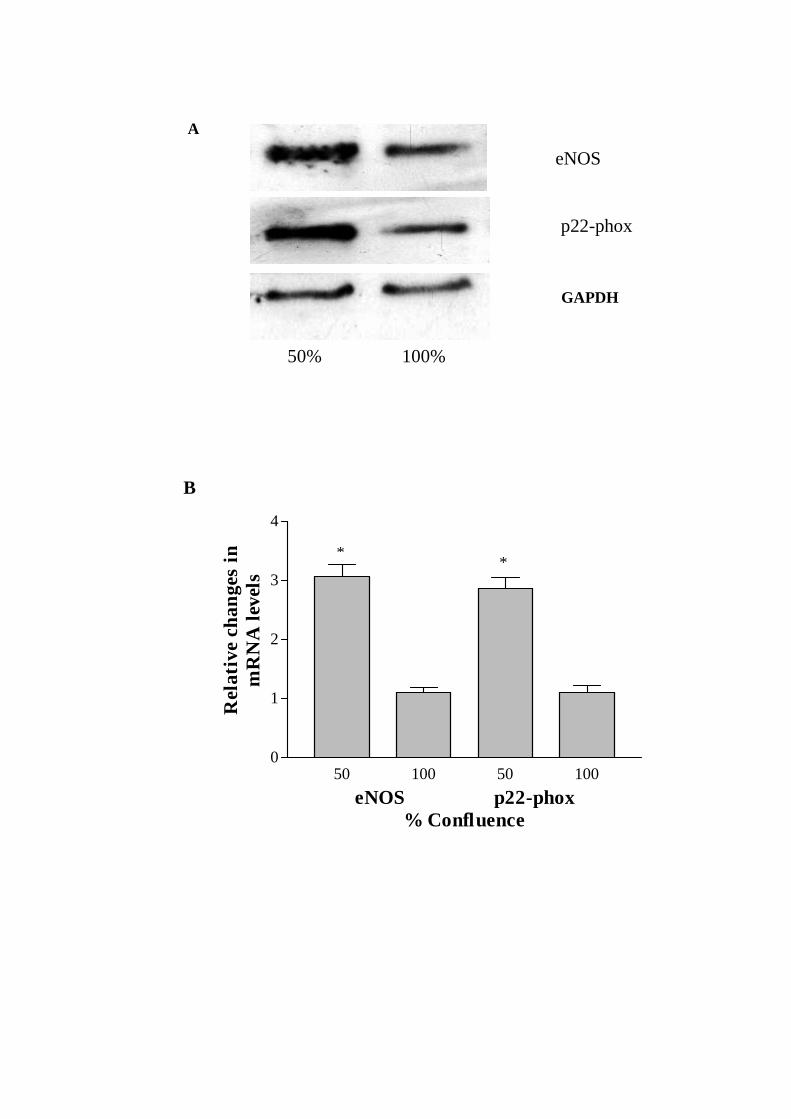
3.1. Effect of cell growth on eNOS and p22-phox mRNA and protein expressions
Northern blot analyses detected a single expected size transcript, namely, 4.3 kb
(eNOS), 0.8 kb (p22-phox) and 1.8 kb (GAPDH), for each gene of interest (Fig. 1A). The
densitometric analysis of autoradiograms revealed an approximately 3-fold increase in
mRNA expressions of both eNOS and p22-phox in proliferating versus resting CMEC
after normalizing to GAPDH mRNA levels (Fig. 1B).
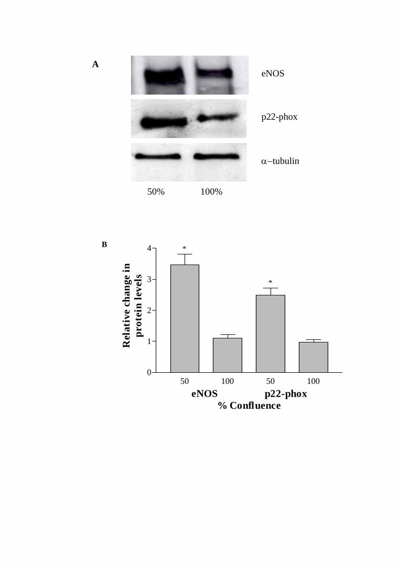
Since the alterations in mRNA levels may not necessarily reflect the changes in
corresponding proteins, the levels of protein expression were investigated by Western
analyses which revealed expected size products for both eNOS (140 kDa) and p22-phox
(~22 kDa) (Fig. 2A). Analyses of the Western autoradiograms showed 3.5- and 2.5-fold
increases in eNOS and p22-phox protein levels in 50% versus 100% confluent CMEC
after normalizing to -tubulin protein levels (Fig. 2B).
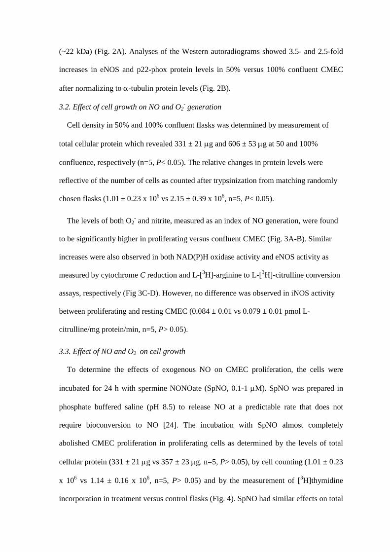
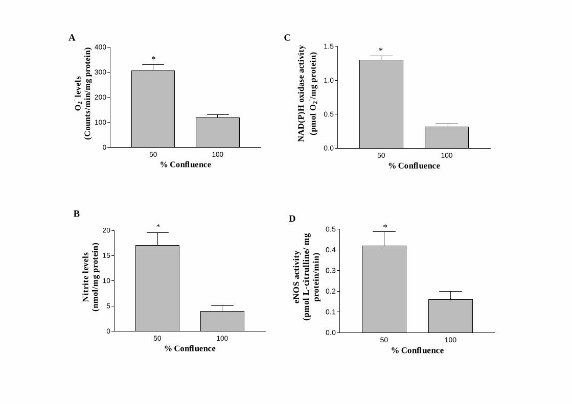
3.2. Effect of cell growth on NO and O2- generation
Cell density in 50% and 100% confluent flasks was determined by measurement of
total cellular protein which revealed 331 ± 21 g and 606 ± 53 g at 50 and 100%
confluence, respectively (n=5, P< 0.05). The relative changes in protein levels were
reflective of the number of cells as counted after trypsinization from matching randomly
chosen flasks (1.01 ± 0.23 x 10⁶ vs 2.15 ± 0.39 x 10⁶, n=5, P< 0.05).
The levels of both O2- and nitrite, measured as an index of NO generation, were found
to be significantly higher in proliferating versus confluent CMEC (Fig. 3A-B). Similar
increases were also observed in both NAD(P)H oxidase activity and eNOS activity as
measured by cytochrome C reduction and L-[3H]-arginine to L-[3H]-citrulline conversion
assays, respectively (Fig 3C-D). However, no difference was observed in iNOS activity
between proliferating and resting CMEC (0.084 ± 0.01 vs 0.079 ± 0.01 pmol L-
citrulline/mg protein/min, n=5, P> 0.05).
3.3. Effect of NO and O2- on cell growth
To determine the effects of exogenous NO on CMEC proliferation, the cells were
incubated for 24 h with spermine NONOate (SpNO, 0.1-1 M). SpNO was prepared in
phosphate buffered saline (pH 8.5) to release NO at a predictable rate that does not
require bioconversion to NO [24]. The incubation with SpNO almost completely
abolished CMEC proliferation in proliferating cells as determined by the levels of total
cellular protein (331 ± 21 g vs 357 ± 23 g. n=5, P> 0.05), by cell counting (1.01 ± 0.23
x 106 vs 1.14 ± 0.16 x 10⁶, n=5, P> 0.05) and by the measurement of [3H]thymidine
incorporation in treatment versus control flasks (Fig. 4). SpNO had similar effects on total
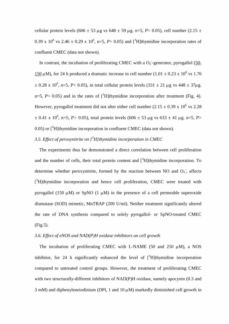
cellular protein levels (606 ± 53 g vs 648 ± 59 g. n=5, P> 0.05), cell number (2.15 ±
0.39 x 10⁶ vs 2.46 ± 0.29 x 10⁶, n=5, P> 0.05) and [3H]thymidine incorporation rates of
confluent CMEC (data not shown).
In contrast, the incubation of proliferating CMEC with a O2--generator, pyrogallol (50-
150 M), for 24 h produced a dramatic increase in cell number (1.01 ± 0.23 x 10⁶ vs 1.76
± 0.28 x 10⁶, n=5, P< 0.05), in total cellular protein levels (331 ± 21 g vs 448 ± 37g.
n=5, P< 0.05) and in the rates of [3H]thymidine incorporation after treatment (Fig. 4).
However, pyrogallol treatment did not alter either cell number (2.15 ± 0.39 x 10⁶ vs 2.28
± 0.41 x 10⁶, n=5, P> 0.05), total protein levels (606 ± 53 g vs 633 ± 41 g. n=5, P>
0.05) or [3H]thymidine incorporation in confluent CMEC (data not shown).
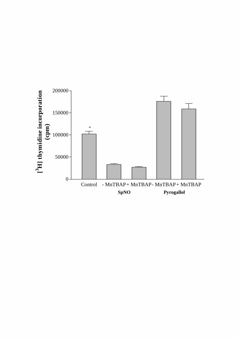
3.5. Effect of peroxynitrite on [3H]thymidine incorporation in CMEC
The experiments thus far demonstrated a direct correlation between cell proliferation
and the number of cells, their total protein content and [3H]thymidine incorporation. To
determine whether peroxynitrite, formed by the reaction between NO and O2-, affects
[3H]thymidine incorporation and hence cell proliferation, CMEC were treated with
pyrogallol (150 M) or SpNO (1 M) in the presence of a cell permeable superoxide
dismutase (SOD) mimetic, MnTBAP (200 U/ml). Neither treatment significantly altered
the rate of DNA synthesis compared to solely pyrogallol- or SpNO-treated CMEC
(Fig.5).
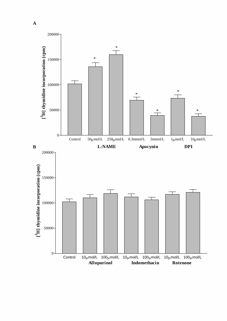
3.6. Effect of eNOS and NAD(P)H oxidase inhibitors on cell growth
The incubation of proliferating CMEC with L-NAME (50 and 250 M), a NOS
inhibitor, for 24 h significantly enhanced the level of [3H]thymidine incorporation
compared to untreated control groups. However, the treatment of proliferating CMEC
with two structurally-different inhibitors of NAD(P)H oxidase, namely apocynin (0.3 and
3 mM) and diphenyleneiodinium (DPI, 1 and 10 M) markedly diminished cell growth in
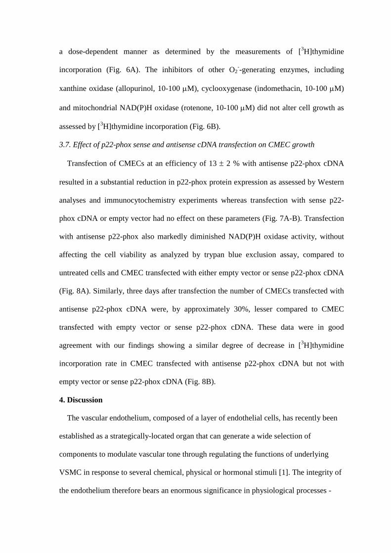
a dose-dependent manner as determined by the measurements of [3H]thymidine
incorporation (Fig. 6A). The inhibitors of other O2--generating enzymes, including
xanthine oxidase (allopurinol, 10-100 M), cyclooxygenase (indomethacin, 10-100 M)
and mitochondrial NAD(P)H oxidase (rotenone, 10-100 M) did not alter cell growth as
assessed by [3H]thymidine incorporation (Fig. 6B).
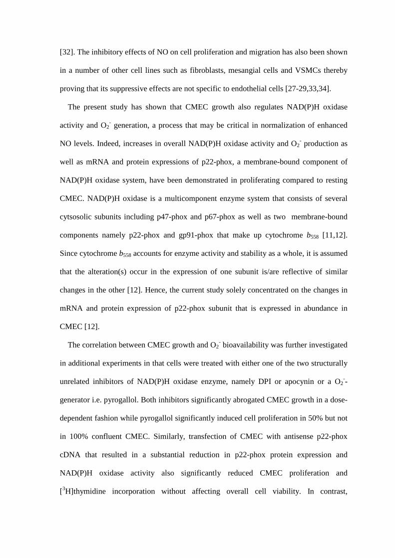
3.7. Effect of p22-phox sense and antisense cDNA transfection on CMEC growth
Transfection of CMECs at an efficiency of 13 2 % with antisense p22-phox cDNA
resulted in a substantial reduction in p22-phox protein expression as assessed by Western
analyses and immunocytochemistry experiments whereas transfection with sense p22-
phox cDNA or empty vector had no effect on these parameters (Fig. 7A-B). Transfection
with antisense p22-phox also markedly diminished NAD(P)H oxidase activity, without
affecting the cell viability as analyzed by trypan blue exclusion assay, compared to
untreated cells and CMEC transfected with either empty vector or sense p22-phox cDNA
(Fig. 8A). Similarly, three days after transfection the number of CMECs transfected with
antisense p22-phox cDNA were, by approximately 30%, lesser compared to CMEC
transfected with empty vector or sense p22-phox cDNA. These data were in good
agreement with our findings showing a similar degree of decrease in [3H]thymidine
incorporation rate in CMEC transfected with antisense p22-phox cDNA but not with
empty vector or sense p22-phox cDNA (Fig. 8B).
4. Discussion
The vascular endothelium, composed of a layer of endothelial cells, has recently been
established as a strategically-located organ that can generate a wide selection of
components to modulate vascular tone through regulating the functions of underlying
VSMC in response to several chemical, physical or hormonal stimuli [1]. The integrity of
the endothelium therefore bears an enormous significance in physiological processes -

e.g. wound healing after an injury to the vascular wall - where controlled proliferation and
migration of endothelial cells are required [2]. However, several pathological conditions
including coronary atherosclerosis, a major cause of morbidity and mortality in the
Western World, are associated with an uncontrolled as well as accelerated proliferation
and migration of both endothelial and VSMCs [5].
It is well-documented that NO, generated by eNOS, inhibits proliferation of a number
of cell types including VSMC and lymphocytes thus prevents the initiation and/or
progression of atherosclerotic disease [25,26]. It is also well-known that ROS, produced
by NAD(P)H oxidase, determine bioavailability of NO in the cardiovascular system and
are implicated in the pathogenesis of atherosclerosis through their ability to enhance
VSMC proliferation and migration [13,14]. However, the current data as to the effects of
NO on endothelial cell growth are rather contradictory in that both growth-stimulating
[3,10] and growth-inhibiting effects have been reported [27-29]. Besides, the effects of
endothelial cell growth on the activity and expression of NAD(P)H oxidase remains to be
fully elucidated, although the correlation between cell growth and enzymatic regulation of
eNOS has recently been well-documented [20]. The present study was therefore designed
to investigate the reciprocal relationship between coronary endothelial cell growth and the
concurrent expression and activity of the enzymes, namely, eNOS and NAD(P)H oxidase
that are associated with the regulation of endothelial cell redox state. All the experiments
outlined in the current study were carried out using early-passage cultured rat CMEC in
order to avoid the effects of changes in coronary endothelial cell phenotype on enzyme
expression and activity [30]. Significant increases in eNOS mRNA and protein
expressions, eNOS activity and consequently in nitrite generation, the ultimate
breakdown product of NO, were observed in proliferating versus confluent CMEC.
Similar increases in these parameters have recently been reported in rat CMEC and aortic

endothelial cells in which the effects of angiotensin II on NO production and eNOS
mRNA/protein expressions were investigated in relation to the state of cell growth
[20,31]. However, in the current study no marked difference was observed in iNOS
activity between growing and quiescent rat CMEC in contrast to elevation in eNOS
activity. This result confirms that eNOS serves as the endogenous source of NO during
coronary endothelial cell growth under conditions where no external stimulus like shear
stress, cyclic strain or hyperglycemia is imposed on cells.
In order to determine the reciprocal effect of NO on CMEC growth and indeed to
ensure whether endogenously-produced and exogenously-added NO elicit different
effects on cell growth, the CMEC were treated with a NOS inhibitor, L-NAME or a NO
donor, SpNO. The former treatment led to a significant increase in cell growth while the
latter dramatically inhibited proliferation as assessed by three different experimental
procedures, namely, cell counting and by measurements of total cellular proteins and
[3H]thymidine incorporation rates. Taken together, these data suggest that endothelial
cells produce physiologically relevant concentrations of NO to preserve the integrity of
endothelium and perhaps vascular wall as a whole. However, once the adequate synthesis
of endogenous NO is attenuated, in this case via inactivation of NOS, the intracellular
oxidative balance may tilt in favor of ROS that in turn accelerate endothelial cell
proliferation. On the contrary, the exogenous supplementation of NO via NO-donors at
higher concentrations that is normally associated with disease conditions like sepsis and
inflammation generates a pool of NO that cannot be neutralized by O2- and therefore
results in the suppression of cell proliferation. In support of our results and hypothesis, a
previous study has also shown that NO-generating compounds namely, S-nitroso-
acetylpenicillamine (SNAP), sodium nitroprusside (SNP) and S-nitroso-glutathione
(GSNO) inhibit human umbilical vein EC and human coronary artery EC proliferation
[32]. The inhibitory effects of NO on cell proliferation and migration has also been shown
in a number of other cell lines such as fibroblasts, mesangial cells and VSMCs thereby
proving that its suppressive effects are not specific to endothelial cells [27-29,33,34].
The present study has shown that CMEC growth also regulates NAD(P)H oxidase
activity and O2- generation, a process that may be critical in normalization of enhanced
NO levels. Indeed, increases in overall NAD(P)H oxidase activity and O2- production as
well as mRNA and protein expressions of p22-phox, a membrane-bound component of
NAD(P)H oxidase system, have been demonstrated in proliferating compared to resting
CMEC. NAD(P)H oxidase is a multicomponent enzyme system that consists of several
cytsosolic subunits including p47-phox and p67-phox as well as two membrane-bound
components namely p22-phox and gp91-phox that make up cytochrome b558 [11,12].
Since cytochrome b558 accounts for enzyme activity and stability as a whole, it is assumed
that the alteration(s) occur in the expression of one subunit is/are reflective of similar
changes in the other [12]. Hence, the current study solely concentrated on the changes in
mRNA and protein expression of p22-phox subunit that is expressed in abundance in
CMEC [12].
The correlation between CMEC growth and O2- bioavailability was further investigated
in additional experiments in that cells were treated with either one of the two structurally
unrelated inhibitors of NAD(P)H oxidase enzyme, namely DPI or apocynin or a O2--
generator i.e. pyrogallol. Both inhibitors significantly abrogated CMEC growth in a dose-
dependent fashion while pyrogallol significantly induced cell proliferation in 50% but not
in 100% confluent CMEC. Similarly, transfection of CMEC with antisense p22-phox
cDNA that resulted in a substantial reduction in p22-phox protein expression and
NAD(P)H oxidase activity also significantly reduced CMEC proliferation and
[3H]thymidine incorporation without affecting overall cell viability. In contrast,

transfection of CMEC with sense p22-phox cDNA or empty vector had no effect on these
parameters. It is noteworthy in this context that, the inhibitors of xanthine oxidase,
cyclooxygenase and mitochondrial NAD(P)H oxidase did not affect CMEC growth.
These results were in agreement with a previous study using three different human
endothelial cell lines and showing ROS derived from NAD(P)H oxidase but not eNOS or
xanthine oxidase as critical elements in endothelial cell growth [35].
The reaction between NO and O2- generates OONO- at a rate of 6.7 x 10⁹ ms-1 and this
rate is three times faster than the reaction between O2- and SOD [36,37]. The formation of
OONO- is a double-edged sword; on one hand potentially deleterious O2- is neutralized,
on the other hand the most potent vasodilator NO is consumed in the process. Hence,
OONO- has been proposed as a toxic compound causing tissue damage, lipid peroxidation
or as a protective molecule improving cellular vitality and relaxation of VSMC [38,39].
The involvement of OONO- in CMEC proliferation or its inhibition was therefore rather
important. In the current study the proliferating CMEC were treated with SpNO or
pyrogallol alone or in the presence of a cell-permeable SOD mimetic, MnTBAP. It has
been demonstrated that the addition of SOD did not interfere with the effects of NO or O2-
per se thereby indicating that NO donors and pyrogallol mediate their effects by releasing
NO and O2-, respectively.
The elucidation of the regulatory effects of endothelial cell growth on eNOS and
NAD(P)H oxidase expression and activities may have significant implications in the
pathogenesis of atherosclerosis in that proliferation and migration of VSMCs contribute
to formation of atheroma. Since NO inhibits these effects, enhanced NO production by
proliferating CMEC may attenuate the development of coronary artherosclerosis. In
contrast, activation of NAD(P)H oxidase enzyme and therefore generation of excess
quantities of O2- in the same cells may diminish the beneficial effects of NO and

accelerate disease formation. These findings imply that EC growth is tightly coupled to
the redox state of the cell and suppression of NAD(P)H oxidase activity may provide a
foundation for the therapy of atherosclerotic disease.
Acknowledgements
This study was in part supported by the grants to Dr Bayraktutan from the Royal Society
UK and Northern Ireland Chest Heart & Stroke Association.
References
1. Bayraktutan, U. Free radicals, diabetes and endothelial dysfunction. Diabet Obes
Metabol 2002; 4: 224-238.
2. Lee PC, Salyapongse AN, Bragdon GA, Shears LL, Watkins SC, Edington HD,
Billiar TR. Impaired wound healing and angiogenesis in eNOS-deficient mice. Am J
Physiol 1999; 277: H1600-1608.
3. Ziche M, Morbidelli L, Masini E, Amerini S, Granger HJ, Maggi CA, et al. Nitric
oxide mediates angiogenesis in vivo and endothelial cell growth and migration in
vitro promoted by substance P. J Clin Invest 1994; 94: 2036-2044.
4. Vacca A, Bruno M, Boccarelli A, Coluccia M, Ribatti D, Bergamo A, et al. Inhibition
of endothelial cell functions and of angiogenesis by the metastasis inhibitor NAMI-A.
Br J Cancer 2002; 86: 993-998.
5. Werba JP, Martinez V, Abulafia DP, Levy R, Magarinos G, Rey RH, et al. Marked
neointimal lipoprotein lipase increase in distinct models of proclivity to
atherosclerosis: a feature independent of endothelial layer integrity. Atherosclerosis
2001; 156: 91-101.
6. Ranjan V, Xiao Z, Diamond SL. Constitutive NOS expression in cultured endothelial
cells is elevated by fluid shear stress. Am J Physiol 1995; 269: H550-H555.
7. McQuillan LP, Leung GK, Marsden PA, Kostyk SK, Kourembanas S. Hypoxia
inhibits expression of eNOS via transcriptional and posttranscriptional mechanisms.
Am J Physiol 1994; 267: H1921-H1927.
8. Kojda G, Noack, E. Effects of pentaerythrityl-tetranitrate and isosorbide-5-
mononitrate in experimental atherosclerosis. Agents Actions Suppl 1995; 45: 201-
206.
9. von der Leyen HE, Gibbons GH, Morishita R, Lewis NP, Zhang L, Nakajima M, et al.
Gene therapy inhibiting neointimal vascular lesion: in vivo transfer of eNOS gene.
Proc Natl Acad Sci USA 1995; 92: 1137-1141.
10. Morbidelli L, Chang CH, Douglas JG, Granger HJ, Ledda F, Ziche M. Nitric oxide
mediates mitogenic effect of VEGF on coronary venular endothelium. Am J Physiol
1996; 270: H411-H415
11. Thrasher AJ, Keep NH, Wientjes F, Segal AW. Chronic granulomatous disease.
Biochim Biophys Acta 1994; 1227: 1-24.
12. Bayraktutan U, Blayney L, Shah AM. Molecular characterisation and localisation of
the NAD(P)H oxidase components gp91-phox and p22-phox in endothelial cells.
Arterioscler Thromb Vasc Biol 2000; 20: 1903-1911.
13. Li PF, Dietz R, von Harsdorf R. Differential effect of hydrogen peroxide and
superoxide anion on apoptosis and proliferation of vascular smooth muscle cells.
Circulation 1997; 96: 3602-3609.
14. Lee SL, Wang WW, Fanburg BL. Superoxide as an intermediate signal for serotonin-
induced mitogenesis. Free Radic Biol Med 1998; 24: 855-888.
15. Kim KS, Takeda K, Sethi R, Pacyk JB, Tanaka K, Zhou YF, et al. Protection from
reoxygenation injury by inhibition of rac1. J Clin Invest 1998; 101: 1821-1826.

16. De Keulenaer GW, Chappell DC, Ishizaka N, Nerem RM., Alexander RW,
Griendling KK. Oscillatory and steady laminar shear stress differentially affect human
endothelial cell redox state: role of a superoxide-producing NADH oxidase. Circ Res
1998; 82: 1094-1101.
17. Matsubara T, Ziff M. Increased superoxide anion release from human endothelial
cells in response to cytokines. J Immunol 1986; 137: 3295-3298.
18. Nishida M, Carley WW, Gerritsen ME, Ellingsen O, Kelly RA. Isolation and
characterization of human and rat cardiac microvascular endothelial cells.
Am J Physiol 1993; 264: H639-H652.
19. Chomczynski P, Sacchi N. Single-step method of RNA isolation by acid guanidinium
thiocyanate-phenol-chloroform extraction. Anal Biochem 1987; 162: 156-159.
20. Bayraktutan U, Ulker S. Effects of ang II on nitric oxide generation in proliferating
and quiescent rat coronary microvascular endothelial cells. Hypertens Res 2003; 26:
749-757.
21. Green LC, Wagner DA, Glogowski J, Skipper PL, Wishnok JS, Tannenbaum SR.
Analysis of nitrate, nitrite and [15N]nitrate in biological fluids. Anal Biochem 1982;
126: 131-138.
22. Li Y, Zhu H, Kuppusamy P. Validation of lucigenin as a chemiluminogenic probe for
detecting superoxide anion radical production by enzymatic and cellular systems. J
Biol Chem 1998; 273: 2015-2023.
23. Griendling KK, Minieri CA, Ollerenshaw JD, Wayne AR. Angiotensin II stimulates
NADH and NADPH oxidase activity in cultured vascular smooth muscle cells. Circ
Res 1994; 74: 1141-1148.
24. Maragos CM, Morley DA, Wink TM, Dunams JE, Keefer LK. Complexes of NO with
nucleophiles as agents for the controlled biological release of nitric oxide:
vasorelaxant effects. J Med Chem 1991; 34: 3242-3247.
25. Kosonen O, Kankanranta H, Vuorinen P, Moilanen E. Inhibition of lymphocyte
proliferation by nitric oxide-releasing oxatriazole derivatives. Eur J Pharmacol 1997;
337: 55-61.
26. Dubey RK, Overbeck HW. Culture of rat mesenteric arteriolar smooth muscle cells:
effects of platelet –derived growth factor, angiotensin and nitric oxide on growth. Cell
Tissue Res 1994; 275: 133-141.
27. Sarkar R, Webb RC, Stanley JC. Nitric oxide inhibition of endothelial cell
mitogenesis and proliferation. Surgery 1995; 118: 274-279.
28. Raychaudhury A, Frischer H, Malik AB. Inhibition of endothelial cell proliferation
and bGFG-induced phenotypic modulation by nitric oxide. J Cell Biochem 1996; 63:
125-134.
29. Lopez-Farre A, de Miguel LS, Caramelo C, Gomez-Macias J, Garcia R, Mosquera JR,
et al. Role of nitric oxide in autocrine control of growth and apoptosis of endothelial
cells. Am J Physiol 1997; 272: H760-H768.
30. Lang D, Bell JP, Bayraktutan U, Small GR, Shah AM, Lewis MJ. Phenotypic changes
in rat and guinea pig coronary microvascular endothelium after culture: loss of NO
synthase activity. Cardiovsc Res 1999; 42: 794-804.
31. Bayraktutan U. Effects of ang II on nitric oxide generation in growing and resting rat
aortic endothelial cells. J Hypertens 2003; 21: (in press)
32. Heller R, Polack T, Grabner R, Till U. Nitric oxide inhibits proliferation of human
endothelial cells via a mechanism independent of cGMP. Atheroscler 1999; 144: 49-
57.

33. Garg UC, Hassid A. Nitric oxide-generating vasodilators and 8-bromo-cyclic
guanosine monophosphate inhibit mitogenesis and proliferation of cultured rat
VSMCs. J Clin Invest 1989; 83: 1774-1777.
34. Garg UC, Hassid A. NO-generating vasodilators inhibit mitogenesis and proliferation
of BALB/C 3T3 fibroblasts by a cGMP-independent mechanism. Biochem Biophys
Res Commun 1990; 171: 474-479.
35. Abid R, Kachra Z, Spokes KC, Aird WC. NADPH oxidase activity is required for
endothelial cell proliferation and migration. FEBS Lett 2000; 486: 252-256.
36. Huie RE, Padmaja S. The reaction of NO with superoxide. Free Radic Res Commun
1993; 18: 195-199.
37. Beckman JS, Koppenol WH. NO, superoxide and peroxynitrite: the good, the bad and
the ugly. Am J Physiol 1996; 271: C1424-C1437.
38. Radi R, Beckman JS, Bush KM, Freeman BA. Peroxynitrite-induced membrane lipid
peroxidation: The cytotoxic potential of superoxide and NO. Arch Biochem Biophys
1991; 288: 481-487.
39. Mayer B, Schrammel A, Klatt P, Koesling D, Schmidt K. Peroxynitrite-induced
accumulation of cGMP in endothelial cells and stimulation of purified soluble
guanylyl cyclase. Dependence on glutathione and possible role of S-nitrosylation. J
Biol Chem 1995; 270: 17355-17360.
Figure legends
Fig. 1. (A) Representative Northern blots of endothelial nitric oxide synthase (eNOS),
p22-phox and GAPDH transcripts amplified from 50 and 100% confluent coronary
microvascular endothelial cells (CMEC). (B) Histogram showing the fold increases in
eNOS and p22-phox mRNA in 50% versus 100% confluent CMEC, after normalization
against GAPDH. Data are expressed as mean ± SEM, n=5, * P< 0.05, 50 vs 100%
confluent CMEC.
Fig. 2. (A) Representative Western blots showing endothelial nitric oxide synthase
(eNOS), p22-phox and -tubulin protein levels in 50 and 100% confluent coronary
microvascular endothelial cells (CMEC). (B) Histogram showing the fold increases in
eNOS and p22-phox protein levels in 50% confluent CMEC, after normalization against
-tubulin. Data are expressed as mean ± SEM, n=5, * P< 0.05, 50 vs 100% confluent
CMEC.
Fig. 3. Effect of varying coronary microvascular endothelial cell (CMEC) confluence on
superoxide anion (O2-) (A) and nitrite (B) generations and on NAD(P)H oxidase (C) and
eNOS activities (D). Data are expressed as mean ± SEM, n=5, * P< 0.05, 50 vs 100%
confluent CMEC.
Fig. 4. Effects of a NO donor i.e. sepiapterin NO (SpNO) and a superoxide anion-
generator, pyrogallol on [3H]thymidine incorporation rates in coronary microvascular
endothelial cells (CMEC). Data are expressed as mean ± SEM, n=5, * P< 0.05, treated vs
untreated CMEC.
Fig. 5. Effects of pyrogallol or SpNO in the absence and presence of a cell permeable
superoxide dismutase (SOD) mimetic, MnTBAP (200 U/ml) on [3H]thymidine
incorporation in proliferating coronary microvascular endothelial cells (CMEC). Data are
expressed as mean ± SEM, n=5, * P< 0.05, treated vs untreated CMEC.
Fig. 6. (A) Effects of different doses of L-NAME, an endothelial nitric oxide synthase
(eNOS) inhibitor and two structurally different inhibitors of NAD(P)H oxidase namely,
apocynin and diphenyleneiodinium (DPI) on [3H]thymidine incorporation in coronary
microvacular endothelial cells (CMEC). (B) Effects of different doses of inhibitors of
xanthine oxidase (allopurinol), cyclooxygenase (indomethacine) and mitochondrial
NAD(P)H oxidase system (rotenone) on [3H]thymidine incorporation in CMEC. Data are
expressed as mean ± SEM, n=5, * P< 0.05, treated vs untreated proliferating CMEC.
Fig. 7. (A) Representative Western blots showing p22-phox protein levels in coronary
microvascular endothelial cells (CMEC) and in CMEC transfected with sense, empty
vector and antisense p22-phox cDNA. (B) Immunofluoresence microscopy showing
localization of p22-phox protein in normal CMEC and CMEC transfected with sense,
empty vector or antisense p22-phox cDNA.
Fig. 8. (A) Effects of sense, empty vector and antisense p22-phox cDNA transfected
coronary microvascular endothelial cells (CMEC) on superoxide anion (O2-) generation in
comparison with untreated proliferating CMEC. (B) Histogram showing the fold
differences in [3H]incorporation in untreated (control) CMEC and CMEC transfected
with sense, empty vector and antisense p22-phox cDNA. Data are expressed as mean ±
SEM, n=5, * P< 0.05, treated vs untreated proliferating CMEC.
50% 100%
A
50 100 500
1
2
3
4
eNOS p22-ph% Confluence
Rel
ativ
ech
ange
sin
mR
NA
leve
ls
B
**
eNOS
p22-phox
GAPDH
100ox
eNOS
p22-phox
tubulin
A
Rel
ativ
ech
ange
in
B
0
1
2
3
4
prot
ein
leve
ls
50% 100%
50 100eNOS
% Conflue
*
50 100p22-phoxnce
*
50 1000
100
200
300
400
*
% Confluence
O2-
leve
ls(C
ount
s/m
in/m
gpr
otei
n)
50 1000
5
10
15
20 *
% Confluence
Nit
rite
leve
ls(n
mol
/mg
prot
ein)
50 1000.0
0.5
1.0
1.5 *
% Confluence
NA
D(P
)Hox
idas
eac
tivi
ty(p
mol
O2- /m
gpr
otei
n)
50 1000.0
0.1
0.2
0.3
0.4
0.5 *
% Confluence
eNO
Sac
tivi
ty(p
mol
L-c
itru
llin
e/m
gpr
otei
n/m
in)
A
B
C
D

Control 0.10
50000
100000
150000
200000
[3 H]
thym
idin
ein
corp
orat
ion
(cpm
)
*
*
mol/L 1mol/L 50mol/L 150mol/LSpNO Pyrogallol
**
Control - MnTBAP+ MnTBAP- MnTBAP+ MnTBAP0
50000
100000
150000
200000
[3 H]
thym
idin
ein
corp
orat
ion
(cpm
)
SpNO Pyrogallol
*
Control 10mol/L 100mol/L 10mol/L 100mol/L 10mol/L 100mol/L0
50000
100000
150000
200000
Allopurinol Indomethacin Rotenone
[3 H]
thym
idin
ein
corp
orat
ion
(cpm
)
A
B
Control 50mol/L 250mol/L 0.3mmol/L 3mmol/L 1mol/L 10mol/L0
50000
100000
150000
200000
L-NAME Apocynin DPI
[3 H]
thym
idin
ein
corp
orat
ion
(cpm
)
*
*
*
*
*
*

B
A
EARAb
Sense
Empty vector
Control Sense Empty Antisensevector
22 kDa
Antisense

A
B
Control Sense Empty Vec. Antisense0
50000
100000
150000
[3 H]
thym
idin
ein
corp
orat
ion
(cpm
)
Control Sense Empty Vec. Antisense0
1
2
3
4
5
6
7
pmol
O- 2/
mg
prot
ein
*
*