Embed Size (px)
Citation preview

Histol Histopathol (1998) 13: 743-749
001: 10.14670/HH-13.743
http://www.hh.um.es
ApoptosiS in adenoma and
Histology and Histopathology
From Cell Biology to Tissue Engineering
early adenocarcinoma of the colon T. Yamamoto l , N. Igarashil, Y. Kato l , M. Kobayashi1 and M. Kawakami2
Department of 1 Pathology and 2Surgical Pathology, Tokyo Women 's Medical College, Tokyo, Japan
Summary. Twenty-six specimens of tubular adenoma and 7 specimens of adenocarcinoma in adenoma of the colon were examined to evaluate apoptosis between adenoma and early adenocarcinoma. Cell proliferation and cell death seemed to be balanced in adenoma with mild and moderate atypia, but unbalanced in adenoma with severe atypia and cancer. Apoptosis was considered to be suppressed at cancer in some cases. However, a number of apoptosis increased at cancer in other cases. Necrosis was seen only in cancer areas. The ratio of cells simultaneously stained by anti-Ki-67 antibody (MIB-l) and terminal deoxynucIeotidyl transferase-mediated deoxyuridine triphosphate-nick end labeling (TUNEL) tended to be high from adenoma with moderate atypia to cancer, suggesting the unstableness of DNA. It is possible that cancer cells having highly unstable DNA easily underwent apoptosis as well as necrosis, accidentally. The p53 protein was positive only in cancer areas of three cases. One of these three cases showed decreased apoptosis in a cancer area, but the other two cases showed increased apoptosis. Furthermore, certain numbers of cancer cells were double-stained by p53 immunohi s tochemistry and TUNEL. These results suggest that the p53 protein may contribute to suppress apoptosis in the last stage of carcinogenesis of the colonic adenocarcinoma, but other factors including ex trinsic stimulation may cause apoptosis despite the mutation of p53 protein.
Key words: Apopto s is, p53, Adenoma, Adenocarcinoma, Colon
Introduction
Growth or homeostasis of tissues depends on the balance of cell proliferation and cell death (Kerr et aI., 1972; Willie et aI., 1980). Apoptosis and necrosis have been considered for cell death (Wyllie et aI., 1980; Buja et aI., 1993; Cummings et aI., 1997), and a cell loss by apoptosis seems to be important for the regulation of cell
Offprint requests to: Dr. Tomoko Yamamoto, Department of Pathology,
Tokyo Women's Medical College, Kawada-cho 8-1 , Shinjuku-ku, Tokyo
162·0054, Japan. Fax: 81·3·5269·7408.
number in the mammalian gastrointestinal tract (Hall et aI., 1994). The number of proliferating cells significantly increases in proportion to the severity of dyspl as ia in colonic epithelial neoplasms (Johnston et aI., 1989; Diebold et aI., 1992; Risio and Rossini , 1993). On the other hand, the number of apoptosis in colonic tumors is still obscure, although correlation between mitotic and apoptotic indices has been reported in colonic adenoma (Arai and Kino , 1995). We investigated apoptosis in adenoma and early adenocarcinoma of the colon to clarify whether any difference is seen between adenoma and cancer. For this purpose, terminal deoxynucleotidyl transferase (TdT)-mediated deoxyuridine triphosphate (dUTP)-nick end labeling (TUNEL) (Gavrieli et aI., 1992) was used to detect apoptosis in addition to counting apoptotic bodies in hematoxylin and eosin staining (HE). The close relation between cell cycle and apoptosis has been considered, although there may be an another apoptotic pathway occurring without entering cell cycle (Coates et aI., 1996). An anti-Ki-67 antibody (MIB-l) detects cells existing in G 1, S, G2, and M phases of cell cycle (Gerdes et aI., 1984; Cattoretti et aI., 1992). Cells simultaneously positive for MIB-J and TUNEL may indicate that cells entering in cell cycle die (Coates et aI., 1996), and that these cells have the unstable DNA. Various genes are involved in the regulation of cell cycle, and some of them are also necessary to regulate apoptosis (Coates et aI., 1996). The p53 prot e in, which normally induces apoptosis depending on cell cycle (Kobayashi et aI., 1995), is presumed to playa role during carcinogenesis close to the stage of the final malignant transformation in colonic adenoma-carcinoma sequence (Fearon and Vogelstein, 1990; Kikuchi-Yanoshita et aI., 1992; Auer et aI., 1994). Immunohistochemistry of p53 protein , including double staining with TUNEL, would reveal whether p53 expression is a major factor for the regulation of apoptosis in the last stage of carcinogenesis.
Materials and methods
Patients and specimens
Thirty-three specimens of colonic tubular adenoma and adenocarcinoma in adenoma obtained by endoscopic

744
Apoptosis in colon tumours
resection were examined (Table 1). Cases were from the files of the Department of Surgical Pathology, Tokyo Women's Medical College. Specimens were fixed in 10% buffered formalin, embedded in paraffin and cut into serial sections at 3 ,urn thick. Sections were stained with HE. The grading and mapping of adenoma was performed according to the classification of the Japanese Society for Cancer of the Colon and Rectum (Japanese Society for Cancer of the Colon and Rectum , 1994): adenomas were divided into mild, moderate and severe atypia according to the grade of cytological and structural atypia, and glands consisting of cytologically malignant cells were regarded as cancer (Fig. 1). In 25 cases that contained more than two areas showing different grades of atypia in one specimen (Table 1), analysis was performed in each lesion.
Immunohistochemical staining
Immunostaining for MIB-l (1: 100 dilution, monoclonal; Immunotech, Marseilles, France) and p53 protein (ready-to-use, monoclonal ; Immunotech) was performed on 33 cases and 28 cases, respectively. The avidin-biotin complex method was employed. Microwave antigen
Table 1. Characteristics of Patients and Polyps.
CASES AGE SEX SIZE CONTAINED LESIONS
1 48 M 0.3 Mild 2 72 M 0.8 Moderate, severe 3 43 M 1.1 Mild, moderate 4 67 F 0.7 Mild, moderate 5 62 M 0.5 Moderate 6 61 M 0.3 Moderate 7 76 M 0.8 Moderate, severe 8 65 F 0.4 Mild, moderate 9 35 F 1.0 Mild, moderate
10 51 M 1.8 Mild, Moderate, severe 11 56 F 0.4 Mild 12 67 M 0.7 Mild, moderate 13 72 M 1.2 Mild, moderate, severe 14 71 M 0.4 Moderate 15 71 M 0.7 Mild , moderate 16 65 M 0.6 Moderate, severe 17 65 M 0.6 Mild , moderate 18 56 M 0.3 Mild 19 40 F 0.8 Moderate 20 69 M 1.3 Mild, moderate 21 61 M 0.9 Mild , moderate 22 60 M 0.5 Mild 23 60 M 0.6 Mild, moderate 24 62 M 0.2 Mild 25 79 F 0.5 Mild, moderate 26 59 M 1.0 Moderate, severe. cancer 27 60 M 1.1 Mild, moderate, severe, cancer 28 59 F 0.7 Mild , moderate, severe, cancer 29 64 M 0.6 Severe, cancer 30 46 M 0.9 Mild, moderate, severe, cancer 31 49 M 0.9 Severe, cancer 32 70 M 1.2 Moderate, severe, cancer 33 53 M 0.7 Severe, cancer
Mild , moderate and severe refer to the degree of atypia in adenoma.
retrieval for 10 minutes at 95 °C in O.OlM citrate buffer, pH 6.0, was performed after deparaffinization and elimination of endogenous peroxidase activity. Sections were incubated with the primary antibody overnight at 4 0c. Reaction products were developed with 3,3'diaminobenzidine tetrahydrochloride (DAB), and sections were counterstained with hematoxylin. A negative control study was performed by eliminating the primary antibody.
TUNEL
Sections were deparaffinized in a routine manner, digested with 20 ,uglml proteinase K (Sigma Chemical Co. Saint Louis, MO) for 30 min at room temperature (RT), and then immersed in 2% hydrogen peroxide for 5 min at RT to quench endogenous peroxidase. After washing, they were incubated with TdT (0.15 units/,ul; GIBCO BRL, Gaithersburg, MD) and digoxigenin-lldUTP (0.02 nm/,u l, Boehringer-Mannheim/Yamanouchi , Tokyo) in TdT buffer (GIBCO BRL) in humidified atmosphere at 37°C for 60 min. The reaction was stopped by immersing them in TB buffer (300 mM sodium chloride, 30 mM sodium citrate) for 15 min at 37°C. Anti-digoxigenin-peroxidase (1:50 dilution, Boehringer Mannheim) was applied and sections were incubated for 30 min, RT. Reaction products were developed with DAB, and sections were counterstained with hematoxylin. Sections digested with DNAse I (10 units/,ul , Boehringer Mannheim) after deparaffinization were used as positive controls. Elimination of TdT in the reaction solution was done as a negative control.
Double staining
TUNEL with successive MIB-1 stainIng was performed in five cases (cases 10, 12, 23, 28 and 33), and TUNEL with successive p53 staining was done in 11 cases (cases 3, 10, 12, 23, 26-28, and 30-33). Reaction products of TUNEL were developed with DAB, and those of MIB-l and p53 were developed with nickel DAB. Sections were counterstained with methyl green.
Evaluation
Upon HE staining, the ratio of mitotic figures and that of apoptotic bodies were regarded as the mitotic index and apoptotic index, respectively. The MIB-1 and TUNEL indices were calculated by counting positive cells. To obtain above indices, 1000 to 2000 cells were counted. Each index was counted in the same area that included glands from upper to lower level and showed fine results in both MIB-l immunohistochemistry and TUNEL. The ratio of double (MIB-1 and TUNEL)positive cells per TUNEL-positive cells was also calculated in the same area. The result of p53 immunostaining was evaluated as positive when most of the glands were stained, focally positive when a few glands were stained, and negative when no glands were stained.

745
Apoptosis in c%n tumours
For double staining of p53 and TUNEL, the ratio of double-positive cells per TUNEL positive cells was calculated in the areas that showed positive immunoreaction against the anti-p53 protein antibody. Apoptotic bodies in the lumen or nuclear debris that could not be identified with certainty as apoptotic bodies were not counted. Although necrosis was positive for TUNEL, this was excluded from counting. Immunostaining to an antibody for leukocyte common antigen was performed to exclude intraepithelial leukocytes. In addition to apoptosis, necrosis was also evaluated. The extent of necrosis in tumor was classified negative (including none and a very little) and positive.
Results
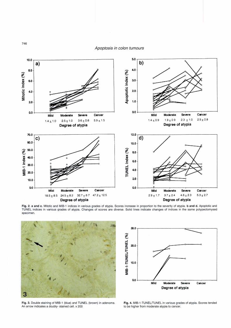
Sixty-seven lesions were mapped in 33 polypectomized specimens (Table 1). Cancer invaded into submucosa in three cases (Table 2). A significant correlation was found between the mitotic and MIB-1 indices, and between the apoptotic and TUNEL indices (data not shown). In specimens with areas showing different grades of atypia, the mitotic and MIB-1 indices increased in proportion to the severity of atypia (Fig 2a,c). Scores of apoptotic and TUNEL indices were diverse in each case (Fig. 2b,d). However, these indices tended to increase between mild and moderate atypia. Scores peaked at moderate or severe atypia in some cases, and either increased or decreased betwen severe atypia and cancer. Necrosis was exceptional in adenoma,
and was generally seen in areas where cancer showed submucosal invasion (Table 2). Some cells were doubly sta ined by both MIB-1 and TUNEL (Fig. 3) . MIB-1-TUNELffUNEL tended to increase between mild and moderate atypia, and to keep high scores from moderate atypia to cancer (Fig. 4).
Adenoma areas were negative for p53 protein in 22 cases. Only a few glands or cells were positively immunostained in 6 cases. Cancer areas were positive in 3 cases (Fig. 5) and focally positive in 4 cases (Table 2). Among cases showing positive immunoreaction against the anti-p53 protein antibody in cancer areas (cases 27, 30 and 31), apoptotic and TUNEL indices between severe atypia and cancer increased in cases 30 and 31, while they decreased in case 27 (Table 2). Cells doublestained by p53 and TUNEL were hardly found in areas where p53 was focally positive. p53-TUNEL/TUNEL was 47.8 % (case 27), 50.0 % (case 30) and 39.2 % (case 31) in the areas where p53 was positive (Fig. 6).
Discussion
The significant relationship between the number of proliferating cells and the severity of dysplasia has been reported with regard to colonic neoplasms (Johnston et a!., 1989; Diebold et a!., 1992; Risio and Rossini, 1993), and s imilar results were obtained in our study. In contrast, the apoptotic or TUNEL index showed no positive relation like the mitotic or MIB-1 index. However, some interesting findings were observed as
Fig. 1. Adenoma with mild (a) , moderate (b) and severe (c) atypia, and cancer (d) . Cells are tall columnar and have an elongated nucleus in the adenoma. Cytoplasmic mucin is reduced and nuclei are more stratified in adenoma with higher grades of atypia. Cancer cells have large, oval shaped nucleus sometimes with conspicuous nucleoli. a, b, x 125; c, d, x 200
746
Apoptosis in c%n tumours
-, ,
,.
~
• , ,
3 Fig. 3. Double staining of MIB-1 (blue) and TUNEL (brown) in adenoma. An arrow indicates a doubly- stained cell. x 200
~.O .---------------------~~------------,
~ ..J UJ Z 20.0 ::l
5 UJ Z ::l l- 10.0
I .... en :E
no ~----~~----~------~----~------~ Mild Moderate Severe C.,cer
Degree of atypia
Fig. 4. MIB-1-TUNELJTUNEL in various grades of atypia. Scores tended to be higher from moderate atypia to cancer.

747
Apoptosis in colon tumours
follows. All four indices tended to increase between mild and
moderate atypia, indicating that cell renewal is accerelated, but that the balance of cell proliferation and cell death is still preserved. On the other hand, changes of these indices were rather diverse from moderate
Table 2. Apoptosis and p53 expression in cases containing cancer.
atypia to cancer suggesting unbalanced cell proliferation and cell death in adenoma with severe atypia and cancer. Apoptotic and TUNEL indices peaked at severe atypia in some cases containing cancer. Apoptosis may be suppressed in acute leukemia (MundIe et aI. , 1994) and malignant lymphoma (Hollowood and Macartney, 1991)
CASES DEGREE OF ATYPIA APOPTOTIC INDEX (%) TUNEL INDEX (%) NECROSIS p53 INVASION TO SUBMUCOSA AREA OF CANCER
Cancer 2.1 3.0 26 Severe 3.0 5.7
Moderate 1.6 2.2
Cancer 2.5 4.2 27 Severe 3.8 8.4
Moderate 2.9 4.5 Mild 2.7 4.0
Cancer 3.1 4.7 28 Severe 1.3 4.1
Moderate 2.4 4.6 Mild 1.2 3.6
29 Cancer 2.5 2.3 Severe 2.2 1.8
Cancer 2.0 4.0 30 Severe 1.7 2.7
Moderate 3.7 6.5 Mild 2.0 3.1
31 Cancer 4.2 9.6 Severe 2.2 2.9
Cancer 3.6 5.3 32 Severe 2.9 5.4
Moderate 1.9 3.0
33 Cancer 2.9 9.2 Severe 4.8 9.6
NE: not examined, +: positive, ±: focally positive, -: negative.
Fig. 5. p53. Positive immunoreaction is seen in a cancer area, but not in an adenoma area. x 80
± ± <50%
+ ± <50%
± ± <50%
NE >50% NE
+ ± <50%
+ + + >50%
+ ± + >50%
+ ± + >50% ±
Fig. 6. Double staining of p53 (blue) and TUNEL (brown) in cancer. An arrow indicates a doubly-stained cell. x 250

748
Apoptosis in c%n tumours
in comparison with the pre-malignant or non-neoplastic state. Similar conditions can be suspected in adenoma with severe atypia and cancer of the colon. Furthermore, apoptosis is supposed to be already supressed in adenoma with severe atypia in a couple of cases that showed the peak of apoptotic and TUNEL indices at moderate atypia. By histology it is sometimes difficult to distinguish adenoma with severe atypia from cancer. The lesions classified as severe atypia might have acquired cell kinetics close to cancer.
On the other hand, some cases showed increased apoptosis between severe atypia and cancer. Necrosis was seen in cancer areas showing submucosal invasion, whereas it was hardly observed in adenomas. Double staining of MIB-1 and TUNEL reveal s that both cell cycle-related and unrelated apoptosis exist in the colonic adenoma and adenocarcinoma. Generally, proliferating cells are located at the lower part of the crypt (Johnston et aI., 1989; Diebold et aI., 1992), while apoptotic cells are at the upper part of crypt in the normal colonic mucosa (Hall et aI., 1994). Double-stained cells were exceptional in the normal colonic mucosa (data not shown). MIB-1-TUNEL/TUNEL tended to increase between mild and moderate atypia and to show high scores between moderate atypia and cancer. Aneuploidy and multiple genetic alterations, more frequent in colonic neoplasm s with higher grades of dysplasia, suggest the increase of genomic instability (Fearon and Vogelstein, 1990; Auer et aI., 1994). Thus, the existence of double-stained cells may indicate abnormal cell kinetics, probably the unstableness of DNA in the colonic epithelial cells. Apoptosis can occur in physiological and pathological conditions (Kerr et aI., 1972; Wyllie et aI., 1980) and extrinsic stimulation can cause either apoptosis or necrosis (Wyllie et aI., 1980; Hockenbery, 1995; Cummings et aI., 1997). The degree of extrinsic stimulation would affect whether cells undergo apoptosis or necrosis (Lennon et aI., 1990; Hockenbery, 1995). It is possible in this context that cells showing remarkable genomic instability such as cancer cells easily undergo apoptosis accidentally, even though intrinsic regulatory mechanisms would direct cells to reduce apoptosis, because the colon is one of the organs that has extrinsic stimulation with ease.
The p53 protein normally induces apoptosis depending on cell cycle (Kobayashi et aI., 1995), and mutations of the p53 gene have been frequently observed in colonic adenocarcinoma (Fearon and Vogelstein, 1990; Kikuchi-Yanoshita et aI., 1992). Accumulation of mutant p53 proteins can be visualized by immunohistochemistry (Kikuchi-Yanoshita et aI., 1992; Auer et aI., 1994; Kobayashi et aI., 1995) and diffusely positive immunoreaction probably results from the mutants (Kobayashi et aI., 1995), although not all p53 gene mutations can be detected by immunohistochem istry (Wadayama et aI., 1993). Immunoreaction against the anti-p53 protein antibody turned positive in cancer areas of three cases, suggesting that p53 gene mutations concern the last stage of carcinogenesis in adenoma-
carcinoma se quence in some cases (Fearon and Vogelstein, 1990; Kikuchi-Yanoshita et aI., 1992; Auer et aI., 1994). Apoptosis was decreased between severe atypia and cancer in one of these three cases that showed positive immunoreaction against the anti-p53 protein antibody in cancer areas, but increased in the other two cases. Furthermore, certain numbers of cells were doubly stained by TUNEL and p53 immunohistochemistry. These results indicate that apoptosis can occur in cells having the mutant p53 protein. Other intrinsic mechanisms besides p53, such as bcl-2 (Nakamura et aI., 1995; Kaklamanis et aI., 1996) and adenomatous polyposis coli (APC) genes (Morin et aI., 1996) are known to contribute to the regulation of apoptosis in colonic tumors, and it is also reported that sodium butylate derived from dietary fibers can cause apoptosis (Hague et aI., 1993). Accumulation of the mutant p53 protein may relate to the suppression of apoptosis in the last stage of carcinogenesis, but this may not be the only factor for the regulation of apoptosis.
Acknowledgments. The authors thank F. Muramatsu, and H. Takeiri for excellent technical assistance. We also thank all the members of the Department of Surgical Pathology for their kind collaboration in this study.
References
Arai T. and Kino I. (1995). Role of apoptosis in modulation olthe growth of human colorectal tubular and villous adenomas. J. Pathol. 176, 37-44.
Auer G.U., Heselmeyer K.M. , Steinbeck R.G., Munck-Wikland E. and Zetterberg A.D. (1994). The relationship between aneuploidy and p53 overexpression during genesis of colorectal adenocarcinoma. Virchows Archiv. 424, 343-347.
Buja L.M. , Eigenbrodt M.L. and Eigenbrodt E.H. (1993). Apoptosis and necrosis. Basic types and mechanisms of cell death. Arch. Pathol. Lab. Med.117, 1208-1214.
Cattoretti G., Becker M.H.G. , Key G., Duchrow M., Schluter C., Galle J. and Gerdes J. (1992). Monoclonal antibodies against recombinant parts of the Ki-67 antigen (MIB 1 and MIB 3) detect proliferating cells in microwave-processed formalin-fixed paraffin sections. J. Pathol. 168, 357-363.
Coates P.J., Hales SA and Hall PA (1996). The association between cell proliferation and apoptosis: studies using the cell cycleassociated protein Ki67 and DNA polymerase alpha. J Pathol. 178, 71-77.
Cummings M.C. , Winterford C.M. and Walker N.I. (1997). Apoptosis. Am. J. Surg. Pathol. 21, 88-101.
Diebold J., Lai M.D. and Liihrs U. (1992) . Analysis of proliferative activity in colorectal mucosa by immunohistochemical detection of proliferating cell nuclear antigen (PCNA). Virchows Arch. (B) 62, 283-289.
Fearon E.R. and Vogelstein B. (1990). A genetic model for colorectal tumorigenesis. Cell 61, 759-767.
Gavrieli Y., Sherman Y. and Ben-Sasson SA (1992). Identification of programmed cell death in situ via specific labeling of nuclear DNA fragmentation. J. Cell BioI. 119, 493-501 .
749
Apoptosis in colon tumours
Gerdes J., Lemke H., Baisch H., Wacker H.-H., Schwab U. and Stein H. (1984) . Cell cycle analysis of a cell proliferation-associated human
nuclear antigen defined by the monoclonal antibody Ki-67. J. Immunol. 133, 1710-1715.
Hague A., Manning A.M., Hanlon K.A., Huschtscha L.I., Hart D. and Paraskeva C. (1993). Sodium butyrate induces apoptosis in human colonic tumour cell lines in a p53-independent pathway: implications
for the possible role of dietary fibre in the prevention of large-bowel cancer. Int. J. Cancer 55, 498-505.
Hall P.A., Coates P.J., Ansari B. and Hopwood D. (1994). Regulation of cell number in the mammalian gastrointestinal tract: the importance
of apoptosis. J. Cell Sci. 107,3569-3577. Hockenbery D. (1995). Defining apoptosis. Am. J. Pathol. 146, 16-19. Hollowood K. and Macartney J.C. (1991). Reduced apoptotic cell death
in follicular lymphoma. J. Pathol. 163, 337-342. Japanese Society for Cancer of the Colon and Rectum. (1994). General
rules for clinical and pathological studies on cancer of the colon, rectum and anus. Kanehara. Tokyo (In Japanese) .
Johnston P.G. , O'Brien M.J., Dervan PA and Carney D.N. (1989) . Immunohistochemical analysis of cell kinetic parameters in colonic
adenocarcinomas, adenomas , and normal mucosa. Hum. Pathol.
20,696-700. Kaklamanis L. , Savage A., Moetensen N. , Tsiotos P., Doussis
Anagnostopoulou I, Biddolph S., Whitehouse R., Harris A.L. and Gatter K. C. (1996). Early expression of bcl-2 protein in the adenoma-carcinoma sequence of colorectal neoplasia. J. Pathol.
179, 10-14. Kerr J.F.R., Wyllie A.H. and Currie A.A. (1972) . Apoptosis: a basic
biological phenomenon with wide ranging implications in tissue
kinetics. Br. J. Cancer 26, 239-257. Kikuchi-Yanoshita R., Konishi M., Ito S., Seki M. , Tanaka K., Maeda Y.,
lino H., Fukayama M. , Koike M. , Mori T., Sakuraba H. , Fukunari H.,
Iwama T. and Miyaki M. (1992). Genetic changes of both p53 alleles associated with the conversion from colorectal adenoma to early carcinoma in familial adenomatous polyposis and non-familial
adenomatous polyposis patients. Cancer Res. 52, 3965-3971. Kobayashi M., Watanabe H., Ajioka Y., Yoshida M, Hitomi J. and
Asakura H. (1995) . Correlation of p53 protein expression with apoptotic incidence in colorectal neoplasia. Virchows Arch. 427, 27-32.
Lennon S.v. , Martin S.J. and Cotter T.G. (1990). Induction of apoptosis (programmed cell death) in tumour cell lines by widely diverging stimuli. Biochem. Soc. Trans. 18, 343-345.
Morin P.J. , Vogelstein B. and Kinzler KW. (1996). Apoptosis and APC in colorectal tumorigenesis. Proc. Natl. Acad. Sci. USA 93, 7950-7954.
Mundie S. , Iftikhar A. , Shetty V. , Dameron S., Wright-Quinones V., Marcus B., Loew J., Gregory S. and Raza A. (1994). Novel in situ double labeling for simultaneous detection of proliferation and apoptosis. J. Histochem. Cy1ochem. 42, 1533-1537.
Nakamura T., Sakai T. and Nariya S. (1995). Cell death in colorectal polyp as evaluated by in situ 3'-tailing reaction and its relationship to BCL-2 expression. Pathol. Int. 45, 721-728.
Risio M. and Rossini F.P . (1993) . Cell proliferation in colorectal adenomas containing invasive carcinoma. Anticancer Res. 13, 43-48.
Wadayama B. , Toguchida J. , Yamaguchi T., Sasaki M.S., Kotoura Y. and Yamamuro T. (1993). p53 expression and its relationsh ip to DNA alterations in bone and soft-tissue sarcomas. Brit. J. Cancer 68, 1134-1139.
Wyllie A .H. , Kerr J.F.R. and Currie A.R . (1980) . Cell death : the significance of apoptosis. Int. Rev. Cy1ol. 68, 251-306.
Accepted January 5, 1998