Embed Size (px)
Citation preview

See discussions, stats, and author profiles for this publication at: https://www.researchgate.net/publication/326506623
Anticancer Potential of Quercetin: A Comprehensive Review
Article · July 2018
CITATIONS
3READS
923
5 authors, including:
Some of the authors of this publication are also working on these related projects:
Dryopteris cycadena View project
Healing of partial tear of supraspinatus tendon after low level laser therapy View project
Abdur Rauf
University of Swabi
312 PUBLICATIONS 1,917 CITATIONS
SEE PROFILE
Muhammad Imran
University of Veterinary and Animal Sciences
227 PUBLICATIONS 1,315 CITATIONS
SEE PROFILE
Syed Amir Gilani
University of Lahore
620 PUBLICATIONS 245 CITATIONS
SEE PROFILE
All content following this page was uploaded by Syed Amir Gilani on 07 July 2019.
The user has requested enhancement of the downloaded file.

Received: 1 February 2018 Revised: 18 June 2018 Accepted: 21 June 2018
DOI: 10.1002/ptr.6155
R E V I EW
Anticancer potential of quercetin: A comprehensive review
Abdur Rauf1 | Muhammad Imran2 | Imtiaz Ali Khan3 | Mujeeb‐ ur‐Rehman4 |
Syed Amir Gilani5 | Zaffar Mehmood5 | Mohammad S. Mubarak6
1Department of Chemistry, University of
Swabi, Ambar, Pakistan
2University Institute of Diet and Nutritional
Sciences, Faculty of Allied Health Sciences,
The University of Lahore‐Pakistan3Department of Agriculture, University of
Swabi, Sayed, Pakistan
4H.E.J. Research Institute of Chemistry,
International Center for Chemical and
Biological Sciences, University of Karachi,
Karachi, Pakistan
5Faculty of Allied and Health Sciences,
University of Lahore, Lahore, Pakistan
6Department of Chemistry, The University of
Jordan, Amman, Jordan
Correspondence
Dr. Abdur Rauf, Department of Chemistry,
University of Swabi, Anbar 23430, Khyber
Pakhtunkhwa, Pakistan.
Email: [email protected]
Prof. Mohammad S. Mubarak, Department of
Chemistry, The University of Jordan, Amman
11942, Jordan.
Email: [email protected]
Phytotherapy Research. 2018;1–22.
Diet plays a key role to maintaining healthy life. Many natural products present in our
diet, such as flavonoids, can prevent the progression of cancer. Quercetin, a distinc-
tive bioactive flavonoid, is a dietary component that has attracted the attention of
dietitians and medicinal chemists due to its numerous health‐promoting effects. It is
an outstanding antioxidant that has a well‐documented role in reducing different
human cancers. Quercetin exhibits direct proapoptotic effects on tumor cells and thus
can inhibit the progress of numerous human cancers. The anticancer effect of querce-
tin has been documented in numerous in vitro and in vivo studies that involved sev-
eral cell lines and animal models. On the other hand, the high toxic effect of quercetin
against cancer cells is accompanied with little or no side effects or harm to normal
cells. Accordingly, this review presents an overview of recent developments on the
use of quercetin against different types of cancer along with mechanisms of action.
In addition, the present review summarizes the literature pertaining to quercetin as
an anticancer agent and provides an assessment of the potential utilization of this nat-
ural compound as a complimentary or alternative medicine for preventing and treating
cancer.
KEYWORDS
cancer prevention, human cancers, mechanisms of action, quercetin
1 | INTRODUCTION
Flavonoids are abundantly present in nature in the form of benzo‐γ‐
pyrone derivatives. Plants, vegetables, and flowers are the major
sources of these compounds. Structurally, flavonoids have diverse
frameworks with interesting biological properties and can play an
important role in the body's defense system. The beneficial effects
of flavonoid‐rich foods have been demonstrated by various studies
(Benavente‐Garcia & Castillo, 2008). There are more than 4,000 types
of various flavonoids in nature with diverse subcategories, such as fla-
vones, isoflavones, flavanones, and chalcones. Amongst the various
promising health benefits, flavonoids possess important biological
activities, such as anti‐inflammatory, antioxidant, hepato‐protective,
and antimicrobial properties (Kanadaswami et al., 2005).
Quercetin (Figure 1), chemically known as 3,3′,4′,5,7‐
pentahydroxyflavone (C15H10O7), is a naturally occurring polyphenolic
flavonoid that is commonly found in different fruits and vegetables
wileyonlinelibrary.com/
such as capers, lovage, dill, cilantro, onions, apples, and berries as in
chokeberries, cranberries, and lingonberries. Perhaps, the most impor-
tant property of this flavonoid is its antioxidant effect. In addition,
quercetin can be useful in cancer prevention (Iacopetta et al., 2017)
and is known to have antiallergic, anti‐inflammatory, and antiviral
activities (Y. Liu et al., 2017). Most importantly, quercetin impedes
the propagation of various types of cancers, such as lung, prostate,
liver, breast, colon, and cervical (Y. Liu et al., 2017); these anticancer
properties are exerted through various mechanisms that involve cellu-
lar signaling and the ability to inhibit enzymes responsible for the acti-
vation of carcinogens. Quercetin displays anticancer effects based on
its binding to cellular receptors and proteins (Murakami, Ashida, &
Terao, 2008; Shih, Pickwell, & Quattrochi, 2000). Furthermore, quer-
cetin has been recently reported to have synergistic effects when
combined with chemotherapeutic agents such as cisplatin, which
may further improve the outcomes of the traditional chemotherapy
(Brito et al., 2015).
© 2018 John Wiley & Sons, Ltd.journal/ptr 1
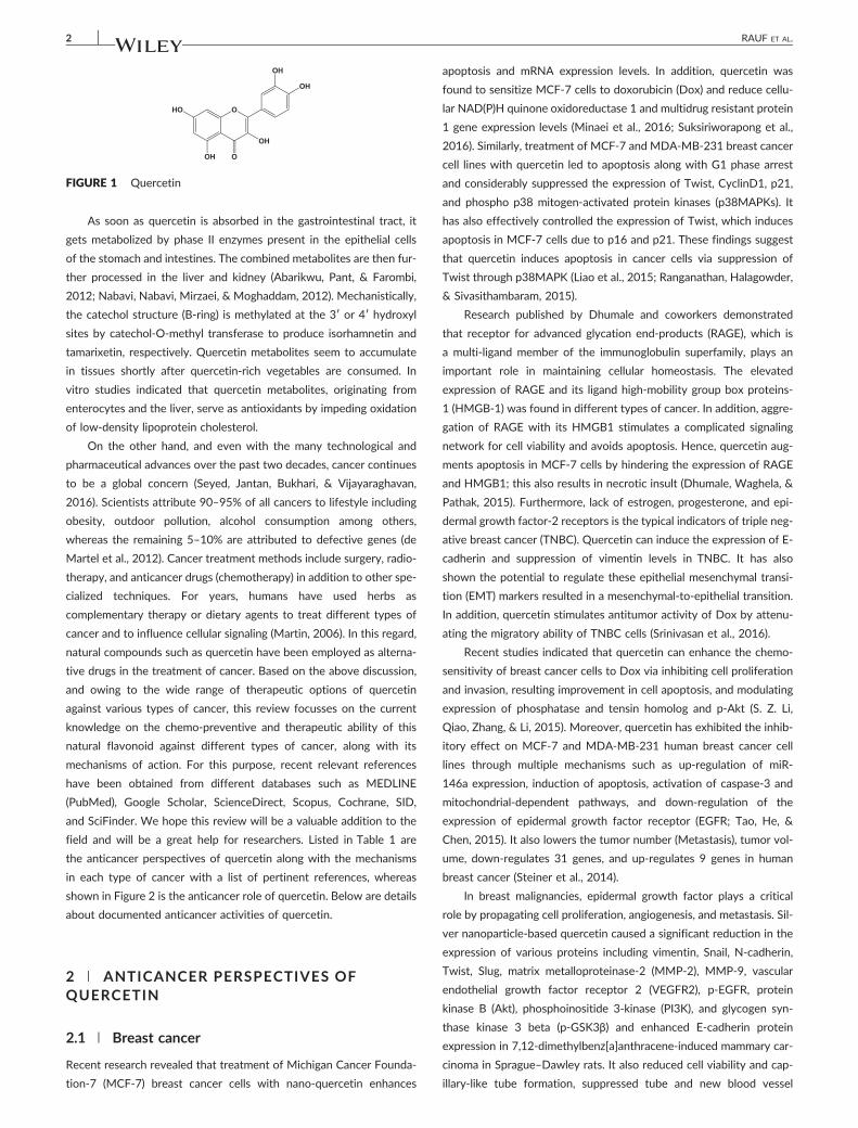
FIGURE 1 Quercetin
2 RAUF ET AL.
As soon as quercetin is absorbed in the gastrointestinal tract, it
gets metabolized by phase II enzymes present in the epithelial cells
of the stomach and intestines. The combined metabolites are then fur-
ther processed in the liver and kidney (Abarikwu, Pant, & Farombi,
2012; Nabavi, Nabavi, Mirzaei, & Moghaddam, 2012). Mechanistically,
the catechol structure (B‐ring) is methylated at the 3′ or 4′ hydroxyl
sites by catechol‐O‐methyl transferase to produce isorhamnetin and
tamarixetin, respectively. Quercetin metabolites seem to accumulate
in tissues shortly after quercetin‐rich vegetables are consumed. In
vitro studies indicated that quercetin metabolites, originating from
enterocytes and the liver, serve as antioxidants by impeding oxidation
of low‐density lipoprotein cholesterol.
On the other hand, and even with the many technological and
pharmaceutical advances over the past two decades, cancer continues
to be a global concern (Seyed, Jantan, Bukhari, & Vijayaraghavan,
2016). Scientists attribute 90–95% of all cancers to lifestyle including
obesity, outdoor pollution, alcohol consumption among others,
whereas the remaining 5–10% are attributed to defective genes (de
Martel et al., 2012). Cancer treatment methods include surgery, radio-
therapy, and anticancer drugs (chemotherapy) in addition to other spe-
cialized techniques. For years, humans have used herbs as
complementary therapy or dietary agents to treat different types of
cancer and to influence cellular signaling (Martin, 2006). In this regard,
natural compounds such as quercetin have been employed as alterna-
tive drugs in the treatment of cancer. Based on the above discussion,
and owing to the wide range of therapeutic options of quercetin
against various types of cancer, this review focusses on the current
knowledge on the chemo‐preventive and therapeutic ability of this
natural flavonoid against different types of cancer, along with its
mechanisms of action. For this purpose, recent relevant references
have been obtained from different databases such as MEDLINE
(PubMed), Google Scholar, ScienceDirect, Scopus, Cochrane, SID,
and SciFinder. We hope this review will be a valuable addition to the
field and will be a great help for researchers. Listed in Table 1 are
the anticancer perspectives of quercetin along with the mechanisms
in each type of cancer with a list of pertinent references, whereas
shown in Figure 2 is the anticancer role of quercetin. Below are details
about documented anticancer activities of quercetin.
2 | ANTICANCER PERSPECTIVES OFQUERCETIN
2.1 | Breast cancer
Recent research revealed that treatment of Michigan Cancer Founda-
tion‐7 (MCF‐7) breast cancer cells with nano‐quercetin enhances
apoptosis and mRNA expression levels. In addition, quercetin was
found to sensitize MCF‐7 cells to doxorubicin (Dox) and reduce cellu-
lar NAD(P)H quinone oxidoreductase 1 and multidrug resistant protein
1 gene expression levels (Minaei et al., 2016; Suksiriworapong et al.,
2016). Similarly, treatment of MCF‐7 and MDA‐MB‐231 breast cancer
cell lines with quercetin led to apoptosis along with G1 phase arrest
and considerably suppressed the expression of Twist, CyclinD1, p21,
and phospho p38 mitogen‐activated protein kinases (p38MAPKs). It
has also effectively controlled the expression of Twist, which induces
apoptosis in MCF‐7 cells due to p16 and p21. These findings suggest
that quercetin induces apoptosis in cancer cells via suppression of
Twist through p38MAPK (Liao et al., 2015; Ranganathan, Halagowder,
& Sivasithambaram, 2015).
Research published by Dhumale and coworkers demonstrated
that receptor for advanced glycation end‐products (RAGE), which is
a multi‐ligand member of the immunoglobulin superfamily, plays an
important role in maintaining cellular homeostasis. The elevated
expression of RAGE and its ligand high‐mobility group box proteins‐
1 (HMGB‐1) was found in different types of cancer. In addition, aggre-
gation of RAGE with its HMGB1 stimulates a complicated signaling
network for cell viability and avoids apoptosis. Hence, quercetin aug-
ments apoptosis in MCF‐7 cells by hindering the expression of RAGE
and HMGB1; this also results in necrotic insult (Dhumale, Waghela, &
Pathak, 2015). Furthermore, lack of estrogen, progesterone, and epi-
dermal growth factor‐2 receptors is the typical indicators of triple neg-
ative breast cancer (TNBC). Quercetin can induce the expression of E‐
cadherin and suppression of vimentin levels in TNBC. It has also
shown the potential to regulate these epithelial mesenchymal transi-
tion (EMT) markers resulted in a mesenchymal‐to‐epithelial transition.
In addition, quercetin stimulates antitumor activity of Dox by attenu-
ating the migratory ability of TNBC cells (Srinivasan et al., 2016).
Recent studies indicated that quercetin can enhance the chemo‐
sensitivity of breast cancer cells to Dox via inhibiting cell proliferation
and invasion, resulting improvement in cell apoptosis, and modulating
expression of phosphatase and tensin homolog and p‐Akt (S. Z. Li,
Qiao, Zhang, & Li, 2015). Moreover, quercetin has exhibited the inhib-
itory effect on MCF‐7 and MDA‐MB‐231 human breast cancer cell
lines through multiple mechanisms such as up‐regulation of miR‐
146a expression, induction of apoptosis, activation of caspase‐3 and
mitochondrial‐dependent pathways, and down‐regulation of the
expression of epidermal growth factor receptor (EGFR; Tao, He, &
Chen, 2015). It also lowers the tumor number (Metastasis), tumor vol-
ume, down‐regulates 31 genes, and up‐regulates 9 genes in human
breast cancer (Steiner et al., 2014).
In breast malignancies, epidermal growth factor plays a critical
role by propagating cell proliferation, angiogenesis, and metastasis. Sil-
ver nanoparticle‐based quercetin caused a significant reduction in the
expression of various proteins including vimentin, Snail, N‐cadherin,
Twist, Slug, matrix metalloproteinase‐2 (MMP‐2), MMP‐9, vascular
endothelial growth factor receptor 2 (VEGFR2), p‐EGFR, protein
kinase B (Akt), phosphoinositide 3‐kinase (PI3K), and glycogen syn-
thase kinase 3 beta (p‐GSK3β) and enhanced E‐cadherin protein
expression in 7,12‐dimethylbenz[a]anthracene‐induced mammary car-
cinoma in Sprague–Dawley rats. It also reduced cell viability and cap-
illary‐like tube formation, suppressed tube and new blood vessel

TABLE 1 Anticancer perspectives of quercetin, along with mechanisms of action
Cancer types Mechanisms References
Breast cancer Increases cell apoptosis and inhibits cell cycle progressionIncreases FasL mRNA expression and p51, p21, and GADD45 signaling
activities. Induces protein level, transcriptional activity, and nucleartranslocation of Foxo3a
Nguyen et al. (2017)
Reduces downstream genes including NQO1 and MRP1 Minaei, Sabzichi, Ramezani, Hamishehkar, and Samadi(2016) and Suksiriworapong et al. (2016)
Down‐regulates the vimentin levels and modulates the epithelialmesenchymal transition (EMT) markers
Srinivasan et al. (2016)
Reduces the expression of vimentin, Snail, N‐cadherin, Twist, Slug,metalloproteinase‐2 (MMP‐2), MMP‐9, VEGFR‐2, p‐EGFR, Akt,p‐phosphoinositide 3‐kinase (PI3K), and p‐GSK3β
Enhances E‐cadherin protein expression
Balakrishnan et al. (2016) and Quagliariello et al.(2016)
Causes cell cycle arrest and apoptosis in breast cancer cells viaregulation of Akt and Bax signaling mechanistic pathways
Sarkar, Ghosh, Chowdhury, Pandey, and Sil (2016)
Upregulates the levels of cleaved caspase‐8 and caspase‐3Suppresses the expression of phospho‐JAK1 and phospho‐STAT3Decreases STAT3‐dependent luciferase reporter gene activity (BT‐474
cells)
Seo et al. (2016)
Inhibits the expression of P‐glycoprotein Lv et al. (2016)
Colon cancer Inhibits the cell viability of CT26 and MC38 colon cancer cellsInduces apoptosis through the mitogen‐activated protein kinases (MAPKs)
pathwayRegulates the expression of EMT markers, such as E‐,
N‐cadherin, β‐catenin, and snail
Kee et al. (2016)
Causes G2 phase arrestInduces autophagic cell death through ERK activation
Y. Zhao, Fan, et al. (2017) and J. Zhao, Liu, et al.(2017)
Enhances the expression of E‐cadherin proteinDecreases the expression of metastasis‐related proteins of MMP‐2 and
MMP‐9Reduces the production of different inflammation factors including
TNF‐α, IL‐6, and Cox‐2
M. Han, Song, and Zhang (2016)
Pancreatic cancer Reduces the expression levels of cellular FLICE‐like inhibitory proteinActivates c‐Jun N‐terminal kinase (JNK)
J. H. Kim, Kim, Choi, and Son (2016) and Nwaeburuet al. (2016)
Reduces the tumor growth and drug resistance Cao et al. (2015)
Suppresses epidermal growth factor‐induced movement actionInhibits the EGFR‐mediated FAK, AKT, MEK1/2, and ERK1/2 signaling
pathway
J. Lee, Han, et al. (2015), W. J. Lee, Hsiao, et al.(2015),Y. J. Lee, Lee, and Lee (2015), and S. H. Lee, Lee,Min, et al. (2015)
Activates caspase‐3, ‐8, and ‐9 and reduces the mitochondrialmembrane potential
Inhibits extracellular signal‐regulated kinase (ERK) phosphorylation andpromotes JNK phosphorylation
F. Y. Chen, Cao, et al. (2015), X. Chen, Dong, et al.(2015), and Q. Chen, Li, et al. (2015)
Liver cancer Induces apoptosis Guan, Gao, Xu, et al. (2016)
Down‐regulates the expression of PI3K, PKC, COX‐2, and ROSEnhances the expression of p53 and BAX
Maurya and Vinayak (2015)
Activates p53‐ROS crosstalk and induces epigenetic modifications Bishayee, Khuda‐Bukhsh, and Huh (2015)
Lung cancer Triggers BCL2/BAX‐mediated apoptosis, as well as necrosis andmitotic catastrophe
Inhibits the migratory potential of A549 cells
Klimaszewska‐Wiśniewska et al. (2017)
Inhibits aurora B activitiesReduces the phosphorylation of histone 3
Xingyu et al. (2016)
Enhances expressions of nm23‐H1 and tissue inhibitor ofmetalloproteinase
Inhibits the protein expression of MMP‐2. GW9662, a PPAR‐γ antagonist
Chuang et al. (2016) and Warnakulasuriya, Ziaullah,and Rupasinghe (2016)
Increases miR‐21 expression and causes inhibition of PDCD4 inducedby [Cr(VI)]
Pratheeshkumar et al. (2017)
Prostatecancer
Decreases tumor improvement, down‐regulates Ki67, and enhancescaspase 7
Down‐regulates growth factors such as VEGF and EGF
Sharma et al. (2016), P. Wang, Henning, et al. (2016),and Y. Wang, Zhang, et al. (2016)
Prevents TGF‐β‐induced expression of vimentin and N‐cadherinDecreases TGF‐β‐induced expression of Twist, Snail, and Slug in
prostate cancer‐3 cell line
Baruah, Khandwekar, and Sharma (2016)
(Continues)
RAUF ET AL. 3

TABLE 1 (Continued)
Cancer types Mechanisms References
Bladder cancer Inhibits cell proliferation and colony formation of human bladder cancercells by inducing DNA damage
Oršolić et al. (2016)
Gastric cancer Inhibits EBV viral protein expressions, including EBNA‐1 and LMP‐2proteins
Prompts p53‐subordinate apoptosisInduces the expression of p53, Bax, and PumaCleaves caspase‐3 and ‐9 and Parp
J. Lee, Lee, Kim, et al. (2016) and H. H. Lee, Lee, Shin,et al. (2016)
Causes mitochondrial apoptotic‐dependent growth inhibition via theblockadeof PI3K‐Akt signaling
Activates caspase‐3 and ‐9Down‐regulates the Bcl‐2 and up‐regulates the Bax and cytochrome cCauses mitochondrial apoptotic‐dependent growth inhibition via the
blockadeof PI3K‐Akt signaling
Shen et al. (2016)
Bone cancer Decreases cyclin D1 expression in SKOV3 and U2OSPt cells Catanzaro, Ragazzi, Vianello, Caparrotta, andMontopoli (2015)
Inhibits 143B proliferation and up‐regulates the expression of miR‐217 X. Zhang, Guo, et al. (2015), J. Y. Zhang, Lin, et al.(2015), and X. A. Zhang, Zhang, et al. (2015)
Blood cancer Activates caspase‐3, ‐8, and ‐9 and promotes leukemic cell apoptosisReduced expression of the antiapoptotic proteins B‐cell, lymphoma (Bcl)‐2.
Enhances expression of the proapoptotic proteins Bcl‐2‐interactingmediatorof cell death
F. Y. Chen, Cao, et al. (2015), X. Chen, Dong, et al.(2015), and Q. Chen, Li, et al. (2015)
Brain cancer Suppresses COX‐2 expression by Hsp27 inhibition and acts as both COX‐2andHsp27 inhibitor
Reduces MMP‐2 expression
Q. C. Li, Liang, Hu, and Tian (2016) and J. Li, Tang, Li,Li, and Fan (2016)
Santos et al. (2015)
Decreases mitochondria and rough endoplasmic reticulum injuryReduces filopodia‐like structures on the cell surface
Induces necrotic cell death and down‐regulates the Bcl‐2 mRNAsexpression.Enhances mitochondrial mRNAs expression
Modulates the mitochondrial pathway and the JAK2/STAT3 signaling
Wang et al. (2013)
Head and neckcancer
Retards colony growth of HSC‐3 cellsSuppresses the MMP‐2 and MMP‐9
Chan, Lien, Lee, and Huang (2016)
Causes cells arrest at the G1 phaseInduces apoptosis, suppresses the expression of Bax, and activates the
expression of Caspase‐3 and Bcl‐2Reverses gene‐encoded Pglycoprotein‐mediated MDR
Z. Yuan et al. (2015)
Cervical cancer Inhibits antiapoptotic AKT and Bcl‐2 expressionIncreases mitochondrial cytochrome‐c levelCauses cell cycle arrest at G2/M
Bishayee et al. (2013)
Induces apoptosis via PI3k/Akt pathways Xiang, Fang, and Wang (2014)
Significantly inhibits UBE2S expression Lin et al. (2017)
Skin cancer Blocks UVB irradiation‐induced COX‐2 up‐expression and NF‐kBactivation in Hacat cell line
Caddeo et al. (2016)
Reduces the tumor size and the cumulative number of papillomas.Decreasesthe serum levels of glutamate oxalate transaminase, glutamate pyruvatetransaminase, alkaline phosphatase, and bilirubin
Ali and Dixit (2015)
Inhibits PI3K and MAPK signalingAttenuates MEK–ERK signaling and influences PI3K/Akt pathway
Rafiq et al. (2015)
Eye cancer Decreases dose‐dependently the RPE cell proliferation, migration, andsecretion of VEGF
Inhibits the secretion of VEGF evoked by CoCl2‐induced hypoxia
R. Chen et al. (2014)
Thyroid cancer Lowers the cell proliferation and increases rate of apoptosis bycaspase activation
Downregulates the levels of Hsp90Decreases chymotrypsin‐like proteasome activity
Mutlu Altundağ et al. (2016) andQuagliariello et al. (2016)
Ovarian cancer Suppresses ROS‐induced injury and increases the expression ofendogenous antioxidant enzymes
W. Li, Liu, et al. (2014), N. Li, Sun, et al. (2014), X. Li,Wang, et al. (2014), and W. Li, Zhao, et al. (2014)
(Continues)
4 RAUF ET AL.
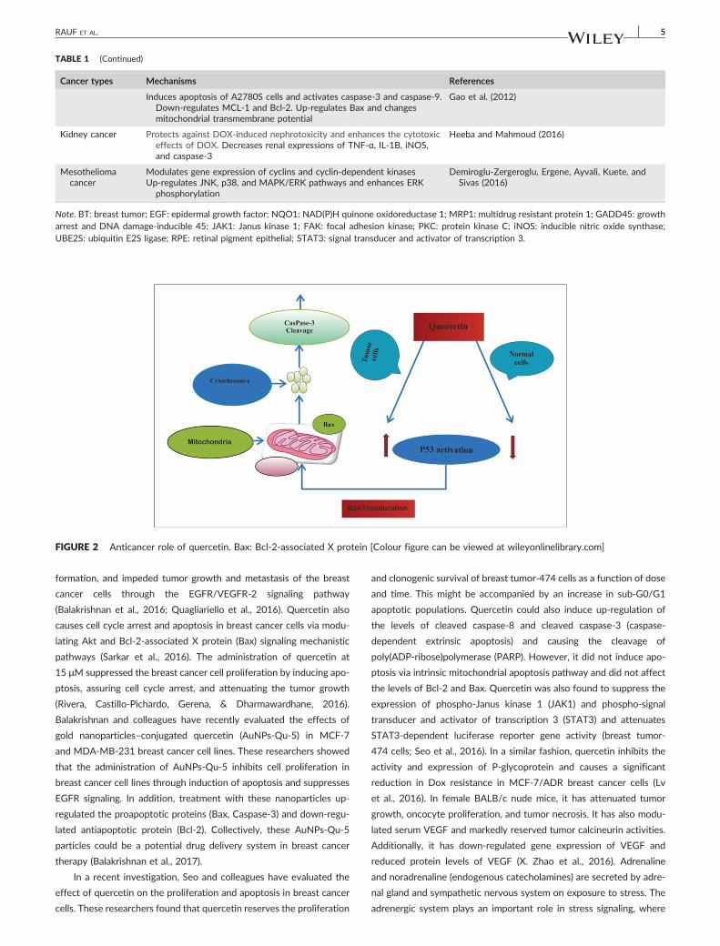
TABLE 1 (Continued)
Cancer types Mechanisms References
Induces apoptosis of A2780S cells and activates caspase‐3 and caspase‐9.Down‐regulates MCL‐1 and Bcl‐2. Up‐regulates Bax and changesmitochondrial transmembrane potential
Gao et al. (2012)
Kidney cancer Protects against DOX‐induced nephrotoxicity and enhances the cytotoxiceffects of DOX. Decreases renal expressions of TNF‐α, IL‐1B, iNOS,and caspase‐3
Heeba and Mahmoud (2016)
Mesotheliomacancer
Modulates gene expression of cyclins and cyclin‐dependent kinasesUp‐regulates JNK, p38, and MAPK/ERK pathways and enhances ERK
phosphorylation
Demiroglu‐Zergeroglu, Ergene, Ayvali, Kuete, andSivas (2016)
Note. BT: breast tumor; EGF: epidermal growth factor; NQO1: NAD(P)H quinone oxidoreductase 1; MRP1: multidrug resistant protein 1; GADD45: growtharrest and DNA damage‐inducible 45; JAK1: Janus kinase 1; FAK: focal adhesion kinase; PKC: protein kinase C; iNOS: inducible nitric oxide synthase;UBE2S: ubiquitin E2S ligase; RPE: retinal pigment epithelial; STAT3: signal transducer and activator of transcription 3.
FIGURE 2 Anticancer role of quercetin. Bax: Bcl‐2‐associated X protein [Colour figure can be viewed at wileyonlinelibrary.com]
RAUF ET AL. 5
formation, and impeded tumor growth and metastasis of the breast
cancer cells through the EGFR/VEGFR‐2 signaling pathway
(Balakrishnan et al., 2016; Quagliariello et al., 2016). Quercetin also
causes cell cycle arrest and apoptosis in breast cancer cells via modu-
lating Akt and Bcl‐2‐associated X protein (Bax) signaling mechanistic
pathways (Sarkar et al., 2016). The administration of quercetin at
15 μM suppressed the breast cancer cell proliferation by inducing apo-
ptosis, assuring cell cycle arrest, and attenuating the tumor growth
(Rivera, Castillo‐Pichardo, Gerena, & Dharmawardhane, 2016).
Balakrishnan and colleagues have recently evaluated the effects of
gold nanoparticles–conjugated quercetin (AuNPs‐Qu‐5) in MCF‐7
and MDA‐MB‐231 breast cancer cell lines. These researchers showed
that the administration of AuNPs‐Qu‐5 inhibits cell proliferation in
breast cancer cell lines through induction of apoptosis and suppresses
EGFR signaling. In addition, treatment with these nanoparticles up‐
regulated the proapoptotic proteins (Bax, Caspase‐3) and down‐regu-
lated antiapoptotic protein (Bcl‐2). Collectively, these AuNPs‐Qu‐5
particles could be a potential drug delivery system in breast cancer
therapy (Balakrishnan et al., 2017).
In a recent investigation, Seo and colleagues have evaluated the
effect of quercetin on the proliferation and apoptosis in breast cancer
cells. These researchers found that quercetin reserves the proliferation
and clonogenic survival of breast tumor‐474 cells as a function of dose
and time. This might be accompanied by an increase in sub‐G0/G1
apoptotic populations. Quercetin could also induce up‐regulation of
the levels of cleaved caspase‐8 and cleaved caspase‐3 (caspase‐
dependent extrinsic apoptosis) and causing the cleavage of
poly(ADP‐ribose)polymerase (PARP). However, it did not induce apo-
ptosis via intrinsic mitochondrial apoptosis pathway and did not affect
the levels of Bcl‐2 and Bax. Quercetin was also found to suppress the
expression of phospho‐Janus kinase 1 (JAK1) and phospho‐signal
transducer and activator of transcription 3 (STAT3) and attenuates
STAT3‐dependent luciferase reporter gene activity (breast tumor‐
474 cells; Seo et al., 2016). In a similar fashion, quercetin inhibits the
activity and expression of P‐glycoprotein and causes a significant
reduction in Dox resistance in MCF‐7/ADR breast cancer cells (Lv
et al., 2016). In female BALB/c nude mice, it has attenuated tumor
growth, oncocyte proliferation, and tumor necrosis. It has also modu-
lated serum VEGF and markedly reserved tumor calcineurin activities.
Additionally, it has down‐regulated gene expression of VEGF and
reduced protein levels of VEGF (X. Zhao et al., 2016). Adrenaline
and noradrenaline (endogenous catecholamines) are secreted by adre-
nal gland and sympathetic nervous system on exposure to stress. The
adrenergic system plays an important role in stress signaling, where

6 RAUF ET AL.
excessive stress can be linked to increased production of reactive oxy-
gen species (ROS). Overproduction of ROS induces oxidative damage
and causes the development of diseases such as cancer. Research
findings revealed that quercetin suppresses (a) generation of ROS, (b)
activation of cyclic adenosine monophosphate and reticular activating
system (RAS), and (c) phosphorylation of extracellular signal‐regulated
kinases 1/2 (ERK1/2) and the expression of HMOX1, MMP‐2, and
MMP‐9 genes. It also suppresses invasion of breast cancer cells by
controlling β2‐adrenergic signaling (Yamazaki, Miyoshi, Kawabata,
Yasuda, & Shimoi, 2014). The (3‐(4,5‐dimethylthiazol‐2‐yl)‐2,5‐diphe-
nyltetrazolium bromide) assay has recently been used to investigate
the anticancer effect of quercetin and its underlying mechanisms in
triple‐negative breast cancer cells. Results indicated that quercetin
increases cell apoptosis, inhibits cell cycle progression, and increases
FasL mRNA expression and p51, p21, and growth arrest and DNA
damage‐inducible 45 (GADD45) signaling activities. These results sug-
gest that quercetin induces apoptosis and cell cycle arrest via modifi-
cation of Foxo3a signaling in triple‐negative breast cancer cells
(Nguyen et al., 2017).
2.2 | Colon cancer
Diet is an important factor associated with colon cancer. Diets that are
low in fiber and high in fat, calories, and red meat and processed meats
increase the risk of developing colon cancer. Cancer treatment
depends on the type of cancer, the stage of the cancer (how much it
has spread), age, health status, and additional personal characteristics
(NCI, 2014).
Numerous studies have dealt with the effect of quercetin on
colon cancer. Kee and coworkers used the water‐soluble tetrazolium
salts assay, annexin V assay, real‐time polymerase chain reaction,
western blot analysis, and gelatin zymography to study the inhibitory
effect of quercetin on colorectal lung metastasis. These researchers
found that quercetin can (a) inhibit the cell viability of colon 26
(CT26) and colon 38 (MC38) cells, (b) induce apoptosis through the
MAPKs pathway in CT26 cells, (c) regulate the expression of EMT
markers, such as E‐, N‐cadherin, β‐catenin, and snail, by nontoxic con-
centrations of quercetin, and (d) inhibit the migration and invasion
abilities of CT26 cells through expression of MMPs and tissue inhibi-
tor of metalloproteinases (TIMPs) regulation. They concluded from
this investigation that quercetin can inhibit the survival and metastatic
ability of CT26 cells, and can suppress colorectal lung metastasis in the
mouse model, and may be a potent therapeutic agent for the treat-
ment of metastatic colorectal cancer (Kee et al., 2016). Similarly, an
investigation by Zhao et al. concluded that 8‐C‐(E‐phenylethenyl)
quercetin, a novel quercetin derivative, triggers G2 phase arrest in
colon cancer cells and suppresses propagation. It also induces autoph-
agic cell death through ERK stimulation (Y. Zhao, Fan, et al., 2017; J.
Zhao, Liu, et al., 2017). Similarly, quercetin at a concentration of
5 μM could markedly suppress the migratory and invasive capacity
of Caco‐2 cells. In addition, results from this investigation revealed
that the expression of E‐cadherin protein was increased by quercetin,
whereas metastasis‐related proteins of MMP‐2, MMP‐9 expression
got decreased by it in a dose‐dependent manner.
The anti‐toll‐like receptor 4 (TLR4) antibody of pyrrolidine dithio-
carbamate might influence the inhibition of quercetin on cell migration
and invasion and the expression of various proteins such as E‐
cadherin, MMP‐2, MMP‐9, NF‐κB p65, and TLR4. Moreover, querce-
tin could lessen the production of different inflammation factors
including TNF‐α, Cox‐2, and interleukin 6 (IL‐6). Hence, quercetin
might exert its anti‐colon cancer activity via the TLR4‐ and/or
NF‐κB‐mediated signaling pathway (M. Han et al., 2016). The 1,2‐
dimethyl hydrazine‐induced colon cancer causes nephrotoxicity
and further increases the blood urea nitrogen, urea, creatinine, and
eventually result in a number of aberrant crypts and foci formation.
The potential protective effect of quercetin on cisplatin‐induced
nephrotoxicity was assessed through lowing the blood urea nitro-
gen, urea, and creatine and also reduced the aberrant crypt foci
number (Q. C. Li, Liang, et al., 2016; J. Li, Tang, et al., 2016). Similar
results were obtained by Saleem et al. (2015) who found that treat-
ment of mice either with quercetin, sodium gluconate, or with the
combination has a positive effect against 1,2‐dimethyl hydrazine‐
induced colon cancer.
In human colon adenocarcinoma cells, quercetin significantly
enhanced the expression of the endocannabinoids receptor (CB1‐R)
and further suppressed PI3K/Akt/mTOR. It also induced JNK/JUN
pathways and modified the metabolism of β‐catenin, either directly
or via activation of CB1‐R (Refolo et al., 2015). These findings were
confirmed by other researchers (Xu et al., 2015). The research work
conducted by Zhang et al. indicated that quercetin significantly pre-
vents the proliferation of human colon cancer in CACO‐2 and SW‐
620 cells by suppressing the NF‐κB pathway, down‐regulation of B‐
cell lymphoma 2, and up‐regulation of Bax (X. Zhang, Guo, Chen, &
Chen, 2015; J. Y. Zhang, Lin, et al., 2015; X. A. Zhang, Zhang, Yin, &
Zhang, 2015). In addition, quercetin was found to have an inhibitory
effect on Wnt/β‐catenin in colon cancer cells SW480, DLD‐1, and
HCT116 cancer cells (Amado et al., 2014). Similarly, a study by Kim
et al. demonstrated that the inhibitory role of quercetin in colon can-
cer cell lines through enhancing the apoptotic cell death via generating
intracellular ROS and through enhancing sestrin 2 expression is
accompanied by activated protein kinase (AMPK) activation.
Moreover, these researchers found that the quercetin‐induced apo-
ptosis involves sestrin 2/AMPK/mTOR pathway by regulating
increases intracellular ROS (Kim, Lee, & Kim, 2013; Kim, Moon, Ahn,
& Cho, 2013).
For HT‐29 colon cancer cells, Kim and coworkers found that quer-
cetin induces apoptosis by attenuating membrane potential of the
mitochondria producing intracellular ROS and elevating the expression
of sestrin 2 via the AMPK/p38 mechanistic pathway (G. T. Kim, Lee,
Kim, & Kim, 2014; M. C. Kim, Lee, Lim, et al., 2014). Similarly, several
research groups independently showed that quercetin increases the
antioxidant activity, increases PARP cleavage, and induces caspase‐
3‐cleavage (twofold) in HT‐29 colon cancer cells. It also lowers the
expressions of specificity proteins (Sp) such as Sp1, Sp3, and Sp4
mRNA; this expression was accompanied by a decreased protein
expression. In addition, the Sp‐dependent antiapoptotic survival gene
was also significantly decreased, both at mRNA and protein levels. It
also attenuates microRNA‐27a and induces a Sp‐repressor, zinc finger
protein ZBTB10 (Atashpour et al., 2015; Cho, Kim, Park, Choo, &

RAUF ET AL. 7
Chong, 2013; Del Follo‐Martinez, Banerjee, Li, Safe, & Mertens‐
Talcott, 2013).
2.3 | Pancreatic cancer
Research revealed that quercetin induces apoptosis in tumor necrosis
factor‐related apoptosis‐inducing ligand (TRAIL) in resistant pancreatic
cancer cells. It was also found that a BH3‐only protein BID consider-
ably reduces attenuated TRAIL/quercetin‐induced apoptosis. Querce-
tin has also the ability to reduce the expression levels of cellular
FLICE‐like inhibitory protein and to strongly save pancreatic cancer
cells from TRAIL/quercetin‐induced apoptosis, in a dose‐dependent
manner. Additionally, quercetin stimulates JNK, which influences the
proteasomal degradation of cellular FLICE‐like inhibitory protein,
followed by sensitized pancreatic cancer cells to TRAIL‐induced apo-
ptosis (J. H. Kim et al., 2016; Nwaeburu et al., 2016). In human pancre-
atic cancer cell lines CFPAC‐1 and SNU‐213, quercetin‐3‐O‐glucoside
suppresses the migratory activity induced by transforming growth fac-
tor‐beta (TGF‐β) and vascular endothelial growth factor A even at rel-
atively low dosages in CFPAC‐1, but not in bFGF‐activated SNU‐213
cells. In addition, co‐treatment with low dose of gemcitabine and
quercetin‐3‐O‐glucoside exhibited synergistic inhibition effects on
the infiltrate activity induced by bFGF in CFPAC‐1 and SNU‐213 cells
(J. Lee, Lee, Kim, & Kim, 2016; H. H. Lee, Lee, Shin, et al., 2016).
Cao et al. demonstrated that quercetin in combination with
gemcitabine suppresses proliferation, invasion and self‐renewal capac-
ity, and cancer stem cells surface markers expression, with alterations
of β‐catenin in pancreatic cancer stem‐like cells. In addition, it reduces
tumor growth and drug resistance in pancreatic cancer (Cao et al.,
2015). In a similar fashion, Lee et al. found that quercetin suppresses
epidermal growth factor‐induced migration activity and inhibits the
infiltration activity of pancreatic cancer cells in a dose‐dependent
manner in human pancreatic cancer cell lines. Furthermore, these
researchers found that antitumor effects of quercetin are mediated
by selectively inhibiting the EGFR‐mediated focal adhesion kinase,
protein kinase B (AKT), MEK1/2, and ERK1/2 signaling pathway (J.
Lee, Han, Yun, & Kim, 2015; W. J. Lee, Hsiao, et al., 2015; Y. J. Lee,
Lee, & Lee, 2015; S. H. Lee, Lee, Min, et al., 2015). Similarly, quercetin
significantly inhibits proliferation, promotes apoptosis, and induces cell
cycle arrest within the G1 phase in pancreatic cancer cells. It can also
activate caspase‐3, ‐8, and ‐9 and reduces the mitochondrial mem-
brane potential and can inhibit the expression level of the δ opioid
receptor, whereas isoquercitrin was found to have no effect on the κ
and μ opioid receptors. Furthermore, quercetin can inhibit ERK phos-
phorylation, promote JNK phosphorylation, and significantly inhibit
xenograft growth in nude mice (F. Y. Chen, Cao, et al., 2015; X. Chen,
Dong, et al., 2015; Q. Chen, Li, et al., 2015).
On the other hand, research conducted by Appari and coworkers
revealed that quercetin significantly inhibits viability, migration,
expression of MMP‐2 and ‐9, aldehyde dehydrogenase 1 activity, col-
ony, and spheroid formation and triggers apoptosis in pancreatic duc-
tal adenocarcinoma. It also induces the expression of miR‐let7‐a and
causes inhibition of K‐ras in cancer cells (Appari, Babu, Kaczorowski,
Gross, & Herr, 2014). Similarly, it induces apoptosis of PANC‐1, char-
acterized as nucleic acid and genomic DNA fragmentation, chromatin
condensation, and sub‐G0/G1 fraction of cell cycle increase. It also
increases the buildup of intracellular Ca2+ ions and Grp78/Bip and
GADD153/CCAAT enhancer‐binding protein homologous protein
(CHOP) protein expression and triggers mitochondrial dysfunction.
Quercetin exerts this cytotoxicity against human pancreatic cancer
cells trough endoplasmic reticulum stress‐mediated apoptotic signal-
ing including pathway, as well as ROS production and mitochondrial
dysfunction (J. H. Lee et al., 2013). Other researchers found that quer-
cetin exerts its antiproliferative effects in pancreatic cancer cells by
inducing apoptosis and attenuating the growth of orthotopically
transplanted pancreatic xenografts (Angst et al., 2013). In MIA PaCa‐
2 pancreatic adenocarcinoma cells, quercetin at 100 μM inhibits tracer
glucose‐derived glycogen labeling (Σm), slows down glycogen synthe-
sis, and manages tumor cell proliferation (Harris et al., 2012).
2.4 | Liver cancer
The leading cause of liver cancer is cirrhosis due to either hepatitis B,
hepatitis C, or excess alcohol intake (Naghavi, Wang, Lozano, et al.,
2015). Other causes include aflatoxin, nonalcoholic fatty liver disease,
and liver flukes. The most common types are hepatocellular carci-
noma, which makes up to 80% of cases, and cholangiocarcinoma
(NCI, 2016).
Previous studies demonstrated that treatment with nano‐capsu-
lated quercetin restricts all changes in diethyl nitrosamine‐mediated
development of hepatocarcinogenesis, suggesting that this nano‐cap-
sulated natural product may be accepted as a potent therapeutic agent
in preventing diethyl nitrosamine‐mediated hepatocarcinogenesis
(Mandal et al., 2014). In addition, fatty acid esters of quercetin‐3‐O‐
glucoside were found to exhibit significant inhibition of HepG2 cell
proliferation. Effect of this novel compound was associated with cell
ycle arrest in S‐phase and apoptosis. Furthermore, quercetin‐3‐O‐glu-
coside esters showed significant low toxicity to normal liver cells than
sorafenib, a chemotherapy drug used in the treatment of hepatocellu-
lar carcinoma (Sudan & Rupasinghe, 2015). Treatment with quercetin
at a dose of 50 mg/kg in mice showed a protective effect on cis-
platin‐induced DNA damage in normal cells, without interfering with
the antitumor efficacy of the combined treatment. These results sug-
gest that quercetin can protect the blood, liver, and kidney cells of
mice against HIPEC‐induced injury and can increase survival of mice
by improving the antitumor adaptive immunity with hyperthermia
(Oršolić & Car, 2014).
Quercetin inhibits the growth of cancer cells, which can be attrib-
uted to various mechanisms, such as the induction of cell cycle arrest
and/or apoptosis, as well as its antioxidant functions. In this respect,
Zhao and coworkers evaluated the activity of quercetin in human liver
cancer HepG2 cells. These workers found that quercetin can induce
apoptosis in human liver cancer HepG2 cells with overexpression of
fatty acid synthase. These results suggest that apoptosis is induced
by quercetin via the inhibition of fatty acid synthase. Additionally,
findings by these researchers suggest that quercetin may be useful
for preventing human liver cancer (P. Zhao et al., 2014). Furthermore,
it was demonstrated by a number of researchers that intake of querce-
tin seems to play a minor regulatory role, whereas supplement doses
may have great effects on gene expression in hepatocytes. Further

8 RAUF ET AL.
work is certainly required in handling of quercetin supplements
(Waizenegger et al., 2015a).
Controlled release of medications remains the most convenient
way to deliver drugs. Bishayee and coworkers examined the effect
of gold‐quercetin loaded into poly(DL‐lactide‐co‐glycolide) nanoparti-
cles (NQ) on HepG2 hepatocarcinoma cells. Results revealed that
quercetin loaded on the nanoparticles preferentially kill cancer cells,
compared with normal cells. In addition, NQ interacted with HepG2
cell DNA and reduces histone deacetylases to manage cell prolifera-
tion and arrest the cell cycle at the sub‐G stage. These nanoparticles
induce apoptosis in HepG2 cells by activating p53‐ROS crosstalk and
by enhancing epigenetic modifications leading to inhibited prolifera-
tion and cell cycle arrest (Bishayee et al., 2015). Protein kinase C is a
key regulator of cell growth in mammalian cells and is linked with
tumor succession. Quercetin, on the other hand, exhibits antitumor
activity both in vitro and in vivo in HepG2 cells. It down‐regulates
the expression of PI3K, protein kinase C, COX‐2, and ROS. Addition-
ally, it enhances the expression of p53 and BAX in HepG2 cells
(Maurya & Vinayak, 2015). One of the shortcomings of quercetin in
clinics is its poor solubility. To overcome these disadvantages, Guan
and coworkers prepared quercetin (QT) as QT‐loaded PLGA‐TPGS
nanoparticles (QPTN) and evaluated its therapeutic efficacy for liver
cancer. Results indicated that QPTN could induce HepG2 cell apopto-
sis in a dose‐dependent manner and that QPTN could suppress tumor
growth by 59.07%. These researchers concluded that QPTN could be
used as a potential intravenous dosage form for the treatment of liver
cancer owing to the enhanced pharmacological effects of quercetin
with increased liver targeting (Guan et al., 2016).
2.5 | Lung cancer
Several research papers have dealt with quercetin as a chemothera-
peutic agent against lung cancer. An investigation by W. Chen, Wang,
Zhuang, Zhang, and Lin (2007) revealed that quercetin significantly
enhances TRAIL‐induced cytotoxicity in non‐small cell lung cancer
cells. It also increases expression of death receptor (DR) 5 and has
no effect on other components of the death‐inducing signaling com-
plex. In addition, these researchers demonstrated that quercetin can
sensitizeTRAIL‐induced cytotoxicity in lung cancer cells via two mech-
anisms: (a) by induction of DR5 and (b) by suppression of survivin
expression; these mechanisms may explain the lung cancer preventive
activity of quercetin. Furthermore, researchers found that treatment
of human lung cancer H‐520 cells with quercetin increases the cis-
platin‐induced apoptosis by 30.2%, down‐regulates Bcl‐XL and Bcl‐2,
and up‐regulates Bax (Kuhar, Sen, & Singh, 2006).
For JB6 Cl41 cells and A549 lung cancer cells, researchers
showed that quercetin inhibits aurora B activities and reduces the
phosphorylation of histone 3 (Xingyu et al., 2016). Quercetin also
reduces ROS production induced by exposure to hexavalent chro-
mium [Cr(VI)] in BEAS‐2B cells. It also suppresses the malignant cell
transformation, improves miR‐21 expression, and causes inhibition of
PDCD4 induced by [Cr(VI)] in a dose‐dependent manner. Further-
more, quercetin reduces the tumor occurrence and suppresses the
Cr(VI)‐induced malignant transformation and tumorigenesis in nude
mice injected with BEAS‐2B cells (Pratheeshkumar et al., 2017). In
human lung carcinoma A549 cells, researchers demonstrated that
quercetin appreciably suppresses cell invasion and migration. It
inhibits the activity and expression of MMPs‐2 in a dose‐dependent
manner. It also increases the expressions of nm23‐H1 and TIMP‐2
and inhibits the protein expression of MMP‐2. GW9662, a PPAR‐γ
antagonist (Chuang et al., 2016; Warnakulasuriya et al., 2016).
In their work on lung carcinogenesis, Chen and colleagues demon-
strated that benzo[a]pyrene‐induced human bronchial epithelial cell
(HBEC) transformation is improved by IL‐6 in vitro. The carcinogen/
IL‐6‐transformed cells exhibit higher expression of signal transducer
and activator of STAT3 when compared with cells transformed by
BPDE alone. Furthermore, these researchers showed that treatment
with quercetin (a) considerably decreases BPDE‐stimulated IL‐6 secre-
tion from human lung fibroblasts via inhibition of the NF‐κB and ERK
pathways, (b) blocks IL‐6‐induced STAT3 activation in HBECs, and (c)
abolishes IL‐6 enhancement of HBEC transformation by BPDE (W.
Chen et al., 2016). On the other hand, Zhao and coworkers examined
the inhibitory effect of quercetin on the growth of A549 lung cancer
cells and found that it induces apoptosis, decreases the levels of
MMP‐9 (mRNA and protein) and TGF‐β1 protein, and reduces the
number of tumor cells. These researchers also found that the combi-
nation of quercetin (at low concentrations) with TIMP‐1 shows syner-
gistic inhibitory effect on the growth of A549 cells (X. Zhao & Zhang,
2015). Quercetin also decreases claudin‐2 expression in lung adeno-
carcinoma A549 cells in a time‐ and concentration‐dependent manner,
lowers the stability of claudin‐2 mRNA, and increases the expression
of miR‐16. Categorically, quercetin reduces claudin‐2 level through
up‐regulation of miR‐16 expression (Sonoki et al., 2015). In lung can-
cer cells (A549 and H460 cells), quercetin reduced cell viability and
inhibited heat shock protein 70 (HSP70) expression in both cell lines
in a dose‐dependent manner. Addition of a fixed quercetin dose
improved gemcitabine‐induced cell death, which was linked to
increased caspase‐3 and caspase‐9 activities (J. Lee, Han, et al.,
2015; W. J. Lee, Hsiao, et al., 2015; Y. J. Lee, Lee, & Lee, 2015; S.
H. Lee, Lee, Min, et al., 2015; Y. Liu, Wu, & Zhang, 2015).
Similarly, quercetin prevented tumor proliferation by (a) initiating
cell cycle arrest, (b) improving TRAIL‐induced tumor cell death, (c) low-
ering the p62 protein expression, and (d) increasing GFP‐LC3B in
human lung cancer cells in a dose‐dependent fashion (Moon, Eo,
Lee, & Park, 2015; J. Wang, Zhang, Cheng, Zhang, & Li, 2015). It also
induces apoptosis in A549 cells via mitochondrial depolarization by
triggering an imbalance in B‐cell lymphoma 2/Bcl2 antagonist X
(Bcl2/Bax) ratio and by down‐regulating the IL‐6/STAT3 signaling
pathway. Additionally, quercetin could block nuclear factor kappa‐
light‐chain‐enhancer of activated B cells (NF‐κB) action at early hours,
which might cause a down‐regulation of the IL‐6 titer, and the IL‐6
expression, in turn, could inhibit p‐STAT3 expression. Down‐regula-
tion of both the STAT3 and NF‐κB expressions might, consequently,
causes down‐regulation of Bcl2 because both are upstream effectors
of Bc12. In A549 cells, modification in Bcl2 reactions may result in
an imbalance in the Bcl2/Bax ratio, which could eventually lead mito-
chondria mediated apoptosis (Mukherjee & Khuda‐Bukhsh, 2015).
Nair and colleagues examined the cumulative effects of curcumin
and quercetin in inducing apoptosis in benzo(a)pyrene (100 mg/kg
body weight)‐induced lung carcinogenesis in mice. In benzo(a)

RAUF ET AL. 9
pyrene‐treated animals, supplementation of curcumin (60 mg/kg body
weight) and quercetin (40 mg/kg body weight), separate as well as
combined, considerably reduced the protein expression of Bcl‐2 and
amplified the protein expression of Bax. Supplementation also
improved the enzyme activities of caspase 9 and caspase 3 (Nair,
Malhotra, & Dhawan, 2015). In addition, a peer group of investigators
(Lam et al., 2012; Youn, Jeong, Jeong, Kim, & Um, 2013) determined
that quercetin strongly inhibits cell production and enhanced sub‐G1
and apoptosis despite of p53 status in H460 cells. It also improved
the expression of genes linked with DR signaling TRAIL receptor, cas-
pase‐10, IL 1R DNA fragmentation factor 45, tumor necrosis factor
receptor 1, FAS, inhibitor of kappa‐B‐alpha (IκBα), and cell cycle inhi-
bition GADD45, p21 (Cip1). However, it reduced the expression of
genes involved in activation of NF‐κB and IKKα. It also suppressed
the NF‐κB and additionally improved the expression of DRs and cell
cycle inhibitors (Lam et al., 2012; Youn et al., 2013). In A549 non‐small
cell lung cancer cells, Klimaszewska‐Wiśniewska and colleagues have
recently employed the methyl‐thiazol‐diphenyl‐tetrazolium (MTT)
assay, annexin V/propidium iodide test, electron microscopic examina-
tion, cell cycle analysis based on DNA content, real‐time polymerase
chain reaction assays, in vitro scratch wound‐healing assay, fluores-
cence staining of F‐actin, β‐tubulin, and vimentin to examine the
effect of quercetin on microfilaments, microtubules, and vimentin
intermediate filaments. Results revealed that quercetin triggers
BCL2/BAX‐mediated apoptosis, necrosis, and mitotic catastrophe
and suppresses the migratory potential of A549 cells. These findings
suggest that quercetin‐induced mitotic catastrophe involves the per-
turbation of mitotic microtubules, which results in monopolar spindle
formation and to failure of cytokinesis (Klimaszewska‐Wiśniewska
et al., 2017)
2.6 | Prostate cancer
Numerous researchers have investigated the effect of quercetin, alone
or in combination with other drugs, on colon cancer. Standard treat-
ment for metastatic and castration‐resistant prostate cancer includes
chemotherapy with docetaxel (Doc). However, chemoresistance and
side effects of Doc limit its clinical success. In this respect, different
research groups investigated the effect of natural products such as
quercetin on the efficacy of androgen‐independent prostate cancer
cells. These researchers found that quercetin (a) improves the healing
practicality of Doc, (b) considerably lessens tumor progression, (c) cut
down the Ki67, (d) increases cleavage of caspase 7, (e) lowers blood
concentrations of growth factors, such as VEGF and epidermal growth
factor, and (f) substantially lifts the levels of tumor silencer mir15a and
mir330 (P. Wang, Henning, et al., 2016; Y. Wang, Zhang, Lv, Zhang, &
Zhu, 2016). In castration‐resistant prostate cancer cells, quercetin
improves the therapeutic effect of Doc in through multiple mecha-
nisms including down‐regulation of chemoresistance‐related proteins
(P. Wang, Henning, et al., 2016; Y. Wang, Zhang, et al., 2016). Addi-
tionally, in PC3 and DU145 prostate cancer cell lines, a combined
treatment with quercetin and curcumin, two known dietary
phytocompounds with described DNMT‐inhibitory activity, was much
more effective than either of them in both inhibition of DNMT and in
triggering apoptosis via mitochondrial depolarization. These two
natural compounds have the potential for use as chemopreventive
agents of androgen resistance in prostate cancer (Sharma et al., 2016).
The therapeutic potential of novel quercetin‐loaded nanomicelles
(to enhance the solubility of quercetin in water) for prostate cancer
treatment was recently evaluated. Results indicated that quercetin
can be efficiently encapsulated into micelles up to 1 mg per ml, which
corresponds to a 450‐fold increase of its water solubility. Additionally,
a nanomicelle‐based drug delivery system could be a promising and
effective therapeutic strategy for clinical treatment of prostate cancer
(X. Zhao et al., 2016). In LNCaP and PC‐3 cells, Song et al. examined
the effects of quercetin combined with 2‐methoxyestradiol on the
proliferation of androgen‐dependent LNCaP and androgen‐indepen-
dent PC‐3 human prostate cancer cells lines. Both quercetin and 2‐
methoxyestradiol could inhibit the growth of prostate cancer cells in
a dose‐dependent manner. In addition, different concentrations of
quercetin ranging from 0 to 200 μmol/L suppress the growth rates
of LNCaP and PC‐3 cells by inducing apoptosis and triggering cell
cycle arrest (Song, Wang, Wang, & Xing, 2016). Unmistakably, querce-
tin induces apoptosis, which leads to cytochrome c release, cleavage
of caspase 3, and PARP. Quercetin also impedes generation of ROS
and Akt/mTOR cell survival pathways in PC‐3 cells (Hamidullah et al.,
2015; Paller et al., 2015). The hyperoside and quercetin in blend
inhibited the development of prostate cancer cells. It induced apopto-
sis, cell cycle arrest, and reduced invasive capacity, through inhibition
of the miR‐21 signaling pathway (F. Q. Yang, Liu, Li, et al., 2015; Z.
Yang, Liu, Liao, et al., 2015; F. Yang, Song, et al., 2015).
In LAPC‐4‐AI and PC‐3 prostate cancer cells, quercetin at a con-
centration of 5 μM significantly enhanced cell cycle arrest at G2/M
phase and increased apoptosis. Quercetin also increased the inhibition
of PI3K/Akt and the STAT3 signaling pathways compared with Doc
alone and decreased the protein expression of multidrug resistance‐
related protein (Y. Wang, Han, et al., 2015; P. Wang, Henning, Heber,
& Vadgama, 2015; P. Wang, Phan, et al., 2015; J. Wang, Zhang, Cheng,
Zhang, & Li, 2015). Y. Wang, Han, et al. (2015), P. Wang, Henning,
et al. (2015), P. Wang, Phan, et al. (2015) and J. Wang, Zhang, et al.
(2015) evaluated the effect of a combination of arctigenin and querce-
tin, two promising natural chemo‐preventive agents, on Androgen‐
dependent LAPC‐4 and LNCaP prostate cancer cells. Results from this
investigation revealed that the combination of the aforementioned
compounds inhibits both androgen receptor and PI3K/Akt pathways.
In addition, results showed that the mixture inhibitions cell migration
in both cell lines compared with individual compounds tested (Y.
Wang, Han, et al., 2015; P. Wang, Henning, et al., 2015; P. Wang,
Phan, et al., 2015; J. Wang, Zhang, et al., 2015). In androgen‐depen-
dent LNCaP and androgen‐independent PC‐3 human prostate cancer
cell lines of male BALB/c nude, the inhibitory effect of a combination
of quercetin and 2‐methoxyestradiol was investigated. Results
showed that the combination appreciably inhibits prostate cancer
xenograft tumor growth for both cell lines as compared with control,
which suggests that the combination can serve as a novel clinical
treatment regimen for prostate cancer (F. Q. Yang, Liu, Li, et al.,
2015; Z. Yang, Liu, Liao, et al., 2015; F. Yang, Song, et al., 2015).
Finally, a review by Baruah et al. (2016) concluded that quercetin
can prevent TGF‐β‐induced expression of vimentin and N‐cadherin
and expand the outflow of E‐cadherin in PC‐3 cells, along these lines
10 RAUF ET AL.
forestalling TGF‐β‐initiated EMT. Besides, the relative expression of
Twist, Snail, and Slug demonstrates that quercetin essentially dimin-
ishes TGF‐β‐induced expression of Twist, Snail, and Slug in PC‐3 cell
line (Baruah et al., 2016).
2.7 | Bladder cancer
Bladder cancer is one of the most common cancers of the urinary tract
and a major cause of cancer‐related mortality. Risk factors include
smoking, occupational exposure to polycyclic aromatic hydrocarbons
and aromatic amines, and, possibly, environmental pollution. On the
other hand, fruits and vegetables intake may exert a protective effect
(Di Lorenzo et al., 2016 and references therein). The cytotoxic and
genotoxic effects of quercetin on human bladder cancer T24 cells
have recently been investigated by Oršolić and colleagues by means
of MTT test, clonogenic assay, and DNA damaging effect by comet
assay. These researchers showed that quercetin at doses of 1 and
50 μM for introduction times (24, 48, and 72 hr) has cytotoxic and
genotoxic impacts on human bladder T24 cells. These results suggest
that quercetin may be an effective chemopreventive and chemothera-
peutic agent and could prevent cell propagation and colony formation
of human bladder cancer cells by expansion of DNA damage of T24
cells (Oršolić et al., 2016).
Similarly, the role of autophagy in quercetin‐induced apoptosis in
human bladder carcinoma BIU‐87 cells in vitro was examined. Querce-
tin considerably inhibited proliferation of BIU‐87 cells in a time‐ and
dose‐dependent fashion and that autophagy is induced earlier than
apoptosis. Hence, autophagy may play a protective role at the initia-
tion phase by delaying apoptosis and reducing the quercetin‐induced
death of BIU‐87 cells (Wei et al., 2012). Additionally, in bladder cancer
253J cells, Y. Kim, Kim, and Cha (2011) found that large conductance
Ca2+‐activated K+ (BK(Ca)) or MaxiK channels are expressed and that
quercetin increases BK(Ca) current in a concentration‐dependent and
reversible manner. On the other hand, Su and coworkers have recently
examined the mechanisms of quercetin on inhibition of bladder can-
cer. They employed MTT and cologenic assays to test the inhibitory
sensitivity in vitro against two human and one murine bladder cancer
cell lines and used western blot to examine AMPK pathway including
4E‐BP1 and S6K. These researchers found that quercetin induces apo-
ptosis and inhibits migration via activation of AMPK (Su et al., 2016).
2.8 | Gastric or stomach cancer
A group of researchers have recently examined the anticancer effects
of quercetin and isoliquiritigenin in xenograft animals implanted with
Epstein–Barr virus (EBV)(+) or EBV(−) human gastric carcinoma. These
researchers found that quercetin exhibits anticancer effect in these
cells by means of hindered EBV viral protein expressions, including
EBNA‐1 and LMP‐2 proteins in tumor tissues from mice infused with
EBV(+) human gastric carcinoma. Quercetin viably prompted p53‐
dependent apoptosis than isoliquiritigenin in EBV(+) human gastric
carcinoma, and this enlistment was related with expanded expressions
of the separated types of caspase‐3, ‐9, and Parp. In EBV(−) human
gastric carcinoma (MKN74), quercetin instigated the expressions of
p53, Bax, and Puma and the separated types of caspase‐3 and ‐9
and Parp at comparative levels (J. Lee, Lee, Kim, et al., 2016; H. H.
Lee, Lee, Shin, et al., 2016). In gastric cancer stem cells, quercetin
induced cell apoptosis in a mitochondrial‐dependent approach
through (a) lessening in mitochondrial membrane potential, (b)
enhancement of caspase‐3 and ‐9, (c) down‐regulation of Bcl‐2, and
(d) up‐regulation of Bax and cytochrome c. It, likewise, caused mito-
chondrial apoptotic‐dependent growth inhibition by diminishing the
PI3K‐Akt signaling and suppressed the overexpression of Bcl‐2 and
kept the reduction in mitochondrial film potential. It similarly aug-
mented the levels of caspases, Bax, and cytochrome c (Shen et al.,
2016). In human gastric cancer MGC‐803 cells, X. Zhang, Guo, et al.
(2015), J. Y. Zhang, Lin, et al. (2015), and X. A. Zhang, Zhang, et al.
(2015) demonstrated that a combined treatment with curcumin and
quercetin essentially suppresses cell multiplication, accompanied by
loss of mitochondrial membrane potential (ΔΨm), release of cyto-
chrome c and diminished phosphorylation of AKT and ERK. On the
other hand, Kim and colleagues employed western blot analysis and
MTT assay to investigate the signaling pathway of quercetin‐induced
apoptosis in the AGS cells, a commonly used human gastric adenocar-
cinoma cell line. They found that quercetin exerts its effect against
AGS cells through inducing apoptosis and suppressing the transient
receptor potential melastatin (TRPM7) streams. Additionally, treat-
ment with quercetin extended the apoptosis of HEK293 cells, which
overexpress TRPM7. These researchers then concluded that quercetin
might play an important pathophysiological role in AGS cells through
MAPK signaling pathways and TRPM7 channels (G. T. Kim, Lee, Kim,
et al., 2014; M. C. Kim, Lee, Lim, et al., 2014).
In human gastric carcinoma, the EPG85‐257P cell line and its dau-
norubicin‐resistant variation EPG85‐257RDB, quercetin exerted anti-
proliferative impact (with an IC50 value of 12 μM after 72 hr),
predominantly through induction of apoptosis, abatement of P‐glyco-
protein expression, hindrance of medication transport, and down‐reg-
ulation of ABCB1 gene expression (Borska et al., 2012). Similarly,
quercetin could inhibit the proliferation of human gastric cancer by
down‐regulation of the expressions of leptin and leptin receptor pro-
tein, leptin mRNA, and leptin receptor mRNA through the JAK–STAT
pathway in MGC‐803 cells (Qin et al., 2012).
In an effort to identify an effective drug as a potential candidate for
gastric cancer,Wang and colleagues investigated the effect of quercetin
on the apoptosis and morphology of gastric carcinoma BGC‐823 cells,
as well as a plausible mechanism of action. Results indicated that quer-
cetin can induce apoptosis of the BGC‐823 cells, accompanied by a
decrease in Bcl‐2/Bax ratio with increased expression of caspase‐3.
This implies that quercetin‐induced apoptosismay bemediated through
the mitochondrial pathway (K. Wang et al., 2011). Additionally, treat-
ment of human gastric cancer cells MGC‐803 with quercetin at
40 μmol/L considerably decreased the expression of VEGF‐C and
VEGFR‐3 compared with the control group after 48 hr. This indicates
that quercetin can down‐regulate the expression of VEGF‐C and
VEGFR‐3 in human gastric cancer cells MGC‐803 (Yu et al., 2009).
2.9 | Bone cancer
Osteosarcoma is becoming the most common malignant bone tumor
in children and young adults. The main difficulties in osteosarcoma

RAUF ET AL. 11
treatment are the occurrence of metastases, severe side effects, and
chemoresistance. Treatment of human osteosarcoma cell line 143B
with quercetin substantially caused growth inhibition, G2/M phase
arrest, and prompted apoptosis (Berndt et al., 2013). Quercetin
blocked the extension of human methotrexate safe osteosarcoma cell
U‐2OS/MTX300 in a dose and time‐dependent route through inciting
cell apoptosis, cutting down mitochondrial layer potential, releasing of
mitochondrial cytochrome c to cytosol, and dephosphorylating of Akt
(Yin et al., 2012). Similarly, the examination disclosures of Sekeroğlu
and Sekeroğlu (2012) demonstrated that treatment with quercetin at
a dose of 50 mg/kg bw/day for 10 days brought in an immense
MTX‐induced chromosomal aberrations from the norm in mouse
bone‐marrow cells of mice. It basically cut down the chromosomal
aberrations and variation cells (Sekeroğlu & Sekeroğlu, 2012). Querce-
tin (50 or 100 mg/kg for 2 days) was neither clastogenic nor
apoptogenic in mice inside and out reduced cisplatin‐induced
clastogenesis and apoptosis in the bone marrow cells in dose‐ and
time‐dependent manner. These researchers concluded that quercetin
has a protective role in the abatement of cisplatin‐induced
clastogenesis and apoptosis in the bone marrow cells of mice and that
quercetin can be a good choice to decrease the deleterious effects of
cisplatin in the bone marrow cells of cancer patients treated with this
drug (Attia, 2010).
In a similar fashion, treatment with quercetin at 10–50 μM doses
for 48 achieved clear changes in the scattering of cell cycle phases in
the CDDP‐resistant SKOV3/CDDP ovarian cell line. The cyclin D1
expression decreased after quercetin treatment in SKOV3 and
U2OSPt cells. (Catanzaro et al., 2015). Similarly, administration of
quercetin (10 μM) inhibited 143B proliferation and up‐regulated the
expression of miR‐217, whereas the target KRAS was down‐regulated
both at mRNA and protein levels. Quercetin also regulated cisplatin
sensitivity by modulating the miR‐217‐KRAS axis (X. Zhang, Guo,
et al., 2015; J. Y. Zhang, Lin, et al., 2015; X. A. Zhang, Zhang, et al.,
2015). In human osteosarcoma cell line (MG‐63), quercetin (a) induced
the loss of mitochondrial membrane potential, (b) down‐regulated the
expression of antiapoptotic protein, Bcl‐2, (c) up‐regulated the expres-
sion of the proapoptotic proteins, Bax, and cytochrome C, and (d) acti-
vated caspase‐3 and caspase‐9 (Liang et al., 2011). It further induced
apoptosis and significantly reduced mitochondrial membrane poten-
tial, caused the release of mitochondrial cytochrome c to the cytosol
and activation of caspase‐3, down‐regulated the Bcl‐2, p‐Bad, up‐reg-
ulated the Bax, and caused dephosphorylating of Akt (Xie et al., 2011).
2.10 | Blood cancer
HSP27 enhances the growth of leukemia by shielding cancer cells of
apoptosis. In U937 human leukemia cells, quercetin synergistically
inhibits cell propagation and induces apoptosis via lessening the
Bcl2‐to‐Bax ratio. It considerably suppresses the penetration of tumor
cells and the expression of angiogenesis‐associated proteins HIF1α
and VEGF. It additionally reduces the protein expression of cyclin
D1 and therefore blocks the cell cycle at G 1 phase. Quercetin consid-
erably reduced 2Notch 1 expression and the phosphorylation stages
of the downstream signaling proteins AKT and mTOR. These results
suggest that inhibition of HSP27 expression improves the anticancer
effects of quercetin in U937 human leukemia cells (W. Chen et al.,
2016). In BALB/c nude mice of P388 leukemic cells, quercetin and
the antileukemic drug Adriamycin could significantly extend the sur-
vival of mice. Quercetin additionally might decrease the ratio of G0/
G1 phase and increase the cell proportion in S phase and G2/M phase
in mice. It additionally activates caspase‐3 and promotes leukemic cell
apoptosis, down‐regulates the expression of BCL‐2 and NF‐κB gene,
and up‐regulates the expression of Bax gene. These findings suggest
that quercetin can inhibit leukemia cell proliferation, promote apopto-
sis, and enhance the chemotherapeutic effects of adriamycin through
controlling the expression of apoptosis‐related genes (Y. Q. Han,
Hong, Su, & Wang, 2014).
Similarly, treatment of human leukemic multidrug resistance
K562/adriamycin cells with quercetin promoted cell apoptosis in a
dose‐dependent fashion, whereas treatment with a combination of
quercetin and adriamycin resulted in synergistic enhancement of the
apoptotic effect. In addition, treatment of K562/adriamycin cells with
quercetin alone or in combination with adriamycin led to (a) loss of
mitochondrial membrane potential, (b) activation of caspase‐8, ‐9,
and ‐3, (c) reduction in the antiapoptotic proteins Bcl‐2 and Bcl‐
extra‐large expression, and (d) improved expression of the
proapoptotic proteins Bcl‐2‐interacting mediator of cell death, Bcl‐2‐
associated death promoter, and Bax in the cells. These findings dem-
onstrate that quercetin is important in multidrug resistance and might
be developed into a new reversal agent for cancer chemotherapy (F. Y.
Chen, Cao, et al., 2015; X. Chen, Dong, et al., 2015; Q. Chen, Li, et al.,
2015). Similar results were obtained by Han and coworkers who
explored the potential antileukemia effects of quercetin along with
its mechanism of action. These researchers demonstrated that the
combination of adriamycin, an anthracycline antibiotic widely applied
in the chemotherapy for leukemia, and quercetin shows prolonged
survival time and less peripheral white blood cells. Quercetin could
improve the antileukemic effect of adriamycin through inhibiting the
proliferation of white blood cells by trapping the cells at the S phase
and activating caspase‐3 via the expressional regulation of Bcl‐2,
Bax, and NF‐κB (Y. Han et al., 2015).
In vitro and in vivo studies on P39 leukemia cells revealed that
quercetin exhibits marked apoptosis, down‐regulation of Bcl‐2, Bcl‐
xL, and myeloid cell leukemia (Mcl)‐1, up‐regulation of Bax, and mito-
chondrial translocation, and activating cytochrome c discharge and
caspases activation. Additionally, it moreover induced the expression
of FasL protein and amplified cell arrest in G1 phase of the cell cycle,
with noticeable reduction in cyclin‐dependent kinase 2 (CDK2), CDK6,
cyclin D, cyclin E, and cyclin A proteins (Maso et al., 2014). In acute
HL‐60 myeloid leukemia (AML) cells, quercetin considerably activated
caspase‐8, caspase‐9, and caspase‐3, initiated PARP cleavage, and
caused mitochondrial membrane depolarization. Initiation of PARP
cleavage by quercetin was additionally observed in THP‐1, MV4‐11,
and U937 cell lines. Moreover, treatment with quercetin prompted
continued activation of ERK and inhibition of ERK in HL‐60 cells (J.
Lee, Han, et al., 2015; W. J. Lee, Hsiao, et al., 2015; Y. J. Lee, Lee, &
Lee, 2015; S. H. Lee, Lee, Min, et al., 2015).
In human K562 chronic myeloid leukemia cells, treatment with
quercetin considerably reduced both the proportion of apoptotic cells
and caspase‐3 activity. It also altered the cell cycle profile, particularly

12 RAUF ET AL.
after 48 hr of exposure. In addition, it increased the Bcl‐2 protein
expression and stopped quercetin‐induced down‐regulation of Mcl‐1
and Bcl‐xL (Brisdelli et al., 2014). Interestingly, the combination
quercetin and menadione (vitamin K3) can improve the outcome of
conventional leukemia therapies mediated by opening of the mito-
chondrial permeability transition pore (Baran et al., 2014). In EBV‐neg-
ative Burkitt's lymphoma cells, quercetin reduced c‐Myc expression
and inhibited the PI3K/AKT/mTOR activity. It additionally induced
absolute autophagy flux in Burkitt's lymphoma cells that contributes
to c‐Myc reduction in some of these cells (Granato et al., 2016).
The effects of quercetin on Hedgehog signaling in chronic mye-
loid leukemia KBM7 cells were recently examined by Li and
coworkers. These workers showed that quercetin significantly inhibits
KBM7 cell proliferation, induces cell apoptosis, and blocks cell cycle at
G1 phase, in dose‐dependent manners. It can also increase p53 and
Caspase‐3 expression (W. Li, Liu, et al., 2014; N. Li, Sun, et al., 2014;
X. Li, Wang, et al., 2014; W. Li, Zhao, et al., 2014). In a diffuse large
B‐cell lymphoma cell line, quercetin synergistically improved rituxi-
mab‐induced growth inhibition and apoptosis. It additionally, exerted
inhibitory activity against STAT3 pathway and down‐regulated the
expression of survival genes (W. Li, Liu, et al., 2014; N. Li, Sun, et al.,
2014; X. Li, Wang, et al., 2014; W. Li, Zhao, et al., 2014). In human
myeloma cell lines U266, KM3 and RPMI8226, and malignant meso-
thelioma (MM) derived cells, Ma and colleagues found that quercetin
inhibits the propagation of MM cells in a dose‐ and time‐dependent
manner, accompanied by reduction of IQGAP1 expression at mRNA
and protein levels and reduction in ERK1/2 activation. Furthermore,
it inhibits the interaction between IQGAP1 and ERK1/2 in RPMI8226
cells (Ma et al., 2014).
Quercetin restores TRAIL‐induced cell death in resistant trans-
formed follicular lymphoma B‐cell lines, despite the high Bcl‐2 expres-
sion levels owing to the chromosomal translocation. It rescues
mitochondrial activity by inducing the proteasomal degradation of
Mcl‐1 and by hindering survivin expression at the mRNA level, regard-
less of p53 (Jacquemin et al., 2012). Chang and coworkers have
recently investigated the molecular mechanisms by which quercetin
exerts its anticancer effects against HL‐60 AML cells. Quercetin sup-
presses cell proliferation in the HL‐60 cell line in vitro and in vivo,
and quercetin‐induced G0/G1‐phase arrest occurs when expressions
of CDK2/4 are inhibited and the CDK inhibitors, p16 and p21, are
induced. These researchers concluded that quercetin induces
cytoprotective autophagy in HL‐60 cells, besides promoting apoptosis.
This inhibition of autophagy can be an effective strategy to enhance
the anticancer activity of quercetin in AML (J.‐L. Chang et al., 2017).
2.11 | Brain cancer
Chae and coworkers evaluated the apoptotic effect of quercetin on
human malignant pleural mesothelioma. Quercetin at 20–80 μM con-
centrations considerably reduced the mesothelioma cell viability and
induced apoptotic cell death in human malignant pleural mesothelioma
(MSTO‐211H). It additionally enhanced the sub‐G₁ cell population,
and was found to interact with Sp1, and considerably inhibited its
expression at the protein and mRNA levels. Quercetin also reduced
the levels of Sp1 regulatory genes, such as cyclin D1, Mcl‐1, and
survivin. Interestingly, apoptotic signaling cascades are activated via
the cleavage of Bid, caspase‐3, and PARP, and by the down‐regulation
of Bcl‐xL and the up‐regulation of Bax. These results strongly suggest
that Sp1 might be a novel molecular target of quercetin in human
malignant pleural mesothelioma (Chae et al., 2012). In U87 glioma cells
in a time‐ and dose‐dependent approach, quercetin significantly sup-
pressed the expression of PLD1 at the transcriptional level and addi-
tionally reduced the NFκB‐induced PLD1 expression via inhibition of
NFkB transactivation. It also suppressed stimulation of MMP‐2 (Park
& Min do, 2011). Q. C. Li, Liang, et al. (2016) and J. Li, Tang, et al.
(2016) have recently studied the effects and interactions of Hsp27
inhibitor, quercetin, and trans‐4‐[4‐(3‐adamantan‐1‐yl‐ureido)‐
cyclohexyloxy]‐benzoic acid on glioblastoma cells and showed that a
combination of quercetin and trans‐4‐[4‐(3‐adamantan‐1‐yl‐ureido)‐
cyclohexyloxy]‐benzoic acid synergistically impedes glioblastoma
growth in vitro and in vivo. Quercetin additionally suppressed COX‐
2 expression by inhibiting Hsp27; hence, it acts as both COX‐2 and
Hsp27 inhibitor (Q. C. Li, Liang, et al., 2016; J. Li, Tang, et al., 2016).
Within human GL‐15 glioblastoma cells, quercetin, and other fla-
vonoids, reduced the number of feasible cells and the mitochondrial
metabolism. Additionally, it damaged mitochondria and rough endo-
plasmic reticulum and induced apoptosis. These polyphenols also initi-
ated delay cell migration, which is linked to a lessening in filopodia‐like
structures on the cell surface, decrease in MMP‐2 expression and
action, and an enhancement in intracellular and extracellular expres-
sion of fibronectin, and intracellular expression of laminin (Santos
et al., 2015). In multiform glioblastoma U87 cells, diverse concentra-
tions of quercetin (50, 100, and 150 μmol/L) induced apoptosis in a
concentration‐dependent fashion by considerably enhancing the
expression of MDM2 mRNA and active caspase‐3 protein but
decreasing the expression of p53 in the cells (H. Wang et al., 2014).
In human anaplastic astrocytoma (MOGGCCM) and glioblastoma mul-
tiform (T98G) cell lines, Jakubowicz‐Gil, Langner, Bądziul, Wertel, and
Rzeski (2013, 2014) have recently investigated the effect of sorafenib
and quercetin on the induction of apoptosis and autophagy. Sorafenib
and quercetin were effective cell death inducers especially in those
cells where the expression of heat shock proteins was blocked.
Similarly, Pozsgai and coworkers employed techniques such as cell
viability assay, flow cytometry analysis, colony formation assay, and
western blot analysis to investigate the efficacy of treatment with irra-
diation, temozolomide, and quercetin, alone, or in combinations, on
two glioblastoma cell lines, DBTRG‐05 and U‐251. A combination of
the agents, including quercetin, greatly reduced cell viability and col-
ony formation. Quercetin alone, or in combination with irradiation,
increased the breakdown of caspase‐3 and PARP‐1, considerably
reduced the level of phospho‐Akt, and raised the levels of phospho‐
ERK, phospho‐JNK, phospho‐p38, and phospho‐RAF1. These findings
suggest that supplementation of standard therapy with quercetin
enhances the efficiency of treatment of experimental glioblastoma
by prompting apoptosis via the cleavage of caspase‐3 and PARP‐1
and by suppressing the activation of Akt pathway (Pozsgai et al.,
2013). In a similar fashion, in C6 glioma cells, quercetin nanoliposomes
initiated necrotic cell death and down‐regulated the expression of Bcl‐
2 mRNAs, and enhance the expression of mitochondrial mRNAs
through STAT3‐mediated signaling pathways in C6 glioma cells. These

RAUF ET AL. 13
nanoliposomes additionally modulated the mitochondrial and the
JAK2/STAT3 signaling pathway (Wang et al., 2013).
On the other hand, quercetin in several cells, including U87‐MG
glioblastoma, U251, and SHG44 glioma cell lines, suppressed cell via-
bility in a dose‐dependent approach. It considerably reduced glioma
cell migration and enhanced cell senescence and apoptosis. Further-
more, treatment with quercetin significantly lowered the protein
intensities of p‐AKT, p‐ERK, MMP‐9, Bcl‐2, and fibronectin. It addi-
tionally suppressed the Ras/MAPK/ERK and PI3K/AKT signaling path-
ways (Pan et al., 2015). Similarly, in U251 and U87 human
glioblastoma cells, administration of 200 or 400 μmol/L of TMZ alone
efficiently inhibited cell viability, whereas the combination of querce-
tin (30 μmol/L) with TMZ (100 μmol/L) considerably suppressed the
cell viability and enhanced the inhibition rate of TMZ. Additionally,
the combined effect significantly increased caspase‐3 activity and
prompted cell apoptosis. Taken all together, treatment with a combi-
nation of TMZ and quercetin can competently suppress human glio-
blastoma cell survival in vitro (Sang, Li, & Lan, 2014).
In a similar fashion, quercetin significantly inhibited propagation
of U373MG cells in a concentration‐dependent approach after 48
and 72 hr of incubation. It additionally induced cell death through apo-
ptosis and further (a) enlarged number of cells in the sub‐G1 phase, (b)
reduced mitochondrial membrane potential, (c) activated caspase‐3
and caspase‐7, (d) increased caspase‐3 and 9 activities, and (f) trig-
gered degradation of PARP. Quercetin also activates JNK, enhances
p53 expression, and initiates autophagy (G. T. Kim, Lee, et al., 2013;
H. Kim, Moon, et al., 2013). Moreover, in peripheral T‐cells, quercetin
(50 mg/kg) exhibited a small decrease in lymphocytic permeation, a
marker of good quality diagnosis in gliomas, and a small reduction in
cell feasibility, in a time‐dependent fashion (Zamin et al., 2014). In
human glioblastoma multiformT98G cells, quercetin induces apoptosis
accompanied with activation of caspase 3 and 9 activation, cyto-
chrome c release from the mitochondrion, and a drop in the mitochon-
drial membrane potential. Increased expression of caspase 12 and the
presence of several granules in the cytoplasm after temozolomide
treatment with or without quercetin may propose that apoptosis is ini-
tiated by endoplasmic reticulum stress (Jakubowicz‐Gil et al., 2013).
2.12 | Head and neck cancer
These cancers are more common among men than they are among
women and are diagnosed more often among people over age 50 than
they are among younger people. Treatment for head and neck cancer
can include surgery, radiation therapy, chemotherapy, targeted ther-
apy, or a combination of treatments (American Cancer Society,
2017a, 2017b). Quercetin in SAS human oral cancer cells has an anti-
cancer role by inhibiting the expression and activity of MMP‐2 and
MMP‐9 and reducing the protein levels of MMP‐2, ‐7, ‐9, and ‐10,
VEGF, and NF‐κB p65. It can also reduce inducible nitric oxide syn-
thase, COX‐2, urokinase‐type plasminogen activator, PI3K, IKBα,
IKB‐α/β, phosphorylated nuclear factor of kappa light polypeptide
gene enhancer in B‐cells inhibitor kinase, alpha/beta (p‐IKKα/β), focal
adhesion kinase, son of sevenless homolog‐1, growth factor receptor‐
bound protein‐2, mitogen‐activated protein kinase kinase kinase‐3,
mitogen‐activated protein kinase kinase kinase‐7, ERK1/2, p‐ERK1/
2, JNK1/2, p38, p‐p38, Jun proto‐oncogene (c‐JUN), and p‐c‐JUN
(Lai et al., 2013). In EGFR‐overexpressing HSC‐3 and TW206 oral
cancer cells, quercetin treatment suppressed cell growth by
inducing G2 arrest and apoptosis. It additionally suppresses the
EGFR/Akt activation with associated initiation of FOXO1 activation.
FOXO1 knockdown reduced quercetin‐induced p21 and
FasL expression, and subsequent G2 arrest and apoptosis, respectively
(C. Y. Huang et al., 2013).
Furthermore, in oral squamous cell carcinoma (SCC), quercetin
reduced the cell feasibility and colony‐forming potential in a dose‐
dependent fashion. It also inhibited the production of SCC‐25 cells
by means of both G1 phase cell cycle arrest and mitochondria‐medi-
ated apoptosis and decreased the abilities of movement and invasion
of SCC‐25 cells in a dose‐dependent approach (S. F. Chen et al.,
2012; S. F. Chen et al., 2013). Quercetin significantly down‐regulated
the aldehyde dehydrogenase 1 activity and productions of Twist, N‐
cadherin, and vimentin in head and neck cancer‐derived sphere cells
in a dose‐dependent fashion (W. W. Chang et al., 2013). In DMBA‐
induced hamster buccal pouch (HBP) carcinogenesis, quercetin has
chemopreventive and chemotherapeutic special effects on cyto-
chrome P450 (CYP)‐mediated ROS production, ROS‐induced cellular
damage, and activation of the NFκB‐signaling circuit. Administration
of quercetin suppressed the growth of DMBA‐induced HBP carcino-
mas by down‐regulation of CYP‐mediated ROS production via
down‐regulation of the expression of CYP1A1 and CYP1B1 and up‐
regulation of antioxidant defenses. It also mitigates ROS generation
and abolishes NFκB signaling by stopping the phosphorylation and
breaking down of IκB, nuclear translocation of NFκB, and
transactivation of its target genes related to cell propagation and apo-
ptosis evasion (Priyadarsini & Nagini, 2012). Similarly, in DMBA‐
induced HBP carcinomas, quercetin (a) lessens tumor occurrence and
tumor liability, (b) considerably defers tumor growth, and (c) triggers
cell cycle arrest and apoptosis and blocks invasion and angiogenesis
(Priyadarsini, Vinothini, Murugan, Manikandan, & Nagini, 2011).
In numerous oral cancer cell lines (SCC‐1483, SCC‐25, and SCC‐
QLL1), administration of quercetin at a concentration of 40 μM signif-
icantly induced apoptosis and exhibited cleavage of PARP. Further-
more, Caspase‐3 activity assay revealed that induction of apoptosis
by quercetin was caspase‐3‐dependent (Kang et al., 2010). Similarly,
in EGFR‐overexpressing HSC‐3 and FaDu head and neck squamous cell
carcinoma (HNSCC) cells in HNSCC, quercetin at 10 μM suppressed
cell migration and invasion. It also inhibits the colony growth of HSC‐
3 cells implanted in a Matrigel matrix and suppresses the expression
and proteolytic activity of MMP‐2 and MMP‐9 in EGFR‐overexpress-
ing HNSCC. These results indicate that quercetin is an effective anti-
cancer agent against MMP‐2 and MMP‐9‐mediated metastasis in
EGFR‐overexpressing HNSCC (Chan et al., 2016). Yuan and colleagues
have recently shown that treatment of KB/VCR oral cancer cells with
quercetin at 25 to 100 μmol/L effectively inhibits the migration and
invasion of cells and causes cells arrest at the G1 phase and decreases
the amount of cells in the S and G2 phase. These researchers found
that quercetin induces apoptosis, suppresses the expression of Bax,
activates the expression of Caspase‐3 and Bcl‐2, and reverses gene‐
encoded Pglycoprotein‐mediated MDR in KB/VCR cells by inhibiting
the expression of Pglycoprotein (Z. Yuan et al., 2015).
14 RAUF ET AL.
2.13 | Cervical cancer
Cervical cancers are some of the principal causes of cancer‐related
death among women in developing countries (Ojesina et al., 2014).
Surgery is still the first choice of cervical cancer treatments; however,
chemotherapy has been widely suggested to avoid recurrence in post-
operative management of cervical cancers (S. Y. Liu & Zheng, 2013).
Due to drug resistance and sever toxicities, there is a need to explore
more reliable and less toxic therapeutic approaches to treat cervical
cancers. Quercetin proved to be a multipurpose anticancer molecule.
In a recent publication, Zhang et al. examined the effect of quercetin
on the expression of heparanase in HeLa and Caski cervical cancer
cells in addition to the molecular mechanism of action. Quercetin less-
ened mRNA expression level of HPA, thus causing its reticence in a
dose‐ and time‐dependent (W. T. Zhang, Zhang, Zhong, Lü, & Cheng,
2013). Quercetin intercalated with calf thymus cell DNA and HeLa cell
DNA and suppressed antiapoptotic AKT and Bcl‐2 expression. It has
also been reported for the increase of mitochondrial cytochrome‐c
level and depolarization of mitochondrial membrane potential with
rise of ROS. In the same way, it was found to control the p53 and cas-
pase‐3 actions. These variations in signaling proteins and externaliza-
tion of phosphotidyl serine residues are involved with initiation of
apoptosis. Decreased AKT expression proposed in cell production
and metastasis potential are accompanied with arrest of the cell cycle
at G2/M (Bishayee et al., 2013).
A group of researchers showed that in HeLa cells, quercetin
appreciably retards the growth and induces apoptosis in vitro in a
time‐ and dose‐dependent fashion. It causes cell cycle arrest at G0/
G1 phase and further down‐regulates the expression of the PI3K
and p‐Akt. It could additionally down‐regulate the expression of bcl‐
2 and up‐regulate Bax. These findings suggest that quercetin induces
apoptosis in HeLa cells through PI3k/Akt pathways (Xiang et al.,
2014). In a similar fashion, Wang and coworkers employed MTT, flow
cytometry, and MDC staining to evaluate proliferation, apoptosis, and
autophagy, respectively, after treatment with quercetin in HeLa cells.
In addition, they used western blot assay to detect LC3‐I/II, Beclin 1,
active caspase‐3, and S6K1 phosphorylation. Results revealed that
quercetin can inhibit HeLa cell production and initiate protective
autophagy at low concentrations (P. Wang, Henning, et al., 2016; Y.
Wang, Zhang, et al., 2016). In human cervical carcinoma HeLa cells,
quercetin caused introduction of apoptosis and remained efficient
for extended period of time (48 hr), decreased Hsp27 and Hsp72
expression, and raised caspases actions (Bądziul, Jakubowicz‐Gil,
Paduch, Głowniak, & Gawron, 2014; Luo et al., 2016).
In human cervical cancer (HeLa) cells, quercetin suppressed cell
viability in a dose‐dependent fashion by initiation of G2/M phase cell
cycle arrest and mitochondrial apoptosis via a p53‐dependent mecha-
nism (Vidya Priyadarsini et al., 2010). Additionally, in transplanted car-
cinoma in BALB/C nude mice, quercetin at different concentrations
(6.25, 12.5, 25, and 50 μmol/L) for 24 hr (a) increased the suppression
rate of the cells, (b) induced apoptosis, (c) considerably lessened mito-
chondrial membrane potential, (d) effectively enhanced the [Ca2+]i,
and (e) activated the caspase‐3 in a dose‐dependent manner (L. Q.
Huang, Zhang, Yang, & Tao, 2009). Furthermore, in Hela cells, querce-
tin at concentrations of 20 to 80 μM increased the cell inhibition rate
from 37% to 83% and increased rate of cell apoptosis from
18.71% ± 2.61% to 70.00% ± 4.05% (W. Zhang & Zhang, 2009).
Finally, recent research by Lin et al. (2017) revealed that luteolin and
quercetin considerably prevent ubiquitin E2S ligase expression in cer-
vical cancer and that high ubiquitin E2S ligase in malignant cancers
contributes to cell motility through EMT signaling.
2.14 | Skin cancer
Skin cancer is by far the most common type of cancer. It includes mel-
anoma, basal and squamous cell, Merkel cell carcinoma, and lymphoma
of the skin. Treatment normally includes surgery, radiotherapy, and
chemotherapy, in addition to other specialized techniques. On the
other hand, ultraviolet (UV) radiation has harmful effects and acts as
a tumor maker and promoter. In Hacat cell line, quercetin‐loaded
nanoparticles can significantly protect against UVB irradiation and
blocks UVB‐induced COX‐2 up‐expression and NF‐kB activation in
Hacat. In addition, poly(d,l‐lactide‐co‐glycolide)‐d‐α‐tocopheryl poly-
ethylene glycol 1000 succinate (PLGA‐TPGS) nanoparticles could pen-
etrate epidermis and reach dermis. Treatment of mice with quercetin‐
loaded NPs diminishes UVB irradiation‐related macroscopic and histo-
pathological variations in mice skin. These results demonstrate that
copolymer PLGA‐TPGS could be employed as drug nanocarriers
against skin damage and disease and provide an external use of
PLGA‐TPGS in the treatment of skin diseases (Caddeo et al., 2016;
Zhu et al., 2016).
Quercetin is a well‐known inhibitor of PI3K and MAP kinase sig-
naling. In UV‐B‐irradiated B16F10 melanoma cells, treatment with
quercetin caused a decrease in cell viability and amplified apoptosis
in a dose‐dependent fashion. The proapoptotic effects of quercetin
in UVB‐irradiated B16F10 cells are mediated through (a) promotion
of intracellular ROS formation, (b) calcium homeostasis disparity, (c)
variation of antioxidant defense response, and (d) depolarization of
mitochondrial membrane potential (ΔΨM). Additionally, enhancement
of UVB‐induced cell death by quercetin was revealed by breaking
down of chromosomal DNA, caspase activation, PARP cleavage, and
a rise in sub‐G1 cells. Furthermore, quercetin significantly reduces
MEK–ERK signaling, effects PI3K/Akt pathway, and improves the
UVB‐induced NF‐κB nuclear translocation. Similarly, a combined treat-
ment with UVB and quercetin decreased the ratio of Bcl‐2 to that of
Bax and up‐regulated the expression of Bim and apoptosis‐inducing
factor. These results suggest the possibility of using quercetin in com-
bination with UVB as a promising treatment alternative for melanoma
in the future (Rafiq et al., 2015).
In a skin carcinogenesis mouse model, skin tumor was induced by
DMBA and croton oil in Swiss albino mouse. Oral administration of
quercetin, at a concentration of 200 and 400 mg/kg body weight daily
for 16 weeks, reduced the tumor size and the number of papillomas. In
addition, quercetin significantly reduced the serum levels of glutamate
oxalate transaminase, glutamate pyruvate transaminase, and alkaline
phosphatase and bilirubin and considerably elevated the levels of glu-
tathione, superoxide dismutase, and catalase. It additionally prevented
lipid peroxides production and reduced DNA damage as compared
with DMBA and croton oil‐treated mice (Ali & Dixit, 2015). On the
other hand, in a DMBA‐tetradecanoyl phorbol‐13‐acetate two stage
RAUF ET AL. 15
mouse skin carcinogenesis protocol, the quercetin diet (0.02% wt/wt)
for 20 weeks extraordinarily deferred the occurrence of skin tumor by
2 weeks and reduced tumor growth by 35%. Additionally, quercetin
supplementation substantially inhibited skin hyperplasia in Tg mice
and suppressed (a) IGF‐1 induced phosphorylation of the IGF‐1 recep-
tor, (b) insulin receptor substrate‐1, (c) Akt and S6K, and (d) IGF‐1
stimulated cell production, in a dose‐dependent approach. These
results suggest that quercetin exerts its anticancer activity through
the inhibition of IGF‐1 signaling (Jung, Bu, Tak, Park, & Kim, 2013).
2.15 | Eye cancer
In human retinal pigment epithelial cells, quercetin, and other flavo-
noids, reduced, in a dose‐dependent fashion, the retinal pigment epi-
thelial cell propagation, migration, and secretion of VEGF. It
additionally inhibited the secretion of VEGF prompted by CoCl2‐
induced hypoxia and substantially decreased cell viability by triggering
cellular necrosis (R. Chen et al., 2014).
2.16 | Thyroid cancer
Radioactive iodine (best known as 131I) is commonly employed in
treating patients with thyroid diseases, including thyroid cancer and
Graves' disease. Side effects of this treatment include DNA damage
and chromosomal breakdowns, which could lead to cell death (Robbins
& Schlumberger, 2005). Moreover, 131I treatment is linked with
increased genetic damage, and the occurrence of secondary malignan-
cies and leukemia might increase with higher doses of radioiodine
(Chow, 2005). Accordingly, there is a pressing need for protection of
normal cells, which may mitigate side effects induced by 131I. In human
papillary thyroid cancer cells, quercetin has been shown to signifi-
cantly lower cell proliferation and improves the rate of apoptosis by
caspase activation. It, additionally, induces cell apoptosis by down‐reg-
ulation of Hsp90 expression. The decrease of chymotrypsin‐like pro-
teasome activity suppresses growth and causes cell death in thyroid
cancer cells (Mutlu Altundağ et al., 2016). In human medullary and
papillary thyroid cancer cells, Quagliariello et al. showed that quercetin
delivered from hydrogel displays a time‐ and CD44‐dependent inter-
action with both cell lines with significant anti‐inflammatory effects.
On the other hand, a combination of quercetin and SNS‐314 results
in a synergistic cytotoxic effect on medullary TT and papillary BCPAP
cell lines with a significant decrease of the IC50 value (Quagliariello
et al., 2016). Similarly, treatment of thyroid papillary cancer cell lines
with different concentrations of quercetin (between 10 and 75 μM)
for 24 hr induces apoptosis by inhibiting HSP production on various
cancer cell lines (Mutlu Altundag et al., 2014).
2.17 | Ovarian cancer
Ovarian cancer, one of the most common female malignancies,
accounts for the leading death rate among the gynecologic cancers.
Risk factors include age, null parity, early menarche, late menopause,
and family history, whereas pregnancy and breastfeeding decrease
this risk (Gao et al., 2012).
In the quest to develop drugs for cancer chemotherapy, Nessa
and colleagues examined the use of combination of drugs acting
synergistically to overcome drug resistance. In their study, two known
anticancer phytochemicals, quercetin and thymoquinone, were com-
bined with two platinum drugs, cisplatin and oxaliplatin, which are
commonly used to treat different types of cancer, including ovarian
cancer. This combination was tried against two human epithelial ovar-
ian cancer cell lines, A2780 and its cisplatin‐resistant form A2780.
Results revealed that the greatest synergism is achieved when the
phytochemical is added first followed by platinum drug 2 hr later,
and the least synergism is observed when the two compounds were
administered as a bolus. It was then suggested that addition of the
phytochemical 2 hr before platinum drug may sensitize cancer cells
to platinum action, thus offering a means to overcome drug resistance
(Nessa, Beale, Chan, Yu, & Huq, 2011). Similarly, researchers encapsu-
lated quercetin into biodegradable monomethoxy poly(ethylene gly-
col)‐poly (ε‐caprolactone) micelles for treating ovarian cancer, in an
effort to increase its solubility in water. This nano‐formulation of quer-
cetin dose‐dependently prevented the growth of A2780S ovarian can-
cer cells. In addition, treatment with quercetin induced apoptosis of
A2780S cells and stimulated caspase‐3 and caspase‐9, down‐regu-
lated MCL‐1 and Bcl‐2, and up‐regulated Bax and changed mitochon-
drial transmembrane potential. These events suggest that quercetin
may induce apoptosis of A2780S cells via the mitochondrial apoptotic
pathway (Gao et al., 2012).
Maciejczyk and Surowiak explored the effect of low doses of
quercetin on the sensitivity of human ovarian cancer cell lines,
SKOV‐3, EFO27, OVCAR‐3, and A278OP, and the effect on the sen-
sitivity of these cell lines to cisplatin and paclitaxel. These researchers
demonstrated that low doses of quercetin increase sensitivity of ovar-
ian cancer cells to cisplatin and paclitaxel (Maciejczyk & Surowiak,
2013). In cisplatin‐sensitive and cisplatin‐resistant A2780s and
A2780cp human ovarian cancer cell lines, the mixture liposomal‐quer-
cetin considerably suppressed tumor progression in both cell lines
compared with free liposomes or quercetin. Furthermore, it induced
apoptosis, decreased microvessel density, and inhibited proliferation
of tumors in both cell lines (Long et al., 2013). Additionally, quercetin
could sensitize human ovarian cancer cells to TRAIL and induced
expression of DR5. Stimulation of DR5 was mediated via activation
of JNK and up‐regulation of a transcription factor CHOP. Up‐regula-
tion of DR5 was also mediated by production of ROS. These results
suggest that quercetin enhancement of TRAIL‐mediated inhibition of
tumor growth of human SKOV‐3 xenograft is associated with apopto-
sis and activation of caspase‐3, CHOP, and DR5 (Yi et al., 2014).
In human ovarian cancer C13* and SKOV3 cells, quercetin at con-
centrations of 40–100 μM displayed proapoptotic effect. In addition,
it suppressed ROS‐induced injury, reduced intracellular ROS level,
and improved the expression of endogenous antioxidant enzymes.
These results suggest an ROS‐mediated mechanism of reducing anti-
neoplastic drugs. Furthermore, quercetin triggered a considerable
reduction of therapeutic efficiency of cisplatin and ROS‐induced dam-
age in xenograft tumor tissue (W. Li, Liu, et al., 2014; N. Li, Sun, et al.,
2014; X. Li, Wang, et al., 2014; W. Li, Zhao, et al., 2014). Similarly,
quercetin repressed the proliferation of SKOV‐3 cells in a time‐ and
dose‐dependent approach, prompted cell apoptosis, and initiated
ovarian cancer SKOV‐3 cell cycle arrest in the G0/G1 phase and a sub-
stantial reduction in the percentage of cells at the G2/M phase
16 RAUF ET AL.
(Arzuman, Beale, Yu, & Huq, 2015; Ren, Deng, Ai, Yuan, & Song,
2015). Additionally, treatment of SKOV‐3 and OVCAR‐8 ovarian can-
cer cells with quercetin induced apoptosis, activated caspase‐3 and
increased sensitivity to cisplatin, and reduced expression and activa-
tion of EGFR.
In addition, quercetin deactivated MAPK–ERK pathway; induced
down‐regulation of cyclin D1, DNA‐PK, phospho‐histone H3, and
up‐regulation of p21; and arrested cell cycle development (Y. Wang,
Han, et al., 2015; P. Wang, Henning, et al., 2015; P. Wang, Phan,
et al., 2015; J. Wang, Zhang, et al., 2015). Similarly, quercetin signifi-
cantly improved the expression levels of cleaved caspase‐3 and
prompted overexpression of miR‐145 in SKOV‐3 and A2780 ovarian
cancer cells (Zhou, Gong, Ding, & Chen, 2015). Furthermore, in ovarian
cancer, pretreatment with quercetin considerably increased cisplatin
cytotoxicity and activated the three branches of endoplasmic reticu-
lum stress. It additionally suppressed STAT3 phosphorylation, leading
to down‐regulation of the BCL‐2 gene downstream of STAT3, and
improved the antitumor effect of cisplatin in a xenograft mouse model
of ovarian cancer (Arzuman, Beale, Chan, Yu, & Huq, 2014; F. Q. Yang,
Liu, Li, et al., 2015; Z. Yang, Liu, Liao, et al., 2015; F. Yang, Song, et al.,
2015). Finally, a review pertaining to quercetin and ovarian cancer has
been recently published (Parvaresh et al., 2016).
2.18 | Kidney cancer
Kidney cancer, also known as renal cancer, is a type of cancer that
starts in the cells in the kidney. Factors that increase the risk of kidney
cancer include smoking, which can double the risk of the disease, reg-
ular use of nonsteroidal anti‐inflammatory drugs such as ibuprofen
and naproxen, obesity, faulty genes, a family history of kidney cancer,
having kidney disease that needs dialysis, being infected with hepatitis
C, and previous treatment for testicular cancer or cervical cancer
(Lipworth, Tarone, & McLaughlin, 2006).
Protective effect of quercetin against cisplatin nephrotoxicity in a
rat tumor model was evaluated by a number of scientists. Co‐treat-
ment with quercetin can partially prevent all renal effects of cisplatin
without affecting its antitumor activity (Sanchez‐Gonzalez, Lopez‐
Hernandez, Perez‐Barriocanal, Morales, & Lopez‐Novoa, 2011). On
the other hand, the antitumor effect of quercetin, combined with anti-
sense oligo gene therapy (inhibiting Snail gene), was recently investi-
gated by Meng et al. (2015). Results revealed that in a Caki‐2 clear
cell renal cell carcinoma (ccRCC) cell line, each of the investigated
therapeutic agents considerably suppressed cell proliferation and
migration and triggered cell cycle arrest and apoptosis. However, the
combination of both agents provided even strong inhibitory effects
toward these cancer cells. This study clearly highlights the use of a
combination of natural products and gene therapy for the treatment
of renal cancer (Meng et al., 2015). Heeba and Mahmoud have probed
the effects of different doses of quercetin on Dox‐induced nephrotox-
icity in rats. Results showed that oral administration of quercetin to
adult male Albino rats for 14 days preserved renal function by reduc-
ing blood urea nitrogen, serum creatinine, renal malondialdehyde,
nitric oxide, reduced glutathione, catalase activity, and renal expres-
sions of TNF‐a, IL‐1B, inducible nitric oxide synthase, and caspase‐3
(Heeba & Mahmoud, 2016). In a similar fashion, Li and colleagues
investigated the anticancer activity of a 1:1 combination of quercetin
and hyperoside on 786‐O renal cancer cells. The combination
decreased the production of ROS by up to 2.25‐fold and increased
the antioxidant ability by up to threefold in these cells. In addition, it
induced caspase‐3 cleavage (twofold), increased PARP cleavage, and
reduced the expression of specificity protein Sp1, Sp3, and Sp4 mRNA
(W. Li, Liu, et al., 2014; N. Li, Sun, et al., 2014; X. Li, Wang, et al., 2014;
W. Li, Zhao, et al., 2014).
2.19 | Mesothelioma cancer
In MM SPC212 and SPC111 cell lines, quercetin substantially sup-
pressed the propagation of cancer, modified the cell cycle distribution,
and increased the level of caspase 9 and caspase 3 in a concentration
and time‐dependent fashion (Demiroglu‐Zergeroglu, Basara‐Cigerim,
Kilic, & Yanikkaya‐Demirel, 2010). Quercetin and quercetin in combi-
nation with cisplatin also moderated gene expression of cyclins,
cyclin‐dependent kinases, and cyclin‐dependent kinases inhibitors
and up‐regulated genes involved in JNK, p38, and MAPK/ERK path-
ways. Moreover, quercetin + cisplatin increased phosphorylations of
p38 and JNK and decreased and that of ERK (Demiroglu‐Zergeroglu
et al., 2016).
In MM MSTO‐211H and H2452 cells, Lee and coworkers found
that quercetin treatment suppresses cell growth by up‐regulating
Nrf2 at both the mRNA and protein levels. Reduction of Nrf2 expres-
sion with siRNA improved cytotoxicity, as demonstrated by (a) an
increase in the level of proapoptotic Bax, (b) a decline in the extent
of antiapoptotic Bcl‐2 with improved cleavage of caspase‐3 and PARP
proteins, and (c) the appearance of a sub‐G0/G1 peak in the flow
cytometric assay. These findings highlight the importance of Nrf2 in
cytoprotection, survival, and drug resistance, in addition to the poten-
tial significance of targeting Nrf2 as a promising strategy for overcom-
ing resistance to chemotherapeutics in MM (J. Lee, Han, et al., 2015;
W. J. Lee, Hsiao, et al., 2015; Y. J. Lee, Lee, & Lee, 2015; S. H. Lee,
Lee, Min, et al., 2015).
3 | CONCLUSIONS
Diet in combination with chemotherapeutics agents has been gaining
popularity in the fight against diseases such as cardiovascular disor-
ders, cancer insurgence, and immune dysfunction. In addition, utiliza-
tion of conventional therapies such as natural products, particularly
in treating cancer, has attracted the attention of the scientific and
medical communities due to their lesser side effects and cost. Querce-
tin, a flavonoid antioxidant found in plant foods, such as leafy greens,
tomatoes, berries, broccoli, onions, and apples, is considered as one of
the most abundant antioxidants in the human diet and plays an impor-
tant role in fighting free radical damage, the effects of aging, and
inflammation. Its wide accessibility, efficacy, and a broad range of
activity, and low toxicity as compared with other examined com-
pounds, make it an attractive chemical in the fight against diseases
including cancer. It has been recognized and employed as an alterna-
tive drug in treating different cancers alone or in combination with
other chemotherapeutic drugs. Certainly, a variety of evidences have

RAUF ET AL. 17
been presented in its favor in combatting cancer; however, some
reports demand that further scientific research is needed. In this
review, we have shown that quercetin provides a wide range of pre-
ventive and therapeutic options against different types of cancer,
along with a description of the various mechanisms by which this com-
pound exerts its action. In summary, this review reveals that quercetin
can be an important complementary medicine for the prevention and
treatment of different types of cancers, owing to its natural origin,
safety, and low cost relative to synthetic cancer drugs. However, fur-
ther studies are needed on this natural compound. Furthermore,
because most of the findings cited in the current review are based
on in vitro and in vivo studies, which do not necessarily represent
the effect of quercetin in human, more investigations that involve dif-
ferent pharmacokinetic parameter are recommended in the future
before this substance hits the market as a prescribed drug. Moreover,
development of standardized extract or dosage could also be pursued
in clinical trials.
CONFLICTS OF INTEREST
Authors declare no conflict of interest.
ORCID
Mohammad S. Mubarak http://orcid.org/0000-0002-9782-0835
REFERENCES
Abarikwu, S. O., Pant, A. B., & Farombi, E. O. (2012). Dietary antioxidant,quercetin, protects sertoli‐germ cell coculture from atrazine‐inducedoxidative damage. Journal of Biochemical and Molecular Toxicology, 26,477–485.
Ali, H., & Dixit, S. (2015). Quercetin attenuates the development of 7,12‐dimethyl benz (a) anthracene (DMBA) and croton oil‐induced skin can-cer in mice. Journal of Biomedical Research, 29, 139–144.
Amado, N. G., Predes, D., Fonseca, B. F., Cerqueira, D. M., Reis, A. H.,Dudenhoeffer, A. C., … Abreu, J. G. (2014). Isoquercitrin suppressescolon cancer cell growth in vitro by targeting the Wnt/β‐catenin signal-ing pathway. The Journal of Biological Chemistry, 289, 35456–35467.
American Cancer Society. (2017a). Cancer Facts and Figures 2017 Exit Dis-claimer. Atlanta, GA.
American Cancer Society (2017b). Retrieved March 29, 2017.
Angst, E., Park, J. L., Moro, A., Lu, Q. Y., Lu, X., Li, G., … Hines, O. J. (2013).The flavonoid quercetin inhibits pancreatic cancer growth in vitro andin vivo. Pancreas, 42, 223–229.
Appari, M., Babu, K. R., Kaczorowski, A., Gross, W., & Herr, I. (2014). Sulfo-raphane, quercetin and catechins complement each other inelimination of advanced pancreatic cancer by miR‐let‐7 induction andK‐ras inhibition. International Journal of Oncology, 45, 1391–1400.
Arzuman, L., Beale, P., Chan, C., Yu, J. Q., & Huq, F. (2014). Synergism fromcombinations of tris (benzimidazole) monochloroplatinum (II) chloridewith capsaicin, quercetin, curcumin and cisplatin in human ovarian can-cer cell lines. Anticancer Research, 34, 5453–5464.
Arzuman, L., Beale, P., Yu, J. Q., & Huq, F. (2015). Monofunctional plati-num‐containing pyridine‐based ligand acts synergistically incombination with the phytochemicals curcumin and quercetin inhuman ovarian tumor models. Anticancer Research, 35, 2783–2794.
Atashpour, S., Fouladdel, S., Movahhed, T. K., Barzegar, E., Ghahremani, M.H., Ostad, S. N., & Azizi, E. (2015). Quercetin induces cell cycle arrestand apoptosis in CD133(+) cancer stem cells of human colorectalHT29 cancer cell line and enhances anticancer effects of doxorubicin.Iranian Journal of Basic Medical Sciences, 18, 635–643.
Attia, S. M. (2010). The impact of quercetin on cisplatin‐inducedclastogenesis and apoptosis in murine marrow cells. Mutagenesis, 25,281–288.
Bądziul, D., Jakubowicz‐Gil, J., Paduch, R., Głowniak, K., & Gawron, A.(2014). Combined treatment with quercetin and imperatorin as apotent strategy for killing HeLa and Hep‐2 cells. Molecular and CellularBiochemistry, 392, 213–227.
Balakrishnan, S., Bhat, F. A., Raja Singh, P., Mukherjee, S., Elumalai, P., Das,S., … Arunakaran, J. (2016). Gold nanoparticle‐conjugated quercetininhibits epithelial‐mesenchymal transition, angiogenesis, and invasive-ness via EGFR/VEGFR‐2‐mediated pathway in breast cancer. CellProliferation, 49, 678–697.
Balakrishnan, S., Mukherjee, S., Das, S., Bhat, F. A., Raja Singh, P., Patra, C.R., & Arunakaran, J. (2017). Gold nanoparticles–conjugated quercetininduces apoptosis via inhibition of EGFR/PI3K/Akt–mediated pathwayin breast cancer cell lines (MCF‐7 and MDA‐MB‐231). Cell Biochemistryand Function, 35, 217–231.
Baran, I., Ionescu, D. A., Filippi, A., Mocanu, M. M., Iftime, A., Babes, R., …Ganea, C. (2014). Novel insights into the antiproliferative effects andsynergism of quercetin and menadione in human leukemia Jurkat Tcells. Leukemia Research, 38, 836–849.
Baruah, M. M., Khandwekar, A. P., & Sharma, N. (2016). Quercetin modu-lates Wnt signaling components in prostate cancer cell line byinhibiting cell viability, migration, and metastases. Tumor Biology, 37,14025–14034.
Benavente‐Garcia, O., & Castillo, J. (2008). Update on uses and propertiesof citrus flavonoids: New findings in anticancer, cardiovascular, andanti‐inflammatory activity. Journal of Agricultural and Food Chemistry,56, 6185–6205.
Berndt, K., Campanile, C., Muff, R., Strehler, E., Born, W., & Fuchs, B.(2013). Evaluation of quercetin as a potential drug in osteosarcomatreatment. Anticancer Research, 33, 1297–1306.
Bishayee, K., Ghosh, S., Mukherjee, A., Sadhukhan, R., Mondal, J., & Khuda‐Bukhsh, A. R. (2013). Quercetin induces cytochrome‐c release and ROSaccumulation to promote apoptosis and arrest the cell cycle in G2/M,in cervical carcinoma: Signal cascade and drug‐DNA interaction. CellProliferation, 46, 153–163.
Bishayee, K., Khuda‐Bukhsh, A. R., & Huh, S. O. (2015). PLGA‐Loaded gold‐nanoparticles precipitated with quercetin downregulate HDAC‐Aktactivities controlling proliferation and activate p53‐ROS crosstalk toinduce apoptosis in hepatocarcinoma cells. Molecules and Cells, 38,518–527.
Borska, S., Chmielewska, M., Wysocka, T., Drag‐Zalesinska, M., Zabel, M.,& Dziegiel, P. (2012). In vitro effect of quercetin on human gastric car-cinoma: Targeting cancer cells death and MDR. Food and ChemicalToxicology, 50, 3375–3383.
Brisdelli, F., Bennato, F., Bozzi, A., Cinque, B., Mancini, F., & Iorio, R. (2014).ELF‐MF attenuates quercetin‐induced apoptosis in K562 cells throughmodulating the expression of Bcl‐2 family proteins. Molecular and Cel-lular Biochemistry, 397, 33–43.
Brito, A. F., Ribeiro, M., Abrantes, A. M., Pires, A. S., Teixo, R. J., Tralhão, J.G., & Botelho, M. F. (2015). Quercetin in cancer treatment, alone or incombination with conventional therapeutics? Current Medicinal Chemis-try, 22, 3025–3039.
Caddeo, C., Nacher, A., Vassallo, A., Armentano, M. F., Pons, R., Fernàndez‐Busquets, X., … Manconi, M. (2016). Effect of quercetin and resveratrolco‐incorporated in liposomes against inflammatory/oxidative responseassociated with skin cancer. International Journal of Pharmacology, 513,153–163.
Cao, C., Sun, L., Mo, W., Sun, L., Luo, J., Yang, Z., & Ran, Y. (2015). Querce-tin‐mediates β‐catenin in pancreatic cancer stem‐like cells. Pancreas,44, 1334–1339.
Catanzaro, D., Ragazzi, E., Vianello, C., Caparrotta, L., & Montopoli, M.(2015). Effect of quercetin on cell cycle and cyclin expression in ovariancarcinoma and osteosarcoma cell lines. Natural Product Communica-tions, 10, 1365–1368.
18 RAUF ET AL.
Chae, J. I., Cho, J. H., Lee, K. A., Choi, N. J., Seo, K. S., Kim, S. B., … Shim, J.H. (2012). Role of transcription factor Sp1 in the quercetin‐mediatedinhibitory effect on human malignant pleural mesothelioma. Interna-tional Journal of Molecular Medicine, 30, 835–841.
Chan, C. Y., Lien, C. H., Lee, M. F., & Huang, C. Y. (2016). Quercetin sup-presses cellular migration and invasion in human head and necksquamous cell carcinoma (HNSCC). Biomedicine (Taipei), 6, 15.
Chang, J.‐L., Chow, J.‐M., Chang, J.‐H., Wen, Y. C., Lin, Y. W., Yang, S. F., …Chien, M. H. (2017). Quercetin simultaneously induces G0/G1‐phasearrest and caspase‐mediated crosstalk between apoptosis and autophagyin human leukemia HL‐60 cells. Environmental Toxicology, 32, 1857–1868.
Chang, W. W., Hu, F. W., Yu, C. C., Wang, H. H., Feng, H. P., Lan, C., …Chang, Y. C. (2013). Quercetin in elimination of tumor initiating stem‐like and mesenchymal transformation property in head and neck can-cer. Head & Neck, 35, 413–419.
Chen, F. Y., Cao, L. F., Wan, H. X., Zhang, M. Y., Cai, J. Y., Shen, L. J., …Zhong, H. (2015). Quercetin enhances adriamycin cytotoxicity throughinduction of apoptosis and regulation of mitogen‐activated proteinkinase/extracellular signal‐regulated kinase/c‐Jun N‐terminal kinasesignaling in multidrug‐resistant leukemia K562 cells. Molecular MedicineReports, 11, 341–348.
Chen, Q., Li, P., Li, P., Xu, Y., Li, Y., & Tang, B. (2015). Isoquercitrin inhibitsthe progression of pancreatic cancer in vivo and in vitro by regulatingopioid receptors and the mitogen‐activated protein kinase signallingpathway. Oncology Reports, 33, 840–848.
Chen, R., Hollborn, M., Grosche, A., Reichenbach, A., Wiedemann, P.,Bringmann, A., & Kohen, L. (2014). Effects of the vegetable polyphe-nols epigallocatechin‐3‐gallate, luteolin, apigenin, myricetin, quercetin,and cyanidin in primary cultures of human retinal pigment epithelialcells. Molecular Vision, 20, 242–258.
Chen, S. F., Nieh, S., Jao, S.‐W., Liu, C.‐L., Wu, C.‐H., Chang, Y.‐C., … Lin, Y.‐S.(2012). Quercetin suppresses drug‐resistant spheres via the p38 MAPK–Hsp27 apoptotic pathway in oral cancer cells. PLoS One, 7, e49275.
Chen, S. F., Nien, S., Wu, C. H., Liu, C. L., Chang, Y. C., & Lin, Y. S. (2013).Reappraisal of the anticancer efficacy of quercetin in oral cancer cells.Journal of the Chinese Medical Association, 76(3), 146–152.
Chen, W., Padilla, M. T., Xu, X., Desai, D., Krzeminski, J., Amin, S., & Lin, Y.(2016). Quercetin inhibits multiple pathways involved in interleukin 6secretion from human lung fibroblasts and activity in bronchial epithe-lial cell transformation induced by benzo[a]pyrene diol epoxide.Molecular Carcinogenesis, 55, 1858–1866.
Chen, W., Wang, X., Zhuang, J., Zhang, L., & Lin, Y. (2007). Induction ofdeath receptor 5 and suppression of survivin contribute to sensitiza-tion of TRAIL‐induced cytotoxicity by quercetin in non‐small cell lungcancer cells. Carcinogenesis, 28(10), 2114–2121.
Chen, X., Dong, X. S., Gao, H. Y., Jiang, Y. F., Jin, Y. L., Chang, Y. Y., …Wang, J. H. (2015). Suppression of HSP27 increases the anti‐tumoreffects of quercetin in human leukemia U937 cells. Molecular MedicineReports, 13, 689–696.
Cho, S. Y., Kim, M. K., Park, K. S., Choo, H., & Chong, Y. (2013). Quercetin‐POC conjugates: Differential stability and bioactivity profiles betweenbreast cancer (MCF‐7) and colorectal carcinoma (HCT116) cell lines.Bioorganic and Medicinal Chemistry, 21, 1671–1679.
Chow, S. M. (2005). Side effects of high‐dose radioactive iodine for abla-tion or treatment of differentiated thyroid carcinoma. Hong KongJournal of Radiology, 8, 127–135.
Chuang, C. H., Yeh, C. L., Yeh, S. L., Lin, E. S., Wang, L. Y., & Wang, Y. H.(2016). Quercetin metabolites inhibit MMP‐2 expression in A549 lungcancer cells by PPAR‐γ associated mechanisms. Journal of NutritionalBiochemistry, 33, 45–53.
de Martel, C., Ferlay, J., Franceschi, S., Vignat, J., Bray, F., Forman, D., &Plummer, M. (2012). Global burden of cancers attributable to infectionsin 2008: A review and synthetic analysis. Lancet Oncology, 13, 607–615.
Del Follo‐Martinez, A., Banerjee, N., Li, X., Safe, S., & Mertens‐Talcott, S.(2013). Resveratrol and quercetin in combination have anticancer
activity in colon cancer cells and repress oncogenic microRNA‐27a.Nutrition and Cancer, 65, 494–504.
Demiroglu‐Zergeroglu, A., Basara‐Cigerim, B., Kilic, E., & Yanikkaya‐Demirel, G. (2010). The investigation of effects of quercetin and itscombination with cisplatin on malignant mesothelioma cells in vitro.Journal of Biomedicine and Biotechnology, 2010, 851589.
Demiroglu‐Zergeroglu, A., Ergene, E., Ayvali, N., Kuete, V., & Sivas, H.(2016). Quercetin and cisplatin combined treatment altered cell cycleand mitogen activated protein kinase expressions in malignantmesotelioma cells. BMC Complementary and Alternative Medicine, 16,281.
Dhumale, S. S., Waghela, B. N., & Pathak, C. (2015). Quercetin protectsnecrotic insult and promotes apoptosis by attenuating the expressionof RAGE and its ligand HMGB1 in human breast adenocarcinoma cells.IUBMB Life, 67, 361–373.
Di Lorenzo, G., Pagliuca, M., Perillo, T., Zarrella, A., Verde, A., De Placido, S.,& Buonerba, C. (2016). Complete response and fatigue improvementwith the combined use of cyclophosphamide and quercetin in a patientwith metastatic bladder cancer: A case report. Medicine (Baltimore), 95,e2598.
Gao, X., Wang, B., Wei, X., Men, K., Zheng, F., Zhou, Y., … Wei, Y. (2012).Anticancer effect and mechanism of polymer micelle‐encapsulatedquercetin on ovarian cancer. Nanoscale, 4, 7021–7030.
Granato, M., Rizzello, C., Romeo, M. A., Yadav, S., Santarelli, R., D'Orazi, G.,… Cirone, M. (2016). Concomitant reduction of c‐Myc expression andPI3K/AKT/mTOR signaling by quercetin induces a strong cytotoxiceffect against Burkitt's lymphoma. The International Journal of Biochem-istry and Cell Biology, 79, 393–400.
Guan, X., Gao, M., Xu, H., Zhang, C., Liu, H., Lv, L., … Tian, Y. (2016). Quer-cetin‐loaded poly(lactic‐co‐glycolic acid)‐d‐α‐tocopheryl polyethyleneglycol 1000 succinate nanoparticles for the targeted treatment of livercancer. Drug Delivery, 23, 3307–3318.
Hamidullah, Kumar, R., Saini, K. S., Kumar, A., Kumar, S., Ramakrishna, E., …Chattopadhyay, N. (2015). Quercetin‐6‐C‐β‐D‐glucopyranoside, natu-ral analog of quercetin exhibits anti‐prostate cancer activity byinhibiting Akt‐mTOR pathway via aryl hydrocarbon receptor. Biochimie,119, 68–79.
Han, M., Song, Y., & Zhang, X. (2016). Quercetin suppresses the migrationand invasion in human colon cancer caco‐2 cells through regulatingtoll‐like receptor 4/nuclear factor‐kappa B pathway. PharmacognosyMagazine, 12, S237–S244.
Han, Y., Yu, H., Wang, J., Ren, Y., Su, X., & Shi, Y. (2015). Quercetin allevi-ates myocyte toxic and sensitizes anti‐leukemic effect of adriamycin.Hematology, 20, 276–283.
Han, Y. Q., Hong, Y., Su, X. L., & Wang, J. R. (2014). Quercetin enhancesthe anti‐leukemic effect of Adriamycin. Zhongguo Shi Yan Xue Ye XueZa Zhi, 22, 1555–1560.
Harris, D. M., Li, L., Chen, M., Lagunero, F. T., Go, V. L. W., & Boros, L. G.(2012). Diverse mechanisms of growth inhibition by luteolin, resvera-trol, and quercetin in MIA PaCa‐2 cells: A comparative glucose tracerstudy with the fatty acid synthase inhibitor C75. Metabolomics, 8,201–210.
Heeba, G. H., & Mahmoud, M. E. (2016). Dual effects of quercetin in doxo-rubicin‐induced nephrotoxicity in rats and its modulation of thecytotoxic activity of doxorubicin on human carcinoma cells. Environ-mental Toxicology, 31, 624–636.
Huang, C. Y., Chan, C. Y., Chou, I. T., Lien, C. H., Hung, H. C., & Lee, M. F.(2013). Quercetin induces growth arrest through activation of FOXO1transcription factor in EGFR‐overexpressing oral cancer cells. The Jour-nal of Nutritional Biochemestry, 24, 1596–1603.
Huang, L. Q., Zhang, W., Yang, Y., & Tao, L. (2009). Effects and its mecha-nism of quercetin on cervical cancer HeLa cells. Zhonghua Fu Chan KeZa Zhi, 44, 436–439.
Iacopetta, D., Grande, F., Caruso, A., Mordocco, R. A., Plutino, M. R.,Scrivano, L., … Sinicropi, M. S. (2017). New insights for the use of
RAUF ET AL. 19
quercetin analogs in cancer treatment. Future Medicinal Chemistry,9(17), 2011–2028.
Jacquemin, G., Granci, V., Gallouet, A. S., Lalaoui, N., Morle, A., Iessi, E., …Micheau, O. (2012). Quercetin‐mediated Mcl‐1 and survivin downreg-ulation restores TRAIL‐induced apoptosis in non‐Hodgkin's lymphomaB cells. Haematologica, 97, 38–46.
Jakubowicz‐Gil, J., Langner, E., Bądziul, D., Wertel, I., & Rzeski, W. (2013).Silencing of Hsp27 and Hsp72 in glioma cells as a tool for programmedcell death induction upon temozolomide and quercetin treatment. Tox-icology and Applied Pharmacology, 273, 580–589.
Jakubowicz‐Gil, J., Langner, E., Bądziul, D., Wertel, I., & Rzeski, W. (2014).Quercetin and sorafenib as a novel and effective couple in pro-grammed cell death induction in human gliomas. NeurotoxicityResearch, 26, 64–77.
Jung, M., Bu, S. Y., Tak, K. H., Park, J. E., & Kim, E. (2013). Anticarcinogeniceffect of quercetin by inhibition of insulin‐like growth factor (IGF)‐1signaling in mouse skin cancer. Nutrition Research and Practice, 7,439–445.
Kanadaswami, C., Lee, L. T., Lee, P. P., Hwang, J. J., Ke, F. C., Huang, Y. T., &Lee, M. T. (2005). The antitumor activities of flavonoids. In Vivo, 19,895–909.
Kang, J. W., Kim, J. H., Song, K., Kim, S. H., Yoon, J. H., & Kim, K. S. (2010).Kaempferol and quercetin, components of Ginkgo biloba extract (EGb761), induce caspase‐3‐dependent apoptosis in oral cavity cancer cells.Phytotherapy Research, 24(Suppl 1), S77–S82.
Kee, J. Y., Han, Y. H., Kim, D. S., Mun, J. G., Park, J., Jeong, M. Y., … Hong,S. H. (2016). Inhibitory effect of quercetin on colorectal lung metastasisthrough inducing apoptosis, and suppression of metastatic ability.Phytomedicine, 23(13), 1680–1690.
Kim, G. T., Lee, S. H., Kim, J. I., & Kim, Y. M. (2014). Quercetin regulates thesestrin 2‐AMPK‐p38 MAPK signaling pathway and induces apoptosisby increasing the generation of intracellular ROS in a p53‐independentmanner. International Journal of Molecular Medicine, 33(4), 863–869.
Kim, G. T., Lee, S. H., & Kim, Y. M. (2013). Quercetin regulates sestrin 2‐AMPK‐mTOR signaling pathway and induces apoptosis via increasedintracellular ROS in HCT116 colon cancer cells. Journal of Cancer Pre-vention, 18, 264–270.
Kim, H., Moon, J. Y., Ahn, K. S., & Cho, S. K. (2013). Quercetin inducesmitochondrial mediated apoptosis and protective autophagy in humanglioblastoma U373MG cells. Oxidative Medicine and Cellular Longevity,2013, 596496, 1–10.
Kim, J. H., Kim, M. J., Choi, K. C., & Son, J. (2016). Quercetin sensitizes pan-creatic cancer cells to TRAIL‐induced apoptosis through JNK‐mediatedcFLIP turnover. The International Journal of Biochemestry & Cell Biology,78, 327–334.
Kim, M. C., Lee, H. J., Lim, B., Ha, K. T., Kim, S. Y., So, I., & Kim, B. J. (2014).Quercetin induces apoptosis by inhibiting MAPKs and TRPM7 chan-nels in AGS cells. International Journal of Molecular Medicine, 33,1657–1663.
Kim, Y., Kim, W. J., & Cha, E. J. (2011). Quercetin‐induced growth inhibi-tion in human bladder cancer cells is associated with an increase inCa‐activated K channels. Korean Journal of Physiology and Pharmacol-ogy, 15, 279–283.
Klimaszewska‐Wiśniewska, A., Hałas‐Wiśniewska, M., Izdebska, M., Gagat, M.,Grzanka, A., & Grzanka, D. (2017). Antiproliferative and antimetastaticaction of quercetin on A549 non‐small cell lung cancer cells through itseffect on the cytoskeleton. Acta Histochemica, 119, 99–112.
Kuhar, M., Sen, S., & Singh, N. (2006). Role of mitochondria in quercetin‐enhanced chemotherapeutic response in human non‐small cell lungcarcinoma H‐520 cells. Anticancer Research, 26, 1297–1303.
Lai, W. W., Hsu, S. C., Chueh, F. S., Chen, Y. Y., Yang, J. S., Lin, J. P., … Chung,J. G. (2013). Quercetin inhibits migration and invasion of SAS human oralcancer cells through inhibition of NF‐κB and matrix metalloproteinase‐2/‐9 signaling pathways. Anticancer Research, 33, 1941–1950.
Lam, T. K., Shao, S., Zhao, Y., Marincola, F., Pesatori, A., Bertazzi, P. A., …Landi, M. T. (2012). Influence of quercetin‐rich food intake on
microRNA expression in lung cancer tissues. Cancer Epidemiology, Bio-markers & Prevention, 21, 2176–2184.
Lee, H. H., Lee, S., Shin, Y. S., Cho, M., Kang, H., & Cho, H. (2016). Anti‐can-cer effect of quercetin in xenograft models with EBV‐associatedhuman gastric carcinoma. Molecules, 21, 1286.
Lee, J., Han, S. I., Yun, J. H., & Kim, J. H. (2015). Quercetin 3‐O‐glucosidesuppresses epidermal growth factor‐induced migration by inhibitingEGFR signaling in pancreatic cancer cells. Tumour Biology, 36,9385–9393.
Lee, J., Lee, J., Kim, S. J., & Kim, J. H. (2016). Quercetin‐3‐O‐glucoside sup-presses pancreatic cancer cell migration induced by tumor‐deterioratedgrowth factors in vitro. Oncology Reports, 35, 2473–2479.
Lee, J. H., Lee, H. B., Jung, G. O., Oh, J. T., Park, D. E., & Chae, K. M. (2013).Effect of quercetin on apoptosis of PANC‐1 cells. Journal of the KoreanSurgical Society, 85, 249–260.
Lee, S. H., Lee, E. J., Min, K. H., Hur, G. Y., Lee, S. H., Lee, S. Y., … Lee, S. Y.(2015). Quercetin enhances chemosensitivity to gemcitabine in lungcancer cells by inhibiting heat shock protein 70 expression. ClinicalLung Cancer, 16, e235–e243.
Lee, W. J., Hsiao, M., Chang, J. L., Yang, S. F., Tseng, T. H., Cheng, C. W., …Chien, M. H. (2015). Quercetin induces mitochondrial‐derived apopto-sis via reactive oxygen species‐mediated ERK activation in HL‐60leukemia cells and xenograft. Archives of Toxicology, 89, 1103–1117.
Lee, Y. J., Lee, D. M., & Lee, S. H. (2015). Nrf2 expression and apoptosis inquercetin‐treated malignant mesothelioma cells. Molecules and Cells,38, 416–425.
Li, J., Tang, C., Li, L., Li, R., & Fan, Y. (2016). Quercetin sensitizes glioblas-toma to t‐AUCB by dual inhibition of Hsp27 and COX‐2 in vitro andin vivo. Journal of Experimental & Clinical Cancer Research, 35, 61.
Li, N., Sun, C., Zhou, B., Xing, H., Ma, D., Chen, G., & Weng, D. (2014). Lowconcentration of quercetin antagonizes the cytotoxic effects of anti‐neoplastic drugs in ovarian cancer. PLoS One, 9, e100314.
Li, Q. C., Liang, Y., Hu, G. R., & Tian, Y. (2016). Enhanced therapeutic effi-cacy and amelioration of cisplatin‐induced nephrotoxicity by quercetinin 1,2‐dimethyl hydrazine‐induced colon cancer in rats. Indian Journal ofPharmacology, 48(2), 168–171.
Li, S. Z., Qiao, S. F., Zhang, J. H., & Li, K. (2015). Quercetin increase thechemosensitivity of breast cancer cells to doxorubicin via PTEN/Aktpathway. Anticancer Agents in Medicinal Chemistry, 15, 1185–1189.
Li, W., Liu, M., Xu, Y. F., Feng, Y., Che, J. P., Wang, G. C., & Zheng, J. H.(2014). Combination of quercetin and hyperoside has anticancereffects on renal cancer cells through inhibition of oncogenicmicroRNA‐27a. Oncology Reports, 31, 117–124.
Li, W., Zhao, Y., Tao, B., & Zhang, Y. (2014). Effects of quercetin on hedge-hog signaling in chronic myeloid leukemia KBM7 cells. Chinese Journalof Integrative Medicine, 20, 776–781.
Li, X., Wang, X., Zhang, M., Li, A., Sun, Z., & Yu, Q. (2014). Quercetin poten-tiates the antitumor activity of rituximab in diffuse large B‐celllymphoma by inhibiting STAT3 pathway. Cell Biochemistry and Biophys-ics, 70, 1357–1362.
Liang, W., Li, X., Li, C., Liao, L., Gao, B., Gan, H., … Chen, X. (2011). Quer-cetin‐mediated apoptosis via activation of the mitochondrial‐dependent pathway in MG‐63 osteosarcoma cells. Molecular MedicineReports, 4(5), 1017–1023.
Liao, H., Bao, X., Zhu, J., Qu, J., Sun, Y., Ma, X., … Zhen, Y. (2015). O‐Alkylated derivatives of quercetin induce apoptosis of MCF‐7 cellsvia a caspase‐independent mitochondrial pathway. Chemico‐BiologicalInteractions, 242, 91–98.
Lin, T. H., Hsu, W. H., Tsai, P. H., Huang, Y. T., Lin, C. W., Chen, K. C., …Cheng, C. H. (2017). Dietary flavonoids, luteolin and quercetin, inhibitinvasion of cervical cancer by reduction of UBE2S through epithelial‐mesenchymal transition signaling. Food & Function, 8, 1558–1568.
Lipworth, L., Tarone, R. E., & McLaughlin, J. K. (2006). The epidemiology ofrenal cell carcinoma. The Journal of Urology, 176, 2353–2358.

20 RAUF ET AL.
Liu, S. Y., & Zheng, P. S. (2013). High aldehyde dehydrogenase activityidentifies cancer stem cells in human cervical cancer. Oncotarget, 4,2462–2475.
Liu, Y., Tang, Z.‐G., Lin, Y., Qu, X. G., Lv, W., Wang, G. B., & Li, C. L. (2017).Effects of quercetin on proliferation and migration of human glioblas-toma U251 cells. Biomedicine & Pharmacotherapy, 92, 33–38.
Liu, Y., Wu, Y. M., & Zhang, P. Y. (2015). Protective effects of curcumin andquercetin during benzo(a)pyrene induced lung carcinogenesis in mice.European Review for Medical and Pharmacological Sciences, 19,1736–1743.
Long, Q., Xiel, Y., Huang, Y., Wu, Q., Zhang, H., Xiong, S., … Gong, C.(2013). Induction of apoptosis and inhibition of angiogenesis byPEGylated liposomal quercetin in both cisplatin‐sensitive and cis-platin‐resistant ovarian cancers. Journal of Biomedical Nanotechnology,9, 965–975.
Luo, C. L., Liu, Y. Q., Wang, P., Song, C. H., Wang, K. J., Dai, L. P., … Ye, H.(2016). The effect of quercetin nanoparticle on cervical cancer progres-sion by inducing apoptosis, autophagy, and anti‐proliferation via JAK2suppression. Biomedicine & Pharmacotherapy, 82, 595–605.
Lv, L., Liu, C., Chen, C., Yu, X., Chen, G., Shi, Y., … Li, G. (2016). Quercetinand doxorubicin co‐encapsulated biotin receptor‐targeting nanoparti-cles for minimizing drug resistance in breast cancer. Oncotarget, 7,32184–32199.
Ma, Y., Jin, Z., Huang, J., Zhou, S., Ye, H., Jiang, S., & Yu, K. (2014). Quer-cetin suppresses the proliferation of multiple myeloma cells by down‐regulating IQ motif‐containing GTPase activating protein 1 expressionand extracellular signal‐regulated kinase activation. Leukemia & Lym-phoma, 55, 2597–2604.
Maciejczyk, A., & Surowiak, P. (2013). Quercetin inhibits proliferation andincreases sensitivity of ovarian cancer cells to cisplatin and paclitaxel.Ginekologia Polska, 84, 590–595.
Mandal, A. K., Ghosh, D., Sarkar, S., Ghosh, A., Swarnakar, S., & Das, N.(2014). Nanocapsulated quercetin downregulates rat hepatic MMP‐13 and controls diethylnitrosamine‐induced carcinoma. Nanomedicine,9, 2323–2337.
Martin, K. R. (2006). Targeting apoptosis with dietary bioactive agents.Experimental Biology and Medicine, 231, 117–129.
Maso, V., Calgarotto, A. K., Franchi, G. C., Nowill, A. E., Filho, P. L., Vassallo,J., & Saad, S. T. O. (2014). Multitarget effects of quercetin in leukemia.Cancer Prevention Research, 7, 1240–1250.
Maurya, A. K., & Vinayak, M. (2015). Anticarcinogenic action of quercetinby downregulation of phosphatidylinositol 3‐kinase (PI3K) and proteinkinase C (PKC) via induction of p53 in hepatocellular carcinoma(HepG2) cell line. Molecular Biology Reports, 42, 1419–1429.
Meng, F. D., Li, Y., Tian, X., Ma, P., Sui, C. G., Fu, L. Y., & Jiang, Y. H. (2015).Synergistic effects of snail and quercetin on renal cell carcinoma Caki‐2by altering AKT/mTOR/ERK1/2 signaling pathways. International Jour-nal of Clinical and Experimental Pathology, 8, 6157–6168.
Minaei, A., Sabzichi, M., Ramezani, F., Hamishehkar, H., & Samadi, N.(2016). Co‐delivery with nano‐quercetin enhances doxorubicin‐medi-ated cytotoxicity against MCF‐7 cells. Molecular Biology Reports, 43,99–105.
Moon, J. H., Eo, S. K., Lee, J. H., & Park, S. Y. (2015). Quercetin‐inducedautophagy flux enhances TRAIL‐mediated tumor cell death. OncologyReports, 34, 375–381.
Mukherjee, A., & Khuda‐Bukhsh, A. R. (2015). Quercetin down‐regulatesIL‐6/STAT‐3 signals to induce mitochondrial‐mediated apoptosis in anonsmall‐cell lung‐cancer cell line A549. Journal of Pharmacopuncture,18, 19–26.
Murakami, A., Ashida, H., & Terao, J. (2008). Multitargeted cancer preven-tion by quercetin. Cancer Letters, 269, 315–325.
Mutlu Altundag, E., Mine Yilmaz, A., Kasaci, T., Corek, C., Taga, Y., & SuhaYalçin, A. (2014). The role of HSP90 in Quercetin‐induced apoptosis inhuman papillary thyroid (B‐CPAP) cancer cells. Free Radical Biology &Medicine, 75(Suppl 1), S43.
Mutlu Altundag, E., Tolga, K., Mine, Y. A., Karademir, B., Koçtürk, S., Taga,Y., & Suha, Y. A. (2016). Quercetin‐induced cell death in human papil-lary thyroid cancer (B‐CPAP) cells. Journal of Thyroid Research, 2016.9843675
Nabavi, S. F., Nabavi, S. M., Mirzaei, M., & Moghaddam, A. H. (2012). Pro-tective effect of quercetin against sodium fluoride induced oxidativestress in rat's heart. Food Function, 3, 437–441.
Naghavi, M., Wang, H., Lozano, R., Davis, A., Liang, X., Zhou, M., … Murry,C. J. L. (2015). Global, regional, and national age‐sex specific all‐causeand cause‐specific mortality for 240 causes of death, 1990–2013: Asystematic analysis for the global burden of disease study 2013. Lancet,385, 117–171.
Nair, P., Malhotra, A., & Dhawan, D. K. (2015). Curcumin and quercetintrigger apoptosis during benzo(a)pyrene‐induced lung carcinogenesis.Molecular and Cellular Biochemistry, 400, 51–56.
NCI. Colon cancer treatment. Retrieved 29 June 2014.
NCI. Adult Primary Liver Cancer Treatment (PDQ®)—Patient version. NCI.6 July 2016. Retrieved 29 September 2016.
Nessa, M. U., Beale, P., Chan, C., Yu, J. Q., & Huq, F. (2011). Synergismfrom combinations of cisplatin and oxaliplatin with quercetin andthymoquinone in human ovarian tumor models. Anticancer Research,31, 3789–3797.
Nguyen, L. T., Lee, Y. H., Sharma, A. R., Park, J. B., Jagga, S., Sharma, G., …Nam, J. S. (2017). Quercetin induces apoptosis and cell cycle arrest intriple‐negative breast cancer cells through modulation of Foxo3a activ-ity. The Korean Journal of Physiology & Pharmacology, 21, 205–213.
Nwaeburu, C. C., Bauer, N., Zhao, Z., Abukiwan, A., Gladkich, J., Benner, A.,& Herr, I. (2016). Up‐regulation of microRNA Let‐7c by quercetininhibits pancreatic cancer progression by activation of Numbl.Oncotarget, 7, 58367–58380.
Ojesina, A. I., Lichtenstein, L., Freeman, S. S., Pedamallu, C. S., Imaz‐Rosshandler, I., Pugh, T. J., … Meyerson, M. (2014). Landscape of geno-mic alterations in cervical carcinomas. Nature, 506, 371–375.
Oršolić, N., & Car, N. (2014). Quercetin and hyperthermia modulate cis-platin‐induced DNA damage in tumor and normal tissues in vivo.Tumour Biology, 35, 6445–6454.
Oršolić, N., Karač, I., Sirovina, D., Kukolj, M., Kunštić, M., Gajski, G., … Štajcar,D. (2016). Chemotherapeutic potential of quercetin on human bladdercancer cells. Journal of Environmental Science and Health. Part a, Toxic/Hazardous Substances & Environmental Engineering, 51, 776–781.
Paller, C. J., Kanaan, Y. M., Beyene, D. A., Naab, T. J., Copeland, R. L., Tsai,H. L., … Hudson, T. S. (2015). Risk of prostate cancer in African‐Amer-ican men: Evidence of mixed effects of dietary quercetin by serumvitamin D status. AA men. Prostate, 75, 1376–1383.
Pan, H. C., Jiang, Q., Yu, Y., Mei, J. P., Cui, Y. K., & Zhao, W. J. (2015). Quer-cetin promotes cell apoptosis and inhibits the expression of MMP‐9and fibronectin via the AKT and ERK signaling pathways in human gli-oma cells. Neurochemistry International, 80, 60–71.
Park, M. H., & Min do, S. (2011). Quercetin‐induced downregulation of phos-pholipase D1 inhibits proliferation and invasion in U87 glioma cells.Biochemical and Biophysical Research Communications, 412, 710–715.
Parvaresh, A., Razavi, R., Rafie, N., Ghiasvand, R., Pourmasoumi, M., &Miraghajani, M. (2016). Quercetin and ovarian cancer: An evaluation basedon a systematic review. Journal of Research in Medical Sciences, 21, 34.
Pozsgai, E., Bellyei, S., Cseh, A., Boronkai, A., Racz, B., Szabo, A., … Hocsak,E. (2013). Quercetin increases the efficacy of glioblastoma treatmentcompared to standard chemoradiotherapy by the suppression of PI‐3‐kinase‐Akt pathway. Nutrition and Cancer, 65, 1059–1066.
Pratheeshkumar, P., Son, Y. O., Divya, S. P., Wang, L., Turcios, L., Roy, R. V.,… Shi, X. (2017). Quercetin inhibits Cr (VI)‐induced malignant cell trans-formation by targeting miR‐21‐PDCD4 signaling pathway. Oncotarget,8, 52118–52131.
Priyadarsini, R. V., Murugan, R. S., Maitreyi, S., Ramalingam, K.,Karunagaran, D., & Nagini, S. (2010). The flavonoid quercetin inducescell cycle arrest and mitochondria‐mediated apoptosis in human
RAUF ET AL. 21
cervical cancer (HeLa) cells through p53 induction and NF‐κB inhibi-tion. European Journal of Pharmacology, 649(2010), 84–91.
Priyadarsini, R. V., & Nagini, S. (2012). Quercetin suppresses cytochromeP450 mediated ROS generation and NFκB activation to inhibit thedevelopment of 7,12‐dimethylbenz[a]anthracene (DMBA) inducedhamster buccal pouch carcinomas. Free Radical Rearch, 46(1), 41–49.
Priyadarsini, R. V., Vinothini, G., Murugan, R. S., Manikandan, P., & Nagini,S. (2011). The flavonoid quercetin modulates the hallmark capabilitiesof hamster buccal pouch tumors. Nutrition and Cancer, 63, 218–226.
Qin, Y., He, L. Y., Chen, Y., Wang, W. Y., Zhao, X. H., & Wu, M. Y. (2012).Quercetin affects leptin and its receptor in human gastric cancerMGC‐803 cells and JAK‐STAT pathway. Xi Bao Yu Fen Zi Mian Yi XueZa Zhi, 28(1), 12–16.
Quagliariello, V., Armenia, E., Aurilio, C., Rosso, F., Clemente, O., de Sena,G., … Barbarisi, A. (2016). New treatment of medullary and papillaryhuman thyroid cancer: Biological effects of hyaluronic acid hydrogelloaded with quercetin alone or in combination to an inhibitor of aurorakinase. The Journal of Cellular Physiology, 231, 1784–1795.
Rafiq, R. A., Quadri, A., Nazir, L. A., Peerzada, K., Ganai, B. A., & Tasduq, S.A. (2015). A potent inhibitor of phosphoinositide 3‐kinase (PI3K) andmitogen activated protein (MAP) kinase signalling. Quercetin(3,3′,4′,5,7‐pentahydroxyflavone) promotes cell death in ultraviolet(UV)‐B‐irradiated B16F10 melanoma cells. PLoS One, 10, e0131253.
Ranganathan, S., Halagowder, D., & Sivasithambaram, N. D. (2015). Quer-cetin suppresses twist to induce apoptosis in MCF‐7 breast cancercells. PLoS One, 10, e0141370.
Refolo, M. G., D'Alessandro, R., Malerba, N., Laezza, C., Bifulco, M., Messa,C., … Tutino, V. (2015). Anti‐proliferative and pro apoptotic effects offlavonoid quercetin are mediated by CB1 receptor in human colon can-cer cell Lines. Journal of Cellular Physiology, 230(12), 2973–2980.
Ren, M. X., Deng, X. H., Ai, F., Yuan, G. Y., & Song, H. Y. (2015). Effect ofquercetin on the proliferation of the human ovarian cancer cell lineSKOV‐3 in vitro. Experimental and Therapeutic Medicine, 10(2),579–583.
Rivera, R. A., Castillo‐Pichardo, L., Gerena, Y., & Dharmawardhane, S.(2016). Anti‐breast cancer potential of quercetin via the akt/ampk/mammalian target of rapamycin (mTOR) signaling cascade. PLoS One,11, e0157251.
Robbins, R. J., & Schlumberger, M. J. (2005). The evolving role of 131I forthe treatment of differentiated thyroid carcinoma. Journal of NuclearMedicine, 46, 28S–37S.
Saleem, T. H., Attya, A. M., Ahmed, E. A., Ragab, S. M. M., Abdallah, M. A.A., & Omar, H. M. (2015). Possible protective effects of quercetin andsodium gluconate against colon cancer induction by dimethylhydrazinein mice. Asian Pacific Journal of Cancer Prevention, 16, 5823–5828.
Sanchez‐Gonzalez, P. D., Lopez‐Hernandez, F. J., Perez‐Barriocanal, F.,Morales, A. I., & Lopez‐Novoa, J. M. (2011). Quercetin reduces cisplatinnephrotoxicity in rats without compromising its anti‐tumour activity.Nephrology Dialysis Transplantation, 26, 3484–3495.
Sang, D. P., Li, R. J., & Lan, Q. (2014). Quercetin sensitizes human glioblas-toma cells to temozolomide in vitro via inhibition of Hsp27. ActaPharmacoginica Sinica, 35, 832–838.
Santos, B. L., Oliveira, M. N., Coelho, P. L., Pitanga, B. P., da Silva, A. B.,Adelita, T., … Costa, S. L. (2015). Flavonoids suppress human glioblas-toma cell growth by inhibiting cell metabolism, migration, and byregulating extracellular matrix proteins and metalloproteinases expres-sion. Chemico‐Biological Interactions, 242, 123–138.
Sarkar, A., Ghosh, S., Chowdhury, S., Pandey, B., & Sil, P. C. (2016).Targeted delivery of quercetin loaded mesoporous silica nanoparticlesto the breast cancer cells. Biochimica et Biophysica Acta, 1860,2065–2075.
Sekeroğlu, Z. A., & Sekeroğlu, V. (2012). Effects of Viscum album L. extractand quercetin on methotrexate‐induced cyto‐genotoxicity in mousebone‐marrow cells. Mutation Research, 746, 56–59.
Seo, H. S., Ku, J. M., Choi, H. S., Choi, Y. K., Woo, J. K., Kim, M., … Ko, S. G.(2016). Quercetin induces caspase‐dependent extrinsic apoptosis
through inhibition of signal transducer and activator of transcription3 signaling in HER2‐overexpressing BT‐474 breast cancer cells. Oncol-ogy Reports, 36, 31–42.
Seyed, M. A., Jantan, I., Bukhari, S. N. A., & Vijayaraghavan, K. (2016). Acomprehensive review on the chemotherapeutic potential ofpiceatannol for cancer treatment, with mechanistic insights. Journal ofAgricultural and Food Chemistry, 64, 725–737.
Sharma, V., Kumar, L., Mohanty, S. K., Maikhuri, J. P., Rajender, S., & Gupta,G. (2016). Sensitization of androgen refractory prostate cancer cells toanti‐androgens through re‐expression of epigenetically repressedandrogen receptor—Synergistic action of quercetin and curcumin.Molecular and Cellular Endocrinology, 431, 12–23.
Shen, X., Si, Y., Wang, Z., Wang, J., Guo, Y., & Zhang, X. (2016). Quercetininhibits the growth of human gastric cancer stem cells by inducingmitochondrial‐dependent apoptosis through the inhibition of PI3K/Akt signaling. International Journal of Molecular Medicine, 38, 619–626.
Shih, H., Pickwell, G. V., & Quattrochi, L. C. (2000). Differential effects offlavonoid compounds on tumor promoter‐induced activation of thehuman CYP1A2 enhancer. Archives of Biochemistry, 373, 287–294.
Song, L. M., Wang, G. D., Wang, H. P., & Xing, N. Z. (2016). Effect of 2‐methoxyestradiol combined with quercetin on prostate cancerin vitro. Zhonghua Yi Xue Za Zhi, 96, 95–99.
Sonoki, H., Sato, T., Endo, S., Matsunaga, T., Yamaguchi, M., Yamazaki, Y.,… Ikari, A. (2015). Quercetin decreases claudin‐2 expression mediatedby up‐regulation of microRNA miR‐16 in lung adenocarcinoma A549cells. Nutrients, 7, 4578–4592.
Srinivasan, A., Thangavel, C., Liu, Y., Shoyele, S., den, R. B., Selvakumar, P.,& Lakshmikuttyamma, A. (2016). Quercetin regulates β‐catenin signal-ing and reduces the migration of triple negative breast cancer.Molecular Carcinogenesis, 55, 743–756.
Steiner, J., Davis, J., McClellan, J., Enos, R. T., Carson, J. A., Fayad, R., …Murphy, E. A. (2014). Dose‐dependent benefits of quercetin on tumor-igenesis in the C3(1)/SV40Tag transgenic mouse model of breastcancer. Cancer Biology & Therapy, 15, 1456–1467.
Su, Q., Peng, M., Zhang, Y., Xu, W., Darko, K. O., Tao, T., … Yang, X. (2016).Quercetin induces bladder cancer cells apoptosis by activation of AMPKsignaling pathway. American Journal of Cancer Research, 6, 498–508.
Sudan, S., & Rupasinghe, H. V. (2015). Antiproliferative activity of longchain acylated esters of quercetin‐3‐O‐glucoside in hepatocellular car-cinoma HepG2 cells. Experimental Biology and Medicine, 240,1452–1464.
Suksiriworapong, J., Phoca, K., Ngamsom, S., Sripha, K., Moongkarndi, P., &Junyaprasert, V. B. (2016). Comparison of poly (ε‐caprolactone) chainlengths of poly (ε‐caprolactone)‐co‐d‐α‐tocopheryl‐poly (ethylene gly-col) 1000 succinate nanoparticles for enhancement of quercetindelivery to SKBR3 breast cancer cells. European Journal ofPharmaceutics and Biopharmaceutics, 101, 15–24.
Tao, S. F., He, H. F., & Chen, Q. (2015). Quercetin inhibits proliferation andinvasion acts by up‐regulating miR‐146a in human breast cancer cells.Molecular and Cellular Biochemistry, 402, 93–100.
Waizenegger, J., Lenze, D., Luckert, C., Seidel, A., Lampen, A., & Hessel, S.(2015a). Dose‐dependent induction of signaling pathways by the flavo-noid quercetin in human primary hepatocytes: A transcriptomic study.Molecular Nutrition & Food Research, 59, 1117–1129.
Wang, G., Wang, J. J., Chen, X. L., Du, S. M., Li, D. S., Pei, Z. J., Lan, H., &Wu, L. B. (2013). The JAK2/STAT3 and mitochondrial pathways areessential for quercetin nanoliposome‐induced C6 glioma cell death. CellDeath & Disease, 4, e746.
Wang, H., Yuan, Z., Chen, Z., Yao, F., Hu, Z., & Wu, B. (2014). Effect ofquercetin on glioma cell U87 apoptosis and feedback regulation ofMDM2‐p53. Nan Fang Yi Ke Da Xue Xue Bao, 34, 686–689.
Wang, J., Zhang, Y. Y., Cheng, J., Zhang, J. L., & Li, B. S. (2015). Preventive andtherapeutic effects of quercetin on experimental radiation induced lunginjury in mice. Asian Pacific Journal of Cancer Prevention, 16, 2909–2914.
Wang, K., Liu, R., Li, J., Mao, J., Lei, Y., Wu, J., … Wei, Y. (2011). Quercetininduces protective autophagy in gastric cancer cells: Involvement of

22 RAUF ET AL.
Akt‐mTOR‐ and hypoxia‐induced factor 1α‐mediated signaling. Autoph-agy, 7, 966–978.
Wang, P., Henning, S. M., Heber, D., & Vadgama, J. V. (2015). Sensitizationto docetaxel in prostate cancer cells by green tea and quercetin. TheJournal of Nutritional Biochemistry, 26, 408–415.
Wang, P., Henning, S. M., Magyar, C. E., Elshimali, Y., Heber, D., &Vadgama, J. V. (2016). Green tea and quercetin sensitize PC‐3 xeno-graft prostate tumors to docetaxel chemotherapy. Journal ofExperimental & Clinical Cancer Research, 35, 73.
Wang, P., Phan, T., Gordon, D., Chung, S., Henning, S. M., & Vadgama, J. V.(2015). Arctigenin in combination with quercetin synergisticallyenhances the antiproliferative effect in prostate cancer cells. MolecularNutrition & Food Research, 59, 250–261.
Wang, Y., Han, A., Chen, E., Singh, R. K., Chichester, C. O., Moore, R. G., …Vorsa, N. (2015). The cranberry flavonoids PAC DP‐9 and quercetinaglycone induce cytotoxicity and cell cycle arrest and increase cisplatinsensitivity in ovarian cancer cells. International Journal of Oncology, 46,1924–1934.
Wang, Y., Zhang, W., Lv, Q., Zhang, J., & Zhu, D. (2016). The critical role ofquercetin in autophagy and apoptosis in HeLa cells. Tumour Biology, 37,925–929.
Warnakulasuriya, S. N., Ziaullah, & Rupasinghe, H. P. (2016). Novel longchain fatty acid derivatives of quercetin‐3‐O‐glucoside reduce cytotox-icity induced by cigarette smoke toxicants in human fetal lungfibroblasts. European Journal of Pharmacology, 781, 128–138.
Wei, L., Liu, J. J., Cao, J., Du, N. C., Ji, L. N., & Yang, X. L. (2012). Role ofautophagy in quercetin‐induced apoptosis in human bladder carcinomaBIU‐87 cells. Zhonghua Zhong Liu Za Zhi, 34, 414–418.
Xiang, T., Fang, Y., & Wang, S. X. (2014). Quercetin suppresses HeLa cellsby blocking PI3K/Akt pathway. Journal of Huazhong University of Sci-ence and Technology. Medical Sciences, 34, 740–744.
Xie, X., Yin, J., Jia, Q., Wang, J., Zou, C., Brewer, K. J., … Shen, J. (2011).Quercetin induces apoptosis in the methotrexate‐resistant osteosar-coma cell line U2‐OS/MTX300 via mitochondrial dysfunction anddephosphorylation of Akt. Oncology Reports, 26, 687–693.
Xingyu, Z., Peijie, M., Dan, P., Youg, W., Daojun, W., Xinzheng, C., …Yangrong, S. (2016). Quercetin suppresses lung cancer growth bytargeting Aurora B kinase. Cancer Medicine, 5, 3156–3165.
Xu, G., Shi, H., Ren, L., Gou, H., Gong, D., Gao, X., & Huang, N. (2015).Enhancing the anti‐colon cancer activity of quercetin by self‐assembledmicelles. International Journal of Nanomedicine, 10, 2051–2063.
Yamazaki, S., Miyoshi, N., Kawabata, K., Yasuda, M., & Shimoi, K. (2014).Quercetin‐3‐O‐glucuronide inhibits noradrenaline‐promoted invasionof MDA‐MB‐231 human breast cancer cells by blocking β₂‐adrenergicsignaling. Archives of Biochemistry and Biophysics, 557, 18–27.
Yang, F., Song, L., Wang, H., Wang, J., Xu, Z., & Xing, N. (2015). Combina-tion of quercetin and 2‐methoxyestradiol enhances inhibition of humanprostate cancer LNCaP and PC‐3 cells xenograft tumor growth. PLoSOne, 10, e0128277.
Yang, F. Q., Liu, M., Li, W., Che, J. P., Wang, G. C., & Zheng, J. H. (2015).Combination of quercetin and hyperoside inhibits prostate cancer cellgrowth and metastasis via regulation of microRNA‐21. Molecular Med-icine Reports, 11, 1085–1092.
Yang, Z., Liu, Y., Liao, J., Gong, C., Sun, C., Zhou, X., … Chen, G. (2015).Quercetin induces endoplasmic reticulum stress to enhance cDDPcytotoxicity in ovarian cancer: Involvement of STAT3 signaling. TheFEBS Journal, 282, 1111–1125.
Yi, L., Zongyuan, Y., Cheng, G., Lingyun, Z., Guilian, Y., & Wei, G. (2014).Quercetin enhances apoptotic effect of tumor necrosis factor‐relatedapoptosis‐inducing ligand (TRAIL) in ovarian cancer cells through reac-tive oxygen species (ROS) mediated CCAAT enhancer‐binding proteinhomologous protein (CHOP)‐death receptor 5 pathway. Cancer Science,105, 520–527.
View publication statsView publication stats
Yin, J., Xie, X., Jia, Q., Wang, J., Huang, G., Zou, C., & Shen, J. (2012). Effectand mechanism of quercetin on proliferation and apoptosis of humanosteosarcoma cell U‐2OS/MTX300. Zhongguo Zhong Yao Za Zhi, 37,611–614.
Youn, H., Jeong, J. C., Jeong, Y. S., Kim, E. J., & Um, S. J. (2013). Quercetinpotentiates apoptosis by inhibiting nuclear factor‐kappaB signaling inH460 lung cancer cells. Biological and Pharmaceutical Bulletin, 36,944–951.
Yu, Z. J., He, L. Y., Chen, Y., Wu, M. Y., Zhao, X. H., & Wang, Z. Y. (2009).Effects of quercetin on the expression of VEGF‐C and VEGFR‐3 inhuman cancer MGC‐803 cells. Xi Bao Yu Fen Zi Mian Yi Xue Za Zhi,25, 678–680.
Yuan, Z., Wang, H., Hu, Z., Huang, Y., Yao, F., Sun, S., & Wu, B. (2015).Quercetin inhibits proliferation and drug resistance in KB/VCR oralcancer cells and enhances its sensitivity to vincristine. Nutrition andCancer, 67, 126–136.
Zamin, L. L., Filippi‐Chiela, E. C., Vargas, J., Demartini, D. R., Meurer, L.,Souza, A. P., … Lenz, G. (2014). Quercetin promotes glioma growth ina rat model. Food and Chemical Toxicology, 63, 205–211.
Zhang, J. Y., Lin, M. T., Zhou, M. J., Yi, T., Tang, Y. N., Tang, S. L., … Chen, H.B. (2015). Combinational treatment of curcumin and quercetin againstgastric cancer MGC‐803 cells in vitro. Molecules, 20, 11524–11534.
Zhang, W., & Zhang, F. (2009). Effects of quercetin on proliferation, apo-ptosis, adhesion and migration, and invasion of HeLa cells. EuropeanJournal of Gynaecological Oncology, 30, 60–64.
Zhang, W. T., Zhang, W., Zhong, Y. J., Lü, Q. Y., & Cheng, J. (2013). Impactof quercetin on the expression of heparanase in cervical cancer cells.Zhonghua Fu Chan Ke Za Zhi, 48, 198–203.
Zhang, X., Guo, Q., Chen, J., & Chen, Z. (2015). Quercetin enhances cis-platin sensitivity of human osteosarcoma cells by modulatingmicroRNA‐217‐KRAS axis. Molecules and Cells, 38, 638–642.
Zhang, X. A., Zhang, S., Yin, Q., & Zhang, J. (2015). Quercetin induceshuman colon cancer cells apoptosis by inhibiting the nuclear factor‐kappa B Pathway. Pharmacognosy Magazine, 11, 404–409.
Zhao, J., Liu, J., Wei, T., Ma, X., Cheng, Q., Huo, S., … Liang, X. J. (2017).Quercetin‐loaded nanomicelles to circumvent human castration‐resis-tant prostate cancer in vitro and in vivo. Nanoscale, 8, 5126–5138.
Zhao, P., Mao, J. M., Zhang, S. Y., Zhou, Z. Q., Tan, Y., & Zhang, Y. (2014).Quercetin induces HepG2 cell apoptosis by inhibiting fatty acid biosyn-thesis. Oncology Letters, 8, 765–769.
Zhao, X., Wang, Q., Yang, S., Chen, C., Li, X., Liu, J., … Cai, D. (2016). Quercetininhibits angiogenesis by targeting calcineurin in the xenograft model ofhuman breast cancer. European Journal of Pharmacology, 781, 60–68.
Zhao, X., & Zhang, J. (2015). Mechanisms for quercetin in prevention oflung cancer cell growth and metastasis. Zhong Nan Da Xue Xue Bao.Yi Xue Ban, 40, 592–597.
Zhao, Y., Fan, D., Zheng, Z. P., Li, E. T. S., Chen, F., Cheng, K. W., & Wang,M. (2017). 8‐C‐(E‐phenylethenyl) quercetin from onion/beef soupinduces autophagic cell death in colon cancer cells through ERK activa-tion. Molecular Nutrition &Food Research, 61(2). https://doi.org/10.1002/mnfr.201600437
Zhou, J., Gong, J., Ding, C., & Chen, G. (2015). Quercetin induces the apo-ptosis of human ovarian carcinoma cells by upregulating the expressionof microRNA‐145. Molecular Medicine Reports, 12, 3127–3131.
Zhu, X., Zeng, X., Zhang, X., Cao, W., Wang, Y., Chen, H., … Shi, X. (2016).The effects of quercetin‐loaded PLGA‐TPGS nanoparticles on ultravio-let B‐induced skin damages in vivo. Nanomedicine, 12, 623–632.
How to cite this article: Rauf A, Imran M, Khan IA, et al. Anti-
cancer potential of quercetin: A comprehensive review.
Phytotherapy Research. 2018;1–22. https://doi.org/10.1002/
ptr.6155


![Quercetin attenuates reduced uterine perfusion pressure ...Quercetin could be widely found in vegetables, fruits, and soybeans [9]. Various studies reported the effect of quercetin](https://img.dokumen.tips/doc/110x75/60fc3df128e11010ab38e9f6/quercetin-attenuates-reduced-uterine-perfusion-pressure-quercetin-could-be-widely.jpg)