Embed Size (px)
Citation preview
Anticancer drugs that target metabolism: is dichloroacetatethe new paradigm?
Ioanna Papandreou, Tereza Goliasova, and Nicholas C. Denko
Department of Radiation Oncology, Division of Radiation and Cancer Biology, Stanford University School of Medicine, Stanford, CA
Recent findings in the fields of oncogenic regulation of metabolism, mitochondrial function and macromolecular synthesis
have brought tumor metabolism and the Warburg effect back into the scientific limelight. A number of metabolic pathways
that seem to be important for tumor growth are being touted as novel targets for anticancer drug development. One of the
candidates in this class of drugs being investigated is dichloroacetate (DCA), a molecule used for over 25 years in the
treatment of children with inborn errors in mitochondrial function. This pyruvate mimetic compound stimulates mitochondrial
function by inhibiting the family of regulatory pyruvate dehydrogenase kinases (PDK1–4). The stimulation of mitochondrial
function, at the expense of glycolysis, reverses the Warburg effect and is thought to block the growth advantage of highly
glycolytic tumors. Interestingly, some of the recent in vitro findings have shown very modest ‘‘antitumor cell activity’’ of DCA
when cells are treated in a dish. However, several studies have reported ‘‘antitumor activity’’ in model tumors. This apparent
paradox raises the question, how do we evaluate cancer drugs designed to target tumor metabolism? Traditional approaches
in cancer drug development have used in vitro assays as a first pass to evaluate potential lead compounds. The fact that DCA
has better in vivo activity than in vitro activity suggests that there are unique aspects of solid tumor growth and metabolism
that are difficult to recapitulate in vitro and may be important in determining the effectiveness of this class of drugs.
In the past 20 years, the number of articles containing ‘‘tumormetabolism’’ has increased from 3 to 28 per year, and the num-ber of times these articles have been cited has increased from23 to 929 per year (ISI, Thompson Reuters statistics). Therenewed interest in understanding the mechanisms and conse-quences of altered tumor metabolism has clearly captured theimagination of the scientific community. The idea that tumorshave altered metabolism was first recognized by Nobel Prize-winning biochemist Otto Warburg when describing glucosemetabolism.1 More recently, the concept that tumors are meta-bolically different has grown to encompass other characteris-tics, such as glutaminolysis, fatty acid oxidation and lipid bio-genesis. There is clearly a different metabolic demand thatdrives these changes in cells that are continuously dividingwhen compared with terminally differentiated cells. The discov-
ery of these alterations has raised the possibility that they maybe therapeutically targeted because of their unique importanceto cancer cells.2
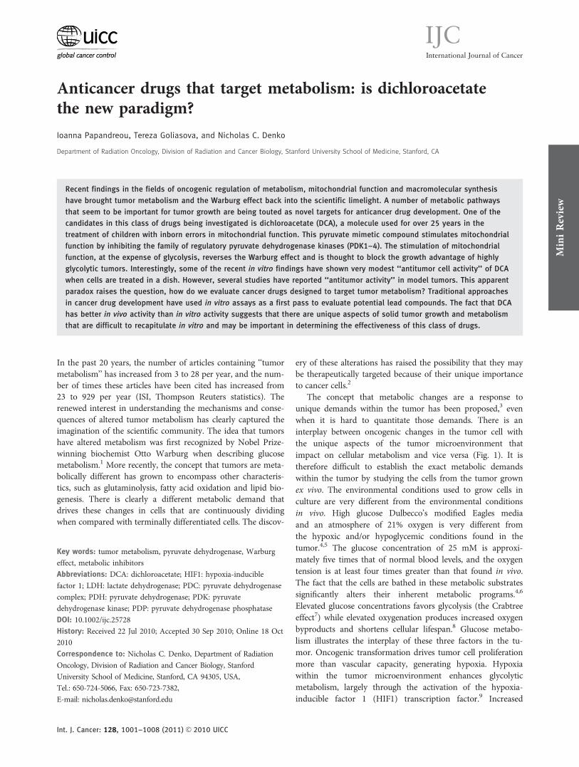
The concept that metabolic changes are a response tounique demands within the tumor has been proposed,3 evenwhen it is hard to quantitate those demands. There is aninterplay between oncogenic changes in the tumor cell withthe unique aspects of the tumor microenvironment thatimpact on cellular metabolism and vice versa (Fig. 1). It istherefore difficult to establish the exact metabolic demandswithin the tumor by studying the cells from the tumor grownex vivo. The environmental conditions used to grow cells inculture are very different from the environmental conditionsin vivo. High glucose Dulbecco’s modified Eagles mediaand an atmosphere of 21% oxygen is very different fromthe hypoxic and/or hypoglycemic conditions found in thetumor.4,5 The glucose concentration of 25 mM is approxi-mately five times that of normal blood levels, and the oxygentension is at least four times greater than that found in vivo.The fact that the cells are bathed in these metabolic substratessignificantly alters their inherent metabolic programs.4,6
Elevated glucose concentrations favors glycolysis (the Crabtreeeffect7) while elevated oxygenation produces increased oxygenbyproducts and shortens cellular lifespan.8 Glucose metabo-lism illustrates the interplay of these three factors in the tu-mor. Oncogenic transformation drives tumor cell proliferationmore than vascular capacity, generating hypoxia. Hypoxiawithin the tumor microenvironment enhances glycolyticmetabolism, largely through the activation of the hypoxia-inducible factor 1 (HIF1) transcription factor.9 Increased
Key words: tumor metabolism, pyruvate dehydrogenase, Warburg
effect, metabolic inhibitors
Abbreviations: DCA: dichloroacetate; HIF1: hypoxia-inducible
factor 1; LDH: lactate dehydrogenase; PDC: pyruvate dehydrogenase
complex; PDH: pyruvate dehydrogenase; PDK: pyruvate
dehydrogenase kinase; PDP: pyruvate dehydrogenase phosphatase
DOI: 10.1002/ijc.25728
History: Received 22 Jul 2010; Accepted 30 Sep 2010; Online 18 Oct
2010
Correspondence to: Nicholas C. Denko, Department of Radiation
Oncology, Division of Radiation and Cancer Biology, Stanford
University School of Medicine, Stanford, CA 94305, USA,
Tel.: 650-724-5066, Fax: 650-723-7382,
E-mail: [email protected]
MiniReview
Int. J. Cancer: 128, 1001–1008 (2011) VC 2010 UICC
International Journal of Cancer
IJC
glycolysis leads to increased production of lactate, which con-tributes to an acidic extracellular pH and further changes ingene expression.10 Both hypoxia and acidosis can contributeto increased levels of somatic mutation that can further drivetumor progression.11,12 It is clearly difficult to reproduce thesecomplex interactions in cells grown in vitro.
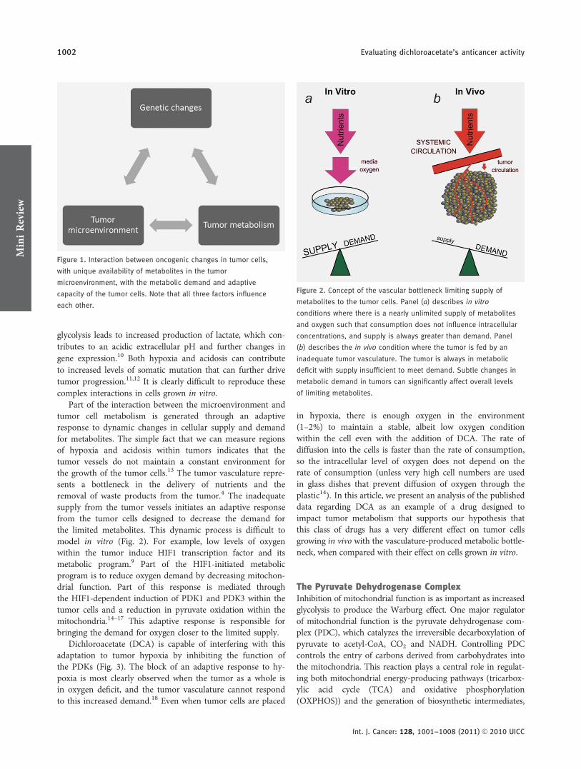
Part of the interaction between the microenvironment andtumor cell metabolism is generated through an adaptiveresponse to dynamic changes in cellular supply and demandfor metabolites. The simple fact that we can measure regionsof hypoxia and acidosis within tumors indicates that thetumor vessels do not maintain a constant environment forthe growth of the tumor cells.13 The tumor vasculature repre-sents a bottleneck in the delivery of nutrients and theremoval of waste products from the tumor.4 The inadequatesupply from the tumor vessels initiates an adaptive responsefrom the tumor cells designed to decrease the demand forthe limited metabolites. This dynamic process is difficult tomodel in vitro (Fig. 2). For example, low levels of oxygenwithin the tumor induce HIF1 transcription factor and itsmetabolic program.9 Part of the HIF1-initiated metabolicprogram is to reduce oxygen demand by decreasing mitochon-drial function. Part of this response is mediated throughthe HIF1-dependent induction of PDK1 and PDK3 within thetumor cells and a reduction in pyruvate oxidation within themitochondria.14–17 This adaptive response is responsible forbringing the demand for oxygen closer to the limited supply.
Dichloroacetate (DCA) is capable of interfering with thisadaptation to tumor hypoxia by inhibiting the function ofthe PDKs (Fig. 3). The block of an adaptive response to hy-poxia is most clearly observed when the tumor as a whole isin oxygen deficit, and the tumor vasculature cannot respondto this increased demand.18 Even when tumor cells are placed
in hypoxia, there is enough oxygen in the environment(1–2%) to maintain a stable, albeit low oxygen conditionwithin the cell even with the addition of DCA. The rate ofdiffusion into the cells is faster than the rate of consumption,so the intracellular level of oxygen does not depend on therate of consumption (unless very high cell numbers are usedin glass dishes that prevent diffusion of oxygen through theplastic14). In this article, we present an analysis of the publisheddata regarding DCA as an example of a drug designed toimpact tumor metabolism that supports our hypothesis thatthis class of drugs has a very different effect on tumor cellsgrowing in vivo with the vasculature-produced metabolic bottle-neck, when compared with their effect on cells grown in vitro.
The Pyruvate Dehydrogenase ComplexInhibition of mitochondrial function is as important as increasedglycolysis to produce the Warburg effect. One major regulatorof mitochondrial function is the pyruvate dehydrogenase com-plex (PDC), which catalyzes the irreversible decarboxylation ofpyruvate to acetyl-CoA, CO2 and NADH. Controlling PDCcontrols the entry of carbons derived from carbohydrates intothe mitochondria. This reaction plays a central role in regulat-ing both mitochondrial energy-producing pathways (tricarbox-ylic acid cycle (TCA) and oxidative phosphorylation(OXPHOS)) and the generation of biosynthetic intermediates,
Figure 1. Interaction between oncogenic changes in tumor cells,
with unique availability of metabolites in the tumor
microenvironment, with the metabolic demand and adaptive
capacity of the tumor cells. Note that all three factors influence
each other.
Figure 2. Concept of the vascular bottleneck limiting supply of
metabolites to the tumor cells. Panel (a) describes in vitro
conditions where there is a nearly unlimited supply of metabolites
and oxygen such that consumption does not influence intracellular
concentrations, and supply is always greater than demand. Panel
(b) describes the in vivo condition where the tumor is fed by an
inadequate tumor vasculature. The tumor is always in metabolic
deficit with supply insufficient to meet demand. Subtle changes in
metabolic demand in tumors can significantly affect overall levels
of limiting metabolites.
MiniReview
1002 Evaluating dichloroacetate’s anticancer activity
Int. J. Cancer: 128, 1001–1008 (2011) VC 2010 UICC
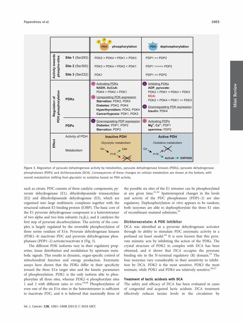
such as citrate. PDC consists of three catalytic components, py-ruvate dehydrogenase (E1), dihydrolipoamide transacetylase(E2) and dihydrolipoamide dehydrogenase (E3), which areorganized into large multimeric complexes together with thestructural subunit E3 binding protein (E3BP). The basic core ofthe E1 pyruvate dehydrogenase component is a heterotetramerof two alpha and two beta subunits (a2b2), and it catalyzes thefirst step of pyruvate decarboxylation. The activity of the com-plex is largely regulated by the reversible phosphorylation ofthree serine residues of E1a. Pyruvate dehydrogenase kinases(PDK1–4) inactivate PDC and pyruvate dehydrogenase phos-phatases (PDP1–2) activate/reactivate it (Fig. 3).
The different PDK isoforms vary in their regulatory prop-erties, tissue distribution and modulation by upstream meta-bolic signals. This results in dynamic, organ-specific control ofmitochondrial function and energy production. Enzymaticassays have shown that the PDKs differ in their specificitytoward the three E1a target sites and the kinetic parametersof phosphorylation. PDK1 is the only isoform able to phos-phorylate all three sites, whereas PDK2–4 phosphorylate sites1 and 2 with different rates in vitro.19,20 Phosphorylation ofeven one of the six E1a sites in the heterotetramer is sufficientto inactivate PDC, and it is believed that maximally three of
the possible six sites of the E1 tetramer can be phosphorylatedat any given time.21,22 Spatiotemporal changes in the levelsand activity of the PDC phosphatases (PDP1–2) are alsoregulatory. Dephosphorylation in vitro appears to be random;both isozymes are able to dephosphorylate the three E1 sitesof recombinant mutated substrates.23
Dichloroacetate: A PDK InhibitorDCA was identified as a pyruvate dehydrogenase activatorthrough its ability to stimulate PDC enzymatic activity in aperfused rat heart model.24 It is now known that this pyru-vate mimetic acts by inhibiting the action of the PDKs. Thecrystal structure of PDK2 in complex with DCA has beenobtained, and it shows that DCA occupies the pyruvatebinding site in the N-terminal regulatory (R) domain.25 Thefour isozymes vary considerably in their sensitivity to inhibi-tion by DCA. PDK2 is the most sensitive, PDK3 the mostresistant, while PDK1 and PDK4 are relatively sensitive.26,27
Treatment of lactic acidosis with DCA
The safety and efficacy of DCA has been evaluated in casesof congenital and acquired lactic acidosis. DCA treatmenteffectively reduces lactate levels in the circulation by
Figure 3. Regulation of pyruvate dehydrogenase activity by metabolites, pyruvate dehydrogenase kinases (PDKs), pyruvate dehydrogenase
phosphatases (PDPs) and dichloroacetate (DCA). Consequences of these changes on cellular metabolism are shown at the bottom, with
overall metabolism shifting from glycolytic to oxidative based on PDH activity.
MiniReview
Papandreou et al. 1003
Int. J. Cancer: 128, 1001–1008 (2011) VC 2010 UICC
stimulating oxidation of pyruvate; however, it remains notknown if DCA can improve the prognosis of patients withthese syndromes.28,29 It has been proposed that young chil-dren with PDH deficiency may benefit the most from chronicDCA treatment.30 The most significant adverse effect oflong-term DCA administration is a reversible peripheralneuropathy.31,32 The severity of the toxicity appears to be agedependent, with adult patients being more susceptible thanchildren.31,33 The reasons for this discrepancy are not entirelyclear, but they are possibly related to the different pharmacoki-netics and metabolism of DCA in the two age groups.34 DCAhas also been used in clinical trials for heart disease, includingcongestive heart failure and ischemic heart disease, showingpositive results and improving myocardial performance.35,36
DCA as a potential cancer therapeutic
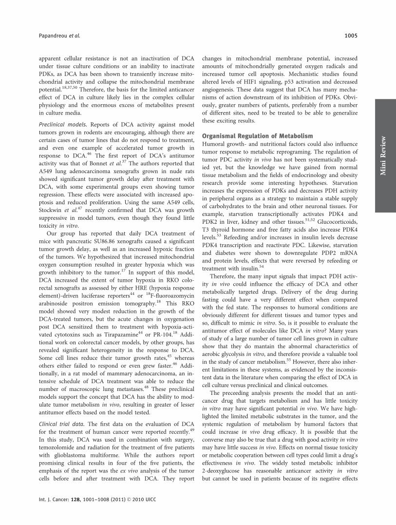
In recent years, DCA has attracted attention as a potentiallysimple and economical means to target glycolytic tumorswhile producing limited side effects in the oxidative healthyorgans. The interest in this drug by the scientific community,cancer patients and the media was kindled in 2007, after agroup from the University of Alberta reported that DCA wasuniquely toxic to human cancer cell lines and inhibited thegrowth of A549 lung tumor xenograft tumors in rats.37 Sincethen, the emerging reports on the efficacy of DCA in vitroand in vivo reveal some interesting and also puzzling charac-teristics, which distinguish the case of DCA from the majorityof drugs developed as anticancer agents (Table 1). The num-
ber of different cancer types and experimental strategies testedto date is too limited to allow for generalized conclusionsabout the efficacy of DCA against all kinds of tumors. Withthis caveat, a qualitative comparison of the literature suggeststhat DCA shows more anticancer effect in vivo thananticancer cell effect in vitro.
In vitro studies. DCA has been reported to have cytotoxiceffects in vitro,37–40 with some responses at clinically relevantconcentrations (0.5–1 mM), while others require supraphar-macologic levels (10–100 mM) and still other groups havefound no direct toxicity in vitro.18,41,42,47 One condition thathas been identified that sensitizes cells to DCA is mutationsthat perturb mitochondrial respiratory function,41,47 suggest-ing that enforced utilization of defective OXPHOS is toxic.As oxygen deprivation also downregulates mitochondrialfunction, it seemed reasonable to hypothesize that hypoxiccells would be more sensitive to DCA. However, this hypoth-esis has not been supported, at least in the limited number ofcell lines tested to date. Moderate in vitro hypoxia did notinfluence the cell cycle profile of DCA-treated colorectalcells43 or the reproductive viability of DCA-treated pancreaticcancer cells (our unpublished observations). Interestingly,another study found severe hypoxia (anoxia?) can be protec-tive against DCA-induced apoptosis in colorectal cancercells.46 The reason for these discrepancies is not clear.
Overall, the majority of the data support the idea thatclinically relevant concentrations of DCA (less than 1 mM)are not directly cytotoxic in vitro. The reason for this
Table 1. Summary of published in vitro, preclinical and clinical studies evaluating the anticancer effects of DCA. Some studies have bothin vitro and model tumors, such as Bonnet et al. 2007
Cancer type References Effect on survival and growth
In vitro studies
Lung, Glioblastoma, Breast 37 Apoptosis in vitro, xenograft growth inhibition
Prostate 38 Growth inhibition in vitro, moderate radiosensitization
Endometrial 39 Growth inhibition
Cervical 40 Growth advantage under hypoxia in vitro
Head and Neck 41 In vitro growth inhibition only in mutND2 overexpressing cells
Pediatric 42 Apoptosis at high concentrations in vitro, some influence on response to chemotherapy
Colorectal 43 Apoptosis at very high concentrations
Preclinical models
Colorectal 44 Little effect on growth, increased hypoxia by HRE-luciferase, sensitized to hypoxic cytotoxins
Colorectal 18 Little effect on growth, increased hypoxia by 18F-FAZA PET, sensitized to hypoxic cytotoxin
Pancreatic 17 Xenograft growth inhibition
Colorectal 45 Xenograft growth inhibition
Colorectal 46 Protected from anoxia in vitro, promoted xenograft growth of SW480
Colorectal, Breast, PML, Prostate 47 Active only against cells with defective electron transport chain
Breast 48 Inhibition of xenograft growth and metastasis
Human patient data
Glioblastoma 49 Clinically stable disease in vivo, decreased HIF1, increased p53 ex vivo
MiniReview
1004 Evaluating dichloroacetate’s anticancer activity
Int. J. Cancer: 128, 1001–1008 (2011) VC 2010 UICC
apparent cellular resistance is not an inactivation of DCAunder tissue culture conditions or an inability to inactivatePDKs, as DCA has been shown to transiently increase mito-chondrial activity and collapse the mitochondrial membranepotential.18,37,50 Therefore, the basis for the limited anticancereffect of DCA in culture likely lies in the complex cellularphysiology and the enormous excess of metabolites presentin culture media.
Preclinical models. Reports of DCA activity against modeltumors grown in rodents are encouraging, although there arecertain cases of tumor lines that do not respond to treatment,and even one example of accelerated tumor growth inresponse to DCA.46 The first report of DCA’s antitumoractivity was that of Bonnet et al.37 The authors reported thatA549 lung adenocarcinoma xenografts grown in nude ratsshowed significant tumor growth delay after treatment withDCA, with some experimental groups even showing tumorregression. These effects were associated with increased apo-ptosis and reduced proliferation. Using the same A549 cells,Stockwin et al.47 recently confirmed that DCA was growthsuppressive in model tumors, even though they found littletoxicity in vitro.
Our group has reported that daily DCA treatment ofmice with pancreatic SU86.86 xenografts caused a significanttumor growth delay, as well as an increased hypoxic fractionof the tumors. We hypothesized that increased mitochondrialoxygen consumption resulted in greater hypoxia which wasgrowth inhibitory to the tumor.17 In support of this model,DCA increased the extent of tumor hypoxia in RKO colo-rectal xenografts as assessed by either HRE (hypoxia responseelement)-driven luciferase reporters44 or 18F-fluoroazomycinarabinoside positron emission tomography.18 This RKOmodel showed very modest reduction in the growth of theDCA-treated tumors, but the acute changes in oxygenationpost DCA sensitized them to treatment with hypoxia-acti-vated cytotoxins such as Tirapazamine44 or PR-104.18 Addi-tional work on colorectal cancer models, by other groups, hasrevealed significant heterogeneity in the response to DCA.Some cell lines reduce their tumor growth rates,45 whereasothers either failed to respond or even grew faster.46 Addi-tionally, in a rat model of mammary adenocarcinoma, an in-tensive schedule of DCA treatment was able to reduce thenumber of macroscopic lung metastases.48 These preclinicalmodels support the concept that DCA has the ability to mod-ulate tumor metabolism in vivo, resulting in greater of lesserantitumor effects based on the model tested.
Clinical trial data. The first data on the evaluation of DCAfor the treatment of human cancer were reported recently.49
In this study, DCA was used in combination with surgery,temozolomide and radiation for the treatment of five patientswith glioblastoma multiforme. While the authors reportpromising clinical results in four of the five patients, theemphasis of the report was the ex vivo analysis of the tumorcells before and after treatment with DCA. They report
changes in mitochondrial membrane potential, increasedamounts of mitochondrially generated oxygen radicals andincreased tumor cell apoptosis. Mechanistic studies foundaltered levels of HIF1 signaling, p53 activation and decreasedangiogenesis. These data suggest that DCA has many mecha-nisms of action downstream of its inhibition of PDKs. Obvi-ously, greater numbers of patients, preferably from a numberof different sites, need to be treated to be able to generalizethese exciting results.
Organismal Regulation of MetabolismHumoral growth- and nutritional factors could also influencetumor response to metabolic reprograming. The regulation oftumor PDC activity in vivo has not been systematically stud-ied yet, but the knowledge we have gained from normaltissue metabolism and the fields of endocrinology and obesityresearch provide some interesting hypotheses. Starvationincreases the expression of PDKs and decreases PDH activityin peripheral organs as a strategy to maintain a stable supplyof carbohydrates to the brain and other neuronal tissues. Forexample, starvation transcriptionally activates PDK4 andPDK2 in liver, kidney and other tissues.51,52 Glucocorticoids,T3 thyroid hormone and free fatty acids also increase PDK4levels.53 Refeeding and/or increases in insulin levels decreasePDK4 transcription and reactivate PDC. Likewise, starvationand diabetes were shown to downregulate PDP2 mRNAand protein levels, effects that were reversed by refeeding ortreatment with insulin.54
Therefore, the many input signals that impact PDH activ-ity in vivo could influence the efficacy of DCA and othermetabolically targeted drugs. Delivery of the drug duringfasting could have a very different effect when comparedwith the fed state. The responses to humoral conditions areobviously different for different tissues and tumor types andso, difficult to mimic in vitro. So, is it possible to evaluate theantitumor effect of molecules like DCA in vitro? Many yearsof study of a large number of tumor cell lines grown in cultureshow that they do mantain the abnormal characteristics ofaerobic glycolysis in vitro, and therefore provide a valuable toolin the study of cancer metabolism.55 However, there also inher-ent limitations in these systems, as evidenced by the inconsis-tent data in the literature when comparing the effect of DCA incell culture versus preclinical and clinical outcomes.
The preceeding analysis presents the model that an anti-cancer drug that targets metabolism and has little toxicityin vitro may have significant potential in vivo. We have high-lighted the limited metabolic substrates in the tumor, and thesystemic regulation of metabolism by humoral factors thatcould increase in vivo drug efficacy. It is possible that theconverse may also be true that a drug with good activity in vitromay have little success in vivo. Effects on normal tissue toxicityor metabolic cooperation between cell types could limit a drug’seffectiveness in vivo. The widely tested metabolic inhibitor2-deoxyglucose has reasonable anticancer activity in vitrobut cannot be used in patients because of its negative effects
MiniReview
Papandreou et al. 1005
Int. J. Cancer: 128, 1001–1008 (2011) VC 2010 UICC

on normal tissues that rely on glucose consumption. Thedose-limiting neurologic toxicity occurs at a drug level wellbelow that needed for anticancer effects in rodent tumors.56,57
Alternatively, it is possible that metabolic cooperation betweencell types or normal and tumor cells may bypass the drug-induced metabolic block. For example, while lactate is oftenviewed as a metabolic waste product, it can be used in somecells as a fuel to power mitochondrial function.58,59
With respect to combining DCA with existing therapies,the preclinical data so far do not show an obvious pattern ofinteraction that would allow for an easy and rational selec-tion of therapeutic regiments. Animal models will continueto be the best means of testing to determine empirically themost promising combinations. We have shown that becauseof its ability to increase oxygen consumption, DCA increasedtumor hypoxia and sensitized xenografted pancreatic andcolon tumors to hypoxic cytotoxins,18,44 so it is intriguing toenvision a treatment plan including these two classes of drugsthat are both designed to exploit the unique hypoxic micro-environment of the tumor.
The interaction of DCA with other metabolic modulatorshas not been reported. A potential target for combinationtherapy is lactate dehydrogenase A (LDHA). Genetic or phar-macologic inhibition of LDHA has been shown to increasemitochondrial function and inhibit model tumors’ formationand progression.60,61 In this combination scheme, the LDHAinhibitor would block the conversion of pyruvate to lactateand DCA would divert the accumulated pyruvate toward mi-tochondrial oxidation. If DCA’s antitumor effect is derivedfrom increased mitochondrial function, it is possible thatcombining PDK and LDHA inhibitors would force an evengreater rate of mitochondrial oxidation and impair tumorgrowth more efficiently.
Nodal points of key survival pathways, such as the PI3K-Akt-mTOR pathway are also the subject of intense drugdevelopment efforts.62 Part of the growth-promoting proper-ties of this pathway comes from its ability to regulate metabo-lism and energy production by direct or indirect mechanisms.For example, oncogenic activation of PI3K-Akt stimulatesglucose uptake and aerobic glycolysis,63,64 whereas activation ofAkt and mTORC1 increases the translation of Hif-1a mRNAunder hypoxia.65 Promising Akt inhibitors and new generationmTOR inhibitors are being tested in clinical trials66,67 andrepresent viable candidates for combination therapy with DCAto modulate both aerobic and hypoxic metabolism.
ConclusionsIn conclusion, recent understanding about the unique metab-olism of the solid tumor has identified several novel, drug-gable pathways that may be preferentially used in tumor cellscompared with normal cells. Analysis of candidate anticancerdrugs designed to target these metabolic pathways willrequire careful experimental design, both in vitro and in vivo.Analysis of the published reports studying DCA shows a con-fusing, and sometime contradictory, range of in vitro andin vivo effects. Genetic studies in model tumors offer compel-ling evidence that this pathway is a good candidate for thera-peutic targeting.68 It would be very helpful in the analysis ofthe potential utility of DCA if there could be some molecularsignature that could predict for drug senstitivity, both inmodel tumors and eventually in patients. Perhaps a carefulanalysis of the presumed target of DCA, the phosphorylationof pyruvate dehydrogenase E1a subunit may offer thissignature.
References
1. Warburg O, Wind F, Negelein E. Themetabolism of tumors in the body. J GenPhysiol 1927;8:519–30.
2. Pan JG, Mak TW. Metabolic targeting asan anticancer strategy: dawn of a new era?Sci STKE 2007;2007:pe14.
3. Deberardinis RJ, Sayed N, Ditsworth D,Thompson CB. Brick by brick: metabolismand tumor cell growth. Curr Opin GenetDev 2008;18:54–61.
4. Vaupel P. Tumor microenvironmentalphysiology and its implications forradiation oncology. Semin Radiat Oncol2004;14:198–206.
5. Walenta S, Chau TV, Schroeder T, LehrHA, Kunz-Schughart LA, Fuerst A,Mueller-Klieser W. Metabolic classificationof human rectal adenocarcinomas: a novelguideline for clinical oncologists? J CancerRes Clin Oncol 2003;129:321–6.
6. Gstraunthaler G, Seppi T, Pfaller W.Impact of culture conditions, culture mediavolumes, and glucose content on metabolic
properties of renal epithelial cell cultures.Are renal cells in tissue culture hypoxic?Cell Physiol Biochem 1999;9:150–72.
7. Bloch-Frankenthal L, Ram D. Therelationship between the Crabtree effectand the oxidative metabolism of glucoseand carbohydrate intermediates in ascitestumor cells. Cancer Res 1959;19:835–42.
8. Parrinello S, Samper E, Krtolica A,Goldstein J, Melov S, Campisi J. Oxygensensitivity severely limits the replicativelifespan of murine fibroblasts. Nat Cell Biol2003;5:741–7.
9. Denko NC. Hypoxia, HIF1 and glucosemetabolism in the solid tumour. Nat RevCancer 2008;8:705–13.
10. Chen JL, Lucas JE, Schroeder T, Mori S,Wu J, Nevins J, Dewhirst M, West M, ChiJT. The genomic analysis of lactic acidosisand acidosis response in human cancers.PLoS Genet 2008;4:e1000293.
11. Bindra RS, Gibson SL, Meng A,Westermark U, Jasin M, Pierce AJ, Bristow
RG, Classon MK, Glazer PM. Hypoxia-induced down-regulation of BRCA1expression by E2Fs. Cancer Res 2005;65:11597–604.
12. Gatenby RA, Smallbone K, Maini PK, RoseF, Averill J, Nagle RB, Worrall L, GilliesRJ. Cellular adaptations to hypoxia andacidosis during somatic evolution of breastcancer. Br J Cancer 2007;97:646–53.
13. Dewhirst MW, Cao Y, Moeller B. Cyclinghypoxia and free radicals regulateangiogenesis and radiotherapy response.Nat Rev Cancer 2008;8:425–37.
14. Papandreou I, Cairns RA, Fontana L, LimAL, Denko NC. HIF-1 mediates adaptationto hypoxia by actively downregulatingmitochondrial oxygen consumption. CellMetab 2006;3:187–97.
15. Lu CW, Lin SC, Chen KF, Lai YY, Tsai SJ.Induction of pyruvate dehydrogenasekinase-3 by hypoxia-inducible factor-1promotes metabolic switch and drugresistance. J Biol Chem 2008;283:28106–14.
MiniReview
1006 Evaluating dichloroacetate’s anticancer activity
Int. J. Cancer: 128, 1001–1008 (2011) VC 2010 UICC
16. Kim JW, Tchernyshyov I, Semenza GL,Dang CV. HIF-1-mediated expression ofpyruvate dehydrogenase kinase: a metabolicswitch required for cellular adaptation tohypoxia. Cell Metab 2006;3:177–85.
17. Chen Y, Cairns R, Papandreou I, Koong A,Denko NC. Oxygen consumption canregulate the growth of tumors, a newperspective on the Warburg effect. PLoSOne 2009;4:e7033.
18. Cairns RA, Bennewith KL, Graves EE,Giaccia AJ, Chang DT, Denko NC.Pharmacologically increased tumor hypoxiacan be measured by 18F-fluoroazomycinarabinoside positron emission tomographyand enhances tumor response to hypoxiccytotoxin PR-104. Clin Cancer Res 2009;15:7170–4.
19. Kolobova E, Tuganova A, Boulatnikov I,Popov KM. Regulation of pyruvatedehydrogenase activity throughphosphorylation at multiple sites. BiochemJ 2001;358:69–77.
20. Korotchkina LG, Patel MS. Site specificityof four pyruvate dehydrogenase kinaseisoenzymes toward the threephosphorylation sites of human pyruvatedehydrogenase. J Biol Chem 2001;276:37223–9.
21. Sugden PH, Randle PJ. Regulation of pigheart pyruvate dehydrogenase byphosphorylation. Studies on the subunitand phosphorylation stoicheiometries.Biochem J 1978;173:659–68.
22. Korotchkina LG, Patel MS. Mutagenesisstudies of the phosphorylation sites ofrecombinant human pyruvatedehydrogenase. Site-specific regulation.J Biol Chem 1995;270:14297–304.
23. Karpova T, Danchuk S, Kolobova E, PopovKM. Characterization of the isozymes ofpyruvate dehydrogenase phosphatase:implications for the regulation of pyruvatedehydrogenase activity. Biochim BiophysActa 2003;1652:126–35.
24. Whitehouse S, Randle PJ. Activation ofpyruvate dehydrogenase in perfused ratheart by dichloroacetate (ShortCommunication). Biochem J 1973;134:651–3.
25. Knoechel TR, Tucker AD, Robinson CM,Phillips C, Taylor W, Bungay PJ, KastenSA, Roche TE, Brown DG. Regulatory rolesof the N-terminal domain based on crystalstructures of human pyruvatedehydrogenase kinase 2 containingphysiological and synthetic ligands.Biochemistry 2006;45:402–15.
26. Bowker-Kinley MM, Davis WI, Wu P,Harris RA, Popov KM. Evidence forexistence of tissue-specific regulation of themammalian pyruvate dehydrogenasecomplex. Biochem J 1998;329(Pt 1):191–6.
27. Baker JC, Yan X, Peng T, Kasten S, RocheTE. Marked differences between two
isoforms of human pyruvatedehydrogenase kinase. J Biol Chem 2000;275:15773–81.
28. Stacpoole PW, Greene YJ. Dichloroacetate.Diabetes Care 1992;15:785–91.
29. Stacpoole PW, Harman EM, Curry SH,Baumgartner TG, Misbin RI. Treatment oflactic acidosis with dichloroacetate. N EnglJ Med 1983;309:390–6.
30. Stacpoole PW, Kurtz TL, Han Z, LangaeeT. Role of dichloroacetate in the treatmentof genetic mitochondrial diseases. AdvDrug Deliv Rev 2008;60:1478–87.
31. Kaufmann P, Engelstad K, Wei Y, Jhung S,Sano MC, Shungu DC, Millar WS, HongX, Gooch CL, Mao X, Pascual JM, HiranoM, et al. Dichloroacetate causes toxicneuropathy in MELAS: a randomized,controlled clinical trial. Neurology 2006;66:324–30.
32. Stacpoole PW, Henderson GN, Yan Z,Cornett R, James MO. Pharmacokinetics,metabolism and toxicology ofdichloroacetate. Drug Metab Rev 1998;30:499–539.
33. Stacpoole PW, Gilbert LR, Neiberger RE,Carney PR, Valenstein E, Theriaque DW,Shuster JJ. Evaluation of long-termtreatment of children with congenital lacticacidosis with dichloroacetate. Pediatrics2008;121:e1223–8.
34. Shroads AL, Guo X, Dixit V, Liu HP,James MO, Stacpoole PW. Age-dependentkinetics and metabolism of dichloroacetate:possible relevance to toxicity. J PharmacolExp Ther 2008;324:1163–71.
35. Wargovich TJ, MacDonald RG, Hill JA,Feldman RL, Stacpoole PW, Pepine CJ.Myocardial metabolic and hemodynamiceffects of dichloroacetate in coronary arterydisease. Am J Cardiol 1988;61:65–70.
36. Bersin RM, Wolfe C, Kwasman M, Lau D,Klinski C, Tanaka K, Khorrami P,Henderson GN, de Marco T, Chatterjee K.Improved hemodynamic function andmechanical efficiency in congestive heartfailure with sodium dichloroacetate. J AmColl Cardiol 1994;23:1617–24.
37. Bonnet S, Archer SL, Allalunis-Turner J,Haromy A, Beaulieu C, Thompson R, LeeCT, Lopaschuk GD, Puttagunta L, BonnetS, Harry G, Hashimoto K, et al. Amitochondria-Kþ channel axis issuppressed in cancer and its normalizationpromotes apoptosis and inhibits cancergrowth. Cancer Cell 2007;11:37–51.
38. Cao W, Yacoub S, Shiverick KT, NamikiK, Sakai Y, Porvasnik S, Urbanek C, RosserCJ. Dichloroacetate (DCA) sensitizes bothwild-type and over expressing Bcl-2prostate cancer cells in vitro to radiation.Prostate 2008;68:1223–31.
39. Wong JY, Huggins GS, Debidda M,Munshi NC, De Vivo I. Dichloroacetateinduces apoptosis in endometrial cancer
cells. Gynecol Oncol 2008;109:394–402.
40. Anderson KM, Jajeh J, Guinan P,Rubenstein M. In vitro effects ofdichloroacetate and CO2 on hypoxic HeLacells. Anticancer Res 2009;29:4579–88.
41. Sun W, Zhou S, Chang SS, McFate T,Verma A, Califano JA. Mitochondrialmutations contribute to HIF1alphaaccumulation via increased reactive oxygenspecies and up-regulated pyruvatedehydrogenease kinase 2 in head and necksquamous cell carcinoma. Clin Cancer Res2009;15:476–84.
42. Heshe D, Hoogestraat S, Brauckmann C,Karst U, Boos J, Lanvers-Kaminsky C.Dichloroacetate metabolically targetedtherapy defeats cytotoxicity of standardanticancer drugs. Cancer ChemotherPharmacol 2010.
43. Madhok BM, Yeluri S, Perry SL, HughesTA, Jayne DG. Dichloroacetate inducesapoptosis and cell-cycle arrest in colorectalcancer cells. Br J Cancer 2010;102:1746–52.
44. Cairns RA, Papandreou I, Sutphin PD,Denko NC. Metabolic targeting of hypoxiaand HIF1 in solid tumors can enhancecytotoxic chemotherapy. Proc Natl AcadSci USA 2007;104:9445–50.
45. Sanchez-Arago M, Chamorro M, CuezvaJM. Selection of cancer cells with repressedmitochondria triggers colon cancerprogression. Carcinogenesis 2010;31:567–76.
46. Shahrzad S, Lacombe K, Adamcic U,Minhas K, Coomber BL. Sodiumdichloroacetate (DCA) reduces apoptosis incolorectal tumor hypoxia. Cancer Lett2010;287:75–83.
47. Stockwin LH, Yu SX, Borgel S, Hancock C,Wolfe TL, Phillips LR, Hollingshead MG,Newton DL. Sodium dichloroacetate(DCA) selectively targets cells with defectsin the mitochondrial ETC. Int J Cancer2010;127:2510–19.
48. Sun RC, Fadia M, Dahlstrom JE, ParishCR, Board PG, Blackburn AC. Reversal ofthe glycolytic phenotype by dichloroacetateinhibits metastatic breast cancer cellgrowth in vitro and in vivo. Breast CancerRes Treat 2010;120:253–60.
49. Michelakis ED, Sutendra G, Dromparis P,Webster L, Haromy A, Niven E, MaguireC, Gammer TL, Mackey JR, Fulton D,Abdulkarim B, McMurtry MS, et al.Metabolic modulation of glioblastoma withdichloroacetate. Sci Transl Med 2010;2:31ra34.
50. Rardin MJ, Wiley SE, Naviaux RK,Murphy AN, Dixon JE. Monitoringphosphorylation of the pyruvatedehydrogenase complex. Anal Biochem2009;389:157–64.
51. Holness MJ, Sugden MC. Pyruvatedehydrogenase activities during thefed-to-starved transition and on re-feeding
MiniReview
Papandreou et al. 1007
Int. J. Cancer: 128, 1001–1008 (2011) VC 2010 UICC
after acute or prolonged starvation.Biochem J 1989;258:529–33.
52. Huang B, Wu P, Bowker-Kinley MM,Harris RA. Regulation of pyruvatedehydrogenase kinase expression byperoxisome proliferator-activated receptor-alpha ligands, glucocorticoids, and insulin.Diabetes 2002;51:276–83.
53. Attia RR, Connnaughton S, Boone LR,Wang F, Elam MB, Ness GC, Cook GA,Park EA. Regulation of pyruvatedehydrogenase kinase 4 (PDK4) by thyroidhormone: role of the peroxisomeproliferator-activated receptor gammacoactivator (PGC-1 alpha). J Biol Chem2010;285:2375–85.
54. Huang B, Wu P, Popov KM, Harris RA.Starvation and diabetes reduce the amountof pyruvate dehydrogenase phosphatase inrat heart and kidney. Diabetes 2003;52:1371–6.
55. Wu M, Neilson A, Swift AL, Moran R,Tamagnine J, Parslow D, Armistead S,Lemire K, Orrell J, Teich J, Chomicz S,Ferrick DA. Multiparameter metabolicanalysis reveals a close link betweenattenuated mitochondrial bioenergeticfunction and enhanced glycolysisdependency in human tumor cells. AmJ Physiol Cell Physiol 2007;292:C125–36.
56. Singh D, Banerji AK, Dwarakanath BS,Tripathi RP, Gupta JP, Mathew TL,Ravindranath T, Jain V. Optimizing cancerradiotherapy with 2-deoxy-d-glucose doseescalation studies in patients withglioblastoma multiforme. StrahlentherOnkol 2005;181:507–14.
57. Maher JC, Krishan A, Lampidis TJ. Greatercell cycle inhibition and cytotoxicityinduced by 2-deoxy-D-glucose in tumorcells treated under hypoxic vs aerobicconditions. Cancer Chemother Pharmacol2004;53:116–22.
58. Sonveaux P, Vegran F, Schroeder T,Wergin MC, Verrax J, Rabbani ZN, DeSaedeleer CJ, Kennedy KM, Diepart C,Jordan BF, Kelley MJ, Gallez B, et al.Targeting lactate-fueled respirationselectively kills hypoxic tumor cells inmice. J Clin Invest 2008;118:3930–42.
59. Koukourakis MI, Giatromanolaki A, HarrisAL, Sivridis E. Comparison of metabolicpathways between cancer cells and stromalcells in colorectal carcinomas: a metabolicsurvival role for tumor-associated stroma.Cancer Res 2006;66:632–7.
60. Fantin VR, St-Pierre J, Leder P.Attenuation of LDHA expression uncoversa link between glycolysis, mitochondrialphysiology, and tumor maintenance.Cancer Cell 2006;9:425–34.
61. Le A, Cooper CR, Gouw AM, Dinavahi R,Maitra A, Deck LM, Royer RE, Vander JagtDL, Semenza GL, Dang CV. Inhibition oflactate dehydrogenase A induces oxidativestress and inhibits tumor progression. ProcNatl Acad Sci USA 2010;107:2037–42.
62. Mitsiades CS, Mitsiades N, Koutsilieris M.The Akt pathway: molecular targets foranti-cancer drug development. Curr CancerDrug Targets 2004;4:235–56.
63. Elstrom RL, Bauer DE, Buzzai M,Karnauskas R, Harris MH, Plas DR,Zhuang H, Cinalli RM, Alavi A, Rudin
CM, Thompson CB. Akt stimulates aerobicglycolysis in cancer cells. Cancer Res 2004;64:3892–9.
64. Buzzai M, Bauer DE, Jones RG,Deberardinis RJ, Hatzivassiliou G,Elstrom RL, Thompson CB. The glucosedependence of Akt-transformed cells canbe reversed by pharmacologic activation offatty acid beta-oxidation. Oncogene 2005;24:4165–73.
65. Laughner E, Taghavi P, Chiles K, MahonPC, Semenza GL. HER2 (neu) signalingincreases the rate of hypoxia-induciblefactor 1alpha (HIF-1alpha) synthesis: novelmechanism for HIF-1-mediated vascularendothelial growth factor expression.Mol Cell Biol 2001;21:3995–4004.
66. Ghobrial IM, Gertz M, Laplant B,Camoriano J, Hayman S, Lacy M, ChumaS, Harris B, Leduc R, Rourke M, AnsellSM, Deangelo D, et al. Phase II trial of theoral mammalian target of rapamycininhibitor everolimus in relapsed orrefractory Waldenstrommacroglobulinemia. J Clin Oncol2010;28:1408–14.
67. Meric-Bernstam F, Gonzalez-Angulo AM.Targeting the mTOR signaling network forcancer therapy. J Clin Oncol 2009;27:2278–87.
68. McFate T, Mohyeldin A, Lu H, Thakar J,Henriques J, Halim ND, Wu H, Schell MJ,Tsang TM, Teahan O, Zhou S, CalifanoJA, et al. Pyruvate dehydrogenase complexactivity controls metabolic and malignantphenotype in cancer cells. J Biol Chem2008;283:22700–8.
MiniReview
1008 Evaluating dichloroacetate’s anticancer activity
Int. J. Cancer: 128, 1001–1008 (2011) VC 2010 UICC