Embed Size (px)
Citation preview

4162 | Phys. Chem. Chem. Phys., 2019, 21, 4162--4175 This journal is© the Owner Societies 2019
Cite this:Phys.Chem.Chem.Phys.,
2019, 21, 4162
Anticancer drug impact on DNA – a studyby neutron spectroscopy coupled withsynchrotron-based FTIR and EXAFS†
Ana L. M. Batista de Carvalho, a Adriana P. Mamede, a Asha Dopplapudi,b
Victoria Garcia Sakai,b James Doherty,cd Mark Frogley,c Gianfelice Cinque,c
Peter Gardner, d Diego Gianolio,c Luı́s A. E. Batista de Carvalho *a andM. Paula M. Marques ae
Complementary structural and dynamical information on drug–DNA interplay has been achieved at a molecular
level, for Pt/Pd-drugs, allowing a better understanding of their pharmacodynamic profile which is crucial for the
development of improved chemotherapeutic agents. The interaction of two cisplatin-like dinuclear Pt(II) and Pd(II)
complexes with DNA was studied through a multidisciplinary experimental approach, using quasi-elastic neutron
scattering (QENS) techniques coupled with synchrotron-based extended X-ray absorption fine structure
(SR-EXAFS) and Fourier-Transform Infrared Spectroscopy-Attenuated Total Reflectance (SR-FTIR-ATR).
DNA extracted from drug-exposed human triple negative breast cancer cells (MDA-MB-231) was used, with a
view to evaluate the effect of the unconventional antineoplastic agents on this low prognosis type of cancer.
The drug impact on DNA’s dynamical profile, via its hydration layer, was provided by QENS, a drug-triggered
enhanced mobility having been revealed. Additionally, an onset of anharmonicity was detected for dehydrated
DNA, at room temperature. Far- and mid-infrared measurements allowed the first simultaneous detection of
the drugs and their primary pharmacological target, as well as the drug-prompted changes in DNA’s
conformation that mediate cytotoxicity. The local environment of the absorbing Pd(II) and Pt(II) centers in the
drugs’ adducts with adenine, guanine and glutathione was attained by EXAFS.
Introduction
Cancer is a leading cause of death worldwide, with a growingincidence: 12.7 million people per year are diagnosed withcancer, from which more than 50% die from the disease. Earlydiagnosis and chemotherapeutic treatment can lower thismortality rate by 30 to 40%, and may lead to the cure of ca.30% of the patients. Anticancer platinum drugs have beenintroduced in clinics since the serendipitous discovery ofcisplatin (cis-dichlorodiamine platinum(II), cis-(NH3)2PtCl2) inthe 1970s, which was the first inorganic compound displaying
high antineoplastic activity towards human tumours.1,2 The threecurrently approved platinum agents – cisplatin, carboplatin (cis-diamine(1,1-cyclobutanedicarboxylato)platinum(II)) and oxaliplatin([(1R,2R)-cyclohexane-1,2-diamine](ethanedioato-O,O0)platinum(II)) –are applied worldwide in chemotherapeutic regimes. However, theirclinical application is still restricted by dose-limiting deleterious sideeffects and acquired resistance upon prolonged administration,3
as well as by a lack of specificity against several cancer types(e.g. metastatic). Hence, considerable effort has been put into thedevelopment of novel metal-based drugs, including cisplatin-likeagents, aiming at an improved antitumor efficiency.4–6 Among these,polynuclear multifunctional Pt(II) and Pd(II) chelates with flexiblebiogenic polyamines as bridging ligands have been synthetized bythe authors and evaluated as to their effect on several types ofhuman neoplasias, namely the low prognosis, highly metastatictriple negative breast cancer against which a Pd(II) agent (Pd2Spm,Spm = spermine, H2N(CH2)3NH(CH2)4NH(CH2)3NH2) has yieldedparticularly promising results.7
A number of studies have been conducted in the last fewyears on these Pt- and Pd-based complexes, reporting confor-mational profiles8–11 and pharmacokinetic/pharmacodynamicbehaviour,12,13 and the effect on different human cancers7,14–22
a Quı́mica-Fı́sica Molecular, Department of Chemistry, University of Coimbra,
3004-535 Coimbra, Portugal. E-mail: [email protected] ISIS Facility, STFC Rutherford Appleton Laboratory, Chilton, Didcot,
Oxfordshire OX11 0QX, UKc Diamond Light Source, Harwell Science and Innovation Campus, Chilton, Didcot,
Oxfordshire OX11 0DE, UKd Manchester Institute of Biotechnology, University of Manchester, Manchester,
M1 7DN, UKe Department of Life Sciences, University of Coimbra, 3000-456 Coimbra, Portugal
† Electronic supplementary information (ESI) available. See DOI: 10.1039/c8cp05881d
Received 18th September 2018,Accepted 2nd January 2019
DOI: 10.1039/c8cp05881d
rsc.li/pccp
PCCP
PAPER
Ope
n A
cces
s A
rtic
le. P
ublis
hed
on 0
3 Ja
nuar
y 20
19. D
ownl
oade
d on
3/2
3/20
22 7
:02:
22 P
M.
Thi
s ar
ticle
is li
cens
ed u
nder
a C
reat
ive
Com
mon
s A
ttrib
utio
n 3.
0 U
npor
ted
Lic
ence
.
View Article OnlineView Journal | View Issue
This journal is© the Owner Societies 2019 Phys. Chem. Chem. Phys., 2019, 21, 4162--4175 | 4163
including their impact on the cellular biochemical profile.23–25
Unconventional pathways of cytotoxicity were disclosed, whichare recognized to lead to an improved therapeutic efficiencycoupled with a lower toxicity. In addition, the data gathered sofar suggest distinct mechanisms of action for the Pt(II) versusthe Pd(II) agents, as well as for the dinuclear polyamine chelatesrelative to the mononuclear lead drug cisplatin. The activity ofthis kind of agents was found to be mediated by selectivecovalent binding of the metal centres to DNA bases (mainlythe purines at their most nucleophilic nitrogen atom, N7),yielding long-range intra- and interstrand adducts responsiblefor cell growth arrest and apoptotic death. Adducts with DNAare formed upon drug activation, according to the followingsteps: transport into the cell, hydrolysis (in the cytoplasm) bysequential loss of the chloride ligands and aquation, uptakeinto the nucleus and reaction of the unstable diaquo specieswith DNA (the drug’s main pharmacological target).26 However,apart from this interplay with DNA, during the pharmacokineticstage drug interaction may also occur with amine and sulfur groupsfrom proteins and other cellular constituents. In particular,endogenous thiols such as glutathione (L-g-glutamyl-cysteinyl-glycine (Glu-Cys-Gly), GSH, a key cellular antioxidant) are knownto intercept this type of agents owing to the high affinity of thesoft Pt(II) and Pd(II) cations towards S-donor ligands,27,28 whichmay significantly lower the drug’s bioavailability at the maintarget and lead to acquired resistance. Since the mechanismsunderlying drug resistance as well as the molecular basis ofcytotoxicity are still not fully understood for these DNA-damagingagents, the present study aims at contributing to their elucidationby monitoring the interaction of spermine Pt(II) and Pd(II) com-plexes (Pt2Spm and Pd2Spm) with DNA.
DNA extracted from human triple negative breast cancercells (MDA-MB-231 cell line) and commercial DNA were used astarget models, while cisplatin was taken as a drug reference.Drug concentrations and incubation times were chosen accordingto previous results, corresponding to an optimal cytotoxic effecttowards this particular neoplastic cell line:7 4 and 8 mM (up to ca.the 2 � IC50 level), for a 48 h exposure time. A multidisciplinaryexperimental approach was followed, through the application of:(i) quasi-elastic neutron scattering (QENS), suitable for directlyaccessing different spatially resolved dynamical processes (at asubnanometer lengthscale and subnanosecond timescale), underdistinct conditions, mainly for hydrogen-rich systems;25,29–31
(ii) synchrotron-radiation Fourier-Transform Infrared Spectroscopy-Attenuated Total Reflectance (SR-FTIR-ATR), recognized as acutting-edge non-destructive tool for obtaining spectral signaturesof molecular components in biological samples, so as to relatestructural to functional data;32,33 and (iii) synchrotron-basedExtended X-ray Absorption Fine Structure (EXAFS) and X-rayAbsorption Near-Edge Structure (XANES), which are methods ofchoice for obtaining detailed information on the local structure ofbioinorganic non-crystalline materials.34,35
Water supports vital biochemical processes in living organisms,and is responsible for the maintenance of the functional three-dimensional architecture of biopolymers through a tight interplaywithin their hydration shells.36 Actually, mobility changes in these
hydration layers may affect the biopolymer’s conformational anddynamical profiles, which rule biofunctionality. It is a well-recognized fact that water within hydration sheets is retarded withrespect to the bulk, although its dynamical properties are not yetcompletely understood. Hence, elucidation of water dynamics inbiological systems and its impact on activity and function is ofthe utmost relevance in drug development, for an improvedunderstanding of a drugs’ mode of action via interaction withall its possible pharmacological targets that may includethe water molecules wrapping their conventional receptors.Neutron techniques such as high resolution quasielastic scatteringare particularly suited for selectively probing water dynamicalbehaviour, namely within biomolecular hydration shells, on anano- to picosecond timescale (ca. 10�9 to 10�13 s) and a 1 to30 Å lengthscale (corresponding to inter- and intramoleculardistances, e.g. H-bonding),37 yielding results not achievableby any other methods. QENS measurements at differenttemperatures allow one to characterize the translational androtational modes of water hydrogens within a biological matrix(e.g. biomolecules, cells or even tissues), and determine howthis dynamical behaviour may be disturbed by the presence ofan external entity such as a drug. A few QENS experiments onwater dynamics in living cells have been reported,29 but drugeffects were first investigated by the authors in cisplatin-exposed human cancer cells using QENS and inelastic neutronscattering spectroscopy (INS).25
FTIR spectroscopy is an extremely powerful, sensitive andnon-invasive analytical tool for interrogating the chemicalcomposition of biological systems, delivering unique spectralsignatures for a particular biomolecule at specific conditions.The synchrotron beam available at the MIRIAM (MultimodeInfraRed Imaging And Microspectroscopy) beamline from theDiamond Light Source (DLS, United Kingdom), currently used,provides stable, broadband and extremely bright IR radiationand delivers FTIR data with an unmatched signal-to-noise ratio.In addition, MIRIAM spans the largest infrared spectral range –extending from the near up to the far-IR (or THz) region – and isone to two orders of magnitude brighter in the mid-/far-IR thanany other conventional thermal IR source. Furthermore, theFTIR attenuated total reflection (ATR) mode used in the presentmeasurements allows one to directly probe DNA without anyparticular sample preparation, with the added advantages ofhigh spatial resolution and an absence of resonant Mie scatteringeffects on the collected spectral data.38 In particular, probing thefar-infrared region (o300 cm�1) enabled us to detect H-bonds andweak non-bonding interactions within the nucleic acid, with veryhigh specificity, thus unveiling specific drug-induced conforma-tional changes that underlie cytotoxicity.
EXAFS, in turn, allows the direct observation of metalcoordination and elucidation of the local environment of theabsorbing metal centre in inorganic compounds, particularly whengood quality crystals are unavailable, and has been successfullyapplied to Pt(II) compounds specifically for assessing their degrada-tion in the presence of S-containing molecules.34,35 In the presentstudy, synchrotron-based EXAFS/XANES measurements yieldeddetailed information on the first coordination shell of Pd(II) and
Paper PCCP
Ope
n A
cces
s A
rtic
le. P
ublis
hed
on 0
3 Ja
nuar
y 20
19. D
ownl
oade
d on
3/2
3/20
22 7
:02:
22 P
M.
Thi
s ar
ticle
is li
cens
ed u
nder
a C
reat
ive
Com
mon
s A
ttrib
utio
n 3.
0 U
npor
ted
Lic
ence
.View Article Online
4164 | Phys. Chem. Chem. Phys., 2019, 21, 4162--4175 This journal is© the Owner Societies 2019
Pt(II) within the drugs’ adducts with adenine (A), guanine (G) andglutathione (GSH).
Complementary structural and dynamical information wastherefore achieved for the drug–DNA systems presently studied.These results provide a more comprehensive understanding of thepharmacodynamic profile of the dinuclear Pt- and Pd-anticanceragents, at a molecular level, particularly regarding their effect onvital biomolecules through an impact on their hydration water,apart from direct perturbation of their native conformation. Thisknowledge is paramount for rational design of novel metal-basedanticancer compounds with an enhanced chemotherapeutic effi-ciency coupled with lower deleterious side effects.
Experimental
The list of chemicals, the experimental details regarding the synthesisand characterization of the Pt2Spm and Pd2Spm complexes, thepreparation of drug solutions, and the cell culture protocol areextensively described in the ESI,† as well as the pre-processing andanalysis procedures of the QENS, FTIR-ATR and EXAFS/XANES data.
Synthesis and characterization of the drug–purine anddrug–glutathione adducts
Drug–purine adducts. The (1 : 4) drug–adenine and drug–guanine adducts (MA4 and MG4, M = Pt(II) or Pd(II)) weresynthesized following a synthetic route previously optimizedby the authors39,40 (see details in the ESI,† Fig. S1 and S2).
Drug–glutathione adducts. In order to prepare (1 : 4) drug–GSH adducts, 4 molar equivalents of glutathione were added toa solution of each of the drugs – Pt2Spm-0.45 mM or Pd2Spm-0.25 mM – and stirring was maintained in the dark, at roomtemperature, for 24 h. The initial colourless reaction mixturebecame orange over time (a characteristic colour of complexescontaining Pt–S covalent bonds). The resulting drug–GS4 solu-tions (MGS4, M = Pt(II) or Pd(II)) were then concentrated byrotary evaporation under vacuum and lyophilized to obtain anorange powder.
Titration of drug–purine adducts with glutathione, adenineor guanine. The Mbase4 (M = Pt(II) or Pd(II), base = A or G)adducts were titrated with GSH. A glutathione solution wasadded, in a (1 : 4) molar ratio, to an aqueous solution of 10 mgof each Mbase4 adduct. The reaction mixtures were kept in thedark with stirring, at 30 1C for 24 hours, after which they wereconcentrated under vacuum at 30 1C until precipitation occurredand then vacuum filtered through a 0.22 mm filter to removeprecipitated adenine or guanine. Finally, they were concentratedunder vacuum (at 30 1C) and lyophilized to yield the solidproducts (hereafter denominated MA4 + GSH and MG4 + GSH).
Preparation of cell pellets for QENS measurements
Cell pellets (100 mg/1 cm3, ca. 5 � 108 cells per sample) wereprepared by cell harvesting (through trypsinisation) followed byrepeated (2�) PBS washing and centrifugation (at 195 � g, for15 min). PBS was used as an isotonic medium in order to avoidwater exchange from the inside to the outside of the cell
(leading to cell shrinkage). The drugs (cisplatin, Pt2Spm andPd2Spm at 4 and 8 mM) were added to the cells during theirlogarithmic phase of growth, and left to incubate for 48 h. Inorder to completely remove the extracellular water component(less than 5%), the cell pellets were washed with deuterated PBSby resuspension (1�) followed by centrifugation (at 195 � g) for5 min, which was then repeated for 15 min (after removal of thefirst supernatant).
DNA extraction and preparation of drug–DNA samples
DNA was extracted from MDA-MB-231 cells (henceforth denomi-nated DNApellet), both untreated and treated with either cisplatinor Pd2Spm (see details in the ESI†). Fibrous commercial DNA(from the calf-thymus) was also analysed – samples without andwith drug were prepared, the latter by solubilizing 100 mg ofDNA fibres in 50 mL of either cisplatin, Pd2Spm or Pt2Spm at8 mM, with stirring (at 4 1C) for ca. 24 h. Aqueous drug solutionswere used (instead of saline solutions) in order to ensure prompthydrolysis of the chlorides, which is essential for drug activationprior to DNA binding. 5 mL of 3 M-sodium acetate and 150 mL ofethanol (Z99.8%) were then added, followed by a 2 h incubation(at �20 1C). The solutions were centrifuged at 4075 � g for20 min (4 1C), and the pellets were washed twice with ethanol(70%) and centrifuged again (under the same conditions). Afterdiscarding the supernatant, the obtained DNA samples wereair dried completely in a desiccator – hereafter denominatedDNAdehyd. Apart from this dehydrated DNA, H2O– and D2O–hydrated DNA were prepared at a r.h. 480%, in order to ensurethe stability of the native B conformation – yielding the sampleshenceforth denoted as DNAhyd (either H2O–DNAhyd or D2O–DNAhyd). This was achieved by placing DNAdehyd in a desiccator(closed environment) with a saturated KCl solution (in eitherH2O or D2O) until attaining a stable weight (corresponding to ar.h. of 84.34–83.62%, at 25 1C).41
QENS measurements
QENS data were acquired at the ISIS Pulsed Neutron and MuonSource of the Rutherford Appleton Laboratory (UnitedKingdom),42 on the OSIRIS spectrometer43 (see details in theESI†). H2O–DNAhyd and D2O–DNAhyd samples were measured,as well as dehydrated DNA (DNAdehyd), both untreated anddrug-exposed – to cisplatin or Pd2Spm at 8 mM (for a 48 hincubation time). The samples were mounted in indium-sealed0.1 mm-thick (3 � 5 cm) flat Al cans (the beam size at thesample being 2.2 � 4.4 cm), and were oriented at �301 withrespect to the incident beam. A vanadium sample (a purelyincoherent elastic scatterer) was also measured, to define theinstrument resolution and correct for detector efficiency.Experiments were carried out at 150 and 298 K.
Synchrotron-based FTIR-ATR measurements
FTIR-ATR spectra were recorded at the MIRIAM beamline B22of DLS,44,45 in a Bruker Vertex 80v Fourier Transform IRinterferometer, in both the far-IR (FIR) and mid-IR (MIR)ranges (see details in the ESI,† Fig. S3). Three sets of experi-ments were carried out (at room temperature): (1) pure drugs
PCCP Paper
Ope
n A
cces
s A
rtic
le. P
ublis
hed
on 0
3 Ja
nuar
y 20
19. D
ownl
oade
d on
3/2
3/20
22 7
:02:
22 P
M.
Thi
s ar
ticle
is li
cens
ed u
nder
a C
reat
ive
Com
mon
s A
ttrib
utio
n 3.
0 U
npor
ted
Lic
ence
.View Article Online
This journal is© the Owner Societies 2019 Phys. Chem. Chem. Phys., 2019, 21, 4162--4175 | 4165
(solid powders); (2) DNA control samples, in the absence of adrug – both commercial (DNAdehyd fibres) and extracted fromuntreated MDA-MB-231 cells (DNApellet); and (3) drug-containingDNA samples, extracted from drug-treated MDA-MB-231 cells(drug–DNApellet).
EXAFS experiments
X-ray absorption experiments (simultaneous EXAFS and XANES)were performed (in both the solid state and in solution) at theB18 beamline of DLS46 (see details in the ESI†). Apart from thefree Pd2Spm and Pt2Spm complexes, their (1 : 4) adducts withadenine (A) and guanine (G) were measured (MA4 and MG4), aswell as the (1 : 4) adducts with reduced glutathione (MAS4 andMGS4) and the adducts titrated with GSH (MA4 + GSH andMG4 + GSH) (M = Pt(II) or Pd(II)).
Results and discussion
The present study aimed at obtaining detailed information onthe pharmacokinetic and pharmacodynamic profiles of twopolynuclear cisplatin-like Pt(II) and Pd(II) anticancer agents: theirimpact on DNA’s dynamical and conformational preferences wasevaluated by QENS techniques and synchrotron-radiation FTIR-ATR, while their interaction with DNA purine bases, as well ascompetition from glutathione, was tackled by synchrotron-basedEXAFS/XANES measurements (Scheme 1).
QENS
It is a well-recognized fact that biomolecules are subject to astrict relationship between structure/conformation and activity,as well as between dynamical behaviour and function. Hydra-tion is critical for bioactivity, the first hydration shell being anessential part of a biomolecule’s structure, strictly regulating itsconformational and dynamical preferences and consequentlyits physiological role.47,48 Regarding DNA, interactions withwater are crucial to ensure the native B-conformation of thedouble helix, thus modulating functionality.49 The dynamics ofwater within the hydration layer differs from that of bulk water,and is dependent on the biomolecule’s conformational fluctua-tions as well as on the chemical and topological heterogeneityof its surface (e.g. phosphate groups or DNA minor vs. majorgrooves). Conversely, even minor variations in hydration waterdynamics may prompt significant rearrangements in DNA that
can severely distress the normal cellular function and lead togrowth inhibition and cell death. An external entity such as adrug may affect the H-bonding network of water in these firsthydration shells, both amid water molecules and between waterand the biomolecule. Hydration water (as well as intracellular water,in a cellular or tissue matrix) may thus constitute a potentialsecondary pharmacological target, namely for anticancer chemo-therapeutic compounds.25 This concept was currently appliedfor the elucidation of the drug–target interaction of metal-basedagents, which is an innovative approach to monitor pharmaco-dynamics and better interpret a drug’s mechanism of action,with a view to improve cytotoxicity.
DNA hydration water is mostly adsorbed, in a cooperativeway, at the outer double helix surface, with a higher densityaround the phosphate groups and more ordered near the bases(ca. 18–30 water molecules per nucleotide50). In this firsthydration shell, H-bond interactions are stronger than in bulkwater and water mobility is significantly reduced, with resi-dence times about 10 times larger.51 Although relaxation withinDNA and its hydration layer is slower than for RNA andproteins, a dynamical transition was detected for this nucleicacid at 200–230 K, similarly to other hydrated biopolymers(globular proteins, GFP, RNA).47,52,53 Below this temperature,flexibility is significantly reduced and functionality is lost. Thistransition is thought to be triggered by strong coupling with thehydration water molecules, which undergo mobility changes atthe same temperature (via H-bonding and translational pro-cesses). Consequently, it is strongly affected by the properties ofthe surrounding medium, which may vary with temperature,pressure, pathological conditions or the presence of non-endogenous compounds such as a drug.52,54 Despite the stillpoorly understood nature of this dynamical transition, it isgenerally acknowledged as essential for biological activity.
Building on the success of the first QENS experimentperformed by the authors on a drug’s impact in human breastcancer cells, which unveiled a noticeable dose-dependentcisplatin effect on intracellular water dynamics (both for thecytoplasm and hydration layers),25 the present study appliedquasi-elastic neutron scattering spectroscopy (with isotopelabelling) to probe hydrated DNA, upon chemotherapeuticexposure. This allowed us to detect drug-elicited dynamicalchanges, which were assessed through variations in the mobilityof the labile protons from the macromolecule and its hydrationlayer – observable within the time- and lengthscales of theOSIRIS spectrometer, ca. 4–200 ps and 4–20 Å. DNA samples(prepared from fibrous calf thymus DNA) were measured beforeand after incubation with either cisplatin or Pd2Spm. Both H2O–and D2O–hydrated samples were probed (DNAhyd), with a view todifferentiate between the drug effects on: (i) DNA’s exchangeablehydrogens and hydration water; and (ii) DNA’s backbone andnon-labile hydrogens. The hydration degree was kept constant(r.h. 480%), ensuring that the measured variations in the QENSprofiles were solely due to the effect of the drug and not totransitions induced by differences in the macromolecule’s hydra-tion. The H2O–DNAhyd samples comprised all dynamical contribu-tions (hydration water, exchangeable and non-exchangeable Hs
Scheme 1 Schematic representation of the samples presently studiedand of the techniques used for their analysis. DNA samples: hyd –hydrated; dehyd – dehydrated; pellet – isolated from MDA-MB-231 cells;MA4, MG4 and MGS4 – drug adducts with adenine (A), guanine (G) orglutathione (GSH) (M = Pt or Pd).
Paper PCCP
Ope
n A
cces
s A
rtic
le. P
ublis
hed
on 0
3 Ja
nuar
y 20
19. D
ownl
oade
d on
3/2
3/20
22 7
:02:
22 P
M.
Thi
s ar
ticle
is li
cens
ed u
nder
a C
reat
ive
Com
mon
s A
ttrib
utio
n 3.
0 U
npor
ted
Lic
ence
.View Article Online

4166 | Phys. Chem. Chem. Phys., 2019, 21, 4162--4175 This journal is© the Owner Societies 2019
and DNA’s skeleton), while the slower motions from the non-labilehydrogens and DNA phosphoribose backbone were retrieved fromthe D2O–DNA samples. Dehydrated DNA (DNAdehyd) was alsoanalysed, with and without the tested compounds. Any effectof the experimental protocol on the dynamical behaviour of thedrug-treated DNAdehyd samples was ruled out by comparing theQENS profiles of DNAdehyd as received (from Sigma) and a corres-ponding sample prepared in the same way as those exposed toeither cisplatin or Pd2Spm (see the Experimental section). For thesedehydrated samples, the main foreseen dynamical processes arethe global modes encompassing the macromolecule’s heavy atoms(slow backbone motions) not expected to be detected within theOSIRIS time window. The experiments were performed at 150 and298 K, which span below and above the dynamical transitiontemperature reported for B-DNA (222 K52). In contrast to roomtemperature, no significant differences were observed between thedynamical profiles of H2O– and D2O–DNAhyd at 150 K, i.e. below thedynamical crossover temperature the hydration layer is more rigidand the H2O–hydrated nucleic acid gradually reaches a dynamicalprofile similar to that of the D2O–hydrated molecule (the internalDNA dynamics being undetectable in OSIRIS).
Fig. 1 depicts the QENS profiles (Fig. 1(A)) and elastic scanplots (elastic intensity vs. temperature for the whole tempera-ture range probed, Fig. 1(B)–(D)) for H2O– and D2O–DNAhyd aswell as for DNAdehyd, clearly showing a dynamical transitiontaking place at ca. 225 K for H2O–DNAhyd and at ca. 260 K forD2O–DNAhyd. A gentle linear-like temperature dependence wasdetected below the cross-over temperature, while above it differentbehaviours were observed for each sample: (i) for D2O–DNAhyd adynamic component was observed, driven by its D2O–hydrationshell, and an anticipated mobility reduction relative to H2O–DNAhyd
was verified (Fig. 1(B)); and (ii) regarding dehydrated DNA
(lyophilized DNA, lacking a hydration layer), some dynamicalcontribution was still detected within the OSIRIS timescale(Fig. 1(C) and (D)). This was also evidenced through the QENSprofiles for H2O–DNAhyd and D2O–DNAhyd versus DNAdehyd
samples (Fig. 1(A)). This dynamic behaviour currently unveiledfor dehydrated DNA is surprising, and not in complete accordancewith previous studies that reported a harmonic (temperatureindependent) profile for dry DNA (commercially available lyophi-lized sample)55,56 and identified hydration water motions as solelyresponsible for the dynamical transition in this system. However,anharmonicity has been formerly reported for dry lysozyme andassigned to methyl group relaxation-like motions.57,58 Accordingly,we tentatively ascribe the onset of anharmonicity presently detectedfor dehydrated DNA to intrinsic relaxation processes, namelyrotational motions of CH3 as well as of H-bond free NH2 groupswithin the nucleic acid molecule (at the nitrogen bases). In fact,methyls have been recognized as plasticizers of proteins, with ahigh impact on their dynamics and activity.56 Although DNAcontains a much lower amount of CH3 moieties as compared toproteins (one CH3 (in thymine) per each four nitrogen bases), itseffect should be similar even if to a much lower extent. Sincemethyl groups are symmetric tops (C3v symmetry), they have a verylow rotational energy barrier and therefore facilitate the dynamicsof biomolecules, namely DNA, even for low hydration levels.
The drug influence through an effect on DNA’s hydrationlayer was only detected at 298 K, which appears to indicate thathydration water and DNA motions, on the ps timescale, are notdirectly coupled at low temperatures. Furthermore, at 150 K(well below the dynamical transition temperature) the hydra-tion shell is considerably less fluid and its dynamics are tooslow to be probed with the OSIRIS instrument (Fig. 2(A)). Also,no drug effect was found for dehydrated DNA (Fig. 2(B)).
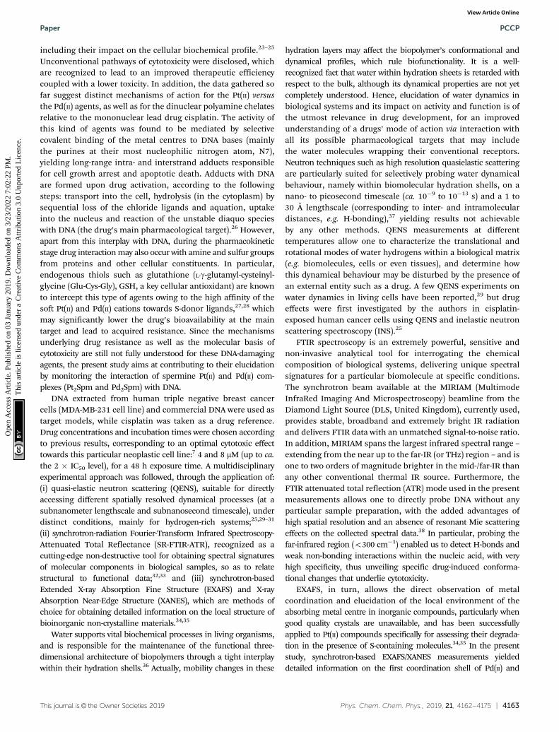
For the hydrated nucleic acid, drug exposure was found toprompt faster DNA dynamics, justified by the disruption of theordered hydration shell and of the native conformation of thenucleic acid upon drug binding, known to affect the nativebase-pair and base packing arrangements: for drug-incubatedDNA, a broader QENS profile was measured (Fig. 3(A)), as wellas a slight deviation to faster kinetics above the dynamicaltransition temperature. The effect of cisplatin was found to be
Fig. 1 QENS data for hydrated versus dehydrated DNA. (A) QENS profiles(for all Q values, at 298 K, logarithmic scale). Elastic scan plots (20–298 K):(B) D2O–DNAhyd vs. H2O–DNAhyd; (C) DNAdehyd vs. H2O–DNAhyd;(D) DNAdehyd vs. D2O–DNAhyd (in (A), the spectra were normalised to maximumpeak intensity. The black line represents the instrument resolution).
Fig. 2 QENS profiles (for all Q values, logarithmic scale): (A) Pd2Spm-treated H2O–DNAhyd, at 150 and 298 K; (B) untreated and Pd2Spm-treatedDNAdehyd, at 298 K (the spectra were normalised to maximum peakintensity. The black line represents the instrument resolution).
PCCP Paper
Ope
n A
cces
s A
rtic
le. P
ublis
hed
on 0
3 Ja
nuar
y 20
19. D
ownl
oade
d on
3/2
3/20
22 7
:02:
22 P
M.
Thi
s ar
ticle
is li
cens
ed u
nder
a C
reat
ive
Com
mon
s A
ttrib
utio
n 3.
0 U
npor
ted
Lic
ence
.View Article Online
This journal is© the Owner Societies 2019 Phys. Chem. Chem. Phys., 2019, 21, 4162--4175 | 4167
somewhat stronger than that of Pd2Spm (Fig. 3(A) vs. Fig. 3(B)),which is in accordance with previous studies by vibrationalmicrospectroscopy on these drugs’ influence on the cellularmetabolic profile.23 Interestingly enough, the drug impact onDNA’s hydration layer was not found to affect the biomolecule’sdynamical transition temperature, neither for cisplatin nor forthe dinuclear Pd-agent. It should be emphasised that drugexposure, either to cisplatin or to Pd2Spm, was found to havea negligible effect on DNA’s hydration level (Fig. S4, ESI†), thusascribing the measured mobility increase exclusively to a drug-mediated effect on the nucleic acid.
No drug effect was found for either the D2O–hydrated DNApellet or DNAdehyd (Fig. 2(B)), suggesting that the slow motionsof the nucleic acid skeleton (backbone dynamics), althoughexpected to be affected by the DNA-binding compounds pre-sently studied, are not detected within the OSIRIS timescale. Inaddition, deuteration of the hydration shell in D2O–DNAhyd
samples and the absence of an hydration layer in DNAdehyd
appear to hinder a significant drug impact on the macro-molecule, evidencing the key role of mobile H’s and hydrationwater molecules as key mediators in external perturbations to thebiomolecule. Nevertheless, these are only tentative conclusionsthat require confirmation on a higher resolution QENS spectro-meter, allowing the detection of the slower dynamical processes(in the nanoscale timescale) that cannot be accessed in OSIRIS.
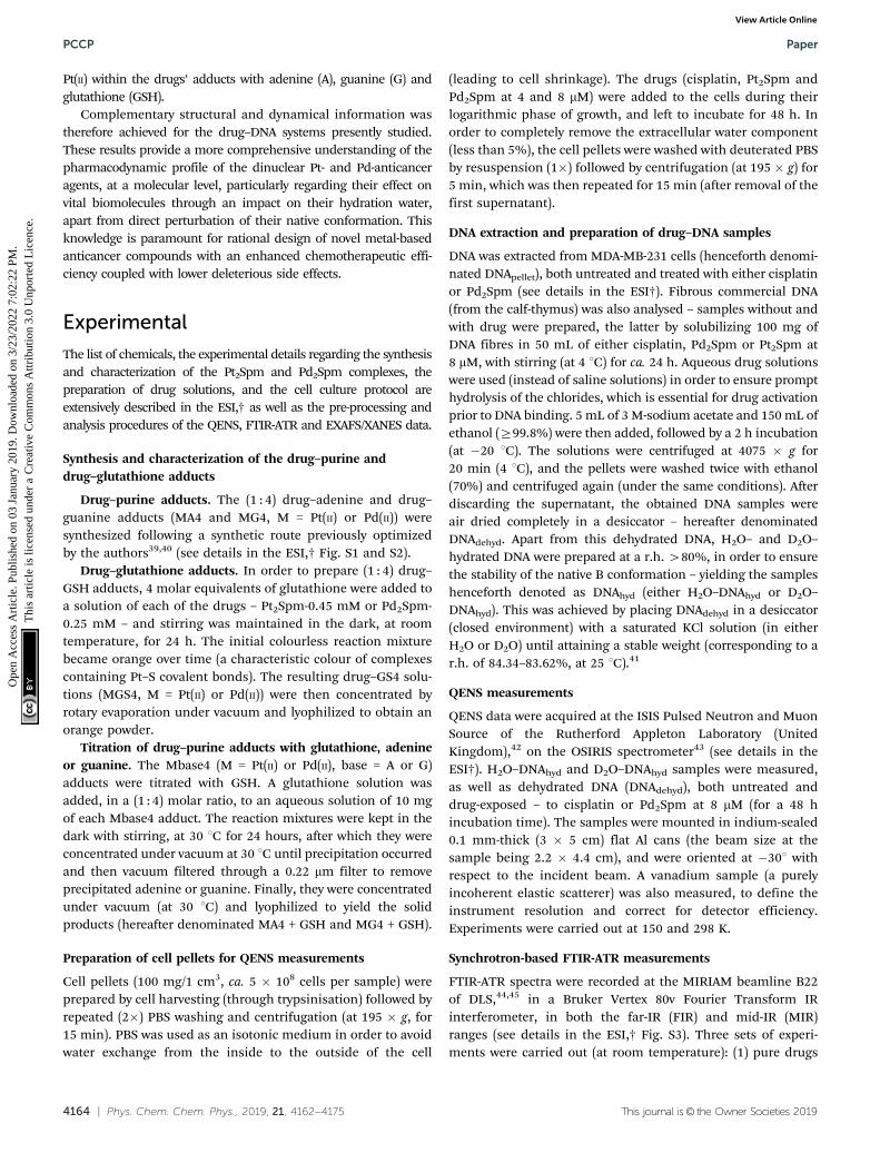
The neutron diffraction plots obtained for H2O–DNAhyd
display two noticeable Bragg peaks (at ca. 0.35 and 0.55 Å�1,detected at both 298 and 150 K, Fig. 4) that reflect the orderedstructure of the nucleic acid molecule. In the presence of thedrug (either cisplatin or Pd2Spm), the most intense peak isclearly shifted from 0.35 to 0.3 Å�1, which corresponds to avariation in distance of ca. 3 Å (from 17.9 to 20.9 Å), that may berelated to both the gap between stacked bases (ca. 3.3 Å) andthe distance between H-bonded base pairs (ca. 2.8 to 2.95 Å) indsDNA helices. This effect (found to be independent of tem-perature) supports a drug-elicited structure disruption of theB-DNA molecule, associated with a perturbation of the nucleicacid’s base packing and pairing arrangements.
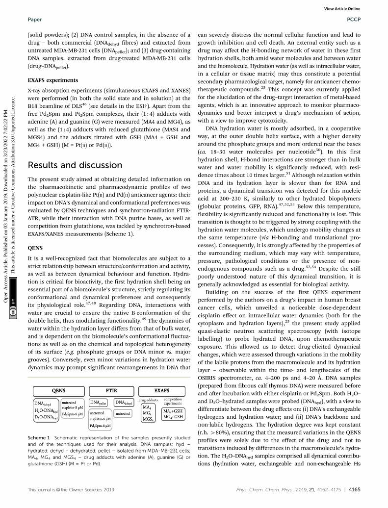
The experimental data was best fit using one d-function(elastic component) convoluted with two Lorentzians (quasie-lastic contributions) (eqn (5), Fig. 5). The very slow globalmotions of the macromolecule are defined by the delta func-tion (slower than the longest observable time defined by theinstrument resolution), while the narrow (Gglobal) and broader
Fig. 3 QENS profiles (298 K, for all Q values) for untreated and drug-exposed H2O–DNAhyd: (A) cisplatin-treated. (B) Pd2Spm-treated (thespectra were normalised to maximum peak intensity. The black linerepresents the instrument resolution).
Fig. 4 Diffraction plots (at 298 K) for untreated and cisplatin-exposedH2O–hydrated DNA.
Fig. 5 QENS spectra (298 K) for untreated (A) and Pd2Spm-treated/8 mM(B) H2O–hydrated DNA, fitted using two Lorentzians and one delta func-tion, at some typical Q values.
Paper PCCP
Ope
n A
cces
s A
rtic
le. P
ublis
hed
on 0
3 Ja
nuar
y 20
19. D
ownl
oade
d on
3/2
3/20
22 7
:02:
22 P
M.
Thi
s ar
ticle
is li
cens
ed u
nder
a C
reat
ive
Com
mon
s A
ttrib
utio
n 3.
0 U
npor
ted
Lic
ence
.View Article Online

4168 | Phys. Chem. Chem. Phys., 2019, 21, 4162--4175 This journal is© the Owner Societies 2019
(Glocal) Lorentzians represent, respectively: (i) slow motions (Q-dependent) from the biopolymer’s hydration water and H-bondrestricted moieties located at the molecule’s surface (side-chainmotions); and (ii) fast localized motions (Q-independent)ascribed to the rotation of CH3 groups and also of aminemoieties (not involved in H-bonds).
The Q-dependent dynamical processes were consistent witha jump-diffusion reorientation model (via large-amplitudecooperative jumps, see ESI†),59,60 as reflected in the plotsdepicting the corresponding FWHM (Gglobal) as a function ofQ2 for both treated and untreated H2O–hydrated DNA, whichshow an asymptotic approach to a plateau at high Q values(Fig. 6(A)). This behaviour agrees with the water distribution inthe first hydration layer of nucleic acids predominantly bindingto their outer surface (unlike for proteins where it maybe enclosed between polypeptide layers or in hydrophobicpockets) and therefore allowing the formation and breakingof H-bonds between the biopolymer and its neighbouringwaters. In turn, the fast motions of the nucleic acid’s amineand hydroxyl groups (not restricted by H-bonding) were foundto be independent of the scattering vector (Fig. 6(B)), evidencinga localized dynamical process with relaxation times given byt = (Glocal)
�1.Table 1 comprises the values presently obtained for the
translational jump-diffusion coefficients (DT) and residencetimes between jumps (tT), as well as for the correlation timesof the fast localized processes, for each system studied. Theseresults reflect the effect of the tested Pt- and Pd-agents onDNA’s dynamical behaviour. For the dynamical processes asso-ciated with the hydration layer, an increase of the diffusioncoefficient (0.72 to 0.90 � 10�5 cm2 s�1) coupled with a
decrease of the residence time (10.13 to 7.40 ps) for controlversus Pd2Spm- and cisplatin-exposed DNA reveals drug-triggered enhanced mobility. The same trend was observedfor the local (faster) motions, unveiled by a decrease in thecorresponding correlation time (3.59 to 3.27 ps). A slightly moresignificant effect was observed for cisplatin as compared to thePd-spermine agent, mainly regarding their effect on DNA’shydration shell, as already revealed by the corresponding QENSprofiles.
The tT value (10 ps) currently obtained for the dynamicalprocesses taking place within DNA’s first hydration layer, in theabsence of a drug, agrees with previously reported data36,49 andis one order of magnitude higher than the value for bulk water(1.1 ps61). This clearly evidences the restricted dynamics ofwater molecules in the close vicinity of DNA. This retardationrelative to bulk water (up to a 6-fold slowdown at roomtemperature) is due to interactions of the water molecules withhydrophobic moieties and phosphate groups of thebiopolymer,49,62 a small number of particularly slow watermolecules having been found in DNA’s minor groove (bridgingthe nitrogen and oxygen atoms of complementary bases), withreorientation times between 60 and 85 ps – the so-called ‘‘spineof hydration’’.63 In addition, the lower residence times pre-sently measured for drug-incubated DNA may be partially dueto a charge screening effect of the metal complexes under study(comprising partial positive charges on the Pt(II) and Pd(II)ions), that may weaken the electrostatic interactions betweenhydration water and negatively charged phosphates at the DNAsurface, thus allowing increased flexibility of the system. Infact, apart from the major role of hydration dynamics, electro-static factors have also been suggested to influence the dyna-mical behaviour of biopolymers.58
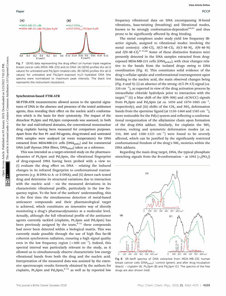
In sum, drug exposure was found to disrupt the nativeordered B-conformation of DNA, prompting a higher flexibilityof the nucleic acid. In the light of the results currently gathered,this is suggested to be associated with a drug impact on thebiomolecule’s first hydration layer, which is prompted intofaster dynamics. This corroborates and complements the effectobserved for cisplatin25 and currently measured for Pd2Spm onthe intracellular milieu, in human breast MDA-MB-231 cancercells: cytoplasmic water was rendered more rigid by thepresence of the metal complexes, while hydration water wasdriven into a more mobile state, as presently verified for H2O–DNAhyd (Fig. 7). Additionally, the more moderate effect on DNAdynamics revealed for Pd2Spm relative to cisplatin is in accor-dance with the influence on intracellular water that was alsoshown to be more significant for the mononuclear Pt-agent.
Fig. 6 Variation of the full widths at half-maximum (FWHM) with Q2 foruntreated, and Pd2Spm- and cisplatin-treated H2O–hydrated DNA (at298 K): (A) Lorentzian function representing the slow translational motions(Gglobal). (B) Lorentzian function representing the fast localised motions(Glocal).
Table 1 Translational diffusion coefficients (DT) and relaxation times (tT) of water for untreated, and Pd2Spm- and cisplatin-treated H2O–hydrated DNA(at 298 K), corresponding to the slow and fast dynamical processes within these systems (represented by Gglobal and Glocal, respectively)
Sample
Gglobal Glocal
DT (�10�5 cm2 s�1) tT (ps) tT (ps)
H2O–DNA 0.72 � 0.0018 10.1274 � 0.3171 3.5927 � 0.0055H2O–DNA + Pd2Spm-8 mM 0.74 � 0.0041 8.3746 � 0.8635 3.3769 � 0.0023H2O–DNA + cisplatin-8 mM 0.90 � 0.0042 7.3974 � 0.3605 3.2721 � 0.0030
PCCP Paper
Ope
n A
cces
s A
rtic
le. P
ublis
hed
on 0
3 Ja
nuar
y 20
19. D
ownl
oade
d on
3/2
3/20
22 7
:02:
22 P
M.
Thi
s ar
ticle
is li
cens
ed u
nder
a C
reat
ive
Com
mon
s A
ttrib
utio
n 3.
0 U
npor
ted
Lic
ence
.View Article Online
This journal is© the Owner Societies 2019 Phys. Chem. Chem. Phys., 2019, 21, 4162--4175 | 4169
Synchrotron-based FTIR-ATR
SR-FTIR-ATR measurements allowed access to the spectral signa-tures of DNA in the absence and presence of the tested antitumoragents, thus revealing their effect on the nucleic acid’s conforma-tion which is the basis for their cytotoxicity. The impact of thedinuclear Pt2Spm and Pd2Spm compounds was assessed, in boththe far- and mid-infrared domains, the conventional mononucleardrug cisplatin having been measured for comparison purposes.Apart from the free Pt- and Pd-agents, drug-treated and untreatedDNA samples were analysed (at room temperature): for DNAextracted from MDA-MB-231 cells (DNApellet) and for commercialDNA (calf thymus DNA fibres, DNAdehyd) taken as a reference.
This was intended as a target-oriented study on the pharmaco-dynamics of Pt2Spm and Pd2Spm, the vibrational fingerprintof drug-exposed DNA having been probed with a view to:(i) evaluate the drug effect on DNA – relating the inducedchanges in its infrared fingerprint to conformational rearran-gements (e.g. B-DNA to A- or Z-DNA); and (ii) detect each testeddrug and determine its structural variations due to interactionwith the nucleic acid – via the measured deviations in itscharacteristic vibrational profile, particularly in the low fre-quency region. To the best of the authors’ understanding, thisis the first time the simultaneous detection of metal-basedanticancer compounds and their pharmacological targetis achieved, which constitutes an innovative way of directlymonitoring a drug’s pharmacodynamics at a molecular level.Actually, although the full vibrational profile of the antitumoragents currently tackled (cisplatin, Pt2Spm and Pd2Spm) hasbeen previously assigned by the team,8–11 these compoundshad never been detected within a biological matrix. This wascurrently made possible through the use of high flux far-IRcoherent synchrotron radiation, ensuring a high signal qualityeven in the low frequency region (o600 cm�1). Indeed, thisspectral interval was particularly relevant to the study, as itallowed us to simultaneously observe characteristic low energyvibrational bands from both the drug and the nucleic acid.Interpretation of the measured data was assisted by the exten-sive spectroscopic results formerly obtained by the authors forcisplatin, Pt2Spm and Pd2Spm,8–11 as well as by reported low
frequency vibrational data on DNA encompassing H-bondvibrations, base-twisting (breathing) and librational modes,known to be strongly conformation-dependent64,65 and thusprone to be significantly affected by drug binding.
The metal complexes under study yield low frequency IR-active signals, assigned to vibrational modes involving themetal centre(s): n(M–Cl), d(Cl–M–Cl), d(Cl–M–N), d(N–M–N)and d(N–M–Cl).8–11,66 Some of these distinctive features werepresently detected in the DNA samples extracted from drug-exposed MDA-MB-231 cells (DNApellet), with clear changes rela-tive to the bands from the isolated drugs owing to DNAcoordination (Fig. 8). This constitutes solid evidence of thedrug’s cellular uptake and conformational rearrangement uponbinding to the nucleic acid, the main observed changes being(Fig. 8 and 9): (i) an absence of the strong n(Cl–Pt–Cl) signal (ca.320 cm�1), as expected in view of the drug activation process byintracellular chloride hydrolysis prior to interaction with thetarget;13 (ii) a blue shift of the d(Pt–NH) and n(CN/CC) signalsfrom Pt2Spm and Pd2Spm (at ca. 1050 and 1070–1085 cm�1,respectively); and (iii) shifts of the CH2 and NH2 deformationbands from the spermine ligand (at 1150–1460 and 1585 cm�1),more noticeable for the Pd(II) system and reflecting a conforma-tional reorganization of the alkylamine chain upon formationof the drug–DNA adduct. Similarly, for cisplatin the NH3
torsion, rocking and symmetric deformation modes (at ca.210, 800 and 1300–1325 cm�1) were found to be severelyaffected, which can be justified by the significantly restrictedconformational freedom of the drug’s NH3 moieties within theDNA adducts.
Regarding the main drug target, DNA, the typical phosphatestretching signals from the B-conformation – at 1092 (ns(PO2))
Fig. 7 QENS data representing the drug effect on human triple negativebreast cancer cells (MDA-MB-231) and on DNA: (A) QENS profiles (for all Qvalues) for untreated and Pd2Spm-treated cells. (B) QENS profiles (for all Qvalues) for untreated and Pd2Spm-exposed H2O–hydrated DNA (thespectra were normalized to maximum peak intensity. The black linerepresents the instrument resolution).
Fig. 8 SR-farIR spectra of DNA extracted from MDA-MB-231 humanbreast cancer cells (DNApellet): control (green), and after drug incubation(black) – cisplatin (A), Pt2Spm (B) and Pd2Spm (C). The spectra of the freedrugs are also shown (red).
Paper PCCP
Ope
n A
cces
s A
rtic
le. P
ublis
hed
on 0
3 Ja
nuar
y 20
19. D
ownl
oade
d on
3/2
3/20
22 7
:02:
22 P
M.
Thi
s ar
ticle
is li
cens
ed u
nder
a C
reat
ive
Com
mon
s A
ttrib
utio
n 3.
0 U
npor
ted
Lic
ence
.View Article Online
4170 | Phys. Chem. Chem. Phys., 2019, 21, 4162--4175 This journal is© the Owner Societies 2019
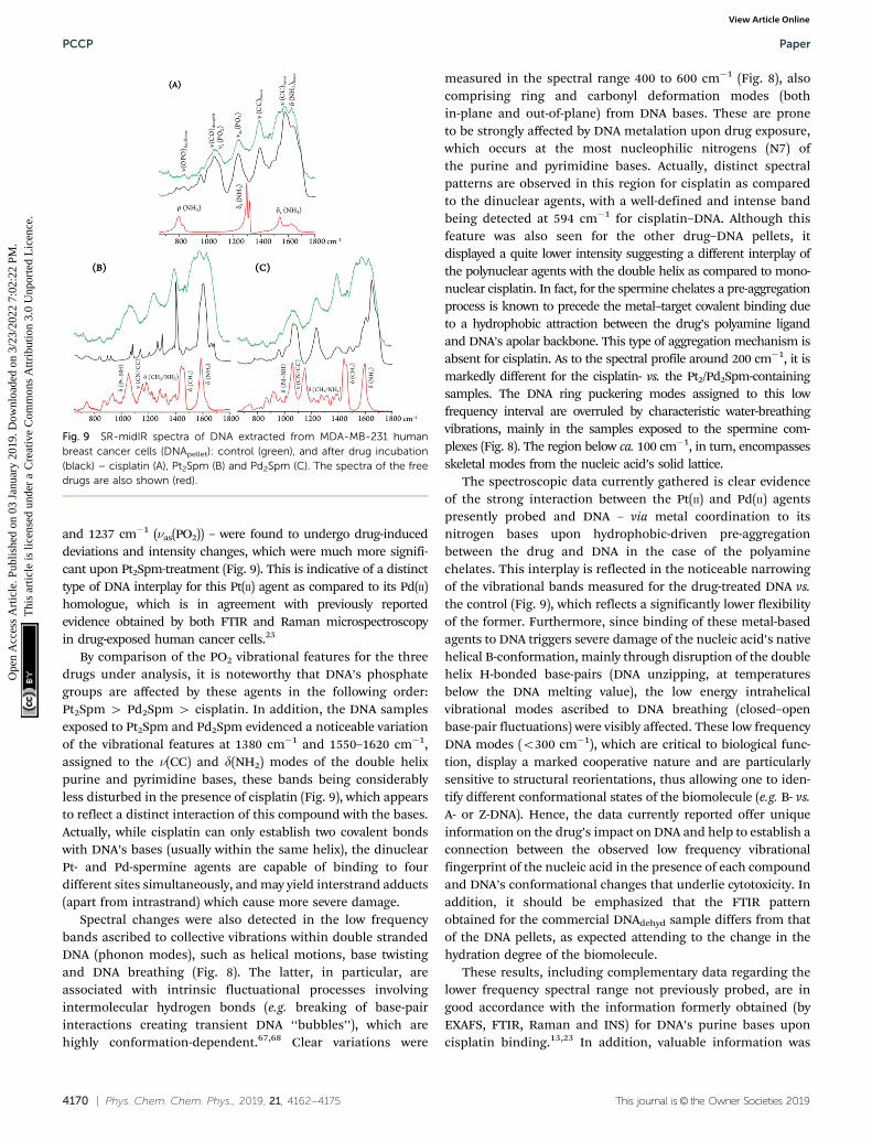
and 1237 cm�1 (nas(PO2)) – were found to undergo drug-induceddeviations and intensity changes, which were much more signifi-cant upon Pt2Spm-treatment (Fig. 9). This is indicative of a distincttype of DNA interplay for this Pt(II) agent as compared to its Pd(II)homologue, which is in agreement with previously reportedevidence obtained by both FTIR and Raman microspectroscopyin drug-exposed human cancer cells.23
By comparison of the PO2 vibrational features for the threedrugs under analysis, it is noteworthy that DNA’s phosphategroups are affected by these agents in the following order:Pt2Spm 4 Pd2Spm 4 cisplatin. In addition, the DNA samplesexposed to Pt2Spm and Pd2Spm evidenced a noticeable variationof the vibrational features at 1380 cm�1 and 1550–1620 cm�1,assigned to the n(CC) and d(NH2) modes of the double helixpurine and pyrimidine bases, these bands being considerablyless disturbed in the presence of cisplatin (Fig. 9), which appearsto reflect a distinct interaction of this compound with the bases.Actually, while cisplatin can only establish two covalent bondswith DNA’s bases (usually within the same helix), the dinuclearPt- and Pd-spermine agents are capable of binding to fourdifferent sites simultaneously, and may yield interstrand adducts(apart from intrastrand) which cause more severe damage.
Spectral changes were also detected in the low frequencybands ascribed to collective vibrations within double strandedDNA (phonon modes), such as helical motions, base twistingand DNA breathing (Fig. 8). The latter, in particular, areassociated with intrinsic fluctuational processes involvingintermolecular hydrogen bonds (e.g. breaking of base-pairinteractions creating transient DNA ‘‘bubbles’’), which arehighly conformation-dependent.67,68 Clear variations were
measured in the spectral range 400 to 600 cm�1 (Fig. 8), alsocomprising ring and carbonyl deformation modes (bothin-plane and out-of-plane) from DNA bases. These are proneto be strongly affected by DNA metalation upon drug exposure,which occurs at the most nucleophilic nitrogens (N7) ofthe purine and pyrimidine bases. Actually, distinct spectralpatterns are observed in this region for cisplatin as comparedto the dinuclear agents, with a well-defined and intense bandbeing detected at 594 cm�1 for cisplatin–DNA. Although thisfeature was also seen for the other drug–DNA pellets, itdisplayed a quite lower intensity suggesting a different interplay ofthe polynuclear agents with the double helix as compared to mono-nuclear cisplatin. In fact, for the spermine chelates a pre-aggregationprocess is known to precede the metal–target covalent binding dueto a hydrophobic attraction between the drug’s polyamine ligandand DNA’s apolar backbone. This type of aggregation mechanism isabsent for cisplatin. As to the spectral profile around 200 cm�1, it ismarkedly different for the cisplatin- vs. the Pt2/Pd2Spm-containingsamples. The DNA ring puckering modes assigned to this lowfrequency interval are overruled by characteristic water-breathingvibrations, mainly in the samples exposed to the spermine com-plexes (Fig. 8). The region below ca. 100 cm�1, in turn, encompassesskeletal modes from the nucleic acid’s solid lattice.
The spectroscopic data currently gathered is clear evidenceof the strong interaction between the Pt(II) and Pd(II) agentspresently probed and DNA – via metal coordination to itsnitrogen bases upon hydrophobic-driven pre-aggregationbetween the drug and DNA in the case of the polyaminechelates. This interplay is reflected in the noticeable narrowingof the vibrational bands measured for the drug-treated DNA vs.the control (Fig. 9), which reflects a significantly lower flexibilityof the former. Furthermore, since binding of these metal-basedagents to DNA triggers severe damage of the nucleic acid’s nativehelical B-conformation, mainly through disruption of the doublehelix H-bonded base-pairs (DNA unzipping, at temperaturesbelow the DNA melting value), the low energy intrahelicalvibrational modes ascribed to DNA breathing (closed–openbase-pair fluctuations) were visibly affected. These low frequencyDNA modes (o300 cm�1), which are critical to biological func-tion, display a marked cooperative nature and are particularlysensitive to structural reorientations, thus allowing one to iden-tify different conformational states of the biomolecule (e.g. B- vs.A- or Z-DNA). Hence, the data currently reported offer uniqueinformation on the drug’s impact on DNA and help to establish aconnection between the observed low frequency vibrationalfingerprint of the nucleic acid in the presence of each compoundand DNA’s conformational changes that underlie cytotoxicity. Inaddition, it should be emphasized that the FTIR patternobtained for the commercial DNAdehyd sample differs from thatof the DNA pellets, as expected attending to the change in thehydration degree of the biomolecule.
These results, including complementary data regarding thelower frequency spectral range not previously probed, are ingood accordance with the information formerly obtained (byEXAFS, FTIR, Raman and INS) for DNA’s purine bases uponcisplatin binding.13,23 In addition, valuable information was
Fig. 9 SR-midIR spectra of DNA extracted from MDA-MB-231 humanbreast cancer cells (DNApellet): control (green), and after drug incubation(black) – cisplatin (A), Pt2Spm (B) and Pd2Spm (C). The spectra of the freedrugs are also shown (red).
PCCP Paper
Ope
n A
cces
s A
rtic
le. P
ublis
hed
on 0
3 Ja
nuar
y 20
19. D
ownl
oade
d on
3/2
3/20
22 7
:02:
22 P
M.
Thi
s ar
ticle
is li
cens
ed u
nder
a C
reat
ive
Com
mon
s A
ttrib
utio
n 3.
0 U
npor
ted
Lic
ence
.View Article Online
This journal is© the Owner Societies 2019 Phys. Chem. Chem. Phys., 2019, 21, 4162--4175 | 4171
gathered on a different interplay with the nucleic acid regard-ing the dinuclear agents Pt2Spm and Pd2Spm as compared tothe mononuclear (clinically used) drug cisplatin.
EXAFS
EXAFS/XANES analysis provided a molecular picture of drug–DNAinterplay for Pt2Spm and Pd2Spm, specifically regarding theirinteraction with DNA purine bases recognized to be the mainbinding sites for cisplatin-like agents. The major goal of theproposed study was attained: to unequivocally determine the localenvironment of the absorbing Pt(II) and Pd(II) centres in the drugs’adducts with adenine and guanine (1 : 4), with a view to determinethe precise first coordination sphere content in these entities.Additionally, interaction with glutathione was assessed, since thisubiquitous sulfur-containing tripeptide can compete for the drugrelative to its major pharmacological target (DNA), thus beingimplicated in acquired resistance to metal-based chemotherapeuticdrugs.28 The current experiments were performed for both the solidsamples and the aqueous solutions, the results thus obtainedshowing no significant differences in accordance with previousEXAFS measurements for the homologous cisplatin-adducts.13
Regarding the effect of temperature, it was verified that the dataat 90 K agreed well with that measured at room temperature,corroborating the stability of these spermine complexes underphysiological conditions, a major requirement for their in vivoapplication as anticancer agents.
Interpretation of the data was based on spectra measured forsolid PtA4, PdA4, PtGS4 and PdGS4, taken as references for themetal centre with distinct first coordination spheres, respectively(4N) and (4S). EXAFS fitting of these standards allowed us tooptimise the bond length values and obtain the amplitude (S0
2),energy shift (E0) and Debye–Waller (s2) factors that were thenused for fitting the metal adducts (displaying coordinationenvironments with either equal or different ligands).
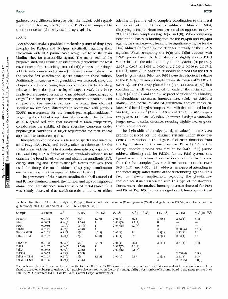
The parameters of the nearest coordination shell around Pdand Pt were obtained, namely the number and type of neighbouratoms, and their distance from the selected metal (Table 2). Itwas clearly observed that stoichiometric amounts of either
adenine or guanine led to complete coordination to the metalcentres in both the Pt and Pd adducts – MA4 and MG4,displaying a (4N) environment per metal as opposed to (2N +2Cl) in the free complexes (Fig. 10(A) and (B)). When comparingboth purine bases as binding sites for the Pt2Spm and Pd2Spmagents, the symmetry was found to be significantly higher for thePt(II) adducts (reflected by the stronger intensity of the EXAFSsignals). When comparing the Pt(II) and Pd(II) adducts withDNA’s purine bases, the latter displayed slightly shorter Pd–Nvalues in both the adenine and guanine systems (respectively2.027 � 0.007 vs. 2.039 � 0.005 and 2.035 � 0.006 vs. 2.047 �0.005 Å, Table 2). In addition, it should be noted that the Pd–Nbond lengths within PdA4 and PdG4 were also shortened relativeto the Pt(NH3)4 reference sample previously measured13 (2.039�0.004 Å). For the drug–glutathione (1 : 4) adducts, a (4S) firstcoordination shell was detected for each of the metal centres(Fig. 10(A) and (B) and Table 2), as proof of efficient drug bindingto glutathione molecules (monodentate, solely via GSH’s Satoms). Both for the Pt– and Pd–glutathione adducts, the calcu-lated M–S bond lengths compare well with that obtained for thePt(GSH)4 reference13 (2.308 � 0.006 and 2.334 � 0.004, respec-tively, vs. 2.313 � 0.006 Å). PdGS4, however, displays a somewhatlonger metal-to-sulfur distance, revealing slightly weaker gluta-thione coordination.
The slight shift of the edge (to higher values) in the XANESprofiles observed for the distinct systems under study evi-denced a variation in the degree of electron donation fromthe ligand atoms to the metal centre (Table 3). While thischarge transfer process was similar for both Pd(II)–purineadducts differing only for PdGS4, for the Pt(II) systems thisligand-to-metal electron delocalisation was found to increasefrom the free complex ((2N + 2Cl) environment) to the PtA4/PtG4 ((4N)) and PtGS4 ((4S)) adducts, as expected attending tothe increasingly softer nature of the surrounding ligands. Thisfact has relevant implications regarding the glutathione-induced resistance associated with this type of metal-agents.Furthermore, the marked intensity increase detected for PtA4and PtGS4 (Fig. 10(C)) reflects a significantly lower symmetry of
Table 2 Results of EXAFS fits for Pt2Spm, Pd2Spm, their adducts with adenine (MA4), guanine (MG4) and glutathione (MGS4), and the [adducts +glutathione] (MA4 + GSH and MG4 + GSH) (M = Pt(II) or Pd(II))
Sample R-Factor S02 E0 (eV) CNN (Å) RN (Å) sN
2 (10�3 Å2) CNCl (Å) RCl (Å) sCl2 (10�3Å2)
Pt2Spm 0.0148 0.74(6) 9(2) 2.2(6) 2.06(3) 3(2) 1.8(6) 2.32(1) 3(1)PtA4 0.0043 0.84(4) 9.5(6) 4 2.039(5) 2.9(5) 0 — —PtG4 0.0086 1.01(4) 10.7(6) 4 2.047(5) 4.1(7) 0 — —PtGS4 0.0141 0.87(6) 6.2(8) 0 — — 4 2.308(6) 5.1(7)PtA4 + GSH 0.0103 0.88(3) 8(1) 1.2(2) 2.03(2) 3* 2.8(2) 2.32(1) 5*PtG4 + GSH 0.0197 0.90(4) 7(2) 0.8(3) 2.03(4) 3* 3.2(3) 2.32(1) 5*
Pd2Spm 0.0108 0.83(6) 6(2) 1.8(7) 2.06(3) 2(2) 2.2(7) 2.31(1) 3(1)PdA4 0.0107 0.84(5) 5.5(8) 4 2.027(7) 2.3(8) — — —PdG4 0.0062 0.86(4) 5.7(6) 4 2.035(6) 2.8(7) — — —PdGS4 0.0065 0.89(4) 5.6(5) — — — 4 2.334(4) 3.3(4)PdA4 + GSH 0.0203 0.87(4) 5(1) 2.6(2) 2.03(1) 2.5* 1.4(2) 2.31(1) 3.3*PdG4 + GSH 0.0106 0.79(4) 5.3(6) — — — 4 2.328(5) 3.0(5)
For each sample, the fit was performed on the first shell of the EXAFS signal with all parameters free (first row) and with coordination numbersfixed to expected values (second row). S0
2: passive electron reduction factor; E0: energy shift; CNX: number of X atoms bond to the metal (either Pt orPd); RX: M–X distances (M = Pt or Pd); sX
2: X atom Debye–Waller factor.
Paper PCCP
Ope
n A
cces
s A
rtic
le. P
ublis
hed
on 0
3 Ja
nuar
y 20
19. D
ownl
oade
d on
3/2
3/20
22 7
:02:
22 P
M.
Thi
s ar
ticle
is li
cens
ed u
nder
a C
reat
ive
Com
mon
s A
ttrib
utio
n 3.
0 U
npor
ted
Lic
ence
.View Article Online
4172 | Phys. Chem. Chem. Phys., 2019, 21, 4162--4175 This journal is© the Owner Societies 2019
these species as compared to PtG4 (and to the free drug), sincethis pre-edge intensity is strongly dependent on the size andgeometry of the molecular cage surrounding the metal absor-ber. For the Pd(II) systems, in turn, this symmetry loss washardly noticeable, which may suggest a more labile coordina-tion of this cation relative to Pt(II), the latter displaying a clearpreference for guanine – yielding PtG4 stable adducts with adefined and quite rigid geometry.
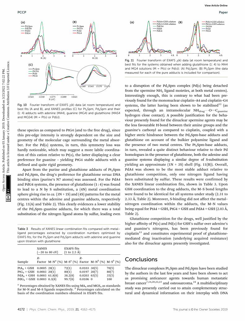
Apart from the purine and glutathione adducts of Pt2Spmand Pd2Spm, the drug’s preference for glutathione versus DNAbinding sites (purine’s N7 atoms) was assessed. For the PdA4and PdG4 systems, the presence of glutathione (1 : 4) was foundto lead to a N by S substitution, a (4N) metal coordinationsphere giving rise to ca. (3N + 1S) and (4S) patterns for the metalcentres within the adenine and guanine adducts, respectively(Fig. 11(A) and Table 2). This clearly evidences a lower stabilityof the Pd2Spm–guanine adducts, for which there was a totalsubstitution of the nitrogen ligand atoms by sulfur, leading even
to a disruption of the Pd2Spm complex (Pd(II) being detachedfrom the spermine NH2 ligand moieties, at both metal centres).Interestingly enough, this is contrary to what had been pre-viously found for the mononuclear cisplatin–A4 and cisplatin–G4systems, the latter having been shown to be stabilised13 (asexpected, through an intramolecular NHdrug� � �OQCguanine
hydrogen close contact). A possible justification for the beha-viour presently found for the dinuclear spermine agents may bethe less favourable H-bond between their amine groups and theguanine’s carbonyl as compared to cisplatin, coupled with ahigher steric hindrance between the Pd2Spm-base adducts andglutathione on account of the bulkier polyamine ligand andthe presence of two metal centres. The Pt2Spm-base adducts,in turn, revealed a quite distinct behaviour relative to their Pdhomologues in the presence of glutathione, both the adenine andguanine systems displaying a similar degree of S-substitutionyielding an approximate (1N + 3S) shell (Fig. 11(B)). Overall,PdA4 was shown to be the most stable adduct relative toglutathione competition, only one nitrogen ligand havingbeen substituted by sulfur. These results were confirmed bythe XANES linear combination fits, shown in Table 3. UponGSH coordination to the drug adducts, the M–S bond lengthswere found to be identical for all systems under study (2.31 to2.33 Å, Table 2). Moreover, S-binding did not affect the metal–nitrogen coordination within the adducts, the M–N valuesbeing equal for PtA4 + GSH, PtG4 + GSH and PdA4 + GSH (2.03 Å,Table 2).
Glutathione competition for the drugs, well justified by thehigher affinity of Pt(II) and Pd(II) for GSH’s sulfur over adenine’sand guanine’s nitrogens, has been previously found forcisplatin13 and constitutes experimental proof of glutathione-mediated drug inactivation (underlying acquired resistance)also for the dinuclear agents presently investigated.
Conclusions
The dinuclear complexes Pt2Spm and Pd2Spm have been studiedby the authors in the last few years and have been shown to actas promising anticancer agents towards human metastaticbreast cancer7,14,20,22,23 and osteosarcoma.24 A multidisciplinarystudy was presently carried out to attain complementary struc-tural and dynamical information on their interplay with DNA
Fig. 10 Fourier transform of EXAFS w(k) data (at room temperature) andbest fits (A and B), and XANES profiles (C) for Pt2Spm, Pd2Spm and their(1 : 4) adducts with adenine (MA4), guanine (MG4) and glutathione (MAS4and MGS4) (M = Pt(II) or Pd(II)).
Table 3 Results of XANES linear combination fits compared with metal–ligand percentages extracted by coordination numbers optimised byEXAFS fits, for the Pt2Spm and Pd2Spm adducts with adenine and guanineupon titration with glutathione
Sample
XANES(�20 to 80 eV)
EXAFS fits(1 to 2.5 Å)
R-Factor M–Na (%) M–Sa (%)
R-Factor M–Nb (%) M–Sb (%)
PtA4 + GSH 0.0005 28(1) 72(1) 0.0103 30(5) 70(5)PtG4 + GSH 0.0003 20(1) 80(1) 0.0197 20(7) 80(7)PdA4 + GSH 0.0003 61.8(8) 38.2(8) 0.0203 65(5) 35(5)PdG4 + GSH 0.0003 0.3(8) 99.7(8) 0.0106 0 100
a Percentages obtained by XANES fits using MA4 and MGS4 as standardsfor M–N and M–S ligands respectively. b Percentages calculated on thebasis of the coordination numbers obtained in EXAFS fits.
Fig. 11 Fourier transform of EXAFS w(k) data (at room temperature) andbest fits for the systems obtained when adding glutathione (1 : 4) to MA4and MG4 solutions (M = Pt(II) or Pd(II)): (A) Pd2Spm; (B) Pt2Spm (the datameasured for each of the pure adducts is included for comparison).
PCCP Paper
Ope
n A
cces
s A
rtic
le. P
ublis
hed
on 0
3 Ja
nuar
y 20
19. D
ownl
oade
d on
3/2
3/20
22 7
:02:
22 P
M.
Thi
s ar
ticle
is li
cens
ed u
nder
a C
reat
ive
Com
mon
s A
ttrib
utio
n 3.
0 U
npor
ted
Lic
ence
.View Article Online
This journal is© the Owner Societies 2019 Phys. Chem. Chem. Phys., 2019, 21, 4162--4175 | 4173
(their main pharmacological target), using QENS measurements,SR-FTIR and SR-EXAFS/XANES.
The QENS experiments currently performed provided accurateand unique data on the impact of metal-based anticancer drugs onDNA via the biopolymer’s first hydration layer, which is known tobe closely coupled to DNA function. Indeed, any drug-triggeredperturbation of the nucleic acid’s hydration shell is expected toinfluence the biomolecule’s dynamical profile and consequently itsbiological function. Measurements for drug-treated and untreatedH2O–hydrated DNA, at room temperature, revealed a clear effect ofboth cisplatin and Pd2Spm on the nucleic acid’s hydration shelldynamics, reflected in increased flexibility for the drug-incubatedsamples. Two dynamical processes were discriminated (on the pstimescale), ascribed to: (i) the biopolymer’s hydration water; and(ii) fast localized rotations of specific groups within DNA – CH3,as well as NH2 not restricted by hydrogen-bonds. The intrinsicstiffness of native DNA appears to be disrupted by the drug,underlining the influence of the water molecules from the firsthydration sheath. It should be emphasized that this effect was onlyobserved (on the ps timescale) for H2O–hydrated DNA and not forthe D2O–hydrated nucleic acid, which also evidenced distinctdynamical transition temperatures. Apart from the hydration-induced flexibility increase, an electrostatic-mediated dynamicenhancement was unveiled for drug-exposed H2O–hydrated DNA.These conclusions are in good agreement with the previouslyreported drug impact on the intracellular milieu – cytoplasmicwater and biomolecules’ hydration layers:25 while the formershowed restrained dynamics upon drug exposure, the hydrationshells were prompted into a more mobile state, as presentlycorroborated for DNA. These combined results support the pro-nounced effect of metal-based chemotherapeutic agents on waterdynamics, and should help to gain a more thorough understandingof the drug-induced cytotoxicity via the water molecules both in theintracellular medium and in the close vicinity of biopolymers. Inaddition, an interesting onset of anharmonicity was detected fordehydrated DNA (on the ps timescale) in contrast with previousstudies on dehydrated biopolymers (e.g. proteins, RNA and DNA),and it was tentatively assigned to rotational motions of methylgroups (1 per 4 bases within the double helix) and, to a smallerextent, of H-bond free NH2 moieties.
Infrared spectroscopy with synchrotron radiation wasconfirmed as a powerful non-invasive molecular probe ofbiosamples, allowing one to interrogate and obtain accurateinsight into the biochemical impact of chemotherapy drugs atthe cellular level. Following previous studies on the effect of thepresently tested compounds in cancer cells,23 their impact onDNA was currently probed, for DNA samples extracted fromtreated and untreated human MDA-MB-231 cells. The twofoldpurpose of this study was accomplished, since the simulta-neous detection of the drug and its pharmacological target wasachieved, specific spectral biomarkers having been identifiedreflecting changes in both the metal agent and DNA, promptedby drug–target interaction. Furthermore, the vibrationalmodes comprising the far-infrared region, which are a spectralsignature of the nucleic acid conformational state, allowed usto unveil drug-elicited perturbations in this biomolecule
recognized to be the basis of cytotoxicity: an altered patternwas detected for the bands ascribed to DNA breathing, asa consequence of drug-triggered disruption of the double helixH-bonded base-pairs. To the best of the authors’ knowledge,this is the first experimental observation of the DNA breathingprocess in the presence of a chemotherapeutic agent. Addition-ally, analysis of the variations occurring in the characteristicbands of the complexes provided evidence of structural varia-tions upon accumulation at the biological target. Moreover,sampling DNA extracted from drug-exposed human cancer cellsenabled us to qualitatively probe the drug’s cellular uptake andits activation process (through intracellular chloride hydrolysis)prior to DNA interaction, apart from its impact on the nucleicacid upon binding to the purine bases. These results couldonly be successfully achieved through FTIR spectroscopy withcoherent synchrotron radiation that guarantees both broad-band spectral coverage and high signal quality. The SR-FTIRresults presently gathered complement previous SR-basedinfrared data obtained at the MIRIAM beamline for the samecompounds and cell line (at equal concentrations and incuba-tion time):23 comparing these results on whole cells with thosemeasured for DNA isolated from cells pre-incubated with thechemotherapeutic agents is of key biological significance fordiscriminating drug effects regarding other potential cellulartargets (e.g. DNA vs. proteins, membrane lipids or intracellularwater).
The successful EXAFS/XANES results gathered for theadenine, guanine and glutathione adducts with Pt2Spm andPd2Spm allowed an accurate assessment of their pharmaco-dynamics (interaction with DNA) and pharmacokinetics: thelocal environment of the metal centres was obtained, for eachsystem, and GSH-metal binding (solely via glutathione’s sulfuratom) was clearly evidenced thus justifying the glutathione-mediated drug resistance mechanisms occurring in vivo.Slightly weaker glutathione coordination was unveiled forPd2Spm as compared to Pt2Spm, which may be relevant witha view to overcome in vivo GSH-mediated drug resistance.Additionally, EXAFS measurements for the adducts in thepresence of glutathione revealed a lower stability of thePd2Spm–guanine adducts relative to their adenine homologues(contrary to what had been previously found for cisplatin13),while their Pt counterparts were shown to have identicalstability to the mononuclear clinical drug. Coupled with thedata previously reported for cisplatin,13 these results providedaccurate information on the chemical composition, structureand relative stability of the adducts formed between thePd2Spm and Pt2Spm complexes and DNA purine bases, leadingto a better understanding of the mode of action of this type ofpolynuclear metal agents at the molecular level.
The present multidisciplinary study on the impact of cispla-tin and cisplatin-like dinuclear agents on DNA’s dynamical andstructural profiles constitutes an innovative way of tackling adrug’s mode of action. A comprehensive and reliable set of datawas gathered on the molecular basis of their cytotoxicitytowards the very low prognosis human metastatic breast cancer,allowing us to better clarify the unconventional interaction
Paper PCCP
Ope
n A
cces
s A
rtic
le. P
ublis
hed
on 0
3 Ja
nuar
y 20
19. D
ownl
oade
d on
3/2
3/20
22 7
:02:
22 P
M.
Thi
s ar
ticle
is li
cens
ed u
nder
a C
reat
ive
Com
mon
s A
ttrib
utio
n 3.
0 U
npor
ted
Lic
ence
.View Article Online
4174 | Phys. Chem. Chem. Phys., 2019, 21, 4162--4175 This journal is© the Owner Societies 2019
of Pt2Spm and Pd2Spm with their main target. Combinedwith biological assays for evaluation of antitumor activity, thecurrent results are expected to provide valuable clues for therational design of metal-based compounds with improvedtherapeutic properties, acting through a multistep process thatleads to function loss of vital biomolecules and ultimately tocell death: (i) direct binding to DNA, causing disruption of itsnative conformation and prompting biofunctional disability;(ii) a continuing effect on the nucleic acid’s hydration layer,which is prompted into faster dynamics, triggering changes inthe biopolymer with consequences at the functional level; and(iii) an impact on intracellular water (cytosol), with an expectedglobal effect on essential cellular components thus hinderingnormal cellular function.
Conflicts of interest
There are no conflicts to declare.
Acknowledgements
The authors thank financial support from the PortugueseFoundation for Science and Technology – UID/MULTI/00070/2013 and PTDC/QEQ-MED/1890/2014 (within Project 3599-PPCDT – jointly financed by the European Community FundFEDER). The STFC Rutherford Appleton Laboratory is thankedfor access to the Research Complex at Harwell (cell culture labs)and to the neutron beam facilities. The neutron work wassupported by the European Commission under the 7th Frame-work Programme through the Key Action: Strengthening theEuropean Research Area, Research Infrastructures (contract no.CP-CSA_INFRA-2008-1.1.1 Number 226507-NMI3). DiamondLight Source (UK) is acknowledged for access to the B22/MIRIAM (SM14895) and B18/Core EXAFS (SP16058) beamlines.A. Fitzpatrick is thanked for technical support at B22.
References
1 B. Rosenberg, L. Vancamp and T. Krigas, Nature, 1965,205, 698.
2 B. Rosenberg, L. VanCamp, J. E. Trosko and V. H. Mansour,Nature, 1969, 222, 385.
3 A. M. Florea and D. Busselberg, Cancers, 2011, 3, 1351.4 L. Kelland, Nat. Rev. Cancer, 2007, 7, 573.5 N. J. Wheate, S. Walker, G. E. Craig and R. Oun, Dalton
Trans., 2010, 39, 8113.6 M. P. M. Marques, ISRN Spectrosc., 2013, 2013, 29.7 S. M. Fiuza, J. Holy, L. A. E. Batista de Carvalho and
M. P. M. Marques, Chem. Biol. Drug Des., 2011, 77, 477.8 L. A. E. Batista de Carvalho, M. P. M. Marques, C. Martin,
S. F. Parker and J. Tomkinson, ChemPhysChem, 2011,12, 1334.
9 M. P. M. Marques, R. Valero, S. F. Parker, J. Tomkinson andL. A. E. Batista de Carvalho, J. Phys. Chem. B, 2013,117, 6421.
10 S. M. Fiuza, A. M. Amado, S. F. Parker, M. P. M. Marquesand L. A. E. Batista de Carvalho, New J. Chem., 2015,39, 6274.
11 A. L. M. Batista de Carvalho, S. F. Parker, L. A. E. Batista deCarvalho and M. P. M. Marques, Acta Crystallogr., Sect. C:Struct. Chem., 2018, 74, 628.
12 O. Corduneanu, A. M. Chiorcea-Paquim, S. M. Fiuza, M. P. M.Marques and A. M. Oliveira-Brett, Bioelectrochemistry, 2010,78, 97.
13 M. P. M. Marques, D. Gianolio, G. Cibin, J. Tomkinson,S. F. Parker, R. Valero, R. P. Lopes and L. A. E. Batista deCarvalho, Phys. Chem. Chem. Phys., 2015, 17, 5155.
14 M. P. M. Marques, T. Girao, M. C. P. De Lima, A. Gameiro,E. Pereira and P. Garcia, Biochim. Biophys. Acta, Mol. CellRes., 2002, 1589, 63.
15 L. J. Teixeira, M. Seabra, E. Reis, M. T. G. da Cruz,M. C. P. de Lima, E. Pereira, M. A. Miranda andM. P. M. Marques, J. Med. Chem., 2004, 47, 2917.
16 S. M. Fiuza, A. M. Amado, P. J. Oliveira, V. A. Sardao,L. A. E. Batista de Carvalho and M. P. M. Marques, Lett.Drug Des. Discovery, 2006, 3, 149.
17 A. S. Soares, S. M. Fiuza, M. J. Goncalves, L. A. E. Batista deCarvalho, M. P. M. Marques and A. M. Urbano, Lett. DrugDes. Discovery, 2007, 4, 460.
18 A. Tassoni, N. Bagni, M. Ferri, M. Franceschetti,A. Khomutov, M. P. Marques, S. M. Fiuza, A. R. Simonianand D. Serafini-Fracassini, Plant Physiol. Biochem., 2010,48, 496.
19 R. Tummala, P. Diegelman, S. M. Fiuza, L. A. E. Batista deCarvalho, M. P. M. Marques, D. L. Kramer, K. Clark, S. Vujcic,C. W. Porter and L. Pendyala, Oncol. Rep., 2010, 24, 15.
20 T. M. Silva, S. Andersson, S. K. Sukumaran, M. P. Marques,L. Persson and S. Oredsson, PLoS One, 2013, 8, e55651.
21 T. M. Silva, S. M. Fiuza, M. P. M. Marques, L. Persson andS. Oredsson, Amino Acids, 2014, 46, 339.
22 A. L. M. Batista de Carvalho, P. S. Medeiros, F. M. Costa,V. P. Ribeiro, J. B. Sousa, C. Diniz and M. P. M. Marques,PLoS One, 2016, 11, e0167218.
23 A. L. M. Batista de Carvalho, M. Pilling, P. Gardner,J. Doherty, G. Cinque, K. Wehbe, C. Kelley, L. A. E. Batistade Carvalho and M. P. M. Marques, Faraday Discuss., 2016,187, 273.
24 I. Lamego, M. P. Marques, I. F. Duarte, A. S. Martins,H. Oliveira and A. M. Gil, J. Proteome Res., 2017, 16, 1773.
25 M. P. M. Marques, A. L. M. Batista de Carvalho, V. G. Sakai,L. Hatter and L. A. E. Batista de Carvalho, Phys. Chem. Chem.Phys., 2017, 19, 2702.
26 J. Reedijk, Proc. Natl. Acad. Sci. U. S. A., 2003, 100, 3611.27 Y. Kasherman, S. Sturup and D. Gibson, J. Med. Chem., 2009,
52, 4319.28 H. H. Chen and M. T. Kuo, Met. – Based Drugs, 2010, 2010, 1.29 A. Frolich, F. Gabel, M. Jasnin, U. Lehnert, D. Oesterhelt,
A. M. Stadler, M. Tehei, M. Weik, K. Wood and G. Zaccai,Faraday Discuss., 2009, 141, 117.
30 F. Foglia, R. Hazael, G. G. Simeoni, M. S. Appavou,M. Moulin, M. Haertlein, V. Trevor Forsyth, T. Seydel,
PCCP Paper
Ope
n A
cces
s A
rtic
le. P
ublis
hed
on 0
3 Ja
nuar
y 20
19. D
ownl
oade
d on
3/2
3/20
22 7
:02:
22 P
M.
Thi
s ar
ticle
is li
cens
ed u
nder
a C
reat
ive
Com
mon
s A
ttrib
utio
n 3.
0 U
npor
ted
Lic
ence
.View Article Online

This journal is© the Owner Societies 2019 Phys. Chem. Chem. Phys., 2019, 21, 4162--4175 | 4175
I. Daniel, F. Meersman and P. F. McMillan, Sci. Rep., 2016,6, 18862.
31 S. Fujiwara, K. Araki, T. Matsuo, H. Yagi, T. Yamada,K. Shibata and H. Mochizuki, PLoS One, 2016, 11, e0151447.
32 H. Y. Holman, H. A. Bechtel, Z. Hao and M. C. Martin, Anal.Chem., 2010, 82, 8757.
33 M. J. Baker, J. Trevisan, P. Bassan, R. Bhargava, H. J. Butler,K. M. Dorling, P. R. Fielden, S. W. Fogarty, N. J. Fullwood,K. A. Heys, C. Hughes, P. Lasch, P. L. Martin-Hirsch,B. Obinaju, G. D. Sockalingum, J. Sule-Suso, R. J. Strong,M. J. Walsh, B. R. Wood, P. Gardner and F. L. Martin, Nat.Protoc., 2014, 9, 1771.
34 M. Obata, M. Harada, H. Ohi, S. Hirohara, M. Gottchaldtand S. Yano, Chem. Pharm. Bull., 2009, 57, 1107.
35 K. Provost, D. Bouvet-Muller, S. Crauste-Manciet, J. Moscovici,L. Olivi, G. Vlaic and A. Michalowicz, Biochimie, 2009, 91, 1301.
36 D. Laage, T. Elsaesser and J. T. Hynes, Chem. Rev., 2017,117, 106945.
37 E. Mamontov and X. Q. Chu, Phys. Chem. Chem. Phys., 2012,14, 11573.
38 K. L. A. Chan and S. G. Kazarian, Chem. Soc. Rev., 2016, 45, 1850.39 B. Lippert, G. Raudaschl, C. J. L. Lock and P. Pilon, Inorg.
Chim. Acta, 1984, 93, 43.40 H. Schoellhorn, G. Raudaschl-Sieber, G. Mueller, U. Thewalt
and B. Lippert, J. Am. Chem. Soc., 1985, 107, 5932.41 L. Greenspan, J. Res. NBS A: Phys. Chem., 1977, 81, 89.42 ISIS - STFC - OSIRIS, http://www.isis.stfc.ac.uk/instruments/
osiris/, accessed 21/01/2018.43 M. T. F. Telling and K. H. Andersen, Phys. Chem. Chem.
Phys., 2005, 7, 1255.44 G. Cinque, M. Frogley, K. Wehbe, J. Filik and J. Pijanka,
Synchrotron Radiat. News, 2011, 24, 24.45 Diamond Light Source, http://www.diamond.ac.uk/,
accessed 18/05/2018.46 A. J. Dent, G. Cibin, S. Ramos, A. D. Smith, S. M. Scott,
L. Varandas, M. R. Pearson, N. A. Krumpa, C. P. Jones andP. E. Robbins, 14th International Conference on X-RayAbsorption Fine Structure (XAFS14), Proceedings, 2009, 190.
47 A. P. Sokolov, J. H. Roh, E. Mamontov and V. G. Sakai, Chem.Phys., 2008, 345, 212.
48 A. M. Stadler, F. Demmel, J. Ollivier and T. Seydel, Phys.Chem. Chem. Phys., 2016, 18, 21527.
49 E. Duboue-Dijon, A. C. Fogarty, J. T. Hynes and D. Laage,J. Am. Chem. Soc., 2016, 138, 7610.
50 V. Makarov, B. M. Pettitt and M. Feig, Acc. Chem. Res., 2002,35, 376.
51 I. A. Beta, I. Michalarias, R. C. Ford, J. C. Li andM. C. Bellissent-Funel, Chem. Phys., 2003, 292, 451.
52 S. H. Chen, L. Liu, X. Chu, Y. Zhang, E. Fratini, P. Baglioni,A. Faraone and E. Mamontov, J. Chem. Phys., 2006,125, 171103.
53 S. Khodadadi, J. H. Roh, A. Kisliuk, E. Mamontov, M. Tyagi,S. A. Woodson, R. M. Briber and A. P. Sokolov, Biophys. J.,2010, 98, 1321.
54 B. R. Wood, Chem. Soc. Rev., 2016, 45, 1980–1998.55 A. P. Sokolov, H. Grimm, A. Kisliuk and A. J. Dianoux, J. Biol.
Phys., 2001, 27, 313.56 J. D. Nickels, J. E. Curtis, H. O’Neill and A. P. Sokolov, J. Biol.
Phys., 2012, 38, 497.57 J. H. Roh, V. N. Novikov, R. B. Gregory, J. E. Curtis,
Z. Chowdhuri and A. P. Sokolov, Phys. Rev. Lett., 2005,95, 038101.
58 J. H. Roh, Macromol. Chem. Phys., 2016, 217, 256.59 D. Laage and J. T. Hynes, Science, 2006, 311, 832.60 D. Laage, J. Phys. Chem. B, 2009, 113, 2684.61 M. C. Bellissent-Funel, S. H. Chen and J. M. Zanotti, Phys.
Rev. E: Stat. Phys., Plasmas, Fluids, Relat. Interdiscip. Top.,1995, 51, 4558.
62 D. Saha, S. Supekar and A. Mukherjee, J. Phys. Chem. B,2015, 119, 11371.
63 M. L. McDermott, H. Vanselous, S. A. Corcelli andP. B. Petersen, ACS Cent. Sci., 2017, 3, 708.
64 A. Markelz, S. Whitmire, J. Hillebrecht and R. Birge, Phys.Med. Biol., 2002, 47, 3797.
65 T. R. Globus, D. L. Woolard, T. Khromova, T. W. Crowe,M. Bykhovskaia, B. L. Gelmont, J. Hesler and A. C. Samuels,J. Biol. Phys., 2003, 29, 89.
66 S. M. Fiuza, A. M. Amado, M. P. Marques and L. A. E. Batistade Carvalho, J. Phys. Chem. A, 2008, 112, 3253.
67 P. H. von Hippel, N. P. Johnson and A. H. Marcus, Biopoly-mers, 2013, 99, 923.
68 M. Gonzalez-Jimenez, G. Ramakrishnan, T. Harwood,A. J. Lapthorn, S. M. Kelly, E. M. Ellis and K. Wynne, Nat.Commun., 2016, 7, 11799.
Paper PCCP
Ope
n A
cces
s A
rtic
le. P
ublis
hed
on 0
3 Ja
nuar
y 20
19. D
ownl
oade
d on
3/2
3/20
22 7
:02:
22 P
M.
Thi
s ar
ticle
is li
cens
ed u
nder
a C
reat
ive
Com
mon
s A
ttrib
utio
n 3.
0 U
npor
ted
Lic
ence
.View Article Online