Embed Size (px)
Citation preview

REVIEWpublished: 26 October 2017
doi: 10.3389/fphar.2017.00761
Frontiers in Pharmacology | www.frontiersin.org 1 October 2017 | Volume 8 | Article 761
Edited by:
José das Neves,
Instituto de Investigação e Inovação
em Saúde (i3S), Portugal
Reviewed by:
Romina Julieta Glisoni,
Consejo Nacional de Investigaciones
Científicas y Técnicas (CONICET),
Argentina
Pedro Fonte,
CBIOS, Universidade Lusófona
Research Center for Biosciences and
Health Technologies, Portugal
*Correspondence:
Bey-Hing Goh
Lay-Hong Chuah
Specialty section:
This article was submitted to
Experimental Pharmacology and Drug
Discovery,
a section of the journal
Frontiers in Pharmacology
Received: 22 July 2017
Accepted: 09 October 2017
Published: 26 October 2017
Citation:
Wu Y-S, Ngai S-C, Goh B-H,
Chan K-G, Lee L-H and Chuah L-H
(2017) Anticancer Activities of
Surfactin and Potential Application of
Nanotechnology Assisted Surfactin
Delivery. Front. Pharmacol. 8:761.
doi: 10.3389/fphar.2017.00761
Anticancer Activities of Surfactin andPotential Application ofNanotechnology Assisted SurfactinDeliveryYuan-Seng Wu 1, 2, Siew-Ching Ngai 2, Bey-Hing Goh 1, 3, 4*, Kok-Gan Chan 5, 6,
Learn-Han Lee 1, 3, 4 and Lay-Hong Chuah 1, 7*
1 School of Pharmacy, Monash University Malaysia, Bandar Sunway, Malaysia, 2 Faculty of Science, School of Biosciences,
The University of Nottingham Malaysia Campus, Semenyih, Malaysia, 3Centre of Health Outcomes Research and
Therapeutic Safety (Cohorts), School of Pharmaceutical Sciences, University of Phayao, Phayao, Thailand, 4Global Asia in
the 21st Century Platform, Asian Centre for Evidence Synthesis in Population, Implementation and Clinical Outcomes, Health
and Well-being Cluster, Monash University Malaysia, Bandar Sunway, Malaysia, 5Division of Genetics and Molecular Biology,
Faculty of Science, Institute of Biological Sciences, University of Malaya, Kuala Lumpur, Malaysia, 6 Vice Chancellor Office,
Jiangsu University, Zhenjiang, China, 7 Advanced Engineering Platform, Monash University Malaysia, Bandar Sunway,
Malaysia
Surfactin, a cyclic lipopeptide biosurfactant produced by various strains of Bacillus
genus, has been shown to induce cytotoxicity against many cancer types, such as
Ehrlich ascites, breast and colon cancers, leukemia and hepatoma. Surfactin treatment
can inhibit cancer progression by growth inhibition, cell cycle arrest, apoptosis, and
metastasis arrest. Owing to the potent effect of surfactin on cancer cells, numerous
studies have recently investigated the mechanisms that underlie its anticancer activity.
The amphiphilic nature of surfactin allows its easy incorporation nano-formulations, such
as polymeric nanoparticles, micelles, microemulsions, liposomes, to name a few. The
use of nano-formulations offers the advantage of optimizing surfactin delivery for an
improved anticancer therapy. This review focuses on the current knowledge of surfactin
properties and biosynthesis; anticancer activity against different cancer models and the
underlyingmechanisms involved; as well as the potential application of nano-formulations
for optimal surfactin delivery.
Keywords: surfactin, lipopeptide, biosurfactant, anticancer, nano-formulation
INTRODUCTION
Cancer remains the second highest cause of death worldwide, which contributed to 8.8 milliondeaths in 2015 (WHO, 2017) and could potentially reach 17 million new cases by 2020 (Obtelet al., 2015). Chemotherapy remains one of the mainstream anticancer treatments. About60% of anticancer drugs are of natural origins, such as plants, microorganisms, vertebrates,and invertebrates (Demain and Sanchez, 2009). A known limitation of chemotherapy ischemoresistance (Alfarouk et al., 2015). In addition, many studies have proven that mostchemotherapeutic drugs are highly cytotoxic and target highly proliferative cells in a non-specificmanner, resulting in only a trivial improvement in patient survival (Sak, 2012; Dy and Adjei, 2013).This then leads to poor prognosis of cancer patients. In this regard, the search and developmentof new anticancer agents that selectively target cancer cells and sensitize the chemoresistant

Wu et al. Surfactin, Cancer and Nanoformulations
cancer cells are desirable (Gudiña et al., 2016). Among varioussources for anticancer drugs, microorganisms have attractedmuch attention due to their ease in production manipulationand the potential to produce diverse bioactive metabolites,such as antibiotic, enzyme inhibitors, and biosurfactants(Chiewpattanakul et al., 2010). In fact, many studies have shownthat biosurfactants are among the microbial metabolites thatshow promising biological activities (Dey et al., 2015).
Biosurfactants are defined as surface-active structurallydifferent organic compounds produced by prokaryotic andeukaryotic microorganisms. They have been highlighted inrecent years to be used as anticancer agents regulating cancerprogression processes (Gudiña et al., 2013). These compoundsare generally localized on the microbial surface and madeof amphiphilic molecule, comprising both hydrophobic andhydrophilic moieties (Banat et al., 2010). Increasing evidencehave shown that biosurfactants are superior to syntheticsurfactant, owing to their microbial origin, high biodegradabilityand low toxicity, which are proven by their application in variousbiomedical fields (Marchant and Banat, 2012b). On the basis oftheir chemical structure and mode of action, they are dividedinto low (including glycolipids and lipopeptides) and high(including polysaccharides, proteins, lipoproteins, and amongothers) molecular weight biosurfactants (Gudiña et al., 2013; Liuet al., 2015). Comparatively, low molecular weight biosurfactantshave greater surface-active activities as they possess a simplerchemical structure, with surfactin (lipopeptide) being the mostwidely studied biosurfactant (Fracchia et al., 2015).
Surfactin, a highly potent biosurfactant, is produced byBacillus subtilis strains (Carrillo et al., 2003). Since its discoveryas a macrolide lipopeptide, many lipopeptide antibiotics havebeen discovered (Kakinuma et al., 1969). It is still receivingmuch attention for its numerous physiological and biochemicalactivities due to its multifaceted interactions with biologicalsystems (Seydlova and Svobodova, 2008). Surfactin has beenreported to exhibit pharmacological actions, such as antibacterialand antifungal (Das et al., 2008), antimycoplasma (Boettcheret al., 2010), antiviral (Seydlova et al., 2011; Sachdev andCameotra, 2013; Singla et al., 2014), anti-inflammatory (Byeonet al., 2008; Zhang et al., 2015), and thrombolytic (Kikuchi andHasumi, 2002; Singla et al., 2014) properties. More recently, it hasbeen shown to exert cytotoxic effects against many cancer types,such as Ehrlich ascites, breast and colon cancers, leukemia andhepatoma (Gudiña et al., 2016). Given its amphiphilic nature, theanticancer activity is attributed to the hydrophobic interactionof fatty acid moiety and the acyl chain of the membrane-boundphospholipids (Liu X. et al., 2010), while its peptide moietyinteracts with cancer cells by binding to the polar heads ofthe membrane lipids (Liu X. et al., 2010). Furthermore, it wassuggested that the delivery of surfactin in a liposome may reduceits toxicity while enhancing its therapeutic effect (Bouffioux et al.,2007), indicating the potential application of nano-formulationfor surfactin.
This review presents a detailed understanding of surfactinin terms of structure and physicochemical properties; isolationand production; biosynthesis mechanisms and their regulations;and surfactin-membrane interactions. Furthermore, it highlights
the recent findings on anticancer activity and the underlyingmechanisms of surfactin against different cancer types. Thepotential application of nano-formulations for surfactin deliveryis also discussed.
DEFINITION OF BIOSURFACTANT
Biosurfactants are amphiphilic compounds produced by bothprokaryotic and eukaryotic microorganisms (Sen, 2010). Theycontain both hydrophobic and hydrophilic moieties, in whichthe hydrophobic group usually consists of hydrocarbonswhile the hydrophilic group can be non-ionic, negatively orpositively charged, or amphoteric (Sineriz et al., 2001). Due toits amphiphilic property, they are surface-active compounds,commonly referred to as surfactants. Surfactants can modifythe conditions prevailing at interfaces, in which they tendto align naturally at the interface between two phases thatdiffer in polarity and hydrogen bonding, such as oil/water orair/water (Rosenberg and Ron, 1999). In practice, biosurfactantsdecrease the free energy at the interface by replacing the bulkmolecules with higher energy, thus reducing the interfacialtension. Surface tension is a measure of the amount of freeenergy required to bring a molecule from the bulk phase to thesurface, which determines the effectiveness of a biosurfactant(Mulligan, 2005). Furthermore, the alteration of surface tensionis dependent on the critical micelle concentration (CMC).CMC is the minimum concentration of a surfactant requiredto initiate micelle formation, and additional surfactant addedto the system will spontaneously form micelles (Seydlova andSvobodova, 2008). It is also a representation of the maximumconcentration of biosurfactant monomers in water that isinfluenced by pH, temperature, and ionic strength (Seydlova andSvobodova, 2008). Taken together, biosurfactants with low CMChave greater surface tension reducing effect as less biosurfactantis required.
One unique example of such versatile biosurfactant issurfactin, produced by several strains of Bacillus genus duringthe stationary phase where the nutrients and oxygen in theculture media are limited (Seydlova et al., 2011). Accumulatingevidence have demonstrated that surfactin acts as a very powerfulbiosurfactant due to its amphiphilic nature containing a polaramino acid head and a hydrocarbon ring (Heerklotz and Seelig,2001; Carrillo et al., 2003). CMC of surfactin in 200mMNaHCO3
at pH 8.7 and 10mM Tris, 10mM NaCl at pH 8.5 are 9.4and 7.5µM, respectively (Carrillo et al., 2003). On the otherhand, the effectiveness of surfactin as a potent surface-activeagent was elucidated. It was found to reduce the surface tensionof water from 72 to 27 mN m−1 at concentration as low as0.005% (10µM) in distilled water (Arima et al., 1968), whichis significantly lower than CMC in water (23mg/l) and two-folds lower than any other detergents (Heerklotz and Seelig,2001). Moreover, it was also shown to have greater surfaceactivity in comparison to its synthetic counterpart, sodium laurylsulfate (Sen, 2010). Additionally, many investigations have shownthat the biodegradability of most anionic lipopeptides, such assurfactin, is susceptible to chemical reaction and degradation
Frontiers in Pharmacology | www.frontiersin.org 2 October 2017 | Volume 8 | Article 761

Wu et al. Surfactin, Cancer and Nanoformulations
under physiological conditions due to the presence of asparticacid (Asp)-glycine segments in the peptide moiety (Geiger andClarke, 1987; Seydlova and Svobodova, 2008).
The role of surfactin in bacterial physiology has beenstudied extensively by using a defective surfactin productionmachinery, in which many biological functions of B. subtilisstrains were impaired (Kinsinger et al., 2005; Seydlova andSvobodova, 2008). One example was its loss of ability toform swarming colonies on the solid media (Banat et al.,2010). Some other possible roles of surfactin include the abilityto increase the surface area of hydrophobic water-insolublegrowth substrates (for example nutrients), thus leading to higherbioavailability of nutrients and also influences the ability ofmicroorganisms’ attachment and detachment from the growthsubstrates (Rosenberg and Ron, 1999). Besides its role as apotent biosurfactant, surfactin also exhibits some other biologicalactivities, including antibacterial, antifungal, antimycoplasma,antiviral, fibrin clot inhibition, erythrocytes lysis (hemolyticactivity), and more recently anticancer activity (Meena et al.,2016). Presumably, these activities are a direct consequence ofthe interaction of surfactin with its target membrane and thealteration of the phospholipid bilayers (Carrillo et al., 2003).
STRUCTURE AND PHYSICOCHEMICALPROPERTIES OF SURFACTIN
Surfactin is an amphipathic cyclic lipopeptide with the molecularweight of 1,036 Da. It is constituted by a heptapeptide(ELLVDLL) with the chiral sequence LLDLLDL interlinked witha fatty acid chain of (β-hydroxy) of C12-C16 carbon chainwhich forms a close cyclic lactone ring structure (Figure 1).Hydrophobic amino acids of surfactin molecule are located atpositions 2, 3, 4, 6, and 7 while hydrophilic glutamyl and aspartylresidues are located at position 1 and 5, giving the moleculetwo negative charges. Surfactin isoforms coexist in the cells asa mixture of seven peptide variants with a different aliphaticchain length. Themolecular assembly of surfactinmolecule in theaqueous solution or at the interface of air and water exploits theβ-sheet structure, forming a horse-saddle conformation, which isbelieved to contribute to its wide biological activities.
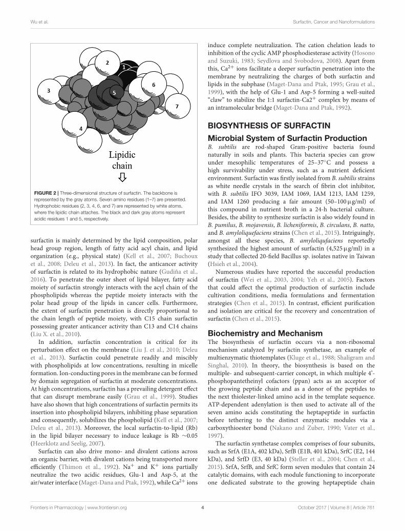
Further exploration of its three-dimensional structure(Figure 2) using high-resolution 1H NMR incorporated withmolecular imaging techniques has identified a minor polardomain at one side of the molecule formed by residues 2 and6 that face each other near acidic Glu-1 and Asp-5 side chains.On the opposite side, residue 4 slightly faces the hydrophobicdomain containing residues 3 and 7, making its amphiphilicnature and strong surface activity. At concentration lower thanCMC, the lipidic chain is freely extended in the solution but it isstill able to strongly involve in hydrophobic interactions, such aslipidmicelles or oligomers at air/water interface. Thus far, there isno report of intrinsic water solubility and lipophilicity (Log P) ofsurfactin. However, surfactin has a hydrophilic-lipophilic balance(HLB) of 10–12, indicating it can freely dissolve in a mixtureof water and oil phases (Gudiña et al., 2013). Furthermore, it isalso soluble in aqueous solution optimally at pH 8–8.5 (alkaline
FIGURE 1 | (A) Primary structure of surfactin, n = 9–11 (indicating the number
of CH2 group in the peptide chain). (B) Chemical structure of surfactin. Seven
amino acids are arranged in the cyclic ring connected with a fatty acid
(β-hydroxy) of the chain lengths 12–16 carbon atoms to form a cyclic lactone
ring.
waster) and organic solvents, such as ethanol, methanol, butanol,chloroform, and dichloromethane (Abdel-Mawgoud et al., 2008).
SURFACTIN-MEMBRANE INTERACTION
The biological activities of surfactin, including anticanceractivities are determined by the interactions with biologicalmembranes. In fact, the profile of membrane lipids in normaland cancer cells differs. For example, phosphatidylcholine, whichplays a key role in stabilizing the bilayer structure, was foundto be the major phospholipid component in cancer cells butnot in normal cells (Preetha et al., 2005; Hilvo et al., 2011).In general, surfactin destabilizes the membrane and disrupts itsintegrity (Bernheimer and Avigad, 1970) via several hypotheticalmechanisms, such as insertion into lipid bilayers, modification ofmembrane permeabilization via channel formation or diffusionof mono- and divalent ions across the membrane barrierand membrane solubilization by a detergent-like mechanism(Bouffioux et al., 2007).
Surfactin penetrates spontaneously into lipid membranes bymeans of hydrophobic interactions, which causes rearrangementof hydrocarbon order and membrane thickness (Maget-Danaand Ptak, 1995). In the lipid bilayer, surfactin displays aconformational change of peptide cycle which further mediatesthe interaction process (Maget-Dana and Ptak, 1995). Manystudies have shown that the sensitivity of lipid membrane to
Frontiers in Pharmacology | www.frontiersin.org 3 October 2017 | Volume 8 | Article 761
Wu et al. Surfactin, Cancer and Nanoformulations
FIGURE 2 | Three-dimensional structure of surfactin. The backbone is
represented by the gray atoms. Seven amino residues (1–7) are presented.
Hydrophobic residues (2, 3, 4, 6, and 7) are represented by white atoms,
where the lipidic chain attaches. The black and dark gray atoms represent
acidic residues 1 and 5, respectively.
surfactin is mainly determined by the lipid composition, polarhead group region, length of fatty acid acyl chain, and lipidorganization (e.g., physical state) (Kell et al., 2007; Buchouxet al., 2008; Deleu et al., 2013). In fact, the anticancer activityof surfactin is related to its hydrophobic nature (Gudiña et al.,2016). To penetrate the outer sheet of lipid bilayer, fatty acidmoiety of surfactin strongly interacts with the acyl chain of thephospholipids whereas the peptide moiety interacts with thepolar head group of the lipids in cancer cells. Furthermore,the extent of surfactin penetration is directly proportional tothe chain length of peptide moiety, with C15 chain surfactinpossessing greater anticancer activity than C13 and C14 chains(Liu X. et al., 2010).
In addition, surfactin concentration is critical for itsperturbation effect on the membrane (Liu J. et al., 2010; Deleuet al., 2013). Surfactin could penetrate readily and misciblywith phospholipids at low concentrations, resulting in micelleformation. Ion-conducting pores in themembrane can be formedby domain segregation of surfactin at moderate concentrations.At high concentrations, surfactin has a prevailing detergent effectthat can disrupt membrane easily (Grau et al., 1999). Studieshave also shown that high concentrations of surfactin permits itsinsertion into phospholipid bilayers, inhibiting phase separationand consequently, solubilizes the phospholipid (Kell et al., 2007;Deleu et al., 2013). Moreover, the local surfactin-to-lipid (Rb)in the lipid bilayer necessary to induce leakage is Rb ∼0.05(Heerklotz and Seelig, 2007).
Surfactin can also drive mono- and divalent cations acrossan organic barrier, with divalent cations being transported moreefficiently (Thimon et al., 1992). Na+ and K+ ions partiallyneutralize the two acidic residues, Glu-1 and Asp-5, at theair/water interface (Maget-Dana and Ptak, 1992), while Ca2+ ions
induce complete neutralization. The cation chelation leads toinhibition of the cyclic AMP phosphodiesterase activity (Hosonoand Suzuki, 1983; Seydlova and Svobodova, 2008). Apart fromthis, Ca2+ ions facilitate a deeper surfactin penetration into themembrane by neutralizing the charges of both surfactin andlipids in the subphase (Maget-Dana and Ptak, 1995; Grau et al.,1999), with the help of Glu-1 and Asp-5 forming a well-suited“claw” to stabilize the 1:1 surfactin-Ca2+ complex by means ofan intramolecular bridge (Maget-Dana and Ptak, 1992).
BIOSYNTHESIS OF SURFACTIN
Microbial System of Surfactin ProductionB. subtilis are rod-shaped Gram-positive bacteria foundnaturally in soils and plants. This bacteria species can growunder mesophilic temperatures of 25–37◦C and possess ahigh survivability under stress, such as a nutrient deficientenvironment. Surfactin was firstly isolated from B. subtilis strainsas white needle crystals in the search of fibrin clot inhibitor,with B. subtilis IFO 3039, IAM 1069, IAM 1213, IAM 1259,and IAM 1260 producing a fair amount (50–100µg/ml) ofthis compound in nutrient broth in a 24-h bacterial culture.Besides, the ability to synthesize surfactin is also widely found inB. pumilus, B. mojavensis, B. licheniformis, B. circulans, B. natto,and B. amyloliquefaciens strains (Chen et al., 2015). Intriguingly,amongst all these species, B. amyloliqufaciens reportedlysynthesized the highest amount of surfactin (4,525µg/ml) in astudy that collected 20-field Bacillus sp. isolates native in Taiwan(Hsieh et al., 2004).
Numerous studies have reported the successful productionof surfactin (Wei et al., 2003, 2004; Yeh et al., 2005). Factorsthat could affect the optimal production of surfactin includecultivation conditions, media formulations and fermentationstrategies (Chen et al., 2015). In contrast, efficient purificationand isolation are critical for the recovery and concentration ofsurfactin (Chen et al., 2015).
Biochemistry and MechanismThe biosynthesis of surfactin occurs via a non-ribosomalmechanism catalyzed by surfactin synthetase, an example ofmultienzymatic thiotemplates (Kluge et al., 1988; Shaligram andSinghal, 2010). In theory, the biosynthesis is based on themultiple- and subsequent-carrier concept, in which multiple 4’-phosphopantetheinyl cofactors (ppan) acts as an acceptor ofthe growing peptide chain and as a donor of the peptides tothe next thiolester-linked amino acid in the template sequence.ATP-dependent adenylation is then used to activate all of theseven amino acids constituting the heptapeptide in surfactinbefore tethering to the distinct enzymatic modules via acarboxythioester bond (Nakano and Zuber, 1990; Vater et al.,1997).
The surfactin synthetase complex comprises of four subunits,such as SrfA (E1A, 402 kDa), SrfB (E1B, 401 kDa), SrfC (E2, 144kDa), and SrfD (E3, 40 kDa) (Steller et al., 2004; Chen et al.,2015). SrfA, SrfB, and SrfC form seven modules that contain 24catalytic domains, with each module functioning to incorporateone dedicated substrate to the growing heptapeptide chain
Frontiers in Pharmacology | www.frontiersin.org 4 October 2017 | Volume 8 | Article 761

Wu et al. Surfactin, Cancer and Nanoformulations
(Peypoux et al., 1999). On the other hand, SrfD is responsiblefor surfactin initiation reaction (Steller et al., 2004). The surfactinsynthetase and the biosynthesis steps of surfactin are as shown inFigure 3.
To initiate surfactin biosynthesis, SrfD protein firstly mediatesthe transfer of the β-hydroxy fatty acid substrate to theN-terminal L-Glu-activating module of surfactin synthetase toinduce β-hydroxyacyl-glutamate formation (Steller et al., 2004).The substrate is then recognized and activated by the adenylationdomain (A domain with about 550 amino acids) (Conti et al.,1997), resulting in aminoacyladenylate formation via Mg2+-dependent hydrolysis and pyrophosphate release (Dieckmannet al., 1995). The initiation products are then elongated bySrfA and SrfB catalyzation via thioester bond cleavages andsimultaneous transpeptidation reactions (Peypoux et al., 1999).The aminoacyladenylate intermediates then bound to the freethiol group of the ppan cofactor before tethering to thiolationdomain (T domain or peptidyl carrier protein with ∼80amino acids) (Weber et al., 2000). Following, the intermediatesbound to the ppan cofactor can undergo subsequent catalyticreactions by other domains. In regard to regulation mechanisms,acyltransferase enzyme eliminates incorrect charging of theppan cofactor from the reaction centers by using its external
thioesterase activity (SrfTE-II) during the initiation process(Schwarzer et al., 2002).
Lastly, SrfC subunit catalyzes the condensation of the lastamino acid residue via peptide bond formation between twoadjacent substrates using condensation domain (C domain with∼450 amino acids) (Peypoux et al., 1999; Kraas et al., 2010).Furthermore, SrfC also catalyzes the release of lipoheptapeptidylintermediate from the surfactin synthetase complex that isstimulated by thioesterase domain (TE with ∼280 amino acids)embedded in SrfC. The resulting peptide is either releasedas a linear acid by hydrolysis or as a cyclic peptide by anintramolecular reaction with a nucleophile (Peypoux et al., 1999).
In addition to A, T, C, and TE domains, there areother optional domains with a distinctive role. For example,epimerization of domain E (with ∼450 amino acids) catalyzesthe racemization of T domain-bound amino acid, whereby Tdomain-bound L-Leu requires two E domains catalyzation frommodules 3 and 6. Then, the adjacent C domain transfers D-amino acid onto the growing peptide chain. In combinationwith D- and L-amino acids, this gives rise to a peptide witha unique conformation that interacts specifically with the cellmembranes (Linne andMarahiel, 2000; Seydlova and Svobodova,2008). Compared to surfactin biosynthesis, its excretion process
FIGURE 3 | Schematic diagram of surfactin synthetase complex for biosynthesis of cyclic surfactin. Surfactin synthetase complex is composed of three-modular SrfA,
three-modular SrfB, mono-modular SrfC and SrfD subunits, which is used to synthesize seven amino acids of surfactin. The peptide chain is elongated from left to
right until the linear product is cyclized by TE domain.
Frontiers in Pharmacology | www.frontiersin.org 5 October 2017 | Volume 8 | Article 761

Wu et al. Surfactin, Cancer and Nanoformulations
and the involved mechanism are still elusive. Nonetheless, anassumption has been suggested that surfactin can be excretedfrom its producers by passively diffusing across the cytoplasmicmembrane as no active transporter has been identified (Tsugeet al., 2001).
Regulation of Biosynthetic Genes inSurfactin BiosynthesisGenetic analysis plays a prerequisite role in controlling orregulating the surfactin biosynthesis (Sen, 2010). In fact, theassembly of surfactin synthetase complex is reflected in thechromosomal organization of its genes (Seydlova and Svobodova,2008). Surfactin synthesis is controlled by two genetic loci,namely srf and sfp (Sen, 2010). The surfactin synthetase complexis coded by the inducible operon, srfA locus (25 kb) with fourmodular open reading frame (ORF), such as ORF1 (srfAA),ORF2 (srfAB), ORF3 (srfAC), and ORF4 (srfAD) that encode thefour surfactin subunits (Hamoen et al., 2003). The srfB locus hasa similar function with comA locus that is responsible for geneticcompetence in B. subtilis (Nakano and Zuber, 1989; Nakanoet al., 1992). The srfA encodes some enzymes that are involvedin catalyzing the surfactin synthesis (Nakano et al., 1991). Forexample, srfAA encodes an enzyme that contains the amino acid-activating domain for Glu, Leu, and D-Leu while the amino-acid domains for Val, Asp, and L-Leu are found in an enzymeencoded by srfAB. In addition, srfAC is responsible for encodingan enzyme that contains L-Leu amino acid-activating domain.Lastly, srfAD encodes an enzyme that resembles the primarystructure of thioesterases family, with more sequence homologieswith type II thioesterases (Fuma et al., 1993; Peypoux et al., 1999;Kraas et al., 2010; Satpute et al., 2010).
Surfactin biosynthesis is also influenced by the expressionof sfp gene that mapped at 4 kb downstream of srfA locus. Itencodes an enzyme with 224 amino acids that belongs to thesuperfamily of 4’-phosphopantetheinases (Nakano et al., 1992).The sfp enzyme has two functions, in which it can be used asthe primers for non-ribosomal peptide synthesis, as well as forproducing cofactors containing holoform from inactive surfactinsynthetase apoform (Lambalot et al., 1996).
In addition to its role in surfactin biosynthesis, srfA geneexpression also correlates with the development of geneticcompetence of B. subtilis in response to exogenous DNA uptakein the stationary phase cultures of glucose-grown cells (D’Souzaet al., 1993). An example is that a nutritional stress duringthe late exponential phase could stimulate the global regulatorymechanisms, ComP-ComA and Spo0A-AbrB, thereby inducingthe expression of srfA (Hamoen et al., 2003). Other than that,comS gene that is located within or outside the srfA gene frameis also involved in B. subtilis competence development (Hamoenet al., 2003).
ANTICANCER PROPERTIES OFSURFACTIN
The structural and amphiphilic properties of surfactin havewidely contributed to its biological activities, including
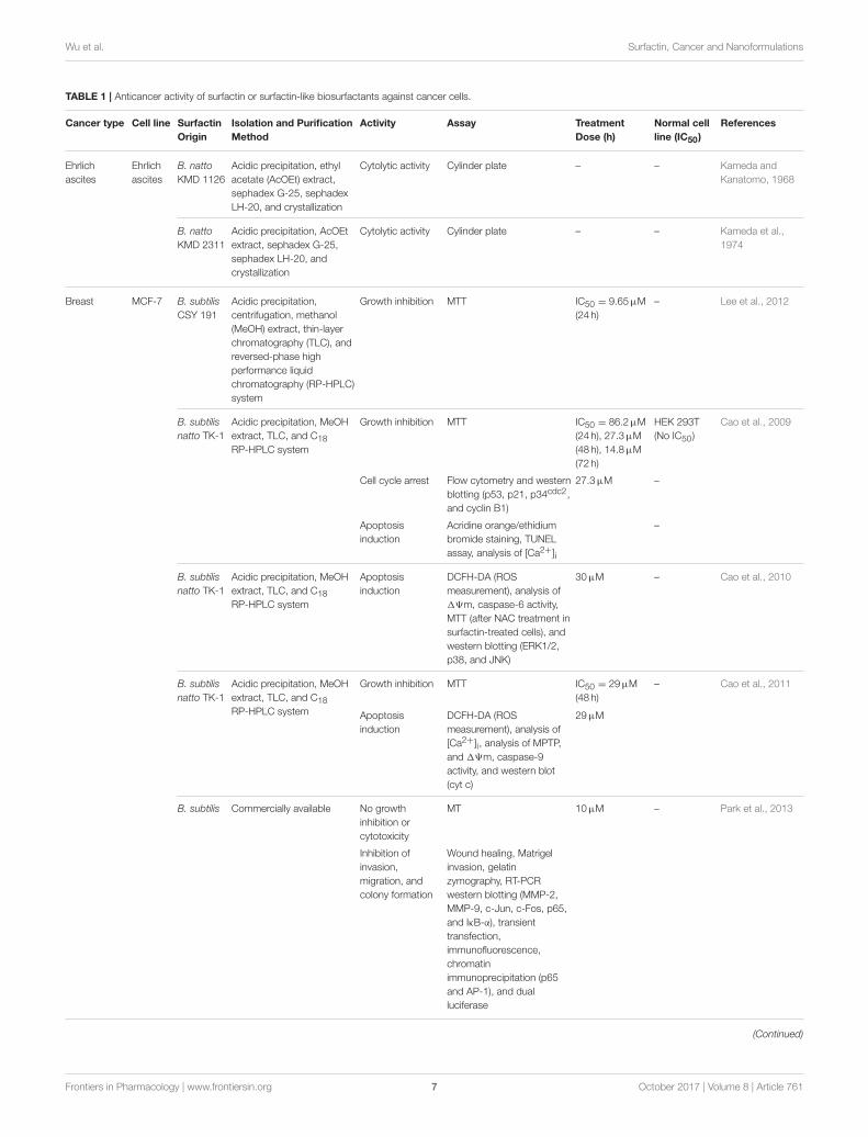
anticancer activity (Wang et al., 2009). In fact, the anticanceractivity of surfactin on a wide range of cancer cell lines has beenstudied recently, including antiproliferative and apoptotic effect,with its inhibitory action on cancer metastasis (Park et al., 2013).Surfactin-mediated programmed cell death is mainly causedby inducing apoptosis, which plays an important role in theregulation of tissue development and homeostasis, and presentsa promising strategy for cancer treatment (Sergeev, 2005).Surfactin-mediated cytotoxicity on different cancer models(Ehrlich ascites, breast and colon cancers, leukemia, hepatic,cervical, etc.) (Table 1) and the molecular mechanisms involvedin inducing apoptosis are presented in Figure 4.
Ehrlich Ascites CarcinomaKameda and his collaborators carried out two studies toinvestigate the anticancer activity of surfactin against Ehrlichascites carcinoma, which was originally established as an ascitestumor in mice. Surfactin isolated from a strain of B. natto(tentatively called KMD 1126) showed in vivo anticancer activityon Ehrlich ascites tumor (Kameda and Kanatomo, 1968). In 1974,Kameda et al. further revealed that B. natto KMD 2311 hadstronger cytolytic activity against Ehrlich ascites carcinoma astested using cylinder plate method among 113 strains of B. nattoisolated from straws in Japan (Kameda et al., 1974).
Breast CancerAmong all cancer types tested, the anticancer activity of surfactinon breast cancer cell lines has beenmost widely studied. B. subtilisCSY 191-derived surfactin was found to inhibit the growth ofhuman MCF-7 breast cancer cells in a dose-dependent manner,with IC50 of 9.65µM at 24 h. Higher surfactin production, whichwas obtained from co-fermentation of cheonggukjang and strainCSY 191, further enhanced the level of anticancer activity from2.6- to 5.1-fold compared to surfactin produced by strain CSY191 alone (Lee et al., 2012). Another strain of B. subtilis 573also significantly reduced the cell viability of T47D and MDA-MB-231 in a dose- and time-dependent manner, with 50%reduction (at 193µM) for T47D and MDA-MD-231 at 48 and72 h, respectively. Furthermore, inhibition of cell proliferationhad a 10% increase in cell cycle arrest at G0/G1 phase aftersurfactin treatment (193µM). It was also reported that 50% ofnormal MC-3T3-E1 breast cell growth were inhibited at the samedose (Duarte et al., 2014).
Furthermore, surfactin-like lipopeptides purified from B.subtilis Hs0121 also showed potent cytotoxicity on human Bcap-37 breast cancer cells. The results showed that C15 surfactin-likelipopeptide exerted the highest inhibition on the cell viability,with IC50 of 29 ± 2.4µM after 24 h exposure. The apoptoticeffect induced was associated with a significant decrease in theunsaturated degree of the cellular fatty acids in Bcap-37 cells dueto a reduction in the amount of fatty acids, thereby enhancingmembrane fluidization (Liu X. et al., 2010).
Surfactin isolated from B. subtilis natto TK-1 strains wasdemonstrated to inhibit the proliferation of MCF-7 cells in adose- and time-dependent manner, with IC50 at 24, 48, and 72 hbeing 86.2, 27.3, and 14.8µM, respectively. The cytotoxicity wastumor-selective as surfactin treatment (100µM) only reduced
Frontiers in Pharmacology | www.frontiersin.org 6 October 2017 | Volume 8 | Article 761
Wu et al. Surfactin, Cancer and Nanoformulations
TABLE 1 | Anticancer activity of surfactin or surfactin-like biosurfactants against cancer cells.
Cancer type Cell line Surfactin
Origin
Isolation and Purification
Method
Activity Assay Treatment
Dose (h)
Normal cell
line (IC50)
References
Ehrlich
ascites
Ehrlich
ascites
B. natto
KMD 1126
Acidic precipitation, ethyl
acetate (AcOEt) extract,
sephadex G-25, sephadex
LH-20, and crystallization
Cytolytic activity Cylinder plate – – Kameda and
Kanatomo, 1968
B. natto
KMD 2311
Acidic precipitation, AcOEt
extract, sephadex G-25,
sephadex LH-20, and
crystallization
Cytolytic activity Cylinder plate – – Kameda et al.,
1974
Breast MCF-7 B. subtilis
CSY 191
Acidic precipitation,
centrifugation, methanol
(MeOH) extract, thin-layer
chromatography (TLC), and
reversed-phase high
performance liquid
chromatography (RP-HPLC)
system
Growth inhibition MTT IC50 = 9.65µM
(24 h)
– Lee et al., 2012
B. subtilis
natto TK-1
Acidic precipitation, MeOH
extract, TLC, and C18
RP-HPLC system
Growth inhibition MTT IC50 = 86.2µM
(24 h), 27.3µM
(48 h), 14.8µM
(72 h)
HEK 293T
(No IC50)
Cao et al., 2009
Cell cycle arrest Flow cytometry and western
blotting (p53, p21, p34cdc2,
and cyclin B1)
27.3µM –
Apoptosis
induction
Acridine orange/ethidium
bromide staining, TUNEL
assay, analysis of [Ca2+]i
–
B. subtilis
natto TK-1
Acidic precipitation, MeOH
extract, TLC, and C18
RP-HPLC system
Apoptosis
induction
DCFH-DA (ROS
measurement), analysis of
19m, caspase-6 activity,
MTT (after NAC treatment in
surfactin-treated cells), and
western blotting (ERK1/2,
p38, and JNK)
30µM – Cao et al., 2010
B. subtilis
natto TK-1
Acidic precipitation, MeOH
extract, TLC, and C18
RP-HPLC system
Growth inhibition MTT IC50 = 29µM
(48 h)
– Cao et al., 2011
Apoptosis
induction
DCFH-DA (ROS
measurement), analysis of
[Ca2+]i, analysis of MPTP,
and 19m, caspase-9
activity, and western blot
(cyt c)
29µM
B. subtilis Commercially available No growth
inhibition or
cytotoxicity
MT 10µM – Park et al., 2013
Inhibition of
invasion,
migration, and
colony formation
Wound healing, Matrigel
invasion, gelatin
zymography, RT-PCR
western blotting (MMP-2,
MMP-9, c-Jun, c-Fos, p65,
and IκB-α), transient
transfection,
immunofluorescence,
chromatin
immunoprecipitation (p65
and AP-1), and dual
luciferase
(Continued)
Frontiers in Pharmacology | www.frontiersin.org 7 October 2017 | Volume 8 | Article 761

Wu et al. Surfactin, Cancer and Nanoformulations
TABLE 1 | Continued
Cancer type Cell line Surfactin
Origin
Isolation and Purification
Method
Activity Assay Treatment
Dose (h)
Normal cell
line (IC50)
References
T47D B. subtilis
573
Acidic precipitation,
centrifugation,
demineralized water
dissolution, and
freeze-drying
Growth inhibition MTS IC50 = 93µM
(48 h)
MC-3 T3-E1
(93µM at
72 h)
Duarte et al., 2014
Cell cycle arrest Flow cytometry –
MDA-
MB-231
B. subtilis Commercially available No growth
inhibition or
cytotoxicity
MT 10µM – Park et al., 2013
Inhibition of
invasion,
migration, and
colony formation
Wound healing, Matrigel
invasion, gelatin
zymography, RT-PCR
western blotting (MMP-2,
MMP-9, c-Jun, c-Fos, p65,
and IκB-α), transient
transfection,
immunofluorescence,
chromatin
immunoprecipitation (p65
and AP-1), and dual
luciferase
B. subtilis
573
Acidic precipitation,
centrifugation,
demineralized water
dissolution, and
freeze-drying
Growth inhibition MTS IC50 = 93µM
(72 h)
MC-3 T3-E1
(93µM at
72 h)
Duarte et al., 2014
Cell cycle arrest Flow cytometry 48µM –
Bcap-37 B. subtilis
Hs0121
Acidic precipitation,
centrifugation, lyophilization,
MeOH extract and C18
RP-HPLC
Growth inhibition MTT IC50 = 29 ±
2.4µM (24 h)
HaCaT
(97µM at
24 h)
Liu X. et al., 2010
Apoptosis
induction (fatty
acid composition
change)
Surface tension
measurement, flow
cytometry (propidium iodine
staining), nuclei staining,
and GC/MS (fatty acid
analysis)
12–96µM -
Colon HCT15 B. circulans
DMS-2
Acidic precipitation, alkaline
water dissolution,
lyophilization, and MeOH
extract, HPLC system
Growth inhibition MTT IC50 = 77µM
(24 h)
NIH/3T3
(482µM at
24 h)
Sivapathasekaran
et al., 2010
HT29 Growth inhibition MTT IC50 = 116µM
(24 h)
NIH/3T3
(482µM at
24 h)
Sivapathasekaran
et al., 2010
LoVo B. subtilis Commercially available Growth inhibition MTT IC50 = 26µM
(48 h)
– Kim et al., 2007
Cell cycle arrest Flow cytometry (Annexin
V/PI staining) and RT-PCR
(p53, p21waf/cip1, CDK2,
and cyclin E)
30µM
Apoptosis
induction
RT-PCR (Fas R, Fas L, Bax)
and western blotting (PARP,
cleaved-caspase 3, ERK,
p38, JNK, p85, and Akt)
Leukemia K562 B. subtilis
natto T-2
Acidic precipitation, MeOH
extract, charcoal treatment,
Pharmadex LH 20, and C18
HPLC system
Growth inhibition MTT IC50 =
10–20µM (24,
36 and 48 h)
– Wang et al., 2007
Cell cycle arrest Flow cytometry and western
blotting (cyclin D1,
p21waf/cip1, and p27)
7.7µM
(Continued)
Frontiers in Pharmacology | www.frontiersin.org 8 October 2017 | Volume 8 | Article 761

Wu et al. Surfactin, Cancer and Nanoformulations
TABLE 1 | Continued
Cancer type Cell line Surfactin
Origin
Isolation and Purification
Method
Activity Assay Treatment
Dose (h)
Normal cell
line (IC50)
Reference
Apoptosis
induction
Nuclei staining, caspase-3
activity, and western blotting
(caspase-3 and PARP)
B. subtilis
natto T-2
Acidic precipitation, MeOH
extract, charcoal treatment,
Pharmadex LH 20, and C18
HPLC system
Apoptosis
induction
TUNEL staining, lactate
dehydrogenase
measurement, analysis of
[Ca2+]i and western blotting
(ERK, p38, JNK, Bax, Bcl-2,
cyt c, and caspase-3)
15.4µM – Wang et al., 2009
Hepatocellular BEL7402 B. subtilis
HSO121
Acidic precipitation,
centrifugation, lyophilization,
MeOH extract, and C18
RP-HPLC system
Growth inhibition MTT IC50 = 35 ±
12µM (24 h)
HaCaT
(97µM at
24 h)
Liu X. et al., 2010
HepG2 B. natto
TK-1
Acidic precipitation, MeOH
extract, TLC, and C18
RP-HPLC system
Apoptosis
induction
DCFH-DA (ROS
measurement) and analysis
of [Ca2+]i,
41µM – Wang et al., 2013
Cervical HeLa B. subtilis
HSO121
Acidic precipitation,
centrifugation, lyophilization,
MeOH extract, and C18
RP-HPLC system
Growth inhibition MTT IC50 = 37 ±
4.5µM (24 h)
HaCaT
(97µM at
24 h)
Liu X. et al., 2010
B. subtilis Commercially available Growth inhibition MTT IC50 = 86.9µM
(16 h), 73.1µM
(24 h), 50.2µM
(48 h)
HaCaT
(97µM at
24 h)
Nozhat et al., 2012
Oral
epidermoid
KB-3-1 B. subtilis
HSO121
Acidic precipitation,
centrifugation, lyophilization,
MeOH extract, and C18
RP-HPLC system
Growth inhibition MTT IC50 = 57 ±
2.6µM (24 h)
HaCaT
(97µM at
24 h)
Liu X. et al., 2010
Pancreatic SW-
1990
B. subtilis
HSO121
Acidic precipitation,
centrifugation, lyophilization,
MeOH extract, and C18
RP-HPLC system
Growth inhibition MTT IC50 = 58 ±
1.6µM (24 h)
HaCaT
(97µM at
24 h)
Liu X. et al., 2010
Rat
melanoma
B16 B. subtilis
HSO121
Acidic precipitation,
centrifugation, lyophilization,
MeOH extract, and C18
RP-HPLC system
Growth inhibition MTT IC50 = 20 ±
1.5µM (24 h)
HaCaT
(97µM at
24 h)
Liu X. et al., 2010
20% viability of normal human HEK 283T embryonic cells.Surfactin also increased intracellular calcium concentration[Ca2+]i and induced apoptosis by arresting cells at G2/Mphase up to 47% at 48 h (Cao et al., 2009). Indeed, surfactinalso led to the accumulation of tumor suppressor p53 andcyclin kinase inhibitor p21waf1/cip1, and inhibited the activity ofG2-specific kinase, cyclin B1/p34cdc2 (Cao et al., 2009), all ofwhich are important for cell cycle phase transition. Cao et al.(2010) further demonstrated that surfactin-induced apoptosisoccurred via a reactive oxygen species/c-Jun N-terminal kinase(ROS/JNK)-mediated mitochondrial/caspase pathway. Surfactintreatment (30µM) stimulated high ROS generation, leading tomitochondrial permeability and membrane potential (19m)loss via JNK phosphorylation. These activities further led to
cytochrome c (cyt c) release and caspase-cascade reaction (Caoet al., 2010).
B. subtilis natto TK-1-purified surfactin can also inducea similar underlying mechanism of ROS/Ca2+-mediatedmitochondrial/caspase pathway to stimulate apoptosis. MCF-7 cells treated with surfactin (29µM) caused 50% viabilityinhibition while 80% inhibition was observed at 68µM.Intriguingly, only 15% inhibition of normal human L-02hepatic cell viability was observed at the highest dose tested(97µM), indicating surfactin’s non-toxicity to normal cells. HighROS generation was observed in surfactin-induced apoptosis,which is reversible by antioxidant N-acetylcysteine. The resultsfurther indicated that surfactin treatment initially induced ROSformation, leading to mitochondrial permeability transition
Frontiers in Pharmacology | www.frontiersin.org 9 October 2017 | Volume 8 | Article 761

Wu et al. Surfactin, Cancer and Nanoformulations
FIGURE 4 | Proposed mechanisms involved in in vitro anticancer activity of surfactin. The anticancer activity of surfactin is associated with growth inhibition, cell cycle
arrest, cell death (apoptosis), and metastasis inhibition. Surfactin treatment can inhibit cancer cell viability by inactivating the cell survival signaling pathways. Besides,
surfactin regulates cell cycle-regulatory proteins, which are pivotal for cell cycle phase transition to block the proliferation of cancer cells. The apoptotic effect (intrinsic
mitochondrial/caspase pathway) of surfactin is mediated by two different pathways that are triggered by high intracellular ROS formation, namely ERS/[Ca2+]i/ERK1/2
and JNK/19m /[Ca2+]i/Bax-to-Bcl-2 ratio/cyt c pathways. Surfactin-induced apoptosis is also associated with the changes in phospholipids composition that leads
to a significant decrease in unsaturated degree of cellular fatty acids. Apart from these, surfactin also inhibits the invasion, migration and colony formation of cancer
cells in the virtue of MMP-9 expression change that involves the inactivation of NF-κB, AP-1, PI3K/Akt, and ERK1/2 signaling pathways.
pore (MPTP) opening and 19m collapse. The resultantincrease in [Ca2+]i caused by the changes in mitochondrialpermeability was inhibited by 1,2-bis (2-aminophenoxy)ethane-N,N,N’,N’-tetraacetic acid (BAPTA-AM, a calciumchelator). These activities led to the release of cyt c andactivated caspase-9, later inducing apoptosis (Cao et al.,2011).
Owing to the efficacy of surfactin-induced growth inhibition,further investigation into breast cancer metastasis using
in vitro model was reported recently. Surfactin inhibited 12-O-tetradecanoylphorbol-13-acetate (TPA)-induced migration,invasion and colony formation by downregulating matrixmetalloprotenaise-9 (MMP-9) expression at 10µM, a dose lowerthan the suboptimal dose that had no cytotoxic effect on MCF-7and MDA-MB-231 cells. Surfactin attenuated TPA-inducedactivation and nuclear localization of nuclear factor-kappa B(NF-κB) and activating protein-1 (AP-1), and strongly repressedphosphatidylinositol 3-kinase (PI3K)/Akt and extracellular
Frontiers in Pharmacology | www.frontiersin.org 10 October 2017 | Volume 8 | Article 761

Wu et al. Surfactin, Cancer and Nanoformulations
signal-regulated kinase (ERK) signaling pathways. In fact,PI3K/Akt and ERK signaling pathways are involved in multiplecellular processes, such as cell survival, proliferation, apoptosis,and cell cycle (Gudiña et al., 2013). Altogether, surfactin-mediated inhibition of cell invasion and MMP-9 expressioninvolves the regulation of NF-κB, AP-1, PI3K/Akt, and ERKpathways (Park et al., 2013).
Colon CancerIn the literature, two investigations into surfactin-mediatedanticancer activity on colon cancer have been reported. Astudy showed that surfactin was able to inhibit the proliferationof LoVo colon cancer cells significantly in a dose- andtime-dependent manner, with IC50 of 26µM at 48 h. Theantiproliferative action induced by surfactin was mediated byapoptotic effect as shown by DNA fragmentation, morphologicalchange, altered levels of apoptosis-, and cell cycle-regulatoryproteins. For example, upregulation was observed for Fasreceptor (Fas R), Fas ligand (Fas L), cleaved poly (ADP-ribose)polymerase (PARP), cleaved caspase-3, p53 and p21waf1/cip1 whileCDK2 and cyclin E were downregulated in a dose- and time-dependent manner. In addition, surfactin treatment led to 10%of G0/G1 phase cell cycle arrest and induction of 40% apoptoticcells. Besides, the phosphorylation levels of PI3K/Akt signalingwere also suppressed upon surfactin treatment. These datasuggest that surfactin-mediated growth inhibition was associatedwith apoptosis induction and cell cycle arrest via the inhibition ofPI3K/Akt cell survival signaling (Kim et al., 2007).
Another study showed that purified surfactin-like lipopeptidesisolated from a marine B. circulans DMS-2 induced moderatecytotoxicity against HCT-15 and HT-29 colon cancer cells, withrespective IC50 of 77 and 116µM after 24 h treatment. At ahigher dose of 290µM, 90% inhibition of cell viability wasobserved. This study also demonstrated the anticancer activityof surfactin is tumor-selective—a high concentration of surfactin(482µM)was required to inhibit 50% of normal mouse NIH/3T3embryo fibroblast cell viability. It was reported that the crudebiosurfactant displayed very low cytotoxicity against cancer celllines, which is possibly due to the relatively low amount ofsurfactin-containing lipopeptides inside (Sivapathasekaran et al.,2010). It was further reported that IC50 induced by purifiedlipopeptides is comparable to the IC50 of surfactin (30µM) (Kimet al., 2007).
LeukemiaCrude cyclic lipopeptides (CLPs) purified from B. subtilis nattoT-2, with surfactin as a main constituent displayed cytotoxicityagainst human K562 leukemia cells at varying concentrations(2–62µM) for 24, 36, and 48 h. CLPs inhibited the growth ofK562 cells in a dose- and time-dependent manner, with the IC50
value between 10 and 20µM for 24–48 h treatment period. Incontrast, the viability of normal HLF primary lung fibroblastwas not affected by CLPs when the concentration is lower than30µM. The antiproliferative activity of surfactin was associatedwith cell cycle arrest and apoptosis induction. Surfactin treatmentresulted in a notable accumulation of cells in the G1 phase (36.5–57.6%) and increased the number of apoptotic cells. Apoptotic
activities induced by surfactin include morphological changeand enhanced the apoptosis-related proteins, such as caspase-3,cleaved PARP, p21waf1/cip1, and p27kip1 while remarkably reducedthe expression of cyclin D1, which plays a role to regulate G1phase progression (Wang et al., 2007).
Wang et al. (2009) further investigated the underlyingmechanism of apoptosis contributing to the anticancer activityof surfactin. They revealed that surfactin induced apoptosisvia [Ca2+]i/ERK-mediated mitochondrial/caspase pathway.Sustained increase in [Ca2+]i was observed after treatment ofsurfactin (15µM) for 3–9 h. Increased [Ca2+]i was associatedwith cell apoptosis and ERK phosphorylation. CLPs-inducedapoptosis in K562 cells was inhibited by PD98059 (an ERKinhibitor), but displayed no effect with p38 and JNK inhibitors,indicating ERK signaling as a critical role in apoptosis induction.It was also shown that apoptosis rate was partially decreasedafter reducing the [Ca2+]i with 1,2-Bis(2-aminophenoxy)ethane-N,N,N′,N′-tetraacetic acid tetrakis(acetoxymethyl ester)(BAPTA-AM) pretreatment. Collectively, this study showedthat the phosphorylation of ERK signaling caused by increased[Ca2+]i activated Bax, cyt c, and caspase 3, leading to apoptosis(Wang et al., 2009).
Hepatocellular CarcinomaSurfactin has also been shown to exhibit cytotoxic effect onhepatocellular carcinoma. Liu X. et al. (2010) demonstratedthat the purified surfactin-like lipopeptides isolated from B.subtilis HSO121 significantly inhibited the cell viability ofhuman Bel-7402 hepatoma cells, with IC50 of 35 ± 12µMcompared to normal human HaCaT keratinocyte cells (IC50 of97µM) at 24 h, indicating the specificity of anticancer actiontoward Bel-7402 cells. Furthermore, Wang et al. (2013) recentlyproposed the signaling network underlying the apoptosis ofHepG2 hepatoma cells induced by surfactin, resembling themechanisms identified in a breast cancer model. Sustained ROSgeneration and [Ca2+]i accumulation upon surfactin treatment(40µM) were responsible for apoptosis induction. The releaseof Ca2+ ions from inositol 1,4,5-trisphosphate and ryanodinereceptors channels contributed to increased [Ca2+]i, which inturn stimulated endoplasmic reticulum stress (ERS). Meanwhile,ERS was also induced by surfactin-produced ROS. Collectively,activated ERS reversibly increased [Ca2+]i and reduced thephosphorylation levels of ERK signaling pathway (Wang et al.,2013). Therefore, these findings suggest that surfactin inducesapoptosis in HepG2 cells via ROS/ERS/Ca2+-mediated ERKpathway.
Cervical CancerThe anticancer activity of surfactin on cervical cancer wasmainly tested on HeLa cell line. The studies were limited toexamining the inhibitory effect of surfactin on cell viabilitywith the underlying mechanism being investigated. Liu X. et al.(2010) showed that purified surfactin-like lipopeptides dose-dependently inhibited the viability of HeLa cancer cells, causing50% reduction at 37 ± 4.5µM after 24 h exposure. Withincreasing attention to the application of diverse biocompatiblenanoparticles in cancer treatment, surfactin C15 nanopeptide has
Frontiers in Pharmacology | www.frontiersin.org 11 October 2017 | Volume 8 | Article 761

Wu et al. Surfactin, Cancer and Nanoformulations
been shown to induce cytotoxicity in HeLa cells (Liu X. et al.,2010). With this approach, the cell viability was reduced withincreasing nanopeptide concentrations and exposure times. TheIC50 of surfactin causing 50% cell viability inhibition for 16,24, and 48 h was 86.9, 73.1, and 50.2µM, respectively. Furtherinvestigations exploring the molecular mechanisms underlyingthe anticancer activity of surfactin on HeLa cells as well as testingthe potential cytotoxic effect of surfactin on other cervical cancercell lines are desirable.
Other Cancer TypesThe inhibitory effect of surfactin or surfactin-like biosurfactantsagainst the growth of other cancer types, such as human oralepidermoid carcinoma, pancreatic, and rat melanoma cancerhas also been investigated. However, the assessment of theanticancer potential for this compound is only limited toexamining the metabolic activity of cancer cells using 3-(4,5-dimethylthiazol-2-yl)-2,5-diphenyl tetrazolium bromide (MTT)assay. For example, surfactin-like lipopeptide was found to causecytotoxicity against oral epidermoid (KB-3-1), pancreatic (SW-1990), and rat melanoma (B16) cancer after 24 h exposure, withtheir respective IC50 was 57 ± 2.6, 58 ± 1.6, and 20 ± 1.6µM(Liu X. et al., 2010).
Based on the studies on surfactin’s anticancer activity againstdifferent cancer models, surfactin is mainly isolated and purifiedfrom several strains of B. subtilis and B. natto, and one strainof B. circulans. Among these Bacillus sp., B. subtilis-producedsurfactin has the highest cytotoxic effect against cancer cellsfollowed by B. natto and B. circulans, which may be due to theirorigin and variations in cultivation, isolation, and purificationmethods. Although some researchers have shown the potentialanticancer promoting activity of surfactin, only few studieshave further investigated the mechanisms underlying surfactin-induced apoptosis. Some of these studies use a single cancercell line to examine the cytotoxicity of surfactin, without usingnormal cell lines as controls. It is inconclusive as to whether theanticancer effect is tumor-selective or only specific against thesingle cell line studied. In addition, a few studies using a singlemethod to evaluate the inhibitory effect of surfactin on cancercell proliferation, such as metabolic measurement assays, whichare mainly used to measure the metabolic activity of viable cellscould not truly represent the proliferation state of cancer cellsafter surfactin treatment. Furthermore, some promising resultsobtained in the in vitro study could not fully claim their potentialanticancer activity, thus further validation by means of in vivoexperiments are warranted.
TOXICITY OF SURFACTIN
There have been several studies on the anticancer effects ofsurfactin against cancer cell lines that indicate its selectivecytotoxicity toward cancer cells. Nonetheless, surfactin purifiedfrom B. subtilis 573 was shown to induce cytotoxicity againsthuman normal MCT-3T3-E1 fibroblast cell line at the sameconcentrations and exposure times that inhibited the viabilityof human T47D and MDA-MB-231 breast cancer cells (Duarteet al., 2014). Hwang et al. (2009) tested the acute toxicity of
surfactin C by oral administration to rats with different doses.At high dose of 1,000 or 2,000mg /kg, increased serum levels ofAlanine transaminase (ALT), Aspartate transaminase (AST), andalkaline phosphatase (ALP) were observed, indicating necrosisof hepatocyte. At lower dose, surfactin C did not show anytoxic effects during treatment period, and the no-observed-adverse-effect level (NOAEL) of surfactin C was determined tobe 500mg/kg (Hwang et al., 2009).
One of the major drawback of surfactin’s use as an anticanceragent is its hemolytic activity above 0.05 g/l (Dehghan-Noudeet al., 2005). To overcome this, linear forms of surfactin havebeen developed, and shown to be non-hemolytic while protectingerythrocytes against the detergent-like action of surfactin(Seydlova and Svobodova, 2008). No significant hemolysis wasobserved when linear surfactin was used up to 1,000µM.Moreover, a protective effect against Triton X-100 inducedhemolysis was found. The concentration at which this protectiveeffect happens is directly correlated to the CMC of the linearsurfactin, and inversely correlated to the acyl chain length of theproduct.
As the toxicity of surfactin may bring about serious sideeffects, cautious use of surfactin at safe doses is crucial. Althoughchemical modification of cyclic surfactin into linear structurehas been shown to have reduced toxicity, the therapeutic effectsof this linear compound is still unknown. In this regard, nano-formulations may be a more promising alternative for surfactindelivery for reduced toxicity and enhanced anticancer effects.
POTENTIAL NANO-DELIVERY OFSURFACTIN
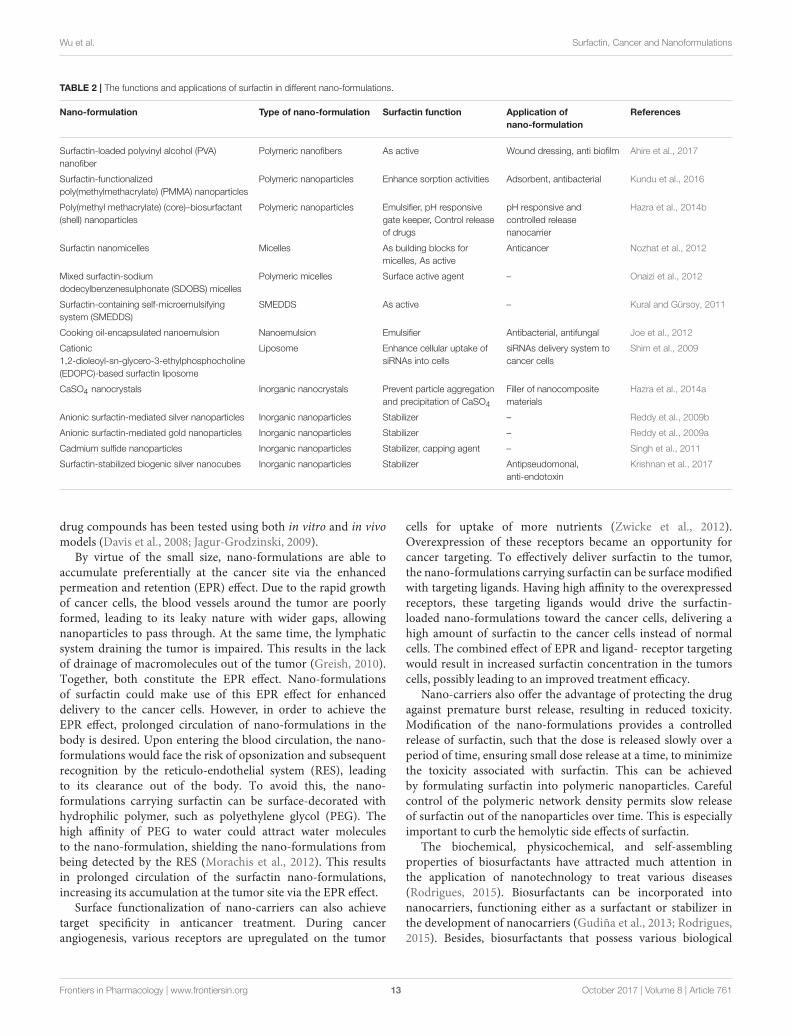
Delivery of surfactin nano-formulations for anticancer treatmenthas been largely unexplored to date. This section provides theinsights of surfactin nano-formulations in the literature, anddiscusses the potential of such formulations to be extended toanticancer therapeutic delivery of surfactin. A summary of thesenano-formulations can be found in Table 2.
Surfactin and Nano-FormulationsThe failure of conventional anticancer treatment are largelydue to non-specific distribution of drugs in the body, leadingto reduced efficient dose to the cancer cells, causing suboptimalresults, and excessive toxicity to normal cells (Khodabandehlooet al., 2016). In recent years, nano-formulations using nano-sizedcarriers (10–400 nm) have made considerable contributions incancer therapy. This claim is based on the advantages of nano-formulations offering high drug loading capacity, improvedbioavailability, prolonged circulation time, better cancertargeting, and the ease of manipulating drug release (Yu et al.,2010). To date, a variety of nanocarriers have been developed,including but not limited to, polymeric nanoparticles, micelles,dendrimers, liposomes, niosomes, solid lipid nanoparticles,gold nanoparticles, nanotubes, magnetic nanoparticles, proteinnanoparticles, micro-, and nano-emulsions (Cho et al., 2008;Yuan et al., 2016). The efficacy of these nanocarriers in delivering
Frontiers in Pharmacology | www.frontiersin.org 12 October 2017 | Volume 8 | Article 761
Wu et al. Surfactin, Cancer and Nanoformulations
TABLE 2 | The functions and applications of surfactin in different nano-formulations.
Nano-formulation Type of nano-formulation Surfactin function Application of
nano-formulation
References
Surfactin-loaded polyvinyl alcohol (PVA)
nanofiber
Polymeric nanofibers As active Wound dressing, anti biofilm Ahire et al., 2017
Surfactin-functionalized
poly(methylmethacrylate) (PMMA) nanoparticles
Polymeric nanoparticles Enhance sorption activities Adsorbent, antibacterial Kundu et al., 2016
Poly(methyl methacrylate) (core)–biosurfactant
(shell) nanoparticles
Polymeric nanoparticles Emulsifier, pH responsive
gate keeper, Control release
of drugs
pH responsive and
controlled release
nanocarrier
Hazra et al., 2014b
Surfactin nanomicelles Micelles As building blocks for
micelles, As active
Anticancer Nozhat et al., 2012
Mixed surfactin-sodium
dodecylbenzenesulphonate (SDOBS) micelles
Polymeric micelles Surface active agent – Onaizi et al., 2012
Surfactin-containing self-microemulsifying
system (SMEDDS)
SMEDDS As active – Kural and Gürsoy, 2011
Cooking oil-encapsulated nanoemulsion Nanoemulsion Emulsifier Antibacterial, antifungal Joe et al., 2012
Cationic
1,2-dioleoyl-sn-glycero-3-ethylphosphocholine
(EDOPC)-based surfactin liposome
Liposome Enhance cellular uptake of
siRNAs into cells
siRNAs delivery system to
cancer cells
Shim et al., 2009
CaSO4 nanocrystals Inorganic nanocrystals Prevent particle aggregation
and precipitation of CaSO4
Filler of nanocomposite
materials
Hazra et al., 2014a
Anionic surfactin-mediated silver nanoparticles Inorganic nanoparticles Stabilizer – Reddy et al., 2009b
Anionic surfactin-mediated gold nanoparticles Inorganic nanoparticles Stabilizer – Reddy et al., 2009a
Cadmium sulfide nanoparticles Inorganic nanoparticles Stabilizer, capping agent – Singh et al., 2011
Surfactin-stabilized biogenic silver nanocubes Inorganic nanoparticles Stabilizer Antipseudomonal,
anti-endotoxin
Krishnan et al., 2017
drug compounds has been tested using both in vitro and in vivomodels (Davis et al., 2008; Jagur-Grodzinski, 2009).
By virtue of the small size, nano-formulations are able toaccumulate preferentially at the cancer site via the enhancedpermeation and retention (EPR) effect. Due to the rapid growthof cancer cells, the blood vessels around the tumor are poorlyformed, leading to its leaky nature with wider gaps, allowingnanoparticles to pass through. At the same time, the lymphaticsystem draining the tumor is impaired. This results in the lackof drainage of macromolecules out of the tumor (Greish, 2010).Together, both constitute the EPR effect. Nano-formulationsof surfactin could make use of this EPR effect for enhanceddelivery to the cancer cells. However, in order to achieve theEPR effect, prolonged circulation of nano-formulations in thebody is desired. Upon entering the blood circulation, the nano-formulations would face the risk of opsonization and subsequentrecognition by the reticulo-endothelial system (RES), leadingto its clearance out of the body. To avoid this, the nano-formulations carrying surfactin can be surface-decorated withhydrophilic polymer, such as polyethylene glycol (PEG). Thehigh affinity of PEG to water could attract water moleculesto the nano-formulation, shielding the nano-formulations frombeing detected by the RES (Morachis et al., 2012). This resultsin prolonged circulation of the surfactin nano-formulations,increasing its accumulation at the tumor site via the EPR effect.
Surface functionalization of nano-carriers can also achievetarget specificity in anticancer treatment. During cancerangiogenesis, various receptors are upregulated on the tumor
cells for uptake of more nutrients (Zwicke et al., 2012).Overexpression of these receptors became an opportunity forcancer targeting. To effectively deliver surfactin to the tumor,the nano-formulations carrying surfactin can be surface modifiedwith targeting ligands. Having high affinity to the overexpressedreceptors, these targeting ligands would drive the surfactin-loaded nano-formulations toward the cancer cells, delivering ahigh amount of surfactin to the cancer cells instead of normalcells. The combined effect of EPR and ligand- receptor targetingwould result in increased surfactin concentration in the tumorscells, possibly leading to an improved treatment efficacy.
Nano-carriers also offer the advantage of protecting the drugagainst premature burst release, resulting in reduced toxicity.Modification of the nano-formulations provides a controlledrelease of surfactin, such that the dose is released slowly over aperiod of time, ensuring small dose release at a time, to minimizethe toxicity associated with surfactin. This can be achievedby formulating surfactin into polymeric nanoparticles. Carefulcontrol of the polymeric network density permits slow releaseof surfactin out of the nanoparticles over time. This is especiallyimportant to curb the hemolytic side effects of surfactin.
The biochemical, physicochemical, and self-assemblingproperties of biosurfactants have attracted much attention inthe application of nanotechnology to treat various diseases(Rodrigues, 2015). Biosurfactants can be incorporated intonanocarriers, functioning either as a surfactant or stabilizer inthe development of nanocarriers (Gudiña et al., 2013; Rodrigues,2015). Besides, biosurfactants that possess various biological
Frontiers in Pharmacology | www.frontiersin.org 13 October 2017 | Volume 8 | Article 761

Wu et al. Surfactin, Cancer and Nanoformulations
activities, including anticancer treatment, could be deliveredto the target site using nanocarriers. Given the amphiphilicnature, surfactin could effortlessly achieve optimal incorporationinto the nanocarriers. The following sections discuss theincorporation of surfactin in different nano-formulations, as wellas the different roles that surfactin play in these formulations.
Surfactin as an Active inNano-FormulationsSurfactin in Polymeric Nanoparticles and NanofibersPolymeric nanoparticles (NP) are submicron-sized colloidalparticles. Loading of drugs onto the NP can be achieved viaadsorption on the NP surface; or encapsulated within the NP.The polymer network could protect the drug against degradationactivities by enzymes found in the body (Bei et al., 2010). Drugrelease from the NP can take place by diffusion, hydrolysis ofthe polymeric network, enzymatic degradation of the polymer,or a combination of different mechanisms. Polymeric materialsused in pharmaceutical applications are often biodegradable innature, which include synthetic polymers such as poly(lactide)(PLA), poly(lactic-co-glycolide) (PLGA), poly(ε-caprolactone)(PCL); and natural polymers like chitosan, alginate, and albumin(Banik et al., 2016).
Surfactin-loaded polyvinyl alcohol (PVA) nanofiber wasformulated for wound dressing as well as to coat prothesticdevices to prevent biofilm formation and secondary infection.The nanofibers were produced by mixing surfactin and PVAsolutions, followed by gravity electrospinning. Increasing levelsof surfactin was found to decrease the diameter of the nanofiber,as shown by the scanning electron micrographs (Figure 5)(Ahire et al., 2017). Although this nanofiber did not exhibitantimicrobial activity, it significantly reduced the adhesion ofListeria monocytogenes EDGe (Ahire et al., 2017).
When synthesizing surfactin-functionalizedpoly(methylmethacrylate) (PMMA) nanoparticles for sorptionproperties, Kundu et al. (2016) found that these nanoparticlesdisplayed significant antibacterial activity against Escherichiacoli at 300µg/ml of the nanoparticles, due to the presenceof surfactin. Hence these nanoparticles can be used forboth sorption properties as well as for antibacterial effects.Moreover, the nanoparticles exhibited low hemolytic activity(Kundu et al., 2016). This shows the possibility of reduced
surfactin-associated toxicity when this biosurfactant isincorporated into a nanocarrier, allowing a safer delivery ofsurfactin to the patients.
Surfactin in Polymeric MicellesPolymeric micelles are made of amphiphilic copolymer witha core-shell structure. At concentration above the CMC,the amphiphilic polymers self-assemble into the micellarstructure, with the outer shell being hydrophilic and inner corehydrophobic. The hydrophobic core could encapsulate poorlywater-soluble drugs while the hydrophilic shell could help tosolubilize the drug. Micelles can appear in various forms, such asspherical, disc-like, and cylindrical structures depending on theconcentration (Akter et al., 2013).
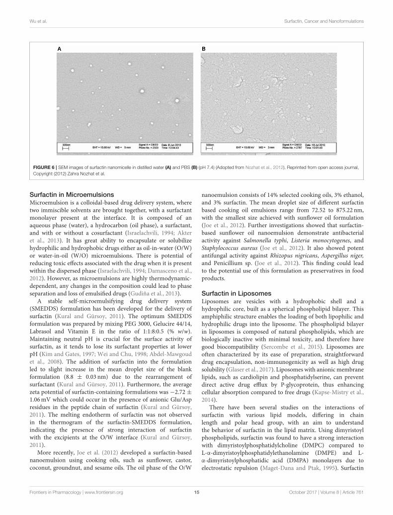
Recently, Nozhat et al. (2012) formulated surfactin C15nanopeptide into nanomicelles, and the scanning electronmicrographs are shown in Figure 6. Apart from the ability toform nanomicelles, surfactin C15 nanopeptide was also shown toarrest the growth of HeLa cell line in a dose- and time-dependentmanner. In fact, the ability of surfactin to form micelles cameas no surprise due to its surfactant properties. Figure 7 showsa proposed micellar structure of surfactin. Other than formingmicelles on its own, surfactin can also form mixed micelles withother surfactant, for example, the synthetic surfactant sodiumdodecylbenzenesulphonate (SDOBS), as demonstrated by Onaizi(Onaizi et al., 2012). The mixed surfactin-synthetic surfactantmay represent a greener and more sustainable formulation,along with reduced costs associated with the exclusive use ofbiosurfactants (Gudiña et al., 2013).
Pluronic block copolymers, which are composed ofpoly(ethylene oxide) (PEO) and poly(propylene oxide) (PPO)and arranged in PEO-PPO-PEO structure, have been widely usedin drug delivery systems (Alakhova and Kabanov, 2014). Uponself-assembly, the core of the micelles will be composed of PPOblocks, which allow for the encapsulation of hydrophobic drugs.The outer shell is composed of hydrated PEO moieties, suitablefor incorporation of hydrophilic drugs (Nambam and Philip,2012). Formulation scientists can make use of this structure forsurfactin delivery, as the compound could fit well in the micellarstructure. Being amphiphilic in nature, the hydrophilic part ofsurfactin could sit in the hydrophilic shell of the micelles, withits hydrophobic part staying in the core.
FIGURE 5 | SEM images of (A) PVA (10%, w/v) loaded with (B) 0.5% (w/v), (C) 1.0% (w/v), and (D) 1.5% (w/v) surfactin (Adopted from Ahire et al., 2017). Reprinted
with permission. Copyright (2017) Elsevier.
Frontiers in Pharmacology | www.frontiersin.org 14 October 2017 | Volume 8 | Article 761
Wu et al. Surfactin, Cancer and Nanoformulations
FIGURE 6 | SEM images of surfactin nanomicelle in distilled water (A) and PBS (B) (pH 7.4) (Adopted from Nozhat et al., 2012). Reprinted from open access journal,
Copyright (2012) Zahra Nozhat et al.
Surfactin in MicroemulsionsMicroemulsion is a colloidal-based drug delivery system, wheretwo immiscible solvents are brought together, with a surfactantmonolayer present at the interface. It is composed of anaqueous phase (water), a hydrocarbon (oil phase), a surfactant,and with or without a cosurfactant (Israelachvili, 1994; Akteret al., 2013). It has great ability to encapsulate or solubilizehydrophilic and hydrophobic drugs either as oil-in-water (O/W)or water-in-oil (W/O) microemulsions. There is potential ofreducing toxic effects associated with the drug when it is presentwithin the dispersed phase (Israelachvili, 1994; Damasceno et al.,2012). However, as microemulsions are highly thermodynamic-dependent, any changes in the composition could lead to phaseseparation and loss of emulsified drugs (Gudiña et al., 2013).
A stable self-microemulsifying drug delivery system(SMEDDS) formulation has been developed for the delivery ofsurfactin (Kural and Gürsoy, 2011). The optimum SMEDDSformulation was prepared by mixing PEG 3000, Gelucire 44/14,Labrasol and Vitamin E in the ratio of 1:1:8:0.5 (% w/w).Maintaining neutral pH is crucial for the surface activity ofsurfactin, as it tends to lose its surfactant properties at lowerpH (Kim and Gates, 1997; Wei and Chu, 1998; Abdel-Mawgoudet al., 2008). The addition of surfactin into the formulationled to slight increase in the mean droplet size of the blankformulation (8.8 ± 0.03 nm) due to the rearrangement ofsurfactant (Kural and Gürsoy, 2011). Furthermore, the averagezeta potential of surfactin-containing formulations was −2.72 ±1.06mV which could occur in the presence of anionic Glu/Aspresidues in the peptide chain of surfactin (Kural and Gürsoy,2011). The melting endotherm of surfactin was not observedin the thermogram of the surfactin-SMEDDS formulation,indicating the presence of strong interaction of surfactinwith the excipients at the O/W interface (Kural and Gürsoy,2011).
More recently, Joe et al. (2012) developed a surfactin-basednanoemulsion using cooking oils, such as sunflower, castor,coconut, groundnut, and sesame oils. The oil phase of the O/W
nanoemulsion consists of 14% selected cooking oils, 3% ethanol,and 3% surfactin. The mean droplet size of different surfactinbased cooking oil emulsions range from 72.52 to 875.22 nm,with the smallest size achieved with sunflower oil formulation(Joe et al., 2012). Further investigations showed that surfactin-based sunflower oil nanoemulsion demonstrate antibacterialactivity against Salmonella typhi, Listeria monocytogenes, andStaphylococcus aureus (Joe et al., 2012). It also showed potentantifungal activity against Rhizopus nigricans, Aspergillus niger,and Penicillium sp. (Joe et al., 2012). This finding could leadto the potential use of this formulation as preservatives in foodproducts.
Surfactin in LiposomesLiposomes are vesicles with a hydrophobic shell and ahydrophilic core, built as a spherical phospholipid bilayer. Thisamphiphilic structure enables the loading of both lipophilic andhydrophilic drugs into the liposome. The phospholipid bilayerin liposomes is composed of natural phospholipids, which arebiologically inactive with minimal toxicity, and therefore havegood biocompatibility (Sercombe et al., 2015). Liposomes areoften characterized by its ease of preparation, straightforwarddrug encapsulation, non-immunogenicity as well as high drugsolubility (Glaser et al., 2017). Liposomes with anionicmembranelipids, such as cardiolipin and phosphatidylserine, can preventdirect active drug efflux by P-glycoprotein, thus enhancingcellular absorption compared to free drugs (Kapse-Mistry et al.,2014).
There have been several studies on the interactions ofsurfactin with various lipid models, differing in chainlength and polar head group, with an aim to understandthe behavior of surfactin in the lipid matrix. Using dimyristoylphospholipids, surfactin was found to have a strong interactionwith dimyristoylphosphatidylcholine (DMPC) compared toL-α-dimyristoylphosphatidylethanolamine (DMPE) and L-α-dimyristoylphosphatidic acid (DMPA) monolayers due toelectrostatic repulsion (Maget-Dana and Ptak, 1995). Surfactin
Frontiers in Pharmacology | www.frontiersin.org 15 October 2017 | Volume 8 | Article 761

Wu et al. Surfactin, Cancer and Nanoformulations
FIGURE 7 | Proposed micelle formation using surfactin as building blocks.
penetration across the lipids is greater when the acyl chainlength of the phospholipids decreases (Maget-Dana and Ptak,1995; Liu X. et al., 2010). Furthermore, the miscibility betweensurfactin and phospholipids is higher for shorter acyl chains andgreater polar head group of phospholipids. Surfactin also has adestabilizing effect on dipalmitoylphosphatidylcholine (DPPC);but it can stabilize dipalmitoylphosphatidylethanolamine(DPPE) and dipalmitoylphospharidylserine (DPPS) molecularinteractions (Bouffioux et al., 2007). These findings indirectlycontribute to design of efficient surfactin in liposomes delivery
systems by manipulating the balance between the phospholipidscomposition in a liposome formulation.
To date, there is no reported development of surfactin-loadedliposome delivery system in the literature. Nonetheless,other lipopeptides such as marine somocystinamide A(ScA) has been successfully encapsulated into liposomesfor anticancer treatment. ScA was able to completely partitioninto phospholipids of 100 nm liposome (nanosome) andalter the membrane structure, which was composed ofcholesterol, 1,2-dioleoyl-sn-glycero-3-phosphoethanolamine,
Frontiers in Pharmacology | www.frontiersin.org 16 October 2017 | Volume 8 | Article 761

Wu et al. Surfactin, Cancer and Nanoformulations
1,2-dioleoyl-sn-glycero-3-phosphocholine (DSPC), scA,1,2-dioleoyl-sn-glycero-3-phosphoethanolamine-N-[methoxy(polyethyleneglycol)-2000] ammonium salt at 1:1:1:0.16:0.16molar ratio (Wrasidlo et al. (2008). Interestingly, ScA-loadedliposome induced cytotoxicity against a panel of cancer celllines in a manner comparable to free ScA. ScA altered thelipid compartment by inducing ceramide formation andaccumulation, which was associated with apoptotic caspase 8induction (Wrasidlo et al., 2008).
The proven interaction between surfactin and phospholipids,along with the successful formulation of lipopeptide ScA intoliposomes, provide a strong foundation for future developmentof surfactin-loaded liposomes. With a better understanding offormulation approaches and the characteristics of surfactin-loaded liposomes, this formulation would have great potential foranticancer treatment.
Surfactin as a Component of Nano-CarrierSurfactants have traditionally been widely used in manydrug delivery applications for their effects as wetting agents,solubilizing agents, surface tension reducer, emulsifier; as wellas use in dispersion and micellization (Lawrence, 1994). Theirapplications are not limited to conventional dosage forms, butalso extend to nano-formulations, such as in nano-micelles,nano-emulsions, niosomes, to name a few. In the quest forgreener, sustainable and renewable resources, biosurfactantsemerged as a decent alternative to replace synthetic surfactants(Marchant and Banat, 2012a). Surfactin, being derived frombacteria and having all desirable properties similar to othersurfactants, present itself as the ideal choice.
Incorporation of surfactin into a cationic 1,2-dioleoyl-sn-glycero-3-ethylphosphocholine (EDOPC)-based liposome wasshown to enhance the cellular delivery of small interfering RNAs(siRNA) to cancer cells. The cationic EDOPC-based liposomeswere formulated using 3, 6, and 14mol % surfactin in the lipidmembrane of liposomes. The size of the lipoplexes was not morethan 200 nm and the Zeta potential values were significantlyreduced following complexation with siRNAs (Shim et al., 2009).The 14% surfactin-containing liposomes did not cause significantreduction in the viability of HeLa cells (84.0 ± 8.1%) comparedto cells treated with surfactin-free liposomes (82.0± 0.5%) (Shimet al., 2009), proving that surfactin is safe at this concentration.The presence of surfactin in the liposomes was found to increasethe extent of cellular delivery of siRNA in the Hela cell line in aconcentration dependent manner. These findings indicated thatcationic surfactin liposomes could be used as a biocompatibledelivery system of nucleic acid based medicines, such as siRNA.
Some biosurfactants have been found to have membranedestabilizing effects at concentrations below their CMC. Theirstructure and nature allows them to interact with the lipidbilayer and cholesterol that sits in the membrane. Hence, somebiosurfactants are used as penetration enhancers to transdermaldelivery. Nicoli et al. (2010) investigated the potential use ofsurfactin to enhance skin delivery of aciclovir. Even thoughsurfactin increased the concentration of aciclovir in the epidermisby 2-fold, enhanced transport of aciclovir across the skin wasnot seen (Nicoli et al., 2010). This shows that surfactin, or other
lipopeptide biosurfactants, might not be suitable as penetrationenhancers for transdermal delivery, despite having the ability tointeract with the membrane.
The use of surfactin was also reported in the synthesis ofcalcium phosphate nanoparticles using reverse microemulsion.As a surface-active agent, surfactin plays a role in theformulation of emulsion to lower the interfacial tension, resultingin nano-sized droplets. Using different water/surfactin ratios,nanoparticles of diverse structures were produced, with theaverage particle size in the range of 16–200 nm (Maity et al.,2011). In surfactant-template/ultrasound-assisted nanocrystalssynthesis, surfactin was used to control the morphology andaspect ratio of nano-calcium sulfate (CaSO4) by adjusting themass ratio of surfactin/H2O and rhamnolipid/surfactin. Withincreasing surfactin concentration, the crystal morphology ofnano-CaSO4 was found to undergo gradual changes fromsubmicrometer-sized long rod to hexagonal plate, and then toplate-like appearance. Surfactin was shown to inhibit calciumsulfate precipitation, stabilizing the intermediate solution phasesduring production of the nanocrystals, resulting in nanocrystalsof different morphology (Hazra et al., 2014a). The same groupof researchers also synthesized PS(core)-biosurfactant (shell)nanoparticles as biocompatible and biodegradable drug deliveryvehicle. Incorporation of biosurfactant improved the process ofclassical emulsion polymerization. The unique surfactin coatedpoly(methyl methacrylate) (nPMMA) have successfully resultedin a pH-responsive nanocarrier, along with a tailored drug releaseprofile (Hazra et al., 2014b).
In general, HLB, critical packing parameter (CPP) andWinsorR ratio could be used to design a successful microemulsion(Gudiña et al., 2013). HLB affects the stability of the emulsion andthe relative contribution of hydrophilic and lipophilic groups ofa surfactant. Surfactants with low HLB (3–6) value indicate thetendency to form W/O microemulsions while high HLB (8–18)tends to form O/W microemulsions. For surfactants with highHLB value (>20), a cosurfactant is usually required to reducetheir effective HLB (Gudiña et al., 2013). The CPP parameter isused tomeasure the ability of a surfactant to form aggregates withrespect to its geometries (Gudiña et al., 2013).Winson R indicatesthe ratio of total energy (per unit area of the interface) for asurfactant to interact in O and W phases and is also dependenton environmental factors (Gudiña et al., 2013).
The ability of surfactin to be formulated into microemulsionscan be determined by its experimental HLB andCPP values (Shenet al., 2011; Gudiña et al., 2013). Based on the HLB (10–12) andCPP (0.1435) values, surfactin could form O/W microemulsionswhen the alkaline condition is maintained. Only a small amountof surfactin is required to formulate a microemulsion for drugdelivery as it has high surface activity (Gudiña et al., 2013).
Surfactin as a Stabilizer in theNano-FormulationsIn addition to acting as a vital component or enhancerduring nanoparticles development, surfactin has also shown tofunction as a good stabilizing and capping agent in developingmetal nanoparticles due to the surface-active properties. Reddyand team used different proton concentrations to cause the
Frontiers in Pharmacology | www.frontiersin.org 17 October 2017 | Volume 8 | Article 761

Wu et al. Surfactin, Cancer and Nanoformulations
conformational change of surfactin that is used to stabilize goldand silver nanoparticles (Reddy et al., 2010). The synthesizednanoparticles were found to be stable for 2 months (Reddyet al., 2009a,b). Besides gold and silver nanoparticles, surfactinproduced by B. amyloliquifaciens KSU-109 was also used as astabilizer in cadmium sulfide nanoparticles (Cds-NPs). The Cds-NPs was synthesized by mixing 0.005% surfactin with 1mM Cdsin 1:1 ratio (v/v) and 10mM sodium sulfide at pH 7.2. The Cds-NPs was found to be stable for 4 months (Singh et al., 2011).More recently, surfactin-producing Brevibacillus brevis KN8(2)was used to stabilize a nanocrystalline silver nanoparticles(AgNPs) (Krishnan et al., 2017). Surfactin-stabilized AgNPsinduced a minimum inhibitory concentration of 10µg/mlagainst Pseudomonas aeruginosa, which is a causative agent ofdiabetic foot infection. Additionally, the AgNPs ameliorated theP. aeruginosa-infected wounds of diabetic mice (Krishnan et al.,2017), suggesting that surfactin-stabilized AgNPs confers thetreatment for gram negative bacteria-infected diabetic wounds.
Surfactin Nano-Formulations forAnticancer TreatmentThe great potential of surfactin for anticancer treatment not onlycomes from its proven cytotoxicity to cancer cells, but also dueits multiple roles when incorporated in nano-formulations. Theamphiphilic structure and surface-active properties of surfactinmake it an ideal candidate to be incorporated into nano-formulations. Polymeric micelles, liposomes, and niosomes areparticularly suitable, as surfactin can easily position itself withinthe hydrophobic/hydrophilic core-shell structure of such nano-formulations. Micro- and nano-emulsions are great choicestoo, where surfactin can be dispersed well in the formulation.Moreover, the use of surfactants is common as stabilizer andemulsifier in emulsions.
Apart from functioning as the active compound used fortherapeutic purposes, surfactin can be incorporated to improvethe formulation. For example, in a nanoemulsion, surfactin canbe added to act as an emulsifier and an active for its anticanceractivities. With this combination, the use of surfactin in thenano-formulation could then generate double actions for betterdelivery and enhanced efficacy of cancer treatment. In effect, itcould even simplify the formulation process as less materials areneeded to produce the nano-formulations, when surfactin can bethe carrier, and the load at the same time.
Drug formulation scientists could also utilize the uniquefeatures of surfactin to incorporate it as one of the components ofthe nanocarrier, such as the use of surfactin as building blocks inthe formulation of micelles, liposomes and niosomes. This can beexemplified by the surfactin nanomicelles mentioned in sectionSurfactin in Polymeric Micelles, where surfactin self assemblesinto micelles for its own delivery. In this regard, pure surfactincould be used without the need of other materials, ensuring themaximum dose of surfactin to be delivered to the cancer cells.
There has been increasing interests in combination therapy ofmultiple drugs incorporated in a single nano-formulation, givingrise to higher therapeutic effect. For example, a mesoporous silicananoparticle was used to encapsulate topotecan and quercetin
against triple negative breast cancer cells (MDA-MB-231) andmultidrug resistance breast cancer cells (MCF-7). The treatmentsignificantly enhanced the drug uptake efficiency by cancer cellswhile thwarted the uptake by normal cells (Murugan et al.,2016). This is also an area to explore for surfactin nano-formulations, whereby a combination of surfactin and anotherchemotherapeutic drug can be loaded into the nano-formulation,with surfactin acting as an adjuvant for anticancer treatment.
Surfactin nano-formulation is an area of great interest andsignificant potential for formulation scientists, yet remainslargely undiscovered. There is ample space for research anddevelopment in utilizing surfactin nano-formulation as animproved nanomedicine strategy to combat cancer.
CONCLUSION
In summary, numerous studies have shown surfactin’s anticancerpromoting activity against a few cancer types, many focusingon breast cancer. Surfactin-induced anticancer activities involvegrowth inhibition, cell cycle arrest, apoptosis, and metastasisarrest (invasion, migration, and colony formation). Growthinhibition caused by surfactin is mediated by reducing thePI3K/Akt and ERK cell survival signaling pathways. Besides,surfactin blocks the proliferation of cancer cells by regulatingcell-cycle regulatory proteins such as tumor suppressor p53 andits downstream effector p21waf1/cip1, CDK2, cyclin B1, cyclinE, p34cdc2, and p27kip1. Surfactin inhibits cancer metastasisby downregulating MMP-9 expression that is mediated by NF-κB, AP-1, PI3K/Akt, and ERK1/2 inactivation. The mechanismsunderlying surfactin-induced apoptosis are mainly associatedwith ROS formation, which could either induce ERS or19m change followed by increasing [Ca2+]i. Increased [Ca2+]istimulates the release of cyt c and activates the caspase-cascadereaction to induce apoptosis. It is notable that surfactin can alsoinduce hemolytic activity and may selectively exert cytotoxicityto normal cells, all of which limits its application as an anticanceragent.
To improve the anticancer effect of surfactin, a promisingstrategy would be the use of nano-formulations due to theirability to achieve the drug selectivity to cancer cells withminimal toxicity to normal cells. Given its amphiphilic nature,surfactin presents itself as a good candidate for incorporationinto various nano-formulations, such as polymeric NPs, micelles,microemulsions, liposomes, to name a few. Numerous surfactinnano-formulations have been reported in the literature. Surfactinnanomicelles (surfactin C15 nanopeptide) have been formulatedand shown to induce cytotoxic effect on HeLa cancer cells.A new surfactin-SMEDDS formulation has been developed,though its anticancer activity requires further confirmation.To date, no liposome-borne surfactin formulation has beendeveloped, even though surfactin could interact with differentlipid models, implying its great potential to be incorporatedinto liposome. There have also been various surfactin nano-formulations reported designed for other purposes, where drugformulation scientists could pick these up and extend them intosurfactin anticancer applications. In fact, this presents a greatopportunity for further advances in surfactin nano-formulations.
Frontiers in Pharmacology | www.frontiersin.org 18 October 2017 | Volume 8 | Article 761

Wu et al. Surfactin, Cancer and Nanoformulations
The potential improved anticancer activity of surfactin nano-formulations thus offers much room for establishment.
AUTHOR CONTRIBUTIONS
The literature review and manuscript writing were performed byYW, LC, and SN, while BG, KC, and LL provided vital guidance
and insight for the writing. The research topic was conceptualizedby LC and BG.
ACKNOWLEDGMENTS
KC thanks the grants from the University of Malaya (GA001-2016, GA002-2016).
REFERENCES
Abdel-Mawgoud, A. M., Aboulwafa, M. M., and Hassouna, N. A. (2008).
Characterization of surfactin produced by Bacillus subtilis isolate BS5. Appl.
Biochem. Biotechnol. 150, 289–303. doi: 10.1007/s12010-008-8153-z
Ahire, J. J., Robertson, D. D., van Reenen, A. J., and Dicks, L. M. T. (2017).
Surfactin-loaded polyvinyl alcohol (PVA) nanofibers alters adhesion of Listeria
monocytogenes to polystyrene. Mater. Sci. Eng. C Mater. Biol. Appl. 77, 27–33.
doi: 10.1016/j.msec.2017.03.248
Akter, N., Radiman, S., Mohamed, F., and Reza, M. I. (2013). Self-assembled
potential bio nanocarriers for drug delivery. Mini Rev. Med. Chem. 13,
1327–1339. doi: 10.2174/1389557511313090007
Alakhova, D. Y., and Kabanov, A. V. (2014). Pluronics and MDR reversal: an
update.Mol. Pharm. 11, 2566–2578. doi: 10.1021/mp500298q
Alfarouk, K. O., Stock, C. M., Taylor, S., Walsh, M., Muddathir, A. K., Verduzco,
D., et al. (2015). Resistance to cancer chemotherapy: failure in drug response
from ADME to P-gp. Cancer Cell Int. 15, 71. doi: 10.1186/s12935-015-
0221-1
Arima, K., Kakinuma, A., and Tamura, G. (1968). Surfactin, a crystalline
peptidelipid surfactant produced by Bacillus subtilis: isolation, characterization
and its inhibition of fibrin clot formation. Biochem. Biophys. Res. Commun. 31,
488–494.
Banat, I. M., Franzetti, A., Gandolfi, I., Bestetti, G., Martinotti, M.
G., Fracchia, L., et al. (2010). Microbial biosurfactants production,
applications and future potential. Appl. Microbiol. Biotechnol. 87, 427–444.
doi: 10.1007/s00253-010-2589-0
Banik, B. L., Fattahi, P., and Brown, J. L. (2016). Polymeric nanoparticles:
the future of nanomedicine. Nanomed. Nanobiotechnol. 8, 271–299.
doi: 10.1002/wnan.1364
Bei, D., Meng, J., and Youan, B. B. (2010). Engineering nanomedicines
for improved melanoma therapy: progress and promises. Nanomedicine 5,
1385–1399. doi: 10.2217/nnm.10.117
Bernheimer, A. W., and Avigad, L. S. (1970). Nature and properties of a
cytolytic agent produced by Bacillus subtilis. J. Gen. Microbiol. 61, 361–369.
doi: 10.1099/00221287-61-3-361
Boettcher, C., Kell, H., Holzwarth, J. F., and Vater, J. (2010). Flexible
loops of thread-like micelles are formed upon interaction of L-alpha-
dimyristoyl-phosphatidylcholine with the biosurfactant surfactin as
revealed by cryo-electron tomography. Biophys. Chem. 149, 22–27.
doi: 10.1016/j.bpc.2010.03.006
Bouffioux, O., Berquand, A., Eeman, M., Paquot, M., Dufrene, Y. F., Brasseur,
R., et al. (2007). Molecular organization of surfactin-phospholipid monolayers:
effect of phospholipid chain length and polar head. Biochim. Biophys. Acta 1768,
1758–1768. doi: 10.1016/j.bbamem.2007.04.015
Buchoux, S., Lai-Kee-Him, J., Garnier, M., Tsan, P., Besson, F., Brisson, A.,
et al. (2008). Surfactin-triggered small vesicle formation of negatively charged
membranes: a novel membrane-lysis mechanism. Biophys. J. 95, 3840–3849.
doi: 10.1529/biophysj.107.128322
Byeon, S. E., Lee, Y. G., Kim, B. H., Shen, T., Lee, S. Y., Park, H. J., et al. (2008).
Surfactin blocks NO production in lipopolysaccharide-activated macrophages
by inhibiting NF-kappaB activation. J. Microbiol. Biotechnol. 18, 1984–1989.
doi: 10.4014/jmb.0800.189
Cao, X. H.,Wang, A. H.,Wang, C. L., Mao, D. Z., Lu, M. F., Cui, Y. Q., et al. (2010).
Surfactin induces apoptosis in human breast cancer MCF-7 cells through a
ROS/JNK-mediated mitochondrial/caspase pathway. Chem. Biol. Interact. 183,
357–362. doi: 10.1016/j.cbi.2009.11.027
Cao, X. H., Zhao, S. S., Liu, D. Y., Wang, Z., Niu, L. L., Hou, L. H., et al.
(2011). ROS-Ca2+ is associated withmitochondria permeability transition pore
involved in surfactin-induced MCF-7 cells apoptosis. Chem. Biol. Interact. 190,
16–27. doi: 10.1016/j.cbi.2011.01.010
Cao, X., Wang, A. H., Jiao, R. Z., Wang, C. L., Mao, D. Z., Yan, L., et al. (2009).
Surfactin induces apoptosis and G(2)/M arrest in human breast cancer MCF-7
cells through cell cycle factor regulation. Cell Biochem. Biophys. 55, 163–171.
doi: 10.1007/s12013-009-9065-4
Carrillo, C., Teruel, J. A., Aranda, F. J., and Ortiz, A. (2003). Molecular mechanism
of membrane permeabilization by the peptide antibiotic surfactin. Biochim.
Biophys. Acta 1611, 91–97. doi: 10.1016/S0005-2736(03)00029-4
Chen, W. C., Juang, R. S., and Wei, Y. H. (2015). Applications of a lipopeptide
biosurfactant, surfactin, produced by microoganisms. Biochem. Eng. J. 103,
158–169. doi: 10.1016/j.bej.2015.07.009
Chiewpattanakul, P., Phonnok, S., Durand, A., Marie, E., and Thanomsub, B. W.
(2010). Bioproduction and anticancer activity of biosurfactant produced by the
dematiaceous fungus Exophiala dermatitidis SK80. J. Microbiol. Biotechnol. 20,
1664–1671. doi: 10.4014/jmb.1007.07052
Cho, K., Wang, X., Nie, S., Chen, Z. G., and Shin, D. M. (2008). Therapeutic
nanoparticles for drug delivery in cancer. Clin. Cancer Res. 14, 1310–1316.
doi: 10.1158/1078-0432.CCR-07-1441
Conti, E., Stachelhaus, T., Marahiel, M. A., and Brick, P. (1997). Structural basis for
the activation of phenylalanine in the non-ribosomal biosynthesis of gramicidin
S. EMBO J. 16, 4174–4183.
Damasceno, B. P., Dominici, V. A., Urbano, I. A., Silva, J. A., Araújo, I. B., Santos-
Magalhães, N. S., et al. (2012). Amphotericin B microemulsion reduces toxicity
and maintains the efficacy as an antifungal product. J. Biomed. Nanotechnol. 8,
290–300. doi: 10.1166/jbn.2012.1374
Das, P., Mukherjee, S., and Sen, R. (2008). Antimicrobial potential of a lipopeptide
biosurfactant derived from a marine Bacillus circulans. J. Appl. Microbiol. 104,
1675–1684. doi: 10.1111/j.1365-2672.2007.03701
Davis, M. E., Chen, Z. G., and Shin, D. M. (2008). Nanoparticle therapeutics: an
emerging treatment modality for cancer. Nat. Rev. Drug Discov. 7, 771–782.
doi: 10.1038/nrd2614
Dehghan-Noude, G., Housaindokht, M., and Bazzaz, B. S. (2005). Isolation,
characterization, and investigation of surface and hemolytic activities of a
lipopeptide biosurfactant produced by Bacillus subtilisATCC 6633. J. Microbiol.
43, 272–276.
Deleu, M., Lorent, J., Lins, L., Brasseur, R., Braun, N., El Kirat, K., et al. (2013).
Effects of surfactin on membrane models displaying lipid phase separation.
Biochim. Biophys. Acta 1828, 801–815. doi: 10.1016/j.bbamem.2012.11.007
Demain, A. L., and Sanchez, S. (2009). Microbial drug discovery: 80 years of
progress. J. Antibiot. 62, 5–16. doi: 10.1038/ja.2008.16
Dey, G., Bharti, R., Sen, R., and Mandal, M. (2015). Microbial amphiphiles: a
class of promising new-generation anticancer agents. Drug Discov. Today 20,
136–146. doi: 10.1016/j.drudis.2014.09.006
Dieckmann, R., Lee, Y. O., van Liempt, H., von Dohren, H., and Kleinkauf,
H. (1995). Expression of an active adenylate-forming domain of peptide
synthetases corresponding to acyl-CoA-synthetases. FEBS Lett. 357, 212–216.
D’Souza, C., Nakano, M. M., Corbell, N., and Zuber, P. (1993). Amino-acylation
site mutations in amino acid-activating domains of surfactin synthetase: effects
on surfactin production and competence development in Bacillus subtilis. J.
Bacteriol. 175, 3502–3510
Duarte, C., Gudina, E. J., Lima, C. F., and Rodrigues, L. R. (2014). Effects of
biosurfactants on the viability and proliferation of human breast cancer cells.
AMB Express 4, 40. doi: 10.1186/s13568-014-0040-0
Frontiers in Pharmacology | www.frontiersin.org 19 October 2017 | Volume 8 | Article 761

Wu et al. Surfactin, Cancer and Nanoformulations
Dy, G. K., and Adjei, A. A. (2013). Understanding, recognizing, and managing
toxicities of targeted anticancer therapies. CA Cancer J. Clin. 63, 249–279.
doi: 10.3322/caac.21184
Fracchia, L., Banat, J. J., Cavallo, M., and Ceresa, C. (2015). Potential therapeutic
applications of microbial surface-active compounds. Bioengineering 2,
144–162. doi: 10.3934/bioeng.2015.3.144
Fuma, S., Fujishima, Y., Corbell, N., D’Souza, C., Nakano, M. M., Zuber, P., et al.
(1993). Nucleotide sequence of 5’ portion of srfA that contains the region
required for competence establishment in Bacillus subtilus. Nucleic Acids Res.
21, 93–97.
Geiger, T., and Clarke, S. (1987). Deamidation, isomerization, and racemization at
asparaginyl and aspartyl residues in peptides. J. Biol. Chem. 262, 785–794.
Glaser, T., Han, I., Wu, L., and Zeng, X. (2017). Targeted
nanotechnology in glioblastoma multiforme. Front. Pharmacol. 8:166.
doi: 10.3389/fphar.2017.00166
Grau, A., Gomez Fernandez, J. C., Peypoux, F., and Ortiz, A. (1999). A study on
the interactions of surfactin with phospholipid vesicles. Biochim. Biophys. Acta
1418, 307–319.
Greish, K. (2010). “Enhanced Permeability and Retention (EPR) effect for
anticancer nanomedicine drug targeting,” in Cancer Nanotechnology: Methods
and Protocols, eds S. R. Grobmyer and B. M. Moudgil (Gainsville, FL: Humana
Press), 25–39.
Gudiña, E. J., Rangarajan, V., Sen, R., and Rodrigues, L. R. (2013). Potential
therapeutic applications of biosurfactants. Trends Pharmacol. Sci. 34, 667–675.
doi: 10.1016/j.tips.2013.10.002
Gudiña, E. J., Teixeira, J. A., and Rodrigues, L. R. (2016). Biosurfactants produced
by marine microorganisms with therapeutic applications. Mar. Drugs 14:E38.
doi: 10.3390/md14020038
Hamoen, L. W., Venema, G., and Kuipers, O. P. (2003). Controlling competence
in Bacillus subtilis: shared use of regulators. Microbiology 149(Pt 1), 9–17.
doi: 10.1099/mic.0.26003-0
Hazra, C., Bari, S., Kundu, D., Chaudhari, A., Mishra, S., and Chatterjee, A.
(2014a). Ultrasound-assisted/biosurfactant-templated size-tunable synthesis
of nano-calcium sulfate with controllable crystal morphology. Ultrason.
Sonochem. 21, 1117–1131. doi: 10.1016/j.ultsonch.2013.12.020
Hazra, C., Kundu, D., Chatterjee, A., Chaudhari, A., and Mishra, S. (2014b).
Poly (methyl methacrylate)(core)–biosurfactant (shell) nanoparticles: Size
controlled sub-100nm synthesis, characterization, antibacterial activity,
cytotoxicity and sustained drug release behavior. Colloids Surf. A. Physicochem.
Eng. Aspects 449, 96–113. doi: 10.1016/j.colsurfa.2014.02.051
Heerklotz, H., and Seelig, J. (2001). Detergent-like action of the antibiotic
peptide surfactin on lipid membranes. Biophys. J. 81, 1547–1554.
doi: 10.1016/S0006-3495(01)75808-0
Heerklotz, H., and Seelig, J. (2007). Leakage and lysis of lipid membranes
induced by the lipopeptide surfactin. Eur. Biophys. J. 36, 305–314.
doi: 10.1007/s00249-006-0091-5
Hilvo, M., Denkert, C., Lehtinen, L., Muller, B., Brockmoller, S., Seppanen-Laakso,
T., et al. (2011). Novel theranostic opportunities offered by characterization of
altered membrane lipid metabolism in breast cancer progression. Cancer Res.
71, 3236–3245. doi: 10.1158/0008-5472.CAN-10-3894
Hosono, K., and Suzuki, H. (1983). Acylpeptides, the inhibitors of cyclic
adenosine 3’,5’-monophosphate phosphodiesterase. III. Inhibition of cyclic
AMP phosphodiesterase. J. Antibiot. 36, 679–683.
Hsieh, F. C., Li, M. C., Lin, T. C., and Kao, S. S. (2004). Rapid detection
and characterization of surfactin-producing Bacillus subtilis and
closely related species based on PCR. Curr. Microbiol. 49, 186–191.
doi: 10.1007/s00284-004-4314-7
Hwang, Y.-H., Kim, M.-S., Song, I.-B., Park, B.-K., Lim, J.-H., Park, S.-C., et al.
(2009). Subacute (28 day) toxicity of Surfactin C, a lipopeptide produced by
Bacillus subtilis, in rats. J. Health Sci. 55, 351–355. doi: 10.1248/jhs.55.351
Israelachvili, J. (1994). The science and applications of emulsions - an overview.
Colloids Surf A. 91, 1–8. doi: 10.1016/0927-7757(94)02743-9
Jagur-Grodzinski, J. (2009). Polymers for targeted and/or sustained drug delivery.
Polym. Adv. Technol. 20, 595–606. doi: 10.1002/pat.1304
Joe, M. M., Bradeeba, K., Parthasarathi, R., Sivakumar, P. K., Chaulan, P. S.,
Tipayno, S., et al. (2012). Development of surfactin based nanoemulsion
formulation from selected cooking oils: evaluation for antimicrobial activity
against food associated microorganisms. J. Taiwan Inst. Chem. Eng. 43,
172–180. doi: 10.1016/j.jtice.2011.08.008
Kakinuma, A., Hori, M., Isono, M., Tamura, G., and Arima, K. (1969).
Determination of amino acid sequence in surfactin, a crystalline peptidolipid
surfactant produced by Bacillus subtilis. Agric. Biol. Chem. 33, 971–977.
doi: 10.1080/00021369.1969.10859408
Kameda, Y., and Kanatomo, S. (1968). “Abstracts papers, The 88th Annual
Meeting of Pharmaceutical Society of Japan,” in The 88th Annual Meeting of
Pharmaceutical Society of Japan (Nagoya), 431.
Kameda, Y., Oira, S., Matsui, K., Kanatomo, S., and Hase, T. (1974). Anti-tumor
activity of Bacillus natto 5. Isolation and characterization of surfactin in the
culture medium of Bacillus natto KMD 2311. Chem. Pharmaceut. Bull. 22,
938–944. doi: 10.1248/cpb.22.938
Kapse-Mistry, S., Govender, T., Srivastava, R., and Yergeri, M. (2014). Nanodrug
delivery in reversing multidrug resistance in cancer cells. Front. Pharmacol.
5:159. doi: 10.3389/fphar.2014.00159
Kell, H., Holzwarth, J. F., Boettcher, C., Heenan, R. K., and Vater, J. (2007).
Physicochemical studies of the interaction of the lipoheptapeptide surfactin
with lipid bilayers of L-alpha-dimyristoyl phosphatidylcholine. Biophys. Chem.
128, 114–124. doi: 10.1016/j.bpc.2007.03.005
Khodabandehloo, H., Zahednasab, H., and Ashrafi Hafez, A. (2016). Nanocarriers
usage for drug delivery in cancer therapy. Iran J. Cancer Prev. 9:e3966.
doi: 10.17795/ijcp-3966
Kikuchi, T., and Hasumi, K. (2002). Enhancement of plasminogen activation by
surfactin C: augmentation of fibrinolysis in vitro and in vivo. Biochim. Biophys.
Acta 1596, 234–245. doi: 10.1016/S0167-4838(02)00221-2
Kim, S. Y., Kim, J. Y., Kim, S. H., Bae, H. J., Yi, H., Yoon, S. H., et al. (2007).
Surfactin from Bacillus subtilis displays anti-proliferative effect via apoptosis
induction, cell cycle arrest and survival signaling suppression. FEBS Lett. 581,
865–871. doi: 10.1016/j.febslet.2007.01.059
Kim, W., and Gates, K. S. (1997). Evidence for thiol-dependent production of
oxygen radicals by 4-methyl-5-pyrazinyl-3H-1,2-dithiole-3-thione (oltipraz)
and 3H-1,2-dithiole-3-thione: possible relevance to the anticarcinogenic
properties of 1,2-dithiole-3-thiones. Chem. Res. Toxicol. 10, 296–301.
doi: 10.1021/tx9601667
Kinsinger, R. F., Kearns, D. B., Hale, M., and Fall, R. (2005). Genetic requirements
for potassium ion-dependent colony spreading in Bacillus subtilis. J. Bacteriol.
187, 8462–8469. doi: 10.1128/JB.187.24.8462-8469.2005
Kluge, B., Vater, J., Salnikow, J., and Eckart, K. (1988). Studies on the biosynthesis
of surfactin, a lipopeptide antibiotic from Bacillus subtilis ATCC 21332. FEBS
Lett. 231, 107–110.
Kraas, F. I., Helmetag, V., Wittmann, M., Strieker, M., and Marahiel, M. A.
(2010). Functional dissection of surfactin synthetase initiation module reveals
insights into the mechanism of lipoinitiation. Chem. Biol. 17, 872–880.
doi: 10.1016/j.chembiol.2010.06.015
Krishnan, N., Velramar, B., Pandiyan, R., and Velu, R. K. (2017).
Anti-pseudomonal and anti-endotoxic effects of surfactin-stabilized
biogenic silver nanocubes ameliorated wound repair in streptozotocin-
induced diabetic mice. Artif. Cells Nanomed. Biotechnol. 14, 1–12.
doi: 10.1080/21691401.2017.1324461
Kundu, D., Hazra, C., Chatterjee, A., Chaudhari, A., Mishra, S., Kharat, A.,
et al. (2016). Surfactin-functionalized poly (methyl methacrylate) as an eco-
friendly nano-adsorbent: from size-controlled scalable fabrication to adsorptive
removal of inorganic and organic pollutants. RSC Adv. 6, 80438–80454.
doi: 10.1039/C6RA10804K
Kural, F. H., and Gürsoy, R. N. (2011). Formulation and characterization
of surfactin-containing self-microemulsifying drug delivery
systems (SF-SMEDDS). Hacettepe Univ. J. Faculty Pharmacy 30,
171–186. Available online at: https://pdfs.semanticscholar.org/6107/
07aafde9f56c2a14d7ee9ad5b0ff5991682f.pdf
Lambalot, R. H., Gehring, A. M., Flugel, R. S., Zuber, P., LaCelle, M., Marahiel,
M. A., et al. (1996). A new enzyme superfamily - the phosphopantetheinyl
transferases. Chem. Biol. 3, 923–936.
Lawrence,M. J. (1994). Surfactant systems: microemulsions and vesicles as vehicles
for drug delivery. Eur. J. Drug Metab. Pharmacokinet. 19, 257–269.
Lee, J. H., Nam, S. H., and Seo,W. T. (2012). The production of surfactin during the
fermentation of cheonggukjang by potential probiotic Bacillus subtilis CSY191
Frontiers in Pharmacology | www.frontiersin.org 20 October 2017 | Volume 8 | Article 761

Wu et al. Surfactin, Cancer and Nanoformulations
and the resultant growth suppression ofMCF-7 human breast cancer cells. Food
Chem. 131, 1347–1354. doi: 10.1016/j.foodchem.2011.09.133
Linne, U., and Marahiel, M. A. (2000). Control of directionality in nonribosomal
peptide synthesis: role of the condensation domain in preventing
misinitiation and timing of epimerization. Biochemistry 39, 10439–10447.
doi: 10.1021/bi000768w
Liu, J. F., Mbadinga, S. M., Yang, S. Z., Gu, J. D., and Mu, B. Z. (2015). Chemical
structure, property and potential applications of biosurfactants produced by
Bacillus subtilis in petroleum recovery and spill mitigation. Int. J. Mol. Sci. 16,
4814–4837. doi: 10.3390/ijms16034814
Liu, J., Zou, A., and Mu, B. (2010). Toluidine blue: aggregation properties and
distribution behavior in surfactin micelle solution. Colloids Surf. B Biointerfaces
75, 496–500. doi: 10.1016/j.colsurfb.2009.09.025
Liu, X., Tao, X., Zou, A., Yang, S., Zhang, L., and Mu, B. (2010). Effect of the
microbial lipopeptide on tumor cell lines: apoptosis induced by disturbing
the fatty acid composition of cell membrane. Protein Cell 1, 584–594.
doi: 10.1007/s13238-010-0072-4
Maget-Dana, R., and Ptak, M. (1992). Interfacial properties of surfactin. J. Colloid
Interface Sci. 153, 285–291. doi: 10.1016/0021-9797(92)90319-H
Maget-Dana, R., and Ptak, M. (1995). Interactions of surfactin with membrane
models. Biophys. J. 68, 1937–1943. doi: 10.1016/S0006-3495(95)80370-X
Maity, J. P., Lin, T. J., Cheng, H. P., Chen, C. Y., Reddy, A. S., Atla, S. B.,
et al. (2011). Synthesis of brushite particles in reverse microemulsions
of the biosurfactant surfactin. Int. J. Mol. Sci. 12, 3821–3830.
doi: 10.3390/ijms12063821
Marchant, R., and Banat, I. M. (2012a). Biosurfactants: a sustainable
replacement for chemical surfactants? Biotechnol. Lett. 34, 1597–1605.
doi: 10.1007/s10529-012-0956-x
Marchant, R., and Banat, I. M. (2012b). Microbial biosurfactants: challenges
and opportunities for future exploitation. Trends Biotechnol. 30, 558–565.
doi: 10.1016/j.tibtech.2012.07.003
Meena, K. R., Dhiman, R., Sharma, A., and Kanwar, S. S. (2016). Applications
of lipopeptide(s) from a Bacillus sp: an overview. Res. J. Recent Sci. 5, 50–54.
Available online at: http://www.isca.in/rjrs/archive/v5/i11/9.ISCA-RJRS-2016-
091.pdf
Morachis, J. M., Mahmoud, E. A., and Almutairi, A. (2012). Physical and chemical
strategies for therapeutic delivery by using polymeric nanoparticles. Pharmacol.
Rev. 64, 505–519. doi: 10.1124/pr.111.005363
Mulligan, C. N. (2005). Environmental applications for biosurfactants. Environ.
Pollut. 133, 183–198. doi: 10.1016/j.envpol.2004.06.009
Murugan, C., Rayappan, K., Thangam, R., Bhanumathi, R., Shanthi, K., Vivek,
R., et al. (2016). Combinatorial nanocarrier based drug delivery approach
for amalgamation of anti-tumor agents in bresat cancer cells: an improved
nanomedicine strategies. Sci. Rep. 6:34053. doi: 10.1038/srep34053
Nakano, M. M., and Zuber, P. (1989). Cloning and characterization of srfB, a
regulatory gene involved in surfactin production and competence in Bacillus
subtilis. J. Bacteriol. 171, 5347–5353.
Nakano, M. M., and Zuber, P. (1990). Molecular biology of antibiotic production
in Bacillus.Crit. Rev. Biotechnol. 10, 223–240. doi: 10.3109/07388559009038209
Nakano, M. M., Corbell, N., Besson, J., and Zuber, P. (1992). Isolation and
characterization of sfp: a gene that functions in the production of the
lipopeptide biosurfactant, surfactin, in Bacillus subtilis. Mol. Gen. Genet. 232,
313–321.
Nakano, M. M., Magnuson, R., Myers, A., Curry, J., Grossman, A. D., and Zuber,
P. (1991). srfA is an operon required for surfactin production, competence
development, and efficient sporulation in Bacillus subtilis. J. Bacteriol. 173,
1770–1778.
Nambam, J. S., and Philip, J. (2012). Effects of interaction of ionic and
nonionic surfactants on self-assembly of PEO-PPO-PEO triblock copolymer
in aqueous solution. J. Phys. Chem. B 116, 1499–1507. doi: 10.1021/
jp208902a
Nicoli, S., Eeman, M., Deleu, M., Bresciani, E., Padula, C., and Santi, P. (2010).
Effect of lipopeptides and iontophoresis on aciclovir skin delivery. J. Pharm.
Pharmacol. 62, 702–708. doi: 10.1211/jpp.62.06.0006
Nozhat, Z., Asadi, A., and Zahri, S. (2012). Properties of Surfactin C-15
nanopeptide and its cytotoxic effect on human cervix cancer (HeLa) cell line. J.
Nanomater. 2012, 1–5. doi: 10.1155/2012/526580
Obtel, M., Lyoussi, B., Tachfouti, N., Pelissier, S. M., and Nejjari, C. (2015). Using
surveillance data to understand cancer trends: an overview in Morocco. Arch.
Public Health 73, 45. doi: 10.1186/s13690-015-0094-8
Onaizi, S. A., Nasser, M. S., and Twaiq, F. A. (2012). Micellization and interfacial
behavior of a synthetic surfactant-biosurfactant mixture. Colloids Surf. A
Physicochem. Eng. Aspects 415, 388–393. doi: 10.1016/j.colsurfa.2012.09.014
Park, S. Y., Kim, J. H., Lee, Y. J., Lee, S. J., and Kim, Y. (2013). Surfactin suppresses
TPA-induced breast cancer cell invasion through the inhibition of MMP-9
expression. Int. J. Oncol. 42, 287–296. doi: 10.3892/ijo.2012.1695
Peypoux, F., Bonmatin, J. M., and Wallach, J. (1999). Recent trends in the
biochemistry of surfactin. Appl. Microbiol. Biotechnol. 51, 553–563.
Preetha, A., Banerjee, R., and Huilgol, N. (2005). Surface activity, lipid profiles
and their implications in cervical cancer. J. Cancer Res. Ther. 1, 180–186.
doi: 10.4103/0973-1482.19600
Reddy, A. S., Chen, C. Y., Chen, C. C., Jean, J. S., Chen, H. R., Tseng, M. J.,
et al. (2010). Biological synthesis of gold and silver nanoparticles mediated
by the bacteria Bacillus subtilis. J. Nanosci. Nanotechnol. 10, 6567–6574.
doi: 10.1166/jnn.2010.2519
Reddy, A. S., Chen, C. Y., Chen, C. C., Jean, J. S., Fan, C. W., Chen, H.
R., et al. (2009a). Synthesis of gold nanoparticles via an environmentally
benign route using a biosurfactant. J. Nanosci. Nanotechnol. 9, 6693–6699.
doi: 10.1166/jnn.2009.1347
Reddy, A. S., Chen, C.-Y., Baker, S. C., Chen, C.-C., Jean, J.-S., Fan, C.-W., et al.
(2009b). Synthesis of silver nanoparticles using surfactin: a biosurfactant as
stabilizing agent.Mater. Lett. 63, 1227–1230. doi: 10.1016/j.matlet.2009.02.028
Rodrigues, L. R. (2015). Microbial surfactants: fundamentals and applicability in
the formulation of nano-sized drug delivery vectors. J. Colloid Interface Sci. 449,
304–316. doi: 10.1016/j.jcis.2015.01.022
Rosenberg, E., and Ron, E. Z. (1999). High- and low-molecular-mass microbial
surfactants. Appl. Microbiol. Biotechnol. 52, 154–162.
Sachdev, D. P., and Cameotra, S. S. (2013). Biosurfactants in agriculture. Appl.
Microbiol. Biotechnol. 97, 1005–1016. doi: 10.1007/s00253-012-4641-8
Sak, K. (2012). Chemotherapy and dietary phytochemical agents. Chemother. Res.
Pract. 2012:282570. doi: 10.1155/2012/282570
Satpute, S. K., Bhuyan, S. S., Pardesi, K. R., Mujumdar, S. S., Dhakephalkar,
P. K., Shete, A. M., et al. (2010). Molecular genetics of biosurfactant
synthesis in microorganisms. Adv. Exp. Med. Biol. 672, 14–41.
doi: 10.1007/978-1-4419-5979-9_2
Schwarzer, D., Mootz, H. D., Linne, U., and Marahiel, M. A. (2002). Regeneration
of misprimed nonribosomal peptide synthetases by type II thioesterases. Proc.
Natl. Acad. Sci. U.S.A. 99, 14083–14088. doi: 10.1073/pnas.212382199
Sen, R. (2010). “Surfactin: biosynthesis, genetics and potential applications,” in
Biosurfactants, ed R. Sen (New York, NY: Springer), 316–323.
Sercombe, L., Veerati, T., Moheimani, F.,Wu, S. Y., Sood, A. K., andHua, S. (2015).
Advances and challenges of liposome assisted drug delivery. Front. Pharmacol.
6:286. doi: 10.3389/fphar.2015.00286
Sergeev, I. N. (2005). Calcium signaling in cancer and vitamin D. J. Steroid
Biochem. Mol. Biol. 97, 145–151. doi: 10.1016/j.jsbmb.2005.06.007
Seydlova, G., and Svobodova, J. (2008). Review of surfactin chemical properties
and the potential biomedical applications. Cent. Eur. J. Med. 3, 123–133.
doi: 10.2478/s11536-008-0002-5
Seydlova, G., Cabala, R., and Svobodova, J. (2011). “Surfactin-novel solutions for
global issues,” in Biomedical Engineering, Trends, Research and Technologies,
(Rijeka: InTech), 306–330.
Shaligram, N. S., and Singhal, R. S. (2010). Surfactin - a review on biosynthesis,
fermentation, purification and applications. Food Technol. Biotechnol. 48,
119. Available online at: http://www.ftb.com.hr/images/pdfarticles/2010/April-
June/48-119.pdf
Shen, H. H., Lin, T. W., Thomas, R. K., Taylor, D. J., and Penfold, J. (2011).
Surfactin structures at interfaces and in solution: the effect of pH and cations. J.
Phys. Chem. B 115, 4427–4435. doi: 10.1021/jp109360h
Shim, G. Y., Kim, S. H., Han, S. E., Kim, Y. B., and Oh, Y. K. (2009).
Cationic surfactin liposomes for enhanced cellular delivery of siRNA. Asian
J. Pharmaceut. Sci. 4, 207–214. Available online at: http://www.asianjps.com:
8080/Jweb_ajps/fileup/PDF/AJPS2009-4(4)-1.pdf
Sineriz, F., Hommel, R., and Kleber, H. (2001). Production of Biosurfactants.
Encyclopedia of Life Support Systems 5.
Frontiers in Pharmacology | www.frontiersin.org 21 October 2017 | Volume 8 | Article 761
Wu et al. Surfactin, Cancer and Nanoformulations
Singh, B. R., Dwivedi, S., Al-Khedhairy, A. A., and Musarrat, J. (2011). Synthesis
of stable cadmium sulfide nanoparticles using surfactin produced by Bacillus
amyloliquifaciens strain KSU-109. Colloids Surf. B Biointerfaces 85, 207–213.
doi: 10.1016/j.colsurfb.2011.02.030
Singla, R. K., Dubey, H. D., and Dubey, A. K. (2014). Therapeutic spectrum of
bacterial metabolites. Indo Global J. Pharmaceut. Sci. 2, 52–64. Available online
at: http://www.iglobaljournal.com/wp-content/uploads/2014/08/3.-Singla-et-
al.-IGJPS-2014.pdf
Sivapathasekaran, C., Das, P., Mukherjee, S., Saravanakumar, J., Mandal, M., and
Sen, R. (2010). Marine bacterium derived lipopeptides: characterization and
cytotoxic activity against cancer cell lines. Int. J. Pept. Res. Ther. 16, 215–222.
doi: 10.1007/s10989-010-9212-1
Steller, S., Sokoll, A., Wilde, C., Bernhard, F., Franke, P., and Vater, J. (2004).
Initiation of surfactin biosynthesis and the role of the SrfD-thioesterase protein.
Biochemistry 43, 11331–11343. doi: 10.1021/bi0493416
Thimon, L., Peypoux, F., Maget-Dana, R., Roux, B., and Michel, G. (1992).
Interactions of bioactive lipopeptides, iturin A and surfactin from Bacillus
subtilis. Biotechnol. Appl. Biochem. 16, 144–151.
Tsuge, K., Ohata, Y., and Shoda, M. (2001). Gene yerP, involved in surfactin self-
resistance in Bacillus subtilis. Antimicrob. Agents Chemother. 45, 3566–3573.
doi: 10.1128/AAC.45.12.3566-3573.2001
Vater, J., Stein, T., Vollenbroich, D., Kruft, V., Wittmann-Liebold, B., Franke, P.,
et al. (1997). The modular organization of multifunctional peptide synthetases.
J. Protein Chem. 16, 557–564.
Wang, C. L., Liu, C., Niu, L. L., Wang, L. R., Hou, L. H., and Cao, X. H. (2013).
Surfactin-induced apoptosis through ROS-ERS-Ca2+-ERK pathways in HepG2
cells. Cell Biochem. Biophys. 67, 1433–1439. doi: 10.1007/s12013-013-9676-7
Wang, C. L., Ng, T. B., Cao, X. H., Jiang, Y., Liu, Z. K., Wen, T. Y., et al. (2009).
CLP induces apoptosis in human leukemia K562 cells through Ca2+ regulating
extracellular-related protein kinase ERK activation. Cancer Lett. 276, 221–227.
doi: 10.1016/j.canlet.2008.11.007
Wang, C. L., Ng, T. B., Yuan, F., Liu, Z. K., and Liu, F. (2007). Induction of
apoptosis in human leukemia K562 cells by cyclic lipopeptide from Bacillus
subtilis natto T-2. Peptides 28, 1344–1350. doi: 10.1016/j.peptides.2007.06.014
Weber, T., Baumgartner, R., Renner, C., Marahiel, M. A., and Holak,
T. A. (2000). Solution structure of PCP, a prototype for the peptidyl
carrier domains of modular peptide synthetases. Structure 8, 407–418.
doi: 10.1016/S0969-2126(00)00120-9
Wei, Y. H., and Chu, I. M. (1998). Enhancement of surfactin production in iron-
enriched media by Bacillus subtilis ATCC 21332. Enzyme Microb. Technol. 22,
724–728. doi: 10.1016/S0141-0229(98)00016-7
Wei, Y. H., Wang, L. F., and Chang, J. S. (2004). Optimizing iron supplement
strategies for enhanced surfactin production with Bacillus subtilis. Biotechnol.
Prog. 20, 979–983. doi: 10.1021/bp030051a
Wei, Y. H., Wang, L. F., Changy, J. S., and Kung, S. S. (2003). Identification
of induced acidification in iron-enriched cultures of Bacillus subtilis during
biosurfactant fermentation. J. Biosci. Bioeng. 96, 174–178.
WHO (2017). Cancer: Key Facts [Online]. World Health Organization. Available
online at: http://www.who.int/mediacentre/factsheets/fs297/en/ (Accessed
April 12, 2017).
Wrasidlo, W., Mielgo, A., Torres, V. A., Barbero, S., Stoletov, K., Suyama,
T. L., et al. (2008). The marine lipopeptide somocystinamide A triggers
apoptosis via caspase 8. Proc. Natl. Acad. Sci. U.S.A. 105, 2313–2318.
doi: 10.1073/pnas.0712198105
Yeh, M. S., Wei, Y. H., and Chang, J. S. (2005). Enhanced production of
surfactin from Bacillus subtilis by addition of solid carriers. Biotechnol. Prog.
21, 1329–1334. doi: 10.1021/bp050040c
Yu, B., Tai, H. C., Xue, W., Lee, L. J., and Lee, R. J. (2010). Receptor-targeted
nanocarriers for therapeutic delivery to cancer.Mol. Membr. Biol. 27, 286–298.
doi: 10.3109/09687688.2010.521200
Yuan, Y., Cai, T., Xia, X., Zhang, R., Chiba, P., and Cai, Y. (2016).
Nanoparticle delivery of anticancer drugs overcomes multidrug resistance
in breast cancer. Drug Deliv. 23, 3350–3357. doi: 10.1080/10717544.2016.
1178825
Zhang, Y., Liu, C., Dong, B., Ma, X., Hou, L., Cao, X., et al.
(2015). Anti-inflammatory activity and mechanism of surfactin in
lipopolysaccharide-activated macrophages. Inflammation 38, 756–764.
doi: 10.1007/s10753-014-9986-y
Zwicke, G. L., Mansoori, G. A., and Jeffery, C. J. (2012). Utilizing the folate
receptor for active targeting of cancer nanotherapeutics. Nano Rev. 3:18496.
doi: 10.3402/nano.v3i0.18496
Conflict of Interest Statement: The authors declare that the research was
conducted in the absence of any commercial or financial relationships that could
be construed as a potential conflict of interest.
Copyright © 2017 Wu, Ngai, Goh, Chan, Lee and Chuah. This is an open-access
article distributed under the terms of the Creative Commons Attribution License (CC
BY). The use, distribution or reproduction in other forums is permitted, provided the
original author(s) or licensor are credited and that the original publication in this
journal is cited, in accordance with accepted academic practice. No use, distribution
or reproduction is permitted which does not comply with these terms.
Frontiers in Pharmacology | www.frontiersin.org 22 October 2017 | Volume 8 | Article 761