Embed Size (px)
Citation preview
Andrographolide sensitizes cancer cells to TRAIL-inducedapoptosis via p53-mediated death receptor 4 up-regulation
Jing Zhou,1 Guo-Dong Lu,1 Chye-Sun Ong,2
Choon-Nam Ong,1 and Han-Ming Shen1
1Department of Community, Occupational and Family Medicine,Yong Loo Lin School of Medicine, National University ofSingapore; 2School of Chemical & Life Sciences, SingaporePolytechnic, Singapore, Republic of Singapore
AbstractTumor necrosis factor–related apoptosis-inducing ligand(TRAIL) is an important member of the tumor necrosis factorsubfamily with great potential in cancer therapy. Andro-grapholide (Andro), a diterpenoid lactone isolated froma traditional herbal medicine Andrographis paniculata, isknown to possess potent anti-inflammatory and anticanceractivities. Here, we showed that pretreatment with Androsignificantly enhances TRAIL-induced apoptosis in varioushuman cancer cell lines, including those TRAIL-resistantcells. Such sensitization is achieved through transcriptionalup-regulation of death receptor 4 (DR4), a death receptor ofTRAIL. In search of the molecular mechanisms responsiblefor DR4 up-regulation, we found that the tumor suppressorp53 plays an essential role in DR4 transcriptional activation.Andro is capable of activating p53 via increased p53phosphorylation and protein stabilization, a processmediated by enhanced reactive oxygen species productionand subsequent c-Jun NH2-terminal kinase activation.Pretreatment with an antioxidant (N-acetylcysteine) or ac-Jun NH2-terminal kinase inhibitor (SP600125) effectivelyprevented Andro-induced p53 activation and DR4 up-regulation and eventually blocked the Andro-inducedsensitization on TRAIL-induced apoptosis. Taken together,these results present a novel anticancer effect of Androand support its potential application in cancer therapyto overcome TRAIL resistance. [Mol Cancer Ther 2008;7(7):2170–80]
IntroductionTumor necrosis factor–related apoptosis-inducing ligand(TRAIL) is a member of the tumor necrosis factor sub-family. TRAIL is a promising anticancer agent for its abilityto selectively induce apoptosis in tumor cells but not innormal cells (1, 2). TRAIL-based therapies are now in phaseI and II clinical trials (3). TRAIL induces apoptosis throughrecognizing and binding to its cognate death receptors,DR4 and DR5 (also named as TRAIL-R1 and TRAIL-R2), onthe cell surface (2, 3). This ligand binding initiates receptorconformational changes and formation of a death-inducingsignaling complex by further recruiting an adaptor mole-cule Fas-associated death domain and caspase-8/10, whichconsequently results in the autoactivation of caspase-8/10to trigger the caspase cascade and eventually leads toapoptosis. However, the anticancer application of TRAIL isunfortunately shadowed by the fact that some types ofcancer cells are resistant to TRAIL-induced cell death (4, 5).This resistance is generally conferred by a few molecularchanges, such as lower expression of cell surface deathreceptors, and/or higher expression of antiapoptoticmolecules [e.g., decoy receptors, FLICE-like inhibitoryprotein (FLIP) and X-linked inhibitors of apoptosis proteins(XIAP); refs. 6, 7]. Thus, identification of sensitizing agentscapable of overcoming such resistance and establishment ofTRAIL-based combination regimens may facilitate animproved treatment of TRAIL-resistant cancers. Severalsensitizing agents have been reported, including histonedeacetylase inhibitors (8), cyclin-dependent kinase inhib-itors (9), proteasome inhibitors (10), and Myc oncoproteinand the Raf kinase inhibitor (11). Previous work in ourlaboratory has also identified several phytochemicals to beeffective in overcoming TRAIL resistance, such as luteolinby degradation of XIAP (12) and 3,3¶-diindolylmethane bydown-regulation of FLIP (13).
Andrographolide (Andro) is a diterpenoid lactone iso-lated from a traditional herbal medicine Andrographispaniculata . This compound is known to possess stronganti-inflammatory activity mainly via its inhibitoryeffect on nuclear transcription factor nuclear factor-nB(14). We have reported previously that Andro is capable ofusing the death receptor–mediated apoptotic pathwayto induce apoptosis in human cancer cells (15). In thepresent study, we show that Andro sensitizes TRAIL-induced apoptosis in TRAIL-resistant human cancer cells.Such sensitization effect is achieved through p53-depen-dent transcriptional up-regulation of DR4, a processmediated by several sequential events including reactiveoxygen species (ROS) production, c-Jun NH2-terminalkinase (JNK) activation, p53 phosphorylation, and stabili-zation. Results from this study thus present a novelfunction of Andro as a potent sensitizer for TRAIL-inducedapoptotic cell death.
Received 1/21/08; revised 3/10/08; accepted 3/18/08.
Grant support: National University of Singapore research scholarship(J. Zhou), Singapore National Medical Research Council research grantNMRC/0949/2005 and Biomedical Research Council research grantBMRC04/1/21/19/333 (H-M. Shen), and Office of Life Science ToxicologyProgram (H-M. Shen and C-N. Ong).
The costs of publication of this article were defrayed in part by thepayment of page charges. This article must therefore be hereby markedadvertisement in accordance with 18 U.S.C. Section 1734 solely toindicate this fact.
Requests for reprints: Han-Ming Shen, Department of Community,Occupational and Family Medicine, Yong Loo Lin School of Medicine,National University of Singapore, 16 Medical Drive, Singapore 117597,Republic of Singapore. Phone: 65-6516-4998; Fax: 65-6779-1489.E-mail: [email protected]
Copyright C 2008 American Association for Cancer Research.
doi:10.1158/1535-7163.MCT-08-0071
2170
Mol Cancer Ther 2008;7(7). July 2008
Research. on January 12, 2019. © 2008 American Association for Cancermct.aacrjournals.org Downloaded from

Materials andMethodsChemicals, Reagents, and AntibodiesAndro, 4¶,6-diamidino-2-phenylindole (DAPI), and other
common chemicals were all purchased from Sigma-Aldrich. Human recombinant TRAIL (carrier free) wasfrom R&D Systems. Pan-caspase inhibitor Z-VAD-FMK,caspase-8 inhibitor Z-IETD-CHO, and caspase-3 inhibitorZ-DEVD-CHO were from Biomol. JNK inhibitor SP600125was from Calbiochem. Trizol RNA extraction kit andLipofectAMINE 2000 transfection reagent were fromInvitrogen. Anti-p53, anti-phosphorylated p53 (Thr81 andSer20), anti-caspase-3, anti-caspase-8, anti-survivin, anti-Bcl-2, anti-Bcl-xL, and anti-Mcl-1 antibodies were from CellSignaling. The anti-XIAP and anti-poly(ADP-ribose) poly-merase (PARP) antibody were from BD PharMingen. Anti-DR4, anti-DcR1, anti-DcR2, anti-c-IAP1, anti-c-IAP2, andanti-a-tubulin antibodies were from Santa Cruz Biotech-nology. Anti-DR5 antibody was from Chemicon. Anti-FLIPantibody, anti-DR4 and anti-DR5 antibodies used forreceptor blockage were purchased form Alexis. Antibodiesagainst DR4 and DR5 used for flow cytometry werefrom eBioscience. The secondary antibodies (horseradishperoxidase–conjugated goat anti-mouse IgG and rabbitanti-goat IgG) and the enhanced chemiluminescencesubstrate were from Pierce.
Cell Culture andTreatmentsHuman liver cancer cells HepG2 and Hep3B, human
cervical cancer cells HeLa, and human colorectal cancercells HCT116 were all obtained from the American TypeCulture Collection. An isogenic pair of HCT116 coloncancer cell lines was kindly provided by Dr. Bert Vogelstein(Johns Hopkins University; ref. 16). All cell lines weremaintained in DMEM (Sigma-Aldrich) supplemented with100 units/mL penicillin, 100 Ag/mL streptomycin, and 10%fetal bovine serum (Hyclone) under standard incubatorcondition (37jC, 5% CO2). Andro was dissolved in DMSOat 100 mmol/L as stock solution, and human recombinantTRAIL was prepared in PBS containing 0.1% bovine serumalbumin at 50 mg/mL.
Detection of ApoptosisCells undergoing apoptosis were evaluated by DAPI
staining for morphologic changes including chromatincondensation and nuclear shrinkage as reported previously(12). Briefly, after various designated treatments, mediumwas removed, and cells were fixed with 70% ethanol atroom temperature for 10 min. This was followed bystaining the fixed cells with 0.3 Ag/mL DAPI (in PBS) atroom temperature for another 10 min and visualized underan inverted fluorescence microscope. The cell death wasquantified by counting the percentage of cell withcondensed nuclear among 200 randomly selected cells.
RNA InterferenceFor the RNA interference study, synthetic small interfer-
ing RNA (siRNA; scrambled siRNA and p53 siRNA) werefrom Qiagen. The cellular delivery of siRNA was carriedout by using LipofectAMINE 2000 and optimized withvarious doses and post-transfection time and evaluated byWestern blot experiment.
Immunoblot AnalysisFor Western blot, equal amount of protein was fraction-
ated on SDS-polyacrylamide gel in the Mini-PROTEAN IIsystem (Bio-Rad) and blotted onto polyvinylidene difluor-ide membrane (Millipore). After blocking with 5% nonfatmilk in TBST [10 mmol/L Tris-HCl (pH 7.5), 100 mmol/LNaCl, and 0.1% Tween 20], the membrane was probed withvarious antibodies and developed with enhanced chemilu-minescence (Pierce) using a Kodak Image Station (Kodak).
Measurement of Cell Surface Expression of DeathReceptors
The DR4 and DR5 cell surface expression was deter-mined by flow cytometry as described previously withminor modification (17). Briefly, after designated treat-ments, cells in six-well plates were collected with non-enzyme dissociation buffer (Sigma-Aldrich) and washedwith PBS. Cells (5 � 105) were incubated with 100 ALstaining buffer containing saturating amounts of anti-DR4or anti-DR5 antibody at room temperature for 30 min. Afterincubation, cells were washed twice with staining bufferand analyzed with flow cytometer (BD Biosciences).
Measurement of Intracellular ROSProduction of intracellular ROS was examined by CM-
H2DCFDA (Molecular Probe) as described previously (18).Cells were loaded with 10 Amol/L CM-H2DCFDA for30 min in culture medium at 37jC followed with the treat-ments as indicated. The cells were collected, and the fluore-scence intensity was analyzed using the flow cytometer.
RNAExtraction and ReverseTranscription-PCRRNA extraction was carried out using a total RNA
extraction kit (Trizol) following the instructions from themanufacturer. Total RNA (5 Ag) from each sample wassubjected to reverse transcription using Moloney murineleukemia virus reverse transcriptase (Promega). For PCR,the amplification reaction was carried out with 200 pmol ofeach primer, 200 Amol/L of each deoxynucleotide triphos-phates, and 0.5 units Taq DNA polymerase II (Promega).The reverse transcription-PCR primers used for glyceral-dehyde-3-phosphate dehydrogenase, p53, DR4, and DR5were based on our previous report (13). PCR products weresize fractionated using 1.0% agarose gel and visualized byethidium bromide staining.
ResultsAndro SensitizesTRAIL-Induced ApoptosisWe first tested the cytotoxicity of TRAIL in human cancer
cells originated from various tissues, including humanhepatoma cells HepG2, human cervical cancer cells HeLa,and colorectal cancer cells HCT116. As shown in Fig. 1A,these three cell lines show different sensitivity to TRAIL-induced cell death. HeLa and HCT116 cells are sensitiveto as low as 2.5 ng/mL TRAIL. In contrast, HepG2 cellsare rather resistant to TRAIL-induced apoptosis even ata much higher concentration (20 ng/mL). In contrast,Andro alone exhibits a similar dose-dependent cytotoxicityin both TRAIL-resistant cells and TRAIL-sensitive cells(IC50, f60 Amol/L; Fig. 1B). To evaluate whether Andro
Molecular Cancer Therapeutics 2171
Mol Cancer Ther 2008;7(7). July 2008
Research. on January 12, 2019. © 2008 American Association for Cancermct.aacrjournals.org Downloaded from
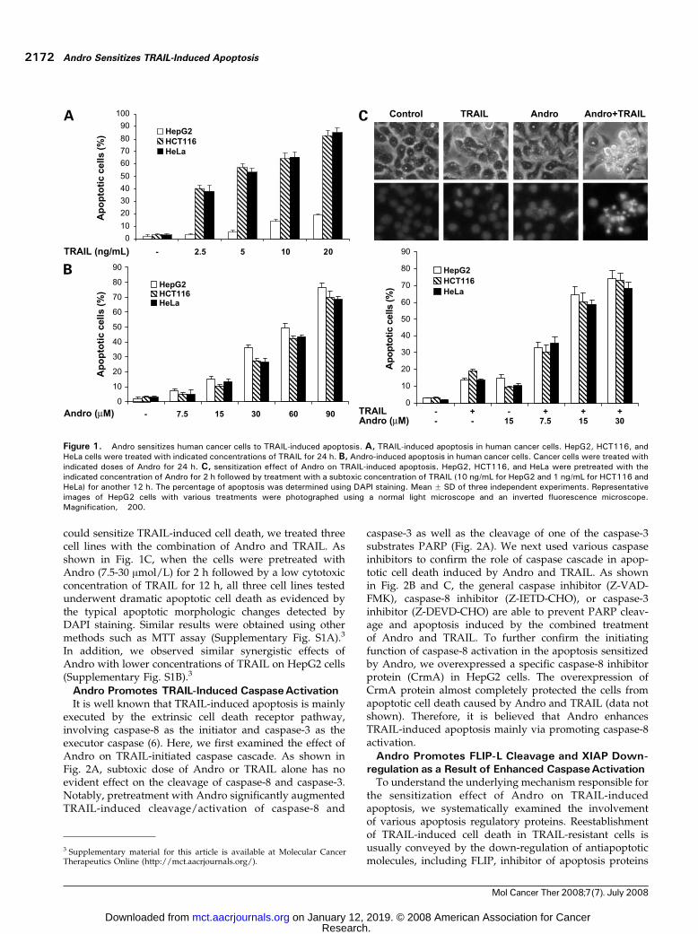
could sensitize TRAIL-induced cell death, we treated threecell lines with the combination of Andro and TRAIL. Asshown in Fig. 1C, when the cells were pretreated withAndro (7.5-30 Amol/L) for 2 h followed by a low cytotoxicconcentration of TRAIL for 12 h, all three cell lines testedunderwent dramatic apoptotic cell death as evidenced bythe typical apoptotic morphologic changes detected byDAPI staining. Similar results were obtained using othermethods such as MTT assay (Supplementary Fig. S1A).3
In addition, we observed similar synergistic effects ofAndro with lower concentrations of TRAIL on HepG2 cells(Supplementary Fig. S1B).3
Andro Promotes TRAIL-Induced CaspaseActivationIt is well known that TRAIL-induced apoptosis is mainly
executed by the extrinsic cell death receptor pathway,involving caspase-8 as the initiator and caspase-3 as theexecutor caspase (6). Here, we first examined the effect ofAndro on TRAIL-initiated caspase cascade. As shown inFig. 2A, subtoxic dose of Andro or TRAIL alone has noevident effect on the cleavage of caspase-8 and caspase-3.Notably, pretreatment with Andro significantly augmentedTRAIL-induced cleavage/activation of caspase-8 and
caspase-3 as well as the cleavage of one of the caspase-3substrates PARP (Fig. 2A). We next used various caspaseinhibitors to confirm the role of caspase cascade in apop-totic cell death induced by Andro and TRAIL. As shownin Fig. 2B and C, the general caspase inhibitor (Z-VAD-FMK), caspase-8 inhibitor (Z-IETD-CHO), or caspase-3inhibitor (Z-DEVD-CHO) are able to prevent PARP cleav-age and apoptosis induced by the combined treatmentof Andro and TRAIL. To further confirm the initiatingfunction of caspase-8 activation in the apoptosis sensitizedby Andro, we overexpressed a specific caspase-8 inhibitorprotein (CrmA) in HepG2 cells. The overexpression ofCrmA protein almost completely protected the cells fromapoptotic cell death caused by Andro and TRAIL (data notshown). Therefore, it is believed that Andro enhancesTRAIL-induced apoptosis mainly via promoting caspase-8activation.
Andro Promotes FLIP-L Cleavage and XIAP Down-regulation as a Result of Enhanced CaspaseActivation
To understand the underlying mechanism responsible forthe sensitization effect of Andro on TRAIL-inducedapoptosis, we systematically examined the involvementof various apoptosis regulatory proteins. Reestablishmentof TRAIL-induced cell death in TRAIL-resistant cells isusually conveyed by the down-regulation of antiapoptoticmolecules, including FLIP, inhibitor of apoptosis proteins
Figure 1. Andro sensitizes human cancer cells to TRAIL-induced apoptosis. A, TRAIL-induced apoptosis in human cancer cells. HepG2, HCT116, andHeLa cells were treated with indicated concentrations of TRAIL for 24 h. B, Andro-induced apoptosis in human cancer cells. Cancer cells were treated withindicated doses of Andro for 24 h. C, sensitization effect of Andro on TRAIL-induced apoptosis. HepG2, HCT116, and HeLa were pretreated with theindicated concentration of Andro for 2 h followed by treatment with a subtoxic concentration of TRAIL (10 ng/mL for HepG2 and 1 ng/mL for HCT116 andHeLa) for another 12 h. The percentage of apoptosis was determined using DAPI staining. Mean F SD of three independent experiments. Representativeimages of HepG2 cells with various treatments were photographed using a normal light microscope and an inverted fluorescence microscope.Magnification, �200.
3 Supplementary material for this article is available at Molecular CancerTherapeutics Online (http://mct.aacrjournals.org/).
Andro Sensitizes TRAIL-Induced Apoptosis2172
Mol Cancer Ther 2008;7(7). July 2008
Research. on January 12, 2019. © 2008 American Association for Cancermct.aacrjournals.org Downloaded from

(XIAP, c-IAP1, and c-IAP2), Bcl-2, Bcl-xL, and Mcl-1(11–13, 19, 20). As shown in Supplementary Fig. S2A,3
Andro significantly enhances TRAIL-induced cleavage ofFLIP-L and reduction of XIAP protein level in HepG2cells, whereas no evident changes are found for otherapoptosis regulatory proteins. Moreover, ectopic expres-sion of either FLIP-L or XIAP protected Andro-sensitizedapoptosis (Supplementary Fig. S2B),3 indicating thatFLIP-L or XIAP plays an important role in the sensitiza-tion effect of Andro on TRAIL-induced apoptosis. Inter-estingly, the pan-caspase inhibitor Z-VAD-FMK almostcompletely reversed the effect of Andro on FLIP-Lcleavage and XIAP down-regulation (SupplementaryFig. S2C).3 It is thus believed that the enhanced FLIP-Lcleavage and XIAP protein down-regulation lie down-stream of the increased caspase activation as the result,but not the cause, of Andro-induced sensitization onTRAIL-induced apoptosis.
Andro Sensitizes TRAIL-Induced Apoptosis via DR4Up-regulation
It has been well established that the decreased expressionof TRAIL receptors DR4 and DR5 and/or up-regulation ofthe decoy receptors DcR1 and DcR2 account for TRAIL resis-tance in certain cancer cell lines (2, 6, 21). To determine thepossible role of TRAIL receptors in Andro-sensitizedapoptosis, we first detected the protein levels of variousTRAIL receptors in cells treated with Andro and TRAIL. Asshown in Fig. 3A, Andro treatment markedly increases the
expression of DR4 in HepG2 cells from 6 h onwards asdetected by Western blot, whereas no evident changes werefound for DR5, DcR1, and DcR2. To confirm the effect ofAndro on DR4, we further detected the DR4 expression levelat cell surface using flow cytometry. Consistently, Andromarkedly increased the cell surface expression of DR4 but notDR5 (Fig. 3B). Similar to a previous report (22), it is noted thatthe basal level of DR5 is much higher than that of DR4detected by either Western blot or flow cytometry (Fig. 3Aand B).
To determine whether the increased DR4 protein levelis due to enhanced gene transcription, we next measuredthe mRNA level of both DR4 and DR5 using RT-PCR(Fig. 3C). Consistent with the changes of the protein level,significant increase of DR4 mRNA level was observedfrom 3 h onwards, preceding the increase of DR4 proteinlevel. Taken together, it is clear that Andro treatmentselectively up-regulates DR4 expression at transcriptionallevel.
DR4 Up-regulation Is Critical for the SensitizationEffect of Andro
To further elucidate the critical role of DR4 in Andro-induced sensitization effects, we neutralized the function ofTRAIL receptors by using DR4 or DR5 blocking antibody asreported previously (23). It is evident that DR4 blockingantibody is able to suppress caspase-8 cleavage (Fig. 3D)and protect against apoptotic cell death induced by thecombined treatment of Andro and TRAIL (Fig. 3E). On
Figure 2. Andro enhances the caspase cascade triggered by TRAIL. A, Andro promotes TRAIL-induced caspase activation. HepG2 cells were pretreatedwith 15 Amol/L Andro for 2 h followed by treatment with TRAIL (10 ng/mL) for indicated periods. Cell lysates were collected and subjected to Western blotfor detecting the cleavage of caspase-8, caspase-3, and PARP. B, caspase inhibitors block PARP cleavage induced by Andro and TRAIL. C, caspaseinhibitors block apoptosis induced by Andro and TRAIL. B and C, HepG2 cells were pretreated with Z-IETD-CHO (25 Amol/L), Z-DEVD-CHO (25 Amol/L), orZ-VAD-FMK (25 Amol/L) for 30 min followed by combined treatment of Andro (15 Amol/L) for 2 h and then TRAIL (10 ng/mL) for another 12 h. Cells werecollected for measurement of PARP cleavage by Western blot (B) or determination of cell death using DAPI staining (C). Mean F SD of three independentexperiments. Magnification, �200.
Molecular Cancer Therapeutics 2173
Mol Cancer Ther 2008;7(7). July 2008
Research. on January 12, 2019. © 2008 American Association for Cancermct.aacrjournals.org Downloaded from

the contrary, these effects could not be detected afterneutralization with the DR5 blocking antibody. Theseresults thus strengthen our argument that DR4 but notDR5 plays a key role in the sensitization effect of Andro onTRAIL-induced apoptotic cell death.
p53 Is Required for the DR4 Up-regulation andEnhanced Apoptosis Induced byAndro
It has been well documented that p53 tumor suppressoris one of the main transcription factors regulating the
expression of both DR4 and DR5 (24–26). Here, we testedwhether Andro-mediated DR4 transcriptional up-regula-tion is p53 dependent. We first showed that Androtreatment increases p53 protein level, which is coincidentwith the temporal change of DR4 protein level (Fig. 4A).Next, we manipulated the p53 expression by using specificsiRNA against p53. As expected, knockdown of p53resulted in the suppression of Andro-induced DR4up-regulation (Fig. 4B) and consequently prevented
Figure 3. Andro up-regulates DR4 transcription. A, time-dependent up-regulation of DR4 protein level by Andro. HepG2 cells were treated with Andro(15 Amol/L) for 2 h followed by TRAIL (10 ng/mL) for indicated periods. Cell lysates were collected for Western blot to detect protein expression level ofvarious TRAIL death receptors. B, effects of Andro on the cell surface expression of DR4 and DR5. HepG2 cells were incubated with Andro (15 Amol/L) for6 h and the cell surface expression of DR4 and DR5 proteins was analyzed by flow cytometry. X axis, fluorescence intensity; Y axis, relative number ofcells. Representative of three independent experiments. C, effects of Andro and TRAIL on DR4 and DR5 mRNA level. HepG2 cells were treated with Andro(15 Amol/L) for 2 h followed by TRAIL (10 ng/mL) for indicated periods. The mRNA levels of DR4 and DR5 were measured by reverse transcription-PCR.Glyceraldehyde-3-phosphate dehydrogenase was used as an internal control. D, DR4 blocking antibody suppresses the cleavage of caspase-8 and PARPinduced by Andro and TRAIL. HepG2 cells were pretreated with DR4 or DR5 blocking antibodies (10 Ag/mL) for 30 min followed by pretreatment withAndro (15 Amol/L � 2 h) and then TRAIL (10 ng/mL) for another 12 h. At the end of treatment, cell lysates were collected for Western blot. E, DR4 blockingantibody prevents Andro-sensitized apoptosis. HepG2 cells were treated as indicated in D. At the end of treatment, cell death was determined using DAPIstaining. Magnification, �200.
Andro Sensitizes TRAIL-Induced Apoptosis2174
Mol Cancer Ther 2008;7(7). July 2008
Research. on January 12, 2019. © 2008 American Association for Cancermct.aacrjournals.org Downloaded from

Andro-sensitized apoptosis (Fig. 4C). These observationsthus establish that p53 is required for Andro-induced DR4up-regulation and sensitization to TRAIL-induced apopto-sis in HepG2 cells.
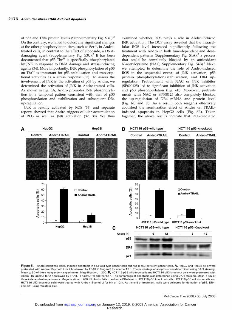
To further confirm the critical role of p53 in Andro-sensitized apoptosis in TRAIL-treated cells, we selectedtwo pairs of cancer cell lines with different genetic featuresof p53: p53 wild-type HepG2 versus p53 mutant Hep3Bcells (27) and p53 wild-type HCT116 versus p53 knockoutHCT116 cells (16). As shown in Fig. 5A, Andro plus TRAILinduce significant apoptosis in HepG2 cells but not inHep3B cells. Similarly, the combined treatment causesobvious apoptotic death in HCT116 p53 wild-type cellsbut shows no effect in HCT116 p53 knockout cells (Fig. 5B).We next examined the DR4 and p21 expression levelsin HCT116 cells treated with Andro. As expected, on Androtreatment, the dramatic enhancement of DR4 could bedetected in HCT116 p53 wild-type but only slight increasein HCT116 p53 knockout cells (Fig. 5C), which is consistentto the change of p21, a well-established p53 target pro-tein. Although p53 is a well-known transcriptional regu-lator of DR4 (24, 25), nuclear factor-nB (28), c-myc (29),and JAK/STAT pathway (30) also have been reportedto be able to regulate the expression of DR4. Therefore,it is possible that other regulatory mechanisms maycontribute to the slight up-regulation of DR4 in HCT116
p53 knockout cells. However, this slight up-regulation ofDR4 is not sufficient to enhance the sensitivity of HCT116p53 knockdown cells to TRAIL- and Andro-inducedapoptosis (Fig. 5B). Therefore, it is believed that up-regulation of DR4 and sensitization of TRAIL-inducedapoptosis by Andro are dependent on the presence offunctional p53.
Andro Elevatesp53 Expression throughJNK-Mediatedp53 Thr81Phosphorylation
Activation of p53 may result from either the increasedtranscriptional level or post-translational modification andstabilization (31). In this study, Andro did not affect themRNA level of p53 (Supplementary Fig. S3A).3 Consistent-ly, it was found that, in the presence of cycloheximide, aprotein synthesis inhibitor, Andro significantly prolongsthe half-life of p53 from 30 min to z2 h (SupplementaryFig. S3B),3 suggesting that accumulation of p53 proteinby Andro is most probably due to its post-transcriptionalregulation.
It has been well established that p53 protein level ismainly regulated via the ubiquitination and proteasomepathway, a process closely associated with the phosphor-ylation status of p53 (32, 33). We next examined the patternof p53 phosphorylation in response to Andro treatment.Notably, dramatic p53 phosphorylation at Thr81 site wasdetected on Andro treatment, with concurrent increase
Figure 4. p53 is required for DR4 up-regulation and enhanced apoptosis by Andro. A, Andro promotes p53 protein accumulation. HepG2 cells weretreated with Andro (15 Amol/L) for 2 h followed by TRAIL (10 ng/mL) for indicated periods. p53 and DR4 protein levels were detected by Western blot. B,p53 knockdown prevents Andro-induced DR4 up-regulation. HepG2 cells were transfected with scrambled siRNA or p53 siRNA for 24 h. Cells were treatedwith Andro (15 Amol/L) for 6 h. At the end of treatment, cells were collected for detection of p53 and DR4 by Western blot. C, p53 knockdown suppressesapoptosis induced by Andro and TRAIL. After p53 knockdown, HepG2 cells were treated with Andro (15 Amol/L) for 2 h followed by TRAIL (10 ng/mL) for12 h. Cells were assessed using DAPI staining. Representative of three independent experiments. Magnification, �200.
Molecular Cancer Therapeutics 2175
Mol Cancer Ther 2008;7(7). July 2008
Research. on January 12, 2019. © 2008 American Association for Cancermct.aacrjournals.org Downloaded from
of p53 and DR4 protein levels (Supplementary Fig. S3C).3
On the contrary, we failed to detect any significant changesat the other phosphorylation sites, such as Ser20, in Andro-treated cells, in contrast to the effect of etoposide, a DNA-damaging agent (Supplementary Fig. S3C).3 It has beendocumented that p53 Thr81 is specifically phosphorylatedby JNK in response to DNA damage and stress-inducingagents (34). More importantly, JNK phosphorylation of p53on Thr81 is important for p53 stabilization and transcrip-tional activities as a stress response (35). To assess theinvolvement of JNK in the activation of p53 by Andro, wedetermined the activation of JNK in Andro-treated cells.As shown in Fig. 6A, Andro promotes JNK phosphoryla-tion in a temporal pattern consistent with that of p53phosphorylation and stabilization and subsequent DR4up-regulation.
JNK is readily activated by ROS (36) and separatereports showed that Andro triggers cellular accumulationof ROS as well as JNK activation (37, 38). We thus
examined whether ROS plays a role in Andro-inducedJNK activation. The DCF assay revealed that the intracel-lular ROS level increased significantly following thetreatment with Andro in both time-dependent and dose-dependent patterns (Supplementary Fig. S4A),3 a processthat could be completely blocked by an antioxidantN-acetylcysteine (NAC; Supplementary Fig. S4B).3 Next,we attempted to determine the role of Andro-inducedROS in the sequential events of JNK activation, p53protein phosphorylation/stabilization, and DR4 up-regulation. Pretreatment with NAC or JNK inhibitor(SP600125) led to significant inhibition of JNK activationand p53 phosphorylation (Fig. 6B). Moreover, pretreat-ments with NAC or SP600125 also completely blockedthe up-regulation of DR4 mRNA and protein level(Fig. 6C and D). As a result, both reagents effectivelyabolished the sensitization effect of Andro on TRAIL-induced apoptosis in HepG2 cells (Fig. 6E). Takentogether, the above results indicate that ROS-mediated
Figure 5. Andro sensitizes TRAIL-induced apoptosis in p53 wild-type cancer cells but not in p53-deficient cancer cells. A, HepG2 and Hep3B cells werepretreated with Andro (15 Amol/L) for 2 h followed by TRAIL (10 ng/mL) for another12 h. The percentage of apoptosis was determined using DAPI staining.Mean F SD of three independent experiments. Magnification, �200. B, HCT116 p53 wild-type cells and HCT116 p53 knockout cells were pretreated withAndro (15 Amol/L) for 2 h followed by TRAIL (1 ng/mL) for another12 h. The percentage of apoptosis was determined using DAPI staining. Mean F SD ofthree independent experiments. Magnification, �200. C, Andro fails to enhance DR4 level in HCT116 p53 knockout cells. HCT116 p53 wild-type cells andHCT116 p53 knockout cells were treated with Andro (15 Amol/L) for 6 h or 12 h. At the end of treatment, cells were collected for detection of p53, DR4,and p21 using Western blot.
Andro Sensitizes TRAIL-Induced Apoptosis2176
Mol Cancer Ther 2008;7(7). July 2008
Research. on January 12, 2019. © 2008 American Association for Cancermct.aacrjournals.org Downloaded from

JNK activation by Andro accounts for the sensitizationeffect of Andro on TRAIL-induced apoptosis via sequen-tial events including p53 phosphorylation/stabilizationand DR4 up-regulation.
DiscussionThe unique property of triggering apoptosis in a varietyof human cancer cells while sparing normal cells makesTRAIL a highly promising cancer therapeutic agent.However, TRAIL resistance is a major limitation in itsclinical application as a cancer therapeutic agent. Oneeffective strategy to overcome this obstacle is to combineTRAIL with other anticancer agents (39). Several chemical
compounds have been identified as effective sensitizingagents to TRAIL-induced apoptosis, including some natu-ral products (8–10, 12, 13). Results from this study providesubstantial evidence that Andro, a diterpenoid lactoneisolated from a traditional medicinal herb, is capable ofsensitizing TRAIL-induced apoptosis in TRAIL-resistantcancer cells. Thus, this study presents a novel anticancereffect of Andro and supports its potential application incancer therapy.
Andro Sensitizes TRAIL-Induced Apoptosis byUp-regulation of DR4
The expression level of death receptors (DR4 and DR5)plays a critical role in determining the cell fate in response
Figure 6. Andro promotes JNK activation, p53 stabilization, and DR4 up-regulation in ROS-dependent manner. A, Andro induces JNK activation. HepG2cells were treated with Andro (15 Amol/L) for 2 h followed by TRAIL (10 ng/mL) for indicated periods. At the end of treatment, cell lysates were collected forthe detection of protein level of phospho-JNK, total JNK, phospho-p53 Thr81, p53, DR4, and a-tubulin using Western blot. B, NAC and SP600125 abrogateAndro-induced JNK activation, p53 phosphorylation, and stabilization. HepG2 cells were pretreated with NAC (2.5 mmol/L for 30 min) or SP600125(20 Amol/L for 30 min) followed by the treatment of Andro (15 Amol/L � 2 h) and then TRAIL (10 ng/mL) for another 6 h. C, NAC and SP600125 preventAndro-induced DR4 up-regulation. Cells were first pretreated with NAC and SP600125 as described inB followed by Andro (15 Amol/L� 2 h) and then TRAIL(10 ng/mL) for another 3 h (for detection of DR4 mRNA level) and 6 h (for detection of DR4 protein level). D, NAC prevents Andro-induced cell surfaceexpression of DR4. HepG2 cells were pretreated with NAC as described above followed by Andro (15 Amol/L) for 6 h. The cell surface expression of DR4was measured using flow cytometry as described in B. Representative of three independent experiments. E, NAC and SP600125 inhibit apoptosis inducedby Andro and TRAIL. HepG2 cells were treated with Andro (15 Amol/L � 2 h) followed by TRAIL (10 ng/mL) for another 12 h. The percentage of apoptosiswas measured with DAPI staining. Mean F SD of three independent experiments. Magnification, �200.
Molecular Cancer Therapeutics 2177
Mol Cancer Ther 2008;7(7). July 2008
Research. on January 12, 2019. © 2008 American Association for Cancermct.aacrjournals.org Downloaded from

to TRAIL (2, 6, 21). Numerous literatures have shownconvincing data that the up-regulation of DR4 and DR5could sensitize TRAIL-resistant cells to TRAIL-induced celldeath (22, 40). In this study, it is believed that Andro-induced sensitization on TRAIL-induced apoptosis ismainly executed via modulation of DR4, but not DR5,based on the following observations. First, Andro selec-tively enhanced the expression level of DR4 in HepG2cells (Fig. 3A and B). Second, the administration of block-ing antibody of DR4, but not DR5, effectively protected thecell death induced by combination of Andro and TRAIL(Fig. 3D and E), further confirming that up-regulation of DR4is functionally important in Andro-sensitized apoptosis.
At present, there are inconsistent reports whether DR4 orDR5 is the main mediator for TRAIL-mediated apoptosisin cancer cells. It has been well reported that, in chroniclymphocytic leukemia cells, DR4 seems to be the principalreceptor in response to TRAIL in the presence of histonedeacetylase inhibitors (41–43). Suppression of DR4 expres-sion or function by homozygous deletion or epigeneticsilencing led to TRAIL resistance (44, 45). Recently,Kurbanov et al. used an established cell culture model forTRAIL resistance and regained TRAIL sensitivity andindicated that significant down-regulation of initiatorcaspase and DR4 was characteristic for TRAIL-resistantmelanoma cells (46). Additionally, another recent reportsuggested that reactivation of DR4 expression contributedto restore sensitivity of melanoma cells to IFN-h or Apo2L/TRAIL (47). Moreover, Aza-Blanc et al. reported that asiRNA targeting DR4 was effective in blocking TRAIL-induced apoptosis, whereas knockdown of DR5 failed toconfer any protection (48). In this regard, DR4 representsthe major determinant of TRAIL sensitivity in certaincancer cells. On the other hand, there is evidence that DR5may play a more prominent role than DR4 in mediatingapoptotic signals emanating from TRAIL in cells expressingboth death receptors (49) and TRAIL binds with a higheraffinity to DR5 than to DR4 in some cells (50). One possibleexplanation for such inconsistent results might be thedifferent expression levels of these two receptors indifferent types of cancer cells. In line with some previousstudies (22), we found that in HepG2 cells DR5 is expressedin a much higher basal level but is more insensitive toregulatory mechanisms. In contrast, DR4 with a very lowbasal level is susceptible of being up-regulated by p53. Sucha difference may well explain the fact that it is DR4 that isresponsible for the sensitization effect of Andro to TRAIL-induced apoptosis.
Andro Up-regulates DR4 via p53-DependentTranscriptional Regulation
The tumor suppressor gene p53 is a key apoptosisregulator in cancer cells and one of its proapoptosisfunctions is achieved via transcriptional activation of deathreceptors (24, 25). In the present study, we found that p53played a critical role in Andro-sensitized apoptosis throughup-regulation of DR4, owing to the following findings.First, RNA interference of p53 could effectively inhibitAndro-induced DR4 up-regulation (Fig. 4B) and abolish the
apoptotic cell death induced by the combined treatment ofAndro and TRAIL (Fig. 4C). Second, evident apoptoticcell death and DR4 up-regulation were only observedin p53 wild-type cancer cells but not in p53-deficient cells(Fig. 5A and B). It is thus believed that Andro is capableof enhancing TRAIL-mediated cancer cell apoptosis viaactivating p53-dependent DR4 transcription.
The involvement of p53 in control of DR4 and DR5transcription remains contradictory. Original work mainlyby El-Deiry’s group identified DR5 as the main target ofp53 as the molecular link between the apoptotic pathwaysmediated by DNA damage and cell death receptors (51, 52).On the other hand, there is convincing evidence showingthat DNA damage-induced p53 activation also leads to DR4up-regulation (24, 25). It is likely that p53 is important forboth DR4 and DR5 depending on the context including thenature of stimuli and the cell type. It remains to be furthertested whether p53-mediated DR4 up-regulation is alsoapplicable to other TRAIL-resistant cancer cells. Oneimplication of such findings is that the therapeutic valueof TRAIL is at least partly dependent on functional p53 incancer cells and the sensitization effect of Andro may onlybe achievable in p53 wild-type cancer cells.
Andro Stabilizes p53 Protein via Oxidative Stress andJNK Activation
It has been well established that p53 activity is mainlyregulated through complex networks of post-translationalmodifications, including phosphorylation, ubiquitination,and proteasome degradation (32, 33). The phosphorylationstatus of p53 directly affects MDM2 binding and subsequentubiquitination and proteasome degradation. At present,more than a dozen phosphorylation sites have been mappedon p53, but only a few have been characterized with respectto the phosphorylating kinase and the relevance to p53activities (32). Among them, activation of JNK has beenlinked with p53 Thr81 phosphorylation in response to DNAdamage and stress-inducing agents (34). Consistently, weobserved p53 phosphorylation at Thr81, but not Ser20 inresponse to Andro treatment, in contrast to the effect ofetoposide, a DNA-damaging agent. Meanwhile, it isintriguing to note that no up-regulation of DR4 was foundin cells treated with etoposide despite the equally enhancedtotal p53 protein level (Supplementary Fig. S3C).3 Themechanism underlying such a discrepancy remains to befurther investigated. More importantly, pretreatment ofSP600125, a specific JNK inhibitor, suppressed Andro-induced JNK activation and thus abrogated p53 phosphor-ylation, total p53 protein accumulation, DR4 up-regulation,and subsequent cell death induced by combined treatmentof Andro and TRAIL (Fig. 6). Collectively, our data reveala sequence of events elicited by Andro, from ROSproduction to JNK activation, p53 phosphorylation, andstabilization to DR4 up-regulation and eventually enhancedcell death.
In conclusion, this study shows a novel anticancer effectof Andro: Andro is capable of activating p53 function viaROS-dependent JNK activation, leading to DR4 up-regula-tion and sensitization to TRAIL-induced apoptosis in
Andro Sensitizes TRAIL-Induced Apoptosis2178
Mol Cancer Ther 2008;7(7). July 2008
Research. on January 12, 2019. © 2008 American Association for Cancermct.aacrjournals.org Downloaded from
TRAIL-resistant cancer cells. Data from this study supportthe further development of Andro as a chemosensitizer incombined therapy with TRAIL to overcome TRAILresistance in p53 wild-type cancers.
Disclosure of Potential Conflicts of InterestThe other authors disclosed no potential conflicts of interest.
References
1. Hall MA, Cleveland JL. Clearing the TRAIL for cancer therapy. CancerCell 2007;12:4–6.
2. Wang S, El-Deiry WS. TRAIL and apoptosis induction by TNF-familydeath receptors. Oncogene 2003;22:8628–33.
3. Huang Y, Sheikh MS. TRAIL death receptors and cancer therapeutics.Toxicol Appl Pharmacol 2007;224:284–9.
4. Duiker EW, Mom CH, de Jong S, et al. The clinical trail of TRAIL. Eur JCancer 2006;42:2233–40.
5. Dyer MJ, MacFarlane M, Cohen GM. Barriers to effective TRAIL-targeted therapy of malignancy. J Clin Oncol 2007;25:4505–6.
6. LeBlanc HN, Ashkenazi A. Apo2L/TRAIL and its death and decoyreceptors. Cell Death Differ 2003;10:66–75.
7. Shankar S, Srivastava RK. Enhancement of therapeutic potential ofTRAIL by cancer chemotherapy and irradiation: mechanisms and clinicalimplications. Drug Resist Updat 2004;7:139–56.
8. Inoue S, MacFarlane M, Harper N, Wheat LM, Dyer MJ, Cohen GM.Histone deacetylase inhibitors potentiate TNF-related apoptosis-inducingligand (TRAIL)-induced apoptosis in lymphoid malignancies. Cell DeathDiffer 2004;11 Suppl 2:S193–206.
9. Palacios C, Yerbes R, Lopez-Rivas A. Flavopiridol induces cellularFLICE-inhibitory protein degradation by the proteasome and promotesTRAIL-induced early signaling and apoptosis in breast tumor cells. CancerRes 2006;66:8858–69.
10. Ganten TM, Koschny R, Haas TL, et al. Proteasome inhibitionsensitizes hepatocellular carcinoma cells, but not human hepatocytes, toTRAIL. Hepatology 2005;42:588–97.
11. Ricci MS, Kim SH, Ogi K, et al. Reduction of TRAIL-induced Mcl-1 andcIAP2 by c-Myc or sorafenib sensitizes resistant human cancer cells toTRAIL-induced death. Cancer Cell 2007;12:66–80.
12. Shi RX, Ong CN, Shen HM. Protein kinase C inhibition and X-linkedinhibitor of apoptosis protein degradation contribute to the sensitiza-tion effect of luteolin on tumor necrosis factor-related apoptosis-inducing ligand-induced apoptosis in cancer cells. Cancer Res 2005;65:7815–23.
13. Zhang S, Shen HM, Ong CN. Down-regulation of c-FLIP contributes tothe sensitization effect of 3,3¶-diindolylmethane on TRAIL-inducedapoptosis in cancer cells. Mol Cancer Ther 2005;4:1972–81.
14. Xia YF, Ye BQ, Li YD, et al. Andrographolide attenuates inflammationby inhibition of NF-nB activation through covalent modification of reducedcysteine 62 of p50. J Immunol 2004;173:4207–17.
15. Zhou J, Zhang S, Ong CN, Shen HM. Critical role of pro-apoptoticBcl-2 family members in andrographolide-induced apoptosis in humancancer cells. Biochem Pharmacol 2006;72:132–44.
16. Bunz F, Dutriaux A, Lengauer C, et al. Requirement for p53and p21 to sustain G2 arrest after DNA damage. Science 1998;282:1497–501.
17. Earel JK, Jr., VanOosten RL, Griffith TS. Histone deacetylaseinhibitors modulate the sensitivity of tumor necrosis factor-relatedapoptosis-inducing ligand-resistant bladder tumor cells. Cancer Res2006;66:499–507.
18. Lu GD, Shen HM, Chung MC, Ong CN. Critical role of oxida-tive stress and sustained JNK activation in aloe-emodin-mediatedapoptotic cell death in human hepatoma cells. Carcinogenesis 2007;28:1937–45.
19. Mirandola P, Sponzilli I, Gobbi G, et al. Anticancer agents sensitizeosteosarcoma cells to TNF-related apoptosis-inducing ligand downmodu-lating IAP family proteins. Int J Oncol 2006;28:127–33.
20. Taniai M, Grambihler A, Higuchi H, et al. Mcl-1 mediates tumor
necrosis factor-related apoptosis-inducing ligand resistance in humancholangiocarcinoma cells. Cancer Res 2004;64:3517–24.
21. Takeda K, Stagg J, Yagita H, Okumura K, Smyth MJ. Target-ing death-inducing receptors in cancer therapy. Oncogene 2007;26:3745–57.
22. Galligan L, Longley DB, McEwan M, Wilson TR, McLaughlin K,Johnston PG. Chemotherapy and TRAIL-mediated colon cancer cell death:the roles of p53, TRAIL receptors, and c-FLIP. Mol Cancer Ther 2005;4:2026–36.
23. Sprick MR, Rieser E, Stahl H, Grosse-Wilde A, Weigand MA,Walczak H. Caspase-10 is recruited to and activated at the native TRAILand CD95 death-inducing signalling complexes in a FADD-dependentmanner but can not functionally substitute caspase-8. EMBO J 2002;21:4520–30.
24. Liu X, Yue P, Khuri FR, Sun SY. p53 upregulates death receptor 4expression through an intronic p53 binding site. Cancer Res 2004;64:5078–83.
25. Guan B, Yue P, Clayman GL, Sun SY. Evidence that the deathreceptor DR4 is a DNA damage-inducible, p53-regulated gene. J CellPhysiol 2001;188:98–105.
26. El-Deiry WS. Insights into cancer therapeutic design based on p53and TRAIL receptor signaling. Cell Death Differ 2001;8:1066–75.
27. Bressac B, Galvin KM, Liang TJ, Isselbacher KJ, Wands JR, Ozturk M.Abnormal structure and expression of p53 gene in human hepatocellularcarcinoma. Proc Natl Acad Sci U S A 1990;87:1973–7.
28. Jin F, Liu X, Zhou Z, et al. Activation of nuclear factor-nB contributesto induction of death receptors and apoptosis by the synthetic retinoidCD437 in DU145 human prostate cancer cells. Cancer Res 2005;65:6354–63.
29. Sussman RT, Ricci MS, Hart LS, Sun SY, El-Deiry WS. Chemotherapy-resistant side-population of colon cancer cells has a higher sensitivity toTRAIL than the non-SP, a higher expression of c-Myc and TRAIL-receptorDR4. Cancer Biol Ther 2007;6:1490–5.
30. Tanaka F, Kawakami A, Tamai M, et al. IFN-g/JAK/STAT pathway-induced inhibition of DR4 and DR5 expression on endothelial cells iscancelled by cycloheximide-sensitive mechanism: novel finding of cyclo-heximide-regulating death receptor expression. Int J Mol Med 2005;15:833–9.
31. Ashcroft M, Taya Y, Vousden KH. Stress signals utilize multiplepathways to stabilize p53. Mol Cell Biol 2000;20:3224–33.
32. Brooks CL, Gu W. p53 ubiquitination: Mdm2 and beyond. Mol Cell2006;21:307–15.
33. Ashcroft M, Vousden KH. Regulation of p53 stability. Oncogene1999;18:7637–43.
34. Wu GS. The functional interactions between the p53 and MAPKsignaling pathways. Cancer Biol Ther 2004;3:156–61.
35. Buschmann T, Potapova O, Bar-Shira A, et al. Jun NH2-terminalkinase phosphorylation of p53 on Thr-81 is important for p53 stabilizationand transcriptional activities in response to stress. Mol Cell Biol 2001;21:2743–54.
36. Shen HM, Liu ZG. JNK signaling pathway is a key modulator in celldeath mediated by reactive oxygen and nitrogen species. Free Radic BiolMed 2006;40:928–39.
37. Li J, Cheung HY, Zhang Z, Chan GK, Fong WF. Andrographo-lide induces cell cycle arrest at G2/M phase and cell death in HepG2cells via alteration of reactive oxygen species. Eur J Pharmacol 2007;568:31–44.
38. Ji L, Liu T, Liu J, Chen Y, Wang Z. Andrographolide inhibits humanhepatoma-derived Hep3B cell growth through the activation of c-JunN-terminal kinase. Planta Med 2007;73:1397–401.
39. Zhang L, Fang B. Mechanisms of resistance to TRAIL-inducedapoptosis in cancer. Cancer Gene Ther 2005;12:228–37.
40. Jin Z, McDonald ER III, Dicker DT, El-Deiry WS. Deficient tumornecrosis factor-related apoptosis-inducing ligand (TRAIL) death receptortransport to the cell surface in human colon cancer cells selectedfor resistance to TRAIL-induced apoptosis. J Biol Chem 2004;279:35829–39.
41. MacFarlane M, Inoue S, Kohlhaas SL, et al. Chronic lymphocyticleukemic cells exhibit apoptotic signaling via TRAIL-R1. Cell Death Differ2005;12:773–82.
42. Natoni A, Macfarlane M, Inoue S, et al. TRAIL signals to apoptosis in
Molecular Cancer Therapeutics 2179
Mol Cancer Ther 2008;7(7). July 2008
Research. on January 12, 2019. © 2008 American Association for Cancermct.aacrjournals.org Downloaded from
chronic lymphocytic leukaemia cells primarily through TRAIL-R1 whereascross-linked agonistic TRAIL-R2 antibodies facilitate signalling via TRAIL-R2. Br J Haematol 2007;139:568–77.
43. MacFarlane M, Kohlhaas SL, Sutcliffe MJ, Dyer MJ, CohenGM. TRAIL receptor-selective mutants signal to apoptosis viaTRAIL-R1 in primary lymphoid malignancies. Cancer Res 2005;65:11265–70.
44. Ozoren N, Fisher MJ, Kim K, et al. Homozygous deletion of the deathreceptor DR4 gene in a nasopharyngeal cancer cell line is associated withTRAIL resistance. Int J Oncol 2000;16:917–25.
45. Horak P, Pils D, Haller G, et al. Contribution of epigenetic silenc-ing of tumor necrosis factor-related apoptosis inducing ligand receptor 1(DR4) to TRAIL resistance and ovarian cancer. Mol Cancer Res 2005;3:335–43.
46. Kurbanov BM, Fecker LF, Geilen CC, Sterry W, Eberle J. Resistance ofmelanoma cells to TRAIL does not result from upregulation of antiapop-totic proteins by NF-nB but is related to downregulation of initiatorcaspases and DR4. Oncogene 2007;26:3364–77.
47. Bae SI, Cheriyath V, Jacobs BS, Reu FJ, Borden EC. Reversal ofmethylation silencing of Apo2L/TRAIL receptor 1 (DR4) expression
overcomes resistance of SK-MEL-3 and SK-MEL-28 melanoma cells tointerferons (IFNs) or Apo2L/TRAIL. Oncogene 2007;27:490–8.
48. Aza-Blanc P, Cooper CL, Wagner K, Batalov S, Deveraux QL, CookeMP. Identification of modulators of TRAIL-induced apoptosis via RNAi-based phenotypic screening. Mol Cell 2003;12:627–37.
49. Kelley RF, Totpal K, Lindstrom SH, et al. Receptor-selectivemutants of apoptosis-inducing ligand 2/tumor necrosis factor-relatedapoptosis-inducing ligand reveal a greater contribution of deathreceptor (DR) 5 than DR4 to apoptosis signaling. J Biol Chem 2005;280:2205–12.
50. Truneh A, Sharma S, Silverman C, et al. Temperature-sensitivedifferential affinity of TRAIL for its receptors. DR5 is the highest affinityreceptor. J Biol Chem 2000;275:23319–25.
51. Wu GS, Kim K, El-Deiry WS. KILLER/DR5, a novel DNA-damageinducible death receptor gene, links the p53-tumor suppressor tocaspase activation and apoptotic death. Adv Exp Med Biol 2000;465:143–51.
52. Takimoto R, El-Deiry WS. Wild-type p53 transactivates the KILLER/DR5 gene through an intronic sequence-specific DNA-binding site.Oncogene 2000;19:1735–43.
Andro Sensitizes TRAIL-Induced Apoptosis2180
Mol Cancer Ther 2008;7(7). July 2008
Research. on January 12, 2019. © 2008 American Association for Cancermct.aacrjournals.org Downloaded from
2008;7:2170-2180. Mol Cancer Ther Jing Zhou, Guo-Dong Lu, Chye-Sun Ong, et al. apoptosis via p53-mediated death receptor 4 up-regulationAndrographolide sensitizes cancer cells to TRAIL-induced
Updated version
http://mct.aacrjournals.org/content/7/7/2170
Access the most recent version of this article at:
Material
Supplementary
http://mct.aacrjournals.org/content/suppl/2008/07/23/7.7.2170.DC1
Access the most recent supplemental material at:
Cited articles
http://mct.aacrjournals.org/content/7/7/2170.full#ref-list-1
This article cites 52 articles, 20 of which you can access for free at:
Citing articles
http://mct.aacrjournals.org/content/7/7/2170.full#related-urls
This article has been cited by 4 HighWire-hosted articles. Access the articles at:
E-mail alerts related to this article or journal.Sign up to receive free email-alerts
Subscriptions
Reprints and
To order reprints of this article or to subscribe to the journal, contact the AACR Publications
Permissions
Rightslink site. (CCC)Click on "Request Permissions" which will take you to the Copyright Clearance Center's
.http://mct.aacrjournals.org/content/7/7/2170To request permission to re-use all or part of this article, use this link
Research. on January 12, 2019. © 2008 American Association for Cancermct.aacrjournals.org Downloaded from