Embed Size (px)
Citation preview

EXCLI Journal 2020;19:936-949 – ISSN 1611-2156
Received: February 05, 2020, accepted: June 25, 2020, published: June 30, 2020
936
Original article:
PHENOXODIOL SENSITIZES METASTATIC COLORECTAL
CANCER CELLS TO 5-FLUOROURACIL- AND OXALIPLATIN-
INDUCED APOPTOSIS THROUGH INTRINSIC PATHWAY
Esra Yaylaci, Hacer Ilke Onen, Atiye Seda Yar Saglam*
Department of Medical Biology and Genetics, Faculty of Medicine, Gazi University,
Besevler, Ankara, Turkey
* Corresponding author: Atiye Seda Yar Saglam, PhD, Department of Medical Biology
and Genetics, Faculty of Medicine, Gazi University, Besevler, Ankara, Turkey.
Phone: +903122024714, e-mail: [email protected]
http://dx.doi.org/10.17179/excli2020-2042
This is an Open Access article distributed under the terms of the Creative Commons Attribution License (http://creativecommons.org/licenses/by/4.0/).
ABSTRACT Colorectal cancer (CRC) is one of the most common types of cancer seen in the world. 5-Fluorouracil (5-Fu) plus
Oxaliplatin (1-OHP) remains the backbone of CRC chemotherapeutics, but with limited success. Phenoxodiol
(Pxd) is an isoflavone analog with antitumor activity against various types of cancers, and sensitizes chemo-
resistant cancer cells to chemotherapeutics including platinum and taxanes. This study was, therefore, undertaken
to examine whether Pxd pre-treatment with conventional chemotherapeutic agent(s) 5-Fu and 1-OHP co-admin-
istration be a therapeutic strategy for CRC. Cell viability and cytotoxicity were evaluated using dimethyl-thiazolyl
diphenyl tetrazolium bromide (MTT) and lactate dehydrogenase assays. The percentage of apoptotic and necrotic
cells were determined by fluorescence microscopy analysis. Besides, active Caspase-3 levels by ELISA and rela-
tive mRNA levels of Caspase 3 (CASP3), CASP8 and CASP9 genes were determined by quantitative real-time
PCR (qPCR) analysis. The pre-treatment of Pxd followed by 5-Fu and 1-OHP co-administration was more effec-
tive at inhibiting cell viability than either chemotherapeutic agents treatment alone. When compared to 5-Fu with
1-OHP alone treatment, Pxd pre-treatment overwhelmingly increased apoptotic Caspase-3 activity levels in CRC
cells. Moreover, qPCR analyses showed that CASP3 and CASP9 mRNA levels significantly increased after pre-
treatment with Pxd followed by 5-Fu and 1-OHP treatments, compared to 5-Fu with 1-OHP alone. Our results
suggested that Pxd enhanced the in vitro antitumor activity of 5-Fu and 1-OHP. Our study also suggested that Pxd
may be a potential candidate agent in advanced CRC and inclusion of Pxd to the conventional chemotherapeutic
agent(s) could be an effective therapeutic strategy for CRC.
Keywords: Apoptosis, colorectal cancer, 5-Fluorouracil, oxaliplatin, phenoxodiol
INTRODUCTION
Colorectal cancer (CRC) is one of the
most common types of cancer seen in the
world in both sexes and one of the leading
causes of cancer-related death in developed
countries (Siegel et al., 2019; Miller et al.,
2019). The incidence rate of CRC in Turkey
(Turkey Cancer Statistics, 2019) is similar to
that reported in the United States (Siegel et
al., 2019) and it is the third most common ma-
lignancy among all cancers.
Chemotherapy, one of the most effective
and powerful strategies used in the treatment
of CRC, is a cornerstone in cancer treatment
for decades. FOLFOX, [5-fluorouracil (5-Fu),
folinic acid, oxaliplatin (1-OHP)] or
FOLFIRI (5-Fu, folinic acid, irinotecan) is
currently standard chemotherapy regimens
EXCLI Journal 2020;19:936-949 – ISSN 1611-2156
Received: February 05, 2020, accepted: June 25, 2020, published: June 30, 2020
937
for the first-line treatment of metastatic CRC
(mCRC) (Simpson et al., 2003; Asmis and
Saltz, 2008; De Gramont et al., 2000). 5-Fu
and 1-OHP remain the backbone of CRC
chemotherapeutics, but with limited success
that may lead to cancer recurrence.
The number of potential drug candidates
which are capable of re-regulating the sensi-
tivity of CRC cells especially in individuals
who relapse or do not respond to treatment,
are increasing in recent years. New drug tar-
gets are being searched to improve treatment
efficacy, and new candidate molecules are be-
ing identified (Aguero et al., 2005; Alvero et
al., 2007; Gamble et al., 2006; Georgaki et al.,
2009; Isono et al., 2018). Therefore, an agent
that can sensitize CRC cells to commonly
used agents will be useful. So, the use of clas-
sical chemotherapy agents, such as 5-Fu and
1-OHP, in combination with newly developed
molecules may shed light on new therapeutic
approaches for effective CRC treatment.
Phenoxodiol (Pxd) (also known as idron-
oxil), is a more potent synthetic analog of
plant isoflavone genistein, which inhibits tu-
mor-associated cell surface ubiquinol
(NADH) oxidase Type 2 (ENOX2) (Brown et
al., 2008). Its inhibition leads to elevation of
intracellular NADH levels, and results in
yielding of ceramide by sphingomyelinase.
Accumulation of ceramide in cell triggers
caspase-mediated apoptosis via degradation
of X-linked inhibitor of apoptosis protein
(XIAP) (Saif et al., 2017).
The effects of Pxd on apoptosis have been
studied in different cancer cells such as ovar-
ian (Kamsteeg et al., 2003), head and neck
(Aguero et al., 2010), melanoma (Yu et al.,
2006), cervical (De Luca et al., 2008), pros-
tate (Aguero et al., 2010; Mahoney at al.,
2012), and renal (Isono et al., 2018) cancer.
Moreover, Pxd can also trigger cell cycle ar-
rest by upregulating expression of p21 in
prostate cancer cell lines (Mahoney at al.,
2014). Another important characteristic of
Pxd is its chemosensitization properties
against chemo-resistant cancer cells. In vitro
studies have been shown that Pxd sensitizes
cancer cells to the antitumor effects of con-
ventional chemotherapeutics in various types
of human malignancies (Sapi et al., 2004;
Kluger et al., 2007; McPherson et al., 2009;
Morré et al., 2009; Yao et al., 2012; Li et al.,
2014; Miyamoto et al., 2018). Furthermore,
previous studies have also indicated that Pxd
can also increase the sensitization of tumor
cells to traditional chemotherapeutic agents in
xenograft models of various cancer types in-
cluding ovarian (Alvero et al., 2006, 2007),
prostate (McPherson et al., 2009), osteosar-
coma (Yao et al., 2012), gallbladder (Li et al.,
2014) cancer.
In our current study, we aimed to find out
whether, Pxd pre-treatment alone and in com-
bination with 5-Fu and 1-OHP can enhance
apoptotic response in both wild type HCT-
116p53+/+ and mutant HCT-116p53-/- cells. The
present study was, therefore, undertaken to
examine whether sensitization of Pxd to 5-Fu
plus 1-OHP is a therapeutic approach for
chemoresistant CRC cells.
MATERIALS AND METHODS
Cell culture conditions and viability assay
The wild type p53 HCT-116 (HCT-
116p53+/+) human CRC cell line (purchase
from ATCC, Rockville, MD, USA) and mu-
tant p53 HCT-116 (HCT-116p53-/-) [gift from
Dr. Bert Vogelstein (Johns Hopkins, Balti-
more, MD)] in DMEM containing 10 % fetal
bovine serum (FBS) and supplemented with
1 % L-Glutamine, 1 % antibiotics/antimy-
cotic agents. Cell culture media and other sup-
plies were obtained from GIBCO (Rockville,
MD, USA). All cells were maintained at 37
°C in a humidified 5 % CO2 incubator and
passaged using trypsin/EDTA solution when
they reached 80 % confluence.
Pxd, 5-Fu [purchase from Sigma-Aldrich
(St. Louis, MO, USA)] and 1-OHP [purchase
from Glentham Life Sciences (Edinburgh,
UK)] were dissolved in 100 % dimethyl sul-
foxide (DMSO) [purchase from Sigma-Al-
drich (St. Louis, MO, USA)] to prepare
proper stock solutions and stored at -20 °C us-
age in experiments. The cells were treated
EXCLI Journal 2020;19:936-949 – ISSN 1611-2156
Received: February 05, 2020, accepted: June 25, 2020, published: June 30, 2020
938
with 0.1 % DMSO as a control in all experi-
ments.
Cells were seeded in a 96-well plate con-
taining DMEM supplemented with 1 % FBS
at 5x10³ cells per well. After overnight cul-
ture, cells were incubated with a serial range
of 5-Fu (1-400 μM) and 1-OHP (1-100 μM)
alone at both 24 and 48 h. According to our
data and other studies (De Angelis et al.,
2004, 2006; Adamsen et al., 2007; Evert et
al., 2018; Li et al., 2018; Guo et al., 2006; Lin
et al., 2012), 5-Fu (200 µM) and 1-OHP (5
µM) combination (FOLFOX) were selected
for 24h due to their intended cytotoxic effect
in cells. Cells also were exposed to fixed con-
centration of Pxd (10 µg/ml) (Kamsteeg et al.,
2003; Alvero et al., 2007; Georgaki et al.,
2009; Gamble et al., 2006) because of its sen-
sitization effect. After 4 h of Pxd pre-treat-
ment, medium was removed and cells were
treated with 5-Fu and 1-OHP for an additional
24 hours. At the end of all incubation periods,
20 μl of a 5 mg/ml stock MTT solution [pur-
chase from Sigma-Aldrich (St. Louis, MO,
USA)] was added to each well and incubated
4h at 37°C. The culture medium was then re-
moved and formazan crystals were dissolved
in 100 µl (in wells) of DMSO. Then, the ab-
sorbance was determined spectrophotometri-
cally at 570 nm using a microplate reader
(Spectramax M3; Molecular Devices, CA,
USA). All assays were performed 6 replicates
in 3 independent experiments.
Evaluation of cell death by the Acridine
orange/Ethidium bromide staining
The Acridine orange/Ethidium bromide
(AO/EtBr) staining method was carried out
whether studied agents induce the CRC cells
to die by apoptosis or necrosis. This staining
method was performed as previously de-
scribed by Rubins et al. (1998). After staining,
cells were observed under a fluorescence mi-
croscope (Olympus, Tokyo, Japan) at 40X
magnification (excitation wavelength of 590
nm). The CRC cells were counted and classi-
fied as viable, apoptotic and necrotic cells.
The selected concentration of the agents was
reproduced independently at least three times.
Quantitation of lactate dehydrogenase
(LDH) release
Cytotoxicity Detection Kit Plus [purchase
from Roche Applied Science (Mannheim,
Germany) was used to determine whether the
agents have any cytotoxic effect on the CRC
cells. To detect the LDH release from necrotic
cells into the extracellular fluid, cells were
cultured in 96-well plates at 1x10⁴ cells/well
and incubated for 24 h. This assay was per-
formed according to the supplier’s instruc-
tion. The optical density was measured at 490
nm with a Spectramax M3 microplate reader
(Molecular Devices, San José, CA, USA).
Concentrations of all agents were performed
6 replicates in 3 independent experiments.
The percent cytotoxicity values was calcu-
lated according to the formula provided by the
manufacturer's protocol.
Detection of active caspase-3 levels
To detect whether apoptosis is caspase-
dependent, cleaved (active) caspase-3 levels
were evaluated by PathScan® Cleaved
Caspase-3 (Asp175) Sandwich Elisa Kit (Cell
Signaling Technology Inc., Danvers, MA,
USA) as described by the suppliers. After
treatment the selected concentration of the
agents on MCF-7 cells, cell lysate was ex-
tracted. Protein content was detected by bicin-
choninic acid (BCA)™ protein assay kit
(Pierce, Rockford, IL, USA). The same pro-
tein amount of each sample was applied to
cleaved caspase-3 coated wells of the pro-
vided plate. At the end of the kit experimental
protocol, the absorbance was measured at 450
nm with an microplate spectrophotometer
(Spectramax M3). Each test was repeated in
duplicate, and mean values were calculated.
Total RNA extraction and quantitative real-
time PCR (qPCR)
As recommendation of supplier, total
RNA was isolated from cells after treatment
with agents by the use of TRIzol reagent
(Invitrogen, UK). The amount of extracted to-
tal RNA in each sample was quantified by
NanoDrop spectrophotometer (NanoDrop
2000, Thermo Scientific, Massachusetts,
EXCLI Journal 2020;19:936-949 – ISSN 1611-2156
Received: February 05, 2020, accepted: June 25, 2020, published: June 30, 2020
939
USA). The cDNA was synthesized from 1 µg
of total RNA using Transcriptor High Fidelity
cDNA synthesis kit (Roche Diagnostics
GmbH) with random hexamers following
manufacturer's instructions. Appropriate gene
specific intron spanning caspase 3 (CASP3),
caspase 8 (CASP8), caspase 9 (CASP9) and
glyceraldehyde-3-phosphate dehydrogenase
(GAPDH) [a housekeeping gene] primers
were designed by the online Universal Probe
Library Assay Design Center (Roche Diag-
nostics GmbH) (https://lifesci-
ence.roche.com/en_tr/brands/universal-
probe-library.html#assay-design-center).
Then, appropriate probe/primer combination
was selected. The selected primer and locked
nucleic acids (LNA) probe sequences are de-
picted in Table 1. Each qPCR reaction was
prepared in triplicate using Light Cycler 480
Probes Master mix (Roche Diagnostics
GmbH) and then run on a LightCycler® 480
instrument (Roche Diagnostics GmbH). After
detection of the Cp values in each sample,
fold changes were calculated using the ΔΔCT
method (Pfaffl et al., 2002).
Statistical analysis
All data were expressed as the mean val-
ues ± standard deviation (SD) and analyzed
via Student’s t-test. P<0.05 was evaluated as
statistically significant difference. Relative
Expression Software Tool 2009 v2.0.13 (Qi-
agen) was used in the analysis of the relative
fold change in the levels of mRNA.
RESULTS
Effects of Pxd, 5-Fu and 1-OHP on the
CRC cell viability
CRC cells were incubated with (1-400
μM) 5-Fu and (1-100 μM) 1-OHP alone at
both 24 and 48 h and cell viability of these
agents was detected by MTT assay. While
HCT-116p53+/+ cell viability was decreased at
10 μM or higher concentrations of 5-Fu for
24 h, HCT-116p53-/- cell viability was dimin-
ished at 5 μM or greater concentrations. After
48 h of incubation, viability of HCT-116p53+/+
and HCT-116p53-/- cells were inhibited at 5 and
1 µM or higher concentrations of 5-Fu, re-
spectively (p<0.05) (Figure 1A-B). After 24 h
of treatment, we found significantly de-
creased cell viability in 1-OHP concentrations
equal to 25 μM or higher in HCT-116p53+/+
cells (p<0.05) (Figure 1C). However, in HCT-
116p53-/- cells, cell viability statistically de-
creased in 1-OHP concentrations 2.5 μM or
greater (p<0.05) (Figure 1D). As shown in
Figure 1C-D, 1-OHP significantly reduced
cell viability in both CRC cells at 1 μM and
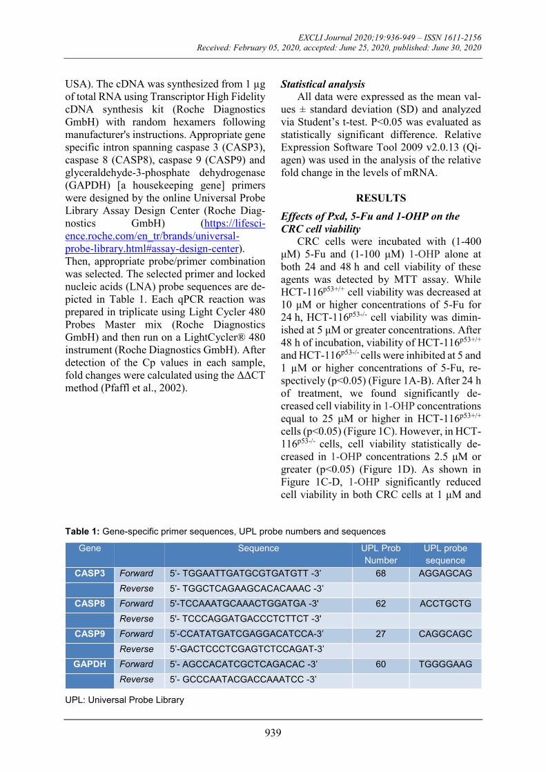
Table 1: Gene-specific primer sequences, UPL probe numbers and sequences
Gene Sequence UPL Prob
Number
UPL probe
sequence
CASP3 Forward 5’- TGGAATTGATGCGTGATGTT -3’ 68 AGGAGCAG
Reverse 5’- TGGCTCAGAAGCACACAAAC -3’
CASP8 Forward 5'-TCCAAATGCAAACTGGATGA -3' 62 ACCTGCTG
Reverse 5'- TCCCAGGATGACCCTCTTCT -3'
CASP9 Forward 5’-CCATATGATCGAGGACATCCA-3’ 27 CAGGCAGC
Reverse 5’-GACTCCCTCGAGTCTCCAGAT-3’
GAPDH Forward 5’- AGCCACATCGCTCAGACAC -3’ 60 TGGGGAAG
Reverse 5’- GCCCAATACGACCAAATCC -3’
UPL: Universal Probe Library

EXCLI Journal 2020;19:936-949 – ISSN 1611-2156
Received: February 05, 2020, accepted: June 25, 2020, published: June 30, 2020
940
Figure 1: Cell viability of 1-400 μM 5-Fu treated HCT-116p53+/+ (A) and HCT-116p53-/- cells (B), 1-400 μM 1-OHP treated HCT-116p53+/+ (C) and HCT-116p53-/- cells (D) at 24 and 48 h. The effects of Pxd, 5-Fu and 1-OHP alone treatment, and 10 µg/ml of Pxd for 4 hours; afterward, Pxd was removed from the media and the cells were treated with 200 µM 5-Fu and 5 µM 1-OHP for an additional 24 hours in HCT-116p53+/+ (E) and HCT-116p53-/- cells (F). *p < 0.05 compared with control group; †p < 0.05 compared with 200 µM 5-Fu and 5 µM 1-OHP co-treated group; Pxd: Phenoxodiol; 5-Fu: 5-Fluorouracil; 1-OHP: Oxal-iplatin
higher concentrations at 48 h (p<0.05). The
sensitivity of both CRC cells to 5-Fu and 1-
OHP treatments was different except incuba-
tion at 48 h with 1-OHP. Compared to un-
treated control cells, the cells treated with 10
μg/ml Pxd for 24 h, both HCT-116p53+/+ and
HCT-116p53-/- cells viability decreased by
82 % and 76 %, respectively (Figure 1E-F).
Furthermore, these cells were also co-admin-
istered with selected concentrations of
chemotherapeutic agents (200 µM 5-Fu and 5
µM 1-OHP) for 24 h. The decline in viable
cells was statistically significant when com-
pared with the untreated control (p < 0.05)
(Figure 1E-F). Co-administration with 5-Fu
and 1-OHP for 24 h overwhelmingly de-
creased the cell viability to 62 % and 69 %,
respectively. To explore whether Pxd could
enhance the chemosensitivity of CRC cells to
co-administration of 5-Fu and/or 1-OHP,
CRC cells were pre-treated Pxd for 4 h; after-
ward, Pxd was removed from the media and

EXCLI Journal 2020;19:936-949 – ISSN 1611-2156
Received: February 05, 2020, accepted: June 25, 2020, published: June 30, 2020
941
the cells were co-administered with 5-Fu and
1-OHP for an additional 24 h and analyzed
cell viability. Our results show that Pxd pre-
treatment was more effective in inhibiting
cancer cell proliferation than cell death in-
duced by co-administration of 5-Fu and 1-
OHP. After Pxd incubation for 4 h, followed
by co-administration with 5-Fu and 1-OHP
for additional 24 h decreased the viable HCT-
116p53+/+ and HCT-116p53-/- cells to 51 and
56 %, respectively. Moreover, Pxd pre-treat-
ment resulted in a strikingly diminished cell
viability compared to 5-Fu and/or 1-OHP
alone treatment in both CRC cells (Figure 1E-
F).
Cytotoxic effect of Pxd, 5-Fu and 1-OHP
on CRC cells
The cytotoxic effects of studied agents on
both HCT-116p53+/+ and HCT-116p53-/- cells
were determined by lactate dehydrogenase
(LDH) activity measurement. Cytotoxicity
was very low and similar in both CRC cells
exposed to selected concentration of Pxd for
24 h (Figure 2E-F). As shown in Figure 2E-F,
5-Fu alone and 1-OHP alone treatments
caused an increase in cytotoxicity at 24 h in
both CRC cells. Whereas the increase was
higher in HCT-116p53+/+ cells than the HCT-
116p53-/-, there was a lack of statistically sig-
nificant differences (p>0.05; Figure 2E-F).
Likewise, both co-administration of 5-Fu
and/or 1-OHP and Pxd pre-treatment fol-
lowed by 5-Fu and/or 1-OHP administration
was more cytotoxic in HCT-116p53+/+ cells
than the HCT-116p53-/-, although no statistical
significance was detected (p>0.05; Figure 2E-
F). In all experiments, cytotoxicity ratio did
not exceed 6 %.
Detection of morphological cellular
changes
In order to detect whether decline in cell
viability was because of apoptosis or necrosis,
CRC cells were stained with AO/EtBr 24 h af-
ter the treatment of cells with the studied
agents. AO/EtBr staining indicated that de-
cline in cell viability found in MTT assay re-
sulted from apoptotic response of the cells
(Figure 2). Representative images of viable,
necrotic and apoptotic cells were depicted in
Figure 2A-B. The apoptosis ratio was statisti-
cally significant for administrations of 5-Fu
alone, 5-Fu and/or 1-OHP co-administration,
and Pxd pre-treatment followed by 5-Fu
and/or 1-OHP in both CRC cells (p<0.05)
(Figure 2C-D). Furthermore, Pxd alone and 1-
OHP alone treatments were also resulted in
statistically significant increases in apoptotic
ratio of HCT-116p53-/- cells (p<0.05) (Figure
2C-D). In addition, when corresponding to 5-
Fu and/or 1-OHP co-administration, Pxd pre-
treatment followed by 5-Fu and/or 1-OHP ad-
ministration elevated apoptotic cells in HCT-
116p53-/- (p<0.05) (Figure 2D).
Detection of caspase-dependent apoptosis In order to detect whether apoptosis is
caspase-dependent or -independent, we meas-
ured the amount of cleaved caspase‐3 in both
CRC cells. Only 5-Fu alone treatment in
HCT-116p53+/+ cells for 24 h, the level of the
cleaved caspase-3 protein increased nearly 2
fold and this elevation was statistically signif-
icant (p<0.05) (Figure 3A). Yet, either single-
agent treatment in HCT-116p53-/- cells for
24 h, there was no statistically significant in-
crease in the levels of cleaved caspase-3 pro-
tein (p>0.05) (Figure 3B). Statistically signif-
icant increase in the amount of the cleaved
caspase-3 protein was detected for admin-
istrations of 5-Fu and/or 1-OHP co-admin-
istration, and Pxd pre-treatment followed by
5-Fu and/or 1-OHP in both CRC cells
(p<0.05) (Figure 3A-B). Moreover, in HCT-
116p53+/+ cells, Pxd pre-treatment followed by
5-Fu and/or 1-OHP strikingly elevated the
level of the cleaved caspase-3 protein when
compared with 5-Fu and/or 1-OHP co-admin-
istration (p<0.05) (Figure 3A). Based on our
findings, it can be interpreted that Pxd pre-
treatment enhanced caspase‐dependent apop-
tosis in both CRC cells.

EXCLI Journal 2020;19:936-949 – ISSN 1611-2156
Received: February 05, 2020, accepted: June 25, 2020, published: June 30, 2020
942
Figure 2: Representative images of viable (1), apoptotic (2), and necrotic (3) cells were determined by AO/EtBr staining using a fluorescence microscope at x400 magnification. HCT-116p53+/+ (A) and HCT-116p53-/- cells (B) co-treated with 200 µM 5-Fu and 5 µM 1-OHP at 24 h. Apoptotic ratios of cells treated with Pxd, 5-Fu and 1-OHP alone treatment, and 10 µg/ml of Pxd for 4 hours; afterward, Pxd was re-moved from the media and the cells were treated with 200 µM 5-Fu and 5 µM 1-OHP for an additional 24 hours in HCT-116p53+/+ (C) and HCT-116p53-/- cells (D). Cytotoxic effects of agents on HCT-116p53+/+ (E) and HCT-116p53-/- cells (F) determined by LDH release into culture medium. The results are ex-pressed as means ± SD from three independent experiments. *p < 0.05 compared with control group; †p < 0.05 compared with 200 µM 5-Fu and 5 µM 1-OHP co-treated group; Pxd: Phenoxodiol; 5-Fu: 5-Fluorouracil; 1-OHP: Oxaliplatin; AO: acridine orange; EtBr: ethidium bromide
Effect of Pxd, 5-Fu and 1-OHP treatment
on CASP3, CASP8 and CASP9 mRNA
levels
In order to detect whether caspase-3 was
activated, by either intrinsic or extrinsic apop-
totic pathways, the relative mRNA expression
levels of CASP3, CASP8 and CASP9 genes
were determined by qPCR. The lack of statis-
tically significant differences in the mRNA
levels of CASP3, CASP8 and CASP9 genes
were observed after single-agent treatments
for 24 h in both CRC cells (p>0.05; Figure

EXCLI Journal 2020;19:936-949 – ISSN 1611-2156
Received: February 05, 2020, accepted: June 25, 2020, published: June 30, 2020
943
3C-D). Similarly, CASP8 mRNA expression
level was not affected after administration of
5-Fu and/or 1-OHP co-administration, and
Pxd pre-treatment followed by 5-FU and/or 1-
OHP in both CRC cells (p>0.05) (Figure 3C-
D). After administration of 5-Fu and/or 1-
OHP co-administration in HCT-116p53+/+
cells, the mRNA levels of CASP3 and CASP9
were induction by 1.88-fold (P<0.05) and
1.83-fold (P<0.05), respectively. Moreover,
in HCT-116p53-/- cells, this treatment resulted
in a 1.91-fold (P<0.05) and a 1.74-fold
(P<0.05) upregulation of CASP3 and CASP9
mRNA levels, respectively. When Pxd pre-
treatment was followed by 5-Fu and/or 1-
OHP administered to the HCT-116p53+/+ cells,
the expression levels of the CASP3 and
CASP9 increased 2.48-fold (P<0.05) and
2.18-fold (P<0.05), respectively. Similarly, in
HCT-116p53-/- cells, this treatment resulted in
elevation of the CASP3 [2.13-fold, (P<0.05)]
and CASP9 [1.96-fold, (P<0.05)] mRNA ex-
pression levels, when compared with the con-
trol cells. Furthermore, when corresponding
to 5-Fu and/or 1-OHP co-administration, Pxd
pre-treatment followed by 5-Fu and/or 1-OHP
administration to both CRC cells increased
the mRNA expression levels of CASP3 and
CASP9 and, these elevations were statistically
important (P<0.05).
Figure 3: Cleaved caspase-3 protein levels of each agent alone treatment and pretreatment of 10 µg/ml of Pxd for 4 hours; afterward, Pxd was removed from the media and the cells were treated with 200 µM 5-Fu and 5 µM 1-OHP for an additional 24 hours in HCT-116p53+/+ (A) and HCT-116p53-/- cells (B). Com-parison of the mRNA expression levels of CASP3, CASP8 and CASP9 genes in HCT-116p53+/+ (C) and HCT-116p53-/- cells (D) treated with agents at different time points. *p < 0.05 compared with control group; †p < 0.05 compared with 200 µM 5-Fu and 5 µM 1-OHP co-treated group; Pxd: Phenoxodiol; 5-Fu: 5-Fluorouracil; 1-OHP: Oxaliplatin; CASP3: Caspase 3; CASP8: Caspase 8; CASP9: Caspase 9; qRT-PCR: quantitative real-time PCR; mRNA: messenger RNA; PCR: polymerase chain reaction
EXCLI Journal 2020;19:936-949 – ISSN 1611-2156
Received: February 05, 2020, accepted: June 25, 2020, published: June 30, 2020
944
DISCUSSION
The fate of CRC treatment depends on the
use of targeted therapy that could disrupt sur-
vival pathways and activate cell death path-
ways. The resistance to anticancer drugs that
arise due to the increase of selective tumor
cell groups during the treatment, in tumor tis-
sues which initially responds to standard
chemotherapy, is one of the major causes of
cancer progress. For this reason, the number
of potential drug candidates which are capa-
ble of re-regulating the sensitivity of CRC
cells especially in individuals who relapse or
do not respond to treatment, are increasing in
recent years. 5-Fu and 1-OHP are frequently
used in combined regimens for CRC (Ad-
amsen et al., 2007; Asmis and Saltz, 2008; De
Angelis et al., 2006; De Gramont et al., 2000;
Ikehata et al., 2014; Simpson et al., 2003). In
particular, chemotherapeutic agent combina-
tions that are currently used are not effective
in patients with unresectable and metastatic
CRC (Van Cutsem et al., 2016). Therefore,
studies investigating newly developed chemi-
cal agents to be used alone or in combination
with classical chemotherapy agents, such as
5-Fu and 1-OHP, are needed for the effective
treatment of CRC. Additionally, the re-
sistance to chemotherapeutic agents and the
cytotoxic effects of high concentrations of
these agents draw attention to the investiga-
tion of different molecules (Berindan-Neagoe
et al., 2013).
Epidemiological studies that showed an
inverse relationship between isoflavone con-
sumption and cancer risk have drawn atten-
tion to the value of these components in can-
cer treatment (Mahoney et al., 2012; Silasi et
al., 2009). Pxd, a novel synthetic isoflavone
derivative, has been shown to have a particu-
larly potent effect on reversing chemothera-
peutic resistance. Pxd inhibits proliferation in
a wide range of human cancer cells such as
leukemia, breast, ovarian and prostate and it
is 20 times more potent than genistein (Jin and
El-Deiry, 2005). Additionally, Pxd has an im-
pact on survival and death pathways and ad-
ministered in combination with standard
chemotherapeutics or monotherapy in solid
cancers. In our study, we aimed to investigate
if Pxd pre-treatment alone and in combination
with 5-Fu and 1-OHP can effect apoptotic, cy-
totoxic and antiproliferative response in both
wild type HCT-116p53+/+ and mutant HCT-
116p53-/- cells.
In our study, it was observed that 5-Fu and
1-OHP decreased the viability in both HCT-
116 cvell lines at 24h. Based on cell viability
results, 200 μM 5-Fu and 5 μM 1-OHP con-
centrations in 5-Fu and 1-OHP combinations
where similar effects were investigated in var-
ious cancer cell lines were determined as ref-
erence (De Angelis et al., 2004, 2006; Ad-
amsen et al., 2007; Evert et al., 2018; Li et al.,
2018; Guo et al., 2006; Lin et al., 2012). For
this purpose, it was shown that the cell viabil-
ity rate for 200 μM 5-Fu was approximately
56 % and 60 %, and for 5 μM 1-OHP was
87 % and 80 % in HCT-116p53+/+ and HCT-
116p53-/- cells, respectively. For Pxd, the via-
bility in both CRC cells was found to be ap-
proximately 80 % with the recommended
concentration and time (10 μg/mL, 4 hours) in
the literature (Kamsteeg et al., 2003; Alvero
et al., 2006; Gamble et al., 2006). After pre-
treatment of 10 μg/ml Pxd to the cells for 4
hours, Pxd was removed from the medium
and 200 μM 5-Fu with 5 μM 1-OHP was co-
administered for 24 hours. When viability
rates of 5-Fu and 1-OHP alone or in combina-
tion with Pxd pre-treated cells were com-
pared, viability rates of pre-treated cells were
significantly reduced. The viability of both
CRC cells decreased significantly after treat-
ment with 5-Fu and 1-OHP followed by Pxd
treatments, compared to 5-Fu with 1-OHP
alone. Accordingly, in both CRC cells, after a
pre-treatment of 10 μg/ml Pxd for 4 hours, the
cell viability rate was approximately 49 %
and 51 % after removal of Pxd from the me-
dium and co-administration of 200 μM 5-Fu
and 5 μM 1-OHP for 24 hours in HCT-
116p53+/+ and HCT-116p53-/- cells, respec-
tively. Based on these data, we can say that
Pxd pre-treatment makes CRC cells more sen-
sitive to the combination of 5-Fu and 1-OHP.
No studies have been found in the literature
regarding the co-administration of 5-Fu and
EXCLI Journal 2020;19:936-949 – ISSN 1611-2156
Received: February 05, 2020, accepted: June 25, 2020, published: June 30, 2020
945
1-OHP following Pxd pre-treatment to HCT-
116p53+/+ and HCT-116p53-/- cells, so we could
not compare our findings. On the other hand,
consistent with our results, in the studies us-
ing various cancer cell lines, it has been re-
ported that Pxd makes these cells more sus-
ceptible to different agents and standard
chemotherapeutic drugs (Kamsteeg et al.,
2003; Alvero et al., 2006; Gamble et al., 2006;
Georgaki et al., 2009; Kluger et al., 2007; Si-
lasi et al., 2009; Aguero et al., 2005). Also,
several studies showed that Pxd rearranges
sensitivity by significantly lowering IC50 val-
ues against paclitaxel, carboplatin, gemcita-
bine, docetaxel and topotecan in ovarian epi-
thelial cancer cells (Kamsteeg et al., 2003; Al-
vero et al., 2006; Silasi et al., 2009). In addi-
tion, when Pxd was removed from the envi-
ronment after 2 hours of pre-treatment of 10
μg/ml in ovarian cancer cells, it increased sen-
sitivity against chemotherapeutic agents such
as carboplatin, paclitaxel and gemcitabine
that were administered for 24 h, by decreasing
cell viability rates and increasing apoptotic ef-
fect (Alvero et al., 2006). Moreover, this
proapoptotic effect has been shown to occur
only in cancer cells and does not cause any
toxicity in normal ovarian epithelial cells
(Kamsteeg et al., 2003; Alvero et al., 2006).
In another study investigating the effects of
Pxd on human umbilical vein endothelial cells
(HUVEC), it was shown that Pxd inhibits tu-
mor cell growth and proliferation as well as
antiangiogenic effect (Gamble et al., 2006).
When the effects of Pxd on different cancer
cells was investigated, it was found that Pxd
stimulates cancer cells to apoptosis, terminal
differentiation and mitotic arrest (G1 phase).
Based on these findings, Pxd was granted Fast
Track status by the Food and Drug Admin-
istration (FDA) as a re-regulator of chemo-
therapeutic susceptibility for platinum and
taxanes in the treatment of recurrent ovarian
and hormone-resistant or non-resistant pros-
tate cancers (Gamble et al., 2006; Wang et al.,
2012). These results indicate that the pre-
treatment of Pxd in combination with other
chemotherapeutic agents may be more bene-
ficial in achieving the most effective concen-
tration and time and in sensitizing chemother-
apeutics with known effects against CRC
cells, rather than by administering it alone in
cancer cells.
The CRC cells were stained with AO/EtBr
to reveal and evaluated morphologically
whether the molecular mechanism underlying
the decrease in cell viability stemmed from
apoptosis or necrosis. Human CRC cells were
stained with AO/EtBr and evaluated morpho-
logically with the fluorescence microscope.
Accordingly, it was determined that Pxd pre-
treatment in both CRC cell lines directed cells
to apoptosis more than necrosis. When the
cell ratios leading to apoptosis were com-
pared, HCT-116p53-/- cells (apoptotic ratio
32.3 %) were found to be more sensitive than
HCT-116p53+/+ cells (apoptotic ratio 29.7 %).
It was observed that the cell viability rates
were consistent with our fluorescence stain-
ing results. These findings were also con-
firmed by LDH release into the medium. Our
results are in line with other studies that
showed the cytotoxic effects of Pxd in both
HCT-116 cells. Results of various studies per-
formed with Pxd application to different can-
cer cell lines showed that cytotoxic effect af-
ter Pxd application did not change signifi-
cantly, similar to our study (Mahoney et al.,
2012; Alvero et al., 2006; Georgaki et al.,
2009).
Targeting the factors regulating apoptosis
is a current approach to the treatment of can-
cer cells (Reed, 2002) and fundamental fea-
ture of a cancer cell is its resistance to undergo
apoptosis (Lowe and Lin, 2000; Mor et al.,
2002), thus, reversing this resistance to apop-
tosis shows the best approach for treatment of
cancer. Apoptosis has been classified into two
main groups: an intrinsic and extrinsic path-
way (Green and Evan, 2002). The proteolytic
active caspase-3 enzyme involved in apopto-
sis belongs to the group of effector caspases
and causes the division of numerous cytoplas-
mic and nuclear components to reveal the
morphological and biochemical properties of
the intrinsic pathway of apoptosis. The en-

EXCLI Journal 2020;19:936-949 – ISSN 1611-2156
Received: February 05, 2020, accepted: June 25, 2020, published: June 30, 2020
946
zyme caspase-3 as a marker of apoptosis con-
firms that the cell has undergone apoptosis at
the protein level (Kamsteeg et al., 2003;
Kluger et al., 2007; Alvero et al., 2006; Gam-
ble et al., 2006; Ikehata et al., 2014). In our
study, 5-Fu and/or 1-OHP co-administration,
and Pxd pre-treatment followed by 5-Fu
and/or 1-OHP in both CRC cells were found
to have more active caspase 3 enzymes than
untreated control cells. When compared to 5-
Fu with 1-OHP alone treatment, Pxd pre-
treatment overwhelmingly increased apop-
totic activity in CRC cells. Increased protein-
caspase-3 activity in both extrinsic and intrin-
sic apoptotic pathways suggests that both cas-
cade pathways lead to proteolytic degradation
using the caspase pathway. Our results are
consistent with various studies investigating
apoptotic responses in different cancer cells
treated with Pxd and show cell-specific apop-
totic response when looking at the effects of
Pxd on different cancer cell lines (Kamsteeg
et al., 2003; Kluger et al., 2007; Alvero et al.,
2006). For example, in a study performed
with ovarian cancer cell lines, it was observed
that the administration of carboplatin,
paclitaxel and docetaxel alone for 24 hours
did not cause any difference in active caspase-
3 activity. After 2 hours of 10 μg/mL Pxd pre-
treatment, Pxd was removed and paclitaxel,
carboplatin and docetaxel were administered
for 24 hours, it was shown that p17 and p19
protein levels, which are the markers of active
caspase-3 forms, and caspase-3 activity were
increased (Alvero et al., 2006). In another in
vitro study, carboplatin alone in melanoma
cells did not show any effect on XIAP and
other components of the apoptotic cascade,
but after pre-treatment of 10 μg/mL Pxd for 2
hours and removal of Pxd was followed by
carboplatin administration for 24 hours,
Caspase 2 and Bid activation and XIAP deg-
radation were observed and a significant in-
crease in caspase-3 activity was reported
(Kluger et al., 2007). These results indicate
that pre-administration of Pxd alone or in
combination with other chemotherapeutic
agents results in a similar effect by increasing
the level of active caspase-3 in different can-
cer cells, thereby activating caspase-depend-
ent apoptosis, leading to the death of cancer
cells. The presence of active caspase-3 in both
CRC cells as a result of Pxd pre-treatment is
indicative of induction of caspase-mediated
apoptosis in these cells.
The expression levels of CASP3, CASP8
and CASP9 genes were also investigated by
qPCR method to determine in both HCT-116
cell lines. We also demonstrated that CASP3
and CASP9 mRNA levels increased after
treatment with 5-Fu and 1-OHP followed by
Pxd treatments, compared to 5-Fu with 1-
OHP alone. Our results show that Pxd is able
to fully activate CASP3, CASP8 and CASP9
in the same cells in which 5-Fu and 1-OHP
failed. This suggests that the apoptotic path-
way is functional in CRC cells but is activated
only in response to Pxd. Although there is no
study in the literature regarding the effect of
Pxd on mRNA expression levels of CASP3,
CASP8 and CASP9 genes HCT-116p53+/+ and
HCT-116p53-/- cells, active caspase-8,
caspase-9 and caspase-3 activities were in-
creased by inducing Fas-dependent apoptotic
pathway of 24 hours of Pxd administration in
various ovarian cancer cells have been shown
to increase (Kamsteeg et al., 2003). In addi-
tion, another in vitro study, showing the
change in apoptotic markers observed at dif-
ferent time intervals after treatment of CP70
ovarian cancer cell line with 10 μg/mL Pxd,
showed the degradation of proapoptotic Bak
protein at 16 hours post-treatment, significant
increase in caspase-8, caspase-9 and caspase-
3 activity and the presence of p30 XIAP (Al-
vero et al., 2006). These data show that Pxd
and 5-Fu with 1-OHP induce caspase-depend-
ent apoptosis by showing a synergistic effect.
To the best of our knowledge, our study is
the first study to evaluate the ability to sup-
press proliferation and stimulate apoptosis by
the co-administration of 5-Fu with 1-OHP
chemotherapeutic agents following Pxd pre-
treatment in both HCT-116 human CRC cells.
Our results indicate that 5-Fu and 1-OHP ren-
der both CRC cells more sensitive to Pxd ef-
fects. Therefore, this study suggests that the
EXCLI Journal 2020;19:936-949 – ISSN 1611-2156
Received: February 05, 2020, accepted: June 25, 2020, published: June 30, 2020
947
addition of Pxd to 5-Fu and 1-OHP could po-
tentially be a therapeutic strategy for CRC. If
our findings are supported by further molecu-
lar analyses in other preclinical in vitro and in
vivo cancer models, we think that Pxd may be
a good candidate molecule for classical
chemotherapeutic agents in the treatment of
CRC.
Acknowledgment
This study was supported by the Gazi
University Projects of Scientific Investiga-
tion, with the project code number 01/2018-
09.
Conflict of interest
The authors declare that they have no con-
flict of interest.
REFERENCES
Adamsen BL, Kravik KL, Clausen OPF, De Angelis
PM. Apoptosis, cell cycle progression and gene expres-
sion in TP53-depleted HCT116 colon cancer cells in
response to short-term 5-fluorouracil treatment. Int J
Oncol. 2007;31:1491-500.
Aguero MF, Facchinetti MM, Sheleg Z, Senderowicz
AM. Phenoxodiol, a novel isoflavone, induces G1 ar-
rest by specific loss in cyclin-dependent kinase 2 activ-
ity by p53-independent induction of p21WAF1/CIP.
Cancer Res. 2005;65:3364-73.
Aguero MF, Venero M, Brown DM, Smulson ME, Es-
pinoza LA. Phenoxodiol inhibits growth of metastatic
prostate cancer cells. Prostate. 2010;70:1211-21.
Alvero AB, O'Malley D, Brown D, Kelly G, Garg M,
Chen W, et al. Molecular mechanism of phenoxodiol-
induced apoptosis in ovarian carcinoma cells. Cancer.
2006;106:599-608.
Alvero AB, Brown D, Montagna M, Matthews M, Mor
G. Phenoxodiol-Topotecan co-administration exhibit
significant anti-tumor activity without major adverse
side effects. Cancer Biol Ther. 2007;6:612-7.
Asmis TR, Saltz L. Systemic therapy for colon cancer.
Gastroenterol Clin N Am. 2008;37:287-95.
Berindan-Neagoe I, Braicu C, Pileczki V, Petric RC,
Miron N, Balacescu O, et al. 5-Fluorouracil potentiates
the anti-cancer effect of oxaliplatin on Colo320 colo-
rectal adenocarcinoma cells. J Gastrointest Liver Dis.
2013;22:37-43.
Brown DM, Heaton A, Husband AJ. Idronoxil. Drugs
Future. 2008;33:844-60.
De Angelis PM, Kravik KL, Tunheim SH, Haug T,
Reichelt WH. Comparison of gene expression in
HCT116 treatment derivatives generated by two differ-
ent 5-fluorouracil exposure protocols. Mol Cancer.
2004;3:11.
De Angelis, PM, Svendsrud DH, Kravik KL, Stokke T.
Cellular response to 5-fluorouracil (5-FU) in 5-FU-re-
sistant colon cancer cell lines during treatment and re-
covery. Mol Cancer. 2006;5:20.
De Gramont A, Figer A, Seymour M, Homerin M,
Hmissi A. Leucovorin and fluorouracil with or without
oxaliplatin as first-line treatment in advanced colorec-
tal cancer. J Clin Oncol. 2000;18:2938-47.
De Luca T, Bosneaga E, Morré DM, Morré DJ. Down-
stream targets of altered sphingolipid metabolism in re-
sponse to inhibition of ENOX2 by phenoxodiol. Bio-
factors. 2008;34:253-60.
Evert J, Pathak S, Sun XF, Zhang H. A study on effect
of oxaliplatin in microRNA expression in human colon
cancer. J Cancer. 2018;9:2046-53.
Gamble JR, Xia P, Hahn CN, Drew JJ, Drogemuller
CJ, Brown D, et al. Phenoxodiol, an experimental anti-
cancer drug, shows potent antiangiogenic properties in
addition to its antitumour effects. Int J Cancer.
2006;18:2412-20.
Georgaki S, Skopeliti M, Tsiatas M, Nicolaou KA, Io-
annou K, Husband A, et al. Phenoxodiol, an anticancer
isoflavene, induces immunomodulatory effects in vitro
and in vivo. J Cell Mol Med. 2009;13:3929-38.
Green DR, Evan GI. A matter of life and death. Cancer
Cell. 2002;1:19-30.
Guo J, Zhou AW, Fu YC, Verma UN, Tripathy D,
Frenkel EP, et al. Efficacy of sequential treatment of
HCT116 colon cancer monolayers and xenografts with
docetaxel, flavopiridol, and 5-florouracil. Acta Phar-
macol Sin. 2006;27:1375-81.
Ikehata M, Ogawa M, Yamada Y, Tanaka S, Ueda K,
Iwakawa S. Different effects of epigenetic modifiers
on the cytotoxicity induced by 5-Fluorouracil, Iri-
notecan or Oxaliplatin in colon cancer cells. Biol
Pharm Bull. 2014;37:67-73.
Isono M, Sato A, Asano T, Okubo K, Asano T. Evalu-
ation of therapeutic potential of phenoxodiol, a novel
isoflavone analog, in renal cancer cells. Anticancer
Res. 2018;38:5709-16.
Jin Z, El-Deiry WS. Overwiew of cell death signaling
pathways. Cancer Biol Ther. 2005;4:139-63.

EXCLI Journal 2020;19:936-949 – ISSN 1611-2156
Received: February 05, 2020, accepted: June 25, 2020, published: June 30, 2020
948
Kamsteeg M, Rutherford T, Sapi E, Hanczaruk B, Sha-
habi S, Flick M, et al. Phenoxodiol - an isoflavone an-
alog - induces apoptosis in chemoresistant ovarian can-
cer cells. Oncogene. 2003;22:2611-20.
Kluger HM, McCarthy MM, Alvero AB, Sznol M, Ari-
yan S, Camp RL, et al. The X-linked inhibitor of apop-
tosis protein (XIAP) is up-regulated in metastatic mel-
anoma, and XIAP cleavage by Phenoxodiol is associ-
ated with Carboplatin sensitization. J Transl Med.
2007;5:6.
Li S, Tian J, Zhang H, Zhou S, Wang X, Zhang L, et
al. Down-regulating IL-6/GP130 targets improved the
anti-tumor effects of 5-fluorouracil in colon cancer.
Apoptosis. 2018;23:356-74.
Li Y, Huang X, Huang Z, Feng J. Phenoxodiol en-
hances the antitumor activity of gemcitabine in
gallbladder cancer through suppressing Akt/mTOR
pathway. Cell Biochem Biophys. 2014;70:1337-42.
Lin YL, Liau JY, Yu SC, Ou DL, Lin LI, Tseng LH, et
al. KRAS mutation is a predictor of oxaliplatin sensi-
tivity in colon cancer cells. PLoS ONE. 2012;7:
e50701.
Lowe SW, Lin AW. Apoptosis in cancer. Carcinogen-
esis. 2000;21:485-95.
Mahoney S, Arfuso F, Rogers P, Hisheh S, Brown D,
Millward M, et al. Cytotoxic effects of the novel iso-
flavone, phenoxodiol, on prostate cancer cell lines. J
Biosci. 2012;37:73-84.
Mahoney S, Arfuso F, Millward M, Dharmarajan A.
The effects of phenoxodiol on the cell cycle of prostate
cancer cell lines. Cancer Cell Int. 2014;14:110.
McPherson RA, Galettis PT, de Souza PL. Enhance-
ment of the activity of phenoxodiol by cisplatin in pros-
tate cancer cells. Br J Cancer. 2009;100:649-55.
Miller KD, Nogueira L, Mariotto AB, Rowland JH,
Yabroff KR, Alfano CM, et al. Cancer treatment and
survivorship statistics. 2019. CA Cancer J Clin.
2019;69:363-85.
Miyamoto M, Takano M, Aoyama T, Soyama H, Ishi-
bashi H, Kato K, et al. Phenoxodiol increases cisplatin
sensitivity in ovarian clear cancer cells through XIAP
down-regulation and autophagy inhibition. Anticancer
Res. 2018;38:301-6.
Mor G, Straszewski S, Kamsteeg M. Role of the
Fas/Fas ligand system in female reproductive organs:
survival and apoptosis. Biochem Pharmacol. 2002;64:
1305-15.
Morré DJ, McClain N, Wu LY, Kelly G, Morré DM.
Phenoxodiol treatment alters the subsequent response
of ENOX2 (tNOX) and growth of hela cells to
paclitaxel and cisplatin. Mol Biotechnol. 2009;42:100-
9.
Pfaffl MW, Horgan GW, Dempfle L. Relative expres-
sion software tool (REST) for group-wise comparison
and statistical analysis of relative expression results in
real-time PCR. Nucl Acids Res. 2002;30:e36.
Reed JC. Apoptosis-based therapies. Nat Rev Drug
Discov. 2002;1:111-21.
Rubins JB, Greatens T, Kratzke RA, Tan AT,
Polunovsky VA, Bitterman P. Lovastatin induces
apoptosis in malignant mesothelioma cells. Am J
Respir Crit Care Med. 1998;157:1616-22.
Saif MW, Heaton A, Lilischkis K, Garner J, Brown
DM. Pharmacology and toxicology of the novel inves-
tigational agent Cantrixil (TRX-E-002-1). Cancer
Chemother Pharmacol. 2017;79:303-14
Sapi E, Alvero AB, Chen W, O'Malley D, Hao XY,
Dwipoyono B, et al. Resistance of ovarian carcinoma
cells to docetaxel is XIAP dependent and reversible by
phenoxodiol. Oncol Res. 2004;14:567-78.
Siegel RL, Miller KD, Jemal A. Cancer statistics. 2019.
CA Cancer J Clin. 2019;69:7-34.
Silasi DA, Alvero AB, Rutherford TJ, Brown D, Mor
G. Phenoxodiol: pharmacology and clinical experience
in cancer monotherapy and in combination with
chemotherapeutic drugs. Exp Opin Pharmacother.
2009;10:1059-67.
Simpson D, Dunn C, Curran M, Goa KL. Oxaliplatin:
a review of its use in combination therapy for advanced
metastatic colorectal cancer. Drugs. 2003;63:2127-56.
Turkey Cancer Statistics, 2019. Available at:
https://hsgm.saglik.gov.tr/tr/kanser-istatistikleri/yil-
lar/2014-yili-turkiye-kanser-istatistikleri.html Ac-
cessed 02 February 2020
Van Cutsem E, Cervantes A, Adam R, Sobrero A, Van
Krieken JH, Aderka D, et al. ESMO consensus guide-
lines for the management of patients with metastatic
colorectal cancer. Ann Oncol. 2016;27:1386-422.
Yao C, Wu S, Li D, Ding H, Wang Z, Yang Y, et al.
Co-administration phenoxodiol with doxorubicin syn-
ergistically inhibit the activity of sphingosine kinase-1
(SphK1), a potential oncogene of osteosarcoma, to sup-
press osteosarcoma cell growth both in vivo and in
vitro. Mol Oncol. 2012;6:392-404.

EXCLI Journal 2020;19:936-949 – ISSN 1611-2156
Received: February 05, 2020, accepted: June 25, 2020, published: June 30, 2020
949
Yu F, Watts RN, Zhang XD, Borrow JM, Hersey P.
Involvement of BH3-only proapoptotic proteins in mi-
tochondrial-dependent Phenoxodiol-induced apoptosis
of human melanoma cells. Anticancer Drugs. 2006;17:
1151-61.
Wang D, Hou L, Wu L, Yu X. Synthesis and anti-tu-
mor activities of novel oxazinyl isoflavonoids. Chem
Pharm Bull. 2012;60:513-20.


![Liver resection for metastatic colorectal cancer - [email protected]](https://img.dokumen.tips/doc/110x75/620633768c2f7b1730055cf8/liver-resection-for-metastatic-colorectal-cancer-emailprotected.jpg)