Embed Size (px)
Citation preview
AMBULATORY DIAGNOSIS OF
ENDOMETRIAL PATHOLOGY
THOMAS JUSTIN CLARK
A thesis submitted to the University of Birmingham for the degree of
DOCTOR OF MEDICINE
Department of Obstetrics & Gynaecology
Faculty of Medicine and Dentistry
University of Birmingham
August 2003

University of Birmingham Research Archive
e-theses repository This unpublished thesis/dissertation is copyright of the author and/or third parties. The intellectual property rights of the author or third parties in respect of this work are as defined by The Copyright Designs and Patents Act 1988 or as modified by any successor legislation. Any use made of information contained in this thesis/dissertation must be in accordance with that legislation and must be properly acknowledged. Further distribution or reproduction in any format is prohibited without the permission of the copyright holder.
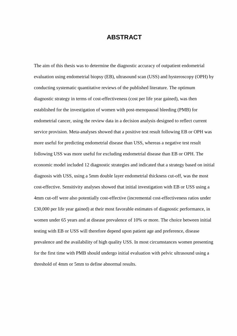
ABSTRACT
The aim of this thesis was to determine the diagnostic accuracy of outpatient endometrial
evaluation using endometrial biopsy (EB), ultrasound scan (USS) and hysteroscopy (OPH) by
conducting systematic quantitative reviews of the published literature. The optimum
diagnostic strategy in terms of cost-effectiveness (cost per life year gained), was then
established for the investigation of women with post-menopausal bleeding (PMB) for
endometrial cancer, using the review data in a decision analysis designed to reflect current
service provision. Meta-analyses showed that a positive test result following EB or OPH was
more useful for predicting endometrial disease than USS, whereas a negative test result
following USS was more useful for excluding endometrial disease than EB or OPH. The
economic model included 12 diagnostic strategies and indicated that a strategy based on initial
diagnosis with USS, using a 5mm double layer endometrial thickness cut-off, was the most
cost-effective. Sensitivity analyses showed that initial investigation with EB or USS using a
4mm cut-off were also potentially cost-effective (incremental cost-effectiveness ratios under
£30,000 per life year gained) at their most favorable estimates of diagnostic performance, in
women under 65 years and at disease prevalence of 10% or more. The choice between initial
testing with EB or USS will therefore depend upon patient age and preference, disease
prevalence and the availability of high quality USS. In most circumstances women presenting
for the first time with PMB should undergo initial evaluation with pelvic ultrasound using a
threshold of 4mm or 5mm to define abnormal results.

DEDICATION
To Chris, Laura, Alice and Joe

ACKNOWLEDGEMENTS
The work forming this thesis was conducted between September 1999 and August 2003 at the
Birmingham Women‟s Hospital, in my capacity as a Research Fellow (funded by the
University of Birmingham Interdisciplinary Fund and Birmingham Women‟s Hospital
Research and Development Fund) and later as a Lecturer in Obstetrics and Gynaecology at
the University of Birmingham. I would like to acknowledge all those people to whom I am
indebted for helping me along the way.
These include Ann Fry-Smith for designing, running and blinding electronic searches for the
hysteroscopy review, and Chris Mann, Patrick Chien and Doris Voit for acting as second
reviewers in the study identification and data abstraction process for the reviews of
endometrial biopsy, ultrasound and hysteroscopy respectively. Thanks to Fujian Song for
supervising the meta-analyses and providing statistical support for all reviews and to Pelham
Barton for his help with constructing the decision tree and directing economic analyses.
Thanks also to Tracy Bingham, my first office companion, for making me laugh from the
outset.
Particular thanks to my academic colleagues and friends „Honest‟ Honest, Janesh Gupta and
Khalid Khan. Honest for his computer wizardry, his appreciation of the frustrations involved
with Health Technology Assessment and for drinking plenty of coffee at my expense. Janesh
for his continuous support, guidance and enthusiasm, in addition to his generosity and
friendship throughout the preparation of this thesis. Special thanks to Khalid for the huge
amount of time and effort given to me at all stages of the research. I am grateful for the

opportunity he gave me, for teaching me about research methodology, supplying ongoing
intellectual advice and forever pointing out my “lack of attention to detail!”
Finally, thanks to my children, Laura, Alice and Joe for their complete lack of interest in this
research and most of all thanks to my beloved wife, Christine, for everything.
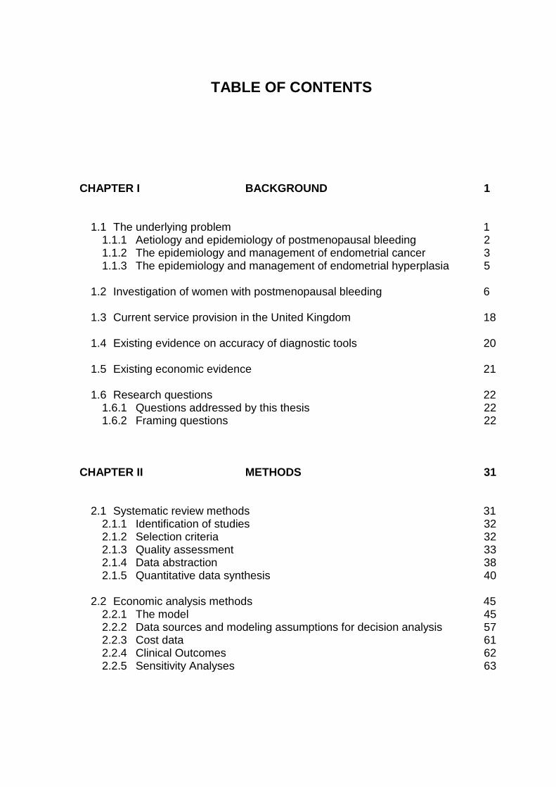
TABLE OF CONTENTS
CHAPTER I BACKGROUND1 1
1.1 The underlying problem 1
1.1.1 Aetiology and epidemiology of postmenopausal bleeding 2 1.1.2 The epidemiology and management of endometrial cancer 3 1.1.3 The epidemiology and management of endometrial hyperplasia 5
1.2 Investigation of women with postmenopausal bleeding 6 1.3 Current service provision in the United Kingdom 18 1.4 Existing evidence on accuracy of diagnostic tools 20 1.5 Existing economic evidence 21 1.6 Research questions 22
1.6.1 Questions addressed by this thesis 22 1.6.2 Framing questions 22
CHAPTER II 2 METHODS 31 2.1 Systematic review methods 31
2.1.1 Identification of studies 32 2.1.2 Selection criteria 32 2.1.3 Quality assessment 33 2.1.4 Data abstraction 38 2.1.5 Quantitative data synthesis 40
2.2 Economic analysis methods 45
2.2.1 The model 45 2.2.2 Data sources and modeling assumptions for decision analysis 57 2.2.3 Cost data 61 2.2.4 Clinical Outcomes 62 2.2.5 Sensitivity Analyses 63
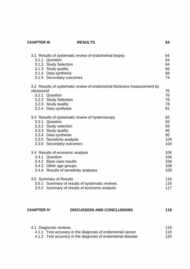
CHAPTER III 3 RESULTS 64
3.1 Results of systematic review of endometrial biopsy 64
3.1.1 Question 64 3.1.2 Study Selection 64 3.1.3 Study quality 66 3.1.4 Data synthesis 68 3.1.5 Secondary outcomes 74
3.2 Results of systematic review of endometrial thickness measurement by ultrasound 76
3.2.1 Question 76 3.2.2 Study Selection 76 3.2.3 Study quality 78 3.2.4 Data synthesis 81
3.3 Results of systematic review of hysteroscopy 93
3.3.1 Question 93 3.3.2 Study selection 93 3.3.3 Study quality 95 3.3.4 Data synthesis 95 3.3.5 Sensitivity analysis 104 3.3.6 Secondary outcomes 104
3.4 Results of economic analysis 106
3.4.1 Question 106 3.4.2 Base case results 106 3.4.3 Other age-groups 108 3.4.4 Results of sensitivity analyses 109
3.5 Summary of Results 116
3.5.1 Summary of results of systematic reviews 116 3.5.2 Summary of results of economic analysis 117
CHAPTER IV 4 DISCUSSION AND CONCLUSIONS 119
4.1 Diagnostic reviews 119 4.1.1 Test accuracy in the diagnosis of endometrial cancer 119 4.1.2 Test accuracy in the diagnosis of endometrial disease 120
4.1.3 Test feasibility 122 4.2 Validity of reviews 123
4.2.1 Heterogeneity 124 4.2.2 Sources of bias 125
4.3 Comparison with other reviews and guidelines 126
4.3.1 Reviews 126 4.3.2 Guidelines 128
4.4 Applicability of reviews 129 4.5 Economic evaluation 134
4.5.1 Base case analysis 135 4.5.2 Sensitivity analysis 135
4.6 Validity of economic evaluation 137
4.6.1 Limitations of economic analysis 138 4.7 Comparison with other economic evaluations and guidelines 141 4.8 Applicability of economic evaluation 141 4.9 Recommendations for practice 144 4.10 Recommendations for future research 145
4.10.1 Diagnostic accuracy 146 4.10.2 Economic evaluation 147
APPENDICES REFERENCES
LIST OF TABLES
CHAPTER I Table 1.1 Diagnostic modalities available to detect endometrial cancer and
hyperplasia in women with postmenopausal bleeding 7 CHAPTER II Table 2.1 Quality assessment and definitions 35 Table 2.2 Hierarchy of evidence for primary research on diagnostic accuracy 37 CHAPTER III Table 3.1 Methodological quality of outpatient EB studies included in meta-
analyses 67 Table 3.2 Pooled estimates of pre-test probabilities, likelihood ratios and post-test
probabilities for diagnostic accuracy of outpatient biopsy in detecting endometrial cancer in women with abnormal uterine bleeding 70
Table 3.3 Sensitivity analyses for meta-analysis of the diagnostic accuracy of
outpatient endometrial biopsy in endometrial hyperplasia with or without atypia and its diagnostic accuracy in detecting endometrial cancer with or without premalignant complex/atypical endometrial hyperplasia 75
Table 3.4 Methodological quality of selected primary studies 80 Table 3.5 Pooled estimates of pre-test probability, likelihood ratio and post-test
probability for ultrasound measurement of endometrial thickness in predicting endometrial cancer 85
Table 3.6 Sensitivity analyses: Studies of ultrasound measurement of both layers
4 mm or 5 mm endometrial thickness for endometrial cancer or
disease with pooled LRs stratified according to study characteristics and quality 86
Table 3.7 Exploration of heterogeneity in estimation of accuracy of ultrasound
(5mm double layer endometrial thickness) for diagnosis of endometrial cancer and disease: Results of meta-regression analysis 88
Table 3.8 Pooled estimates of pretest probability, likelihood ratio and posttest
probability for ultrasound measurement of endometrial thickness in predicting endometrial disease (hyperplasia and/or cancer) 92
Table 3.9 Pooled estimates of pretest probabilities, likelihood ratios and posttest
probabilities for diagnostic accuracy of hysteroscopy in detecting endometrial cancer and disease in women with abnormal uterine bleeding 98
Table 3.10 Exploration of heterogeneity in estimation of accuracy of hysteroscopy
for diagnosis of endometrial cancer and disease: Results of meta-regression analysis 100
Table 3.11 Investigation of postmenopausal bleeding: Incremental cost-
effectiveness ratios for diagnostic strategies, compared in each case to no initial investigation 107
Table 3.12 Investigation of postmenopausal bleeding: Incremental cost-
effectiveness ratios for the non-dominated strategies, compared in each case to a strategy of ultrasound (5mm cut-off) 108
Table 3.13 Investigation of postmenopausal bleeding at different ages of
presentation: Incremental cost-effectiveness ratios of strategies compared to ultrasound (5mm cut-off) 109
Table 3.14 Sensitivity analysis: The effect of delayed diagnosis on the incremental
cost-effectiveness ratios of combination strategies compared to ultrasound (5mm cut-off) 111
Table 3.15 Summary of results of economic evaluation: cost-effectiveness of each
strategy compared with ultrasound scan (5mm cut-off) 118
LIST OF FIGURES
CHAPTER I
Figure 1-1 Outpatient endometrial biopsy devices 10 Figure 1-2 Outpatient endometrial biopsy 11 Figure 1-3 Pelvic ultrasound 12 Figure 1-4 Transvaginal ultrasound 13 Figure 1-5 Endometrial thickness measured by transvaginal ultrasound scan 14 Figure 1-6 Hysteroscopy 15 Figure 1-7 Semi-rigid 2.5mm diameter hysteroscopes 16 Figure 1-8 Hysteroscopic views of the endometrium 17 Figure 1-9 Event pathway (current service provision) for the investigation and
management of women with postmenopausal bleeding 19 CHAPTER II Figure 2-1 Decision analytic model: Strategy utilising initial evaluation with
endometrial biopsy (EB) for the investigation of postmenopausal bleeding for endometrial cancer 48
Figure 2-2 Decision analytic model: Strategy utilising initial evaluation with pelvic
ultrasound scan (USS) using a cut-off of 4mm to signify abnormal endometrial thickness for the investigation of postmenopausal bleeding for endometrial cancer 49
Figure 2-3 Decision analytic model: Strategy utilising initial evaluation with pelvic
ultrasound scan (USS) using a cut-off of 5mm to signify abnormal endometrial thickness for the investigation of postmenopausal bleeding for endometrial cancer 50
Figure 2-4 Decision analytic model: Strategy utilising initial evaluation with outpatient hysteroscopy (OPH) for the investigation of postmenopausal bleeding for endometrial cancer 51
Figure 2-5 Decision analytic model: Strategy utilising initial evaluation with a
combination of pelvic ultrasound and outpatient hysteroscopy (USS_OPH) for the investigation of postmenopausal bleeding for endometrial cancer (both 4mm and 5mm ultrasound cut-offs used to signify abnormal endometrial thickness) 52
Figure 2-6 Decision analytic model: Strategy utilising initial evaluation with a
combination of pelvic ultrasound and endometrial biopsy (USS_EB) for the investigation of postmenopausal bleeding for endometrial cancer (both 4mm and 5mm ultrasound cut-offs used to signify abnormal endometrial thickness) 53
Figure 2-7 Decision analytic model: Strategy utilising initial evaluation with a
combination of endometrial biopsy and outpatient hysteroscopy (EB_OPH) for the investigation of postmenopausal bleeding for endometrial cancer 54
Figure 2-8 Decision analytic model: Strategy utilising initial evaluation with a
combination of pelvic ultrasound, endometrial biopsy and outpatient hysteroscopy (USS_EB_OPH) for the investigation of postmenopausal bleeding for endometrial cancer (both 4mm and 5mm ultrasound cut-offs used to signify abnormal endometrial thickness) 55
Figure 2-9 Decision analytic model: Strategy of no initial evaluation (i.e. diagnostic
work-up only if symptoms recurred) for the investigation of postmenopausal bleeding for endometrial cancer 56
Figure 2-10 Decision analytic model (common pathway for further treatment of
endometrial cancer following initial hysterectomy) 60 CHAPTER III Figure 3-1 Study selection process for systematic review of outpatient EB 65 Figure 3-2 Sensitivity and specificity of endometrial biopsy in the diagnosis of
endometrial cancer and hyperplasia 69 Figure 3-3 Funnel plot endometrial biopsy and cancer 71 Figure 3-4 Funnel plot endometrial biopsy and hyperplasia 73
Figure 3-5 Study selection process for systematic review of ultrasound scan 78 Figure 3-6 Sensitivity and specificity of ultrasound 4mm in the diagnosis of
endometrial cancer and disease 82 Figure 3-7 Sensitivity and specificity of ultrasound 5mm in the diagnosis of
endometrial cancer and disease 83
Figure 3-8 Funnel plot of ultrasound ( 5 mm double layer) and endometrial cancer 90
Figure 3-9 Study selection process for systematic review of hysteroscopy 94 Figure 3-10 Sensitivity and specificity of hysteroscopy in the diagnosis of
endometrial cancer and disease 96 Figure 3-11 Funnel plot of hysteroscopy and endometrial cancer 102 Figure 3-12 The cost-effectiveness plane 112 Figure 3-13 Results of univariate sensitivity analysis on extra cost and survival of
USS 4mm compared to USS 5mm 113 Figure 3-14 Results of univariate sensitivity analysis on extra cost and survival of
EB compared to USS 5mm 114 Figure 3-15 Results of univariate sensitivity analysis on extra cost and survival of
EB compared to USS 4mm 115 CHAPTER IV Figure 4-1 Pooled estimates of pretest probabilities, likelihood ratios and posttest
probabilities for accuracy of outpatient endometrial biopsy in diagnosing endometrial cancer in women with postmenopausal bleeding (Nomogram reproduced with permission)118 131
Figure 4-2 Pooled estimates of pretest probabilities, likelihood ratios and posttest
probabilities for accuracy of endometrial thickness measurement by pelvic ultrasound, using both a 4mm and 5mm cut-offs, in diagnosing endometrial cancer in women with postmenopausal bleeding. (Nomogram reproduced with permission)118 132
Figure 4-3 Pooled estimates of pretest probabilities, likelihood ratios and posttest
probabilities for accuracy of hysteroscopy in diagnosing endometrial cancer in women with postmenopausal bleeding (Nomogram reproduced with permission)118 133
LIST OF APPENDICES
Appendix 1 Search strategy for economic evidence 148 Appendix 2 Economic evaluations in the diagnosis of endometrial cancer in
postmenopausal bleeding 149 Appendix 3 Search strategy for Endometrial biopsy evidence 150 Appendix 4 Search strategy for ultrasound endometrial thickness evidence 151 Appendix 5 Search strategy for hysteroscopy evidence 152 Appendix 6 Data collection checklist for review of outpatient endometrial biopsy
153 Appendix 7 Data collection checklist for review of pelvic ultrasound 155 Appendix 8 Data collection checklist for review of hysteroscopy 157 Appendix 9 Probability estimates used and data sources for the decision tree
used for the investigation of postmenopausal bleeding 162 Appendix 10 Direct medical costs used and data sources for decision tree for the
investigation of postmenopausal bleeding (Base case and sensitivity analyses) 163
Appendix 11 Reference list of excluded studies from systematic reviews of e
endometrial biopsy 164 Appendix 12 Diagnostic accuracy of outpatient endometrial biopsy in detecting
endometrial cancer and hyperplasia in women at risk of abnormal endometrial histology 168
Appendix 13 Procedure feasibility and diagnostic accuracy of outpatient
endometrial biopsy in endometrial cancer and hyperplasia. 169 Appendix 14 Studies included in systematic review of ultrasound measurement of
endometrial thickness for predicting endometrial cancer and hyperplasia 171
Appendix 15 Reference list of excluded studies from systematic reviews of
ultrasound 174
Appendix 16 Likelihood ratios (LR) for predicting endometrial cancer in primary studies 182
Appendix 17 Likelihood ratios (LR) for predicting endometrial disease (hyperplasia
and/or cancer) in primary studies 184 Appendix 18 Reference list of excluded studies from systematic reviews of
hysteroscopy 186 Appendix 19 Diagnostic accuracy of hysteroscopy in detecting endometrial cancer
and hyperplasia in women at risk of abnormal endometrial histology: Methodological details 197
Appendix 20 Procedure feasibility and diagnostic accuracy of hysteroscopy in
endometrial cancer and endometrial disease (cancer and hyperplasia) 200
Appendix 21 Life expectancies of United Kingdom women stratified by age, surgery
and presence of endometrial cancer 203 Appendix 22 Investigation of postmenopausal bleeding: Base-case results for the
decision model with a starting age of 65 years 204 Appendix 23 Incremental cost-effectiveness ratios for diagnostic strategies,
compared to ultrasound (5mm cut-off) assuming endometrial biopsy is performed at the same visit following a positive ultrasound or outpatient hysteroscopy 205
Appendix 24 Investigation of postmenopausal bleeding at different ages of
presentation: Non-discounted incremental cost-effectiveness ratios of strategies compared to ultrasound (5mm cut-off) 206
Appendix 25 Sensitivity analysis for the diagnostic strategy ultrasound using a 4mm
cut-off compared to ultrasound using a 5mm cut-off 207 Appendix 26 Sensitivity analysis for the diagnostic strategy endometrial biopsy
compared to ultrasound using a 5mm cut-off 208 Appendix 27 Sensitivity analysis for the diagnostic strategy endometrial biopsy
compared to ultrasound using a 4mm cut-off 209 Appendix 28 Basis for recommendations about the use of interventions, treatments
or service 210 Appendix 29 Publications from the thesis 212

ABBREVIATIONS
ABS Abdominal Ultrasound Scan
BWH Birmingham Women‟s Hospital
CEA Cost-Effectiveness Analysis
CI Confidence Interval
D&C Dilatation of the cervix and Curettage of the endometrium
D Dominated
DB Directed Biopsy
dOR diagnostic Odds Ratio
EB Endometrial Biopsy
Eca Endometrial cancer
Ehyp Endometrial hyperplasia
ET Endometrial Thickness
FIGO International Federation of Gynaecology and Obstetrics
FNR False Negative Rate
FPR False Positive Rate
HRT Hormone Replacement Therapy
Hyst Hysterectomy
ICER Incremental Cost-Effectiveness Ratio
LR Likelihood Ratio
LYG Life Year Gained
MeSH Medical Subject Heading

NHS National Health Service
NHS EED NHS Economic Evaluation Database
NS Not Specified
OB Outpatient Biopsy
OPH Outpatient Hysteroscopy
QALY Quality Adjusted Life Year
TAH Total Abdominal Hysterectomy
TNR True Negative Rate
TPR True Positive Rate
TVS Transvaginal Ultrasound Scan
tw textword
USS Ultrasound Scan
WMCIU West-Midlands Cancer Intelligence Unit
1
CHAPTER I
1
BACKGROUND
1.1 The underlying problem
Postmenopausal bleeding (PMB), unscheduled bleeding on hormone replacement therapy
(HRT) and menorrhagia are common gynecological problems.71
310
The main aim of
investigations for abnormal uterine bleeding is to exclude serious intrauterine pathology,
namely endometrial cancer and hyperplasia,232
conditions most prevalent in postmenopausal
women.
Traditional investigation of women with postmenopausal bleeding using inpatient blind
dilatation of the cervix and curettage of the endometrium (D&C) is now considered out-dated
practice and has been replaced by initial outpatient endometrial evaluation using miniature
endometrial biopsy (EB) devices, transvaginal ultrasound scan (USS) and outpatient
hysteroscopy (OPH).310
However, despite the widely accepted advantages of outpatient
investigation, there is uncertainty regarding the individual value of these tests and the best
sequence or combination in which to use them. Consequently practice varies throughout
Europe and North America,3,59,70,251,296,326
largely dependent upon prejudice (of individual
clinicians) and pragmatism (resources available to them).
2
The main aim of investigating women with PMB is to exclude endometrial cancer and its
precursor, endometrial hyperplasia. The incidence of endometrial cancer has increased during
the last decade.251,272
Unlike other malignancies affecting women, endometrial cancer often
presents at an early stage with the possibility of curative treatment by hysterectomy.251
Prognosis is increasingly bleak the more advanced the disease. As there have been no recent
advances in the treatment of endometrial cancer that can be expected to increase survival, the
importance of accurate and timely diagnosis of endometrial cancer is paramount in order to
reduce mortality further.
This thesis assesses the diagnostic accuracy of currently available outpatient tests for the
clinical investigation of women with PMB for endometrial cancer and hyperplasia. Moreover,
the thesis examines the cost-effectiveness of strategies utilising EB, USS and OPH for the
diagnosis of endometrial cancer.
1.1.1 Aetiology and epidemiology of postmenopausal bleeding
PMB is a common clinical problem in both general practice and hospital settings.1,89,310
Women are most likely to present with this symptom in the sixth decade of life272
where
consultation rates in primary care for PMB are 14.3/1000 population.1,272
Similarly, in the
hospital setting, abnormal patterns of uterine bleeding account for more than 70% of all
gynaecological consultations in the peri- and post-menopausal years.310
At the Birmingham
Women‟s Hospital (BWH), which serves a female population of 220,000 (of which one can
assume 80,000 are postmenopausal), approximately 1000 women are seen each year with
PMB (incidence 12.5/1000 population).
3
In most instances (90-95%), PMB results from benign causes such as intrauterine structural
pathologies (polyps, fibroids) or prescription of exogenous hormones. Often, bleeding arises
from apparently normal atrophic endometrium and is thought to be due to superficial petechial
haemorrhages and mucosal ulceration.278,342
However, the main aim of investigations for
PMB is to exclude endometrial cancer,232
which presents with this symptom in over 95% of
cases.250
The probability of endometrial cancer in women presenting with PMB is
approximately 5-10%16,146,149,251
and therefore referral of such women for further investigation
in secondary care is mandatory. Published recommendations state that women should be seen
within 2-6 weeks of referral.251
On referral, some additional means of endometrial assessment
are performed, as it is not possible to exclude cancer on clinical assessment alone.
Traditionally, abnormal uterine bleeding has been investigated with blind D&C under general
anaesthetic but now there is a trend towards minimally invasive, outpatient investigations
utilising EB, USS and OPH first (see current service provision below).251
Although cervical cancer can present with PMB, most women with this condition present
below the age of 55 years with intermenstrual bleeding, postcoital bleeding, vaginal discharge
or pain.251,259
The diagnosis is made from clinical examination and cervical biopsy. The
diagnosis of cervical cancer is not considered further in this thesis.
1.1.2 The epidemiology and management of endometrial cancer
Endometrial cancer represents the most common female pelvic genital malignancy in the
western world136
and is increasingly common among more affluent populations272
and
increases with the adoption of more westernised lifestyles.272
The aetiology of endometrial
cancer is unknown, but several factors are known to increase or decrease the likelihood of
4
developing endometrial cancer. The most important of these appear to be age, obesity and
unopposed endogenous or exogenous oestrogen production.272
In England and Wales, there are around 4000 new cases of endometrial cancer per annum
(440 in the West Midlands), representing almost 4% of all cancer cases in women, in whom it
is ranked 5th
.251,272
Incidence rates are approximately 50 per 100,000 population in women
over 60 years. The overall age-standardised rate has remained close to 12/100,000 since the
1970s, but in women aged 55-74 rates have increased slightly in the 1990s.251
The lifetime
risk of developing endometrial cancer has been estimated to be 1.4%. An average general
practitioner with a list size of 2000 would expect to see 1 new case of endometrial cancer
every 6 years. In contrast to the trends in incidence, there have been long-term declines in
mortality from cancer of the uterus. The age-standardised rate has halved – from 6/100,000 in
1950 to 3/100,000 in 1999. In England and Wales survival was only slightly below the
European average, but was well below that in the Netherlands, Germany, France and more
than 10% below rates in the USA.272
Overall 5-year survival is around 77%, and improves
with early stage localised disease. Around 70% of women diagnosed with endometrial cancer
have early stage disease and 5-year survival is around 87%. Survival is worse for later stage
disease at around 60% and is as low as 19% with the most advanced stage of disease.72
If
detected at an early stage, endometrial cancer is curable in most cases, usually by surgery
(hysterectomy) and/or radiotherapy. As there have been no recent advances in the treatment of
endometrial cancer that can be expected to increase survival, the importance of accurate and
timely diagnosis of endometrial cancer is paramount in order to reduce mortality further.
5
1.1.3 The epidemiology and management of endometrial hyperplasia
Endometrial hyperplasia is more prevalent than endometrial cancer and affects both pre and
postmenopausal women.10,199,206
The probability of endometrial hyperplasia in women
presenting with postmenopausal bleeding (with or without HRT) is approximately 15%.63,206
The risk factors for developing hyperplastic endometrium are the same as for endometrial
cancer and thus relate mainly to unopposed oestrogen exposure. Endometrial hyperplasia is
categorised histologically by the degree of architectural disruption (simple or complex
hyperplasia) and by the presence of abnormal cytology (atypia). The importance of
endometrial hyperplasia relates not only to the symptoms of genital tract bleeding it can
cause, but to its oncogenic potential. The natural history of endometrial hyperplasia is not
fully understood.125
What is known is that a proportion of simple and complex hyperplastic
processes will regress without treatment199
although the time scale over which such regression
may occur is unclear. However, a small proportion (estimated to be between 1 and 3%199
) will
progress to frank endometrial cancer. The main prognostic factor is the presence of atypical
cells. Malignant progression has been reported to occur in 30% of atypical endometrial
hyperplasias if left untreated.120,175,199
Management depends upon accurate and timely diagnosis. Endometrial hyperplasia without
atypia can be managed without the need for treatment as the condition may regress
spontaneously, but regular endometrial assessment is required to exclude disease
progression.120,125,199
Medical treatments include systemic (oral/parenteral)120,125,339
or local
(intrauterine) progestogens to reverse the oestrogen dominant milieu.267,288
Traditional
treatment is with hysterectomy.120,339
In the presence of atypical cells, hysterectomy is usually
recommended in view of the potential for malignant transformation.120
Erroneous diagnosis
6
can thus be detrimental to patients. For example, unnecessary surgery may be performed for
false positive diagnoses of hyperplastic endometrium whereas false negative diagnoses can
result in progression to endometrial cancer.
1.2 Investigation of women with postmenopausal bleeding
The traditional investigation for PMB was inpatient dilatation of the cervix and curettage of
the endometrium (D&C).152
This is now considered out dated practice71
and has been largely
replaced by the development of minimally invasive diagnostic tools for use in the outpatient
setting. These new diagnostic modalities include outpatient endometrial biopsy (EB),
transvaginal ultrasonography (USS) and outpatient hysteroscopy (OPH). (Table 1-1).
7
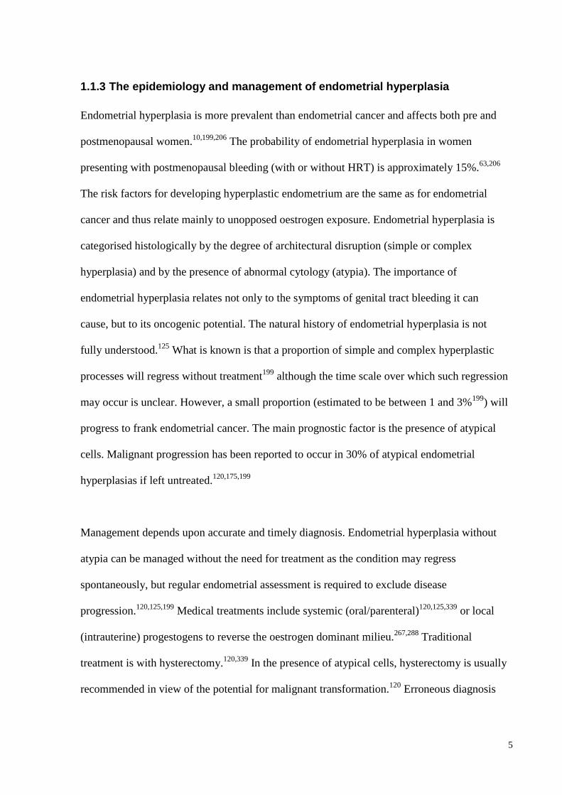
Table 1.1 Diagnostic modalities available to detect endometrial cancer and hyperplasia in women with postmenopausal bleeding
Features Endometrial
Biopsy
Ultrasound Hysteroscopy Prevailing clinical opinion
Safety All safe,
12,62,63,160,303 endometrial
biopsy has more potential for
trauma as it is a blind procedure
Acceptability All acceptable 210109,195
, ultrasound
least painful and invasive,
endometrial biopsy most painful320
Feasibility Failure rates higher in procedures
requiring uterine instrumentation.
Endometrial biopsy higher than
hysteroscopy.38,62,63,164
Other Minimal
expertise
required62
Extracavity /
pelvic
information37
Directed
endometrial
biopsies134
Advances in the technology and
application of ultrasound6,33,291
and
other radiographic imaging
techniques168
gives this modality
the greatest future potential in
diagnosis
invariably typically generally
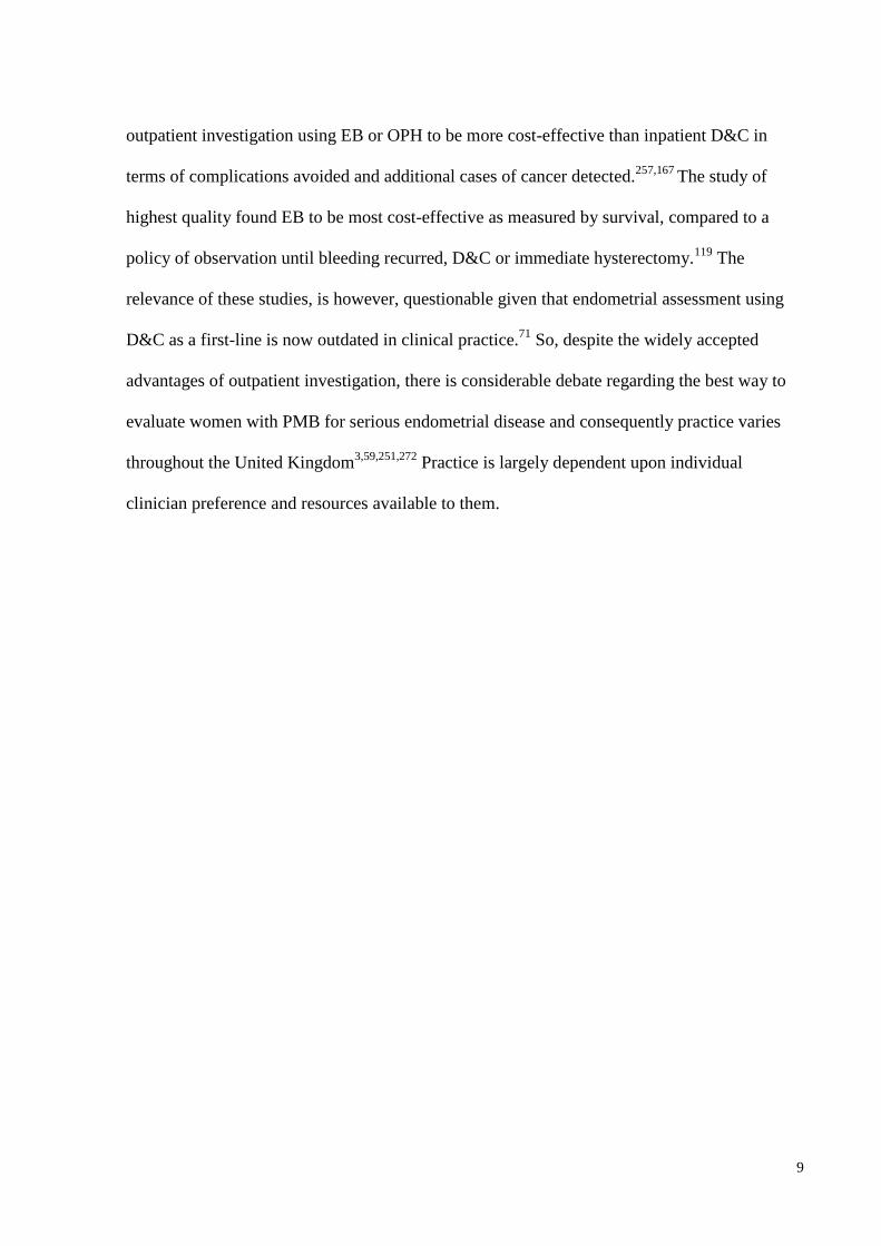
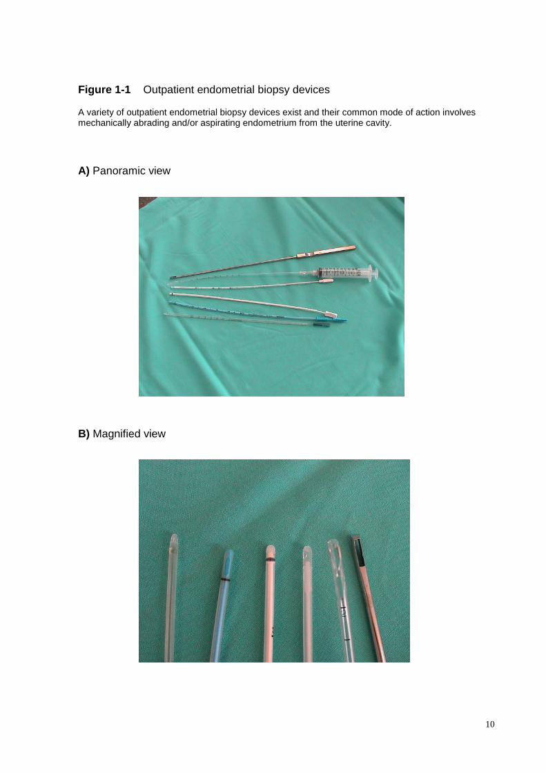
Outpatient EB (Figure 1-1) is a blind procedure where the endometrium is sampled using
small-diameter mechanical or suction devices, which can be easily introduced into the uterine
cavity without the need for anaesthetic. There is concern however, surrounding the non-
representative nature of these blind procedures, which may be related to the small proportion
of the endometrial surface sampled277
and the non-sampling of focal intrauterine lesions.157
8
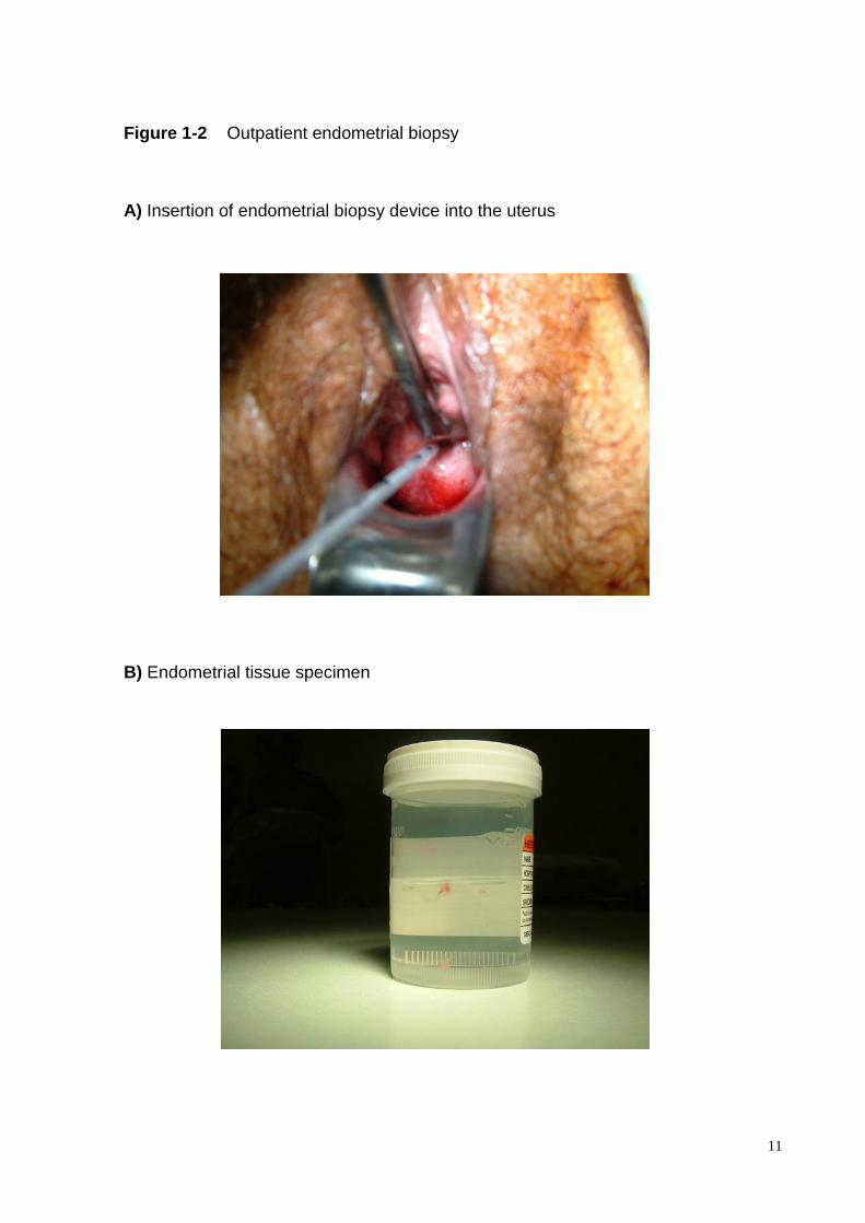
The development of pelvic ultrasound scanning (transabdominal or transvaginal) has allowed
high resolution imaging inside the uterus enabling measurement of the endometrial thickness.
(Figure 1-2 to 1-4) 303
It has been shown that the endometrial thickness of normal atrophic
uterus measures on average 2.3 mm123,146,243,244
. However, advanced endometrial cancer has
also been known to occur in cases without noticeable endometrial thickness on ultrasound96
The expertise and availability of ultrasound to gynaecologists varies throughout the UK,
Europe and North America. Consequently, radiologists or trained radiographers rather than
gynaecologists often perform USS. In the United Kingdom this situation is likely to change as
a result of the recent introduction of specialist gynecological training in USS.280
Hysteroscopy (Figures 1-5 and 1-7) is an endoscopic technique allowing visualisation of the
endometrial cavity. Recent advances in instrumentation have allowed hysteroscopy to be
performed in an outpatient setting, further increasing its use in gynecological practice.296
Various macroscopic features have been suggested as indicative of endometrial disease.
However, there is no consensus and visual interpretation is subjective and operator
dependent.56
Concerns surrounding the role and value of hysteroscopic diagnosis have
therefore arisen.5,25,164,232
All the forgoing outpatient modalities are generally considered to be safe,62,63
simple to use,
62,63 and acceptable to patients.
26,109,194,195 In addition, avoiding the need for an inpatient stay
potentially reduces health resources utilisation. Such considerations have been examined in
three published economic evaluations257,167,119
(Appendix 2), which were based on imprecise
data derived from small primary studies. The use of a single outpatient testing strategy in
comparison to blind inpatient D&C was explored in each study. Two of the studies found
9
outpatient investigation using EB or OPH to be more cost-effective than inpatient D&C in
terms of complications avoided and additional cases of cancer detected.257,167
The study of
highest quality found EB to be most cost-effective as measured by survival, compared to a
policy of observation until bleeding recurred, D&C or immediate hysterectomy.119
The
relevance of these studies, is however, questionable given that endometrial assessment using
D&C as a first-line is now outdated in clinical practice.71
So, despite the widely accepted
advantages of outpatient investigation, there is considerable debate regarding the best way to
evaluate women with PMB for serious endometrial disease and consequently practice varies
throughout the United Kingdom3,59,251,272
Practice is largely dependent upon individual
clinician preference and resources available to them.
10
Figure 1-1 Outpatient endometrial biopsy devices
A variety of outpatient endometrial biopsy devices exist and their common mode of action involves mechanically abrading and/or aspirating endometrium from the uterine cavity.
A) Panoramic view
B) Magnified view
11
Figure 1-2 Outpatient endometrial biopsy
A) Insertion of endometrial biopsy device into the uterus
B) Endometrial tissue specimen
12
Figure 1-3 Pelvic ultrasound
A) Transducers suitable for gynaecologic pelvic ultrasound scans: Transabdominal and transvaginal probes
B) Transvaginal probe (magnified view).
13
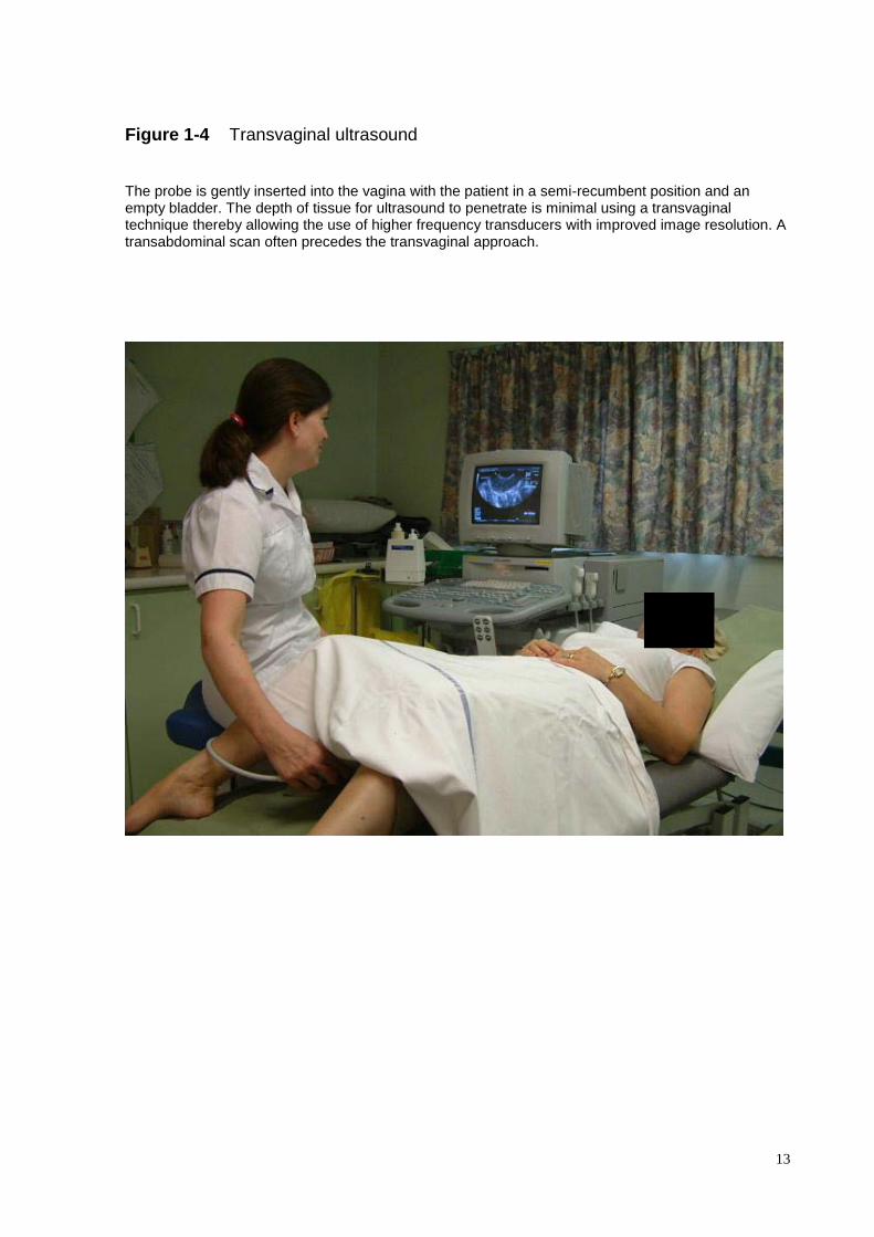
Figure 1-4 Transvaginal ultrasound
The probe is gently inserted into the vagina with the patient in a semi-recumbent position and an empty bladder. The depth of tissue for ultrasound to penetrate is minimal using a transvaginal technique thereby allowing the use of higher frequency transducers with improved image resolution. A transabdominal scan often precedes the transvaginal approach.
14
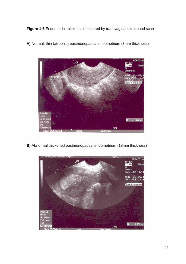
Figure 1-5 Endometrial thickness measured by transvaginal ultrasound scan
A) Normal, thin (atrophic) postmenopausal endometrium (3mm thickness)
B) Abnormal thickened postmenopausal endometrium (18mm thickness)
15
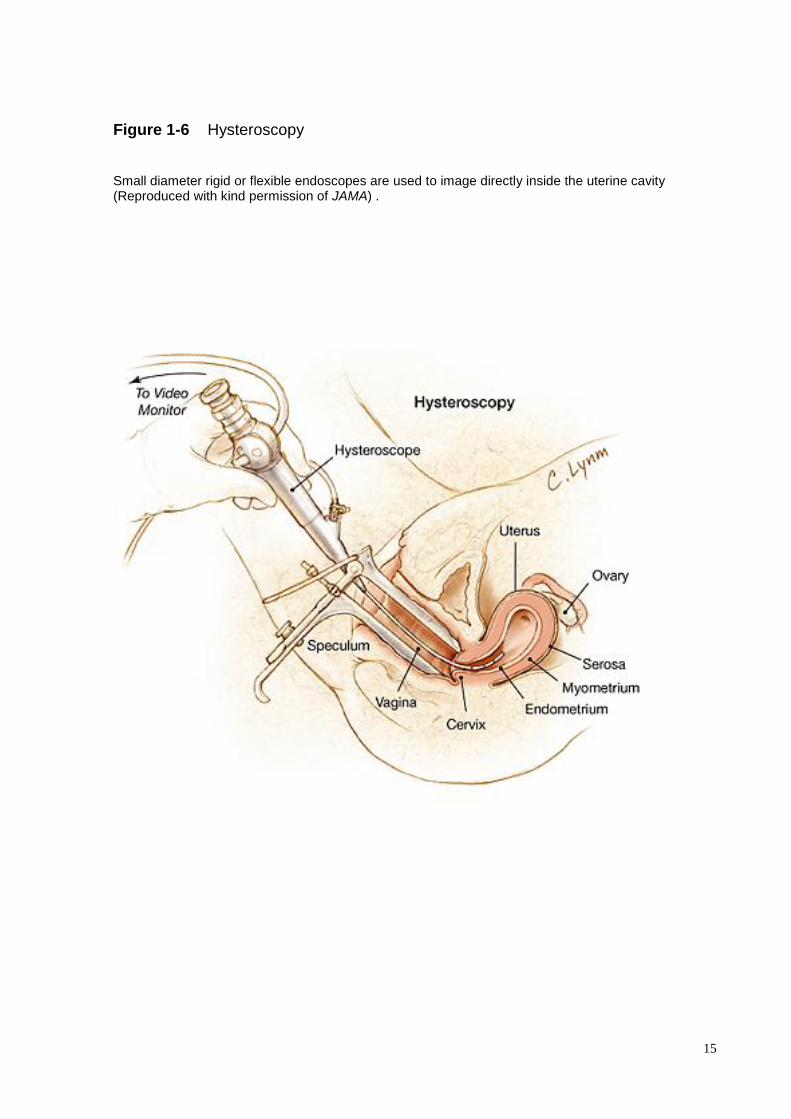
Figure 1-6 Hysteroscopy
Small diameter rigid or flexible endoscopes are used to image directly inside the uterine cavity (Reproduced with kind permission of JAMA) .
16
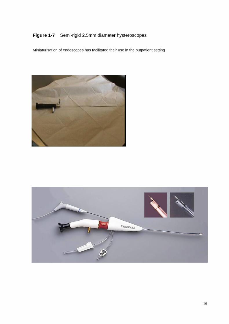
Figure 1-7 Semi-rigid 2.5mm diameter hysteroscopes
Miniaturisation of endoscopes has facilitated their use in the outpatient setting
17
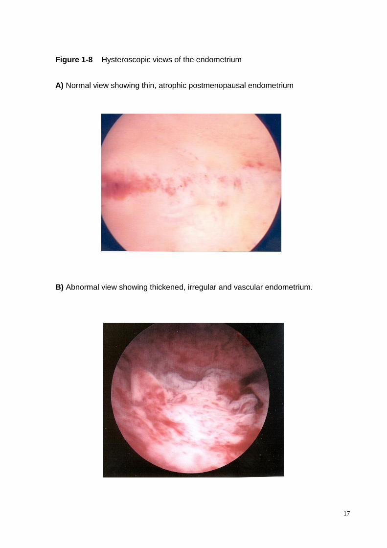
Figure 1-8 Hysteroscopic views of the endometrium
A) Normal view showing thin, atrophic postmenopausal endometrium
B) Abnormal view showing thickened, irregular and vascular endometrium.
18
1.3 Current service provision in the United Kingdom
Referral of all women presenting with PMB in the primary care setting for further
investigation is mandatory251
in order to exclude endometrial cancer. All women referred
should be seen within 2 weeks.251
Endometrial assessment is performed on referral, utilising
the outpatient tests, EB, USS or OPH. This often takes place in a „one stop‟ setting where the
investigation(s) take place during a single consultation with no planned follow up unless
test(s) fail or abnormal results are found. Negative findings result in discharge back to
primary care, whereas a positive diagnosis leads to advanced treatment in most instances.
Treatment for endometrial cancer varies, although in most instances hysterectomy and
surgical staging is performed followed by adjuvant non-surgical treatments where necessary.
279,105 The typical event pathway is shown in Figure 1-9, which is the essential basis of any
cost-effectiveness analyses.
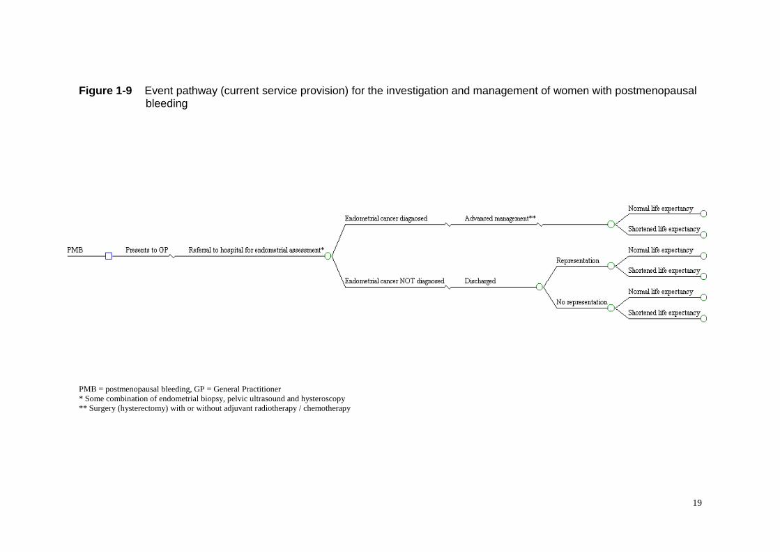
19
Figure 1-9 Event pathway (current service provision) for the investigation and management of women with postmenopausal bleeding
PMB = postmenopausal bleeding, GP = General Practitioner
* Some combination of endometrial biopsy, pelvic ultrasound and hysteroscopy
** Surgery (hysterectomy) with or without adjuvant radiotherapy / chemotherapy
20
1.4 Existing evidence on accuracy of diagnostic tools
The bibliographic databases MEDLINE (1966-2001) and EMBASE (1982-2001) were
searched for existing published evidence addressing the accuracy of investigative tools used
in PMB. This showed that in the last decade, there have been many publications indicating
that outpatient EB, ultrasound measurement of endometrial thickness and ambulatory
hysteroscopy may be useful in predicting endometrial cancer and hyperplasia. However,
individual studies addressing accuracy of these minimally invasive diagnostic tools, are small
leading to imprecise and heterogeneous estimates of accuracy.51
In addition, many studies
have used measures of diagnostic accuracy that are not clinically intuitive. The generation of
conflicting and confusing data has thus hampered clinical interpretation. The absence of a
uniform strategy for the investigation of women with PMB has resulted because of a
deficiency in the rigorous assessment of these newer diagnostic tools.
No systematic reviews of EB, USS or OPH were available at the outset of the research
forming this thesis. However, during the course of my research program, two systematic
reviews of USS and one of EB were published. The results and conclusions of all these
reviews are of limited validity due to potential biases in their methodological approach as
discussed later in the thesis (see section 4.4). I was unable to identify any systematic reviews
addressing the diagnostic accuracy of hysteroscopy. Therefore the need to conduct
comprehensive high quality reviews in this field was clear.
21
1.5 Existing economic evidence
The bibliographic databases MEDLINE (1966-2001) and EMBASE (1988-2001) were
searched for existing published economic evidence addressing the cost-effectiveness of
investigative tools currently used in PMB for detecting endometrial cancer. The search
strategy used is shown in Appendix 1. In addition, the NHS Economic Evaluation Database
(EED) held at the Centre for Reviews and Dissemination at the University of York and the
Cochrane Library were also searched. Following the electronic searches of MEDLINE and
EMBASE, there were 26 potentially eligible studies identified of which one337
was selected
after obtaining the full manuscripts. No manuscripts were selected from the EED out of 22
potentially eligible studies. No relevant studies were found from the Cochrane Library.
This study addressed outpatient investigation using USS or EB, and concluded that initial
evaluation with USS was less costly than initial evaluation with EB in relation to test
feasibility.337
No study was identified that evaluated the cost-effectiveness of all
contemporary outpatient modalities (i.e. EB, USS and OPH) used in sequence or combination
for the investigation of postmenopausal bleeding for endometrial cancer (Appendix 2).
Therefore there is a need to conduct a rigorous economic evaluation of diagnostic tools
current used in investigating women with PMB for endometrial cancer.
22
1.6 Research questions
1.6.1 Questions addressed by this thesis
The following questions were posed in this thesis.
In women presenting with postmenopausal bleeding:
1. What is the accuracy of outpatient endometrial biopsy in the diagnosis of endometrial
cancer and hyperplasia and what are the rates of failure and complications?
2. What is the accuracy of outpatient endometrial ultrasound in the diagnosis of endometrial
cancer and hyperplasia?
3. What is the accuracy of outpatient hysteroscopy in the diagnosis of endometrial cancer
and hyperplasia and what are the rates of failure and complications?
4. Which of the above three tests and their combination is most cost effective in outpatient
diagnosis of endometrial cancer?
1.6.2 Framing questions
Careful formulation of focussed research questions is necessary to aid the appropriate design
of systematic reviews and cost-effectiveness analyses so that specific answers can be
provided. The components of research questions in diagnosis are generally a population, a test
and a reference standard against which the accuracy of the test will be measured. Some other
outcome of interest may also be specified. Breaking down a question into these component
parts facilitates the precise identification of problems needing to be addressed.186
The factors
considered when formulating the questions posed in this thesis are discussed below:
23
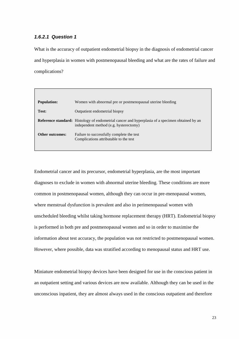
1.6.2.1 Question 1
What is the accuracy of outpatient endometrial biopsy in the diagnosis of endometrial cancer
and hyperplasia in women with postmenopausal bleeding and what are the rates of failure and
complications?
Endometrial cancer and its precursor, endometrial hyperplasia, are the most important
diagnoses to exclude in women with abnormal uterine bleeding. These conditions are more
common in postmenopausal women, although they can occur in pre-menopausal women,
where menstrual dysfunction is prevalent and also in perimenopausal women with
unscheduled bleeding whilst taking hormone replacement therapy (HRT). Endometrial biopsy
is performed in both pre and postmenopausal women and so in order to maximise the
information about test accuracy, the population was not restricted to postmenopausal women.
However, where possible, data was stratified according to menopausal status and HRT use.
Miniature endometrial biopsy devices have been designed for use in the conscious patient in
an outpatient setting and various devices are now available. Although they can be used in the
unconscious inpatient, they are almost always used in the conscious outpatient and therefore
Population: Women with abnormal pre or postmenopausal uterine bleeding
Test: Outpatient endometrial biopsy
Reference standard: Histology of endometrial cancer and hyperplasia of a specimen obtained by an
independent method (e.g. hysterectomy)
Other outcomes: Failure to successfully complete the test
Complications attributable to the test
24
the question of diagnostic performance was restricted to the outpatient setting, where it is
most relevant. Both the test and reference standard (inpatient endometrial curettage or
hysterectomy specimen) involve histological examination of obtained endometrial tissue.
Therefore the disease outcomes of interest (endometrial cancer and hyperplasia) could be
directly compared with one another facilitating ease of data abstraction. This also allowed the
impact of the severity of endometrial hyperplasia on the diagnostic performance of
endometrial biopsy to be examined, by stratifying data according to the presence or absence
of abnormal cytology (atypical cells). Non-endometrial uterine malignancies were not
considered in any of the reviews and where they occurred were excluded from analysis.
Good diagnostic performance depends upon test feasibility and safety in addition to
diagnostic accuracy. This is especially true for tests designed for use in the outpatient setting,
where patient factors may limit successful completion of the test. Unsuccessful sampling
using outpatient endometrial biopsy was categorised as either failed procedures (i.e. failure to
correctly position the device in the uterine cavity) or as histologically inadequate specimens
(failure to obtain adequate endometrial tissue for histological diagnosis from a correctly sited
device within the uterine cavity). Failed procedures may occur because of technical
difficulties (usually unfavourable uterine anatomy) or pain resulting from the outpatient
procedure. Histologically inadequate specimens are important because they may reflect the
normal atrophic postmenopausal endometrial state where obtaining endometrial tissue would
not necessarily be expected, rather than poor test performance. This consideration is important
in clinical practice as it presents problems of interpretation. Failure to successfully complete
the test was thus defined as both including and excluding histologically inadequate specimens
(see data abstraction in chapter 3). Complications were categorised according to whether they
25
arose directly from the test procedure itself (e.g. visceral damage, haemorrhage, infection,
vaso-vagal episodes) or indirectly (e.g. exacerbation of existing health problem such as
angina) and by severity (potentially life threatening or not).
1.6.2.2 Question 2
What is the accuracy of pelvic ultrasound in the diagnosis of endometrial cancer and
hyperplasia in women with postmenopausal bleeding?
Measurement of endometrial thickness by pelvic ultrasound can be performed using
transabdominal probes or more recently using transvaginal probes allowing better image
resolution.128
The procedure is rarely, if ever, required as an inpatient admission.
Measurement of endometrial thickness is useful in postmenopausal women because oestrogen
dependent endometrial thickening does not occur and so thin endometrium is considered
normal. In contrast, thickened endometrium may represent the important diagnoses of cancer
and hyperplasia as well as other benign conditions such as endometrial polyps. There is
considerable debate regarding what constitutes an abnormally thickened endometrium and so
various measurement cut-offs have been used.160,303,319
Different methods for measuring
endometrial thickness by ultrasound exist (e.g. single layer and double layer measurement)
Population: Women with abnormal pre or postmenopausal uterine bleeding
Test: Endometrial thickness measured by pelvic ultrasound
Reference standard: Histological confirmation of endometrial cancer and disease (cancer +
hyperplasia)
26
and this has complicated the situation further. For the purposes of this review, all studies of
symptomatic postmenopausal women undergoing pelvic ultrasound were included, but data
were grouped according to the type of ultrasound probe, cut-off level for abnormality and
measurement method used.
Pelvic ultrasound is not confined to use in postmenopausal women, but is widely employed in
pre-menopausal women with abnormal menstrual bleeding. This is because patient acceptance
is almost universal128,303
and the modality allows the myometrium and ovaries to be imaged,
in addition to the uterine cavity and endometrium. Consequently, the most commonly
occurring gynaecologic pathology in women of reproductive age, uterine fibroids340
and
benign ovarian cysts,28
can be diagnosed with a high degree of accuracy.105,176
However,
measurement of endometrial thickness to diagnose endometrial cancer or hyperplasia is of
limited use in this population.80,92,102,104,106,268
This is because a thickened endometrium is
normally seen in pre-menopausal women as a result of ovarian oestrogen production and the
extent of endometrial thickening varies in accordance with the stage of the menstrual cycle.242
Furthermore, the prevalence of endometrial cancer is very low in premenopausal women with
menstrual dysfunction27,219,271
and benign endometrial polyps, which cause focal endometrial
thickening, are far more common.20,103
The population in my research question was therefore
restricted to postmenopausal women with vaginal bleeding. Exogenous oestrogen given to
postmenopausal women as part of HRT may cause endometrial proliferation to a varying, but
lesser degree than in pre-menopausal women.147
Thus, symptomatic postmenopausal women
on HRT were also included in the population, although data was stratified according to HRT
usage.
27
The review of endometrial biopsy was conducted first, where the diagnostic reference
standard was endometrial histology obtained by inpatient sampling (endometrial curettage,
directed biopsy, endometrial resection and hysterectomy specimens). However, a significant
proportion of primary studies assessing diagnostic accuracy of ultrasound (and hysteroscopy)
used outpatient endometrial biopsy devices to obtain histological samples. The results of the
review of outpatient endometrial biopsy showed high diagnostic accuracy (see chapter IV).
Bias due to misdiagnosis by endometrial biopsy, though considered unlikely to be a
significant problem, was explored with subgroup analyses based on variation in histological
reference standard (see chapter III). In contrast to the research question posed for endometrial
biopsy, the condition of endometrial hyperplasia was not considered, but instead it was
included together with endometrial cancer under the heading „endometrial disease.‟ This
approach was used because ultrasonic (and hysteroscopic) features of hyperplasia are not
clearly distinct from those of endometrial cancer56
and such an approach has previously been
used.134,303
The safety of ultrasound has been well established12
and a previous systematic review303
has
reported negligible failure rates and so these outcomes were not considered.
1.6.2.3 Question 3
What is the accuracy of hysteroscopy in the diagnosis of endometrial cancer and hyperplasia
in women with postmenopausal bleeding and what are the rates of failure and complications?
28
Hysteroscopy can be carried out as an inpatient test under general anaesthetic or as an
outpatient test in the conscious patient. The research question relates to outpatient
investigative tools in women with postmenopausal uterine bleeding. However, as with
endometrial biopsy, hysteroscopy is performed in both pre and postmenopausal women and so
in order to maximise the information about test accuracy, the population was not restricted to
postmenopausal women. However, where possible, data was stratified according to the setting
and menopausal status. Diagnosis of the conditions endometrial cancer or disease was
considered (see question formulation for pelvic ultrasound – see section 1.6.2.2). Where
cancer or hyperplasia was suspected within a focal abnormality, these were categorised under
cancer and/or disease.
Hysteroscopic procedures failing to make a final diagnosis because of technical aspects (e.g.
cervical stenosis, anatomical factors, structural abnormalities), inadequate visualization (e.g.
obscured by bleeding, endometrial debris) or patient factors (e.g. pain, intolerance) were
categorized as failed procedures. Complications were categorised in the same way as for
endometrial biopsy (see section 1.6.2.1).
Population: Women with abnormal pre or postmenopausal uterine bleeding
Test: Hysteroscopy
Reference standard: Histological confirmation of endometrial cancer and disease (cancer +
hyperplasia)
Other outcomes: Failure to successfully complete the test
Complications attributable to the test
29
1.6.2.4 Question 4
What is the most cost-effective outpatient test or combination of outpatient tests for the
diagnosis of endometrial cancer in women with postmenopausal bleeding?
Endometrial cancer is the most important diagnosis to exclude in women with abnormal
uterine bleeding. This potentially life-threatening diagnosis is invariably made in association
with postmenopausal bleeding (PMB), where endometrial cancer is found in 5-10% of such
women.16,146,149,251
Additional means of outpatient endometrial assessment is mandatory251
(traditional investigation with inpatient dilatation and curettage as a first-line is considered
outdated practice),71
but which test or combination of tests is best to use is not established and
consequently is the focus of clinical debate and the reason for eclectic hospital
practice.3,59,251,272
Endometrial hyperplasia is not considered to be premalignant unless found
in association with abnormal cytology.120,175,199
The hyperplastic process is usually reversed
with simple hormonal treatment.120,267,288,339
Endometrial cancer is the only diagnosis
considered to significantly affect survival and therefore the management of this condition has
been chosen for the cost-effectiveness analysis, where effectiveness is measured in terms of
life years gained.
Population: Women with postmenopausal uterine bleeding
Test: Diagnostic approaches utilising various combinations of outpatient endometrial
biopsy, ultrasound and hysteroscopy
Clinical outcome: Survival following diagnosis and treatment of endometrial cancer
Economic outcome: Costs incurred in testing and in treating endometrial cancer
(NHS hospital perspective)

30
The benefits and costs of using an intervention to treat a disease depend upon whose
perspective it is (i.e. patient, hospital, payer, society etc.) The societal perspective is the most
comprehensive one (and encompasses all other perspectives), as it considers all costs and
benefits irrespective of who pays and who benefits. This is of use to government and policy
makers who are interested in allocating resources to improve population welfare.231
However,
our main concern related to maximising health (as opposed to welfare) in the symptomatic,
postmenopausal female population undergoing mandatory investigation for abnormal uterine
bleeding. For this reason we chose to take the perspective of the health service provider (NHS
healthcare system) and considered direct medical costs appropriate to the NHS.
31
CHAPTER II
2 MET
METHODS
This research presented in this thesis was undertaken with two aims:
(1) To summarise the current evidence on the diagnostic accuracy of outpatient endometrial
evaluation using endometrial biopsy (EB), ultrasound scan (USS) and outpatient
hysteroscopy (OPH) using systematic reviews
(2) To determine the most cost-effective combination of these tests for the investigation of
women with post-menopausal bleeding (PMB) for endometrial cancer using decision-
analytic modeling.
2.1 Systematic review methods
To determine the accuracy of the outpatient diagnostic tests used in PMB to predict
endometrial cancer and hyperplasia, quantitative systematic reviews of endometrial biopsy
(EB), pelvic ultrasound (USS) and hysteroscopy (OPH) were conducted. The methodology
used was common to all three reviews, it was based on a prospective protocol considering
widely recommended methods,66,183,189
and followed the stages given below.
32
2.1.1 Identification of studies
General bibliographic databases, MEDLINE and EMBASE, were searched. Language
restrictions were not applied. The electronic search strategies targeted the relevant diagnostic
procedures exclusively, studies addressing the relevant clinical problem (abnormal uterine
bleeding which encompasses both pre and postmenopausal bleeding) were then identified on
completion of the initial search phase by examining all the retrieved citations. Pilot searches
suggested that the chosen search strategies gave reasonable precision without compromising
sensitivity. These search strategies are detailed in Appendices 3 to 5.
In addition, the Cochrane Library and relevant specialist registers of the Cochrane
Collaboration were searched. Reference lists of all known reviews and primary studies were
checked and direct contact with manufacturers of outpatient EB devices and hysteroscopes
was also made.
2.1.2 Selection criteria
The reviews focused on prospective observational studies or comparative cross-sectional
studies in which the results of the diagnostic test of interest were compared with the results of
a reference standard. The following criteria were used to select articles for inclusion:
Population: Women with abnormal pre or postmenopausal bleeding.
Diagnostic tests: Outpatient endometrial biopsy, endometrial thickness
measurement using ultrasound imaging and hysteroscopy.
Reference Standard: Endometrial cancer and endometrial hyperplasia confirmed
histologically.
33
Two reviewers identified the studies in a two-stage process independently (see
Acknowledgments). The titles and abstracts identified as being potentially relevant from the
computer database searches or inspection of bibliographies were scanned and provisionally
included, unless they could definitely be excluded as not addressing the accuracy of EB, USS
or OPH. The full texts of all provisionally included articles from the first stage were retrieved.
The authors and journal titles were removed from the retrieved citations thereby blinding the
reviewers. Final inclusion/exclusion decisions were made with reference to a checklist, the
items of which were based on the selection criteria above. The checklists were piloted and the
repeatability of their use tested and confirmed. The checklists used for each review are shown
in Appendices 6 to 8. Disagreements about inclusion/exclusion were initially resolved by
consensus and where this was not possible it was resolved using arbitration by a third
reviewer (see Acknowledgements). The agreement statistics between reviewers were
computed using percentage agreement and weighted kappa statistics.67
The kappa statistic
provides measurement of agreement obtained beyond chance and weights provide credit for
partial agreement.55
2.1.3 Quality assessment
All papers meeting the eligibility criteria were assessed for their methodological quality.
Quality was defined as the confidence that the study design, conduct and analysis minimized
bias in the estimation of diagnostic accuracy. Based on existing checklists,66,107,163,207,230
quality assessment involved scrutinizing study designs and the relevant features of population,
intervention and outcome. These included method of data collection and patient selection,
details relating to type of abnormal bleeding and menopausal status, description of the

34
diagnostic test and histological reference standard, and presence of verification bias and
blinding (Table 2-1).189
This approach is in line with the recently published STARD criteria
for reporting test accuracy studies34,35
and the recently developed „QUADAS tool‟ for
assessing quality of test accuracy studies (personal communication – see Chapter 4).
35
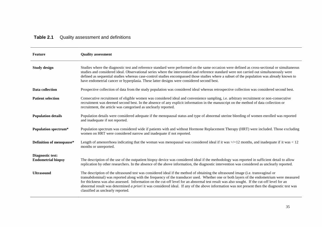
Table 2.1 Quality assessment and definitions
Feature Quality assessment
Study design
Studies where the diagnostic test and reference standard were performed on the same occasion were defined as cross-sectional or simultaneous
studies and considered ideal. Observational series where the intervention and reference standard were not carried out simultaneously were
defined as sequential studies whereas case-control studies encompassed those studies where a subset of the population was already known to
have endometrial cancer or hyperplasia. These latter designs were considered second best.
Data collection
Prospective collection of data from the study population was considered ideal whereas retrospective collection was considered second best.
Patient selection
Consecutive recruitment of eligible women was considered ideal and convenience sampling, i.e. arbitrary recruitment or non-consecutive
recruitment was deemed second best. In the absence of any explicit information in the manuscript on the method of data collection or
recruitment, the article was categorised as unclearly reported.
Population details
Population details were considered adequate if the menopausal status and type of abnormal uterine bleeding of women enrolled was reported
and inadequate if not reported.
Population spectrum* Population spectrum was considered wide if patients with and without Hormone Replacement Therapy (HRT) were included. Those excluding
women on HRT were considered narrow and inadequate if not reported.
Definition of menopause* Length of amenorrhoea indicating that the woman was menopausal was considered ideal if it was >/=12 months, and inadequate if it was < 12
months or unreported.
Diagnostic test:
Endometrial biopsy The description of the use of the outpatient biopsy device was considered ideal if the methodology was reported in sufficient detail to allow
replication by other researchers. In the absence of the above information, the diagnostic intervention was considered as unclearly reported.
Ultrasound The description of the ultrasound test was considered ideal if the method of obtaining the ultrasound image (i.e. transvaginal or
transabdominal) was reported along with the frequency of the transducer used. Whether one or both layers of the endometrium were measured
for thickness was also assessed. Information on the cut-off level for an abnormal test result was also sought. If the cut-off level for an
abnormal result was determined a priori it was considered ideal. If any of the above information was not present then the diagnostic test was
classified as unclearly reported.

36
Table 2-1 continued
Hysteroscopy The description of the hysteroscopic technique and the definition of the hysteroscopic features constituting a diagnosis of endometrial disease
were considered adequate if the methodology was reported in sufficient detail or referenced to allow replication by other researchers. For
hysteroscopic technique to be deemed adequate the method used to inspect the uterine cavity had to be explicit in addition to describing the
setting, type of hysteroscope, distension medium, and imaging system. In the absence of the above information, description of the diagnostic
intervention was considered as inadequate.
Reference standard
For confirmation of diagnosis by a reference standard, histology obtained from inpatient endometrial sampling (hysterectomy, directed biopsy
or D&C were considered ideal and histology obtained from blind outpatient sampling was considered second best (USS and OPH). For the
reviews of EB confirmation of diagnosis by a reference standard, hysterectomy, directed biopsy and dilatation and curettage under anaesthesia
were considered adequate, in that order of importance.
Verification bias† Verification bias was considered to be present if the application of the reference test was dependent upon the result of the hysteroscopy
(differential verification) or if <90% patients originally tested had diagnosis verified (incomplete or partial verification)
Timing of verification‡ The verification of diagnosis following the index test was either performed at the same time (simultaneous) or after a short delay (sequential).
Simultaneous verification was considered ideal whereas sequential verification was considered second best.
Blinding Blinding was considered present if it was clearly reported that the pathologists providing histological diagnoses were kept unaware of the test
(endometrial biopsy, ultrasound or hysteroscopy) diagnosis. If the diagnosis following the test was divulged to the pathologists or in the
absence of any such reporting, blinding was categorized as absent.
Follow up Greater than 90% follow up of the original study population was considered ideal and less than 90% follow up as second best.
* Ultrasound review only
† Ultrasound and hysteroscopy reviews only
‡ Hysteroscopy review only
37
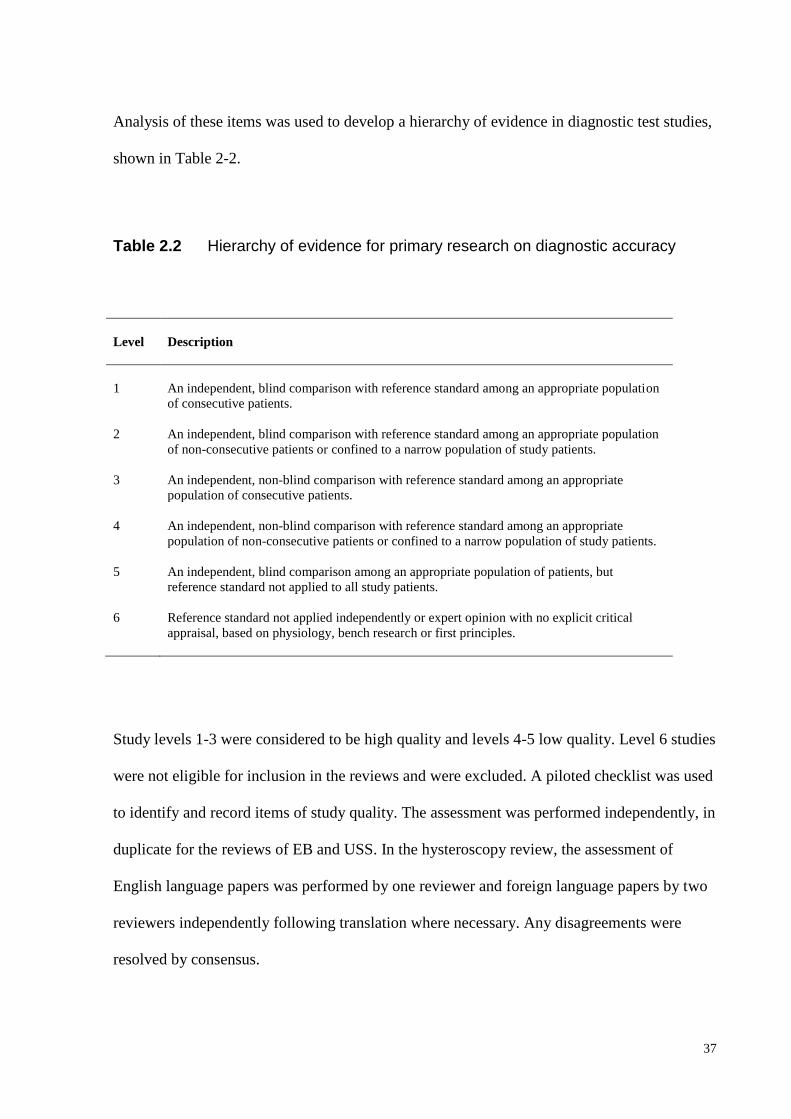
Analysis of these items was used to develop a hierarchy of evidence in diagnostic test studies,
shown in Table 2-2.
Table 2.2 Hierarchy of evidence for primary research on diagnostic accuracy
Level Description
1 An independent, blind comparison with reference standard among an appropriate population
of consecutive patients.
2 An independent, blind comparison with reference standard among an appropriate population
of non-consecutive patients or confined to a narrow population of study patients.
3 An independent, non-blind comparison with reference standard among an appropriate
population of consecutive patients.
4 An independent, non-blind comparison with reference standard among an appropriate
population of non-consecutive patients or confined to a narrow population of study patients.
5 An independent, blind comparison among an appropriate population of patients, but
reference standard not applied to all study patients.
6 Reference standard not applied independently or expert opinion with no explicit critical
appraisal, based on physiology, bench research or first principles.
Study levels 1-3 were considered to be high quality and levels 4-5 low quality. Level 6 studies
were not eligible for inclusion in the reviews and were excluded. A piloted checklist was used
to identify and record items of study quality. The assessment was performed independently, in
duplicate for the reviews of EB and USS. In the hysteroscopy review, the assessment of
English language papers was performed by one reviewer and foreign language papers by two
reviewers independently following translation where necessary. Any disagreements were
resolved by consensus.
38
2.1.4 Data abstraction
Data were extracted independently and in duplicate. Data abstraction forms are given in
Appendices 6 to 8.
2.1.4.1 Diagnostic accuracy data extraction
Three outcomes were considered: endometrial cancer, endometrial hyperplasia and normal
(functional or atrophic endometrium and benign focal abnormalities e.g. intrauterine polyps
and fibroids) for the review of EB. As discussed in section 1.6.2.2, endometrial disease,
defined as including cancer and/or hyperplasia, was examined rather than endometrial
hyperplasia for the reviews of USS and hysteroscopy. Non-endometrial uterine malignancies
were excluded from analysis.
Endometrial cancer was considered the most important diagnosis and, to analyse its
prediction, data were abstracted as two by two tables of the diagnostic test under scrutiny,
result (positive or negative for cancer) and the results of the reference standard histology
(benign or cancer). Similarly contingency tables were produced for USS and hysteroscopy
results and endometrial disease (benign or disease), and EB and endometrial hyperplasia
(hyperplasia or non-hyperplasia). In the review of ultrasound measurement of endometrial
thickness, different cut-off levels for an abnormal test result were adopted by the different
selected studies and 2x2 tables were produced according to these cut-off levels (see also
section 1.6.2.2).
To examine the impact of the severity of endometrial hyperplasia on the diagnostic
performance of EB, endometrial hyperplasia data were stratified by the presence or absence of
39
atypical cells (abnormal cytology) as part of a secondary analysis (as discussed in section
1.6.2.1). This involved constructing two sets of 2x2 tables comparing EB and reference
standard histology. The target disorder (histology) for the first table was hyperplasia without
atypia (normal / negative test result being absence of hyperplasia) and in the second table the
target disorder was hyperplasia with atypia (normal / negative test result being absence of
hyperplasia with atypia). A final analysis was then conducted for endometrial cancer or
precancer (complex or atypical hyperplasia). In this analysis, data were abstracted as 2x2
tables of the outpatient biopsy result (positive or negative for endometrial cancer + precancer)
and the results of the reference standard histology (endometrial cancer/precancer or non-
endometrial cancer/precancer).
2.1.4.2 Secondary outcomes data extraction
Data pertaining to complications, inadequate specimens (EB only) and failures associated
with testing were recorded (see section 1.6). Failures were excluded from two by two tables,
whereas inadequate specimens (precluding a definitive diagnosis following the reference test
in the case of hysteroscopy) were used in sensitivity analyses including them along with
negative results. This is because the inability to obtain a specimen is generally considered a
negative result.19,24
Information on menopausal status, the number of women recruited, and
those whose outcome data were known was also sought from the manuscripts. In addition, the
setting (outpatient or inpatient) and technical details pertaining to the hysteroscopic
examination were sought.

40
2.1.5 Quantitative data synthesis
2.1.5.1 2x2 tables
The tables were constructed as detailed in section 2.1.4.1. True positive rate (sensitivity), false
positive rate (1-specificity) and likelihood ratios (LRs) were calculated for each study along
with their 95% confidence intervals (CIs). Where 2x2 tables contained zero cells, 0.5 was
added to each cell to enable our calculations.287
2.1.5.2 Heterogeneity
Heterogeneity of results between different studies was formally assessed in all reviews
graphically, using sensitivity and specificity plots in addition to the χ2
test. In order to explore
for clinical sources of heterogeneity, the potential explanatory variables were defined a
priori.64
In view of the potential influence of spectrum variability,276,331
menopausal status
and setting were considered to be important. In addition, examination of the impact of study
quality on estimation of accuracy according to individual quality items (patient selection,
reference standard, completeness of verification and blinding) and also according to an overall
quality level (1-5) incorporating these items, was planned.62
2.1.5.2.1 Subgroup and meta-regression analysis
Statistical examination was performed to assess whether estimation of accuracy was different
in the subgroups. This was done by examining if the impact of an explanatory variable on the
log of diagnostic odds ratio (dOR), a measure which accommodates LRs for both positive and
negative test results, in meta-regression analysis.305,311
Univariable analyses were initially
performed followed by multivariable modeling, which controlled for confounding between
41
variables.305
In the review of ultrasound, meta-analyses were performed separately for
subgroups of studies with the same cut-off level for abnormality and the same measurement
techniques (single or both endometrial layers). The effect of HRT use on diagnostic accuracy
was also evaluated by subgroup analysis. Sources of heterogeneity were explored for by
univariate subgroup analyses according to the pre-specified possible explanatory variables
population spectrum (HRT use) and study quality items. Additional subgroup analyses were
performed, stratifying studies according to variation in specific study characteristics (e.g.
population, intervention, and outcome).82,305
Multivariable modeling was then performed as
described for hysteroscopy below.
In the review of hysteroscopy, the models produced by multivariable analysis included
menopausal status (postmenopausal vs. pre-menopausal and mixed population) and clinical
setting (office vs. inpatient) as explanatory variables. The models were adjusted for the effect
of study quality. For this quality was used as a binary variable (levels 1-3 vs. 4-5), which
avoided problems of co-linearity between quality items. By testing only three variables in
meta-regression analysis, it was hoped to avoid spurious results due to “overfitting”. 311
This
approach is in keeping with published recommendations, which advocate a cautious
examination of potential reasons for heterogeneity by specification of a small number of
subgroup analyses in advance. 64,69,82
For the hysteroscopy review, additional post hoc analyses to explore for causes of
heterogeneity were conducted alongside those planned in advance, when certain variables
were considered to be informative or recommended by the peer reviewers. Following
univariable analyses, multivariable meta-regression analyses were performed to evaluate the
42
effect of the explanatory variables on log dOR observed among individual studies.305
The
models produced by multivariable analysis included the independent variables description of
test (adequate vs. inadequate), complications (present vs. absent), timing of verification
(simultaneous vs. sequential), method of data collection (prospective vs. other) and
completeness of follow up (greater than 90% vs. less than 90%), in addition to the variables
defined a priori. The findings of these post hoc analyses were, however, considered in the
context of hypothesis generation.
2.1.5.3 Meta-analysis
Meta-analysis to produce summary pooled estimates of sensitivity and specificity were
performed if these measures were found to behave independently83,170
as indicated by lack of
statistical correlation between them. However, estimates of sensitivity and specificity have
limited value in clinical interpretation.86,162,174,283
Therefore summary likelihood ratios (LRs)
were generated as the principal measures of diagnostic accuracy based on the
recommendations of the various Evidence-based Medicine Groups.82,84,86,150,173,174
The LRs
indicate by how much a given hysteroscopy finding raises or lowers the probability of having
endometrial cancer or disease.21
This is important in clinical decision making because the
estimated probability of disease (or not having disease) is a prime factor determining whether
to withhold treatment, undertake further diagnostic testing or treat without further testing.264
Thus the generation of LRs and post-test probabilities represents a more relevant method of
establishing the utility of a test and reduces the risk of erroneous inferences being
drawn.162,185
.
43
Pooling of LRs was performed by weighting the log LR from each study in inverse proportion
to its variance. The clinical implications of the LRs generated for diagnostic accuracy were
examined to determine post-test probabilities using Bayes‟ theorem using the formula: post-
test probability = likelihood ratio x pre-test probability/[1-pre-test probability x (1-likelihood
ratio)]. An estimate of pre-test probability was obtained by calculating the prevalence of
pathology in the population studied. The post-test probability of endometrial pathology, in
the presence of a particular test result, refers to the probability of this outcome being present
conditional on this test result. In this way, a more clinically useful measure of the diagnostic
performance of the test is obtained as it relates to the actual test result before the presence or
absence of pathology is known. In order to deal with the uncertainty in the estimation, 95%
confidence intervals (CI) around the point estimates were generated. Approximate variance
for the post-test odds were obtained by adding the variances of the combined LRs and pre-test
odds, enabling the calculation of its 95% CI. The 95% CIs for the post-test probabilities were
then generated by converting the limits of the post-test odds to their respective probabilities.
Inferences according to strength of evidence were generated considering estimate of accuracy,
homogeneity of results and study quality.188
2.1.5.3.1 Fixed and random effects models
When heterogeneity was encountered within subgroup meta-analysis for hysteroscopy or
ultrasound, results were initially pooled from individual studies using both a fixed effects and
random effects model. In the presence of heterogeneity across studies, a random effects model
may be considered preferable64,69,82,91
in meta-analysis, as this approach produces wider CIs.
However, this benefit has to be balanced against the potential disadvantage that by weighting
smaller studies preferentially, it may produce biased point estimates of accuracy.82
44
Examination for such bias in meta-analyses was carried out and results reported with a fixed
effects model where a random effects model was associated with higher estimates of
accuracy. This allowed more conservative interpretation of the results. Furthermore, if
heterogeneity remained within the pre-specified clinical subgroups, interpretations were based
on inferences from high quality studies (levels 1-3).
2.1.5.4 Sensitivity analysis
In order to ascertain how uncertainty arising from underlying assumptions influence review
results, sensitivity analyses were conducted where indicated. A sensitivity analysis was
carried out in the review of hysteroscopy, considering inadequate histological specimens,
precluding a definitive diagnosis following the reference test, as negative results. This is
because insufficient tissue samples are generally taken to mean absence of pathology.19,24
Intrauterine polyps were also excluded and fibroids as part of a sensitivity analysis, in order to
examine whether the presence of these focal lesions affected estimates of diagnostic accuracy.
2.1.5.5 Publication bias
For all reviews, exploration for publication bias was carried out, by producing funnel plots
(scatter plot of individual study accuracy against some measure of study information186
) of
diagnostic odds ration (dOR) against corresponding inverse of variance. When no publication
bias is present the plots will be shaped like a funnel because studies of smaller size are
expected to have increased variation in the estimates of accuracy.318
The bigger the study
variance, the lower the weighting of the study and the less information it provides. This means
that in addition to small sample size of included primary studies, those studies reporting very
45
high accuracy (few false positive or negative diagnoses) will also have a relatively big
variance and thus be weighted less. The adjusted rank correlation method was used to test the
correlation between estimated LRs and their standard errors.23,23
2.2 Economic analysis methods
The cost-effectiveness analysis was based on modeling the costs and outcomes of patients
with PMB investigated using various diagnostic strategies. Survival in terms of life years
gained (LYG) was the outcome and cost per LYG was the measure of cost-effectiveness. This
approach is widely used in health economics137,231,262,327
and was conducted according to
recommended methods.99,111,231,240,253,301
2.2.1 The model
A decision model114,198,294
was constructed to reflect current service provision (Figure 2-1). As
there is no consensus regarding how best to investigate women with PMB for endometrial
cancer, initial investigation utilising all tests either alone or in combination were included in
the model. For strategies involving USS, both 4mm and 5mm cut-offs were used to define
abnormal endometrial thickening. This was done to address the ongoing clinical debate
regarding what constitutes the best USS cut-off for abnormal endometrial thickening (4mm or
5mm) and also to reflect varying clinical practice.160,303,319
A further option, of withholding
immediate investigation at initial presentation and only instituting diagnostic work-up if PMB
recurred, was also considered. Thus, 12 outpatient strategies for the clinical investigation of
women with PMB for endometrial cancer were evaluated based on initial evaluation with:
46
1. EB
2. USS (4mm)
3. USS (5mm)
4. OPH
5. USS (4mm) and OPH
6. USS (5mm) and OPH
7. USS (4mm) and EB
8. USS (5mm) and EB
9. EB and OPH
10. USS (4mm) and EB and OPH
11. USS (5mm) and EB and OPH
12. No initial evaluation
In cases of test failure, the default diagnostic procedure was inpatient evaluation of the
endometrium under general anaesthetic utilising blind or directed dilatation of the cervix and
curettage of the endometrium (D&C) (Figures 2-2 to 2-10). As discussed in section 1.2, initial
endometrial assessment by inpatient D&C is outmoded, but is still employed when outpatient
modalities fail.
The model used estimates of probabilities for various test results, life expectancy, direct
medical cost and computed cost-effectiveness as a function of age cohorts (45 years, 55 years,
65 years, 75 years and greater than 80 years of age). Endometrial cancer was divided into

47
localised (FIGO stage I) and more advanced (FIGO stages II-IV) disease. The model also
considered costs arising from morbidity associated with diagnosis by D&C.152
48
Figure 2-1 Decision analytic model: Strategy utilising initial evaluation with endometrial biopsy (EB) for the investigation of postmenopausal bleeding for endometrial cancer
TAHcEB+cTAH+cCancerIMM
EB Pos
pE Btp r
TAHcEB+cRep+cTAH+cCancerDEL
RepresentDischargeEB Neg
#
Success
pE Bsu ccess
TAHcEB+cDC+cTAH+cCancerIMM
D&C Pos
pD C tp r
TAHcEB+cDC+cRep+cTAH+cCancerDEL
RepresentDischargeD&C Neg
#
Failure
#
Cancer
TAHcEB+cTAH
EB Pos
pE Bfpr
DISCHARGEcEB
EB Neg
#
Success
pE Bsu ccess
TAHcEB+cDC+cTAH
D&C Pos
pD C fp r
DISCHARGEcEB+cDC
D&C Neg
#
Failure
#
No Cancer
EB
Cancer IMM = endometrial cancer treatment following immediate diagnosis, Cancer DEL = endometrial cancer treatment following delayed diagnosis, D&C or DC = dilatation and
curettage, EB = endometrial biopsy, fpr = false positive rate, Neg = negative test result, OPH =outpatient hysteroscopy, Pos = positive test result, PMB = postmenopausal bleeding, Rep =
represent, TAH = total abdominal hysterectomy, tpr = true positive rate, USS = ultrasound scan.
Prefix c = cost, prefix p = probability, # = complementary probability
Represent = a combination of all three tests performed (USS and EB and OPH) – see text for details
49
Figure 2-2 Decision analytic model: Strategy utilising initial evaluation with pelvic ultrasound scan (USS) using a cut-off of 4mm to signify abnormal endometrial thickness for the investigation of postmenopausal bleeding for endometrial cancer
Cancer IMM = endometrial cancer treatment following immediate diagnosis, Cancer DEL = endometrial cancer treatment following delayed diagnosis, D&C or DC = dilatation and
curettage, EB = endometrial biopsy, fpr = false positive rate, Neg = negative test result, OPH =outpatient hysteroscopy, Pos = positive test result, PMB = postmenopausal bleeding, Rep =
represent, TAH = total abdominal hysterectomy, tpr = true positive rate, USS = ultrasound scan.
Prefix c = cost, prefix p = probability., # = complementary probability, Represent = a combination of all three tests performed (USS and EB and OPH) – see text for details
TAH cUSS+cEB+cTAH+cCancerIMM EB Pos
pEBtprifUSSPos TAH
cUSS+cEB+cRep+cTAH+cCancerDEL Represent Discharge EB Neg #
EB Success pEBsuccessafterUSS
TAH cUSS+cEBfail+cDC+cTAH+cCancerIMM D&C Pos
pDCtpr TAH
cUSS+cEBfail+cDC+cRep+cTAH+cCancerDEL Represent Discharge D&C Neg #
EB Failure #
USS Pos pUSStpr
TAH cUSS+cRep+cTAH+cCancerDEL Represent Discharge USS Neg
#
Success pUSSsuccess
TAH cUSS+cDC+cTAH+cCancerIMM D&C Pos
pDCtpr TAH
cUSS+cDC+cRep+cTAH+cCancerDEL Represent Discharge D&C Neg #
Failure #
Cancer
TAH cUSS+cEB+cTAH EB Pos
pEBfpr DISCHARGE
cUSS+cEB EB Neg #
EB Success pEBsuccessafterUSS
TAH cUSS+cEBfail+cDC+cTAH D&C Pos
pDCfpr DISCHARGE
cUSS+cEBfail+cDC D&C Neg #
EB Failure #
USS Pos pUSSfpr
DISCHARGE cUSS USS Neg
#
Success pUSSsuccess
TAH cUSS+cDC+cTAH D&C Pos
pDCfpr DISCHARGE
cUSS+cDC D&C Neg #
Failure #
No Cancer
USS 4mm
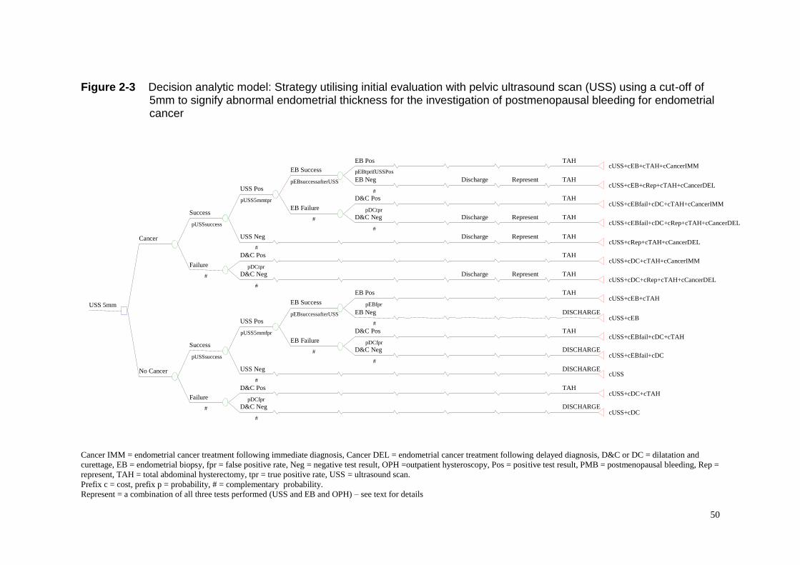
50
Figure 2-3 Decision analytic model: Strategy utilising initial evaluation with pelvic ultrasound scan (USS) using a cut-off of 5mm to signify abnormal endometrial thickness for the investigation of postmenopausal bleeding for endometrial cancer
Cancer IMM = endometrial cancer treatment following immediate diagnosis, Cancer DEL = endometrial cancer treatment following delayed diagnosis, D&C or DC = dilatation and
curettage, EB = endometrial biopsy, fpr = false positive rate, Neg = negative test result, OPH =outpatient hysteroscopy, Pos = positive test result, PMB = postmenopausal bleeding, Rep =
represent, TAH = total abdominal hysterectomy, tpr = true positive rate, USS = ultrasound scan.
Prefix c = cost, prefix p = probability, # = complementary probability.
Represent = a combination of all three tests performed (USS and EB and OPH) – see text for details
TAH cUSS+cEB+cTAH+cCancerIMM EB Pos
pEBtprifUSSPos TAH
cUSS+cEB+cRep+cTAH+cCancerDEL Represent Discharge EB Neg #
EB Success pEBsuccessafterUSS
TAH cUSS+cEBfail+cDC+cTAH+cCancerIMM D&C Pos
pDCtpr TAH
cUSS+cEBfail+cDC+cRep+cTAH+cCancerDEL Represent Discharge D&C Neg #
EB Failure #
USS Pos pUSS5mmtpr
TAH cUSS+cRep+cTAH+cCancerDEL Represent Discharge USS Neg
#
Success pUSSsuccess
TAH cUSS+cDC+cTAH+cCancerIMM D&C Pos
pDCtpr TAH
cUSS+cDC+cRep+cTAH+cCancerDEL Represent Discharge D&C Neg #
Failure #
Cancer
TAH cUSS+cEB+cTAH EB Pos
pEBfpr DISCHARGE
cUSS+cEB EB Neg #
EB Success pEBsuccessafterUSS
TAH cUSS+cEBfail+cDC+cTAH D&C Pos
pDCfpr DISCHARGE
cUSS+cEBfail+cDC D&C Neg #
EB Failure #
USS Pos pUSS5mmfpr
DISCHARGE cUSS USS Neg
#
Success pUSSsuccess
TAH cUSS+cDC+cTAH D&C Pos
pDCfpr DISCHARGE
cUSS+cDC D&C Neg #
Failure #
No Cancer
USS 5mm
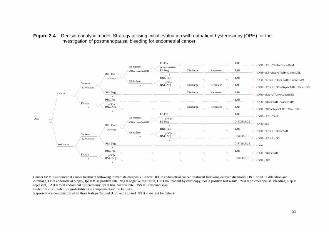
51
Figure 2-4 Decision analytic model: Strategy utilising initial evaluation with outpatient hysteroscopy (OPH) for the investigation of postmenopausal bleeding for endometrial cancer
Cancer IMM = endometrial cancer treatment following immediate diagnosis, Cancer DEL = endometrial cancer treatment following delayed diagnosis, D&C or DC = dilatation and
curettage, EB = endometrial biopsy, fpr = false positive rate, Neg = negative test result, OPH =outpatient hysteroscopy, Pos = positive test result, PMB = postmenopausal bleeding, Rep =
represent, TAH = total abdominal hysterectomy, tpr = true positive rate, USS = ultrasound scan.
Prefix c = cost, prefix p = probability, # = complementary probability.
Represent = a combination of all three tests performed (USS and EB and OPH) – see text for details
TAH cOPH+cEB+cTAH+cCancerIMM EB Pos
pEBtprifOPHPos TAH
cOPH+cEB+cRep+cTAH+cCancerDEL Represent Discharge EB Neg #
EB Success pEBsuccessafterOPH
TAH cOPH+cEBfail+cDC+cTAH+cCancerIMM D&C Pos
pDCtpr TAH
cOPH+cEBfail+cDC+cRep+cTAH+cCancerDEL Represent Discharge D&C Neg #
EB Failure #
OPH Pos pOPHtpr
TAH cOPH+cRep+cTAH+cCancerDEL Represent Discharge OPH Neg
#
Success pOPHsuccess
TAH cOPH+cDC+cTAH+cCancerIMM D&C Pos
pDCtpr TAH
cOPH+cDC+cRep+cTAH+cCancerDEL Represent Discharge D&C Neg #
Failure #
Cancer
TAH cOPH+cEB+cTAH EB Pos
pEBfpr DISCHARGE
cOPH+cEB EB Neg #
EB Success pEBsuccessafterOPH
TAH cOPH+cEBfail+cDC+cTAH D&C Pos
pDCfpr DISCHARGE
cOPH+cEBfail+cDC D&C Neg #
EB Failure #
OPH Pos pOPHfpr
DISCHARGE cOPH OPH Neg
#
Success pOPHsuccess
TAH cOPH+cDC+cTAH D&C Pos
pDCfpr DISCHARGE
cOPH+cDC D&C Neg #
Failure #
No Cancer
OPH
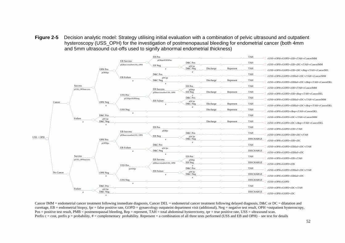
52
Figure 2-5 Decision analytic model: Strategy utilising initial evaluation with a combination of pelvic ultrasound and outpatient hysteroscopy (USS_OPH) for the investigation of postmenopausal bleeding for endometrial cancer (both 4mm and 5mm ultrasound cut-offs used to signify abnormal endometrial thickness)
Cancer IMM = endometrial cancer treatment following immediate diagnosis, Cancer DEL = endometrial cancer treatment following delayed diagnosis, D&C or DC = dilatation and
curettage, EB = endometrial biopsy, fpr = false positive rate, GOPD = gynaecology outpateint department visit (additional), Neg = negative test result, OPH =outpatient hysteroscopy,
Pos = positive test result, PMB = postmenopausal bleeding, Rep = represent, TAH = total abdominal hysterectomy, tpr = true positive rate, USS = ultrasound scan.
Prefix c = cost, prefix p = probability, # = complementary probability. Represent = a combination of all three tests performed (USS and EB and OPH) – see text for details
TAH cUSS+cOPH-cGOPD+cEB+cTAH+cCancerIMM EB Pos
pEBtprifOPHPos TAH
cUSS+cOPH-cGOPD+cEB+cDC+cTAH+cCancerIMM D&C Pos pDCtpr
TAH cUSS+cOPH-cGOPD+cEB+cDC+cRep+cTAH+cCancerDEL Represent Discharge D&C Neg
#
EB Neg #
EB Success pEBsuccessafterUSS_OPH
TAH cUSS+cOPH-cGOPD+cEBfail+cDC+cTAH+cCancerIMM D&C Pos
pDCtpr TAH
cUSS+cOPH-cGOPD+cEBfail+cDC+cRep+cTAH+cCancerDEL Represent Discharge D&C Neg #
EB Failure #
OPH Pos pOPHtpr
TAH cUSS+cOPH-cGOPD+cEB+cTAH+cCancerIMM EB Pos
pEBtpr TAH
cUSS+cOPH-cGOPD+cEB+cRep+cTAH+cCancerDEL Represent Discharge EB Neg #
EB Success pEBsuccessafterUSS_OPH
TAH cUSS+cOPH-cGOPD+cEBfail+cDC+cTAH+cCancerIMM D&C Pos
pDCtpr TAH
cUSS+cOPH-cGOPD+cEBfail+cDC+cRep+cTAH+cCancerDEL Represent Discharge D&C Neg #
EB Failure #
USS Pos pUSStprifOPHneg
TAH cUSS+cOPH-cGOPD+cRep+cTAH+cCancerDEL Represent Discharge USS Neg
#
OPH Neg #
Success pUSS_OPHsuccess
TAH cUSS+cOPH-cGOPD+cDC+cTAH+cCancerIMM D&C Pos
pDCtpr TAH
cUSS+cOPH-cGOPD+cDC+cRep+cTAH+cCancerDEL Represent Discharge D&C Neg #
Failure #
Cancer
TAH cUSS+cOPH-cGOPD+cEB+cTAH EB Pos
pEBfpr TAH
cUSS+cOPH-cGOPD+cEB+cDC+cTAH D&C Pos pDCfpr
DISCHARGE cUSS+cOPH-cGOPD+cEB+cDC D&C Neg
#
EB Neg #
EB Success pEBsuccessafterUSS_OPH
TAH cUSS+cOPH-cGOPD+cEBfail+cDC+cTAH D&C Pos
pDCfpr DISCHARGE
cUSS+cOPH-cGOPD+cEBfail+cDC D&C Neg #
EB Failure #
OPH Pos pOPHfpr
TAH cUSS+cOPH-cGOPD+cEB+cTAH EB Pos
pEBfpr DISCHARGE
cUSS+cOPH-cGOPD+cEB EB Neg #
EB Success pEBsuccessafterUSS_OPH
TAH cUSS+cOPH-cGOPD+cEBfail+cDC+cTAH D&C Pos
pDCfpr DISCHARGE
cUSS+cOPH-cGOPD+cEBfail+cDC D&C Neg #
EB Failure #
USS Pos pUSSfpr
DISCHARGE cUSS+cOPH-cGOPD USS Neg
#
OPH Neg #
Success pUSS_OPHsuccess
TAH cUSS+cOPH-cGOPD+cDC+cTAH D&C Pos
pDCfpr DISCHARGE
cUSS+cOPH-cGOPD+cDC D&C Neg #
Failure #
No Cancer
USS + OPH
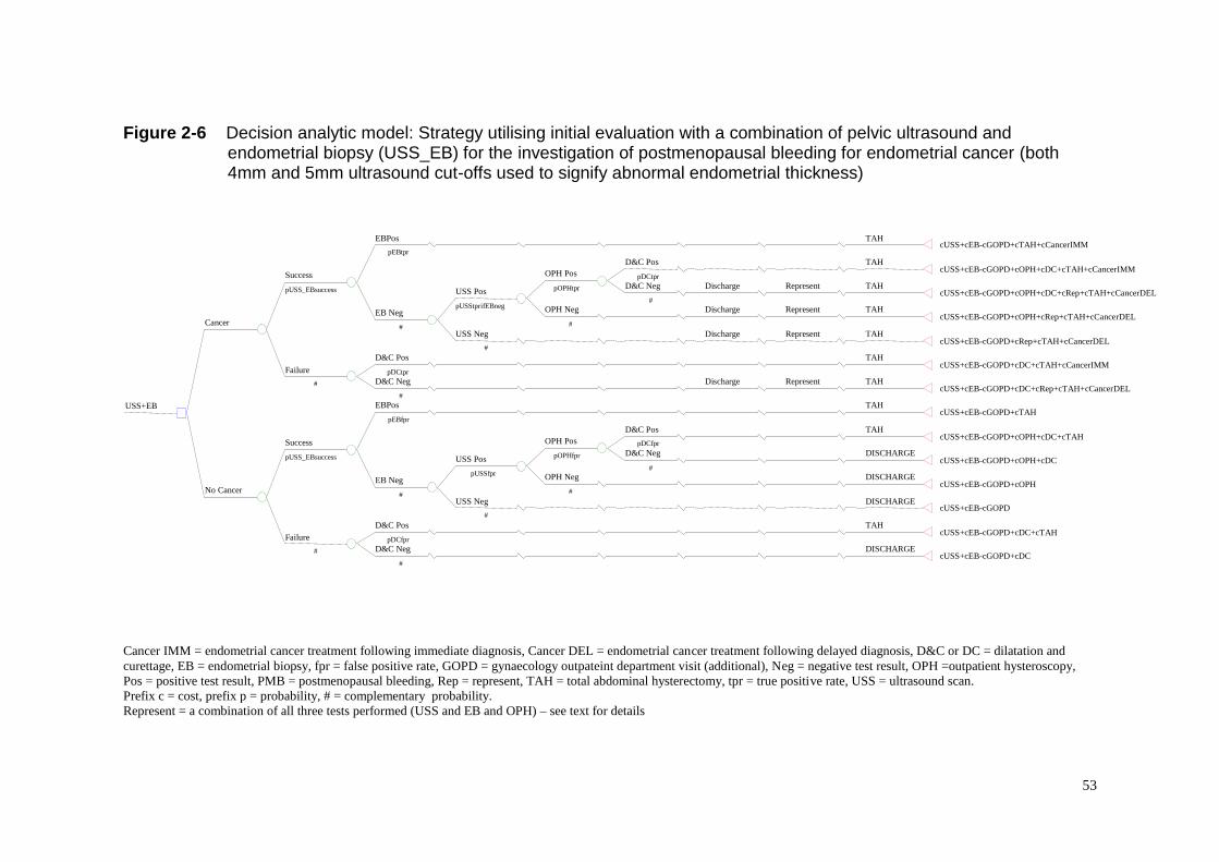
53
Figure 2-6 Decision analytic model: Strategy utilising initial evaluation with a combination of pelvic ultrasound and endometrial biopsy (USS_EB) for the investigation of postmenopausal bleeding for endometrial cancer (both 4mm and 5mm ultrasound cut-offs used to signify abnormal endometrial thickness)
Cancer IMM = endometrial cancer treatment following immediate diagnosis, Cancer DEL = endometrial cancer treatment following delayed diagnosis, D&C or DC = dilatation and
curettage, EB = endometrial biopsy, fpr = false positive rate, GOPD = gynaecology outpateint department visit (additional), Neg = negative test result, OPH =outpatient hysteroscopy,
Pos = positive test result, PMB = postmenopausal bleeding, Rep = represent, TAH = total abdominal hysterectomy, tpr = true positive rate, USS = ultrasound scan.
Prefix c = cost, prefix p = probability, # = complementary probability.
Represent = a combination of all three tests performed (USS and EB and OPH) – see text for details
TAH cUSS+cEB-cGOPD+cTAH+cCancerIMM EBPos
pEBtpr TAH
cUSS+cEB-cGOPD+cOPH+cDC+cTAH+cCancerIMM D&C Pos pDCtpr
TAH cUSS+cEB-cGOPD+cOPH+cDC+cRep+cTAH+cCancerDEL Represent Discharge D&C Neg
# OPH Pos
pOPHtpr TAH
cUSS+cEB-cGOPD+cOPH+cRep+cTAH+cCancerDEL Represent Discharge OPH Neg #
USS Pos pUSStprifEBneg
TAH cUSS+cEB-cGOPD+cRep+cTAH+cCancerDEL Represent Discharge USS Neg
# EB Neg
#
Success pUSS_EBsuccess
TAH cUSS+cEB-cGOPD+cDC+cTAH+cCancerIMM D&C Pos
pDCtpr TAH
cUSS+cEB-cGOPD+cDC+cRep+cTAH+cCancerDEL Represent Discharge D&C Neg #
Failure #
Cancer
TAH cUSS+cEB-cGOPD+cTAH EBPos
pEBfpr TAH
cUSS+cEB-cGOPD+cOPH+cDC+cTAH D&C Pos pDCfpr
DISCHARGE cUSS+cEB-cGOPD+cOPH+cDC D&C Neg
# OPH Pos
pOPHfpr DISCHARGE
cUSS+cEB-cGOPD+cOPH OPH Neg #
USS Pos pUSSfpr
DISCHARGE cUSS+cEB-cGOPD USS Neg
# EB Neg
#
Success pUSS_EBsuccess
TAH cUSS+cEB-cGOPD+cDC+cTAH D&C Pos
pDCfpr DISCHARGE
cUSS+cEB-cGOPD+cDC D&C Neg #
Failure #
No Cancer
USS+EB
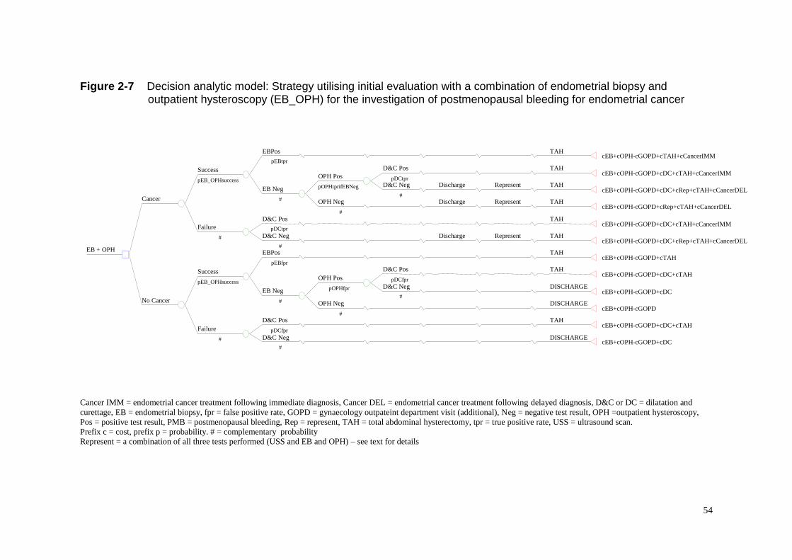
54
Figure 2-7 Decision analytic model: Strategy utilising initial evaluation with a combination of endometrial biopsy and outpatient hysteroscopy (EB_OPH) for the investigation of postmenopausal bleeding for endometrial cancer
Cancer IMM = endometrial cancer treatment following immediate diagnosis, Cancer DEL = endometrial cancer treatment following delayed diagnosis, D&C or DC = dilatation and
curettage, EB = endometrial biopsy, fpr = false positive rate, GOPD = gynaecology outpateint department visit (additional), Neg = negative test result, OPH =outpatient hysteroscopy,
Pos = positive test result, PMB = postmenopausal bleeding, Rep = represent, TAH = total abdominal hysterectomy, tpr = true positive rate, USS = ultrasound scan.
Prefix c = cost, prefix p = probability. # = complementary probability
Represent = a combination of all three tests performed (USS and EB and OPH) – see text for details
TAH cEB+cOPH-cGOPD+cTAH+cCancerIMM EBPos
pEBtpr TAH
cEB+cOPH-cGOPD+cDC+cTAH+cCancerIMM D&C Pos
pDCtpr TAH
cEB+cOPH-cGOPD+cDC+cRep+cTAH+cCancerDEL Represent Discharge D&C Neg
#
OPH Pos pOPHtprifEBNeg
TAH cEB+cOPH-cGOPD+cRep+cTAH+cCancerDEL Represent Discharge OPH Neg
#
EB Neg #
Success pEB_OPHsuccess
TAH cEB+cOPH-cGOPD+cDC+cTAH+cCancerIMM
D&C Pos pDCtpr
TAH cEB+cOPH-cGOPD+cDC+cRep+cTAH+cCancerDEL
Represent Discharge D&C Neg #
Failure #
Cancer
TAH cEB+cOPH-cGOPD+cTAH
EBPos pEBfpr
TAH cEB+cOPH-cGOPD+cDC+cTAH
D&C Pos pDCfpr
DISCHARGE cEB+cOPH-cGOPD+cDC
D&C Neg #
OPH Pos pOPHfpr
DISCHARGE cEB+cOPH-cGOPD
OPH Neg #
EB Neg #
Success pEB_OPHsuccess
TAH cEB+cOPH-cGOPD+cDC+cTAH
D&C Pos pDCfpr
DISCHARGE cEB+cOPH-cGOPD+cDC D&C Neg
#
Failure #
No Cancer
EB + OPH
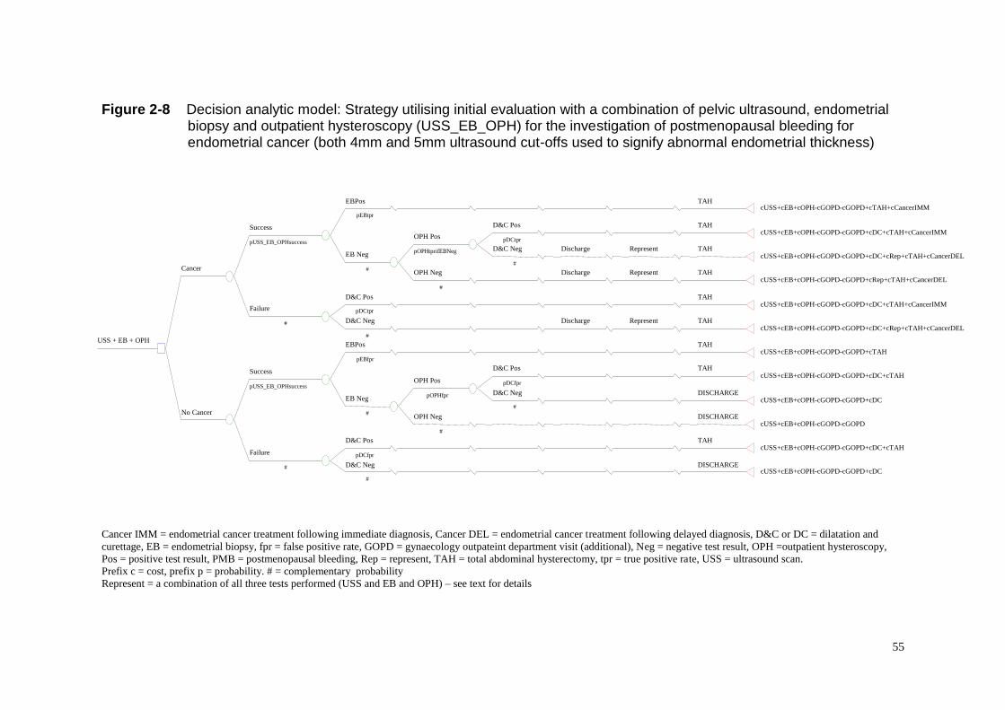
55
Figure 2-8 Decision analytic model: Strategy utilising initial evaluation with a combination of pelvic ultrasound, endometrial biopsy and outpatient hysteroscopy (USS_EB_OPH) for the investigation of postmenopausal bleeding for endometrial cancer (both 4mm and 5mm ultrasound cut-offs used to signify abnormal endometrial thickness)
Cancer IMM = endometrial cancer treatment following immediate diagnosis, Cancer DEL = endometrial cancer treatment following delayed diagnosis, D&C or DC = dilatation and
curettage, EB = endometrial biopsy, fpr = false positive rate, GOPD = gynaecology outpateint department visit (additional), Neg = negative test result, OPH =outpatient hysteroscopy,
Pos = positive test result, PMB = postmenopausal bleeding, Rep = represent, TAH = total abdominal hysterectomy, tpr = true positive rate, USS = ultrasound scan.
Prefix c = cost, prefix p = probability. # = complementary probability
Represent = a combination of all three tests performed (USS and EB and OPH) – see text for details
TAH cUSS+cEB+cOPH-cGOPD-cGOPD+cTAH+cCancerIMM EBPos
pEBtpr TAH
cUSS+cEB+cOPH-cGOPD-cGOPD+cDC+cTAH+cCancerIMM D&C Pos
pDCtpr TAH
cUSS+cEB+cOPH-cGOPD-cGOPD+cDC+cRep+cTAH+cCancerDEL Represent Discharge D&C Neg
#
OPH Pos pOPHtprifEBNeg
TAH cUSS+cEB+cOPH-cGOPD-cGOPD+cRep+cTAH+cCancerDEL
Represent Discharge OPH Neg #
EB Neg #
Success pUSS_EB_OPHsuccess
TAH cUSS+cEB+cOPH-cGOPD-cGOPD+cDC+cTAH+cCancerIMM
D&C Pos pDCtpr
TAH cUSS+cEB+cOPH-cGOPD-cGOPD+cDC+cRep+cTAH+cCancerDEL
Represent Discharge D&C Neg #
Failure #
Cancer
TAH cUSS+cEB+cOPH-cGOPD-cGOPD+cTAH
EBPos pEBfpr
TAH cUSS+cEB+cOPH-cGOPD-cGOPD+cDC+cTAH
D&C Pos pDCfpr
DISCHARGE cUSS+cEB+cOPH-cGOPD-cGOPD+cDC
D&C Neg #
OPH Pos pOPHfpr
DISCHARGE cUSS+cEB+cOPH-cGOPD-cGOPD
OPH Neg #
EB Neg #
Success pUSS_EB_OPHsuccess
TAH cUSS+cEB+cOPH-cGOPD-cGOPD+cDC+cTAH
D&C Pos pDCfpr
DISCHARGE cUSS+cEB+cOPH-cGOPD-cGOPD+cDC D&C Neg
#
Failure #
No Cancer
USS + EB + OPH
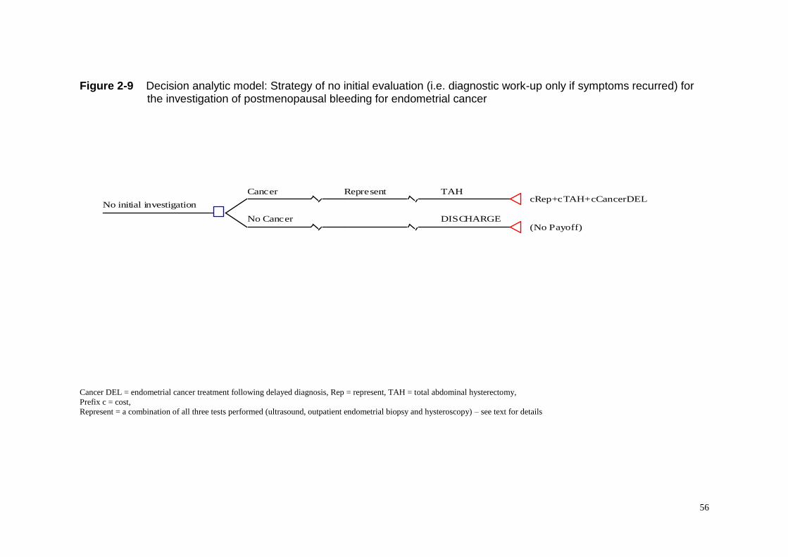
56
Figure 2-9 Decision analytic model: Strategy of no initial evaluation (i.e. diagnostic work-up only if symptoms recurred) for the investigation of postmenopausal bleeding for endometrial cancer
TAHcRep+cTAH+cCancerDEL
RepresentCancer
DISCHARGE(No Payoff)
No Cancer
No initial investigation
Cancer DEL = endometrial cancer treatment following delayed diagnosis, Rep = represent, TAH = total abdominal hysterectomy,
Prefix c = cost,
Represent = a combination of all three tests performed (ultrasound, outpatient endometrial biopsy and hysteroscopy) – see text for details
57
2.2.2 Data sources and modeling assumptions for decision analysis
In the first instance it was assumed that the hypothetical presentation with postmenopausal
bleeding represented the first episode. No postmenopausal woman was assumed to be less
than 45 years old and no other significant aetiology (e.g. other genital tract malignancy) was
considered. The woman was considered to be otherwise healthy with a normal age-adjusted
life expectancy. The probability of endometrial cancer in women presenting with
postmenopausal bleeding is between 5 and 10%16,251
and women are most likely to present
with this symptom in the seventh decade of life.272
Therefore, a 65 year old woman presenting
with PMB and a 5% prevalence of malignant disease were used for the base case analysis.
The initial investigation(s) used in each strategy were assumed to take place in a „one stop‟
setting (i.e. one initial consultation only with no planned follow up unless test(s) failed or
abnormal results were found). It was assumed that a consultant grade specialist performed all
diagnostic and surgical procedures. For the base case analysis it was assumed that an
additional return visit was required following a positive USS in order to perform endometrial
sampling. The impact of performing EB following a positive USS at the same visit was
examined as part of a sensitivity analysis, to reflect the practice of gynaecologists with
expertise in ultrasound. Expert clinical opinion was then obtained independently about
decision-making conditional upon positive or negative test results (i.e. the need for any further
testing or therapeutic intervention). An expert clinical panel was then convened to reach
consensus in cases of disagreement. In this manner a representative body of opinion was
obtained regarding current management pathways in the diagnosis of PMB. It was agreed that
invasive surgery (hysterectomy) for endometrial cancer would not be performed without
58
histological confirmation, whether by EB or inpatient sampling (D&C). Once the decision for
hysterectomy had been made, additional pre-operative investigation by examination under
anaesthesia, fractional curettage, cystoscopy, magnetic resonance imaging and other
radiographic modalities was assumed not to have been necessary in accordance with current
clinical practice.279
Radiotherapy and chemotherapy were assumed to have been provided by
the same medical oncologist.
2.2.2.1 Diagnostic Tests
Failed diagnostic procedures led to investigation by inpatient endometrial assessment under
general anaesthesia (D&C). In the case of outpatient endometrial biopsy, failed procedures
were considered to be cases where technical problems meant that an endometrial specimen
could not be obtained. Histologically inadequate specimens were considered to be negative
tests for both EB and D&C.19,62
Inpatient D&C was assumed to have no technical failure
rate.219
Data for failure rates and estimates of diagnostic accuracy were obtained from high
quality published systematic quantitative reviews of the diagnostic literature for EB, USS and
OPH (presented in this thesis – Appendix 9).62
Failure rates for initial strategies utilising test
combinations were estimated by the consensus panel based on the definition of a failed
strategy as any test making up the strategy failing and on available failure rate data from
individual tests.62
63,160,303
Similarly, failure rates were also adjusted for tests performed in a
diagnostic strategy conditional on the success of preceding tests. The base case true positive
rates for diagnostic tests carried out conditional on a preceding test result were also adjusted
as part of a sensitivity analysis to take account of plausible changes in accuracy due to lack of
complete test independence (Appendix 9).16
57
As over 95% of women with endometrial
cancer present with PMB,250
it was assumed that all women who were erroneously discharged
59
following the initial presentation (i.e. false negatives) remained symptomatic. The interval to
representation was thus taken to be short and all these women were then assumed to undergo
reinvestigation with all outpatient tests where perfect test success and accuracy was assumed.
No serious morbidity was assumed to be associated with any of the ambulatory procedures
(ultrasound, hysteroscopy and endometrial biopsy) based on evidence from systematic
reviews of the available literature.62,63,160,303
For D&C the major complication rate was
assumed to be 1.4% (included haemorrhage 0.4%, infection 0.3%, perforation 0.6% and
emergency laparotomy 0.1%).152
Costs associated with morbidity arising from complications
were incorporated into the model, but no adjustment to life expectancy was made (Appendix
10). Mortality rates were assumed to be negligible for all the diagnostic tests.62,63,152,160,303
The
mortality rate for abdominal hysterectomy in endometrial cancer was assumed to increase
with age (0.4% in a woman aged 45 years, 0.8% at 55 years, 1.4% at 65 years and 3.5% at 75
years)344
and adjustments to survival were made accordingly.
2.2.2.2 Treatment
For the base case analysis, it was assumed that all women not discharged underwent initial
treatment by total abdominal hysterectomy (TAH) and bilateral salpingo-oophorectomy with
or without pelvic node sampling (i.e. all were fit for surgery and none had primary radical
radiotherapy). All women were therefore assumed to be surgically staged.72
There is some
variation in practice in the treatment of endometrial cancer regarding the relative roles of
surgery, radiotherapy / chemotherapy.72,279
The treatment pathways in this model were based
on published recommendations and reports of current practice.2,245,251,279,309
All
epidemiological statistics relating to endometrial cancer were taken from the latest annual
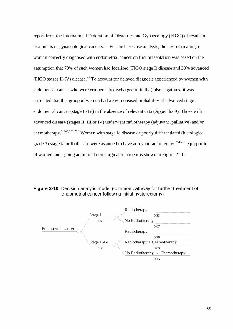
60
report from the International Federation of Obstetrics and Gynaecology (FIGO) of results of
treatments of gynaecological cancers.72
For the base case analysis, the cost of treating a
woman correctly diagnosed with endometrial cancer on first presentation was based on the
assumption that 70% of such women had localised (FIGO stage I) disease and 30% advanced
(FIGO stages II-IV) disease.72
To account for delayed diagnosis experienced by women with
endometrial cancer who were erroneously discharged initially (false negatives) it was
estimated that this group of women had a 5% increased probability of advanced stage
endometrial cancer (stage II-IV) in the absence of relevant data (Appendix 9). Those with
advanced disease (stages II, III or IV) underwent radiotherapy (adjuvant /palliative) and/or
chemotherapy.2,245,251,279
Women with stage Ic disease or poorly differentiated (histological
grade 3) stage Ia or Ib disease were assumed to have adjuvant radiotherapy.251
The proportion
of women undergoing additional non-surgical treatment is shown in Figure 2-10.
Figure 2-10 Decision analytic model (common pathway for further treatment of endometrial cancer following initial hysterectomy)
Radiotherapy
0.33
No Radiotherapy
0.67
Stage I
0.65
Radiotherapy
0.76
Radiotherapy + Chemotherapy
0.09
No Radiotherapy +/- Chemotherapy
0.15
Stage II-IV
0.35
Endometrial cancer
61
Standardised radiotherapy and chemotherapy regimens were assumed regardless of disease
stage, radiotherapy consisted of a 5-week course of external beam radiotherapy giving a total
dose of 50-55 grays in 20-28 fractions. Chemotherapy consisted of standard cytotoxic and/or
hormonal therapies.2,245,251,279
Compliance with treatment was assumed to be 100%. It was
assumed that hormonal treatment using long term oral progestogens was not employed given
there is no evidence of benefit in terms of survival.226
The 5 year survival rates were assumed
to be 87% for stage I disease and 60% for advanced (stage II-IV) disease.72
2.2.3 Cost data
Costs were estimated from the perspective of my base National Health Service (NHS) hospital
and from NHS data provided by the Department of Health. The analysis included all direct
medical costs in UK pounds sterling (Appendix 10). Data for the base case and sensitivity
analyses were obtained from local sources (Birmingham Women‟s Hospital data for
uncomplicated procedures 2000-2001) and national sources (Department of Health, National
Schedule of Reference Costs for the United Kingdom 2000252
and Unit Costs of Health and
Social Care 2000/2001247
). Drug costs were obtained from the British National Formulary
2002. Costs for outpatient investigation included the clinic appointment and other hospital
charges, the relevant procedures (endometrial biopsy, ultrasound scan or outpatient
hysteroscopy) and the specialist(s) fee (consultant gynaecologist +/- consultant pathologist).
Costs for inpatient endometrial assessment under general anaesthesia (hysteroscopy/D&C)
took into account hospital costs for a day-case surgical procedure in addition to the
specialists‟ fees for a consultant gynaecologist and anaesthetist. In addition, a cost associated
with complications arising from D&C was estimated and incorporated (included costs of
unexpected inpatient stay and antibiotic treatment for haemorrhage, uterine infection,
62
perforation and unplanned emergency laparotomy – see Appendix 10). The costs of
reinvestigation by all three outpatient modalities incurred in those women representing after
initial erroneous discharge were included in the model. Hysterectomy was classed as a
complex major laparotomy and costed according to the base hospital charges taking into
account uncomplicated inpatient hospital stay, operating theatre costs and specialist fees.
Radiotherapy charges were estimated from charges for standard outpatient treatment charges
(12-24 fractions of external beam radiotherapy) using national data.252
Chemotherapy was
costed according to national data for day-case treatment of the female reproductive system.252
No adjustments to costs were made for the effects of inflation.
2.2.4 Clinical Outcomes
Baseline values of the probabilities of each test result and treatment outcome, together with
the costs of each diagnostic intervention, were estimated and incorporated into the decision
tree (DATA Professional 2001, Treeage software inc, 1075 Main Street, Williamstown,
United States, MA 01267 [www.treeage.com]. The cost and effectiveness for each of the 12
strategies were calculated. The effectiveness of each competing diagnostic strategy was
determined by comparing survival using the outcome measure cost per life year
gained.137,231,262
Age specific life expectancies were calculated in the following way. For “true negative”
results, normal actuarial age/sex specific death rates177
were used to calculate life expectancy.
For women with stage I or stage II-IV endometrial cancer, international 5-year survival data72
were compared with the expected survival for the general population. The resulting hazard
ratio was assumed to apply constantly over 12 years, after which survival is equivalent to the
63
normal population.166
Finally, for “false positive” results, an age-specific immediate mortality
was applied for the effect of the unnecessary hysterectomy344
, after which the general
population life expectation was used. The base case analysis used an age of 65 years.272
This
age was chosen as endometrial cancer has its peak incidence in this decade.272
The costs, effect in terms of additional life year saved and average cost-effectiveness ratios
(cost per additional life year saved) were determined for each diagnostic strategy.137,231,262,327
Incremental cost-effectiveness ratios were then generated by using the ratio of cost compared
to change in life expectancy relative to the cheapest strategy. In this way improvements in life
expectancy per extra pound spent could be determined.99,253,301
In accordance with Treasury
guidelines, future years of life were discounted at 1.5% per year.88,325
Discounting costs was
not relevant as all costs were assumed to occur in the first year.
2.2.5 Sensitivity Analyses
Extensive sensitivity analyses were performed for all strategies found to be potentially cost-
effective following the base case analysis. One-way analyses were performed over ranges of
age at presentation, disease prevalence, test failure rates, estimates of diagnostic accuracy and
upstaging of endometrial cancer due to delayed diagnosis to explore the robustness of the
analytic model (Appendices 6 and 7). For treatment of endometrial cancer, the costs of local
(FIGO Stage 1) and advanced (FIGO stage II-IV) disease were varied together.
64
CHAPTER III
3 RESULTS
RESULTS
3.1 Results of systematic review of endometrial biopsy
3.1.1 Question
What is the accuracy of outpatient endometrial biopsy in the diagnosis of endometrial cancer
and hyperplasia and what are the rates of failure and complications?
3.1.2 Study Selection
The electronic search generated 1369 citations and of these there were 39
articles8,22,32,42,78,93,100,101,113,117,121,126,135,139,143,157,161,169,181,190,192,201,202,209,213,214,254,257,261,284,289,298
,299,312,313,317,332,346,349 which both reviewers thought were relevant: 37 were published in
English, one in French and one in Spanish. A further 13
articles24,30,124,133,138,182,196,204,206,307,314,315,333
were identified through examination of the
reference lists of the known primary publications and review articles. After independent
review of the 52 manuscripts, 12 articles (11 English,22,24,78,117,133,138,161,192,314,317,332
one French
284) were considered to be eligible for inclusion in the review (Figure 3-1). Agreement
regarding eligibility was 90% (weighted kappa 0.7). The lists of references supplied by the
manufacturers contacted did not add anything to the above search. Excluded studies are listed
in Appendix 11.
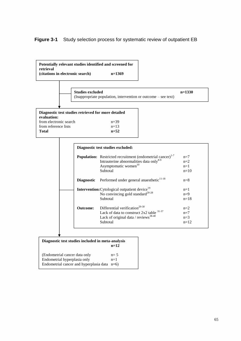
65
Figure 3-1 Study selection process for systematic review of outpatient EB
Potentially relevant studies identified and screened for
retrieval
(citations in electronic search) n=1369
Diagnostic test studies included in meta-analysis
n=12
(Endometrial cancer data only n= 5
Endometrial hyperplasia only n=1
Endometrial cancer and hyperplasia data n=6)
Diagnostic test studies excluded:
Population: Restricted recruitment (endometrial cancer)1-7
n=7
Intrauterine abnormalities data only8-9
n=2
Asymptomatic women10
n=1
Subtotal n=10
Diagnostic Performed under general anaesthetic11-18
n=8
Intervention:Cytological outpatient device19
n=1
No convincing gold standard20-28
n=9
Subtotal n=18
Outcome: Differential verification29-30
n=2
Lack of data to construct 2x2 table 31-37
n=7
Lack of original data / reviews38-40
n=3
Subtotal n=12
Total excluded1-40
n=40
Studies excluded n=1330
(Inappropriate population, intervention or outcome – see text)
Diagnostic test studies retrieved for more detailed
evaluation:
from electronic search n=39
from reference lists n=13
Total n=52
66
There were 1337 subjects in 14 diagnostic evaluations reported in 12 primary studies, which
assessed the diagnostic accuracy of EB in detecting serious endometrial disease: 40 women in
a single evaluation of the Accurette® device,138
70 women in a single evaluation of the
Gynoscann® device,317
176 women in a single evaluation of the Novak® curette,314
865
women in 8 evaluations of the Pipelle® device,22,24,78,133,161,192,284,332
104 women in 2 evaluations of the Vabra® aspirator138,314
and 77 women in a single
evaluation of the Z-sampler®117
device. Of the 12 included studies, 11 studies (1013 women)
assessed the diagnosis of endometrial cancer. Seven of these evaluations contained data
exclusively about postmenopausal women,22,78,117,138,161,332
four about pre and postmenopausal
women24,192,284,317
and in three menopausal status was unclear133,314
. Postmenopausal women
represented 36% of the populations studied.
3.1.3 Study quality
The observer agreement for various items of study quality ranged from 73 to 100%. Kappa
values were 0.5 for population enrolment, 1.0 for biopsy technique description, 0.9 for
blinding of test results and 1.0 for description of outcomes. The methodological quality
criteria of the studies selected for meta-analyses are detailed in Appendix 12 and summarised
in Table 3-1.
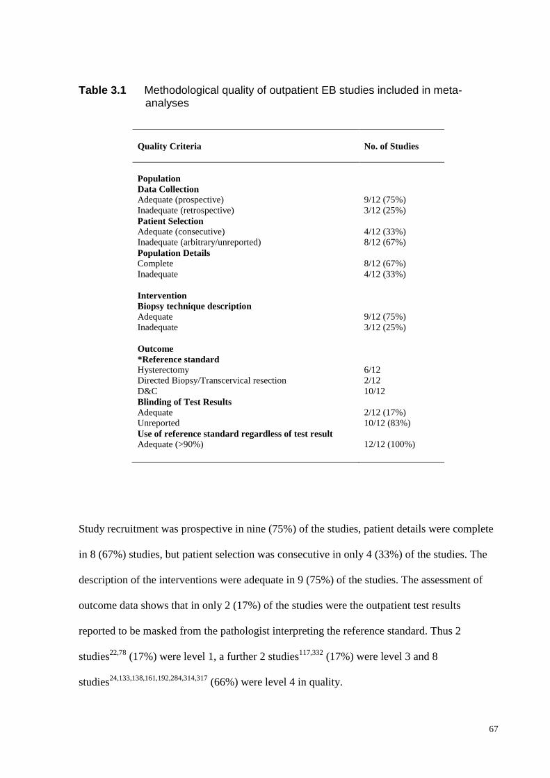
67
Table 3.1 Methodological quality of outpatient EB studies included in meta-analyses
Quality Criteria No. of Studies
Population
Data Collection
Adequate (prospective) 9/12 (75%)
Inadequate (retrospective) 3/12 (25%)
Patient Selection
Adequate (consecutive) 4/12 (33%)
Inadequate (arbitrary/unreported) 8/12 (67%)
Population Details
Complete 8/12 (67%)
Inadequate 4/12 (33%)
Intervention
Biopsy technique description
Adequate 9/12 (75%)
Inadequate 3/12 (25%)
Outcome
*Reference standard
Hysterectomy 6/12
Directed Biopsy/Transcervical resection 2/12
D&C 10/12
Blinding of Test Results
Adequate 2/12 (17%)
Unreported 10/12 (83%)
Use of reference standard regardless of test result
Adequate (>90%) 12/12 (100%)
Study recruitment was prospective in nine (75%) of the studies, patient details were complete
in 8 (67%) studies, but patient selection was consecutive in only 4 (33%) of the studies. The
description of the interventions were adequate in 9 (75%) of the studies. The assessment of
outcome data shows that in only 2 (17%) of the studies were the outpatient test results
reported to be masked from the pathologist interpreting the reference standard. Thus 2
studies22,78
(17%) were level 1, a further 2 studies117,332
(17%) were level 3 and 8
studies24,133,138,161,192,284,314,317
(66%) were level 4 in quality.
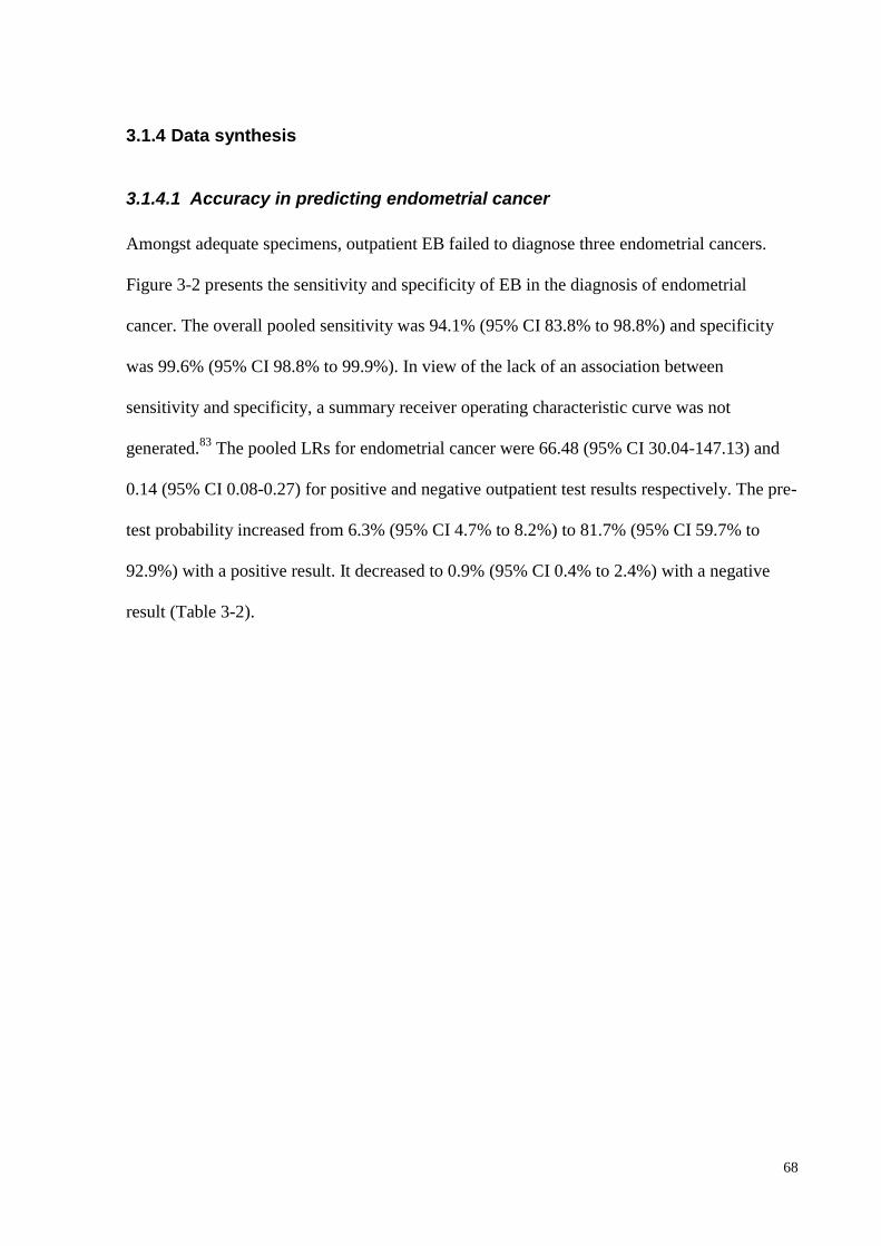
68
3.1.4 Data synthesis
3.1.4.1 Accuracy in predicting endometrial cancer
Amongst adequate specimens, outpatient EB failed to diagnose three endometrial cancers.
Figure 3-2 presents the sensitivity and specificity of EB in the diagnosis of endometrial
cancer. The overall pooled sensitivity was 94.1% (95% CI 83.8% to 98.8%) and specificity
was 99.6% (95% CI 98.8% to 99.9%). In view of the lack of an association between
sensitivity and specificity, a summary receiver operating characteristic curve was not
generated.83
The pooled LRs for endometrial cancer were 66.48 (95% CI 30.04-147.13) and
0.14 (95% CI 0.08-0.27) for positive and negative outpatient test results respectively. The pre-
test probability increased from 6.3% (95% CI 4.7% to 8.2%) to 81.7% (95% CI 59.7% to
92.9%) with a positive result. It decreased to 0.9% (95% CI 0.4% to 2.4%) with a negative
result (Table 3-2).
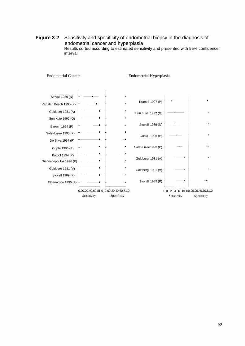
69
Figure 3-2 Sensitivity and specificity of endometrial biopsy in the diagnosis of endometrial cancer and hyperplasia Results sorted according to estimated sensitivity and presented with 95% confidence interval
Endometrial Cancer Endometrial Hyperplasia
Stovall 1989 (N)
Van den Bosch 1995 (P)
Goldberg 1981 (A)
Sun Kuie 1992 (G)
Baruch 1994 (P)
Salet-Lizee 1993 (P)
De Silva 1997 (P)
Gupta 1996 (P)
Batool 1994 (P)
Giannacopoulos 1996 (P)
Goldberg 1981 (V)
Stovall 1989 (P)
Etherington 1995 (Z)
0.00.20.40.60.81.0 0.00.20.40.60.81.0
Sensitivity Specificity
Krampl 1997 (P)
Sun Kuie 1992 (G)
Stovall 1989 (N)
Gupta 1996 (P)
Salet-Lizee1993 (P)
Goldberg 1981 (A)
Goldberg 1981 (V)
Stovall 1989 (P)
0.00.20.40.60.81.00.00.20.40.60.81.0
Sensitivity Specificity
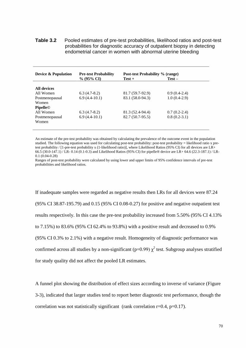
70
Table 3.2 Pooled estimates of pre-test probabilities, likelihood ratios and post-test probabilities for diagnostic accuracy of outpatient biopsy in detecting endometrial cancer in women with abnormal uterine bleeding
Device & Population Pre-test Probability Post-test Probability % (range)
% (95% CI) Test + Test –
All devices
All Women 6.3 (4.7-8.2) 81.7 (59.7-92.9) 0.9 (0.4-2.4)
Postmenopausal
Women
6.9 (4.4-10.1) 83.1 (58.0-94.3) 1.0 (0.4-2.9)
Pipelle©
All Women 6.3 (4.7-8.2) 81.3 (52.4-94.4) 0.7 (0.2-2.4)
Postmenopausal
Women
6.9 (4.4-10.1) 82.7 (50.7-95.5) 0.8 (0.2-3.1)
An estimate of the pre-test probability was obtained by calculating the prevalence of the outcome event in the population
studied. The following equation was used for calculating post-test probability: post-test probability = likelihood ratio x pre-
test probability / [1-pre-test probability x (1-likelihood ratio)], where Likelihood Ratios (95% CI) for all devices are LR+
66.5 (30.0-147.1) / LR- 0.14 (0.1-0.3) and Likelihood Ratios (95% CI) for pipelle® device are LR+ 64.6 (22.3-187.1) / LR-
0.1 (0.04-0.28)
Ranges of post-test probability were calculated by using lower and upper limits of 95% confidence intervals of pre-test
probabilities and likelihood ratios.
If inadequate samples were regarded as negative results then LRs for all devices were 87.24
(95% CI 38.87-195.79) and 0.15 (95% CI 0.08-0.27) for positive and negative outpatient test
results respectively. In this case the pre-test probability increased from 5.50% (95% CI 4.13%
to 7.15%) to 83.6% (95% CI 62.4% to 93.8%) with a positive result and decreased to 0.9%
(95% CI 0.3% to 2.1%) with a negative result. Homogeneity of diagnostic performance was
confirmed across all studies by a non-significant (p=0.99) χ2 test. Subgroup analyses stratified
for study quality did not affect the pooled LR estimates.
A funnel plot showing the distribution of effect sizes according to inverse of variance (Figure
3-3), indicated that larger studies tend to report better diagnostic test performance, though the
correlation was not statistically significant (rank correlation r=0.4, p=0.17).
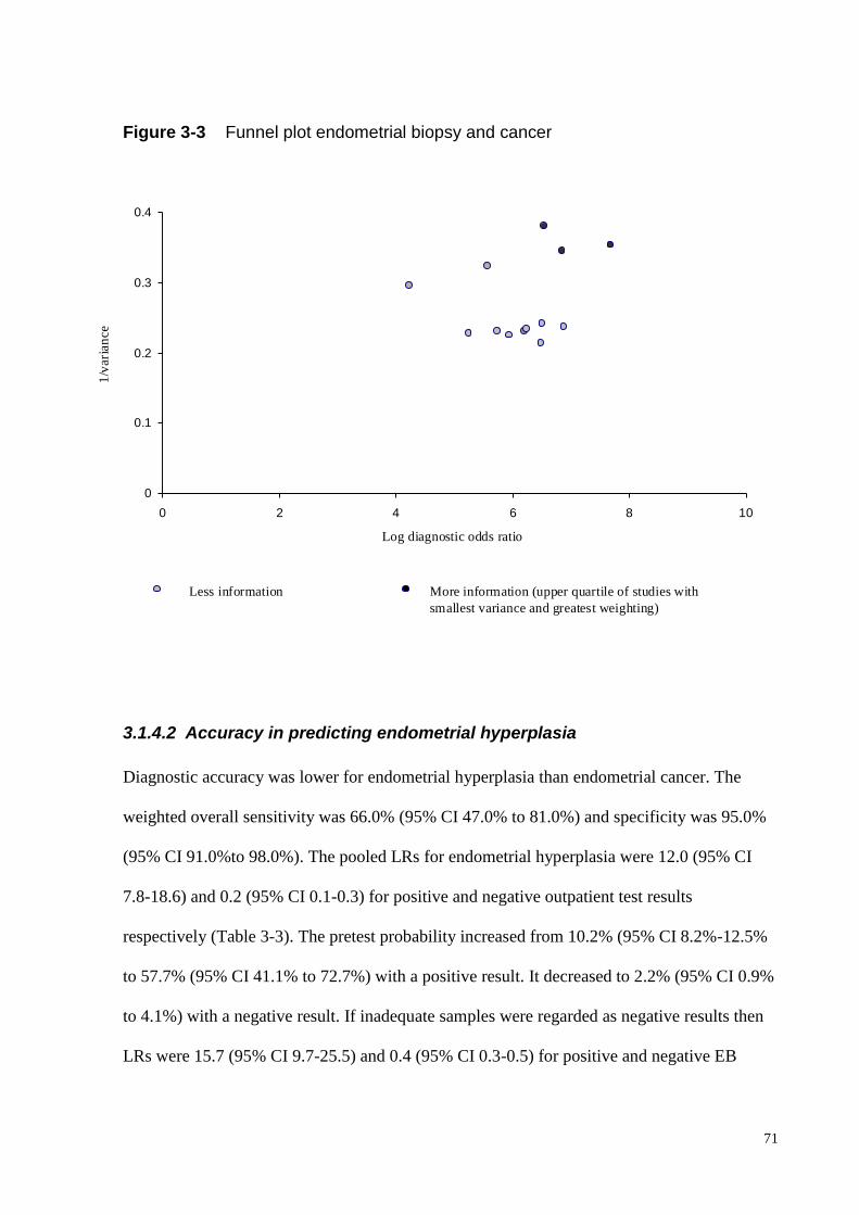
71
Figure 3-3 Funnel plot endometrial biopsy and cancer
3.1.4.2 Accuracy in predicting endometrial hyperplasia
Diagnostic accuracy was lower for endometrial hyperplasia than endometrial cancer. The
weighted overall sensitivity was 66.0% (95% CI 47.0% to 81.0%) and specificity was 95.0%
(95% CI 91.0%to 98.0%). The pooled LRs for endometrial hyperplasia were 12.0 (95% CI
7.8-18.6) and 0.2 (95% CI 0.1-0.3) for positive and negative outpatient test results
respectively (Table 3-3). The pretest probability increased from 10.2% (95% CI 8.2%-12.5%
to 57.7% (95% CI 41.1% to 72.7%) with a positive result. It decreased to 2.2% (95% CI 0.9%
to 4.1%) with a negative result. If inadequate samples were regarded as negative results then
LRs were 15.7 (95% CI 9.7-25.5) and 0.4 (95% CI 0.3-0.5) for positive and negative EB
0
0.1
0.2
0.3
0.4
0 2 4 6 8 10
Log diagnostic odds ratio
1/v
ari
an
ce
Less information More information (upper quartile of studies with
smallest variance and greatest weighting)
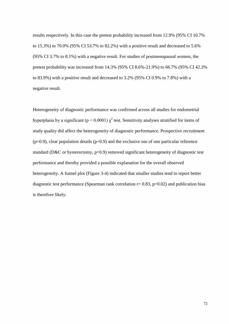
72
results respectively. In this case the pretest probability increased from 12.9% (95% CI 10.7%
to 15.3%) to 70.0% (95% CI 53.7% to 82.2%) with a positive result and decreased to 5.6%
(95% CI 3.7% to 8.1%) with a negative result. For studies of postmenopausal women, the
pretest probability was increased from 14.3% (95% CI 8.6%-21.9%) to 66.7% (95% CI 42.3%
to 83.9%) with a positive result and decreased to 3.2% (95% CI 0.9% to 7.8%) with a
negative result.
Heterogeneity of diagnostic performance was confirmed across all studies for endometrial
hyperplasia by a significant (p = 0.0001) χ2 test. Sensitivity analyses stratified for items of
study quality did affect the heterogeneity of diagnostic performance. Prospective recruitment
(p=0.9), clear population details (p=0.9) and the exclusive use of one particular reference
standard (D&C or hysterectomy, p=0.9) removed significant heterogeneity of diagnostic test
performance and thereby provided a possible explanation for the overall observed
heterogeneity. A funnel plot (Figure 3-4) indicated that smaller studies tend to report better
diagnostic test performance (Spearman rank correlation r= 0.83, p=0.02) and publication bias
is therefore likely.
73
Figure 3-4 Funnel plot endometrial biopsy and hyperplasia
3.1.4.2.1 Severity of endometrial hyperplasia
The data were separated for the presence of endometrial hyperplasia with or without atypia.
Table 3-3 shows the pooled LRs, pre and posttest probabilities for positive and negative
results for these subgroups along with the overall pooled result for comparison. Significant
heterogeneity of diagnostic test performance was present. Possible explanations for this
related to study design and type of reference standard as sensitivity analyses excluding
retrospective studies and studies using more than one reference standard produced
homogeneity of diagnostic performance. In addition the data was analysed to determine the
diagnostic accuracy of outpatient biopsy in detecting premalignant (complex and atypical
0
2
4
6
8
0 2 4 6 8
Log diagnostic odds ratio
1/v
ari
an
ce
Less information More information (upper quartile of studies with
smallest variance and greatest weighting)
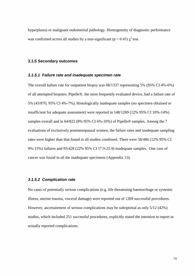
74
hyperplasia) or malignant endometrial pathology. Homogeneity of diagnostic performance
was confirmed across all studies by a non-significant (p = 0.43) χ2
test.
3.1.5 Secondary outcomes
3.1.5.1 Failure rate and inadequate specimen rate
The overall failure rate for outpatient biopsy was 68/1337 representing 5% (95% CI 4%-6%)
of all attempted biopsies. Pipelle®, the most frequently evaluated device, had a failure rate of
5% (43/870, 95% CI 4%-7%). Histologically inadequate samples (no specimen obtained or
insufficient for adequate assessment) were reported in 148/1269 (12% 95% CI 10%-14%)
samples overall and in 64/822 (8% 95% CI 6%-10%) of Pipelle® samples. Among the 7
evaluations of exclusively postmenopausal women, the failure rates and inadequate sampling
rates were higher than that found in all studies combined. There were 58/486 (12% 95% CI
9%-15%) failures and 93/428 (22% 95% CI 17.9-25.9) inadequate samples. One case of
cancer was found in all the inadequate specimens (Appendix 13).
3.1.5.2 Complication rate
No cases of potentially serious complications (e.g. life threatening haemorrhage or systemic
illness, uterine trauma, visceral damage) were reported out of 1269 successful procedures.
However, ascertainment of serious complications may be suboptimal as only 5/12 (42%)
studies, which included 251 successful procedures, explicitly stated the intention to report or
actually reported complications.
75
Table 3.3 Sensitivity analyses for meta-analysis of the diagnostic accuracy of outpatient endometrial biopsy in endometrial hyperplasia with or without atypia and its diagnostic accuracy in detecting endometrial cancer with or without premalignant complex/atypical endometrial hyperplasia
Device (No. Evaluations) Positive test Negative test LR positive LR negative Pretest probability Posttest probability
&Study (Year published) (Sensitivity) (1-Specificity) (95% CI) (95% CI) (95% CI) +ve test (95% CI) -ve test (95% CI)
Endometrial hyperplasia (all 8 evaluations)
Total 21,26,40,44,61,66
82/126 38/679 12.0 (7.8-18.6) 0.2 (0.1-0.3) 10.2% (8.%-12.5%) 57.7% (41.1 - 72.7%) 2.2% (0.9 to 4.1%)
Endometrial hyperplasia without atypia (4 evaluations)
Total 21,26,40,44
55/90 26/427 10.7 (6.1-18.9) 0.45 (0.35-0.57) 15.7% (12.6-19.1%) 86.7% (72.8 to 3.9%) 7.7% (4.8 to 11.9%)
Endometrial hyperplasia with atypia (1 evaluation)
Total 44
2/3 1/51 34 (4.2-277.4) 0.3 (0.07-1.7) 5.6 (1.2-15.4%) 66.9% (48.5 to 8.1%) 2.6% (0.4 to 9.4%)
Endometrial cancer +/- complex/atypical endometrial hyperplasia (8 evaluations)
Total 21,26,40,44,61,66
34/37 2/768 95.1 (41.2-219.4) 0.1 (0.07-0.3) 4.5% (3.2 to 6.1%) 81.8% (57.7 to 93.4%) 0.5% (0.2 to 1.9%)
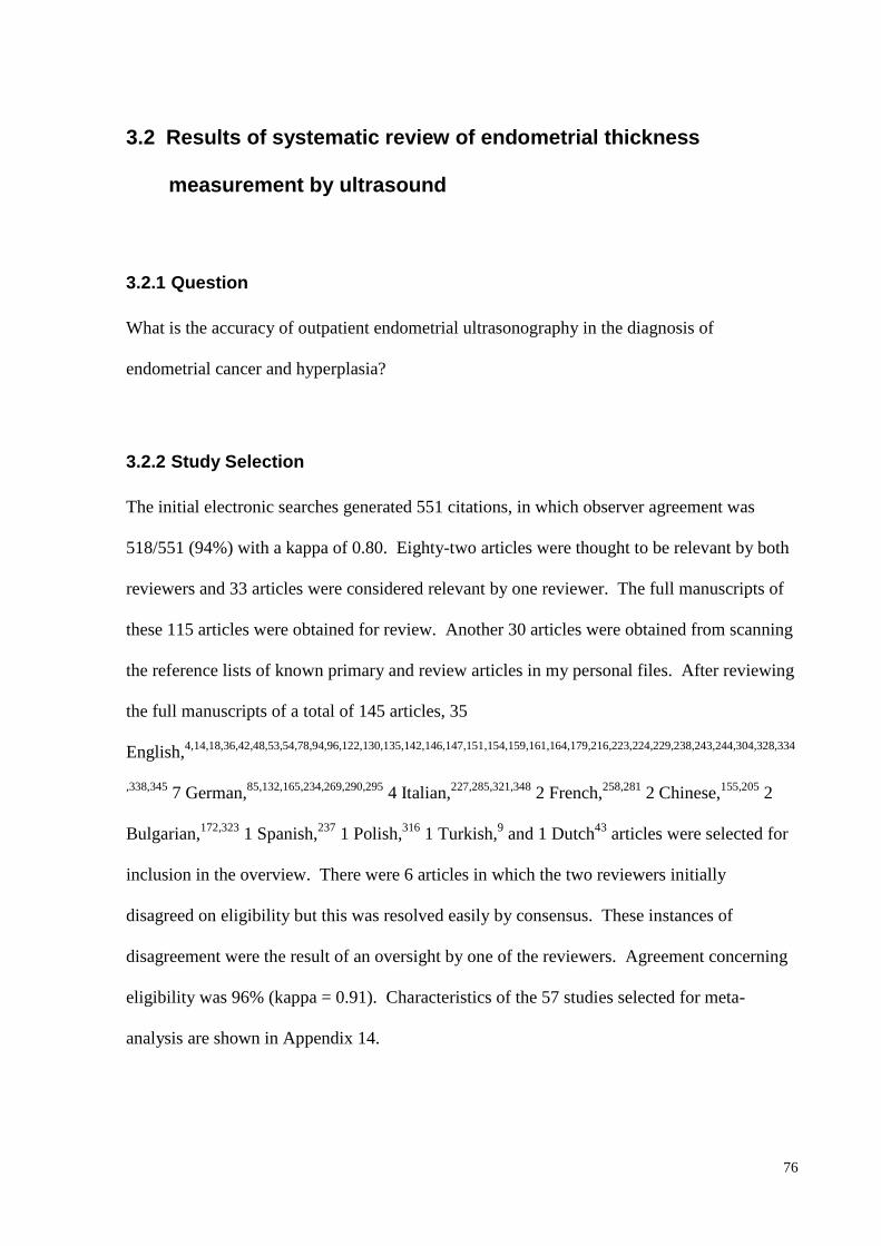
76
3.2 Results of systematic review of endometrial thickness
measurement by ultrasound
3.2.1 Question
What is the accuracy of outpatient endometrial ultrasonography in the diagnosis of
endometrial cancer and hyperplasia?
3.2.2 Study Selection
The initial electronic searches generated 551 citations, in which observer agreement was
518/551 (94%) with a kappa of 0.80. Eighty-two articles were thought to be relevant by both
reviewers and 33 articles were considered relevant by one reviewer. The full manuscripts of
these 115 articles were obtained for review. Another 30 articles were obtained from scanning
the reference lists of known primary and review articles in my personal files. After reviewing
the full manuscripts of a total of 145 articles, 35
English,4,14,18,36,42,48,53,54,78,94,96,122,130,135,142,146,147,151,154,159,161,164,179,216,223,224,229,238,243,244,304,328,334
,338,345 7 German,
85,132,165,234,269,290,295 4 Italian,
227,285,321,348 2 French,
258,281 2 Chinese,
155,205 2
Bulgarian,172,323
1 Spanish,237
1 Polish,316
1 Turkish,9 and 1 Dutch
43 articles were selected for
inclusion in the overview. There were 6 articles in which the two reviewers initially
disagreed on eligibility but this was resolved easily by consensus. These instances of
disagreement were the result of an oversight by one of the reviewers. Agreement concerning
eligibility was 96% (kappa = 0.91). Characteristics of the 57 studies selected for meta-
analysis are shown in Appendix 14.

77
The reasons for excluding the remaining 88 manuscripts (Figure 3-5 and Appendix 15)
included inappropriate study design (18 studies), inappropriate study population (31 studies),
inappropriate clinical outcomes being reported (5 studies), and the inability to extract data
precluding construction of 2x2 tables (31 studies). Three articles were also excluded due to
duplicate publication.
78
Figure 3-5 Study selection process for systematic review of ultrasound scan
3.2.3 Study quality
The observer agreement for the various components of study quality was 89-100%, kappa
values were 0.64 for population enrolment, 1.0 for description of amenorrhoea and HRT use,
1.0 for description of analytical test and cut-off level, 0.88 for number of endometrial layers
used in the ultrasonic measurement of endometrial thickness, 0.69 for blinding of test results
Potentially relevant studies identified and screened for
retrieval (citations in electronic search) n=551
Diagnostic test studies included in meta-analysis
n=57
Diagnostic test studies excluded:
Inappropriate study design (not accuracy) n=18 A7,9,12,25,26,28,33,36,45,49,51,67,70,74,80,85,86,88
Inappropriate study population n=31 A1,4-6,11,14,16-19,31,32,35,39,46,48,54-56,59,62,64,66,68,69,73,75-77,79,83
Inappropriate outcomes (not cancer)A3,22,40,72,87
n=5
Inability to extract for 2x2 table n=31 A2,8,10,13,15,20,21,23,24,27,29,30,34,37,38,42-44,47,50,53,58,60,61,63,65,71,78,81,82,84
Duplicate publicationA41,52,57
n=3
Population: Restricted recruitment (endometrial cancer) 5,18-
20,24-25,30 n=7
Intrauterine abnormalities data only 29,56
n=2
Asymptomatic women 43
n=1
Subtotal n=10
Diagnostic Performed under general anaesthetic 22,41,48,50,54-55,60-61
n=8 Intervention:Cytological outpatient device 59
n=1
No convincing gold standard 13,15-16,23,26,28,31,33,58
n=9
Subtotal n=18
Outcome:
Differential verification 14,27
n=2
Lack of data to construct 2x2 table 34-36,42,45-46,49,53
n=8
Lack of original data / reviews 21,32,51
n=3
Subtotal n=13
TTotal excluded 5,13-17,18-35,38,41-43,45-46,48-51,53-56,58-61
n=88
Studies excluded n=406
(Inappropriate population, intervention or outcome – see text)
Diagnostic test studies retrieved for more detailed
evaluation:
from electronic search n=115
from reference lists n=30
Total n=145

79
and 1.0 for completeness of verification. The instances of disagreement were the result of an
oversight on of one of the reviewers, and were resolved easily by consensus. The main
features of the methodological qualities of those studies selected for meta-analysis are
summarised in Table 3-4. A majority of the studies were quality level 4-5.
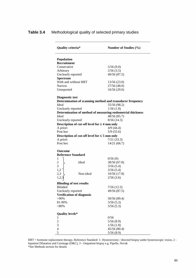
80
Table 3.4 Methodological quality of selected primary studies
Quality criteria* Number of Studies (%)
Population
Recruitment
Consecutive 5/56 (9.0)
Arbitrary 2/56 (3.5)
Unclearly reported 49/56 (87.5)
Spectrum
With and without HRT 13/56 (23.0)
Narrow 27/56 (48.0)
Unreported 16/56 (29.0)
Diagnostic test
Determination of scanning method and transducer frequency
Ideal 55/56 (98.2)
Unclearly reported 1/56 (1.8)
Determination of method of measuring endometrial thickness
Ideal 48/56 (85.7)
Unclearly reported 8/56 (14.3)
Description of cut-off level for 4 mm only
A priori 4/9 (44.4)
Post hoc 5/9 (55.6)
Description of cut-off level for 5 mm only
A priori 7/21 (33.3)
Post hoc 14/21 (66.7)
Outcome
Reference Standard
1 0/56 (0)
2 Ideal 38/56 (67.8)
3 3/56 (5.4)
1,2 3/56 (5.4)
2,3 Non-ideal 10/56 (17.8)
1,2,3 2/56 (3.6)
Blinding of test results
Blinded 7/56 (12.5)
Unclearly reported 49/56 (87.5)
Verification of diagnosis
>90% 50/56 (89.4)
81-90% 3/56 (5.3)
<80% 3/56 (5.3)
Quality levels*
1 0/56
2 5/56 (8.9)
3 1/56 (1.8)
4 45/56 (80.4)
5 5/56 (8.9)
HRT = hormone replacement therapy, Reference Standard: 1- Hysterectomy / directed biopsy under hysteroscopic vision, 2 -
Inpatient Dilatation and Curettage (D&C), 3 - Outpatient biopsy e.g. Pipelle, Novak
*See Methods section for details
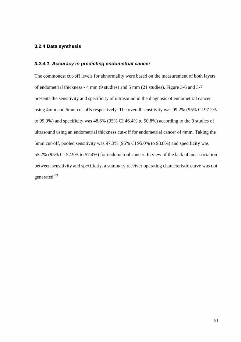
81
3.2.4 Data synthesis
3.2.4.1 Accuracy in predicting endometrial cancer
The commonest cut-off levels for abnormality were based on the measurement of both layers
of endometrial thickness - 4 mm (9 studies) and 5 mm (21 studies). Figure 3-6 and 3-7
presents the sensitivity and specificity of ultrasound in the diagnosis of endometrial cancer
using 4mm and 5mm cut-offs respectively. The overall sensitivity was 99.2% (95% CI 97.2%
to 99.9%) and specificity was 48.6% (95% CI 46.4% to 50.8%) according to the 9 studies of
ultrasound using an endometrial thickness cut-off for endometrial cancer of 4mm. Taking the
5mm cut-off, pooled sensitivity was 97.3% (95% CI 95.0% to 98.8%) and specificity was
55.2% (95% CI 52.9% to 57.4%) for endometrial cancer. In view of the lack of an association
between sensitivity and specificity, a summary receiver operating characteristic curve was not
generated.83
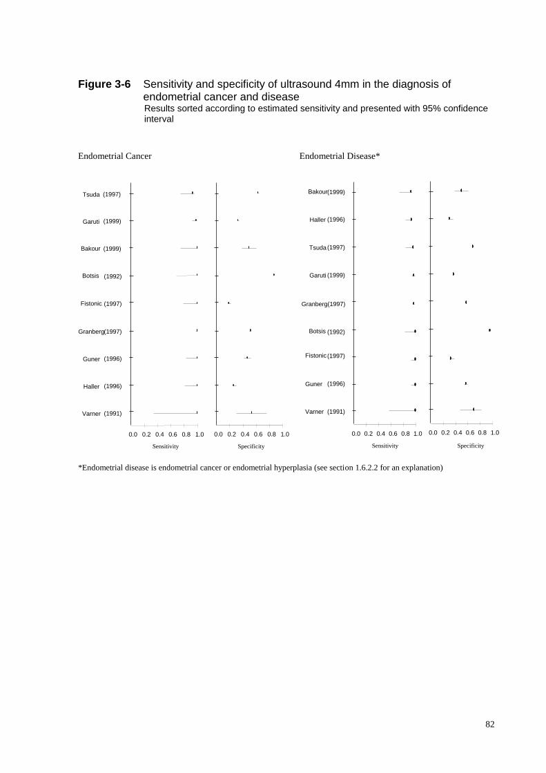
82
Figure 3-6 Sensitivity and specificity of ultrasound 4mm in the diagnosis of endometrial cancer and disease Results sorted according to estimated sensitivity and presented with 95% confidence interval
Endometrial Cancer Endometrial Disease*
*Endometrial disease is endometrial cancer or endometrial hyperplasia (see section 1.6.2.2 for an explanation)
Tsuda (1997)
Garuti (1999)
Bakour (1999)
Botsis (1992)
Fistonic (1997)
Granberg(1997)
Guner (1996)
Haller (1996)
Varner (1991)
0.0 0.2 0.4 0.6 0.8 1.0 0.0 0.2 0.4 0.6 0.8 1.0
Sensitivity Specificity
Bakour(1999)
Haller (1996)
Tsuda(1997)
Garuti (1999)
Granberg(1997)
Botsis (1992)
Fistonic (1997)
Guner (1996)
Varner (1991)
0.0 0.2 0.4 0.6 0.8 1.00.0 0.2 0.4 0.6 0.8 1.0
Sensitivity Specificity
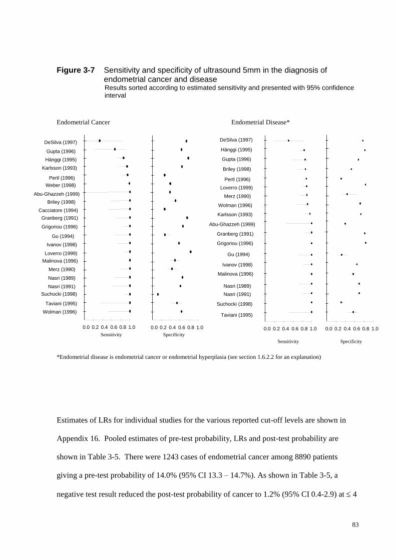
83
Figure 3-7 Sensitivity and specificity of ultrasound 5mm in the diagnosis of endometrial cancer and disease Results sorted according to estimated sensitivity and presented with 95% confidence interval
Endometrial Cancer Endometrial Disease*
*Endometrial disease is endometrial cancer or endometrial hyperplasia (see section 1.6.2.2 for an explanation)
Estimates of LRs for individual studies for the various reported cut-off levels are shown in
Appendix 16. Pooled estimates of pre-test probability, LRs and post-test probability are
shown in Table 3-5. There were 1243 cases of endometrial cancer among 8890 patients
giving a pre-test probability of 14.0% (95% CI 13.3 – 14.7%). As shown in Table 3-5, a
negative test result reduced the post-test probability of cancer to 1.2% (95% CI 0.4-2.9) at 4
DeSilva (1997)
Hänggi (1995)
Gupta (1996)
Briley (1998)
Pertl (1996)
Loverro (1999)
Merz (1990)
Wolman (1996)
Karlsson (1993)
Abu-Ghazzeh (1999)
Granberg (1991)
Grigoriou (1996)
Gu (1994)
Ivanov (1998)
Malinova (1996)
Nasri (1989)
Nasri (1991)
Suchocki (1998)
Taviani (1995)
0.0 0.2 0.4 0.6 0.8 1.0 0.0 0.2 0.4 0.6 0.8 1.0
Sensitivity Specificity
DeSilva (1997)
Gupta (1996)
Hänggi (1995)
Karlsson (1993)
Pertl (1996)
Weber (1998)
Abu-Ghazzeh (1999)
Briley (1998)
Cacciatore (1994)
Granberg (1991)
Grigoriou (1996)
Gu (1994)
Ivanov (1998)
Loverro (1999)
Malinova (1996)
Merz (1990)
Nasri (1989)
Nasri (1991)
Suchocki (1998)
Taviani (1995)
Wolman (1996)
0.0 0.2 0.4 0.6 0.8 1.0 0.0 0.2 0.4 0.6 0.8 1.0
Sensitivity Specificity
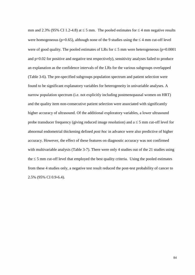
84
mm and 2.3% (95% CI 1.2-4.8) at 5 mm. The pooled estimates for 4 mm negative results
were homogeneous (p=0.65), although none of the 9 studies using the 4 mm cut-off level
were of good quality. The pooled estimates of LRs for 5 mm were heterogeneous (p=0.0001
and p=0.02 for positive and negative test respectively), sensitivity analyses failed to produce
an explanation as the confidence intervals of the LRs for the various subgroups overlapped
(Table 3-6). The pre-specified subgroups population spectrum and patient selection were
found to be significant explanatory variables for heterogeneity in univariable analyses. A
narrow population spectrum (i.e. not explicitly including postmenopausal women on HRT)
and the quality item non-consecutive patient selection were associated with significantly
higher accuracy of ultrasound. Of the additional exploratory variables, a lower ultrasound
probe transducer frequency (giving reduced image resolution) and a 5 mm cut-off level for
abnormal endometrial thickening defined post hoc in advance were also predictive of higher
accuracy. However, the effect of these features on diagnostic accuracy was not confirmed
with multivariable analysis (Table 3-7). There were only 4 studies out of the 21 studies using
the 5 mm cut-off level that employed the best quality criteria. Using the pooled estimates
from these 4 studies only, a negative test result reduced the post-test probability of cancer to
2.5% (95% CI 0.9-6.4).
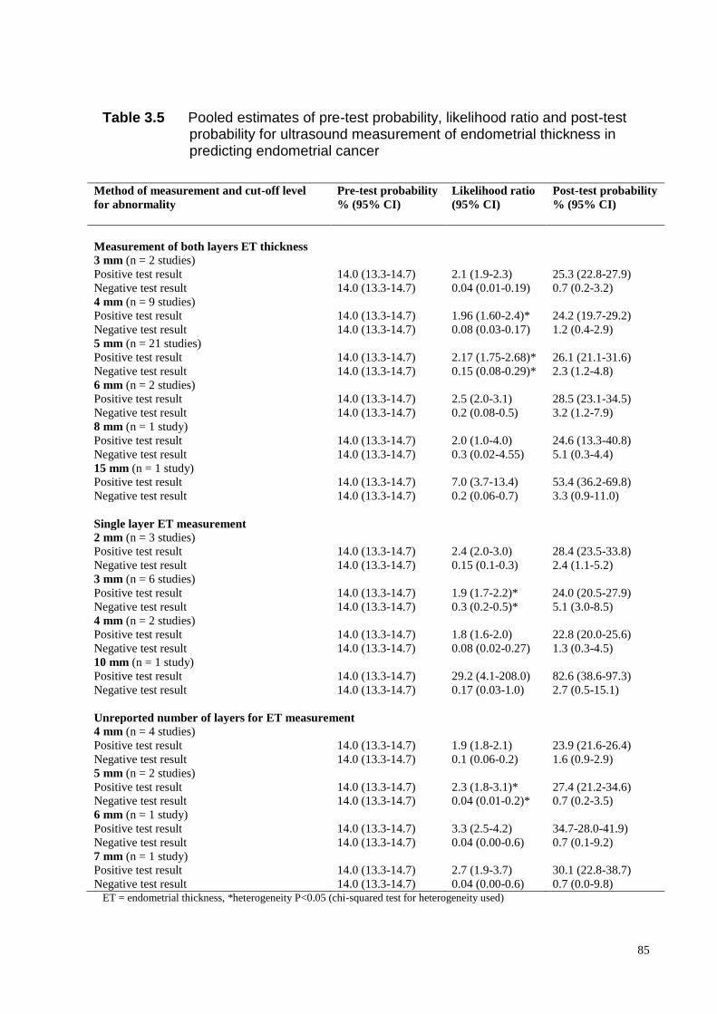
85
Table 3.5 Pooled estimates of pre-test probability, likelihood ratio and post-test probability for ultrasound measurement of endometrial thickness in predicting endometrial cancer
Method of measurement and cut-off level
for abnormality
Pre-test probability
% (95% CI)
Likelihood ratio
(95% CI)
Post-test probability
% (95% CI)
Measurement of both layers ET thickness
3 mm (n = 2 studies)
Positive test result 14.0 (13.3-14.7) 2.1 (1.9-2.3) 25.3 (22.8-27.9)
Negative test result 14.0 (13.3-14.7) 0.04 (0.01-0.19) 0.7 (0.2-3.2)
4 mm (n = 9 studies)
Positive test result 14.0 (13.3-14.7) 1.96 (1.60-2.4)* 24.2 (19.7-29.2)
Negative test result 14.0 (13.3-14.7) 0.08 (0.03-0.17) 1.2 (0.4-2.9)
5 mm (n = 21 studies)
Positive test result 14.0 (13.3-14.7) 2.17 (1.75-2.68)* 26.1 (21.1-31.6)
Negative test result 14.0 (13.3-14.7) 0.15 (0.08-0.29)* 2.3 (1.2-4.8)
6 mm (n = 2 studies)
Positive test result 14.0 (13.3-14.7) 2.5 (2.0-3.1) 28.5 (23.1-34.5)
Negative test result 14.0 (13.3-14.7) 0.2 (0.08-0.5) 3.2 (1.2-7.9)
8 mm (n = 1 study)
Positive test result 14.0 (13.3-14.7) 2.0 (1.0-4.0) 24.6 (13.3-40.8)
Negative test result 14.0 (13.3-14.7) 0.3 (0.02-4.55) 5.1 (0.3-4.4)
15 mm (n = 1 study)
Positive test result 14.0 (13.3-14.7) 7.0 (3.7-13.4) 53.4 (36.2-69.8)
Negative test result 14.0 (13.3-14.7) 0.2 (0.06-0.7) 3.3 (0.9-11.0)
Single layer ET measurement
2 mm (n = 3 studies)
Positive test result 14.0 (13.3-14.7) 2.4 (2.0-3.0) 28.4 (23.5-33.8)
Negative test result 14.0 (13.3-14.7) 0.15 (0.1-0.3) 2.4 (1.1-5.2)
3 mm (n = 6 studies)
Positive test result 14.0 (13.3-14.7) 1.9 (1.7-2.2)* 24.0 (20.5-27.9)
Negative test result 14.0 (13.3-14.7) 0.3 (0.2-0.5)* 5.1 (3.0-8.5)
4 mm (n = 2 studies)
Positive test result 14.0 (13.3-14.7) 1.8 (1.6-2.0) 22.8 (20.0-25.6)
Negative test result 14.0 (13.3-14.7) 0.08 (0.02-0.27) 1.3 (0.3-4.5)
10 mm (n = 1 study)
Positive test result 14.0 (13.3-14.7) 29.2 (4.1-208.0) 82.6 (38.6-97.3)
Negative test result 14.0 (13.3-14.7) 0.17 (0.03-1.0) 2.7 (0.5-15.1)
Unreported number of layers for ET measurement
4 mm (n = 4 studies)
Positive test result 14.0 (13.3-14.7) 1.9 (1.8-2.1) 23.9 (21.6-26.4)
Negative test result 14.0 (13.3-14.7) 0.1 (0.06-0.2) 1.6 (0.9-2.9)
5 mm (n = 2 studies)
Positive test result 14.0 (13.3-14.7) 2.3 (1.8-3.1)* 27.4 (21.2-34.6)
Negative test result 14.0 (13.3-14.7) 0.04 (0.01-0.2)* 0.7 (0.2-3.5)
6 mm (n = 1 study)
Positive test result 14.0 (13.3-14.7) 3.3 (2.5-4.2) 34.7-28.0-41.9)
Negative test result 14.0 (13.3-14.7) 0.04 (0.00-0.6) 0.7 (0.1-9.2)
7 mm (n = 1 study)
Positive test result 14.0 (13.3-14.7) 2.7 (1.9-3.7) 30.1 (22.8-38.7)
Negative test result 14.0 (13.3-14.7) 0.04 (0.00-0.6) 0.7 (0.0-9.8) ET = endometrial thickness, *heterogeneity P<0.05 (chi-squared test for heterogeneity used)
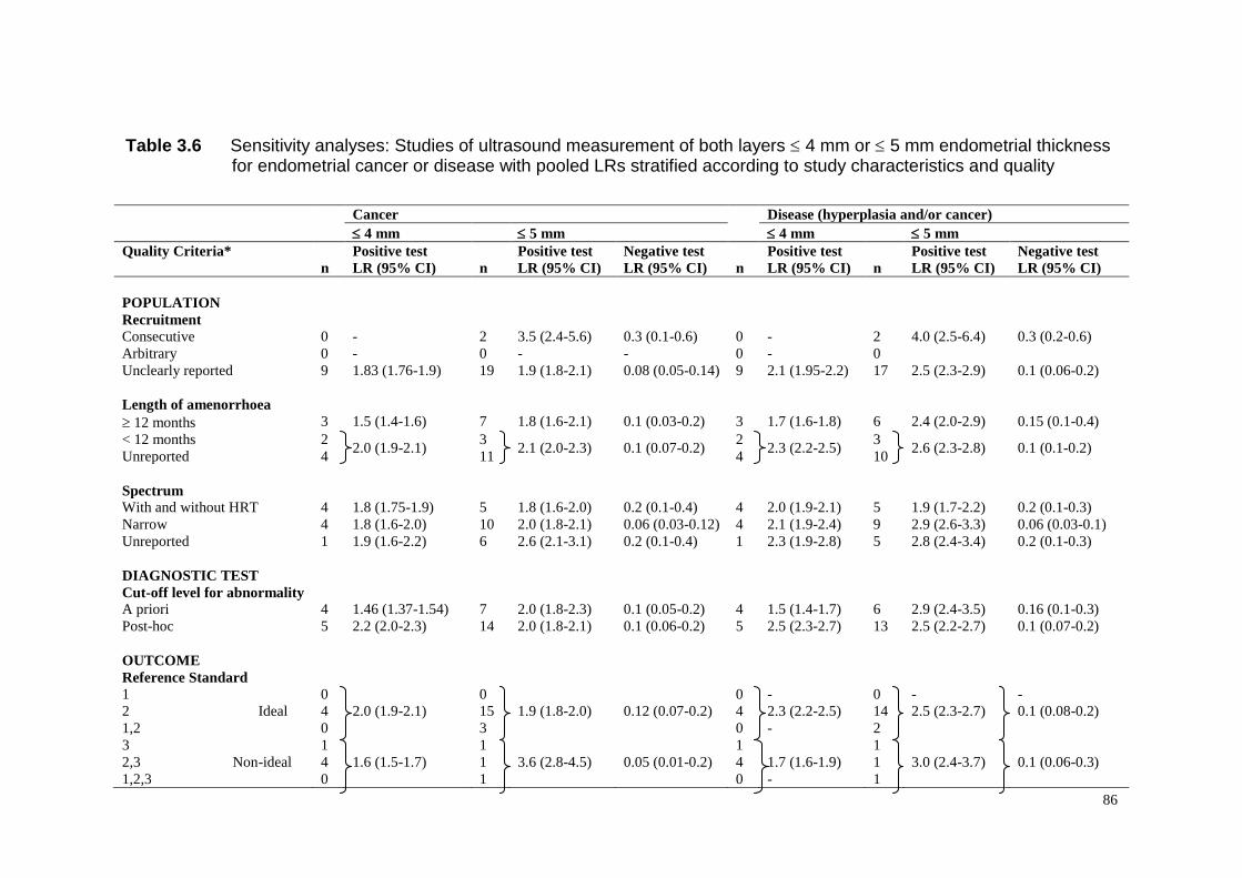
86
Table 3.6 Sensitivity analyses: Studies of ultrasound measurement of both layers 4 mm or 5 mm endometrial thickness for endometrial cancer or disease with pooled LRs stratified according to study characteristics and quality
Cancer Disease (hyperplasia and/or cancer)
4 mm 5 mm 4 mm 5 mm
Quality Criteria*
n
Positive test
LR (95% CI)
n
Positive test
LR (95% CI)
Negative test
LR (95% CI)
n
Positive test
LR (95% CI)
n
Positive test
LR (95% CI)
Negative test
LR (95% CI)
POPULATION
Recruitment
Consecutive 0 - 2 3.5 (2.4-5.6) 0.3 (0.1-0.6) 0 - 2 4.0 (2.5-6.4) 0.3 (0.2-0.6)
Arbitrary 0 - 0 - - 0 - 0
Unclearly reported 9 1.83 (1.76-1.9) 19 1.9 (1.8-2.1) 0.08 (0.05-0.14) 9 2.1 (1.95-2.2) 17 2.5 (2.3-2.9) 0.1 (0.06-0.2)
Length of amenorrhoea
12 months 3 1.5 (1.4-1.6) 7 1.8 (1.6-2.1) 0.1 (0.03-0.2) 3 1.7 (1.6-1.8) 6 2.4 (2.0-2.9) 0.15 (0.1-0.4)
< 12 months 2 2.0 (1.9-2.1)
3 2.1 (2.0-2.3) 0.1 (0.07-0.2)
2 2.3 (2.2-2.5)
3 2.6 (2.3-2.8) 0.1 (0.1-0.2)
Unreported 4 11 4 10
Spectrum
With and without HRT 4 1.8 (1.75-1.9) 5 1.8 (1.6-2.0) 0.2 (0.1-0.4) 4 2.0 (1.9-2.1) 5 1.9 (1.7-2.2) 0.2 (0.1-0.3)
Narrow 4 1.8 (1.6-2.0) 10 2.0 (1.8-2.1) 0.06 (0.03-0.12) 4 2.1 (1.9-2.4) 9 2.9 (2.6-3.3) 0.06 (0.03-0.1)
Unreported 1 1.9 (1.6-2.2) 6 2.6 (2.1-3.1) 0.2 (0.1-0.4) 1 2.3 (1.9-2.8) 5 2.8 (2.4-3.4) 0.2 (0.1-0.3)
DIAGNOSTIC TEST
Cut-off level for abnormality
A priori 4 1.46 (1.37-1.54) 7 2.0 (1.8-2.3) 0.1 (0.05-0.2) 4 1.5 (1.4-1.7) 6 2.9 (2.4-3.5) 0.16 (0.1-0.3)
Post-hoc 5 2.2 (2.0-2.3) 14 2.0 (1.8-2.1) 0.1 (0.06-0.2) 5 2.5 (2.3-2.7) 13 2.5 (2.2-2.7) 0.1 (0.07-0.2)
OUTCOME
Reference Standard
1 0 0 0 - 0 - -
2 Ideal 4 2.0 (1.9-2.1) 15 1.9 (1.8-2.0) 0.12 (0.07-0.2) 4 2.3 (2.2-2.5) 14 2.5 (2.3-2.7) 0.1 (0.08-0.2)
1,2 0 3 0 - 2
3 1 1 1 1
2,3 Non-ideal 4 1.6 (1.5-1.7) 1 3.6 (2.8-4.5) 0.05 (0.01-0.2) 4 1.7 (1.6-1.9) 1 3.0 (2.4-3.7) 0.1 (0.06-0.3)
1,2,3 0 1 0 - 1
87
Table 3-6 continued
Cancer Disease (hyperplasia and/or cancer)
4 mm 5 mm 4 mm 5 mm
Quality Criteria*
n
Positive test
LR (95% CI)
n
Positive test
LR (95% CI)
Negative test
LR (95% CI)
n
Positive test
LR (95% CI)
n
Positive test
LR (95% CI)
Negative test
LR (95% CI)
Blinding of test results
Blinded 0 - 3 2.9 (2.4-3.4) 0.1 (0.02-0.3) 0 - 3 5.0 (3.9-6.4) 0.03 (0.01-0.1)
Unclearly reported 9 1.83 (1.76-1.9) 18 1.9 (1.8-2.0) 0.1 (0.07-0.2) 9 2.1 (1.95-2.2) 16 2.2 (2.0-2.4) 0.015 (0.1-0.2)
Verification of diagnosis
>90% 9 1.83 (1.76-1.9) 17 2.0 (1.9-2.1) 0.1 (0.05-0.2) 9 2.1 (1.95-2.2) 15 2.9 (2.6-3.2) 0.1 (0.05-0.1)
81-90% 0 - 2 2.0 (1.7-2.3) 0.2 (0.1-0.4)
0 - 2 1.9 (1.7-2.2) 0.2 (0.1-0.4)
<80% 0 - 2 0 - 2
Quality Level
I-III 0 - 4 2.8 (2.3-3.3) 0.16 (0.06-0.4) 0 - 4 4.7 (3.7-6.0) 0.08 (0.03-0.2)
IV-VI 9 1.83 (1.76-1.9) 17 1.9 (1.8-2.1) 0.09 (0.05-0.2) 9 2.1 (1.95-2.2) 15 2.2 (2.0-2.4) 0.13 (0.09-0.2)
Reference Standard and Quality Level
Ideal and I-III 0 - 4 2.8 (2.3-3.3) 0.16 (0.06-0.4) 0 - 4 4.7 (3.7-6.0) 0.08 (0.03-0.2)
Ideal and IV-VI 4 2.0 (1.9-2.1) 14 1.8 (1.6-1.9) 0.1 (0.06-0.2) 4 2.3 (2.2-2.5) 12 2.05 (1.9-2.3) 0.1 (0.09-0.2)
Non-ideal and IV-VI 5 1.6 (1.5-1.7) 3 3.6 (2.8-4.5) 0.05 (0.01-0.2) 5 1.7 (1.6-1.9) 3 3.0 (2.4-3.7) 0.1 (0.06-0.3)
Reference Standard: 1- Hysterectomy / directed biopsy under hysteroscopic vision, 2 - Inpatient D&C (D&C), 3 - Outpatient biopsy e.g. Pipelle, Novak, *see Methods section for details
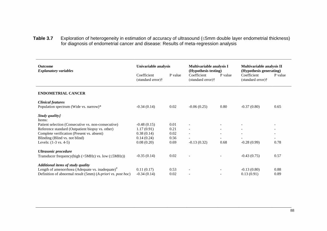
88
Table 3.7 Exploration of heterogeneity in estimation of accuracy of ultrasound (5mm double layer endometrial thickness) for diagnosis of endometrial cancer and disease: Results of meta-regression analysis
Outcome
Explanatory variables
Univariable analysis Multivariable analysis I
(Hypothesis testing)
Multivariable analysis II
(Hypothesis generating)
Coefficient
(standard error)†
P value Coefficient
(standard error)†
P value Coefficient
(standard error)†
P value
ENDOMETRIAL CANCER
Clinical features
Population spectrum (Wide vs. narrow)* -0.34 (0.14) 0.02 -0.06 (0.25) 0.80 -0.37 (0.80) 0.65
Study quality‡
Items:
Patient selection (Consecutive vs. non-consecutive) -0.48 (0.15) 0.01 - - - -
Reference standard (Outpatient biopsy vs. other) 1.17 (0.91) 0.21 - - - -
Complete verification (Present vs. absent) 0.38 (0.14) 0.02 - - - -
Blinding (Blind vs. not blind) 0.14 (0.24) 0.56 - - - -
Levels: (1-3 vs. 4-5) 0.08 (0.20) 0.69 -0.13 (0.32) 0.68 -0.28 (0.99) 0.78
Ultrasonic procedure
Transducer frequency(high (>5MHz) vs. low (5MHz)) -0.35 (0.14) 0.02 - - -0.43 (0.75) 0.57
Additional items of study quality
Length of amenorrhoea (Adequate vs. inadequate)# 0.11 (0.17) 0.53 - - -0.13 (0.80) 0.88
Definition of abnormal result (5mm) (A-priori vs. post hoc) -0.34 (0.14) 0.02 - - 0.13 (0.91) 0.89
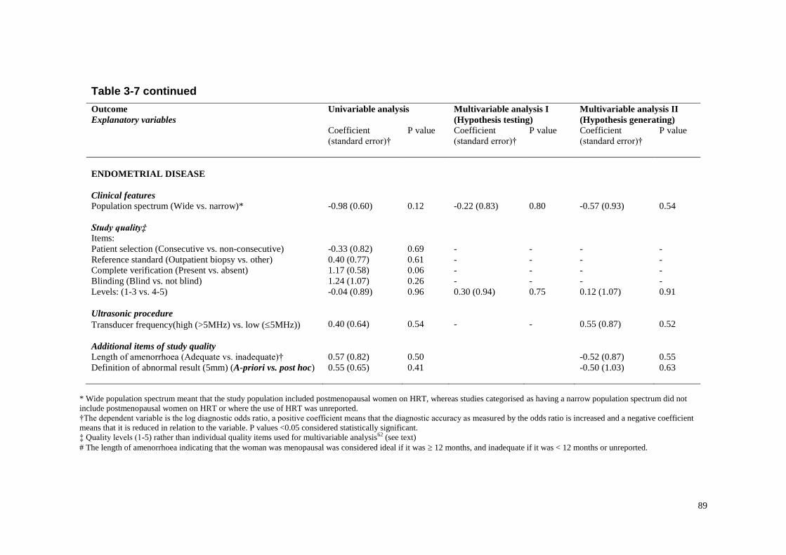
89
Table 3-7 continued
Outcome
Explanatory variables
Univariable analysis Multivariable analysis I
(Hypothesis testing)
Multivariable analysis II
(Hypothesis generating)
Coefficient
(standard error)†
P value Coefficient
(standard error)†
P value Coefficient
(standard error)†
P value
ENDOMETRIAL DISEASE
Clinical features
Population spectrum (Wide vs. narrow)* -0.98 (0.60) 0.12 -0.22 (0.83) 0.80 -0.57 (0.93) 0.54
Study quality‡
Items:
Patient selection (Consecutive vs. non-consecutive) -0.33 (0.82) 0.69 - - - -
Reference standard (Outpatient biopsy vs. other) 0.40 (0.77) 0.61 - - - -
Complete verification (Present vs. absent) 1.17 (0.58) 0.06 - - - -
Blinding (Blind vs. not blind) 1.24 (1.07) 0.26 - - - -
Levels: (1-3 vs. 4-5) -0.04 (0.89) 0.96 0.30 (0.94) 0.75 0.12 (1.07) 0.91
Ultrasonic procedure
Transducer frequency(high (>5MHz) vs. low (5MHz)) 0.40 (0.64) 0.54 - - 0.55 (0.87) 0.52
Additional items of study quality
Length of amenorrhoea (Adequate vs. inadequate)† 0.57 (0.82) 0.50 -0.52 (0.87) 0.55
Definition of abnormal result (5mm) (A-priori vs. post hoc) 0.55 (0.65) 0.41 -0.50 (1.03) 0.63
* Wide population spectrum meant that the study population included postmenopausal women on HRT, whereas studies categorised as having a narrow population spectrum did not
include postmenopausal women on HRT or where the use of HRT was unreported.
†The dependent variable is the log diagnostic odds ratio, a positive coefficient means that the diagnostic accuracy as measured by the odds ratio is increased and a negative coefficient
means that it is reduced in relation to the variable. P values <0.05 considered statistically significant.
‡ Quality levels (1-5) rather than individual quality items used for multivariable analysis62 (see text)
# The length of amenorrhoea indicating that the woman was menopausal was considered ideal if it was 12 months, and inadequate if it was < 12 months or unreported.
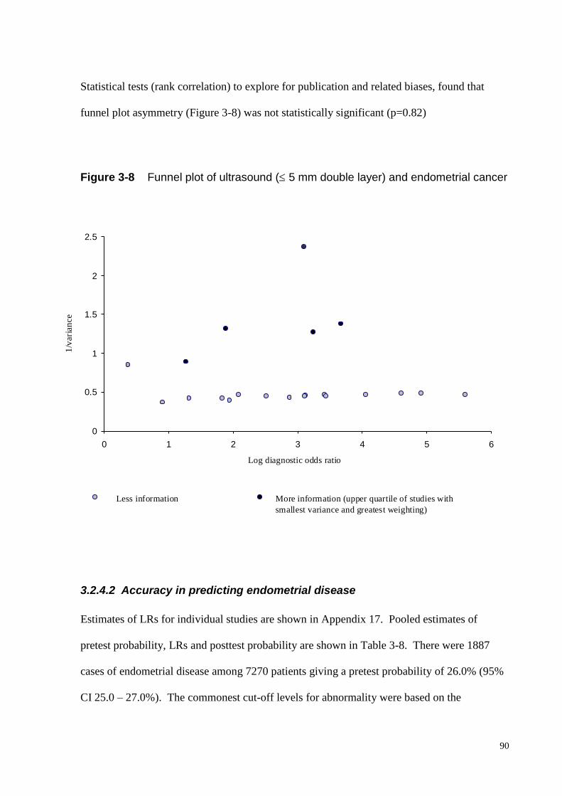
90
Statistical tests (rank correlation) to explore for publication and related biases, found that
funnel plot asymmetry (Figure 3-8) was not statistically significant (p=0.82)
Figure 3-8 Funnel plot of ultrasound ( 5 mm double layer) and endometrial cancer
3.2.4.2 Accuracy in predicting endometrial disease
Estimates of LRs for individual studies are shown in Appendix 17. Pooled estimates of
pretest probability, LRs and posttest probability are shown in Table 3-8. There were 1887
cases of endometrial disease among 7270 patients giving a pretest probability of 26.0% (95%
CI 25.0 – 27.0%). The commonest cut-off levels for abnormality were based on the
0
0.5
1
1.5
2
2.5
0 1 2 3 4 5 6
Log diagnostic odds ratio
1/v
ari
an
ce
Less information More information (upper quartile of studies with
smallest variance and greatest weighting)
91
measurement of both layers of endometrial thickness - 4 mm (9 studies) and 5 mm (19
studies). As shown in Table 3-8, a negative test result reduced the posttest probability of
cancer to 2.4% (95% CI 1.3-3.9) at 4 mm and 5.0% (95% CI 2.9-9.1) at 5 mm. The
pooled estimates for 4 mm negative results were homogeneous. However, none of the 9
studies using the 4 mm cut-off level were of good quality. Although the pooled estimates of
LRs for 5 mm were heterogeneous (p<0.05), sensitivity analyses failed to produce an
explanation as the confidence intervals of the LRs for the various subgroups overlapped
(Table 3-6). An explanation for heterogeneity was not provided by the population spectrum or
items of study quality (Table 3-7) when examined using both univariable and multivariable
analysis. Similarly, the other potential explanatory variables considered did not appear to
influence diagnostic accuracy. There were only 4 studies out of the 21 studies using the 5
mm cut-off level that employed the best quality criteria. Using the pooled estimates from
these 4 studies only, a negative test result reduced the posttest probability of endometrial
disease to 2.7% (95% CI 0.9-6.9).
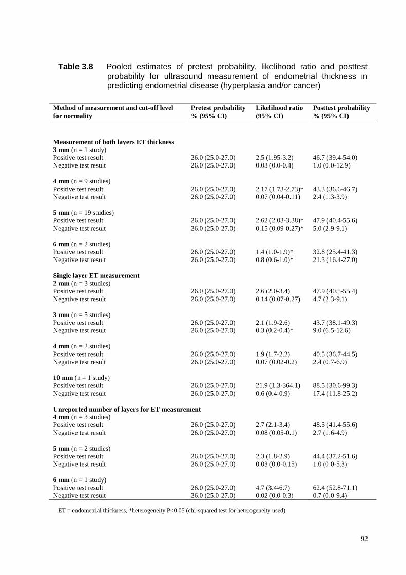
92
Table 3.8 Pooled estimates of pretest probability, likelihood ratio and posttest probability for ultrasound measurement of endometrial thickness in predicting endometrial disease (hyperplasia and/or cancer)
Method of measurement and cut-off level
for normality
Pretest probability
% (95% CI)
Likelihood ratio
(95% CI)
Posttest probability
% (95% CI)
Measurement of both layers ET thickness
3 mm (n = 1 study)
Positive test result 26.0 (25.0-27.0) 2.5 (1.95-3.2) 46.7 (39.4-54.0)
Negative test result 26.0 (25.0-27.0) 0.03 (0.0-0.4) 1.0 (0.0-12.9)
4 mm (n = 9 studies)
Positive test result 26.0 (25.0-27.0) 2.17 (1.73-2.73)* 43.3 (36.6-46.7)
Negative test result 26.0 (25.0-27.0) 0.07 (0.04-0.11) 2.4 (1.3-3.9)
5 mm (n = 19 studies)
Positive test result 26.0 (25.0-27.0) 2.62 (2.03-3.38)* 47.9 (40.4-55.6)
Negative test result 26.0 (25.0-27.0) 0.15 (0.09-0.27)* 5.0 (2.9-9.1)
6 mm (n = 2 studies)
Positive test result 26.0 (25.0-27.0) 1.4 (1.0-1.9)* 32.8 (25.4-41.3)
Negative test result 26.0 (25.0-27.0) 0.8 (0.6-1.0)* 21.3 (16.4-27.0)
Single layer ET measurement
2 mm (n = 3 studies)
Positive test result 26.0 (25.0-27.0) 2.6 (2.0-3.4) 47.9 (40.5-55.4)
Negative test result 26.0 (25.0-27.0) 0.14 (0.07-0.27) 4.7 (2.3-9.1)
3 mm (n = 5 studies)
Positive test result 26.0 (25.0-27.0) 2.1 (1.9-2.6) 43.7 (38.1-49.3)
Negative test result 26.0 (25.0-27.0) 0.3 (0.2-0.4)* 9.0 (6.5-12.6)
4 mm (n = 2 studies)
Positive test result 26.0 (25.0-27.0) 1.9 (1.7-2.2) 40.5 (36.7-44.5)
Negative test result 26.0 (25.0-27.0) 0.07 (0.02-0.2) 2.4 (0.7-6.9)
10 mm (n = 1 study)
Positive test result 26.0 (25.0-27.0) 21.9 (1.3-364.1) 88.5 (30.6-99.3)
Negative test result 26.0 (25.0-27.0) 0.6 (0.4-0.9) 17.4 (11.8-25.2)
Unreported number of layers for ET measurement
4 mm (n = 3 studies)
Positive test result 26.0 (25.0-27.0) 2.7 (2.1-3.4) 48.5 (41.4-55.6)
Negative test result 26.0 (25.0-27.0) 0.08 (0.05-0.1) 2.7 (1.6-4.9)
5 mm (n = 2 studies)
Positive test result 26.0 (25.0-27.0) 2.3 (1.8-2.9) 44.4 (37.2-51.6)
Negative test result 26.0 (25.0-27.0) 0.03 (0.0-0.15) 1.0 (0.0-5.3)
6 mm (n = 1 study)
Positive test result 26.0 (25.0-27.0) 4.7 (3.4-6.7) 62.4 (52.8-71.1)
Negative test result 26.0 (25.0-27.0) 0.02 (0.0-0.3) 0.7 (0.0-9.4)
ET = endometrial thickness, *heterogeneity P<0.05 (chi-squared test for heterogeneity used)
93
3.3 Results of systematic review of hysteroscopy
3.3.1 Question
What is the accuracy of outpatient hysteroscopy in the diagnosis of endometrial cancer and
hyperplasia and what are the rates of failure and complications?
3.3.2 Study selection
A total of 65 primary studies (20 non-English studies), including 26,346 women, assessed the
diagnostic accuracy of hysteroscopy in detecting serious endometrial disease and met the
criteria for inclusion (Figure 3-9 and Appendix 18).
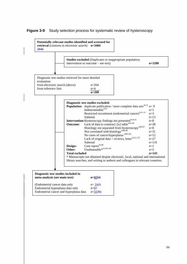
94
Figure 3-9 Study selection process for systematic review of hysteroscopy
Potentially relevant studies identified and screened for
retrieval (citations in electronic search) n=3484
2846
Diagnostic test studies excluded:
Population: duplicate publication / more complete data setsA1-9
n= 9
IndeterminableA10
n=1
Restricted recruitment (endometrial cancer)A11-13
n=3
Subtotal n=13
Intervention:Hysteroscopy findings not presentedA14-21
n=8
Outcome: Lack of data to construct 2x2 tableA22-59
n=38
Histology not separated from hysteroscopyA60-67
n=8
Not correlated with histologyA68-98
n=31
No cases of cancer/hyperplasia A99-101
n=12
Lack of original data = reviews, letterA111-137
n=27
Subtotal n=116
Design: Case reportA138
n=1
Other: Unobtainable*A139-143
n=5
Total excluded n=143
* Manuscripts not obtained despite electronic, local, national and international
library searches, and writing to authors and colleagues in relevant countries.
Diagnostic test studies excluded:
Population: duplicate publication / more complete data setsA1-6
n=6
IndeterminableA7
n=1
Restricted recruitment (endometrial cancer)A8-10
n=3
Subtotal n=10
Intervention: Hysteroscopy findings not presented
A11-18 n=8
Outcome: Lack of data to construct 2x2 tableA19-53
n=35
Histology not separated from hysteroscopyA54-60
n=7
Cannot separate cancer and hyperplasiaA61
n=1
Not correlated with histologyA62-92
n=31
Studies excluded (Duplicates or inappropriate population,
intervention or outcome – see text) n=3280
Diagnostic test studies retrieved for more detailed
evaluation:
from electronic search (above) n=204
from reference lists n=4
n=208
190
Diagnostic test studies included in
meta-analysis (see main text) n=6554
(Endometrial cancer data only n= 2421
Endometrial hyperplasia data only n=97
Endometrial cancer and hyperplasia data n=3226)
95
Agreement regarding eligibility was 96% (weighted kappa 0.8). Of the 65 included studies, 56
studies (24,649 women) assessed the diagnosis of endometrial cancer. Postmenopausal
women represented 29% of the populations studied.
3.3.3 Study quality
Details of the participants, interventions, outcomes and study quality criteria of the studies
selected for meta-analyses are summarized in Appendices 19 and 20. There was a single study
of the highest methodological quality (level 1), one study was classified as level 2, ten studies
(15%) were level 3, 42 studies (65%) were level 4 and 11 studies (17%) were level 5 in
quality.
3.3.4 Data synthesis
3.3.4.1 Accuracy in predicting endometrial cancer
Figure 3-10 presents the sensitivity and specificity of hysteroscopy in the diagnosis of
endometrial cancer. The variations in sensitivity were much greater than the variations in
specificity and there was no significant association between sensitivity and specificity
(Spearman‟s correlation coefficient r=-0.06, P=0.65). Weighted by the number of cases, the
overall sensitivity was 86.4% (95% CI 84.0% to 88.6%) and specificity was 99.2% (95% CI
99.1% to 99.3%) according to 56 studies of hysteroscopy for endometrial cancer. In view of
the lack of an association between sensitivity and specificity, a summary receiver operating
characteristic curve was not generated.83
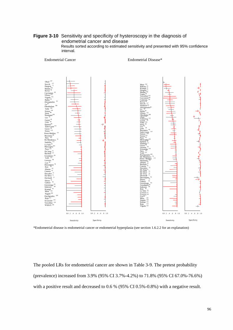
96
Figure 3-10 Sensitivity and specificity of hysteroscopy in the diagnosis of endometrial cancer and disease
Results sorted according to estimated sensitivity and presented with 95% confidence interval.
Endometrial Cancer Endometrial Disease*
*Endometrial disease is endometrial cancer or endometrial hyperplasia (see section 1.6.2.2 for an explanation)
The pooled LRs for endometrial cancer are shown in Table 3-9. The pretest probability
(prevalence) increased from 3.9% (95% CI 3.7%-4.2%) to 71.8% (95% CI 67.0%-76.6%)
with a positive result and decreased to 0.6 % (95% CI 0.5%-0.8%) with a negative result.
1.0.8.6.4.20.0
Ohad107
Sevcik117
Dargent71
Madan100
Kovar90
Cacciatore68
Uhiara122
Vigada125
Haller87
Alexopoulos62
Lo96
Okeahialam108
Tahir 120
Simon118
Iossa88
Neumann106
Liu95
Garuti82
Litta 94
Epstein80
Salet-Lizee114
Gucer 85
Sousa 119
Perez-Medina111
Bocanera66
Neis 105
De Mendonca 74
Sanfeliu 115
La Sala93
Mencaglia103
Paya 110
De Jong 73
Bucholz 67
Grosdanov 84
Valli 123
Loverro98
Luo 99
Descargues78
Kun 92
Altaras63
Caserta70
Davydov72
De Silva 75
Decloedt 77
Elewa 79
Gabrys81
Gorostiaga83
Itzkowic 89
Krampl91
Maia 101
Nagele104
Paschpoulos109
Raju 113
ScwarzlerVercelliniWidrich
116
124
126
1.0.8.6.4.20.0 1.0.8.6.4.20.01.0.8.6.4.20.0
102
65
91
71
100
86
124
68
69
90
117
126
62
96
87
122
108
121
82
120
88
95
94
66
114
118
119
110
103
80
115
85
64
116
105
123
109
74
Perez- Medina 111
106
63
67
70
72
73
75
76
77
78
79
81
83
84
89
92
93
97
98
99
101
104
112
113
MaiaBakourKramplDargentMadanGuptaVercelliniCacciatoreCameronKovarSevcikWidrichAlexopoulosLoHallerUhiaraOkeahialaOhadTodorovaGarutiTahirIossaLiuLittaBocaneraSalet-LizeeSimonSousaPayaMencagliaEpsteinSanfeliuGucerAzzenaScwarzlerNeisValliPaschpoulosDe Mendonca
NeumannAltarasBucholzCasertaDavydovDe JongDe SilvaDe VivoDecloedtDescarguesElewaGabrysGorostiagaGrosdanovItzkowicKunLa SalaLoverroLoverroLuoMaiaNagelePossatiRajuVigada 125
107
SpecificitySensitivitySpecificitySensitivity
97
Heterogeneity of diagnostic performance between studies was present as confirmed by a
statistically significant χ2 test and this remained within the pre-specified clinical subgroups
(setting and menopausal status). An explanation for heterogeneity was not provided by the
study setting, menopausal status or study quality (Table 3-10). Neither did the other potential
explanatory variables defined post hoc significantly influence diagnostic accuracy. The
reported occurrence of complications was associated with reduced accuracy on univariable
analysis, but this was not confirmed on multivariable analysis.
Statistical tests (rank correlation) to explore for publication and related biases, found that
funnel plot asymmetry (Figure 3-11) was not statistically significant (p=0.34)
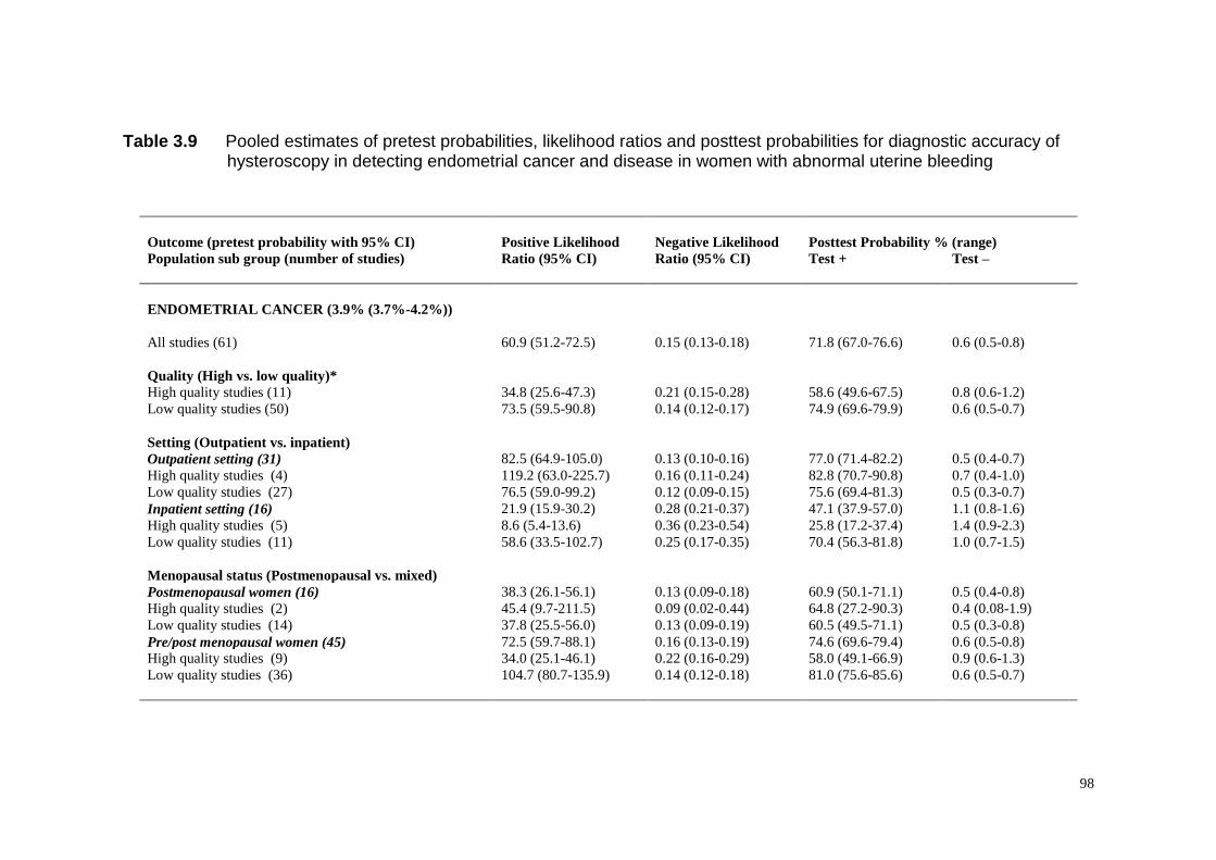
98
Table 3.9 Pooled estimates of pretest probabilities, likelihood ratios and posttest probabilities for diagnostic accuracy of hysteroscopy in detecting endometrial cancer and disease in women with abnormal uterine bleeding
Outcome (pretest probability with 95% CI) Positive Likelihood Negative Likelihood Posttest Probability % (range)
Population sub group (number of studies) Ratio (95% CI) Ratio (95% CI) Test + Test –
ENDOMETRIAL CANCER (3.9% (3.7%-4.2%))
All studies (61) 60.9 (51.2-72.5) 0.15 (0.13-0.18) 71.8 (67.0-76.6) 0.6 (0.5-0.8)
Quality (High vs. low quality)*
High quality studies (11) 34.8 (25.6-47.3) 0.21 (0.15-0.28) 58.6 (49.6-67.5) 0.8 (0.6-1.2)
Low quality studies (50) 73.5 (59.5-90.8) 0.14 (0.12-0.17) 74.9 (69.6-79.9) 0.6 (0.5-0.7)
Setting (Outpatient vs. inpatient)
Outpatient setting (31) 82.5 (64.9-105.0) 0.13 (0.10-0.16) 77.0 (71.4-82.2) 0.5 (0.4-0.7)
High quality studies (4) 119.2 (63.0-225.7) 0.16 (0.11-0.24) 82.8 (70.7-90.8) 0.7 (0.4-1.0)
Low quality studies (27) 76.5 (59.0-99.2) 0.12 (0.09-0.15) 75.6 (69.4-81.3) 0.5 (0.3-0.7)
Inpatient setting (16) 21.9 (15.9-30.2) 0.28 (0.21-0.37) 47.1 (37.9-57.0) 1.1 (0.8-1.6)
High quality studies (5) 8.6 (5.4-13.6) 0.36 (0.23-0.54) 25.8 (17.2-37.4) 1.4 (0.9-2.3)
Low quality studies (11) 58.6 (33.5-102.7) 0.25 (0.17-0.35) 70.4 (56.3-81.8) 1.0 (0.7-1.5)
Menopausal status (Postmenopausal vs. mixed)
Postmenopausal women (16) 38.3 (26.1-56.1) 0.13 (0.09-0.18) 60.9 (50.1-71.1) 0.5 (0.4-0.8)
High quality studies (2) 45.4 (9.7-211.5) 0.09 (0.02-0.44) 64.8 (27.2-90.3) 0.4 (0.08-1.9)
Low quality studies (14) 37.8 (25.5-56.0) 0.13 (0.09-0.19) 60.5 (49.5-71.1) 0.5 (0.3-0.8)
Pre/post menopausal women (45) 72.5 (59.7-88.1) 0.16 (0.13-0.19) 74.6 (69.6-79.4) 0.6 (0.5-0.8)
High quality studies (9) 34.0 (25.1-46.1) 0.22 (0.16-0.29) 58.0 (49.1-66.9) 0.9 (0.6-1.3)
Low quality studies (36) 104.7 (80.7-135.9) 0.14 (0.12-0.18) 81.0 (75.6-85.6) 0.6 (0.5-0.7)
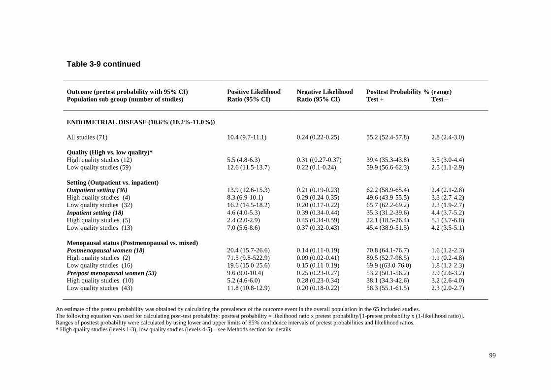
99
Table 3-9 continued
Outcome (pretest probability with 95% CI) Positive Likelihood Negative Likelihood Posttest Probability % (range)
Population sub group (number of studies) Ratio (95% CI) Ratio (95% CI) Test + Test –
ENDOMETRIAL DISEASE (10.6% (10.2%-11.0%))
All studies (71) 10.4 (9.7-11.1) 0.24 (0.22-0.25) 55.2 (52.4-57.8) 2.8 (2.4-3.0)
Quality (High vs. low quality)*
High quality studies (12) 5.5 (4.8-6.3) 0.31 ((0.27-0.37) 39.4 (35.3-43.8) 3.5 (3.0-4.4)
Low quality studies (59) 12.6 (11.5-13.7) 0.22 (0.1-0.24) 59.9 (56.6-62.3) 2.5 (1.1-2.9)
Setting (Outpatient vs. inpatient)
Outpatient setting (36) 13.9 (12.6-15.3) 0.21 (0.19-0.23) 62.2 (58.9-65.4) 2.4 (2.1-2.8)
High quality studies (4) 8.3 (6.9-10.1) 0.29 (0.24-0.35) 49.6 (43.9-55.5) 3.3 (2.7-4.2)
Low quality studies (32) 16.2 (14.5-18.2) 0.20 (0.17-0.22) 65.7 (62.2-69.2) 2.3 (1.9-2.7)
Inpatient setting (18) 4.6 (4.0-5.3) 0.39 (0.34-0.44) 35.3 (31.2-39.6) 4.4 (3.7-5.2)
High quality studies (5) 2.4 (2.0-2.9) 0.45 (0.34-0.59) 22.1 (18.5-26.4) 5.1 (3.7-6.8)
Low quality studies (13) 7.0 (5.6-8.6) 0.37 (0.32-0.43) 45.4 (38.9-51.5) 4.2 (3.5-5.1)
Menopausal status (Postmenopausal vs. mixed)
Postmenopausal women (18) 20.4 (15.7-26.6) 0.14 (0.11-0.19) 70.8 (64.1-76.7) 1.6 (1.2-2.3)
High quality studies (2) 71.5 (9.8-522.9) 0.09 (0.02-0.41) 89.5 (52.7-98.5) 1.1 (0.2-4.8)
Low quality studies (16) 19.6 (15.0-25.6) 0.15 (0.11-0.19) 69.9 ((63.0-76.0) 1.8 (1.2-2.3)
Pre/post menopausal women (53) 9.6 (9.0-10.4) 0.25 (0.23-0.27) 53.2 (50.1-56.2) 2.9 (2.6-3.2)
High quality studies (10) 5.2 (4.6-6.0) 0.28 (0.23-0.34) 38.1 (34.3-42.6) 3.2 (2.6-4.0)
Low quality studies (43) 11.8 (10.8-12.9) 0.20 (0.18-0.22) 58.3 (55.1-61.5) 2.3 (2.0-2.7)
An estimate of the pretest probability was obtained by calculating the prevalence of the outcome event in the overall population in the 65 included studies.
The following equation was used for calculating post-test probability: posttest probability = likelihood ratio x pretest probability/[1-pretest probability x (1-likelihood ratio)].
Ranges of posttest probability were calculated by using lower and upper limits of 95% confidence intervals of pretest probabilities and likelihood ratios.
* High quality studies (levels 1-3), low quality studies (levels 4-5) – see Methods section for details
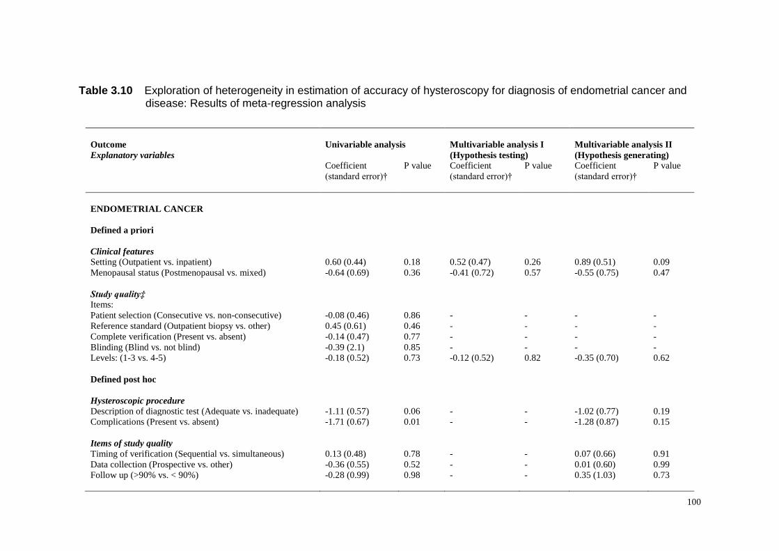
100
Table 3.10 Exploration of heterogeneity in estimation of accuracy of hysteroscopy for diagnosis of endometrial cancer and disease: Results of meta-regression analysis
Outcome
Explanatory variables
Univariable analysis Multivariable analysis I
(Hypothesis testing)
Multivariable analysis II
(Hypothesis generating)
Coefficient
(standard error)†
P value Coefficient
(standard error)†
P value Coefficient
(standard error)†
P value
ENDOMETRIAL CANCER
Defined a priori
Clinical features
Setting (Outpatient vs. inpatient) 0.60 (0.44) 0.18 0.52 (0.47) 0.26 0.89 (0.51) 0.09
Menopausal status (Postmenopausal vs. mixed) -0.64 (0.69) 0.36 -0.41 (0.72) 0.57 -0.55 (0.75) 0.47
Study quality‡
Items:
Patient selection (Consecutive vs. non-consecutive) -0.08 (0.46) 0.86 - - - -
Reference standard (Outpatient biopsy vs. other) 0.45 (0.61) 0.46 - - - -
Complete verification (Present vs. absent) -0.14 (0.47) 0.77 - - - -
Blinding (Blind vs. not blind) -0.39 (2.1) 0.85 - - - -
Levels: (1-3 vs. 4-5) -0.18 (0.52) 0.73 -0.12 (0.52) 0.82 -0.35 (0.70) 0.62
Defined post hoc
Hysteroscopic procedure
Description of diagnostic test (Adequate vs. inadequate) -1.11 (0.57) 0.06 - - -1.02 (0.77) 0.19
Complications (Present vs. absent) -1.71 (0.67) 0.01 - - -1.28 (0.87) 0.15
Items of study quality
Timing of verification (Sequential vs. simultaneous) 0.13 (0.48) 0.78 - - 0.07 (0.66) 0.91
Data collection (Prospective vs. other) -0.36 (0.55) 0.52 - - 0.01 (0.60) 0.99
Follow up (>90% vs. < 90%) -0.28 (0.99) 0.98 - - 0.35 (1.03) 0.73
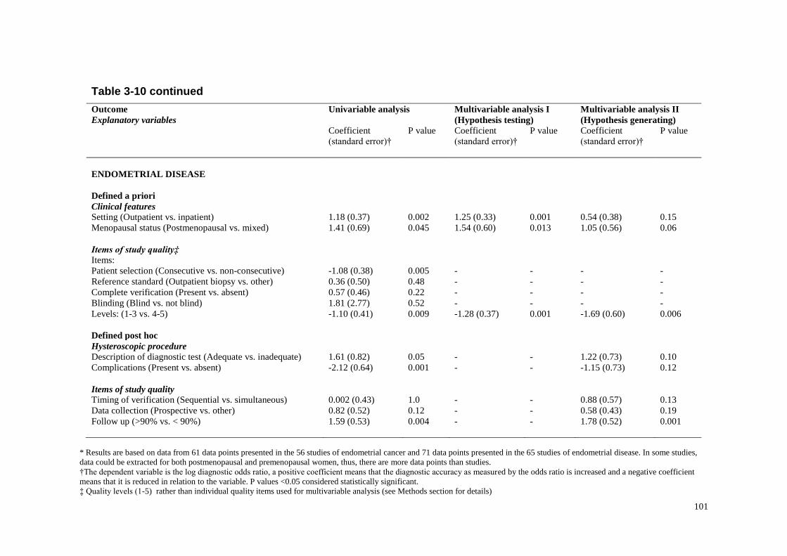
101
Table 3-10 continued
Outcome
Explanatory variables
Univariable analysis Multivariable analysis I
(Hypothesis testing)
Multivariable analysis II
(Hypothesis generating)
Coefficient
(standard error)†
P value Coefficient
(standard error)†
P value Coefficient
(standard error)†
P value
ENDOMETRIAL DISEASE
Defined a priori
Clinical features
Setting (Outpatient vs. inpatient) 1.18 (0.37) 0.002 1.25 (0.33) 0.001 0.54 (0.38) 0.15
Menopausal status (Postmenopausal vs. mixed) 1.41 (0.69) 0.045 1.54 (0.60) 0.013 1.05 (0.56) 0.06
Items of study quality‡
Items:
Patient selection (Consecutive vs. non-consecutive) -1.08 (0.38) 0.005 - - - -
Reference standard (Outpatient biopsy vs. other) 0.36 (0.50) 0.48 - - - -
Complete verification (Present vs. absent) 0.57 (0.46) 0.22 - - - -
Blinding (Blind vs. not blind) 1.81 (2.77) 0.52 - - - -
Levels: (1-3 vs. 4-5) -1.10 (0.41) 0.009 -1.28 (0.37) 0.001 -1.69 (0.60) 0.006
Defined post hoc
Hysteroscopic procedure
Description of diagnostic test (Adequate vs. inadequate) 1.61 (0.82) 0.05 - - 1.22 (0.73) 0.10
Complications (Present vs. absent) -2.12 (0.64) 0.001 - - -1.15 (0.73) 0.12
Items of study quality
Timing of verification (Sequential vs. simultaneous) 0.002 (0.43) 1.0 - - 0.88 (0.57) 0.13
Data collection (Prospective vs. other) 0.82 (0.52) 0.12 - - 0.58 (0.43) 0.19
Follow up (>90% vs. < 90%) 1.59 (0.53) 0.004 - - 1.78 (0.52) 0.001
* Results are based on data from 61 data points presented in the 56 studies of endometrial cancer and 71 data points presented in the 65 studies of endometrial disease. In some studies,
data could be extracted for both postmenopausal and premenopausal women, thus, there are more data points than studies.
†The dependent variable is the log diagnostic odds ratio, a positive coefficient means that the diagnostic accuracy as measured by the odds ratio is increased and a negative coefficient
means that it is reduced in relation to the variable. P values <0.05 considered statistically significant.
‡ Quality levels (1-5) rather than individual quality items used for multivariable analysis (see Methods section for details)
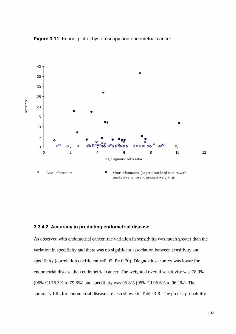
102
Figure 3-11 Funnel plot of hysteroscopy and endometrial cancer
3.3.4.2 Accuracy in predicting endometrial disease
As observed with endometrial cancer, the variation in sensitivity was much greater than the
variation in specificity and there was no significant association between sensitivity and
specificity (correlation coefficient r=0.05, P= 0.70). Diagnostic accuracy was lower for
endometrial disease than endometrial cancer. The weighted overall sensitivity was 78.0%
(95% CI 76.3% to 79.6%) and specificity was 95.8% (95% CI 95.6% to 96.1%). The
summary LRs for endometrial disease are also shown in Table 3-9. The pretest probability
0
5
10
15
20
25
30
35
40
0 2 4 6 8 10 12
Log diagnostic odds ratio
1/v
ari
an
ce
Less information More information (upper quartile of studies with
smallest variance and greatest weighting)
103
increased from 10.6% (95% CI 10.2%-11.0%) to 55.2% (95% CI 52.4%-57.8%) with a
positive result and decreased to 2.8% (95% CI 2.4%-3.0%) with a negative result (Table 3-9).
There was heterogeneity in the overall and subgroup meta-analyses (Table 3-9). Clinical
setting and menopausal status were significant explanatory variables for heterogeneity in
univariable analyses as was the quality item, patient selection (Table 3-10). Poor study
quality, the office setting and postmenopausal women were associated with significantly
higher accuracy of hysteroscopy. The effect of these features on diagnostic accuracy was
confirmed with multivariable analysis (Table 3-10). Of the variables defined post hoc, only
follow up greater than 90% was associated with higher accuracy on both univariable and
multivariable analyses (Table 3-10).
104
3.3.5 Sensitivity analysis
In 12 (18%) studies it was not possible to determine the rate of inadequate specimen due to a
lack of clear reporting and the rate was assumed to be zero for the purpose of analysis. This
gave an inadequate specimen rate on the reference test of 1196/25409 (4.7%, 95% CI 4.5%-
5.0%). The pooled LRs were not altered if inadequate samples were regarded as negative
results. There were 4622 focal lesions (intrauterine polyps of fibroids) detected in 25409
hysteroscopies (prevalence 18%) reported in 55/65 primary studies. In 152 of the 4622 focal
anomalies (prevalence 3%) endometrial cancer (17) or hyperplasia (135) was present.
Estimates of accuracy for endometrial cancer were not affected when focal abnormalities
were excluded as part of a sensitivity analysis (LR for positive and negative test 59.3 (49.2-
71.6) and 0.14 (0.12-0.16).
3.3.6 Secondary outcomes
3.3.6.1 Failure rate
Failure rates were clearly reported in 36/65 (55%) studies. The overall failure rate was
937/26346 (3.6%, 95% CI 3.3%-3.8%) when considering all studies and 937/19323 (4.9%,
95% CI 4.6-5.2%) when studies with unclear reporting were excluded. In those studies
performed exclusively in one setting, the failure rate for an ambulatory procedure was
755/18126 (4.2%, 95% CI 3.9-4.5%) compared to 86/2526 (3.4%, 95% CI 2.7-4.2%) for an
inpatient procedure. However, the underlying reasons for failure varied between settings.
Failed hysteroscopies in the office setting resulted from technical problems (e.g. cervical
stenosis, anatomical factors, structural abnormalities) or patient factors (e.g. pain, intolerance)
more often than in inpatient setting (79% v 9%). By contrast, inadequate visualization (e.g.
105
obscured by bleeding, debris) was more common in the inpatient setting as a reason for failure
(3% v 0.7%). Endometrial cancer was found in 8/927 (0.8%, 95% CI 0.4%-1.7%) failed
procedures reported in the 56 cancer studies and endometrial disease was found in 25/937
(2.7%, 95% CI 1.7%-3.9%) failures reported in all included studies. In those studies where
data for postmenopausal women could be separated, the failure rate of hysteroscopy (67/1948,
3.4%, 95% CI 2.7%-4.4%) was comparable to the overall rate (Appendix 20).
3.3.6.2 Complication rate
Eight cases of potentially serious complications (pelvic infection, uterine perforation (4),
bladder perforation, and precipitation of a hypocalcaemic crisis and an anginal episode) were
reported out of 25,409 successful procedures. However, ascertainment of serious
complications may be suboptimal as only 19/65 (29%) studies, which included 9413
successful procedures, explicitly stated the intention to report or actually reported
complications.
106
3.4 Results of economic analysis
3.4.1 Question
Which of the three available tests (EB, USS and OPH) and their combinations is most cost
effective in outpatient diagnosis of endometrial cancer?
3.4.2 Base case results
Life expectancies adjusted for age, surgery and presence of endometrial cancer are shown in
Appendix 21. All strategies for diagnostic work-up were associated with improved survival
when compared with a strategy of undertaking no initial investigation of women with PMB.
However, there was little difference in expected survival between diagnostic strategies. The
strategies OPH, EB + OPH and USS+EB+OPH were dominated by other strategies (i.e. in
each case there was an alternative strategy that was both cheaper and more effective –
Appendix 22). Incremental cost-effectiveness ratios (ICERs) comparing the cost-effectiveness
of strategies with no initial investigation are shown in Table 3-11.
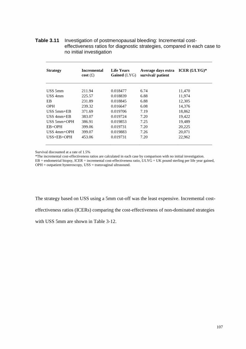
107
Table 3.11 Investigation of postmenopausal bleeding: Incremental cost-effectiveness ratios for diagnostic strategies, compared in each case to no initial investigation
Strategy Incremental
cost (£) Life Years
Gained (LYG) Average days extra
survival/ patient
ICER (£/LYG)*
USS 5mm 211.94 0.018477 6.74 11,470
USS 4mm 225.57 0.018839 6.88 11,974
EB 231.89 0.018845 6.88 12,305
OPH 239.32 0.016647 6.08 14,376
USS 5mm+EB 371.69 0.019706 7.19 18,862
USS 4mm+EB 383.07 0.019724 7.20 19,422
USS 5mm+OPH 386.91 0.019853 7.25 19,489
EB+OPH 399.06 0.019731 7.20 20,225
USS 4mm+OPH 399.07 0.019883 7.26 20,071
USS+EB+OPH 453.06 0.019731 7.20 22,962
Survival discounted at a rate of 1.5%
*The incremental cost-effectiveness ratios are calculated in each case by comparison with no initial investigation.
EB = endometrial biopsy, ICER = incremental cost-effectiveness ratio, £/LYG = UK pound sterling per life year gained,
OPH = outpatient hysteroscopy, USS = transvaginal ultrasound.
The strategy based on USS using a 5mm cut-off was the least expensive. Incremental cost-
effectiveness ratios (ICERs) comparing the cost-effectiveness of non-dominated strategies
with USS 5mm are shown in Table 3-12.
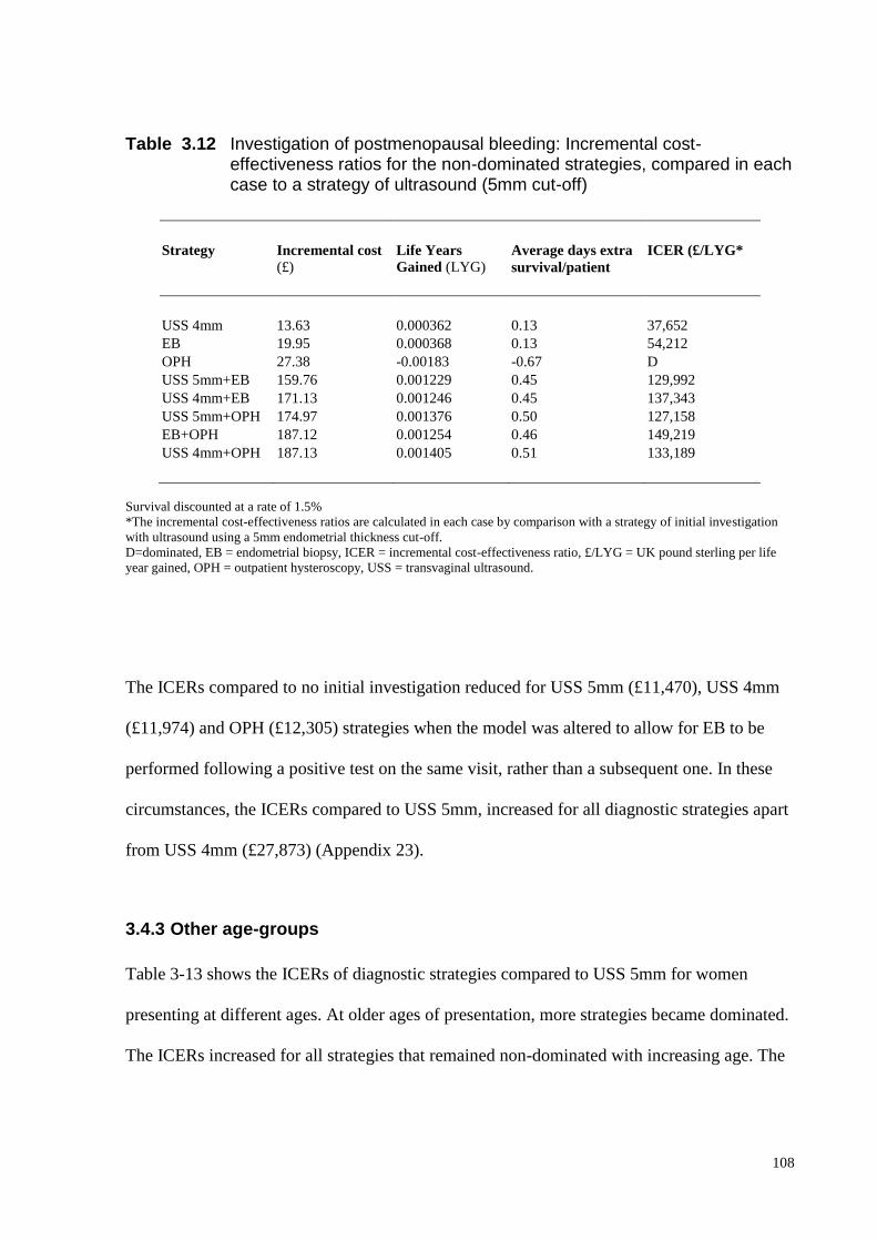
108
Table 3.12 Investigation of postmenopausal bleeding: Incremental cost-effectiveness ratios for the non-dominated strategies, compared in each case to a strategy of ultrasound (5mm cut-off)
Strategy Incremental cost
(£) Life Years
Gained (LYG) Average days extra
survival/patient
ICER (£/LYG*
USS 4mm 13.63 0.000362 0.13 37,652
EB 19.95 0.000368 0.13 54,212
OPH 27.38 -0.00183 -0.67 D
USS 5mm+EB 159.76 0.001229 0.45 129,992
USS 4mm+EB 171.13 0.001246 0.45 137,343
USS 5mm+OPH 174.97 0.001376 0.50 127,158
EB+OPH 187.12 0.001254 0.46 149,219
USS 4mm+OPH 187.13 0.001405 0.51 133,189
Survival discounted at a rate of 1.5%
*The incremental cost-effectiveness ratios are calculated in each case by comparison with a strategy of initial investigation
with ultrasound using a 5mm endometrial thickness cut-off.
D=dominated, EB = endometrial biopsy, ICER = incremental cost-effectiveness ratio, £/LYG = UK pound sterling per life
year gained, OPH = outpatient hysteroscopy, USS = transvaginal ultrasound.
The ICERs compared to no initial investigation reduced for USS 5mm (£11,470), USS 4mm
(£11,974) and OPH (£12,305) strategies when the model was altered to allow for EB to be
performed following a positive test on the same visit, rather than a subsequent one. In these
circumstances, the ICERs compared to USS 5mm, increased for all diagnostic strategies apart
from USS 4mm (£27,873) (Appendix 23).
3.4.3 Other age-groups
Table 3-13 shows the ICERs of diagnostic strategies compared to USS 5mm for women
presenting at different ages. At older ages of presentation, more strategies became dominated.
The ICERs increased for all strategies that remained non-dominated with increasing age. The
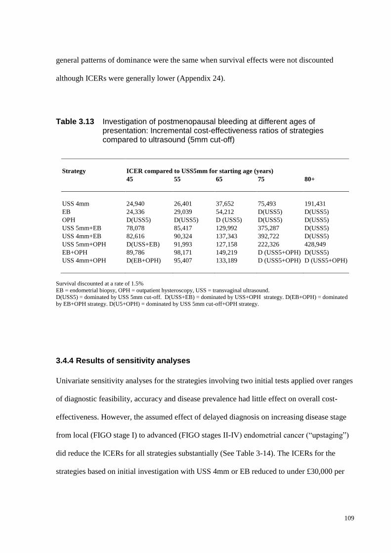
109
general patterns of dominance were the same when survival effects were not discounted
although ICERs were generally lower (Appendix 24).
Table 3.13 Investigation of postmenopausal bleeding at different ages of presentation: Incremental cost-effectiveness ratios of strategies compared to ultrasound (5mm cut-off)
Strategy ICER compared to USS5mm for starting age (years)
45 55 65 75 80+
USS 4mm 24,940 26,401 37,652 75,493 191,431
EB 24,336 29,039 54,212 D(USS5) D(USS5)
OPH D(USS5) D(USS5) D (USS5) D(USS5) D(USS5)
USS 5mm+EB 78,078 85,417 129,992 375,287 D(USS5)
USS 4mm+EB 82,616 90,324 137,343 392,722 D(USS5)
USS 5mm+OPH D(USS+EB) 91,993 127,158 222,326 428,949
EB+OPH 89,786 98,171 149,219 D (USS5+OPH) D(USS5)
USS 4mm+OPH D(EB+OPH) 95,407 133,189 D (USS5+OPH) D (USS5+OPH)
Survival discounted at a rate of 1.5%
EB = endometrial biopsy, OPH = outpatient hysteroscopy, USS = transvaginal ultrasound.
D(USS5) = dominated by USS 5mm cut-off. D(USS+EB) = dominated by USS+OPH strategy. D(EB+OPH) = dominated
by EB+OPH strategy. D(U5+OPH) = dominated by USS 5mm cut-off+OPH strategy.
3.4.4 Results of sensitivity analyses
Univariate sensitivity analyses for the strategies involving two initial tests applied over ranges
of diagnostic feasibility, accuracy and disease prevalence had little effect on overall cost-
effectiveness. However, the assumed effect of delayed diagnosis on increasing disease stage
from local (FIGO stage I) to advanced (FIGO stages II-IV) endometrial cancer (“upstaging”)
did reduce the ICERs for all strategies substantially (See Table 3-14). The ICERs for the
strategies based on initial investigation with USS 4mm or EB reduced to under £30,000 per

110
life year gained when the probability of upstaging endometrial cancer following delay was 6%
and 8% respectively.
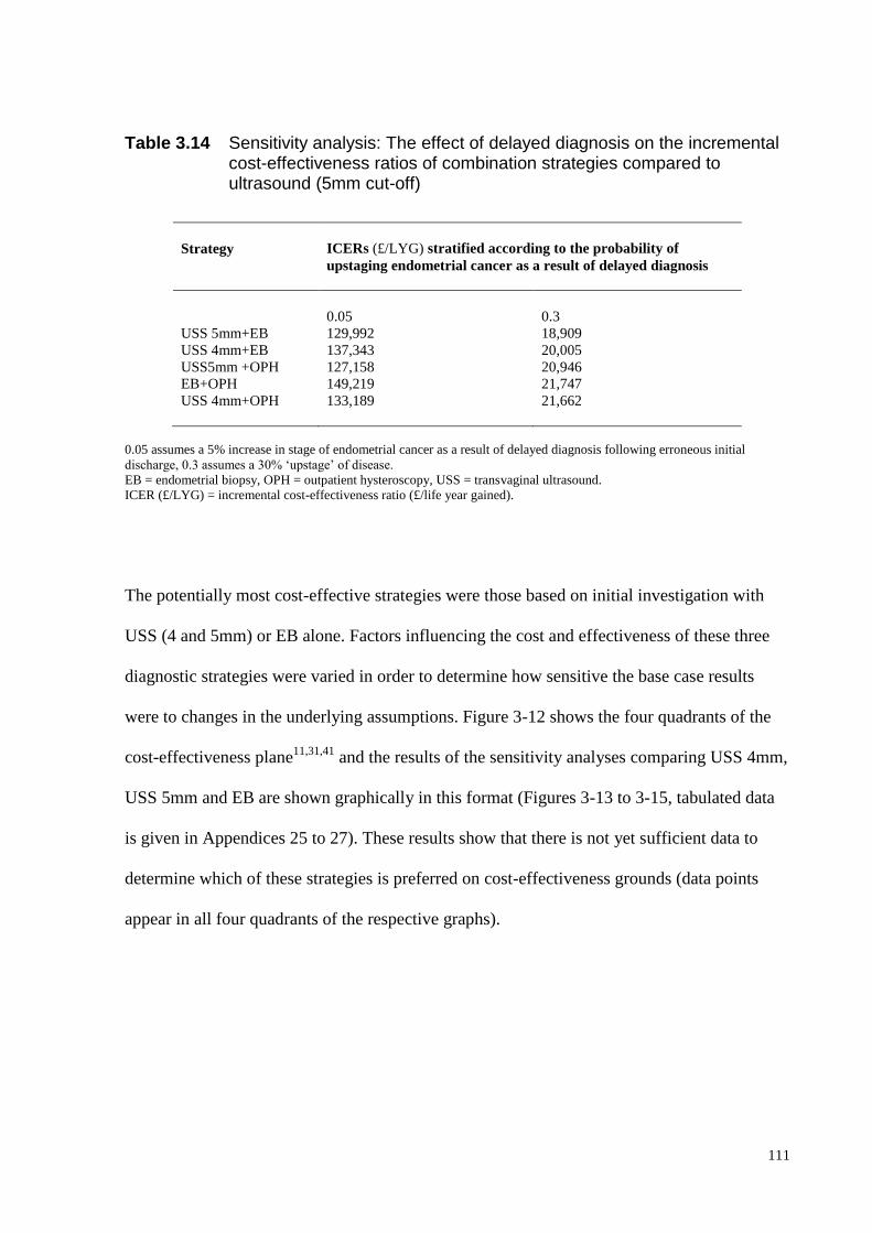
111
Table 3.14 Sensitivity analysis: The effect of delayed diagnosis on the incremental cost-effectiveness ratios of combination strategies compared to ultrasound (5mm cut-off)
Strategy ICERs (£/LYG) stratified according to the probability of
upstaging endometrial cancer as a result of delayed diagnosis
0.05 0.3
USS 5mm+EB 129,992 18,909
USS 4mm+EB 137,343 20,005
USS5mm +OPH 127,158 20,946
EB+OPH 149,219 21,747
USS 4mm+OPH 133,189 21,662
0.05 assumes a 5% increase in stage of endometrial cancer as a result of delayed diagnosis following erroneous initial
discharge, 0.3 assumes a 30% „upstage‟ of disease.
EB = endometrial biopsy, OPH = outpatient hysteroscopy, USS = transvaginal ultrasound.
ICER (£/LYG) = incremental cost-effectiveness ratio (£/life year gained).
The potentially most cost-effective strategies were those based on initial investigation with
USS (4 and 5mm) or EB alone. Factors influencing the cost and effectiveness of these three
diagnostic strategies were varied in order to determine how sensitive the base case results
were to changes in the underlying assumptions. Figure 3-12 shows the four quadrants of the
cost-effectiveness plane11,31,41
and the results of the sensitivity analyses comparing USS 4mm,
USS 5mm and EB are shown graphically in this format (Figures 3-13 to 3-15, tabulated data
is given in Appendices 25 to 27). These results show that there is not yet sufficient data to
determine which of these strategies is preferred on cost-effectiveness grounds (data points
appear in all four quadrants of the respective graphs).
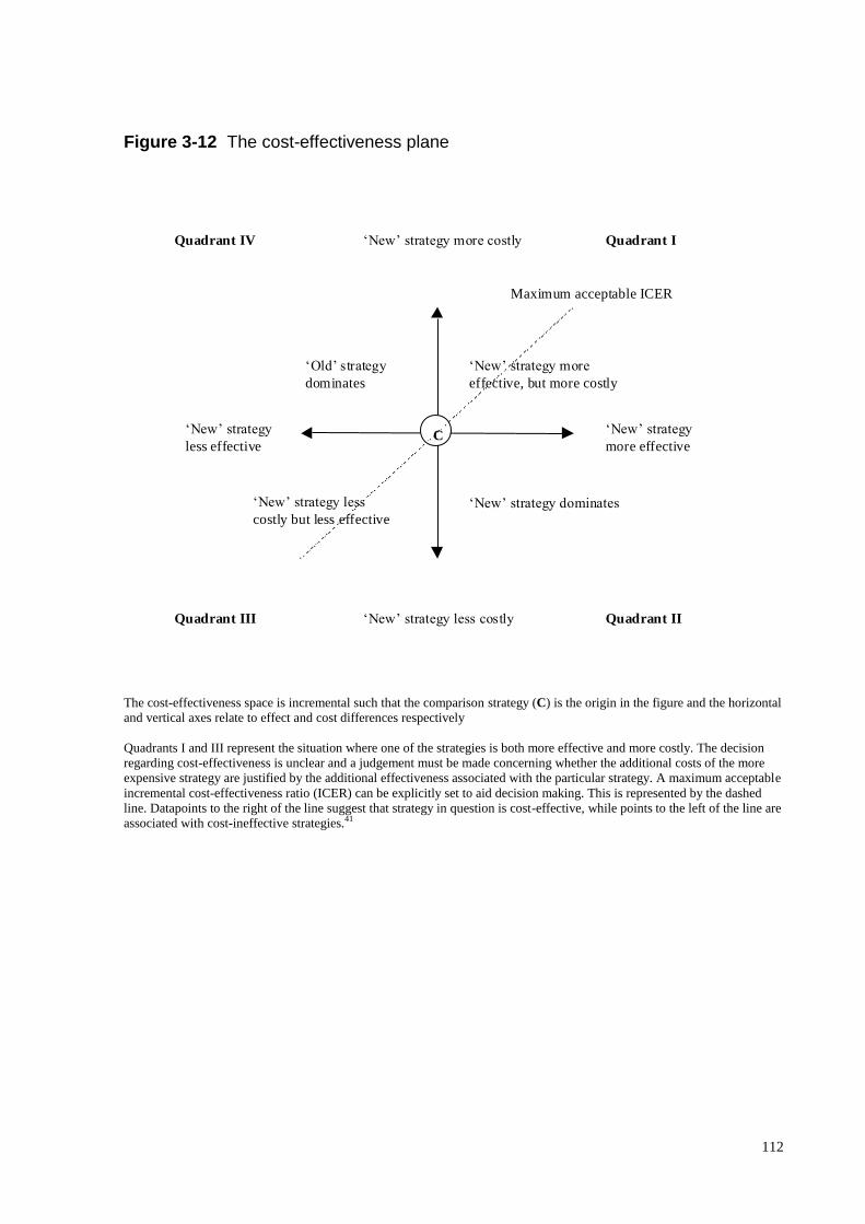
112
Figure 3-12 The cost-effectiveness plane
The cost-effectiveness space is incremental such that the comparison strategy (C) is the origin in the figure and the horizontal
and vertical axes relate to effect and cost differences respectively
Quadrants I and III represent the situation where one of the strategies is both more effective and more costly. The decision
regarding cost-effectiveness is unclear and a judgement must be made concerning whether the additional costs of the more
expensive strategy are justified by the additional effectiveness associated with the particular strategy. A maximum acceptable
incremental cost-effectiveness ratio (ICER) can be explicitly set to aid decision making. This is represented by the dashed
line. Datapoints to the right of the line suggest that strategy in question is cost-effective, while points to the left of the line are
associated with cost-ineffective strategies.41
„Old‟ strategy
dominates
„New‟ strategy more
effective, but more costly
„New‟ strategy less
costly but less effective
„New‟ strategy dominates
„New‟ strategy
more effective
„New‟ strategy
less effective
„New‟ strategy more costly
„New‟ strategy less costly
C
Quadrant III
Quadrant IV
Quadrant II
Quadrant I
Maximum acceptable ICER
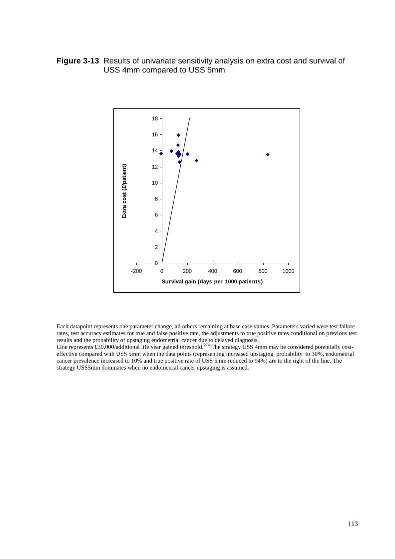
113
Figure 3-13 Results of univariate sensitivity analysis on extra cost and survival of USS 4mm compared to USS 5mm
0
2
4
6
8
10
12
14
16
18
-200 0 200 400 600 800 1000
Survival gain (days per 1000 patients)
Extr
a c
ost
(£/p
ati
en
t)
Each datapoint represents one parameter change, all others remaining at base case values. Parameters varied were test failure
rates, test accuracy estimates for true and false positive rate, the adjustments to true positive rates conditional on previous test
results and the probability of upstaging endometrial cancer due to delayed diagnosis.
Line represents £30,000/additional life year gained threshold.274 The strategy USS 4mm may be considered potentially cost-
effective compared with USS 5mm when the data points (representing increased upstaging probability to 30%, endometrial
cancer prevalence increased to 10% and true positive rate of USS 5mm reduced to 94%) are to the right of the line. The
strategy USS5mm dominates when no endometrial cancer upstaging is assumed.
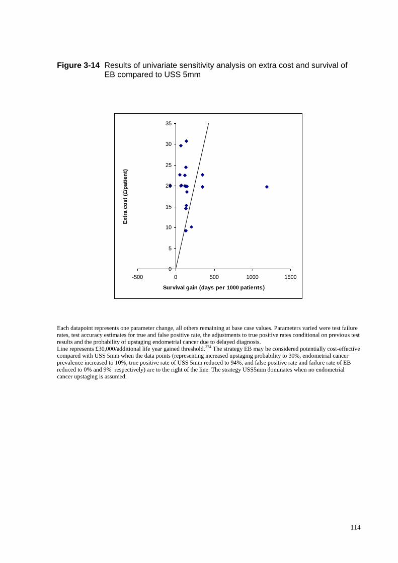
114
Figure 3-14 Results of univariate sensitivity analysis on extra cost and survival of EB compared to USS 5mm
0
5
10
15
20
25
30
35
-500 0 500 1000 1500
Survival gain (days per 1000 patients)
Extr
a c
ost
(£/p
ati
en
t)
Each datapoint represents one parameter change, all others remaining at base case values. Parameters varied were test failure
rates, test accuracy estimates for true and false positive rate, the adjustments to true positive rates conditional on previous test
results and the probability of upstaging endometrial cancer due to delayed diagnosis.
Line represents £30,000/additional life year gained threshold.274 The strategy EB may be considered potentially cost-effective
compared with USS 5mm when the data points (representing increased upstaging probability to 30%, endometrial cancer
prevalence increased to 10%, true positive rate of USS 5mm reduced to 94%, and false positive rate and failure rate of EB
reduced to 0% and 9% respectively) are to the right of the line. The strategy USS5mm dominates when no endometrial
cancer upstaging is assumed.
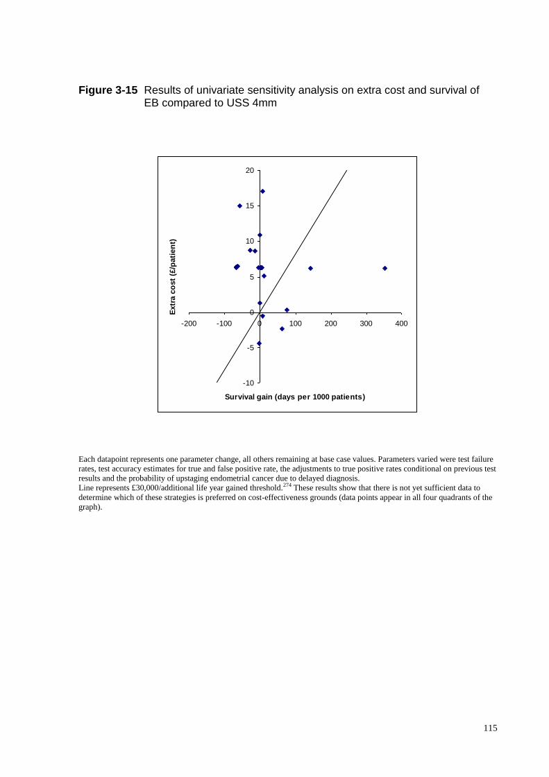
115
Figure 3-15 Results of univariate sensitivity analysis on extra cost and survival of EB compared to USS 4mm
-10
-5
0
5
10
15
20
-200 -100 0 100 200 300 400
Survival gain (days per 1000 patients)
Extr
a c
ost
(£/p
ati
en
t)
Each datapoint represents one parameter change, all others remaining at base case values. Parameters varied were test failure
rates, test accuracy estimates for true and false positive rate, the adjustments to true positive rates conditional on previous test
results and the probability of upstaging endometrial cancer due to delayed diagnosis.
Line represents £30,000/additional life year gained threshold.274 These results show that there is not yet sufficient data to
determine which of these strategies is preferred on cost-effectiveness grounds (data points appear in all four quadrants of the
graph).
116
3.5 Summary of Results
3.5.1 Summary of results of systematic reviews
The literature was of relatively poor methodological quality.
There was statistical heterogeneity in pooling of likelihood ratios, for USS and OPH, but
an explanation for this could not be found in spectrum composition and study quality.
A positive test result on EB diagnosed endometrial cancer with a pooled LR of 66.48
(95% CI 30.04-147.13) while a negative test result had a pooled LR of 0.14 (95% CI 0.08-
0.27).
The commonest USS cut-offs to define abnormal endometrial thickness were 4mm and
5mm, measuring both endometrial layers. Using a 4mm cut-off, a positive test result on
USS diagnosed endometrial cancer with a pooled LR of 1.96 (95% CI 1.6-2.4) while a
negative test result had a pooled LR of 0.08 (95% CI 0.03-0.17). The LRs for positive and
negative ultrasound results for diagnosing endometrial cancer using a 5mm cut-off were
2.17 (95% CI 1.75-2.68) and 0.15 (95% CI 0.08-0.29) respectively.
A positive test result on OPH diagnosed endometrial cancer with a pooled LR of 60.9
(95% CI 51.2-72.5) while a negative test result had a pooled LR of 0.15 (95% CI 0.13-
0.18).
117
3.5.2 Summary of results of economic analysis
Life expectancies were comparable for all diagnostic strategies, but costs varied.
For all ages economic modeling indicated that the strategy based on initial diagnosis with
USS was the least expensive for the investigation of women with PMB.
Strategies based on initial investigation with OPH or all tests combined were dominated
by other strategies, in that in each case there was an alternative strategy that was cheaper
and more effective.
When compared to initial investigation with USS 5mm for a woman aged 65 (base case -
decade of peak incidence of endometrial cancer), the ICERs for the non-dominated
strategies ranged between £37,652 for the initial strategy USS 4mm and £149,219 for the
strategy EB + OPH per additional LYG.
The ICERs increased when considering older ages at presentation and reduced for lower
ages. However, the ICERs were still well above generally recognised thresholds for all
strategies with the exception of USS 4mm and EB under the age of 65 years.
Initial investigation with EB is potentially a cost-effective strategy (ICER reduced below
£30,000 per LYG)) compared to USS, if EB performs at the more favourable estimates of
accuracy and USS at the least favourable estimates of accuracy. Similarly, the ICER
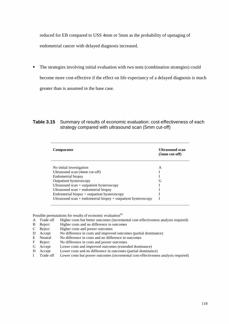
118
reduced for EB compared to USS 4mm or 5mm as the probability of upstaging of
endometrial cancer with delayed diagnosis increased.
The strategies involving initial evaluation with two tests (combination strategies) could
become more cost-effective if the effect on life expectancy of a delayed diagnosis is much
greater than is assumed in the base case.
Table 3.15 Summary of results of economic evaluation: cost-effectiveness of each strategy compared with ultrasound scan (5mm cut-off)
Comparator Ultrasound scan
(5mm cut-off)
No initial investigation A
Ultrasound scan (4mm cut-off) I
Endometrial biopsy I
Outpatient hysteroscopy G
Ultrasound scan + outpatient hysteroscopy I
Ultrasound scan + endometrial biopsy I
Endometrial biopsy + outpatient hysteroscopy I
Ultrasound scan + endometrial biopsy + outpatient hysteroscopy I
Possible permutations for results of economic evaluation82
A Trade off Higher costs but better outcomes (incremental cost-effectiveness analysis required)
B Reject Higher costs and no difference in outcomes
C Reject Higher costs and poorer outcomes
D Accept No difference in costs and improved outcomes (partial dominance)
E Neutral No difference in costs and no difference in outcomes
F Reject No difference in costs and poorer outcomes
G Accept Lower costs and improved outcomes (extended dominance)
H Accept Lower costs and no difference in outcomes (partial dominance)
I Trade off Lower costs but poorer outcomes (incremental cost-effectiveness analysis required)
119
CHAPTER IV
4 DISCUSSION AND CONCLUSIONS
DISCUSSION AND CONCLUSIONS
4.1 Diagnostic reviews
4.1.1 Test accuracy in the diagnosis of endometrial cancer
The reviews of diagnostic hysteroscopy and endometrial biopsy show them to be safe
procedures with a low incidence of serious complications.62,63
Although the review of
ultrasound did not record this data, primary studies have not reported these procedures to be
associated with significant side effects.12
When the uterine cavity is adequately visualised,
hysteroscopy is highly accurate, and thereby clinically useful in the diagnosis of endometrial
cancer. Moreover, performance of the test does not appear to be significantly altered by the
clinical setting or menopausal status. Endometrial biopsy is also highly accurate when
adequate specimens are obtained. For both these diagnostic tests, a positive test result is
highly accurate but a negative test result is of more limited accuracy and thereby only
moderately useful.21,174
As the diagnosis of endometrial cancer is very important, the high
likelihood ratio for a positive test should raise most pre-test probabilities over any threshold
for advanced management.264
In contrast, the likelihood ratio for a negative test may not be
low enough to negate the need for further diagnostic testing (i.e. malignant pathology can be
missed by outpatient biopsy and hysteroscopy), thereby reducing the utility of outpatient
biopsy or hysteroscopy in isolation for excluding cancer.
120
In contrast, these results suggest that ultrasonic measurement of endometrial thickness has
limited diagnostic prediction for endometrial cancer but is a good test for exclusion of
malignancy. A 4 mm or 5 mm cut-off level measuring both layers, can be used to rule out
endometrial cancer with good certainty, as a negative test result reduced the post-test
probability substantially (less than 0.5% using 4mm and less than 1% using 5mm, assuming a
5% pre-test probability). The marginally greater reduction in post-test probability, and the
statistical homogeneity of the pooled LR for a negative test result, may favour use of the
4mm double layer cut-off level. However, all 9 included studies at this cut-off were of poor
methodological quality. The tangible reduction in post-test probability of endometrial cancer
observed at a 5 mm cut-off level remained (4.2% assuming a 5% prevalence) when pooling
only the best quality studies, although no explanation for heterogeneity was found. As the
exclusion of endometrial cancer is very important, one should be wary of relying on the
pooled estimates of only 4 studies, despite them being of good quality. This illustrates the
poor methodological quality of the majority of primary studies on this topic. These findings
concur with a recent Consensus Conference statement, which has also concluded that, an
endometrial thickness greater than 5 mm should be considered as abnormal,141
similar to a
previous systematic review303
(see below).
4.1.2 Test accuracy in the diagnosis of endometrial disease
Abnormal postmenopausal endometrial thickening is a feature of endometrial hyperplasia as
well as endometrial cancer. It is not surprising therefore, that the diagnostic performance of
ultrasound is similar for both conditions and the inferences remain the same i.e. ultrasonic
measurement of endometrial thickness has limited diagnostic prediction for endometrial
hyperplasia or cancer but it is a good test for exclusion of endometrial pathology. However,
121
the diagnostic accuracy of EB and OPH is reduced in endometrial disease (cancer and/or
hyperplasia) compared with estimates of accuracy for endometrial cancer. The modest
estimates of accuracy in diagnosing endometrial disease reduce the usefulness of these two
outpatient modalities in informing clinical decision-making.21
Further testing will be
indicated, especially if symptoms persist or intrauterine structural abnormalities are suspected,
as endometrial disease cannot be ruled in or excluded with a high level of certainty.
Items of poor study quality (retrospective recruitment, unclear reporting) appear to explain the
observed heterogeneity of diagnostic test performance of EB in hyperplasia. Separating the
data for the presence of atypia changed the estimates of diagnostic accuracy, although not
significantly. Diagnostic accuracy was reduced for endometrial hyperplasia without atypia
and increased when atypical cells were present. The presence of complex hyperplasia with
atypia is important in clinical practice, because approximately 33% will progress to
malignancy.199
Simple hyperplasia on the other hand, rarely progresses and the hyperplastic
process can be reversed with local or systemic progestogens.125
Moreover, this review
demonstrates that outpatient endometrial biopsy is highly accurate in diagnosing either
premalignant or malignant endometrial pathology. A positive test result was more accurate
than a negative test result. It is encouraging that the more clinically significant the
endometrial pathology, the better the diagnostic accuracy of outpatient endometrial biopsy
and hence the more clinically useful the test. In contrast to the findings for endometrial
cancer, hysteroscopy is more accurate for the diagnosis of endometrial disease in
postmenopausal women and when undertaken in the office setting. In this review (OPH),
statistically significant differences between these clinical subgroups are quantitative rather
than qualitative. Invariably such differences only explain part of the heterogeneity. Therefore,
122
it may be argued that the overall average estimates may provide the best summary of the
available evidence.347
However, cautious interpretation would demand that one considers the
test‟s performance to vary according to setting and menopausal status. Therefore, inferences
are based on these clinical subgroups and methodological quality in the case of endometrial
disease.
4.1.3 Test feasibility
The results of these systematic reviews show outpatient endometrial biopsy and hysteroscopy
to be successful procedures.62,63
Ultrasonography is the least invasive investigation and has
previously been shown to be associated with a negligible failure rate.303
Failure rates and
inadequate sampling rates were higher for EB in postmenopausal women compared with
premenopausal women. Inadequate endometrial samples, despite successful outpatient
procedures, may result from poor patient compliance or biopsy technique, inherent problems
with non-representative sampling, varied pathological interpretation or be consistent with the
underlying atrophic endometrial state. The review of EB found that single cases of cancer and
hyperplasia were found in inadequate EB specimens, although sensitivity analysis showed
that the effect of these missed cases on overall accuracy estimates was minimal. However,
further means of endometrial evaluation should be considered, particularly when endometrial
imaging or menopausal status is inconsistent with the finding of inadequate tissue.
Hysteroscopy is a successful procedure in both pre and postmenopausal women although the
lack of an effect of menopausal status may be the result of reporting bias, as recording of
failures was unclear in some studies. The office setting appears to have a marginally higher
failure rate compared to the inpatient setting. This is attributable to anatomical and patient
factors rather than inadequate visualization, which is more common in the inpatient setting.
123
The failure rate of office hysteroscopy may represent an underestimate because of more
favourable patient selection. However, selection bias is unlikely to have affected diagnostic
performance in endometrial disease because the ease of visualisation, and hence diagnosis, is
not readily predictable prior to hysteroscopy. Furthermore, the trend towards improved
diagnostic performance was confirmed on multivariable analysis, which adjusted for
menopausal status. Technical failure in performing the EB or OPH should lead to other means
of endometrial assessment.
4.2 Validity of reviews
The strength of this overview is based on its compliance with criteria for performing rigorous
systematic reviews.66,170,235,260
The study focused on explicit research questions and a clear
prospective protocol was formulated. The search strategies were broad and data that were
subject to duplicate publication were excluded from the reviews. Articles were included that
were published in non-English languages. Furthermore, the assessment of methodological
quality and data extraction was performed in a valid107,207
and reproducible fashion. The
evidence was quantitatively summarised and summary LRs were used based on the
recommendations of the various Evidence-based Medicine Working Group's.84,86,150,174
Using
LRs and Bayes‟ theorem allowed the generation of clinically meaningful post-test
probabilities thereby facilitating clinical decision making.174
Since the completion of the
reviews included in this thesis, recommendations have been published for the optimal
reporting of primary studies of diagnostic accuracy.34,35
The vast majority of information
advocated in this „STARD‟ statement has been extracted and presented.
124
4.2.1 Heterogeneity
Sensitivity analyses were performed to investigate for possible sources of heterogeneity,
which were planned a priori. Heterogeneity relates to the presence of differences in results
between individual studies. Homogeneity of results from study to study is one of the criteria
for meta-analysis, but presence of inconsistency itself does not always invalidate a meta-
analysis. In this situation, it is important to consider possible reasons for heterogeneity and so
try and explain it. Exploration for sources of heterogeneity was performed as thoroughly as
possible in accordance with published guidelines,69,82,91
taking into account differences in
methodological quality and study characteristics, using both univariable and multivariable
analytic techniques. However, this approach did not explain the observed variation in the
reviews of ultrasound and hysteroscopy. Such analyses are often restricted due to the number
of available studies.322,341
Although the reviews included numerous studies, the exploration of
underlying sources of heterogeneity may be limited without access to individual patient
data.110
Cautious interpretation of the pooled findings for hysteroscopy and ultrasound is
recommended in this situation. However, in view of the lack of satisfactory explanations for
heterogeneity between studies it may be reasonable to base inferences on the overall pooled
results.347
The methodological quality of the primary studies included in the reviews were generally
poor. Frequent methodological shortcomings included non-consecutive population enrolment
and unclear reporting of patient‟s menopausal status. Another potential source of bias in the
review of ultrasound was the manner in which the cut-off level for abnormal endometrial
thickness was determined. In a majority of studies using the 4 and 5 mm cut-off level, this
125
was determined post hoc i.e. retrospectively following the conduct of the test and outcome
examinations. This would explain the large number of studies in which there was no
incidence of endometrial cancer in the presence of a negative test result. Ideally, the cut-off
level at which a test will perform most optimally should be determined prior to conducting a
study to assess its diagnostic performance.148
Such potential biases may contribute to
heterogeneity, but in this review they did not account for the inconsistency of the results
across studies.
4.2.2 Sources of bias
4.2.2.1 Reference standard
In the reviews of USS and OPH, choice of histological reference standard and lack of blinding
in its assessment could potentially introduce bias. Hysterectomy specimens are regarded as
the „gold‟ standard for verification of endometrial disease, but the exclusive use of this
reference standard in a diagnostic test study is not feasible. Therefore it is not surprising that
many studies included in these reviews obtained endometrial tissue using other methods. It is
worth noting that any test applied in clinical practice to finally direct patient management can
serve as a reference standard. However, evaluating diagnostic tests compared to reference
tests instead of a „gold‟ standard (in this case hysterectomy), will generally underestimate
diagnostic accuracy as a result of misclassification on true disease status by the reference test
chosen.236
Bias due to misdiagnosis by these methods is however, unlikely to be a significant
problem in this thesis. This is because outpatient endometrial sampling methods are
considered to be highly accurate for endometrial cancer.61,62
Moreover, both subgroup
analyses did not show the type of reference standard or blinding to be significant predictors
126
for diagnostic performance. Blinding in this overview may be less important than in other
diagnostic test studies. This is because the histological diagnosis of endometrial cancer, the
primary outcome measure, is an objective one215
and consequently not as susceptible to
expectation bias.
4.2.2.2 Publication bias
The impact of publication bias is another important consideration in all systematic reviews, as
diagnostic accuracy may be overestimated as a result. Here studies with negative or non-
significant results may have been less likely to be published. However, this was only
suggested by funnel plot asymmetry82,306
in the review of EB in endometrial hyperplasia.
Removal of the smaller studies that reported the highest estimates of diagnostic accuracy as
part of a sensitivity analysis did not materially alter the results although it did reduce the
estimates of accuracy slightly, suggesting that the pooled results are reliable.
4.3 Comparison with other reviews and guidelines
4.3.1 Reviews
Two systematic reviews of ultrasound and one review of EB have been recently
published.303,319
Methodological deficiencies arising from the review of EB95
compromise the
internal and external validity of their review findings. These deficiencies include the use of a
limited search and the inappropriate inclusion of data derived from studies restricted to
women known to have endometrial cancer, asymptomatic women, cytological devices and
procedures carried out under general anaesthetic in overall data synthesis. Estimates of
127
diagnostic performance are thus likely to be affected to an unknown degree. However, despite
these limitations, the pooled detection rates and false positive rates for endometrial cancer
were comparable with those derived from the EB review included in this thesis (95% and
0.5% vs. 94% and 1% respectively). The reviews of ultrasound303,319
also had methodological
problems such as restricting the searching to just one database, which is associated with
literature retrieval bias306
and lack of study quality assessment.207
One of the USS reviews303
suggested that an endometrial thickness of 5 mm can reliably exclude endometrial
pathology in postmenopausal women (detection rate 96% for a 39% false positive rate
compared with 97% and 45% respectively for the review in this thesis). They recommended
that a negative test result avoided the need for endometrial sampling for histological
examination. However, the potential biases in the review process raised concerns that this
conclusion was over optimistic and therefore required testing as part of a decision analysis
(see below).
In contrast, the other recently published USS review319
recommended that histological
sampling (D&C) was still required following a negative USS (detection rate 96% for a 50%
false positive rate). The authors used individual patient data from a few centres to demonstrate
that the median USS endometrial thickness in postmenopausal women with and without
endometrial cancer varied between them. They argued that a universal, optimum endometrial
thickness cut-off was not appropriate, but such cut-offs should be individualized according to
local data. The findings of this review are potentially biased because of a narrow and outdated
search restricted to the English language, use of a small data sample and lack of any attempt
to explore the reasons for variation in endometrial thickness measurements (the
reproducibility of this measurement has been demonstrated by others116
) and accuracy
128
between centres. Indeed, 9 of the 11 included centres reported median endometrial thickness
of 5 mm for unaffected women and all reported median endometrial thickness greater than
this for endometrial cancer, in keeping with the findings of both Smith-Bindman et al303
and
the USS review included in this thesis. Applying the accuracy estimates from all three USS
reviews, assuming a 5% pre-test probability of cancer and USS endometrial thickness cut-offs
of 4 or 5mm, the posterior probability of cancer following a negative USS is between 0.4 and
0.8%. Thus, the inference that USS is a good test for exclusion of endometrial malignancy in
PMB remains regardless of which pooled estimate of accuracy is applied. I was unable to
identify any systematic reviews addressing the diagnostic accuracy of hysteroscopy.
4.3.2 Guidelines
The Scottish Intercollegiate Guidelines Network (SIGN) published a clinical guideline for the
investigation of post-menopausal bleeding in September 2002.293
No other such guideline was
identified following searches of electronic bibliographic databases and relevant internet health
sites. This guideline favoured the use of transvaginal ultrasound because of the “…..greater
quantity and higher quality of evidence supporting its use compared with other methods.”
Although the guideline was developed using a standard methodology,292
the acquisition of
evidence was incomplete and important recommendations have been made without due regard
to the supporting evidence, thereby undermining the strength of contained recommendations.
For example, the findings from systematic reviews of pelvic ultrasound160,303
were included in
the SIGN guideline, but those of endometrial biopsy were not.61,62,95
Furthermore, the review
of hysteroscopy presented in this thesis63
was not published until the month following
publication of the SIGN guideline. These omitted reviews show there to be an even greater
quantity of available primary research for other outpatient modalities compared with
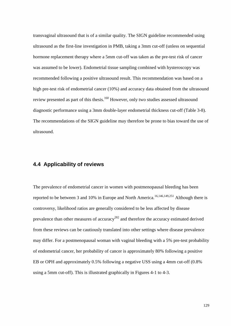
129
transvaginal ultrasound that is of a similar quality. The SIGN guideline recommended using
ultrasound as the first-line investigation in PMB, taking a 3mm cut-off (unless on sequential
hormone replacement therapy where a 5mm cut-off was taken as the pre-test risk of cancer
was assumed to be lower). Endometrial tissue sampling combined with hysteroscopy was
recommended following a positive ultrasound result. This recommendation was based on a
high pre-test risk of endometrial cancer (10%) and accuracy data obtained from the ultrasound
review presented as part of this thesis.160
However, only two studies assessed ultrasound
diagnostic performance using a 3mm double-layer endometrial thickness cut-off (Table 3-8).
The recommendations of the SIGN guideline may therefore be prone to bias toward the use of
ultrasound.
4.4 Applicability of reviews
The prevalence of endometrial cancer in women with postmenopausal bleeding has been
reported to be between 3 and 10% in Europe and North America.16,146,149,251
Although there is
controversy, likelihood ratios are generally considered to be less affected by disease
prevalence than other measures of accuracy282
and therefore the accuracy estimated derived
from these reviews can be cautiously translated into other settings where disease prevalence
may differ. For a postmenopausal woman with vaginal bleeding with a 5% pre-test probability
of endometrial cancer, her probability of cancer is approximately 80% following a positive
EB or OPH and approximately 0.5% following a negative USS using a 4mm cut-off (0.8%
using a 5mm cut-off). This is illustrated graphically in Figures 4-1 to 4-3.

130
The pre-test probability can be individualised in the presence of factors obtained from earlier
in the clinical process. These will include adverse historical features (e.g. unopposed
endogenous or exogenous oestrogen exposure, severity and duration of bleeding, family
history) and adverse examination findings (e.g. obesity, immobile uterus).272
However, the
absolute effect of such factors is unknown and thus difficult to quantify without further
research (see section 4-10).
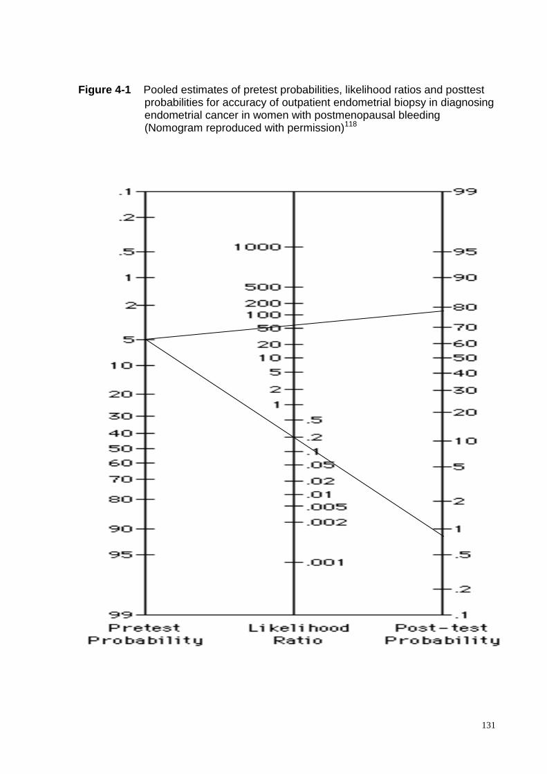
131
Figure 4-1 Pooled estimates of pretest probabilities, likelihood ratios and posttest probabilities for accuracy of outpatient endometrial biopsy in diagnosing endometrial cancer in women with postmenopausal bleeding (Nomogram reproduced with permission)118
132
Figure 4-2 Pooled estimates of pretest probabilities, likelihood ratios and posttest probabilities for accuracy of endometrial thickness measurement by pelvic ultrasound, using both a 4mm and 5mm cut-offs, in diagnosing endometrial cancer in women with postmenopausal bleeding. (Nomogram reproduced with permission)118
4mm cut-off 5mm cut-off

133
Figure 4-3 Pooled estimates of pretest probabilities, likelihood ratios and posttest probabilities for accuracy of hysteroscopy in diagnosing endometrial cancer in women with postmenopausal bleeding (Nomogram reproduced with permission)118
134
4.5 Economic evaluation
These quantitative reviews provide precise estimates of accuracy of EB, USS and OPH in the
diagnosis of endometrial cancer facilitating comparison between diagnostic performance. In
order to further define the roles of respective tests and resolve the debate regarding the best
sequence and combination of tests,44
a decision analysis was conducted based on this data.
114,198,294 The results of this economic approach show that survival is similar regardless of
which initial diagnostic strategy is selected for the investigation of women with PMB for
endometrial cancer. In contrast, costs varied between strategies, being more expensive when
utilising combinations of tests from the outset. Postmenopausal bleeding is a common
condition associated with high resource use,1,89,310
and under such circumstances, small
differences in costs and outcome can be expected to affect healthcare expenditure and disease
burden substantially.
The balance between clinical benefit and economics (cost per life year gained) will influence
recommendations for practice (see Figure 3-12).40,65
Cost-effectiveness analysis is an aid to
decision making. As cost-effectiveness is relative, judicious interpretation involves describing
competing interventions as being more or less cost-effective than others.300
No clear decision
rule exists for cost-effectiveness analyses and therefore absolute statements about the cost-
effectiveness of a particular intervention should be viewed with caution. 40
However, absolute
„threshold‟ values for determining cost-effectiveness that represent the willingness of society
to pay for additional units of health benefit, are often used to make rationale decisions
regarding the implementation of particular health care strategies.41,65,73,203,249,274
135
4.5.1 Base case analysis
One such approach is to consider that a strategy is not cost-effective if the ICER is above a
threshold, generally taken to be £30,000 per life-year gained.274
Application of this standard
threshold suggests that all strategies are cost-effective compared to a policy of undertaking no
initial investigation for first episode of PMB. Of the diagnostic modalities available, initial
investigation with USS using a 5mm cut-off was the least expensive and no other strategy was
found to be cost-effective compared to USS at this cut-off. However, the ICERs for USS 4mm
(£37,652) and EB (£53,212) were close to the £30,000 ceiling. Compared to combination test
strategies, initial investigation with USS 5mm alone remained the most cost-effective strategy
for the diagnosis of endometrial cancer regardless of age at presentation. In women less than
65 years of age, however, initial investigation with USS at a lower 4mm cut-off or EB may be
considered cost-effective, although the additional cost is still over £20,000 to gain one
additional year of life for the very young (aged 45 years) postmenopausal woman.
4.5.2 Sensitivity analysis
Sensitivity analyses showed that initial investigation with USS 4mm or EB were potentially
cost-effective strategies compared to USS 5mm, if they performed at their most favourable
estimates of diagnostic performance (accuracy and success). Despite obtaining precise
estimates of diagnostic performance from high quality secondary research,61-63,160,303
the base
case results were sensitive to small changes in these variables limiting the strength of any
inferences regarding comparison of these three testing protocols. Variation in the prevalence
of endometrial cancer also had an important influence of cost-effectiveness. At higher disease
prevalence (10%), a strategy based on initial testing with EB was potentially more cost-
effective than strategies based on USS (ICER for EB strategy reduced to £1633 and £23,730
136
compared with USS 4mm and 5mm respectively). In contrast, at cancer prevalences below
5% assumed in the base case analysis, USS strategies became more favourable on cost-
effectiveness grounds. The choice between initial testing with EB or USS at a 4 or 5mm cut-
off will therefore depend upon the nature of the clinician‟s practice (including the prevalence
of endometrial cancer in the local population), the availability of high quality USS and patient
preference140
In contrast, the base case findings for combination strategies were robust to changes in the
underlying model assumptions apart from if the effect on life expectancy of a delayed
diagnosis was considered to be much greater than assumed in the base case. This is an
example of uncertainty arising from the evaluative process39
i.e. the need to extrapolate from
a clinical outcome (false negative diagnosis resulting in erroneous discharge) to a health
outcome (reduced survival resulting from upstaging of endometrial cancer due to delayed
diagnosis). However, it is doubtful that the additional proportion of women presenting with
advanced extrauterine disease (i.e. greater than stage I localised disease), as a consequence of
delayed diagnosis, would be significantly greater than 5%. This is because endometrial cancer
presents with PMB in almost all cases and this alarming symptom will persist with an
untreated endometrial tumour. Time to representation following erroneous discharge is
therefore likely to be short, even when taking into account the impact of initial false
reassurance, and so the effect of this delay on disease progression would be limited.
In addition to its cost-effectiveness in terms of survival, there is consistent qualitative
evidence showing ultrasound to be less invasive, better tolerated and preferred by women
when compared with EB and OPH.26,140,320
Furthermore, the base case analysis assumed that
an additional return visit was required following a positive USS in order to perform
137
endometrial sampling. However, USS is increasingly being performed by the consulting
gynaecologist280
(this is common in much of Europe46,180
) rather than radiologists or
radiographers, and in such circumstances return visits for histological testing would not be
necessary. This favours the initial independent USS strategies further as a result of reduced
costs and convenience. This was confirmed by sensitivity analysis, where the ICER for the
EB strategy was in excess of £100,000. An initial strategy employing USS is therefore
recommended for the investigation of women with postmenopausal bleeding. There is
insufficient data however, to recommend whether a 4 or 5mm endometrial thickness cut-off is
preferred. In practice, the choice between initial testing with EB or USS at a 4 or 5mm cut-off
will depend upon the nature of the clinician‟s practice, the availability of high quality USS
and patient preference140
4.6 Validity of economic evaluation
An analytic approach was used to quantify decisions made within the clinical process for the
diagnostic work up of women with PMB. This involved developing a clear decision making
framework based on contemporary clinical practice.114
The design and reporting of the
decision analysis is in keeping with current recommendations for a rigorous economic
analysis.87,98,99,111,253,300,301
The research question, study design and perspective of
analysis47,225
were clearly stated and the decision model described incorporating all alternate
strategies.99
Outcomes of interest were identified and all supporting assumptions and
estimates of test performance and costs comprehensively stated. A basic set of base case test
results (discounted and non-discounted)49
including incremental cost-effectiveness ratios were
138
presented for all alternate non-dominated strategies40,300
and key sensitivity analyses
presented to assess the stability of data assumptions.39,99
Previous economic analyses evaluating the investigation of PMB have been of limited value
because they have used imprecise and heterogeneous estimates of accuracy derived from
particular primary studies published in the medical literature, in addition to evaluating
outmoded tests.119,167,257,337
The economic analysis presented in this thesis used data on
feasibility, accuracy and safety obtained from high quality systematic reviews62,63,160,303
and
survival data from a recognised international source.72
In the few areas where explicit data to
populate the decision tree was unavailable from the literature, probabilities of relevant
outcomes (conditional estimates of test failure and accuracy) were independently estimated
followed by consensus where disagreements arose. In this way it was hoped to represent the
mainstream view.
4.6.1 Limitations of economic analysis
My approach could be criticised firstly in respect of test accuracy assessment. This stems
from the fact that most published accuracy data looks at tests in isolation, but does not take
into account the whole clinical context, such as information available from the preceding
clinical history and examination. Consequently the usefulness of diagnostic tests may be
overestimated16,57
increasing cost-effectiveness ratios to an unknown degree. Furthermore,
without access to precise individual patient data, the accuracy of tests had to be estimated
when used in combination as well as the changes in accuracy, which would be anticipated
when conditional on a prior test result. Another potential limitation relates to the assumption
that women with endometrial cancer who were erroneously discharged (false negatives) all
139
remained symptomatic and all represented within a short time frame where the error was
always detected. Endometrial cancer presents with PMB in the vast majority of cases250
and
so the assumption of persistent symptoms appears to be reasonable. However, the effect of
false reassurance on the likelihood and timing of representation is unknown. We tried to
account for this delay by assuming that some of these women would represent with higher
stage disease. This approach has been used before.119
Sensitivity analysis around the
proportion of women „upstaged‟ in this way increased costs. The strategies involving initial
evaluation with EB or any two tests combined became more favourable in terms of cost-
effectiveness if the effect of a delayed diagnosis was assumed to have a greater impact on
survival.
A third area for possible criticism surrounds the identification, measurement and valuation of
costs.111,273
Precise and comprehensive economic data is not readily available and so the best
routine data that could be acquired from local and national sources was used.247,252
It was felt reasonable to disregard indirect costs (e.g. patient transportation, time off work) as
the viewpoint of this analysis was that of the hospital provider of health care within the
United Kingdom National Health Service (NHS).231
Furthermore, all diagnostic strategies
were based on outpatient investigation with comparably short „recovery times‟ and treatment
following diagnosis (and thereby costs) were common to all strategies. Although microcosting
was used to some extent, gross costing was used in most instances in keeping with available
data sources (e.g. hospital costs at the level of healthcare resource groups).252
Where local
costs were used, these often reflected charges as distinct from real costs.273
Potential litigation
costs were not included for those women erroneously discharged. However, legal proceedings
are likely to continue increasing in the future within the United Kingdom NHS and so such
140
costs may need to be taken into account. However, inferences are unlikely to be altered in
such circumstances because USS has the lowest rate of false negative diagnosis.
Uncertainty in parameters other than costs results from the fact that data are obtained from
finite samples, and is therefore statistically uncertain. Data for the parameters diagnostic
performance and treatment outcomes, were based upon precise confidence interval data
derived from systematic reviews62,63,160
and high quality international cancer registry data
respectively (FIGO).72
In contrast, unit costs for procedures at individual centres are likely to
be known with reasonable certainty, but costs will vary between centres. Thus, it is
appropriate to consider variation in cost parameters in a different way from uncertainty in
other parameters. In effect, there is a new "base case" result for each centre, which is itself
subject to sensitivity analysis on other parameters.
The main results here apply to centres whose patterns of costs are similar to those at the
Birmingham Women's Hospital (BWH). If the patterns of costs at another centre are
substantially different, the analysis must be re-run. For examples of this, we ran the analysis
for one centre whose costs were always at the bottom of the range given in Appendix 10, and
separately for a centre whose costs were consistently at the top of the range. In each case,
using the base case values for other parameters, the results show that EB dominates USS4mm,
although this is not the case for the costs based on BWH. Similarly, the strategy EB
dominated USS 5mm assuming high costs, but was also very cost-effective at low costs
(£962/LYG). The ICER for USS 4mm compared with USS 5mm decreased (£26,129) at low
assumed costs and increased slightly assuming high costs (£42,365). It should also be
appreciated that a best (minimum costs) or worst (maximum costs) case scenario is likely to
141
overestimate any uncertainty associated with the results of economic evaluation, because cost
components are unlikely to be perfectly correlated. 39,41
In view of the aforementioned,
sensitivity analyses around cost data were not presented. As the results of this economic
evaluation are limited to the NHS perspective, their use outside this setting would only be
appropriate if the findings are maintained after application of more relevant local cost data
(see section 4.7). This is also true for NHS centres with markedly different patterns of costs to
those used in the base case analysis.
4.7 Comparison with other economic evaluations and guidelines
No study was identified that evaluated the cost-effectiveness of all contemporary outpatient
modalities (i.e. EB, USS and OPH) used in sequence or combination for the investigation of
postmenopausal bleeding for endometrial cancer (see section 1.5). The only identified
guideline for the investigation of PMB (SIGN guideline)293
highlighted the need for a cost-
effectiveness analysis of different sequences of investigation using available tests and the
effect of using different ultrasound endometrial thickness cut-offs.
4.8 Applicability of economic evaluation
The applicability of findings from this evaluation are limited geographically given that the
perspective of this analysis is that of the United Kingdom National Health Service
(NHS).97,253
However, one would expect that the twelve strategies defined within this decision
142
algorithm would encompass most clinical practices from Europe and North
America.3,59,70,99,251,296,326
The application of more relevant local cost data to this model will
facilitate translation of findings to different healthcare settings.99,145
This analysis is confined to the initial investigation of women with PMB for endometrial
cancer and did not look at women presenting with recurrent episodes of PMB. A recently
published cohort study followed up women for 10 years or more that had been discharged
after original presentation for PMB.158
They found that a quarter of the original cohort of 252
women developed further PMB during this time. Of these symptomatic women, 11% had an
underlying endometrial cancer, which is similar to the 5-10% prevalence generally quoted for
endometrial cancer in first episode PMB.16,146,149,251
Reassuringly, no woman with
endometrial cancer had an endometrial thickness less than 5mm on transvaginal ultrasound
and no asymptomatic women developed endometrial cancer during the period of follow up.158
The interval of recurrent bleeding was wide (2 months to 10 years), stages at diagnosis of the
seven endometrial cancers were not given and data were missing in 14% of the original
cohort. Thus inferences must be cautious. However, as longer periods before representation
are more likely to signify new rather than existing pathology, it appears reasonable to consider
women who develop a recurrent episode of PMB at an interval of at least 6 months or more to
be at similar risk of endometrial cancer as if they presented with a first episode. The findings
of the analysis are thus likely to be generalisable to recurrent PMB in this set of
circumstances.
The baseline estimates of accuracy cannot be reliably extrapolated to include those
postmenopausal women with unscheduled bleeding on hormone replacement therapy (HRT).
143
However, such women bleeding on combined HRT regimens have a lower prior risk of
endometrial cancer16
thereby more in keeping with the lower range of cancer prevalence (3%)
used as part of a sensitivity analysis. This would appear to favour the use of USS, as
competing strategies become less cost-effective at lower disease prevalence compared to
those based on USS. However, optimal cut-offs for endometrial thickness measurement in
women taking HRT are less well defined (false-positive rates are higher)303
140,160
and so
alternative or additional testing with EB or OPH is likely to be necessary in the presence of
this uncertainty. The accuracy of endometrial thickness measurement by USS is also less well
defined in symptomatic women at risk of endometrial cancer due to tamoxifen therapy29,108
and so additional testing is recommended68
In most cases, however, PMB results from benign
endometrial or intra-cavity pathology,60,278,342
which does not require treatment unless
symptoms persist.
This analysis did not consider those women with less common malignant causes of PMB,
such as non-uterine pelvic masses (vulvar, vaginal, cervical and ovarian cancers). More
commonly these conditions are diagnosed after presentation with other symptoms such as
pain or urinary and bowel problems.251
However, one should recommend a clinical
gynaecologic examination in all women with PMB regardless of which diagnostic tests are
used. The place of ultrasound is further strengthened as it is the only modality that has the
advantage of allowing assessment of other pelvic organs68
and in particular opportunistic
ovarian screening.
144
4.9 Recommendations for practice
• Women presenting for the first time with PMB should undergo initial evaluation with
pelvic ultrasound as this represents the most cost-effective strategy for excluding
endometrial cancer. A threshold of 4mm or 5mm with double layer endometrial
thickness may be used to define abnormal results on pelvic ultrasound. Different
approaches to enable the generation of practice recommendations exist, based on the
methodological strength of evidence and the clinical outcomes and associated
costs.73,203,228,274
Applying one such approach (the basis for recommendations about the
use of interventions, treatments or services employed by the West Midlands Health
Technology Assessment Collaboration) would rate the recommendation from this thesis
as Evidence level II – Supported (£3000-£20,000 per life year). Details of this basis for
recommendation are given in Appendix 28.
• Clinical guidelines should be developed and disseminated based on the results from this
analysis.208
This should facilitate more effective and efficient delivery of
gynaecological cancer services in line with current recommendations.251
I have prepared
a West Midlands Health Technology Assessment Report for dissemination within the
region (see Appendix 29).
145
4.10 Recommendations for future research
The remit of any research has to be specified at the outset if it is to be successfully realised.
Time restraints and the availability of resources limit the extent of research undertaken.
Consequently, potentially important areas of research may be left unexplored. For example, in
the reviews included as part of this thesis, it was not feasible to contact authors to acquire
individual patient data. This would have strengthened the reviews by providing clinical data
relating to patient history and examination findings, as well as data pertaining to individual
tests and those used in combination. This would have allowed a more rigorous exploration of
variation in results and more precisely determine the pre-test probability of endometrial
cancer for a particular individual, which may then impact on the usefulness of tests in specific
circumstances. Similarly, the economic analysis would have benefited from acquiring quality
of life data so that non-monetary units of valuation could have been generated (quality
adjusted life years or QALY).137,253,324
This would have then allowed a cost-per-QALY
analysis to be performed taking into account quantitative and qualitative aspects of health. 225
Moreover this approach enables the comparison of diverse competing health care
interventions (allocative efficiency).262
A further improvement would have involved the
acquisition of data about resource use associated with the treatment of endometrial cancer,309
especially the follow up of women with advanced endometrial cancer, as this would have
facilitated more precise estimation of costs.
146
4.10.1 Diagnostic accuracy
• Future research should be aimed at generating estimates of diagnostic test accuracy of
test combinations from individual patient meta-analyses. Such analyses should take into
account the whole clinical process so that the additional information provided by
diagnostic testing is more accurately quantified in the clinical context.16,57,183
The
analysis should be updated in the future to take into account the use of new diagnostic
tools, such as 3D ultrasonography.178
The decision to treat or withhold treatment is determined by the estimated probability of
disease (or not having disease) and the costs and benefits of subsequent clinical
action.236,264
In clinical practice these factors are implicitly integrated into the clinical
decision making process. Synthesizing the available diagnostic evidence in a clinician-
friendly manner185
(generation of pre and post-test probabilities) enables therapeutic
recommendations to be made by explicit consideration of the available evidence,
obviating the need for intuition. However, even in the presence of robust evidence
about disease probability and treatment costs and consequences, the threshold at which
treatment decisions are made will vary between individual clinicians.184
Research
determining the relative values assigned to these outcomes by clinicians will allow
relevant decision frameworks to be produced for application in specific settings.
147
4.10.2 Economic evaluation
• Future decision-models may be improved by incorporation of new diagnostic tools and
collecting data about resource use in treatment follow up and palliative care. The effect
of staging endometrial cancer clinically (e.g. using magnetic resonance imaging), as
opposed to surgically, on therapeutic outcomes may need to be explored if this method
of staging becomes more established.127,239
If the ongoing Medical Research Council
ASTEC trial shows benefit from routine pelvic node dissection, then the effects of this
approach on costs and survival will need to be incorporated into the model.239
The
design of disease specific quality of life instruments58
for women with PMB and
endometrial cancer will allow the collection of meaningful utility data. This will
improve the sensitivity of the model and the effects of a particular diagnostic and
consequent therapeutic intervention will be more usefully and individually quantified in
a cost-utility analysis.208
148
APPENDICES
Appendix 1 Search strategy for economic evidence
Medline (1966 – May 2002) and Embase (1982 – May 2002)
PMB (tw)
Endometrium [pathology]
Endometrial neoplasms [diagnosis,economics]
Uterine haemorrhage [diagnosis,economics]
1 or 2 or 3 or 4
Decision support techniques (tw)
Costs
Cost analysis (tw)
Cost-benefit analysis (tw)
Economics (tw)
Economic evaluation (tw)
Cost effectiveness
Outcome assessment (health care) [economics]
6 or 7 or 8 or 9 or 10 or 11 or 12 or 13
5 and 14
Cochrane Library issue 3 (CCTR)
Postmenopausal bleeding or endometrial cancer or cost-effectiveness or decision analysis
NHS Economic Effectiveness Database, Centre for Reviews and Dissemination (NHS
EED, June 2002) [Available at http://www1.york.ac.uk/inst/crd/welcome.htm
Accessibility verified 13 June 2002]
Postmenopausal bleeding or endometrial cancer or cost-effectiveness or decision analysis
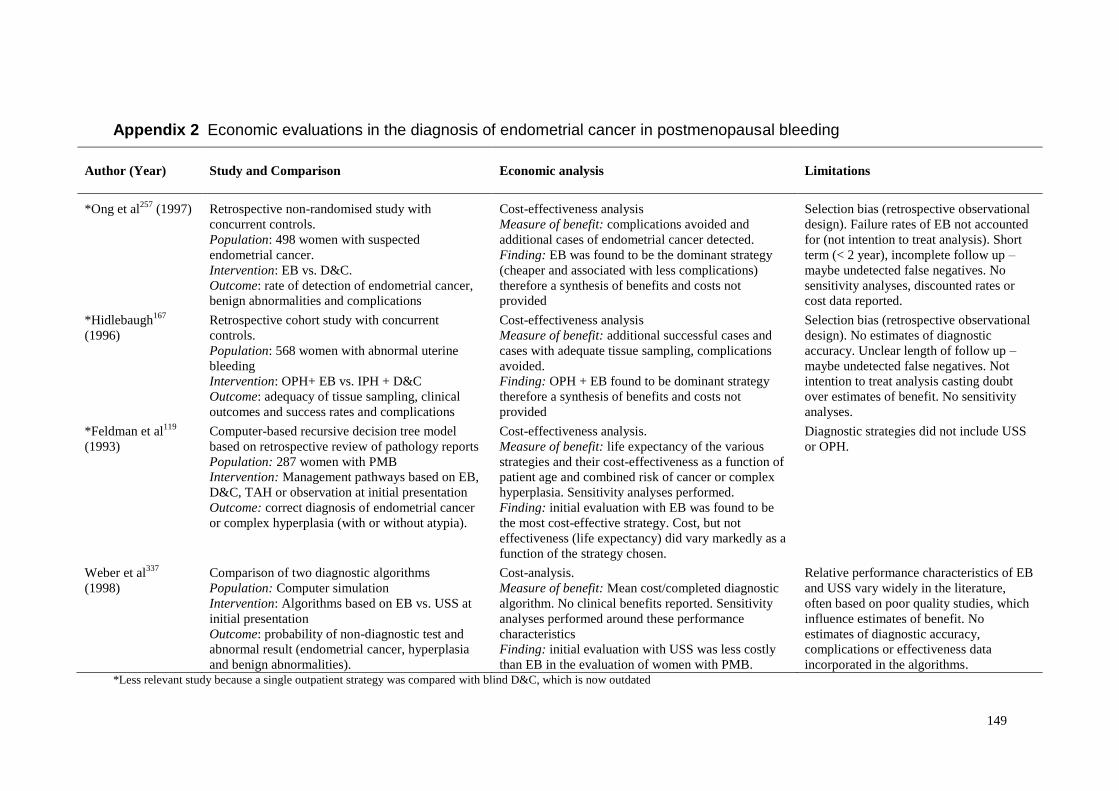
149
Appendix 2 Economic evaluations in the diagnosis of endometrial cancer in postmenopausal bleeding
Author (Year) Study and Comparison Economic analysis Limitations
*Ong et al257
(1997)
Retrospective non-randomised study with
concurrent controls.
Population: 498 women with suspected
endometrial cancer.
Intervention: EB vs. D&C.
Outcome: rate of detection of endometrial cancer,
benign abnormalities and complications
Cost-effectiveness analysis
Measure of benefit: complications avoided and
additional cases of endometrial cancer detected.
Finding: EB was found to be the dominant strategy
(cheaper and associated with less complications)
therefore a synthesis of benefits and costs not
provided
Selection bias (retrospective observational
design). Failure rates of EB not accounted
for (not intention to treat analysis). Short
term (< 2 year), incomplete follow up –
maybe undetected false negatives. No
sensitivity analyses, discounted rates or
cost data reported.
*Hidlebaugh167
(1996)
Retrospective cohort study with concurrent
controls.
Population: 568 women with abnormal uterine
bleeding
Intervention: OPH+ EB vs. IPH + D&C
Outcome: adequacy of tissue sampling, clinical
outcomes and success rates and complications
Cost-effectiveness analysis
Measure of benefit: additional successful cases and
cases with adequate tissue sampling, complications
avoided.
Finding: OPH + EB found to be dominant strategy
therefore a synthesis of benefits and costs not
provided
Selection bias (retrospective observational
design). No estimates of diagnostic
accuracy. Unclear length of follow up –
maybe undetected false negatives. Not
intention to treat analysis casting doubt
over estimates of benefit. No sensitivity
analyses.
*Feldman et al119
(1993)
Computer-based recursive decision tree model
based on retrospective review of pathology reports
Population: 287 women with PMB
Intervention: Management pathways based on EB,
D&C, TAH or observation at initial presentation
Outcome: correct diagnosis of endometrial cancer
or complex hyperplasia (with or without atypia).
Cost-effectiveness analysis.
Measure of benefit: life expectancy of the various
strategies and their cost-effectiveness as a function of
patient age and combined risk of cancer or complex
hyperplasia. Sensitivity analyses performed.
Finding: initial evaluation with EB was found to be
the most cost-effective strategy. Cost, but not
effectiveness (life expectancy) did vary markedly as a
function of the strategy chosen.
Diagnostic strategies did not include USS
or OPH.
Weber et al337
(1998)
Comparison of two diagnostic algorithms
Population: Computer simulation
Intervention: Algorithms based on EB vs. USS at
initial presentation
Outcome: probability of non-diagnostic test and
abnormal result (endometrial cancer, hyperplasia
and benign abnormalities).
Cost-analysis.
Measure of benefit: Mean cost/completed diagnostic
algorithm. No clinical benefits reported. Sensitivity
analyses performed around these performance
characteristics
Finding: initial evaluation with USS was less costly
than EB in the evaluation of women with PMB.
Relative performance characteristics of EB
and USS vary widely in the literature,
often based on poor quality studies, which
influence estimates of benefit. No
estimates of diagnostic accuracy,
complications or effectiveness data
incorporated in the algorithms. *Less relevant study because a single outpatient strategy was compared with blind D&C, which is now outdated

150
Appendix 3 Search strategy for Endometrial biopsy evidence
Medline (1966 – December 1999)
Endometrial biopsy
Endometrial biop$.tw
1 or 2
Exp diagnosis
Diagnos$.tw
di.fs.
4 or 5 or 6
3 and 7
limit 8 to human
Embase (1982 – December 1999)
Endometrial biopsy
Endometrial biop$.tw
1 or 2
Exp diagnosis
Diagnos$.tw
di.fs.
4 or 5 or 6
3 and 7
limit 8 to human
Cochrane Library issue 3 (CCTR)
Endometrial biopsy
Hand searching
Reference lists of included primary studies and review articles

151
Appendix 4 Search strategy for ultrasound endometrial thickness evidence
Medline (1966 – December 2000)
Ultrasound
Sonography
1 or 2
Endometrial thickness
3 and 4
limit 5 to human
Embase (1982 – December 1999)
Ultrasound
Sonography
1 or 2
Endometrial thickness
3 and 4
limit 5 to human
Cochrane Library issue 3 (CCTR)
Ultrasound or sonography
Hand searching
Reference lists of included primary studies and review articles
152
Appendix 5 Search strategy for hysteroscopy evidence
Medline (1966 – December 2001)
Exp hysteroscopy/
Hysteroscop$.ti,ab.
Exp diagnosis
Diagnos$.ti,ab.
di.fs.
or/ 1-2
or/ 3-5
6 and 7
animal/ not human
8 not 9
Embase (1982 – December 2001)
Exp hysteroscopy/
Hysteroscop$.ti,ab.
Exp diagnosis
Diagnos$.ti,ab.
di.fs.
or/ 1-2
or/ 3-5
6 and 7
animal/ not human
8 not 9
Cochrane Library issue 4 (CCTR)
Hysteroscopy
Hand searching
Reference lists of included primary studies and review articles
Specialist journal Gynaecological Endoscopy
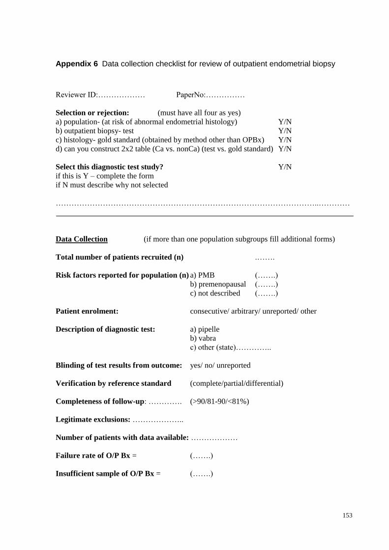
153
Appendix 6 Data collection checklist for review of outpatient endometrial biopsy
Reviewer ID:……………… PaperNo:……………
Selection or rejection: (must have all four as yes)
a) population- (at risk of abnormal endometrial histology) Y/N
b) outpatient biopsy- test Y/N
c) histology- gold standard (obtained by method other than OPBx) Y/N
d) can you construct 2x2 table (Ca vs. nonCa) (test vs. gold standard) Y/N
Select this diagnostic test study? Y/N
if this is Y – complete the form
if N must describe why not selected
………………………………………………………………………………………..…………
Data Collection (if more than one population subgroups fill additional forms)
Total number of patients recruited (n) .…….
Risk factors reported for population (n) a) PMB (…….)
b) premenopausal (…….)
c) not described (…….)
Patient enrolment: consecutive/ arbitrary/ unreported/ other
Description of diagnostic test: a) pipelle
b) vabra
c) other (state)…………..
Blinding of test results from outcome: yes/ no/ unreported
Verification by reference standard (complete/partial/differential)
Completeness of follow-up: …………. (>90/81-90/<81%)
Legitimate exclusions: ………………..
Number of patients with data available: ………………
Failure rate of O/P Bx = (…….)
Insufficient sample of O/P Bx = (…….)
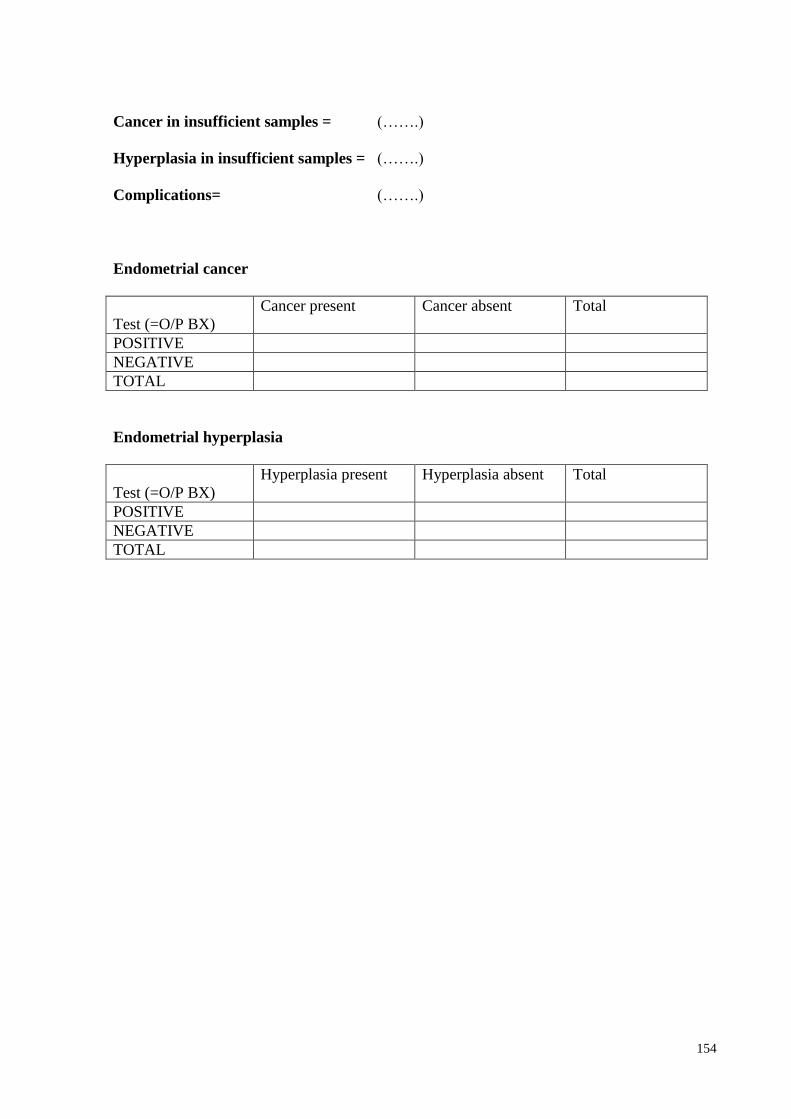
154
Cancer in insufficient samples = (…….)
Hyperplasia in insufficient samples = (…….)
Complications= (…….)
Endometrial cancer
Test (=O/P BX)
Cancer present Cancer absent Total
POSITIVE
NEGATIVE
TOTAL
Endometrial hyperplasia
Test (=O/P BX)
Hyperplasia present Hyperplasia absent Total
POSITIVE
NEGATIVE
TOTAL
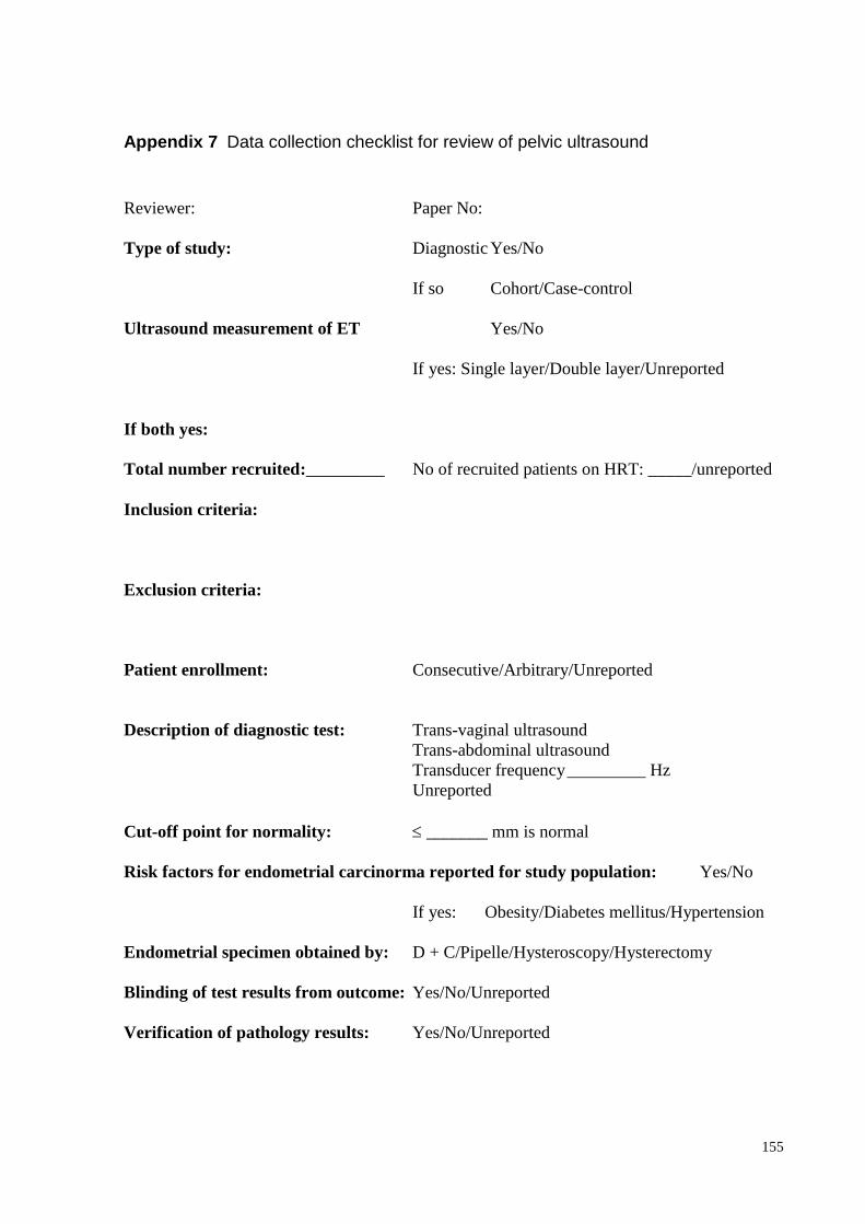
155
Appendix 7 Data collection checklist for review of pelvic ultrasound
Reviewer: Paper No:
Type of study: Diagnostic Yes/No
If so Cohort/Case-control
Ultrasound measurement of ET Yes/No
If yes: Single layer/Double layer/Unreported
If both yes:
Total number recruited:_________ No of recruited patients on HRT: _____/unreported
Inclusion criteria:
Exclusion criteria:
Patient enrollment: Consecutive/Arbitrary/Unreported
Description of diagnostic test: Trans-vaginal ultrasound
Trans-abdominal ultrasound
Transducer frequency _________ Hz
Unreported
Cut-off point for normality: _______ mm is normal
Risk factors for endometrial carcinorma reported for study population: Yes/No
If yes: Obesity/Diabetes mellitus/Hypertension
Endometrial specimen obtained by: D + C/Pipelle/Hysteroscopy/Hysterectomy
Blinding of test results from outcome: Yes/No/Unreported
Verification of pathology results: Yes/No/Unreported

156
Appendix 7 continued
Number of patients with data available: ______________
Number of legitimate exclusions: ______________
Follow-up rate: > 90%/ 81-90%/ <80%
CUT-OFF VALUE ____________________ MM
Endometrial cancer
TEST RESULT
ENDOMETRIAL
CA PRESENT
ENDOMETRIAL
CA ABSENT
TOTAL
POSITIVE
NEGATIVE
TOTAL
Endometrial disease (i.e. endometrial cancer + all types of endometrial hyperplasia but
excluding endometrial polyp and pyometra)
TEST RESULT
ENDOMETRIAL
DIS PRESENT
ENDOMETRIAL
DIS ABSENT
TOTAL
POSITIVE
NEGATIVE
TOTAL
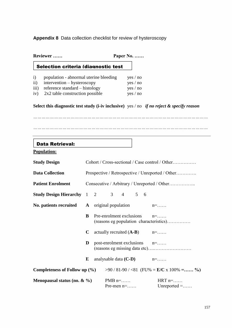
157
Appendix 8 Data collection checklist for review of hysteroscopy
Reviewer …… Paper No. ……
i) population - abnormal uterine bleeding yes / no
ii) intervention – hysteroscopy yes / no
iii) reference standard – histology yes / no
iv) 2x2 table construction possible yes / no
Select this diagnostic test study (i-iv inclusive) yes / no if no reject & specify reason
………………………………………………………………………………………………………………
………………………………………………………………………………………………………………
Population:
Study Design Cohort / Cross-sectional / Case control / Other……………
Data Collection Prospective / Retrospective / Unreported / Other………….
Patient Enrolment Consecutive / Arbitrary / Unreported / Other……………..
Study Design Hierarchy 1 2 3 4 5 6
No. patients recruited A original population n=……
B Pre-enrolment exclusions n=……
(reasons eg population characteristics)……………
C actually recruited (A-B) n=……
D post-enrolment exclusions n=……
(reasons eg missing data etc)….……………………
E analysable data (C-D) n=……
Completeness of Follow up (%) >90 / 81-90 / <81 (FU% = E/C x 100% =…… %)
Menopausal status (no. & %) PMB n=…… HRT n=……
Pre-men n=…… Unreported =……
Selection criteria (diagnostic test
study):
Data Retrieval:
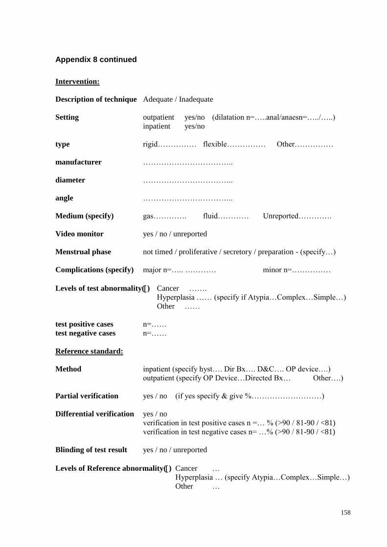
158
Appendix 8 continued
Intervention:
Description of technique Adequate / Inadequate
Setting outpatient yes/no (dilatation n=…..anal/anaesn=…../…..)
inpatient yes/no
type rigid…………… flexible…………… Other……………
manufacturer ……………………………..
diameter ……………………………..
angle ……………………………..
Medium (specify) gas…………. fluid………… Unreported………….
Video monitor yes / no / unreported
Menstrual phase not timed / proliferative / secretory / preparation - (specify…)
Complications (specify) major n=….. ………… minor n=……………
Levels of test abnormality() Cancer …….
Hyperplasia …… (specify if Atypia…Complex…Simple…)
Other ……
test positive cases n=……
test negative cases n=……
Reference standard:
Method inpatient (specify hyst…. Dir Bx…. D&C…. OP device….)
outpatient (specify OP Device…Directed Bx… Other….)
Partial verification yes / no (if yes specify & give %………………………)
Differential verification yes / no
verification in test positive cases n =… % (>90 / 81-90 / <81)
verification in test negative cases n= …% (>90 / 81-90 / <81)
Blinding of test result yes / no / unreported
Levels of Reference abnormality() Cancer …
Hyperplasia … (specify Atypia…Complex…Simple…)
Other …

159
Appendix 8 continued
Outcome:
Failure Rate n / E =…..
Specify reason for failure Technical n=….. Inadequate view n=…..
Patient factor n=…..
Failed hysteroscopies Cancer n=…… Hyperplasia n=……
Insufficient histology n=……
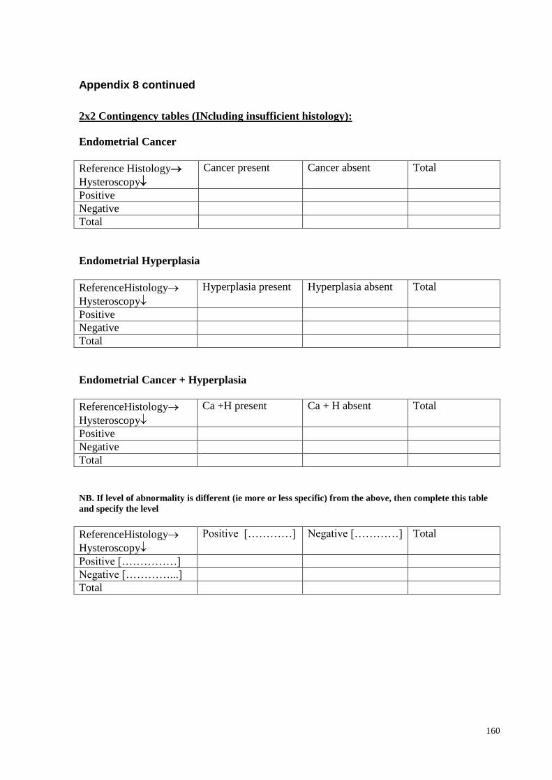
160
Appendix 8 continued
2x2 Contingency tables (INcluding insufficient histology):
Endometrial Cancer
Reference Histology
Hysteroscopy
Cancer present Cancer absent Total
Positive
Negative
Total
Endometrial Hyperplasia
ReferenceHistology
Hysteroscopy
Hyperplasia present Hyperplasia absent Total
Positive
Negative
Total
Endometrial Cancer + Hyperplasia
ReferenceHistology
Hysteroscopy
Ca +H present Ca + H absent Total
Positive
Negative
Total
NB. If level of abnormality is different (ie more or less specific) from the above, then complete this table
and specify the level
ReferenceHistology
Hysteroscopy
Positive […………] Negative […………] Total
Positive [……………]
Negative […………...]
Total

161
Appendix 8 continued
2x2 Contingency tables (EXcluding insufficient histology):
Endometrial Cancer
Reference Histology
Hysteroscopy
Cancer present Cancer absent Total
Positive
Negative
Total
Endometrial Hyperplasia
ReferenceHistology
Hysteroscopy
Hyperplasia present Hyperplasia absent Total
Positive
Negative
Total
Endometrial Cancer + Hyperplasia
ReferenceHistology
Hysteroscopy
Ca +H present Ca + H absent Total
Positive
Negative
Total
NB. If level of abnormality is different (ie more or less specific) from the above, then complete this table
and specify the level
ReferenceHistology
Hysteroscopy
Positive
[…………………]
Negative
[…………………..]
Total
Positive [……………]
Negative [……….…..]
Total
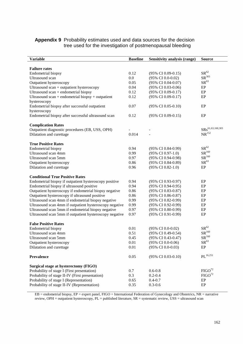
162
Appendix 9 Probability estimates used and data sources for the decision tree used for the investigation of postmenopausal bleeding
Variable Baseline Sensitivity analysis (range) Source
Failure rates
Endometrial biopsy 0.12 (95% CI 0.09-0.15) SR62
Ultrasound scan 0.0 (95% CI 0.0-0.02) SR303
Outpatient hysteroscopy 0.05 (95% CI 0.04-0.07) SR63
Ultrasound scan + outpatient hysteroscopy 0.04 (95% CI 0.03-0.06) EP
Ultrasound scan + endometrial biopsy 0.12 (95% CI 0.09-0.17) EP
Ultrasound scan + endometrial biopsy + outpatient
hysteroscopy
0.12 (95% CI 0.09-0.17) EP
Endometrial biopsy after successful outpatient
hysteroscopy
0.07 (95% CI 0.05-0.10) EP
Endometrial biopsy after successful ultrasound scan 0.12 (95% CI 0.09-0.15) EP
Complication Rates
Outpatient diagnostic procedures (EB, USS, OPH) - - SRs61,63,160,303
Dilatation and curettage 0.014 - NR152
True Positive Rates
Endometrial biopsy 0.94 (95% CI 0.84-0.99) SR62
Ultrasound scan 4mm 0.99 (95% CI 0.97-1.0) SR160
Ultrasound scan 5mm 0.97 (95% CI 0.94-0.98) SR160
Outpatient hysteroscopy 0.86 (95% CI 0.84-0.89) SR63
Dilatation and curettage 0.96 (95% CI 0.82-1.0) EP
Conditional True Positive Rates
Endometrial biopsy if outpatient hysteroscopy positive 0.94 (95% CI 0.93-0.97) EP
Endometrial biopsy if ultrasound positive 0.94 (95% CI 0.94-0.95) EP
Outpatient hysteroscopy if endometrial biopsy negative 0.86 (95% CI 0.83-0.87) EP
Outpatient hysteroscopy if ultrasound positive 0.86 (95% CI 0.86-0.87) EP
Ultrasound scan 4mm if endometrial biopsy negative 0.99 (95% CI 0.82-0.99) EP
Ultrasound scan 4mm if outpatient hysteroscopy negative 0.99 (95% CI 0.92-0.99) EP
Ultrasound scan 5mm if endometrial biopsy negative 0.97 (95% CI 0.80-0.99) EP
Ultrasound scan 5mm if outpatient hysteroscopy negative 0.97 (95% CI 0.91-0.99) EP
False Positive Rates
Endometrial biopsy 0.01 (95% CI 0.0-0.02) SR62
Ultrasound scan 4mm 0.51 (95% CI 0.49-0.54) SR160
Ultrasound scan 5mm 0.45 (95% CI 0.43-0.47) SR160
Outpatient hysteroscopy 0.01 (95% CI 0.0-0.06) SR63
Dilatation and curettage 0.01 (95% CI 0.0-0.03) EP
Prevalence 0.05 (95% CI 0.03-0.10) PL16,251
Surgical stage at hysterectomy (FIGO)
Probability of stage I (First presentation) 0.7 0.6-0.8 FIGO72
Probability of stage II-IV (First presentation) 0.3 0.2-0.4 FIGO72
Probability of stage I (Representation) 0.65 0.4-0.7 EP
Probability of stage II-IV (Representation) 0.35 0.3-0.6 EP
EB = endometrial biopsy, EP = expert panel, FIGO = International Federation of Gynecology and Obstetrics, NR = narrative
review, OPH = outpatient hysteroscopy, PL = published literature, SR = systematic review, USS = ultrasound scan
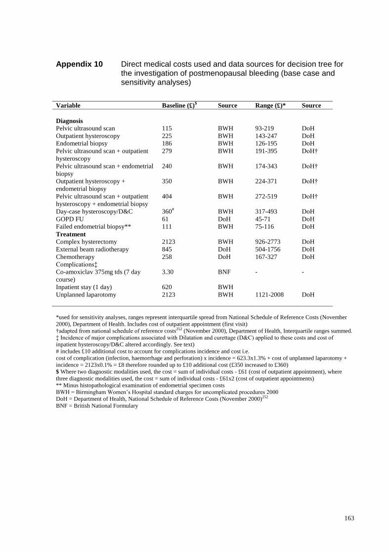
163
Appendix 10 Direct medical costs used and data sources for decision tree for the investigation of postmenopausal bleeding (base case and sensitivity analyses)
Variable Baseline (£)
$ Source Range (£)* Source
Diagnosis
Pelvic ultrasound scan 115 BWH 93-219 DoH
Outpatient hysteroscopy 225 BWH 143-247 DoH
Endometrial biopsy 186 BWH 126-195 DoH
Pelvic ultrasound scan + outpatient
hysteroscopy
279 BWH 191-395 DoH†
Pelvic ultrasound scan + endometrial
biopsy
240 BWH 174-343 DoH†
Outpatient hysteroscopy +
endometrial biopsy
350 BWH 224-371 DoH†
Pelvic ultrasound scan + outpatient
hysteroscopy + endometrial biopsy
404 BWH 272-519 DoH†
Day-case hysteroscopy/D&C 360# BWH 317-493 DoH
GOPD FU 61 DoH 45-71 DoH
Failed endometrial biopsy** 111 BWH 75-116 DoH
Treatment
Complex hysterectomy 2123 BWH 926-2773 DoH
External beam radiotherapy 845 DoH 504-1756 DoH
Chemotherapy 258 DoH 167-327 DoH
Complications‡
Co-amoxiclav 375mg tds (7 day
course)
3.30 BNF - -
Inpatient stay (1 day) 620 BWH
Unplanned laparotomy 2123 BWH 1121-2008 DoH
*used for sensitivity analyses, ranges represent interquartile spread from National Schedule of Reference Costs (November
2000), Department of Health. Includes cost of outpatient appointment (first visit)
†adapted from national schedule of reference costs252 (November 2000), Department of Health, Interquartile ranges summed.
‡ Incidence of major complications associated with Dilatation and curettage (D&C) applied to these costs and cost of
inpatient hysteroscopy/D&C altered accordingly. See text)
# includes £10 additional cost to account for complications incidence and cost i.e.
cost of complication (infection, haemorrhage and perforation) x incidence = 623.3x1.3% + cost of unplanned laparotomy +
incidence = 2123x0.1% = £8 therefore rounded up to £10 additional cost (£350 increased to £360)
$ Where two diagnostic modalities used, the cost = sum of individual costs - £61 (cost of outpatient appointment), where
three diagnostic modalities used, the cost = sum of individual costs - £61x2 (cost of outpatient appointments)
** Minus histopathological examination of endometrial specimen costs
BWH = Birmingham Women‟s Hospital standard charges for uncomplicated procedures 2000
DoH = Department of Health, National Schedule of Reference Costs (November 2000)252
BNF = British National Formulary
164
Appendix 11 Reference list of excluded studies from systematic reviews of e endometrial biopsy
A1. Guido R, Kanbour-Shakir A, Rulin M, Christopherson W. Pipelle endometrial
sampling: sensitivity in the detection of endometrial cancer. J Reprod Med
1995;40:553-55.
A2. Stovall TG, Photopulos GJ, Poston WM, Ling FW, Sandles LG. Pipelle endometrial
sampling in patients with known endometrial cancer. Obstet Gynecol 1991;77:954-56.
A3. Zorlu CG, Cobanoglu O, Isik AZ, Kutluay L, Kuscu E. Accuracy of pipelle
endometrial sampling in endometrial cancer. Gynecol Obstet Invest 1994;38:272-75.
A4. Larson DM, Johnson KK, Broste SK, Krawisz BR, Kresl JJ. Comparison of D&C and
office endometrial biopsy in predicting final histopathologic grade in endometrial
cancer. Obstet Gynecol 1995;86:38-42.
A5. Larson DM, Krawisz BR, Johnson KK, Broste SK. Comparison of the Z-sampler and
Novak endometrial biopsy instruments for in-office diagnosis of endometrial cancer.
Gynecol Oncol 1994;54:64-67.
A6. Bocanera AR, Roncoroni EC, Schlaen I, Ben J, Monteverde R, Gonzalez GM et al. An
articulated rotating brush for office endometrial evaluation of climacteric outpatients.
Maturitas 1994;19:67-76.
A7. Ferry J, Farnsworth A, Webster M, Wren B. The efficacy of the pipelle endometrial
biopsy in detecting endometrial cancer. Aust NZ J Obstet Gynaecol 1993;33:76-78.
A8. Elpek G, Uner M, Elpek M, Sedele M, Karaveli S. The diagnostic accuracy of the
Pipelle endometrial sampler in the presence of endometrial polyps. J Obstet Gynaecol
1998;18:274-75.
A9. Kent A, Haines P, Manners BCP. Blind endometrial biopsies: insufficient for
diagnosis in women with intrauterine pathology. Gynaecol Endosc 1998;7:273-78.
A10. Shipley CF, III, Simmons CL, Nelson GH. Comparison of transvaginal sonography
with endometrial biopsy in asymptomatic postmenopausal women. Journal of
Ultrasound Med 1994;13:99-104.
A11. Goldchmit R, Katz Z, Blickstein I, Caspi B, Dgani R. The accuracy of endometrial
Pipelle sampling with and without sonographic measurement of endometrial thickness.
Obstet Gynecol 1993;82:727-30.
A12. Kavak Z, Ceyhan N, Pekin S. Combination of vaginal ultrasonography and pipelle
sampling in the diagnosis of endometrial disease. Aust NZ J Obstet Gynaecol
1996;36:63-66.
165
A13. Schei B, Bang T, Halgunset J, Haugen O, Haarstad I, Onsrud M. Microcurettage
sampling of the endometrium for histopathological examination - simpler but not safe?
Acta Obstet Gynecol Scand 1994;73:497-501.
A14. Lofgren O, Alm P, Ionescu A, Skjerris J. Uterine microcurettage with combined
endometrial histopathology and cytology. An alternative to conventional curettage.
Acta Obstet Gynecol Scand 1988;67:401-03.
A15. Fothergill DJ, Brown VA, Hill AS. Histological sampling of the endometrium--a
comparison between formal curettage and the Pipelle sampler. Br.J Obstet Gynaecol
1992;99:779-80.
A16. Law J. Histological sampling of the endometrium-acomparison between formal
curettage and the pipelle sampler. Br J Obstet Gynaecol 1993;100:503-04.
A17. Sonnendecker E, Sevitz H, Hofmeyr GJ. Diagnostic accuracy of the accurette
endometrial sampler. S Afr Med J 1982;61:109-13.
A18. Kufahl J, Pederson I, Eriksen PS, Helkjaer PE, Larsen LG, Jensen KL et al.
Transvaginal ultrasound, endometrial cytology sampled by Gynoscann and histology
obtained by Uterine Explora Curette compared to the histology of the uterine
specimen. A prospective study in pre- and postmenopasual women undergoing
elective hysterectomy. Acta Obstet Gynecol Scand 1997;76:790-96.
A19. Bistoletti P, Hjerpe A, Mollerstrom G. Cytological diagnosis of endometrial cancer
and preinvasive endometrial lesions. A comparison of the Endo-Pap sampler with
fractional curettage. Acta Obstet Gynecol Scand 1988;67:343-45.
A20. Dijkhuizen FP, Mol BW, Brolmann HA, Heintz AP. The accuracy of endometrial
sampling in the diagnosis of patients with endometrial cancer and hyperplasia: a meta-
analysis. Cancer 2000;89:1765-72.
A21. Giusa-Chiferi MG, Goncalves WJ, Baracat EC, Albuquerque Neto LC, Bortoletto CC,
de Lima GR. Transvaginal ultrasound, uterine biopsy and hysteroscopy for
postmenopausal bleeding. Int J Gynaecol Obstet 1996;55:39-44.
A22. Ong S, Duffy T, Lenehan P, Murphy J. Endometrial pipelle biopsy compared to
conventional dilatation and curettage. Ir.J Med Sci. 1997;166:47-49.
A23. Briley M, Lindsell DR. The role of transvaginal ultrasound in the investigation of
women with post-menopausal bleeding. Clin Radiol 1998;53:502-05.
A24. Franchi D, Colombo N, Bocciolone L, Maggioni A, Costa D, Sacchini V. Tamoxifen
and the uterus: Potential uterine risks of anti-oestrogens. The approach of the
European Institute of Oncology. Eur J Cancer 1998;34 Suppl 4:S34-S35.
A25. Lidor A, Ismajovich B, Confino E, David M. Histopathological findings in 226
women with postmenopasual bleeding. Acta Obstet Gynecol Scand 1986;65:41-43.
166
A26. Altaras MM, Aviram R, Cohen I, Markov S, Goldberg GL, Beyth Y.
Microhysteroscopy and endometrial biopsy results following failed diagnostic
dilatation and curettage in women with postmenopausal bleeding. Int J Gynaecol
Obstet 1993;42:255-60.
A27. Goncalves MA, Goncalves WJ, Matias M, Novo NF, Baracat EC, de Lima GR. A
hysteroscopic and anatomicopathological study in women with breast cancer. Minerva
Ginecol 1998;50:341-46.
A28. Pal L, Lapensee L, Toth TL, Isaacson KB. Comparison of office hysteroscopy,
transvaginal ultrasonography and endometrial biopsy in evaluation of abnormal
uterine bleeding [published erratum appears in J Soc Laparoendosc Surg 1997 Oct-
Dec;1(4):395]. J Soc Laparoendoscopic Surgeons 1997;1:125-30.
A29. Dubinsky TJ, Parvey HR, Maklad N. The role of transvaginal sonography and
endometrial biopsy in the evaluation of peri- and postmenopausal bleeding. AJR
1997;169:145-49.
A30. Iossa A, Cianferoni L, Ciatto S, Cecchini S, Campatelli C, Lo SF. Hysteroscopy and
endometrial cancer diagnosis: a review of 2007 consecutive examinations in self-
referred patients. Tumori 1991;77:479-83.
A31. O'Connell LP, Fries MH, Zeringue E, Brehm W. Triage of abnormal postmenopausal
bleeding: a comparison of endometrial biopsy and transvaginal sonohysterography
versus fractional curettage with hysteroscopy. Am J Obstet Gynecol 1998;178:956-61.
A32. Suarez RA, Grimes DA, Majmudar B, Benigno BB. Diagnostic endometrial aspiration
with the Karman cannula. J Reprod.Med 1983;28:41-44.
A33. Stovall TG, Ling FW, Morgan PL. A prospective, randomized comparison of the
Pipelle endometrial sampling device with the Novak curette. Am J Obstet Gynecol
1991;165:1287-90.
A34. Koonings PP, Moyer DL, Grimes DA. A randomized clinical trial comparing Pipelle
and Tis-u-trap for endometrial biopsy. Obstet Gynecol 1990;75:293-95.
A35. Loffer FD. Hysteroscopy with selective endometrial sampling compared with D&C
for abnormal uterine bleeding: the value of a negative hysteroscopic view. Obstet
Gynecol 1989;73:16-20.
A36. Dubinsky TJ, Parvey HR, Gormaz G, Curtis M, Maklad N. Transvaginal
hysterosonography: comparison with biopsy in the evaluation of postmenopausal
bleeding. J Ultrasound Med 1995;14:887-93.
A37. Lipscomb GH, Lopatine SM, Stovall TG, Ling FW. A randomized comparison of the
Pipelle, Accurette, and Explora endometrial sampling devices. Am J Obstet Gynecol
1994;170:591-94.
A38. Shapley M, Redman CW. Endometrial sampling and general practice. Br.J Gen Pract.
1997;47:387-91.

167
A39. Youssif SN, McMillan DL. Outpatient endometrial biopsy: the pipelle. Br J Hosp Med
1995;54:198-201.
A40. Van den Bosch T, Vandendael A, Wranz P, Lombard C. Endopap versus Pipelle
sampling in the diagnosis of postmenopausal endometrial disease. European Journal of
Obstet, Gynecol Reprod Biol 1996;64:91-94.
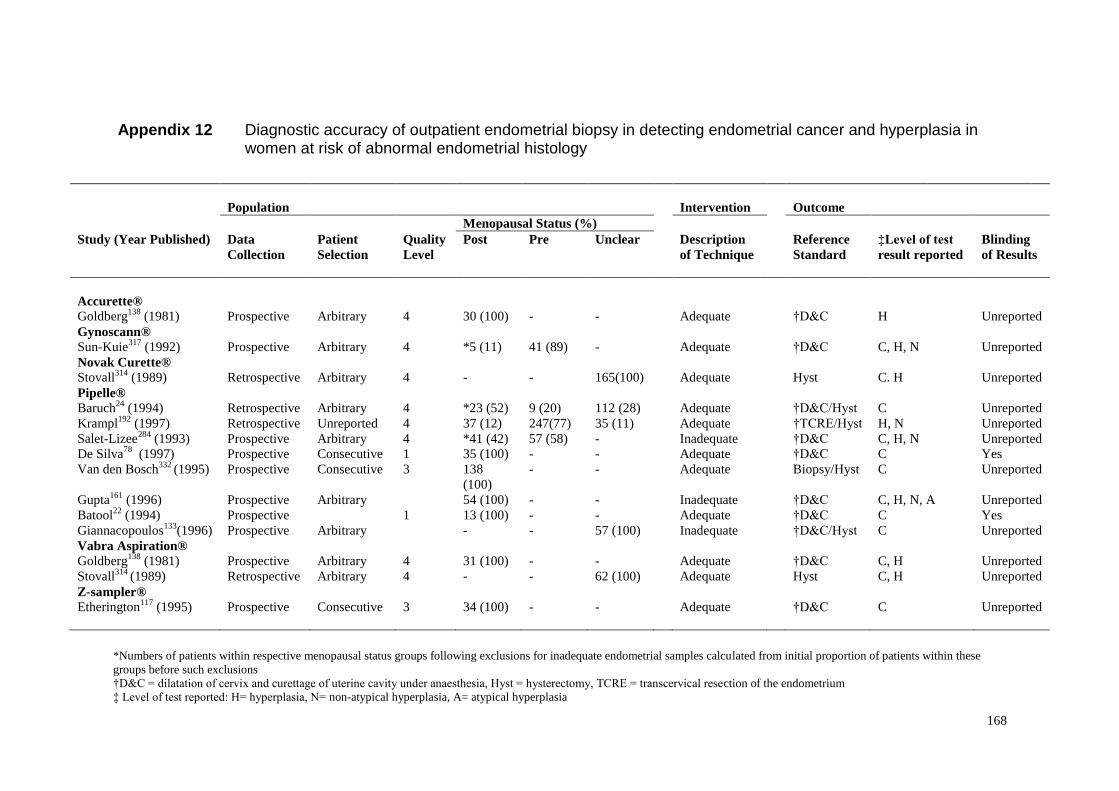
168
Appendix 12 Diagnostic accuracy of outpatient endometrial biopsy in detecting endometrial cancer and hyperplasia in women at risk of abnormal endometrial histology
Population Intervention Outcome
Menopausal Status (%)
Study (Year Published) Data
Collection
Patient
Selection
Quality
Level
Post Pre Unclear Description
of Technique
Reference
Standard
‡Level of test
result reported
Blinding
of Results
Accurette®
Goldberg138
(1981) Prospective Arbitrary 4 30 (100) - - Adequate †D&C H Unreported
Gynoscann®
Sun-Kuie317
(1992) Prospective Arbitrary 4 *5 (11) 41 (89) - Adequate †D&C C, H, N Unreported
Novak Curette®
Stovall314
(1989) Retrospective Arbitrary 4 - - 165(100) Adequate Hyst C. H Unreported
Pipelle®
Baruch24
(1994) Retrospective Arbitrary 4 *23 (52) 9 (20) 112 (28) Adequate †D&C/Hyst C Unreported
Krampl192
(1997) Retrospective Unreported 4 37 (12) 247(77) 35 (11) Adequate †TCRE/Hyst H, N Unreported
Salet-Lizee284
(1993) Prospective Arbitrary 4 *41 (42) 57 (58) - Inadequate †D&C C, H, N Unreported
De Silva78
(1997) Prospective Consecutive 1 35 (100) - - Adequate †D&C C Yes
Van den Bosch332
(1995) Prospective Consecutive 3 138
(100)
- - Adequate Biopsy/Hyst C Unreported
Gupta161
(1996) Prospective Arbitrary 54 (100) - - Inadequate †D&C C, H, N, A Unreported
Batool22
(1994) Prospective 1 13 (100) - - Adequate †D&C C Yes
Giannacopoulos133
(1996) Prospective Arbitrary - - 57 (100) Inadequate †D&C/Hyst C Unreported
Vabra Aspiration®
Goldberg138
(1981) Prospective Arbitrary 4 31 (100) - - Adequate †D&C C, H Unreported
Stovall314
(1989) Retrospective Arbitrary 4 - - 62 (100) Adequate Hyst C, H Unreported
Z-sampler®
Etherington117
(1995) Prospective Consecutive 3 34 (100) - - Adequate †D&C C Unreported
*Numbers of patients within respective menopausal status groups following exclusions for inadequate endometrial samples calculated from initial proportion of patients within these
groups before such exclusions
†D&C = dilatation of cervix and curettage of uterine cavity under anaesthesia, Hyst = hysterectomy, TCRE = transcervical resection of the endometrium
‡ Level of test reported: H= hyperplasia, N= non-atypical hyperplasia, A= atypical hyperplasia
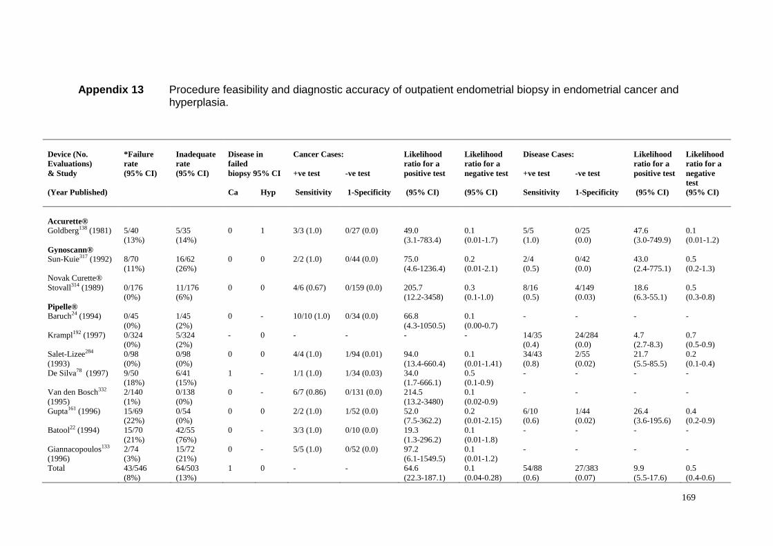
169
Appendix 13 Procedure feasibility and diagnostic accuracy of outpatient endometrial biopsy in endometrial cancer and hyperplasia.
Device (No.
Evaluations)
*Failure
rate
Inadequate
rate
Disease in
failed
Cancer Cases: Likelihood
ratio for a
Likelihood
ratio for a
Disease Cases: Likelihood
ratio for a
Likelihood
ratio for a
& Study (95% CI) (95% CI) biopsy 95% CI +ve test -ve test positive test negative test +ve test -ve test positive test negative
test
(Year Published) Ca Hyp Sensitivity 1-Specificity (95% CI) (95% CI) Sensitivity 1-Specificity (95% CI) (95% CI)
Accurette®
Goldberg138 (1981) 5/40
(13%)
5/35
(14%)
0 1 3/3 (1.0) 0/27 (0.0) 49.0
(3.1-783.4)
0.1
(0.01-1.7)
5/5
(1.0)
0/25
(0.0)
47.6
(3.0-749.9)
0.1
(0.01-1.2)
Gynoscann®
Sun-Kuie317 (1992) 8/70
(11%)
16/62
(26%)
0 0 2/2 (1.0) 0/44 (0.0) 75.0
(4.6-1236.4)
0.2
(0.01-2.1)
2/4
(0.5)
0/42
(0.0)
43.0
(2.4-775.1)
0.5
(0.2-1.3)
Novak Curette®
Stovall314 (1989) 0/176
(0%)
11/176
(6%)
0 0 4/6 (0.67) 0/159 (0.0) 205.7
(12.2-3458)
0.3
(0.1-1.0)
8/16
(0.5)
4/149
(0.03)
18.6
(6.3-55.1)
0.5
(0.3-0.8)
Pipelle®
Baruch24 (1994) 0/45
(0%)
1/45
(2%)
0 - 10/10 (1.0) 0/34 (0.0) 66.8
(4.3-1050.5)
0.1
(0.00-0.7)
- - - -
Krampl192 (1997) 0/324
(0%)
5/324
(2%)
- 0 - - - - 14/35
(0.4)
24/284
(0.0)
4.7
(2.7-8.3)
0.7
(0.5-0.9)
Salet-Lizee284
(1993)
0/98
(0%)
0/98
(0%)
0 0 4/4 (1.0) 1/94 (0.01) 94.0
(13.4-660.4)
0.1
(0.01-1.41)
34/43
(0.8)
2/55
(0.02)
21.7
(5.5-85.5)
0.2
(0.1-0.4)
De Silva78 (1997) 9/50
(18%)
6/41
(15%)
1 - 1/1 (1.0) 1/34 (0.03) 34.0
(1.7-666.1)
0.5
(0.1-0.9)
- - - -
Van den Bosch332
(1995)
2/140
(1%)
0/138
(0%)
0 - 6/7 (0.86) 0/131 (0.0) 214.5
(13.2-3480)
0.1
(0.02-0.9)
- - - -
Gupta161 (1996) 15/69
(22%)
0/54
(0%)
0 0 2/2 (1.0) 1/52 (0.0) 52.0
(7.5-362.2)
0.2
(0.01-2.15)
6/10
(0.6)
1/44
(0.02)
26.4
(3.6-195.6)
0.4
(0.2-0.9)
Batool22 (1994) 15/70
(21%)
42/55
(76%)
0 - 3/3 (1.0) 0/10 (0.0) 19.3
(1.3-296.2)
0.1
(0.01-1.8)
- - - -
Giannacopoulos133
(1996)
2/74
(3%)
15/72
(21%)
0 - 5/5 (1.0) 0/52 (0.0) 97.2
(6.1-1549.5)
0.1
(0.01-1.2)
- - - -
Total 43/546
(8%)
64/503
(13%)
1 0 - - 64.6
(22.3-187.1)
0.1
(0.04-0.28)
54/88
(0.6)
27/383
(0.07)
9.9
(5.5-17.6)
0.5
(0.4-0.6)
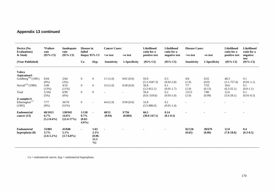
170
Appendix 13 continued
Device (No.
Evaluations)
*Failure
rate
Inadequate
rate
Disease in
failed
Cancer Cases: Likelihood
ratio for a
Likelihood
ratio for a
Disease Cases: Likelihood
ratio for a
Likelihood
ratio for a
& Study (95% CI) (95% CI) biopsy 95% CI +ve test -ve test positive test negative test +ve test -ve test positive test negative
test
(Year Published) Ca Hyp Sensitivity 1-Specificity (95% CI) (95% CI) Sensitivity 1-Specificity (95% CI) (95% CI)
Vabra
Aspiration®
Goldberg138 (1981) 0/64
(0%)
2/64
(3%)
0 0 1/1 (1.0) 0/61 (0.0) 93.0
(5.3-1647.3)
0.3
(0.02-2.8)
6/6
(1.0)
0/25
(0.0)
48.3
(3.1-757.4)
0.1
(0.01-1.1)
Stovall314 (1989) 5/40
(13%)
4/35
(11%)
0 0 3/3 (1.0) 0/28 (0.0) 50.8
(3.2-812.1)
0.1
(0.01-1.7)
7/7
(1.0)
7/55
(0.13)
18.6
(6.3-55.1)
0.1
(0.0-1.1)
Total 5/104
(5%)
6/99
(6%)
0 0 - - 59.4
(6.8.-518.6)
0.2
(0.03-1.0)
13/13
(1.0)
7/80
(0.09)
12.6
(5.6-28.1)
0.1
(0.01-0.5)
Z-sampler®
Etherington117
(1995)
7/77
(9%)
36/70
(51%)
0 - 4/4 (1.0) 0/30 (0.0) 55.8
(3.5-886.0)
0.1
(0.01-1.4)
- - - -
Endometrial
cancer (13)
68/1013
6.7%
(5.2-8.4%)
138/945
14.6%
(12.4-17%)
1/138
0.7%
(0.02-
4.0%)
- 48/51
(0.94)
3/756
(0.004)
66.5
(30.0-147.1)
0.14
(0.1-0.3)
- - - -
Endometrial
hyperplasia (8)
33/881
3.7%
(2.6-5.2%)
43/848
5.1%
(3.7-6.8%)
- 1/43
2.3%
(0.06-
12.3
%)
- - - - 82/126
(0.65)
38/679
(0.06)
12.0
(7.8-18.6)
0.4
(0.3-0.5)
Ca = endometrial cancer, hyp = endometrial hyperplasia
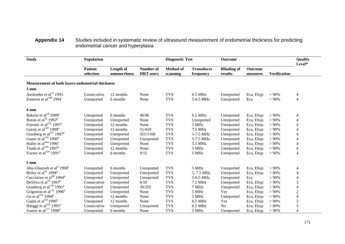
171
Appendix 14 Studies included in systematic review of ultrasound measurement of endometrial thickness for predicting endometrial cancer and hyperplasia
Study Population Diagnostic Test Outcome Quality
Level*
Patient
selection
Length of
amenorrhoea
Number of
HRT users
Method of
scanning
Transducer
frequency
Blinding of
results
Outcome
measures
Verification
Measurement of both layers endometrial thickness 3 mm Auslender et al
14 1993 Consecutive 12 months None TVS 6.5 MHz Unreported Eca, Ehyp > 90% 4
Zannoni et al348
1994 Unreported 6 months None TVS 5-6.5 MHz Unreported Eca > 90% 4
4 mm Bakour et al
18 1999
a Unreported 6 months 46/96 TVS 6.5 MHz Unreported Eca, Ehyp > 90% 4
Botsis et al36
1992p Unreported Unreported None TVS Unreported Unreported Eca, Ehyp > 90% 4
Fistonic et al122
1997a Unreported 12 months None TVS 5 MHz Unreported Eca, Ehyp > 90% 4
Garuti et al130
1999a Unreported 12 months 51/419 TVS 7.5 MHz Unreported Eca, Ehyp > 90% 4
Granberg et al147
1997p Unreported Unreported 351/1168 TVS 5-7.5 MHz Unreported Eca, Ehyp > 90% 4
Guner et al159
1996p Unreported Unreported Unreported TVS 5-7.5 MHz Unreported Eca, Ehyp > 90% 4
Haller et al164
1996a Unreported Unreported None TVS 5.5 MHz Unreported Eca, Ehyp > 90% 4
Tsuda et al328
1997p Unreported 12 months None TVS 5 MHz Unreported Eca, Ehyp > 90% 4
Varner et al334
1991p Unreported 6 months 9/15 TVS 5 MHz Unreported Eca, Ehyp > 90% 4
5 mm Abu-Ghazzeh et al
4 1999
a Unreported 6 months Unreported TVS 5 MHz Unreported Eca, Ehyp > 90% 4
Briley et al42
1998a Unreported Unreported Unreported TVS 5, 7.5 MHz Unreported Eca, Ehyp < 80% 4
Cacciatore et al48
1994p Unreported Unreported Unreported TVS 5-6.5 MHz Unreported Eca > 90% 4
DeSilva et al78
1997p Consecutive Unreported 6/50 TVS 7.5 MHz Unreported Eca, Ehyp > 90% 3
Granberg et al146
1991p Unreported Unreported 30/205 TVS 7 MHz Unreported Eca, Ehyp > 90% 4
Grigoriou et al151
1996p Unreported Unreported None TVS 5 MHz Yes Eca, Ehyp > 90% 2
Gu et al155
1994p Unreported 12 months None TVS 5 MHz Unreported Eca, Ehyp > 90% 4
Gupta et al161
1996p Unreported 12 months None TVS 6.5 MHz Yes Eca, Ehyp > 90% 2
Hänggi et al165
1995a Consecutive Unreported Unreported TVS 6.5 MHz No Eca, Ehyp < 80% 5
Ivanov et al172
1998p Unreported 6 months None TVS 5 MHz Unreported Eca, Ehyp > 90% 4
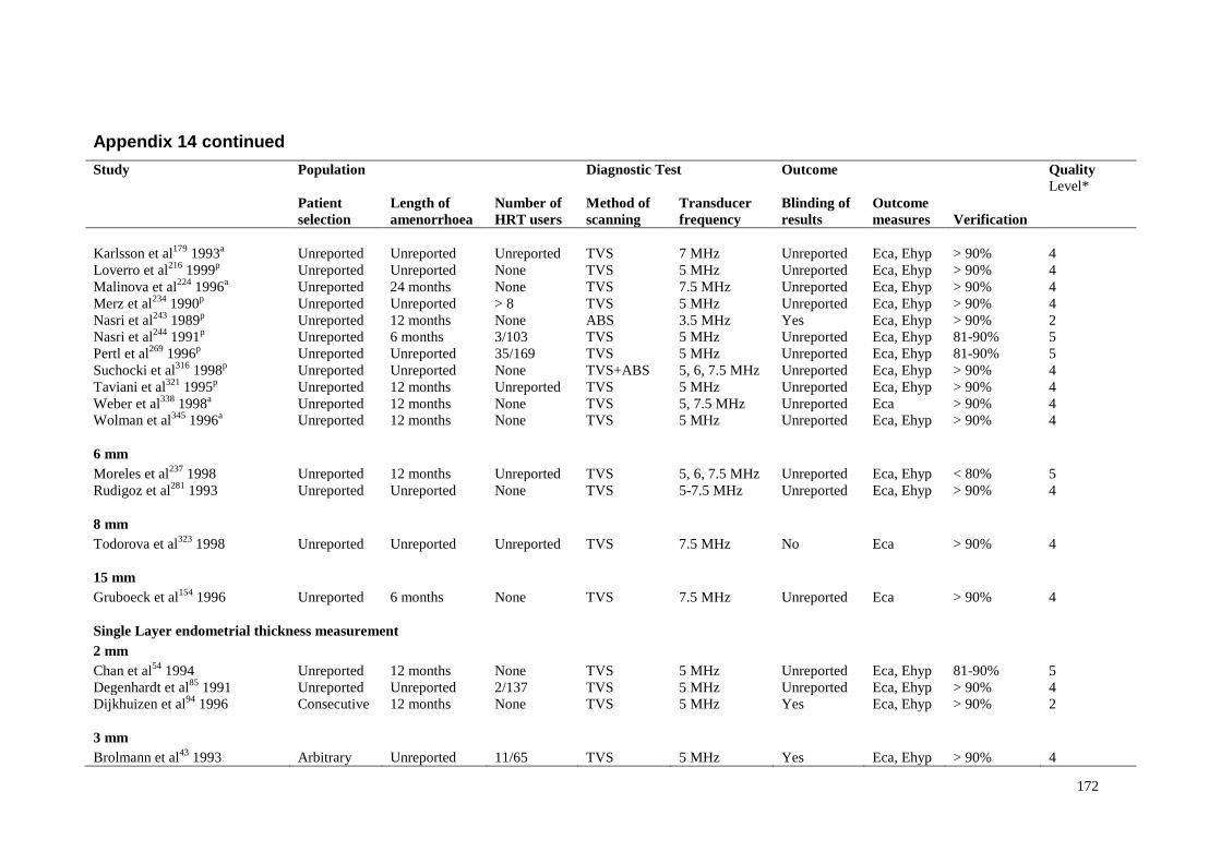
172
Appendix 14 continued
Study Population Diagnostic Test Outcome Quality
Level*
Patient
selection
Length of
amenorrhoea
Number of
HRT users
Method of
scanning
Transducer
frequency
Blinding of
results
Outcome
measures
Verification
Karlsson et al179
1993a Unreported Unreported Unreported TVS 7 MHz Unreported Eca, Ehyp > 90% 4
Loverro et al216
1999p Unreported Unreported None TVS 5 MHz Unreported Eca, Ehyp > 90% 4
Malinova et al224
1996a Unreported 24 months None TVS 7.5 MHz Unreported Eca, Ehyp > 90% 4
Merz et al234
1990p Unreported Unreported > 8 TVS 5 MHz Unreported Eca, Ehyp > 90% 4
Nasri et al243
1989p Unreported 12 months None ABS 3.5 MHz Yes Eca, Ehyp > 90% 2
Nasri et al244
1991p Unreported 6 months 3/103 TVS 5 MHz Unreported Eca, Ehyp 81-90% 5
Pertl et al269
1996p Unreported Unreported 35/169 TVS 5 MHz Unreported Eca, Ehyp 81-90% 5
Suchocki et al316
1998p Unreported Unreported None TVS+ABS 5, 6, 7.5 MHz Unreported Eca, Ehyp > 90% 4
Taviani et al321
1995p Unreported 12 months Unreported TVS 5 MHz Unreported Eca, Ehyp > 90% 4
Weber et al338
1998a Unreported 12 months None TVS 5, 7.5 MHz Unreported Eca > 90% 4
Wolman et al345
1996a Unreported 12 months None TVS 5 MHz Unreported Eca, Ehyp > 90% 4
6 mm Moreles et al
237 1998 Unreported 12 months Unreported TVS 5, 6, 7.5 MHz Unreported Eca, Ehyp < 80% 5
Rudigoz et al281
1993 Unreported Unreported None TVS 5-7.5 MHz Unreported Eca, Ehyp > 90% 4
8 mm Todorova et al
323 1998 Unreported Unreported Unreported TVS 7.5 MHz No Eca > 90% 4
15 mm Gruboeck et al
154 1996 Unreported 6 months None TVS 7.5 MHz Unreported Eca > 90% 4
Single Layer endometrial thickness measurement 2 mm Chan et al
54 1994 Unreported 12 months None TVS 5 MHz Unreported Eca, Ehyp 81-90% 5
Degenhardt et al85
1991 Unreported Unreported 2/137 TVS 5 MHz Unreported Eca, Ehyp > 90% 4
Dijkhuizen et al94
1996 Consecutive 12 months None TVS 5 MHz Yes Eca, Ehyp > 90% 2
3 mm Brolmann et al
43 1993 Arbitrary Unreported 11/65 TVS 5 MHz Yes Eca, Ehyp > 90% 4
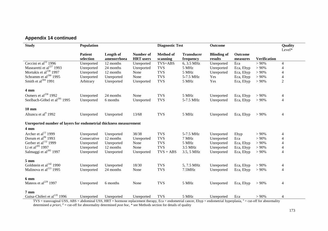
173
Appendix 14 continued
Study Population Diagnostic Test Outcome Quality
Level*
Patient
selection
Length of
amenorrhoea
Number of
HRT users
Method of
scanning
Transducer
frequency
Blinding of
results
Outcome
measures
Verification
Ceccini et al53
1996 Unreported 12 months Unreported TVS+ABS 6, 3.5 MHz Unreported Eca > 90% 4
Masearetti et al227
1993 Unreported 24 months Unreported TVS 5 MHz Unreported Eca, Ehyp > 90% 4
Mortakis et al238
1997 Unreported 12 months None TVS 5 MHz Unreported Eca, Ehyp > 90% 4
Schramm et al290
1995 Unreported Unreported None TVS 5-7.5 MHz Yes Eca, Ehyp > 90% 4
Smith et al304
1991
Arbitrary Unreported Unreported TVS 5 MHz Yes Eca, Ehyp > 90% 2
4 mm Osmers et al
258 1992 Unreported 24 months None TVS 5 MHz Unreported Eca, Ehyp > 90% 4
Seelbach-Göbel et al295
1995 Unreported 6 months Unreported TVS 5-7.5 MHz Unreported Eca, Ehyp > 90% 4
10 mm Altuncu et al
9 1992 Unreported Unreported 13/68 TVS 5 MHz Unreported Eca, Ehyp > 90% 4
Unreported number of layers for endometrial thickness measurement 4 mm Archer et al
13 1999 Unreported Unreported 38/38 TVS 5-7.5 MHz Unreported Ehyp > 90% 4
Dorum et al96
1993 Consecutive 12 months Unreported TVS 7 MHz Unreported Eca > 90% 4
Gerber et al132
1999 Unreported Unreported None TVS 5 MHz Unreported Eca, Ehyp > 90% 4
Li et al205
1997 Unreported 12 months None TVS 3.5 MHz Unreported Eca, Ehyp > 90% 4
Salmaggi et al285
1997 Unreported Unreported Unreported TVS + ABS 3.5, 5 MHz Unreported Eca, Ehyp > 90% 4
5 mm
Goldstein et al142
1990 Unreported Unreported 18/30 TVS 5, 7.5 MHz Unreported Eca, Ehyp > 90% 4
Malinova et al223
1995 Unreported 24 months None TVS 7.5MHz Unreported Eca, Ehyp > 90% 4
6 mm
Mateos et al229
1997 Unreported 6 months None TVS 5 MHz Unreported Eca, Ehyp > 90% 4
7 mm
Guisa-Chiferi et al135
1996 Unreported Unreported Unreported TVS 5 MHz Unreported Eca > 90% 4 TVS = transvaginal USS, ABS = abdominal USS, HRT = hormone replacement therapy, Eca = endometrial cancer, Ehyp = endometrial hyperplasia, a = cut-off for abnormality
determined a priori, p = cut-off for abnormality determined post hoc, * see Methods section for details of quality
174
Appendix 15 Reference list of excluded studies from systematic reviews of ultrasound
A1. Abu Hmeidan F, Bilek K, Baier D, Nuwayhid M, Kade R. Ultrasound assessment of the
endometrium in early detection of endometrial cancer in high risk patients. Zentralbl
Gynakol 1992, 114: 455-8.
A2. Abu Hmeidan F, Bilek K, Baier D, Nuwayhid M, Kade R. Ultrasound image of
endometrial cancer. Ultraschall Med 1992, 13: 178-82.
A3. Alcazar JL, Laparte C. Comparative study of transvaginal ultrasonography and
hysteroscopy in post-menopausal bleeding. Gynecol Obstet Invest 1996, 41: 47-9.
A4. Aleem F, Predanic M, Calame R, Moukhtar M, Pennisi J. Transvaginal color and pulsed
Doppler sonography of the endometrium: a possible role in reducing the number of
D&C procedures J Ultrasound Med 1995, 14: 139-45, 147-8.
A5. Andolf E, Dahlander K, Aspenberg P. Ultrasonic thickness of the endometrium
correlated to body weight in asymptomatic postmenopausal women. Obstet Gynecol
1993, 82: 936-40.
A6. Atri M, Nazarnia S, Aldis AE, Reinhold C, Bret PM, Kintzen G. Transvaginal
ultrasound appearance of endometrial abnormalities. Radiographics 1994, 14: 483-92.
A7. Baiocchi G, Gilardi G. Endometrial cancer: an increasing neoplasm. Screening and
early diagnosis: proposal for a protocol. Minerva Ginecol 1997, 49: 147-52.
A8. Ballester MJ, Girones R, Torres JV, Guillen P, Osborne NG, Bonilla-Musoles F.
Diagnosis of endometrial cancer: Predictive value of transvaginal color Doppler. J
Gynecol Surg 1994, 10: 173-83.
A9. Barakat RR. Benign and hyperplastic endometrial changes associated with tamoxifen
use. Oncol 1997, 11(2 Suppl 1): 35-7.
A10. Bonilla-Musoles F, Ballester MJ, Marti MC, Raga F, Osborne NG. Transvaginal color
Doppler assessment of endometrial status in normal postmenopausal women: the
effect of hormone replacement therapy. J Ultrasound Med 1995, 14: 503-7.
A11. Bonilla-Musoles F, Raga F, Osborne NG, Blanes J, Coelho F. Three-dimensional
hysterosonography for the study of endometrial tumours: comparison with
conventional transvaginal sonography, hysterosalpingography, and hysteroscopy.
Gynecol Oncol 1997, 65: 245-52.
175
A12. Bourne TH, Campbell S, Steer CV, Royston P, Whitehead MI, Collins WP. Detection
of endometrial cancer by transvaginal ultrasonography with color flow imaging and
blood flow analysis: A preliminary report. Gynecol Oncol 1991, 40: 253-9.
A13. Buyuk E, Durmusoglu F, Erenus M, Karakoc B. Endometrial disease diagnosed by
transvaginal ultrasound and D&C. Acta Obstet Gynecol Scand 1999, 78: 419-22.
A14. Carranza-Lira S, Ortiz-Rodriguez ML, Martinez-Chequer JC, Santa-Rita-Escamilla MT,
Garcia-Hernandez E, Romo-Aguirre C. Correlation of histopathologic findings with
ultrasonography variables of the endometrium according to body weight and adipose
tissue distribution in postmenopause. Ginecol Obstet Mex 1996, 64: 517-21.
A15. Carranza-Lira S, Martinez-Chequer JC, Santa-Rita-Escamilla MT, Romo-Aguirre C.
Evaluation of ultrasonographic variables of the endometrium in relation with
histopathologic findings in patients with postmenopausal uterine bleeding. Ginecol
Obstet Mex 1996, 64: 552-5.
A16. Caserta D, Porretta M, Moscarini M. Transvaginal ultrasonography vs hysteroscopy.
Study of 288 cases of abnormal uterine bleeding. Minerva Ginecol 1997, 49: 251-3.
A17. Castelo-Branco C, Puerto B, Duran M, Gratacos E, Torne A, Fortuny A, Vanrell JA.
Transvaginal sonography of the endometrium in postmenopausal women: monitoring
the effect of hormone replacement therapy. Maturitas 1994, 19: 59-65.
A18. Cecchini S, Ciatto S, Bonardi R, Mazzotta A, Grazzini G, Pacini P, Muraca MG.
Screening by ultrasonography for endometrial cancer in postmenopausal breast cancer
patients under adjuvant tamoxifen. Gynecol Oncol 1996, 60: 409-11.
A19. Ciatto S, Cecchini S, Bonardi R, Grazzini G, Mazzotta A, Zappa M. A feasibility study
of screening for endometrial cancer in postmenopausal women by ultrasonography.
Tumori 1995, 81: 334-7.
A20. Cohen I, Perel E, Flex D, Tepper R, Altaras MM, Cordoba M, Beyth Y. Endometrial
pathology in postmenopausal tamoxifen treatment: Comparison between
gynaecologically symptomatic and asymptomatic breast cancer patients. J Clin Pathol
1999, 52: 278-82.
A21. Conoscenti G, Meir Y, Fischer-Tamaro L, Maieron A, Natale R, D' Ottavio G, Rustico
M, Facca MC, Monterosso A, Mandruzzato G. The diagnostic capacities of
transvaginal echography and hysteroscopy in the characterization of endometrial
pathology. Minerva Ginecol 1995, 47: 293-300.
A22. Conoscenti G, Meir YJ, Fischer-Tamaro L, Maieron A, Natale R, D' Ottavio G, Rustico
M, Mandruzzato G. Endometrial assessment by transvaginal sonography and
histological findings after D & C in women with PMB. Ultrasound Obstet Gynecol
1995, 6: 108-15.

176
A23. D' Amelio R, Perrone G, Di Vincenzo F, Casalino S, Capri O, Galoppi P, Zichella L.
Echographic monitoring of the endometrium with a transvaginal probe in the
menopause. A clinical study of 185 women in the menopause. Minerva Ginecol 1994,
46: 551-6.
A24. Dragojevic-Dikic S, Markovic A, Dukic M, Vasiljevic M, Popovic-Lazic J, Rakic S,
Draganic M, Jankovic S. Evaluation of abnormal uterine bleeding by transvaginal
colour Doppler sonography. Arch Oncol 1998, 6: 167-9.
A25. Dubinsky TJ, Parvey HR, Gormaz G, Curtis M, Maklad N. Transvaginal
hysterosonography: comparison with biopsy in the evaluation of PMB. J Ultrasound
Med 1995, 14: 887-93.
A26. El Ahmady O, Gad M, El Sheimy R, Halim AB, Eissa S, Hassan F, Walker R.
Comparative study between sonography, pathology and UGP in women with
perimenopausal bleeding. Anticancer Res 1996, 16: 2309-13.
A27. Emanuel MH, Verdel MJ, Wamsteker K, Lammes FB. A prospective comparison of
transvaginal ultrasonography and diagnostic hysteroscopy in the evaluation of patients
with abnormal uterine bleeding: Clinical implications. Am J Obstet Gynecol 1995,
172: 547-52.
A28. Fleischer AC, Wheeler JE, Yeh IT, Kravitz B, Jensen C, MacDonald B. Sonographic
assessment of the endometrium in osteopenic postmenopausal women treated with
idoxifene. J Ultrasound Med 1999, 18: 503-12.
A29. Franchi M, Ghezzi F, Donadello N, Zanaboni F, Beretta P, Bolis P. Endometrial
thickness in tamoxifen treated patients: An independent predictor of endometrial
disease. Obstet Gynecol 1999, 93:1004-8.
A30. Gaucherand P, Piacenza JM, Salle B, Rudigoz RC. Sonohysterography of the uterine
cavity: Preliminary investigations. J Clin Ultrasound 1995, 23: 339-48.
A31. Georgiev D, Netsov V. A follow-up on the endometrial status of patients in the
postmenopause taking hormonal replacement treatment. Akush Ginekol Sofiia 1996,
35: 29-30.
A32. Goldstein SR, Zeltser I, Horan CK, Snyder JR, Schwartz LB. Ultrasonography-based
triage for perimenopausal patients with abnormal uterine bleeding. Am J Obstet
Gynecol 1997, 177: 102-8.
A33. Granberg S, Bourne TH. Transvaginal ultrasonography of endometrial disorders in
postmenopausal women. Ultrasound Quarterly 1995, 13: 61-74.
A34. Gucer F, Arikan MG, Petru E, Mitterdorfer B, Lahousen M, Lax S. Diagnostic value of
combined vaginal ultrasound and hysteroscopy in peri- and PMB. Gynakol
Geburtshilfliche Rundsch 1996, 36: 9-13.
177
A35. Gull B, Karlsson B, Milsom I, Wikland M, Granberg S. Transvaginal sonography of the
endometrium in a representative sample of postmenopausal women. Ultrasound
Obstet Gynecol 1996, 7: 322-7.
A36. Haines CJ, Chung TK, Lau TK. Sonographic measurement of endometrial thickness as
a predictor of vaginal bleeding in women using continuous combined hormone
replacement therapy. Gynecol Obstet Invest 1997, 44: 187-90.
A37. Hann LE, Giess CS, Bach AM, Tao Y, Baum HJ, Barakat RR. Endometrial thickness in
tamoxifen-treated patients: Correlation with clinical and pathologic findings. AJR
1997, 168: 657-61.
A38. Holbert TR. Transvaginal ultrasonographic measurement of endometrial thickness in
postmenopausal women receiving estrogen replacement therapy. Am J Obstet Gynecol
1997, 176: 1334-9.
A39. Indman PD. Abnormal uterine bleeding. Accuracy of vaginal probe ultrasound in
predicting abnormal hysteroscopic findings. J Reprod Med 1995, 10: 545-8.
A40. Karlsson B, Granberg S, Hellberg P, Wikland M. Comparative study of transvaginal
sonography and hysteroscopy for the detection of pathologic endometrial lesions in
women with PMB. J Ultrasound Med 1994, 13: 757-62.
A41. Karlsson B, Granberg S, Wikland M, Ylostalo P, Torvid K, Marsal K, Valentin L.
Transvaginal ultrasonography of the endometrium in women with PMB - A Nordic
multicenter study. Am J Obstet Gynecol 1995, 172:1488-94.
A42. Kekre AN, Jose R, Seshadri L. Transvaginal sonography of the endometrium in south
Indian postmenopausal women. Aust N Z J Obstet Gynaecol 1997, 37: 449-51.
A43. Klug PW, Leitner G. Comparison of vaginal ultrasound and histologic findings of the
endometrium. Geburtshilfe Frauenheilkd 1989, 49: 797-802.
A44. Kufahl J, Pedersen I, Sindberg-Eriksen P, Helkjaer PE, Larsen LG, Jensen KL, de Nully
P, Philipsen T, Wahlin A. Transvaginal ultrasound, endometrial cytology sampled by
Gynoscann and histology obtained by Uterine Explora Curette compared to the
histology of the uterine specimen. A prospective study in pre- and postmenopausal
women undergoing elective hysterectomy. Acta Obstet Gynecol Scand 1997, 76: 790-
6.
A45. Lerner JP, Timor-Tritsch IE, Monteagudo A. Use of transvaginal sonography in the
evaluation of endometrial hyperplasia and cancer. Obstet Gynecol Surv 1996, 51:
718-25.
A46. Lin MC, Gosink BB, Wolf SI, Feldesman MR, Stuenkel CA, Braly PS, Pretorius DH.
Endometrial thickness after menopause: effect of hormone replacement. Radiology
1991, 180: 427-32.
178
A47. Maia Jr H, Barbosa IC, Marques D, Calmon LC, Ladipo OA, Coutinho EM.
Hysteroscopy and transvaginal sonography in menopausal women receiving hormone
replacement therapy. J Am Assoc Gynecol Laparosc 1996, 4: 13-8.
A48. Maia Jr H, Barbosa IC, Farias JP, Ladipo OA, Coutinho EM. Evaluation of the
endometrial cavity during menopause. Int J Gynecol Obstet 1996, 52: 61-6.
A49. Malpani A, Singer J, Wolverson MK, Merenda G. Endometrial hyperplasia: value of
endometrial thickness in ultrasonographic diagnosis and clinical significance. J Clin
Ultrasound 1990, 18: 173-7.
A50. Marconi D, Exacoustos C, Cangi B, Perroni A, Zupi E, Valli E, Romanini C.
Transvaginal sonographic and hysteroscopic findings in postmenopausal women
receiving tamoxifen. J Am Assoc Gynecol Laparosc 1997, 4: 331-9.
A51. Marty R. Diagnostic fibrohysteroscopic evaluation of perimenopausal and
postmenopausal uterine bleeding: A comparative study with Belgian and Japanese
data. J Am Assoc Gynecol Laparosc 1998, 5: 69-73.
A52. Nasri MN, Shepherd JH, Setchell ME, Lowe DG, Chard T. Sonographic depiction of
postmenopausal endometrium with transabdominal and transvaginal scanning.
Ultrasound Obstet Gynecol 1991, 1: 279-83.
A53. O' Connell LP, Fries MH, Zeringue E, Brehm W. Triage of abnormal PMB: A
comparison of EB and transvaginal sonohysterography versus fractional curettage with
hysteroscopy. Am J Obstet Gynecol 1998, 178: 956-61.
A54. Osmers R, Volksen M, Rath W, Kuhn W. Vaginal sonography: a screening method for
early detection of ovarian tumors and endometrial cancers?. Arch Gynecol Obstet
1989, 245: 602-6.
A55. Osmers R, Volksen M, Rath W, Teichmann A, Kuhn W. Vaginosonographic
measurement of the postmenopausal endometrium in the early detection of
endometrial cancer. Geburtshilfe Frauenheilkd 1989, 49: 262-5.
A56. Osmers R, Volksen M, Rath W, Kuhn W. Vaginosonographic detection of endometrial
cancer in postmenopausal women. Int J Gynecol Obstet 1990, 32: 35-7.
A57. Osmers R, Volksen M, Schauer A. Vaginosonography for early detection of endometrial
cancer? Lancet 1990, 335: 1569-71.
A58. Ozsener S, Ozaran A, Itil I, Dikmen Y. Endometrial pathology of 104 postmenopausal
breast cancer patients treated with tamoxifen. Eur J Gynaecol Oncol 1998, 19: 580-3.
A59. Pal A, Borthaiser Z. Experience with transvaginal ultrasonic examination in patients
with metrorrhagia. Orv Hetil 1994, 135: 1305-7.
179
A60. Pal L, Lapensee L, Toth TL, Isaacson KB. Comparison of office hysteroscopy,
transvaginal ultrasonography and EB in evaluation of abnormal uterine bleeding. J Soc
Laparoendosc Surg 1997, 1: 125-30.
A61. Persiani P, Perotti F, Riccardi A, Gallina D, Polatti F, Zara C Diagnostic accuracy in
transvaginal echography in benign endometrial diseases and its comparison with
hysteroscopic biopsy. Minerva Ginecol 1995, 47: 63-7.
A62. Piegsa K, Calder A, Davis JA, McKay-Hart D, Wells M, Bryden F. Endometrial status
in post-menopausal women on long-term continuous combined hormone replacement
therapy (Kliofem). A comparative study of EB, OPH and transvaginal ultrasound. Eur
J Obstet Gynecol Reprod Biol 1997, 72: 175-80.
A63. Saidi MH, Sadler RK, Theis VD, Akright BD, Farhart SA, Villanueva GR. Comparison
of sonography, sonohysterography and hysteroscopy for evaluation of abnormal
uterine bleeding. J Ultrasound Med 1997, 16: 587-91.
A64. Schmidt T, Rein DT, Romer TH, Straube W, Mallmann P. The role of hysteroscopy in
the management of asymptomatic postmenopausal patients with suspicious ultrasound
findings of the uterine endometrium - Correlation with sonographic and histologic
findings. . Geburtshilfe Frauenheilkd 1999, 59: 163-6.
A65. Schwarzler P, Concin H, Bosch H, Berlinger A, Wohlgenannt K, Collins WP, Bourne
TH. An evaluation of sonohysterography and diagnostic hysteroscopy for the
assessment of intrauterine pathology. Ultrasound Obstet Gynecol 1998, 11: 337-42.
A66. Sheth S, Hamper UM, Kurman R.J. Thickened endometrium in the postmenopausal
woman: sonographic-pathologic correlation. Radiology 1993, 187: 135-9.
A67. Smith-Bindman R, Kerlikowske K, Feldstein VA, Subak L, Scheidler J, Segal M, Brand
R, Grady D. Endovaginal ultrasound to exclude endometrial cancer and other
endometrial abnormalities. JAMA 1998, 280: 1510-7.
A68. Strzyzewski W, Marianowski J. A case of early detection of uterine cancer in a
program of genital screening of postmenopausal patients by transvaginal ultrasonic
scanning. Ginekol Pol 1992, 63: 255-8.
A69. Taipale P, Tarjanne H, Heinonen UM. The diagnostic value of transvaginal sonography
in the diagnosis of endometrial malignancy in women with peri- and PMB published
erratum appears in Acta Obstet Gynecol Scand 1995, 74: 324. Acta Obstet Gynecol
Scand 1994, 73: 819-23.
A70. Tercanli S, Hosli I, Holzgreve W. Ultrasound endometrium diagnosis. Ther Umsch
1996, 53: 455-66.
A71. Tesoro MR, Borgida AF, MacLaurin NA, Asuncion CM. Transvaginal endometrial
sonography in postmenopausal women taking tamoxifen. Obstet Gynecol 1999, 93:
363-6.
180
A72. Towbin NA, Gviazda IM, March CM. Office hysteroscopy versus transvaginal
ultrasonography in the evaluation of patients with excessive uterine bleeding. Am J
Obstet Gynecol 1996, 174: 1678-82.
A73. Tresukosol D, Paosavasdi S, Sirisabya N. Endometrial feature and uterine blood flow in
abnormal uterine bleeding. J Med Assoc Thai 1994, 77: 435-9.
A74. Tsuda H, Kawabata M, Umesaki N, Kawabata K, Ogita S. Vaginal scan for identifying
endometrial abnormalities: limitations in clinical management. Gynecol Obstet Invest
1995, 40: 64-5.
A75. Tsuda H, Kawabata M, Kawabata K, Yamamoto K, Hidaka A, Umesaki N. Comparison
between transabdominal and transvaginal ultrasonography for identifying endometrial
malignancies. Gynecol Obstet Invest 1995, 40: 271-3.
A76. Tsuda H, Kawabata M, Kawabata K, Yamamoto K, Umesaki N. Improvement of
diagnostic accuracy of transvaginal ultrasound for identification of endometrial
malignancies by using cut-off level of endometrial thickness based on length of time
since menopause. Gynecol Oncol 1997, 64: 35-7
A77. Van den Bosch T, Vandendael A, Van Schoubroeck D, Wranz PA, Lombard CJ.
Combining vaginal ultrasonography and office endometrial sampling in the diagnosis
of endometrial disease in postmenopausal women. Obstet Gynecol 1995, 85: 349-52.
A78. Volgger B, Alge A, Windbichler G, Zeimet AG, Muller-Holzner E, Marth C.
Ultrasound morphologic evaluation of the postmenopausal endometrium. A
prospective study. Gynakol Geburtshilfliche Rundsch 1996, 36: 21-8.
A79. Weber G, Merz E, Bahlmann F, Mitze M, Weikel W, Knapstein PG. Assessment of
myometrial infiltration and preoperative staging by transvaginal ultrasound in patients
with endometrial cancer. Ultrasound Obstet Gynecol 1995, 6: 362-7.
A80. Weber AM, Belinson JL, Bradley LD, Piedmonte MR. Vaginal ultrasonography versus
EB in women with PMB. Am J Obstet Gynecol 1997, 177: 924-9.
A81. Weigel M, Schmitt W, Lieder HJ. Inthraphuvasak J. The value of various parameters for
ultrasound assessment of the postmenopausal endometrium with reference to benign
and malignant neoplasms. Geburtshilfe Frauenheilkd 1990, 50: 870-6.
A82. Weigel M, Friese K, Strittmatter HJ, Melchert F. Ultrasound assessment of the
postmenopausal endometrium. Is measuring thickness adequate?. Ultraschall Med
1994, 15: 117-21.
A83. Weigel M, Friese K, Strittmatter HJ, Melchert F. Measuring the thickness - is that all we
have to do for sonographic assessment of endometrium in postmenopausal women?
Ultrasound Obstet Gynecol 1995, 6: 97-102.
181
A84. Weiner Z, Beck D, Rottem S, Brandes JM, Thaler I. Uterine artery flow velocity
waveforms and color flow imaging in women with perimenopausal and PMB.
Correlation to endometrial histopathology. Acta Obstet Gynecol Scand 1993, 72: 162-
6.
A85. Wikland M, Granberg S, Karlsson B. Assessment of the endometrium in the
postmenopausal woman by vaginal sonography. Ultrasound Quarterly 1992, 10: 15-
27.
A86. Wikland M, Granberg S, Karlsson B. Replacing diagnostic curettage by vaginal
ultrasound. Eur J Obstet Gynecol Reprod Biol 1993, 49: 35-8.
A87. Wolman I, Amster R, Hartoov J, Gull I, Kupfermintz M, Lessing JB, Jaffa AJ.
Reproducibility of transvaginal ultrasonographic measurements of endometrial
thickness in patients with PMB. Gynecol Obstet Invest 1998, 46:191-4.
A88. Zacchi V, Zini R, Canino A. Transvaginal sonography as a screening method for the identification of patients at risk of postmenopausal endometrial pathology. Minerva Ginecol 1993, 45: 339-42.
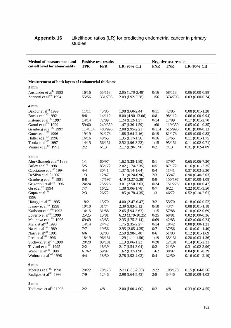
182
Appendix 16 Likelihood ratios (LR) for predicting endometrial cancer in primary studies
Method of measurement and
cut-off level for abnormality
Positive test results Negative test results
TPR FPR LR (95% CI) FNR TNR LR (95% CI)
Measurement of both layers of endometrial thickness 3 mm Auslender et al
14 1993 16/16 55/113 2.05 (1.70-2.48) 0/16 58/113 0.06 (0.00-0.88)
Zannoni et al348
1994 55/56 331/705 2.09 (l.92-2.28) 1/56 374/705 0.03 (0.00-0.24)
4 mm Bakour et al
18 1999 11/11 43/85 1.98 (l.60-2.44) 0/11 42/85 0.08 (0.01-1.28)
Botsis et al36
1992 8/8 14/112 8.00 (4.90-13.06) 0/8 98/112 0.06 (0.00-0.94)
Fistonic et al122
1997 14/14 72/89 1.24 (l.12-1.37) 0/14 17/89 0.17 (0.01-2.70)
Garuti et al130
1999 59/60 240/359 1.47 (l.36-1.59) 1/60 119/359 0.05 (0.01-0.35)
Granberg et al147
1997 114/114 480/996 2.08 (l.95-2.21) 0/114 516/996 0.01 (0.00-0.13)
Guner et al159
1996 19/19 92/173 1.88 (l.64-2.16) 0/19 81/173 0.05 (0.00-0.83)
Haller et al164
1996 16/16 48/65 1.35 (l.17-1.56) 0/16 17/65 0.11 (0.01-1.75)
Tsuda et al328
1997 14/15 56/151 2.52 (l.96-3.22) 1/15 95/151 0.11 (0.02-0.71)
Varner et al334
1991 2/2 6/13 2.17 (l.20-3.90) 0/2 7/13 0.31 (0.02-4.09)
5 mm Abu-Ghazzeh et al
4 1999 1/1 60/97 1.62 (l.38-1.89) 0/1 37/97 0.65 (0.06-7.30)
Briley et al42
1998 5/5 85/172 2.02 (1.74-2.35) 0/5 87/172 0.16 (0.01-2.35)
Cacciatore et al48
1994 4/4 30/41 1.37 (l.14-1.64) 0/4 11/41 0.37 (0.03-5.30)
DeSilva et al78
1997 1/3 12/47 1.31 (0.24-6.96) 2/3 35/47 0.90 (0.40-2.03)
Granberg et al146
1991 8/8 47/197 4.19 (3.27-5.38) 0/8 150/197 0.07 (0.00-1.08)
Grigoriou et al151
1996 24/24 75/226 3.01 (2.50-3.63) 0/24 151/226 0.03 (0.00-0.47)
Gu et al155
1994 7/7 16/22 1.38 (l.06-1.78) 0/7 6/22 0.22 (0.01-3.50)
Gupta et al161
1996
2/3 26/72 1.85 (0.78-4.35) 1/3 46/72 0.52 (0.10-2.61)
Hänggi et al165
1995 18/21 15/70 4.00 (2.47-6.47) 3/21 55/70 0.18 (0.06-0.52)
Ivanov et al172
1998 10/10 31/74 2.39 (l.83-3.12) 0/10 43/74 0.08 (0.01-1.18)
Karlsson et al179
1993 14/15 31/88 2.65 (l.94-3.63) 1/15 57/88 0.10 (0.02-0.69)
Loverro et al216
1999 25/25 13/81 6.23 (3.79-10.25) 0/25 68/81 0.02 (0.00-0.36)
Malinova et al224
1996 69/69 43/85 2.35 (l.75-3.14) 0/69 42/85 0.02 (0.00-0.24)
Merz et al234
1990 14/14 24/42 1.75 (l.35-2.27) 0/14 18/42 0.08 (0.00-1.21)
Nasri et al243
1989 7/7 19/56 2.95 (2.05-4.25) 0/7 37/56 0.10 (0.01-1.40)
Nasri et al244
1991 6/6 32/83 2.59 (l.98-3.40) 0/6 51/83 0.12 (0.01-1.69)
Pertl et al269
1996 18/19 96/131 1.29 (1.11-1.50) 1/19 35/131 0.20 (0.03-1.36)
Suchocki et al316
1998 28/28 89/101 1.13 (l.06-1.22) 0/28 12/101 0.14 (0.01-2.31)
Taviani et al321
1995 2/2 18/39 2.17 (l.54-3.04) 0/2 21/39 0.31 (0.02-3.96)
Weber et al338
1998 61/62 59/97 1.62 (l.37-1.90) 1/62 38/97 0.04 (0.01-0.29)
Wolman et al345
1996 4/4 18/50 2.78 (l.92-4.02) 0/4 32/50 0.16 (0.01-2.19)
6 mm Moreles et al
237 1998 20/22 70/178 2-31 (l.85-2.90) 2/22 108/178 0.15 (0.04-0.56)
Rudigoz et al281
1993 7/9 12/46 2.98 (l.64-5.43) 2/9 34/46 0.30 (0.09-1.03)
8 mm Todorova et al
323 1998 2/2 4/8 2.00 (l.00-4.00) 0/2 4/8 0.33 (0.02-4.55)
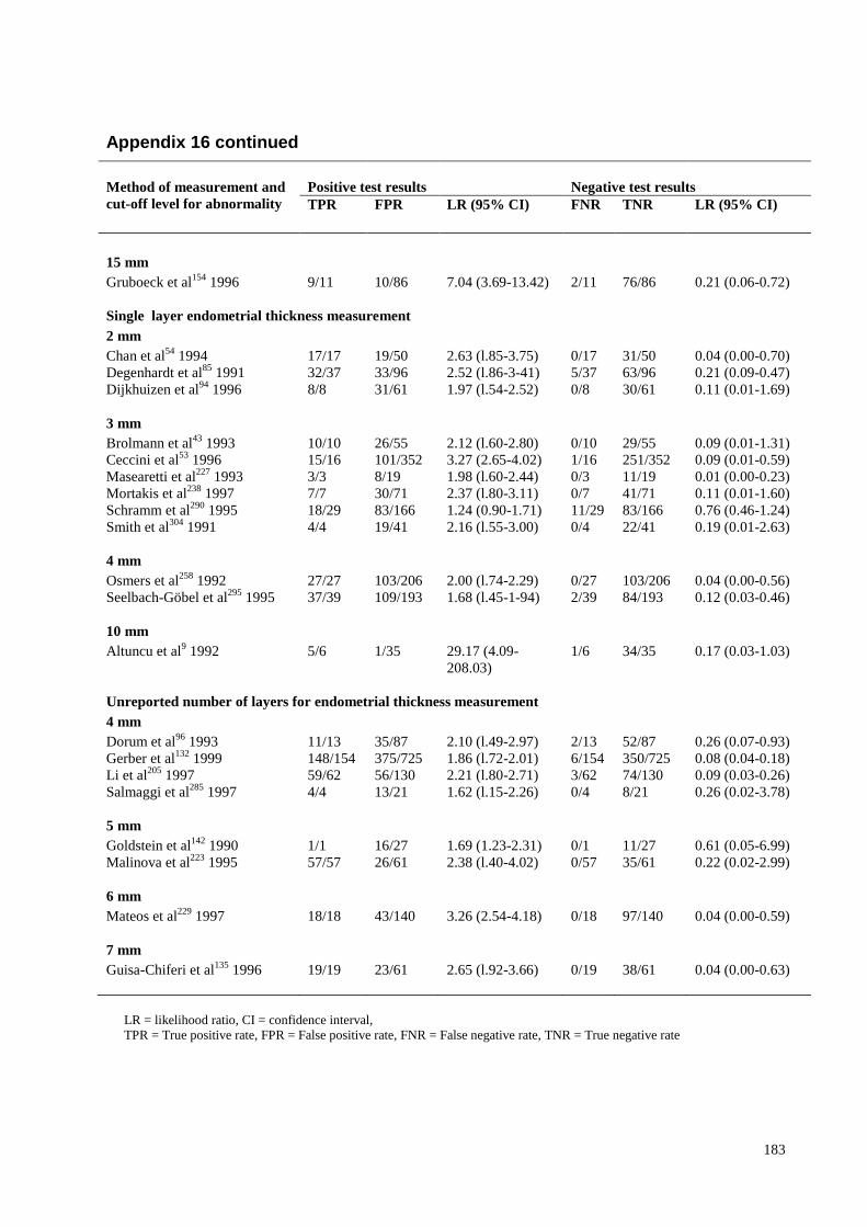
183
Appendix 16 continued
Method of measurement and
cut-off level for abnormality
Positive test results Negative test results
TPR FPR LR (95% CI) FNR TNR LR (95% CI)
15 mm Gruboeck et al
154 1996 9/11 10/86 7.04 (3.69-13.42) 2/11 76/86 0.21 (0.06-0.72)
Single layer endometrial thickness measurement 2 mm Chan et al
54 1994 17/17 19/50 2.63 (l.85-3.75) 0/17 31/50 0.04 (0.00-0.70)
Degenhardt et al85
1991 32/37 33/96 2.52 (l.86-3-41) 5/37 63/96 0.21 (0.09-0.47)
Dijkhuizen et al94
1996 8/8 31/61 1.97 (l.54-2.52) 0/8 30/61 0.11 (0.01-1.69)
3 mm Brolmann et al
43 1993 10/10 26/55 2.12 (l.60-2.80) 0/10 29/55 0.09 (0.01-1.31)
Ceccini et al53
1996 15/16 101/352 3.27 (2.65-4.02) 1/16 251/352 0.09 (0.01-0.59)
Masearetti et al227
1993 3/3 8/19 1.98 (l.60-2.44) 0/3 11/19 0.01 (0.00-0.23)
Mortakis et al238
1997 7/7 30/71 2.37 (l.80-3.11) 0/7 41/71 0.11 (0.01-1.60)
Schramm et al290
1995 18/29 83/166 1.24 (0.90-1.71) 11/29 83/166 0.76 (0.46-1.24)
Smith et al304
1991
4/4 19/41 2.16 (l.55-3.00) 0/4 22/41 0.19 (0.01-2.63)
4 mm Osmers et al
258 1992 27/27 103/206 2.00 (l.74-2.29) 0/27 103/206 0.04 (0.00-0.56)
Seelbach-Göbel et al295
1995 37/39 109/193 1.68 (l.45-1-94) 2/39 84/193 0.12 (0.03-0.46)
10 mm Altuncu et al
9 1992 5/6 1/35 29.17 (4.09-
208.03)
1/6 34/35 0.17 (0.03-1.03)
Unreported number of layers for endometrial thickness measurement 4 mm Dorum et al
96 1993 11/13 35/87 2.10 (l.49-2.97) 2/13 52/87 0.26 (0.07-0.93)
Gerber et al132
1999 148/154 375/725 1.86 (l.72-2.01) 6/154 350/725 0.08 (0.04-0.18)
Li et al205
1997 59/62 56/130 2.21 (l.80-2.71) 3/62 74/130 0.09 (0.03-0.26)
Salmaggi et al285
1997 4/4 13/21 1.62 (l.15-2.26) 0/4 8/21 0.26 (0.02-3.78)
5 mm Goldstein et al
142 1990 1/1 16/27 1.69 (1.23-2.31) 0/1 11/27 0.61 (0.05-6.99)
Malinova et al223
1995 57/57 26/61 2.38 (l.40-4.02) 0/57 35/61 0.22 (0.02-2.99)
6 mm Mateos et al
229 1997 18/18 43/140 3.26 (2.54-4.18) 0/18 97/140 0.04 (0.00-0.59)
7 mm Guisa-Chiferi et al
135 1996 19/19 23/61 2.65 (l.92-3.66) 0/19 38/61 0.04 (0.00-0.63)
LR = likelihood ratio, CI = confidence interval,
TPR = True positive rate, FPR = False positive rate, FNR = False negative rate, TNR = True negative rate
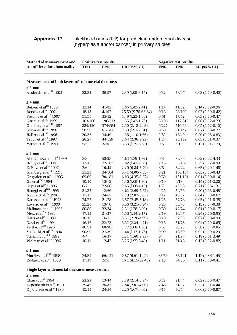
184
Appendix 17 Likelihood ratios (LR) for predicting endometrial disease (hyperplasia and/or cancer) in primary studies
Method of measurement and
cut-off level for abnormality
Positive test results
Negative test results
TPR FPR LR (95% CI) FNR TNR LR (95% CI)
Measurement of both layers of endometrial thickness 3 mm Auslender et al
14 1993 32/32 39/97 2.49 (l.95-3.17) 0/32 58/97 0.03 (0.00-0.40)
4 mm Bakour et al
18 1999 13/14 41/82 1.86 (l.43-2.41) 1/14 41/82 0.14 (0.02-0.96)
Botsis et al36
1992 18/18 4/102 25.50 (9.76-66.64) 0/18 98/102 0.03 (0.00-0.42)
Fistonic et al122
1997 51/51 35/52 1.49 (l.23-1.80) 0/51 17/52 0.03 (0.00-0.47)
Garuti et al130
1999 103/106 196/313 1.55 (l.42-1.70) 3/106 117/313 0.08 (0.02-0.23)
Granberg et al147
1997 220/226 374/884 2.30 (2.12-2.49) 6/226 510/884 0.05 (0.02-0.10)
Guner et al159
1996 50/50 61/142 2.33 (l.93-2.81) 0/50 81/142 0.02 (0.00-0.27)
Haller et al164
1996 30/32 34/49 1.35 (1.10-1.66) 2/32 15/49 0.20 (0.05-0.83)
Tsuda et al328
1997 26/27 44/139 3.04 (2.36-3.93) 1/27 95/139 0.05 (0.01-0.37)
Varner et al334
1991 5/5 3/10 3.33 (l.29-8.59) 0/5 7/10 0.12 (0.01-1.79)
5 mm Abu-Ghazzeh et al
4 1999 3/3 58/95 1.64 (l.39-1.92) 0/3 37/95 0.32 (0.02-4.33)
Briley et al42
1998 13/15 77/162 1.82 (l.41-2.36) 2/15 85/162 0.25 (0.07-0.93)
DeSilva et al78
1997 3/6 10/44 2.20 (0.84-5.79) 3/6 34/44 0.65 (0.29-1.46)
Granberg et al146
1991 21/21 34/184 5.41 (4.00-7.33) 0/21 150/184 0.03 (0.00-0.43)
Grigoriou et al151
1996 69/69 30/181 6.03 (4.35-8.37) 0/69 151/181 0.01 (0.00-0.14)
Gu et al155
1994 10/10 13/19 1.46 (l.08-1.98) 0/10 6/19 0.14 (0.01-2.26)
Gupta et al161
1996 6/7 22/68 2.65 (l.68-4.19) 1/7 46/68 0.21 (0.03-1.31)
Hänggi et al165
1995 21/25 12/66 4.62 (2.69-7.92) 4/25 54/66 0.20 (0.08-0.48)
Ivanov et al172
1998 17/17 24/67 2.79 (2.03-3.85) 0/17 43/67 0.04 (0.00-0.67)
Karlsson et al179
1993 24/25 21/78 3.57 (2.45-5.18) 1/25 57/78 0.05 (0.01-0.38)
Loverro et al216
1999 25/28 13/78 5.36 (3.21-8.94) 3/28 65/78 0.13 (0.04-0.38)
Malinova et al224
1996 80/80 32/74 2.31 (l.78-3.00) 0/80 42/74 0.01 (0.00-0.17)
Merz et al234
1990 17/19 21/37 1.58 (l.14-2.17) 2/19 16/37 0.24 (0.06-0.95)
Nasri et al243
1989 10/10 16/53 3.31 (2.20-4.99) 0/10 37/53 0.07 (0.00-0.99)
Nasri et al244
1991 16/16 22/73 3.32 (2.34-4.71) 0/16 51/73 0.04 (0.00-0.65)
Pertl et al269
1996 46/52 68/98 1.27 (l.08-1.50) 6/52 30/98 0.38 (0.17-0.85)
Suchocki et al316
1998 90/90 27/39 1.44 (l.17-1.78) 0/90 12/39 0.02 (0.00-0.29)
Taviani et al321
1995 4/4 16/37 2.31 (1.60-3.35) 0/4 21/37 0.18 (0.01-2.49)
Wolman et al345
1996 10/11 12/43 3.26 (l.95-5.45) 1/11 31/43 0.13 (0.02-0.82)
6 mm Moreles et al
237 1998 24/59 66/141 0.87 (0.61-1.24) 35/59 75/141 1.12 (0.86-1.45)
Rudigoz et al281
1993 17/19 2/36 16.1 (4.15-62.48) 2/19 34/36 0.11 (0.03-0.41)
Single layer endometrial thickness measurement 2 mm Chan et al
54 1994 23/23 13/44 3.38 (2.14-5.34) 0/23 31/44 0.03 (0.00-0.47)
Degenhardt et al85
1991 39/46 26/87 2.84 (2.01-4.00) 7/46 61/87 0.22 (0.11-0.44)
Dijkhuizen et al94
1996 15/15 24/54 2.25 (l.67-3.03) 0/15 30/54 0.06 (0.00-0.87)
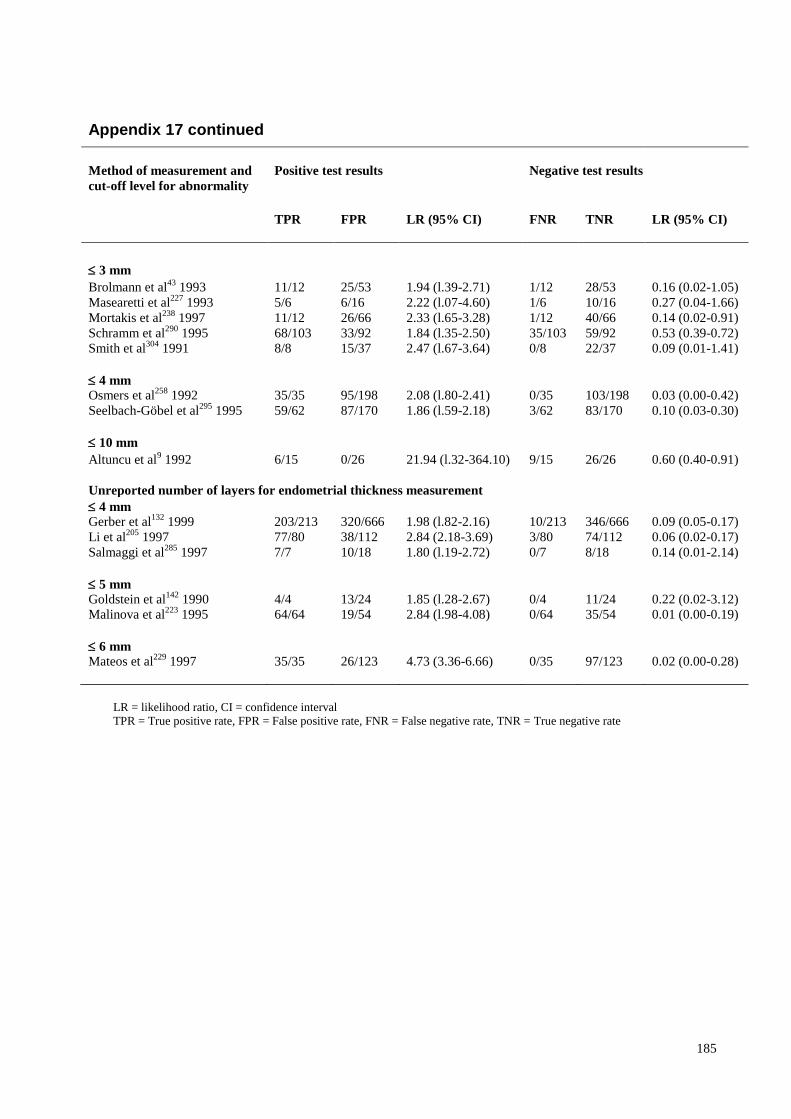
185
Appendix 17 continued
Method of measurement and
cut-off level for abnormality
Positive test results
Negative test results
TPR FPR LR (95% CI) FNR TNR LR (95% CI)
3 mm Brolmann et al
43 1993 11/12 25/53 1.94 (l.39-2.71) 1/12 28/53 0.16 (0.02-1.05)
Masearetti et al227
1993 5/6 6/16 2.22 (l.07-4.60) 1/6 10/16 0.27 (0.04-1.66)
Mortakis et al238
1997 11/12 26/66 2.33 (l.65-3.28) 1/12 40/66 0.14 (0.02-0.91)
Schramm et al290
1995 68/103 33/92 1.84 (l.35-2.50) 35/103 59/92 0.53 (0.39-0.72)
Smith et al304
1991
8/8 15/37 2.47 (l.67-3.64) 0/8 22/37 0.09 (0.01-1.41)
4 mm
Osmers et al258
1992 35/35 95/198 2.08 (l.80-2.41) 0/35 103/198 0.03 (0.00-0.42)
Seelbach-Göbel et al295
1995 59/62 87/170 1.86 (l.59-2.18) 3/62 83/170 0.10 (0.03-0.30)
10 mm Altuncu et al
9 1992 6/15 0/26 21.94 (l.32-364.10) 9/15 26/26 0.60 (0.40-0.91)
Unreported number of layers for endometrial thickness measurement
4 mm
Gerber et al132
1999 203/213 320/666 1.98 (l.82-2.16) 10/213 346/666 0.09 (0.05-0.17)
Li et al205
1997 77/80 38/112 2.84 (2.18-3.69) 3/80 74/112 0.06 (0.02-0.17)
Salmaggi et al285
1997 7/7 10/18 1.80 (l.19-2.72) 0/7 8/18 0.14 (0.01-2.14)
5 mm
Goldstein et al142
1990 4/4 13/24 1.85 (l.28-2.67) 0/4 11/24 0.22 (0.02-3.12)
Malinova et al223
1995 64/64 19/54 2.84 (l.98-4.08) 0/64 35/54 0.01 (0.00-0.19)
6 mm
Mateos et al229
1997 35/35 26/123 4.73 (3.36-6.66) 0/35 97/123 0.02 (0.00-0.28)
LR = likelihood ratio, CI = confidence interval
TPR = True positive rate, FPR = False positive rate, FNR = False negative rate, TNR = True negative rate
186
Appendix 18 Reference list of excluded studies from systematic reviews of hysteroscopy
A1. Valli E, Zupi E, Marconi D, Dini M-L, Di Felice M, Romanini C. A new score for
endometrial lesions based on hysteroscopic parameters: Preliminary results. Gynae
Endosc 1994, 3(3):185-188.
A2. Nagele F, O'Connor H, Baskett TF, Davies A, Mohammed H, Magos AL.
Hysteroscopy in women with abnormal uterine bleeding on hormone replacement
therapy: a comparison with postmenopausal bleeding. Fertil Steril 1996, 65(6):1145-
1150.
A3. La Sala GB, Sacchetti F, Dessanti L. Ambulatory diagnostic hysteroscopy: our
experience with Hamou's microhysteroscopy in 676 patients. Ann Ostet Ginecol Med
Perinat 1984, 105(5):300-307.
A4. La Sala GB, Sacchetti F, Dessanti L, Torelli MG, Sartori F. Panoramic diagnostic
microhysteroscopy: Analysis of results obtained from 976 patients. Acta Eur Fertil
1986, 17(5):369-375.
A5. Garuti G, Sambruni I, Cellani F, Garzia D, Alleva P, Luerti M. Hysteroscopy and
transvaginal ultrasonography in postmenopausal women with uterine bleeding. Int J
Gynaecol Obstet 1999, 65(1):25-33.
A6. Mencaglia L, Perino A. Diagnostic hysteroscopy today. Acta Eur Fertil 1986,
17(6):431-439.
A7. Garuti G, Sambruni I, Iurlaro E, Luerti M. The failure of hysteroscopic view to
differentiate between proliferative disorders of the endometrium. Ital J Obstet
Gynaecol 1999, 11(2):39-46.
A8. Kovar P. Hysteroscopy--a high standard or standard examination method? (Analysis
of 690 diagnostic hysteroscopies). Ceska Gynekol 1998, 63(5):418-422.
A9. Alexopoulos ED, Simonis CD, Kidsley S, Fay TN. The value of outpatient
hysteroscopy in the management of postmenopausal bleeding: A review of 862 cases.
Gynaecol Endosc 2000, 9(2):107-112.
A10. Woolcott R, Petchpud A. The efficacy of hysteroscopy: a comparison of women
presenting with infertility versus other gynaecological symptoms [see comments].
Aust NZ J Obstet Gynaecol 1995, 35(3):310-313.
A11. La Sala GB, Torelli MG, Dessanti L, Cigarini C, Sartori F. Usefulness of hysteroscopy
in staging of endometrial cancer: Analysis of results obtained from 96 patients.
Gynaecol Endosc 1992, 1(2):103-106.
187
A12. Tanizawa O, Miyake A, Sugimoto O. Re-evaluation of hysteroscopy in the diagnosis
of uterine endometrial cancer. Nippon Sanka Fujinka Gakkai Zasshi - Acta Obstet
Gynaecol Jap 1991, 43(6):622-626.
A13. Rullo S, Piccioni MG, Framarino dei Malatesta ML, Silvestrini J, Boni T, Marzetti L.
Sonographic, hysteroscopic, histological correlation in the early diagnosis of
endometrial cancer. Eur J Gynaecol Oncol 1991, 12(6):463-469.
A14. Colafranceschi M, Van Herendael B, Perino A, Bettocchi S, Tantini C, Mencaglia L et
al. Reliability of endometrial biopsy under direct hysteroscopic control. Gynaecol
Endosc 1995, 4(2):119-122.
A15. Conoscenti G, Meir YJ, Fischer-Tamaro L, Maieron A, Natale R, D'Ottavio et al.
Endometrial assessment by transvaginal sonography and histological findings after D
& C in women with postmenopausal bleeding. Ultrasound Obstet Gynecol 1995,
6(2):108-115.
A16. Mortakis AE, Mavrelos K. Transvaginal ultrasonography and hysteroscopy in the
diagnosis of endometrial abnormalities. J Am Assos Gynecol Laparosc 1997, 4(4):449-
452.
A17. Rudigoz RC, Salle B, Piacenza JM, Saint-Hilaire P, Gaucherand P.
Hysterosonographic study of the uterine cavity. J Gynecol Obstet Biol Reprod 1995,
24(7):697-704.
A18. Bronz L, Suter T, Rusca T. The value of transvaginal sonography with and without
saline instillation in the diagnosis of uterine pathology in pre- and postmenopausal
women with abnormal bleeding or suspect sonographic findings. Ultrasound Obstet
Gynecol 1997, 9(1):53-58.
A19. Van den BT, Vandendael A, Van Schoubroeck D, Wranz PA, Lombard CJ.
Combining vaginal ultrasonography and office endometrial sampling in the diagnosis
of endometrial disease in postmenopausal women. Obstet Gynecol 1995, 85(3):349-
352.
A20. Dubinsky TJ, Parvey HR, Gormaz G, Curtis M, Maklad N. Transvaginal
hysterosonography: comparison with biopsy in the evaluation of postmenopausal
bleeding. J Ultrasound Med 1995, 14(12):887-893.
A21. Valenzano M, Costantini S, Cucuccio S, Dugnani MC, Paoletti R, Ragni N. Use of
hysterosonography in women with abnormal postmenopausal bleeding. Eur J
Gynaecol Oncol 1999, 20(3):217-222.
A22. Pace S, Villani C, Lotti G, Labi FL. Hysteroscopy as an elective tool in abnormal
uterine bleeding in perimenopausal women. Eur J Gynaecol Oncol 1992, 13(5):409-
413.
188
A23. Finikiotis G. Hysteroscopy: an analysis of 523 patients. Aust NZ J Obstet Gynaecol
1989, 29(3 Pt 1):253-255.
A24. Giusa-Chiferi MG, Goncalves WJ, Baracat EC, Albuquerque Neto LC, Bortoletto CC,
de Lima GR. Transvaginal ultrasound, uterine biopsy and hysteroscopy for
postmenopausal bleeding. Int J Gynaecol Obstet 1996, 55(1):39-44.
A25. Uno LH, Sugimoto O, Carvalho FM, Bagnoli VR, Fonseca AM, Pinotti JA.
Morphologic hysteroscopic criteria suggestive of endometrial hyperplasia. Int J
Gynaecol Obstet 1995, 49(1):35-40.
A26. Badawy A, Ash A, Nagele F, O'Connor H, Davis A, Magos AL. Is it worth taking a
biopsy of the normal-looking endometrium? Gynaecol Endosc 1996, 5(4):225-229.
A27. Cooper MJ, Broadbent JA, Molnar BG, Richardson R, Magos AL. A series of 1000
consecutive out-patient diagnostic hysteroscopies. J Obstet Gynaecol 1995, 21(5):503-
507.
A28. Vigada G, Malanetto C. [he role of hysteroscopy in the early diagnosis of small, focal
endometrial neoplasms. Minerva Ginecol 1996, 48(11):493-495.
A29. Mencaglia L, Scarselli G, Tantini C. Hysteroscopic evaluation of endometrial cancer.
J Reprod Med 1984, 29(10):701-704.
A30. Kimmig R, Hillemanns P, Hepp H. The diagnostic hysteroscopy - A new standard.
Gynakologe 1997, 30(5):384-391.
A31. Semeraro A, Vecchione A. Rilievi isteroscopica in donne con stillicidio ematico
perimestruale. Pat Clin Ost Gin 1984, 12:497-501.
A32. Surico N, Ragonesi G, Porcelli A, Marengo F, Wierdis T. Hysteroscopic diagnosis
during metrorrhagia. Minerva Ginecol 1985, 37(9):519-521.
A33. Saccucci P, Rigon G, Provenza C, Mastrone M, Are P, Pisani G et al. Hysteroscopic
features in postmenopausal uterine bleeding. Minerva Ginecol 1996, 48(10):401-404.
A34. de Cesare E, Fabrizio L, Polidoro M. Early diagnosis of endometrial cancer.
Introduction of hysteroscopy. Minerva Ginecol 1985, 37(3):93-97.
A35. Gubbini G, Linsalata I, Stagnozzi R, Stefanetti M, Bovicelli A, Vecchio et al.
Outpatient diagnostic hysteroscopy: 14,000 cases. Minerva Ginecol 1996, 48(9):383-
390.
A36. Conoscenti G, Meir Y, Fischer-Tamaro L, Maieron A, Natale R, D'Ottavio et al. The
diagnostic capacities of transvaginal echography and hysteroscopy in the
characterization of endometrial pathology. Minerva Ginecol 1995, 47(7-8):293-300.
189
A37. Persiani P, Perotti F, Riccardi A, Gallina D, Polatti F, Zara C. Diagnostic accuracy in
transvaginal echography in benign endometrial diseases and its comparison with
hysteroscopic biopsy. Minerva Ginecol 1995, 47(3):63-67.
A38. Sajdak S, Michalska M, Kedzia H, Spaczynski M. Usefulness of transvaginal
ultrasound and hysteroscopy in diagnosing endometrial hyperplasia and endometrial
cancer. Ginecol Pol 1993, 64(9):431-437.
A39. Wamsteker K, de Blok S, de Wit W. Diagnostic and therapeutic applications of
hysteroscopy. Ned Tijdschr Geneeskd 1988, 132(45):2041-2044.
A40. Walton SM, Macphail S. The value of hysteroscopy in postmenopausal and
perimenopausal bleeding. J Obstet Gynaecol 1988, 8(4):332-336.
A41. Pace S, Grassi A, Ferrero S, Figliolini N, Catania R, Labi FL et al. Diagnostic
methods of early detection of endometrial hyperplasia and cancer. Eur J Gynaecol
Oncol 1995, 16(5):373-381.
A42. Townsend DE, Fields G, McCausland A, Kauffman K. Diagnostic and operative
hysteroscopy in the management of persistent postmenopausal bleeding. Obstet
Gynecol 1993, 82(3):419-421.
A43. Saidi MH, Sadler RK, Theis VD, Akright BD, Farhart SA, Villanueva GR.
Comparison of sonography, sonohysterography, and hysteroscopy for evaluation of
abnormal uterine bleeding. J Ultrasound Med 1997, 16(9):587-591.
A44. Karlsson B, Granberg S, Hellberg P, Wikland M. Comparative study of transvaginal
sonography and hysteroscopy for the detection of pathologic endometrial lesions in
women with postmenopausal bleeding. J Ultrasound Med 1994, 13(10):757-762.
A45. Torrejon R, Fernandez-Alba JJ, Carnicer I, Martin A, Castro C, Garcia-Cabanillas J et
al. The value of hysteroscopic exploration for abnormal uterine bleeding. J Am Assos
Gynecol Laparosc 1997, 4(4):453-456.
A46. Luo QD. Evaluation of hysteroscopy in the diagnosis of abnormal uterine bleeding.
Chung-Hua Fu Chan Ko Tsa Chih 1991, 21(3):152-154.
A47. Coloma CF, Paya A, V, Diago A, V, Costa CS, Valero, Fenollosa V et al. 2,000 out-
patient diagnostic hyteroscopies: 8 years of experience. Prog Obstet Gin 1998,
41(6):347-352.
A48. Hamou J, Salat-Baroux J. Microcolpohysteroscopie dans les metrorragies. Contracept
Fertil Sex 1985, 13:389-394.
A49. Concin H, Bosch H, Schwarzler P. Hysteroscopy--applications and risks.
Hysteroscopy versus fractionated curettage: therapeutic insufficiency of abrasion.
Gynakol Geburtshilfliche Rundsch 1995, 35(2):114-116.
190
A50. Motashaw ND. Experience with the hysteroscope. Acta Eur Fertil 1986, 17(6):417-
418.
A51. Dexeus S, Labastida R, Marques L. Hysteroscopy in daily gynaecologic practice. Acta
Eur Fertil 1986, 17(6):423-425.
A52. Font-Sastre V, Carabias J, Bonilla-Musoles F, Pellicer A. Office hysteroscopy with
small calibre instruments. Acta Eur Fertil 1986, 17(6):427-429.
A53. Mazzon I, Scotto V, Guidi ML, Vittori G, Ricci G, Crisci G et al. Outpatient
hysteroscopy in the diagnosis of neoplastic and preneoplastic lesions of the
endometrium. Eur J Gynaecol Oncol 1988, 9(3):261-264.
A54. Grozdanov G, Malinova M. Uterine hemorrhage as an indication for performing
contact hysteroscopic examination. Akush Ginekol 1989, 28(1):58-60.
A55. Feng L, Xia E, Duan H. Diagnosis of uterine diseases by combined hysteroscopy and
ultrasonography. Chung-Hua Fu Chan Ko Tsa Chih 1996, 31(6):334-337.
A56. Bielanow T, Gabrys M, Woyton J, Koltowska M, Hirnle L, Zmijewski J. Indications
and results of hysteroscopy in own material. Acta Endosc Pol 1993, 3(1):37-39.
A57. Ben Hmid R, Mahjoub S, Boughizane S, Dakhli R, Smaili L, Lebbi I et al. Value of
ambulatory diagnostic hysteroscopy. A review of a series of 292 cases. Tunisie
Medicale 2000, 78(10):600-606.
A58. Chechia A, Koubaa A, Makhlouf T, Terras K, Miaadi N. Comparison of
ultrasonographic and hysteroscopic results in perimenopausal metrorrhagias. Tunisie
Medicale 2001, 79(4):238-241.
A59. Pasqualotto EB, Margossian H, Price LL, Bradley LD. Accuracy of preoperative
diagnostic tools and outcome of hysteroscopic management of menstrual dysfunction.
J Am Assoc Gynecol Laparosc 2000, 7(2):201-209.
A60. Spiewankiewicz B, Stelmachow J, Sawicki W, Kietlinska Z. Hysteroscopy with
selective endometrial sampling after unsuccessful dilatation and curettage in diagnosis
of symptomatic endometrial cancer and endometrial hyperplasias. Eur J Gynaecol
Oncol 1995, 16(1):26-29.
A61. Gimpelson RJ, Rappold HO. A comparative study between panoramic hysteroscopy
with directed biopsies and dilatation and curettage. A review of 276 cases. Am J
Obstet Gynecol 1988, 158(3 Pt 1):489-492.
A62. Dijkhuizen FP, Brolmann HA, Potters AE, Bongers MY, Heinz AP. The accuracy of
transvaginal ultrasonography in the diagnosis of endometrial abnormalities. Obstet
Gynecol 1996, 87(3):345-349.
191
A63. Loffer FD. Hysteroscopy with selective endometrial sampling compared with D&C
for abnormal uterine bleeding: the value of a negative hysteroscopic view. Obstet
Gynecol 1989, 73(1):16-20.
A64. Emanuel MH, Verdel MJ, Wamsteker K, Lammes FB. A prospective comparison of
transvaginal ultrasonography and diagnostic hysteroscopy in the evaluation of patients
with abnormal uterine bleeding: clinical implications. Am J Obstet Gynecol 1995,
172(2 Pt 1):547-552.
A65. O'Connell LP, Fries MH, Zeringue E, Brehm W. Triage of abnormal postmenopausal
bleeding: a comparison of endometrial biopsy and transvaginal sonohysterography
versus fractional curettage with hysteroscopy. Am J Obstet Gynecol 1998, 178(5):956-
961.
A66. Goldrath MH, Sherman AI. Office hysteroscopy and suction curettage: can we
eliminate the hospital diagnostic dilatation and curettage? Am J Obstet Gynecol 1985,
152(2):220-229.
A67. Mathew M, Gupta R, Krolikowski A. Role of transvaginal ultrasonography and
diagnostic hysteroscopy in the evaluation of patients with abnormal uterine bleeding.
Int J Gynaecol Obstet 2000, 71(3):251-253.
A68. Etherington IJ, Harrison KR, Read MD. A comparison of outpatient endometrial
sampling with hysteroscopy, curettage and cystoscopy in the evaluation of
postmenopausal bleeding. J Obstet Gynaecol 1995, 15(4):259-262.
A69. Indman PD. Abnormal uterine bleeding. Accuracy of vaginal probe ultrasound in
predicting abnormal hysteroscopic findings. J Reprod Med 1995, 40(8):545-548.
A70. Vercellini P, Vendola N, Ragni G, Trespidi L, Oldani S, Crosignani PG. Abnormal
uterine bleeding associated with iron-deficiency anemia. Etiology and role of
hysteroscopy. J Reprod Med 1993, 38(7):502-504.
A71. Cronje HS. Diagnostic hysteroscopy after postmenopausal uterine bleeding. S Afr Med
J 1984, 66(20):773-774.
A72. Suprun HZ, Taendler-Stolero R, Schwartz J, Ettinger M. Experience with Endopap
endometrial sampling in the cytodiagnosis of endometrial cancer and its precursor
lesions. I. A correlative cytologic-histologic-hysteroscopic diagnostic pilot study. Acta
Cytologica 1994, 38(3):319-323.
A73. Marty R. Experience with a new flexible hysteroscope. Int J Gynaecol Obstet 1988,
27(1):97-99.
A74. Goldfarb HA. D&C results improved by hysteroscopy. N J Med 1989, 86(4):277-279.
A75. Gaglione R, Cinque B, Paparatti L, Pistilli E. Hysteroscopy: A milestone in
gynaecology. Gynaecol Endosc 1996, 5(6):319-322.
192
A76. Blanc B, Cravello L, D'Ercole C, Roger V, Porcu G. Investigations of the
endometrium. Contribution of endometrectomy. Rev Fr GynecolObstetr 1998,
93(1):29.
A77. Zavodny P, Kudela M, L'ubusky D, Pilka R. Experience with diagnostic and
therapeutic hysteroscopy. Ceska Gynekol 1998, 63(1):82-83.
A78. Struzziero E, Corbo M. Use of panoramic hysteroscopy in the differential diagnosis of
abnormal uterine bleeding. Minerva Ginecol 1989, 41(7):329-330.
A79. Rudigoz RC, Frobert C, Chassagnard F, Gaucherand P. The place of vaginal
ultrasound in investigation of the normal bleeding patterns during reproductive life. J
Gynecol Obstet Biol Reprod 1992, 21(6):644-650.
A80. Motashaw ND, Dave S. Diagnostic and therapeutic hysteroscopy in the management
of abnormal uterine bleeding. J Reprod Med 1990, 35(6):616-620.
A81. Hidlebaugh D. A comparison of clinical outcomes and cost of office versus hospital
hysteroscopy. J Am Assos Gynecol Laparosc 1996, 4(1):39-45.
A82. Marty R. Diagnostic fibrohysteroscopic evaluation of perimenopausal and
postmenopausal uterine bleeding: a comparative study with Belgian and Japanese data.
J Am Assos Gynecol Laparosc 1998, 5(1):69-73.
A83. Brooks PG, Serden SP. Hysteroscopic findings after unsuccessful dilatation and
curettage for abnormal uterine bleeding. Am J Obstet Gynecol 1988, 158(6 Pt 1):1354-
1357.
A84. Choo YC, Mak KC, Hsu C, Wong TS, Ma HK. Postmenopausal uterine bleeding of
nonorganic cause. Obstet Gynecol 1985, 66(2):225-228.
A85. Downes E, al Azzawi F. The predictive value of outpatient hysteroscopy in a
menopause clinic. Br J Obstet Gynaecol 1993, 100(12):1148-1149.
A86. Valli E, Zupi E, Marconi D, Solima E, Nagar G, Romanini C. Outpatient diagnostic
hysteroscopy. J Am Assos Gynecol Laparosc 1998, 5(4):397-402.
A87. Bradley LD, Widrich T. State-of-the-art flexible hysteroscopy for office gynecologic
evaluation. J Am Assos Gynecol Laparosc 1995, 2(3):263-267.
A88. Chapman JD, Sherman RH. Hysteroscopy: a prospective examination of its value. J
Am Osteopath Assoc 1986, 86(4):219-223.
A89. Schaaps JP, Dubois M, Vosse M, Verheyen M, Lambotte R. Ultrasonic exploration of
the uterine cavity: Which pertinence? Contracept Fertil Sex 1991, 19(11):929-934.
193
A90. Barroco LE, Oliveira LC, Sa-Melo P. "Office hysteroscopy" experience with Hamou
microcolpohysteroscope in 250 patients. Acta Eur Fertil 1986, 17(6):419-421.
A91. Henie O, I, Maltau JM. Hysteroscopy. Tidsskr Nor Laegeforen 1985, 105(22):1397-
1398.
A92. Giannola C, Carducci AF, Musso P. Hysteroscopy and its applications. Considerations
on a year of clinical experience. Minerva Ginecol 1986, 38(4):265-269.
A93. Duncan GR, Weerasinghe DS. The diagnostic possibilities of a modified
hysteroscopic technique. NZ Med J 1985, 98(773):101-103.
A94. Rossetti D, Gerli S, Saab JC, Di Renzo GC. Diagnostic hysteroscopy and endometrial
pathology. Revue Medicale Libanaise 1999, 11(2):69-71.
A95. Makris N, Xygakis A, Michalas S, Dachlythras M, Prevedourakis C. Day clinic
diagnostic hysteroscopy in a state hospital. Clin Exp Obstet Gynecol 1999, 26(2):91-
92.
A96. Roman JD, Trivedi AN. Implementation of an outpatient hysteroscopy clinic at
Waikato Women's Hospital report of the first 60 cases. NZ Med J 1999,
112(1091):253-255.
A97. Labastida R, Ubeda A, Cararach M, Penella J. 5059 diagnostic hysteroscopies:
Method, indications and results. Prog Obstet Gin 1994, 37(6):348-354.
A98. Pagano R, Pou J, Sanchez I, Vanrell JA. Diagnostic hysteroscopy with CO2:
Indications and results. Prog Obstet Gin 1995, 38(8):528-532.
A99. Arnott N, Phillips WDP. An audit of outpatient hysteroscopy clinic at Perth Royal
Infirmary after the introduction of referral guidelines. J Obstet Gynaecol 1998,
18(1):93.
A100. Grio R, Malara D, Curti A, Porpiglia M. Abnormal uterine bleeding during
climacteric. Correlation between transvaginal ultrasonography, hysteroscopy and
histology. Minerva Ginecol 1999, 51(4):125-127.
A101. Wortman M, Daggett A. Hysteroscopic management of intractable uterine bleeding. A
review of 103 cases. J Reprod Med 1993, 38(7):505-510.
A102. Emanuel MH, Wamsteker K, Lammes FB. Is dilatation and curettage obsolete for
diagnosing intrauterine disorders in premenopausal patients with persistent abnormal
uterine bleeding? Acta Obstet Gynecol Scand 1997, 76(1):65-68.
A103. Finikiotis G. The hyperaemic endometrium at hysteroscopy. Aust NZ J Obstet
Gynaecol 1990, 30(4):351-353.
194
A104. Ramsay JE, Calder AL, Hart DM, Habiba MA, Akkad AA, al Azzawi F.
Hysteroscopic investigation of bleeding in women receiving tibolone: A case-control
study. Gynaecol Endosc 1998, 7(3):115-119.
A105. Kent ASH, Haines P, Manners BTB, Coats PM. Blind endometrial biopsies:
Insufficient for diagnosis in women with intrauterine pathology. Gynaecol Endosc
1998, 7(5):273-278.
A106. Gubbini G, Filoni M, Linsalata I, Stagnozzi R, Stefanetti M, Marabini A. The role of
hysteroscopy in the diagnosis and follow-up of endometrial hyperplasia. Minerva
Ginecol 1998, 50(4):125-133.
A107. Alcazar JL, Laparte C. Comparative study of transvaginal ultrasonography and
hysteroscopy in postmenopausal bleeding. Gynecol Obstet Invest 1996, 41(1):47-49.
A108. Towbin NA, Gviazda IM, March CM. Office hysteroscopy versus transvaginal
ultrasonography in the evaluation of patients with excessive uterine bleeding. Am J
Obstet Gynecol 1996, 174(6):1678-1682.
A109. Wieser F, Albrecht A, Kurz C, Wenzl R, Nagele F. Ambulatory hysteroscopy in
evaluation of postmenopausal bleeding. Wien Klin Wochenschr 1999, 111(7):289-293.
A110. Gimpelson RJ. Panoramic hysteroscopy with directed biopsies vs. dilatation and
curettage for accurate diagnosis. J Reprod Med 1984, 29(8):575-578.
A111. Lewis BV. Hysteroscopy in gynaecological practice: a review. J Royal Soc Med 1984,
77(3):235-237.
A112. Lindemann HJ. Hysteroscopy: the state of the art. Eur J Obstet Gynecol Reprod Biol
1994, 53(2):79-80.
A113. Livsey R. The efficacy of hysteroscopy. Aust NZ J Obstet Gynaecol 1996, 36(2):226-
227.
A114. ACOG criteria set. Hysteroscopy, diagnostic, for abnormal uterine bleeding. Number
16, May 1996. Committee on Quality Assessment. American College of Obstetricians
and Gynecologists. Int J Gynaecol Obstet 1996, 54(1):78-79.
A115. Kohorn EI. Hysteroscopy in the management of endometrial cancer. Obstet Gynecol
Clin North Am 1988, 15(1):73-75.
A116. Gerber B, Krause A, Quasmeh A, Rohde E, Reimer T, Friese K. The value of
hysteroscopy and D and C in the assessment of postmenopausal uterine bleeding.
Geburtshilfe Frauenheil 1998, 58(8):440-445.
A117. Bertone C, Osnengo G, Bigano G, Leoni G, De Ambrosis C. Hysteroscopy in the
morphologic and functional study of the fallopian tubes. Minerva Ginecol 1984,
36(12):783-785.
195
A118. Kulakov VI, Adamian LV, Beloglazova SE. Diagnostic and surgical hysteroscopy.
Akush Ginekol 1993,(4):55-59.
A119. Hucke J, Beck L. Diagnostic and therapeutic hysteroscopy. Clin Exp Obstet Gynecol
1992, 19(4):275-276.
A120. Valle RF. Hysteroscopy. Curr Opin Obstet Gynecol 1991, 3(3):422-426.
A121. Saidi MH, Schenken RS. Outpatient diagnostic hysteroscopies. Obstet Gynecol 1996,
88(5):900-901.
A122. Downes E, al Azzawi F. Hysteroscopic views and endometrial behavior. Am J Obstet
Gynecol 1994, 171(6):1673-1674.
A123. Loffer FD. Does hysteroscopy improve upon the sensitivity of dilation and curettage
in the diagnosis of endometrial hyperplasia or cancer? Gynecol Oncol 1999,
73(1):171.
A124. Oladipo A, al Azzawi F. The predictive value of outpatient hysteroscopy in a
menopause clinic. Br J Obstet Gynaecol 1994, 101(12):1101.
A125. De Mendonca R, Kay TT, Emanuel MH, Verdel MJ, Wamsteker K, Lammes FB.
Value of transvaginal ultrasonography and hysteroscopy in the evaluation of
postmenopauaal women with metrorrhagia. Am J Obstet Gynecol 1995, 173(4):1352-
1353.
A126. Yazicioglu HF. A clear hysteroscopic view and the use of other diagnostic modalities
in addition to hysteroscopy to achieve better diagnostic accuracy [letter, comment].
Am J Obstet Gynecol 1997, 176(4):950-951.
A127. Habiba MA. Diagnostic accuracy of outpatient hysteroscopy. Am J Obstet Gynecol
1997, 176(6):1399-1400.
A128. Gimpelson RJ, Whalen TR. Hysteroscopy as gold standard for evaluation of abnormal
uterine bleeding [letter, comment]. Am J Obstet Gynecol 1995, 173(5):1637-1638.
A129. Beillon X. Hysteroscopy. Soins Gynecol Obstet Pueric 1987,(73-74):11-12.
A130. Karateeva AN, Mitrofanov OP. The Hy-VS-1 hysteroscope with fiber light guide. Med
Tekh 1984,(3):45-46.
A131. Rodriguez CA, Giribert RE. Revista de Enfermeria 1996, 19(213):67-70.
A132. Marty R. The use of a new flexible hysteroscope: Our diagnostic and operative
experience over 84 cases. Contracept Fertil Sex 1987, 15(6):593-594.
196
A133. Lavieille F. Hysteroscopy ambulatory diagnosis in 1994. Gynecologie - Revue du
Gynecologue 1994, 2(5):315-317.
A134. Saidi MH. Office hysteroscopy versus transvaginal ultrasonography in the evaluation
of patients with excessive uterine bleeding. Am J Obstet Gynecol 1997, 176(2):492-
493.
A135. Hysteroscopy. Obstet Gynecol Clin North Am 1988, 15(1):1-166.
A136. Vesterdal A. Bleeding disorders. Endoscopic examination. Sygeplejersken 1993,
93(26):14.
A137. Lewis BV. Hysteroscopy for the investigation of abnormal uterine bleeding. Br J
Obstet Gynaecol 1990, 97(4):283-284.
A138. Paraskevaides EC, Ayoade G. Hysteroscopic detection of early endometrial cancer. J
Obstet Gynaecol 1992, 12(3):208.
A139. Bargelli G, Tantini C, Savino L, Venturini N, Noci I, Sarselli G. Hysteroscopy in
perimenopausal women. Cervix & the Lower Female Genital Tract 1988, 6(3):253-
257.
A140. Loverro G, Bettocchi S, Vicino M, Selvaggi L. Diagnosis of endometrial hyperplasia
in women with abnormal uterine bleeding. Acta Eur Fertil 1994, 25(1):23-25.
A141. Piccolboni G, Arlacchi E, Cattani P, Zardini R, Lavanda E, Zardini E. Diagnostic
value of hysteroscopy: correlation with histological findings after dilatation and
curettage and hysterectomy. Acta Eur Fertil 1991, 22(4):233-234.
A142. Pungetti D, Dimicco R, Mattucci M, Nardi M, Maruizio G, Lenzi M et al. A
comparative study between panoramic hysteroscopy and endometrial biopsy. Analysis
of 150 cases. Acta Eur Fertil 1990, 21(4):201-203.
A143. Vardhan S, Mohan S, Ranjan P. Hysteroscopy in postmenopausal bleeding. Medical
Journal Armed Forces India 2001, 57(2):114-116.
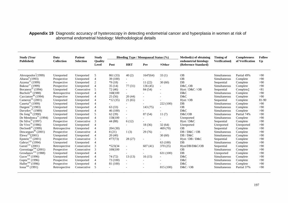
197
Appendix 19 Diagnostic accuracy of hysteroscopy in detecting endometrial cancer and hyperplasia in women at risk of abnormal endometrial histology: Methodological details
Study (Year Data Patient Study Bleeding Type / Menopausal Status (%) Method(s) of obtaining Timing of Completeness Follow
Published) Collection Selection Quality endometrial histology Verification§ of Verification Up
Level Post HRT Pre †Other (Reference Standard)
Alexopoulos7(1999) Unreported Unreported 5 861 (33) 40 (2) 1647(64) 33 (1) OB Simultaneous Partial 49% >90
Altaras8 (1993) Prospective Unreported 4 39 (100) - - - OB Simultaneous Complete >90
Azzena15 (1999) Prospective Unreported 2 *9 (18) - 11 (22) 30 (60) DB Sequential Complete >90
Bakour17 (1999) Prospective Unreported 4 35 (14) 77 (31) 136 (45) - D&C, OB Simultaneous Complete >90
Bocanera32 (1994) Unreported Consecutive 5 72 (46) - 84 (54) - Hyst / D&C / OB Sequential Complete‡ <81
Bucholz45 (1988) Retrospective Unreported 4 168(100 - - - D&C Simultaneous Complete >90
Cacciatore48 (1994) Prospective Unreported 4 25 (56) 20 (44) - - D&C Simultaneous Complete >90
Cameron50 (2001) Unreported Unreported 4 *12 (35) 21 (65) - - Hyst / OB Sequential Complete 81-90
Caserta52 (1999) Unreported Unreported 4 - - - 222 (100) DB Simultaneous Complete >90
Dargent74 (1983) Unreported Unreported 4 63 (33) - 143 (75) - OB Simultaneous Complete >90
Davydov75 (1989) Unreported Unreported 4 46 (100) - - - D&C Simultaneous Complete >90
De Jong76 (1990) Unreported Unreported 5 62 (39) - 87 (54) 11 (7) D&C/OB Simultaneous Partial 74% >90
De Mendonca77 (1994) Unreported Unreported 4 158(100 - - - Unreported Simultaneous Complete >90
De Silva78 (1997) Prospective Consecutive 3 44 (88) 6 (12) - - Hyst / D&C Sequential Complete >90
De Vivo79 (1986) Unreported Unreported 4 - - 18 (36) 32 (64) Unreported Unreported Unreported >90
Decloedt81 (1999) Retrospective Unreported 4 204 (30) - 469 (70) OB Sequential Complete >90
Descargues90 (2001) Prospective Consecutive 4 8 (21) 1 (3) 29 (76) - DB / D&C / OB Simultaneous Complete >90
Elewa112(2001) Unreported Unreported 4 20 (40) 30 (60) DB / D&C Simultaneous Complete >90
Epstein115 (2001) Prospective Consecutive 3 #77(73) 28 (27) - - Hyst / DB / D&C Sequential Complete >90
Gabrys129 (1994) Unreported Unreported 4 63 (100) DB Simultaneous Complete >90
Garuti131 (2001) Retrospective Consecutive 3 *523(34 - 607 (41) 370 (25) Hyst/DB/D&C/OB Sequential Complete >90
Gorostiaga144 (2001) Prospective Consecutive 3 100(100 - - - OB Simultaneous Complete >90
Grosdanov153 (1988) Unreported Unreported 4 - - - 631 (100) DB Unreported Complete >90
Gucer156 (1996) Unreported Unreported 4 74 (72) 13 (13) 16 (15) - D&C Simultaneous Complete >90
Gupta161 (1996) Prospective Unreported 4 73 (100) - - - D&C Simultaneous Complete >90
Haller164 (1996) Prospective Unreported 4 81 (100) - - - D&C Simultaneous Complete >90
Iossa169 (1991) Retrospective Consecutive 5 - - - 815 (100) D&C / OB Simultaneous Partial 37% >90
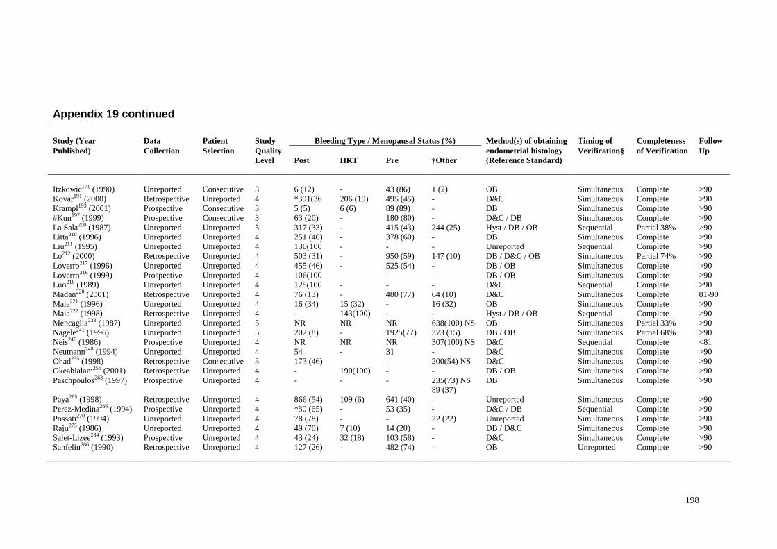
198
Appendix 19 continued
Study (Year Data Patient Study Bleeding Type / Menopausal Status (%) Method(s) of obtaining Timing of Completeness Follow
Published) Collection Selection Quality endometrial histology Verification§ of Verification Up
Level Post HRT Pre †Other (Reference Standard)
Itzkowic171 (1990) Unreported Consecutive 3 6 (12) - 43 (86) 1 (2) OB Simultaneous Complete >90
Kovar191 (2000) Retrospective Unreported 4 *391(36 206 (19) 495 (45) - D&C Simultaneous Complete >90
Krampl193 (2001) Prospective Consecutive 3 5 (5) 6 (6) 89 (89) - DB Simultaneous Complete >90
#Kun197 (1999) Prospective Consecutive 3 63 (20) - 180 (80) - D&C / DB Simultaneous Complete >90
La Sala200 (1987) Unreported Unreported 5 317 (33) - 415 (43) 244 (25) Hyst / DB / OB Sequential Partial 38% >90
Litta210 (1996) Unreported Unreported 4 251 (40) - 378 (60) - DB Simultaneous Complete >90
Liu211 (1995) Unreported Unreported 4 130(100 - - - Unreported Sequential Complete >90
Lo212 (2000) Retrospective Unreported 4 503 (31) - 950 (59) 147 (10) DB / D&C / OB Simultaneous Partial 74% >90
Loverro217 (1996) Unreported Unreported 4 455 (46) - 525 (54) - DB / OB Simultaneous Complete >90
Loverro216 (1999) Prospective Unreported 4 106(100 - - - DB / OB Simultaneous Complete >90
Luo218 (1989) Unreported Unreported 4 125(100 - - - D&C Sequential Complete >90
Madan220 (2001) Retrospective Unreported 4 76 (13) - 480 (77) 64 (10) D&C Simultaneous Complete 81-90
Maia221 (1996) Unreported Unreported 4 16 (34) 15 (32) - 16 (32) OB Simultaneous Complete >90
Maia222 (1998) Retrospective Unreported 4 - 143(100) - - Hyst / DB / OB Sequential Complete >90
Mencaglia233 (1987) Unreported Unreported 5 NR NR NR 638(100) NS OB Simultaneous Partial 33% >90
Nagele241 (1996) Unreported Unreported 5 202 (8) - 1925(77) 373 (15) DB / OB Simultaneous Partial 68% >90
Neis246 (1986) Prospective Unreported 4 NR NR NR 307(100) NS D&C Sequential Complete <81
Neumann248 (1994) Unreported Unreported 4 54 - 31 - D&C Simultaneous Complete >90
Ohad255 (1998) Retrospective Consecutive 3 173 (46) - - 200(54) NS D&C Simultaneous Complete >90
Okeahialam256 (2001) Retrospective Unreported 4 - 190(100) - - DB / OB Simultaneous Complete >90
Paschpoulos263 (1997) Prospective Unreported 4 - - - 235(73) NS
89 (37)
DB Simultaneous Complete >90
Paya265 (1998) Retrospective Unreported 4 866 (54) 109 (6) 641 (40) - Unreported Simultaneous Complete >90
Perez-Medina266 (1994) Prospective Unreported 4 *80 (65) - 53 (35) - D&C / DB Sequential Complete >90
Possati270 (1994) Unreported Unreported 4 78 (78) - - 22 (22) Unreported Simultaneous Complete >90
Raju275 (1986) Unreported Unreported 4 49 (70) 7 (10) 14 (20) - DB / D&C Simultaneous Complete >90
Salet-Lizee284 (1993) Prospective Unreported 4 43 (24) 32 (18) 103 (58) - D&C Simultaneous Complete >90
Sanfeliu286 (1990) Retrospective Unreported 4 127 (26) - 482 (74) - OB Unreported Complete >90
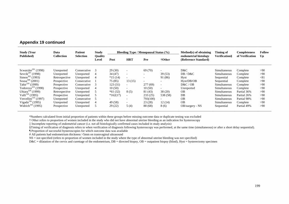
199
Appendix 19 continued
Study (Year Data Patient Study Bleeding Type / Menopausal Status (%) Method(s) of obtaining Timing of Completeness Follow
Published) Collection Selection Quality endometrial histology Verification§ of Verification Up
Level Post HRT Pre †Other (Reference Standard)
Scwarzler291 (1998) Unreported Consecutive 3 29 (30) - 69 (70) - D&C Simultaneous Complete >90
Sevcik297 (1998) Unreported Unreported 4 34 (47) - - 39 (53) DB / D&C Simultaneous Complete >90
Simon302 (1993) Retrospective Unreported 4 *15 (14) - - 91 (86) Hyst Sequential Complete <81
Sousa308 (2001) Prospective Consecutive 1 75 (85) 13 (15) - - Hyst/DB/OB Sequential Complete >90
Tahir320 (1999) Prospective Consecutive 3 123 (31) - 277 (69) - D&C / OB Simultaneous Complete >90
Todorova323 (1998) Prospective Unreported 4 10 (50) - 10 (50) - Unreported Simultaneous Complete >90
Uhiara329 (1999) Retrospective Unreported 5 *61 (32) 8 (5) 81 (43) 38 (20) OB Simultaneous Partial 36% >90
Valli330 (1995) Prospective Unreported 5 *162(17) - 233 (25) 538 (58) DB Simultaneous Partial 26% >90
Vercellini335 (1997) Unreported Consecutive 5 - - 793(100) - OB Simultaneous Partial 98% >90
Vigada336 (1995) Unreported Unreported 4 49 (58) - 23 (28) 12 (14) OB Simultaneous Complete >90
Widrich343 (1995) Prospective Unreported 5 29 (22) 5 (4) 88 (68) 8 (6) OB/surgery - NS Sequential Partial 49% >90
*Numbers calculated from initial proportion of patients within these groups before missing outcome data or duplicate testing was excluded
† Other refers to proportion of women included in the study who did not have abnormal uterine bleeding as an indication for hysteroscopy
‡ Incomplete reporting of endometrial cancer (i.e. not all histologically confirmed cases included in study analysis)
§Timing of verification of diagnosis refers to when verification of diagnosis following hysteroscopy was performed, at the same time (simultaneous) or after a short delay sequential).
¶ Proportion of successful hysteroscopies for which outcome data was available
# All patients had endometrium thickness >5mm on transvaginal ultrasound
NS = not specified (refers to proportion of women included in the study where the type of abnormal uterine bleeding was not specified)
D&C = dilatation of the cervix and curettage of the endometrium, DB = directed biopsy, OB = outpatient biopsy (blind), Hyst = hysterectomy specimen
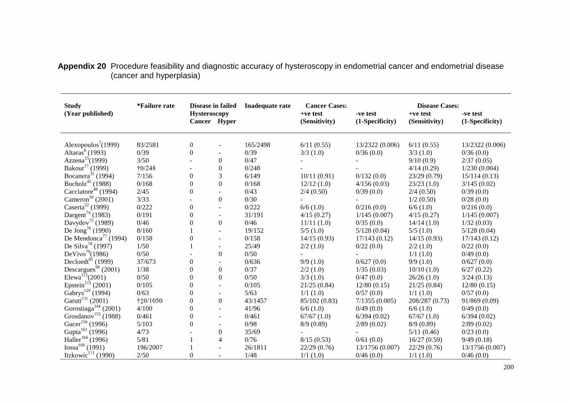
200
Appendix 20 Procedure feasibility and diagnostic accuracy of hysteroscopy in endometrial cancer and endometrial disease (cancer and hyperplasia)
Study *Failure rate Disease in failed Inadequate rate Cancer Cases: Disease Cases:
(Year published) Hysteroscopy
Cancer Hyper
+ve test
(Sensitivity)
-ve test
(1-Specificity)
+ve test
(Sensitivity)
-ve test
(1-Specificity)
Alexopoulos7(1999) 83/2581 0 - 165/2498 6/11 (0.55) 13/2322 (0.006) 6/11 (0.55) 13/2322 (0.006)
Altaras8 (1993) 0/39 0 - 0/39 3/3 (1.0) 0/36 (0.0) 3/3 (1.0) 0/36 (0.0)
Azzena15
(1999) 3/50 - 0 0/47 - - 9/10 (0.9) 2/37 (0.05)
Bakour17
(1999) †0/248 - 0 0/248 - - 4/14 (0.29) 1/230 (0.004)
Bocanera32
(1994) 7/156 0 3 6/149 10/11 (0.91) 0/132 (0.0) 23/29 (0.79) 15/114 (0.13)
Bucholz45
(1988) 0/168 0 0 0/168 12/12 (1.0) 4/156 (0.03) 23/23 (1.0) 3/145 (0.02)
Cacciatore48
(1994) 2/45 0 - 0/43 2/4 (0.50) 0/39 (0.0) 2/4 (0.50) 0/39 (0.0)
Cameron50
(2001) 3/33 - 0 0/30 - - 1/2 (0.50) 0/28 (0.0)
Caserta52
(1999) 0/222 0 - 0/222 6/6 (1.0) 0/216 (0.0) 6/6 (1.0) 0/216 (0.0)
Dargent74
(1983) 0/191 0 - 31/191 4/15 (0.27) 1/145 (0.007) 4/15 (0.27) 1/145 (0.007)
Davydov75
(1989) 0/46 0 0 0/46 11/11 (1.0) 0/35 (0.0) 14/14 (1.0) 1/32 (0.03)
De Jong76
(1990) 8/160 1 - 19/152 5/5 (1.0) 5/128 (0.04) 5/5 (1.0) 5/128 (0.04)
De Mendonca77
(1994) 0/158 0 - 0/158 14/15 (0.93) 17/143 (0.12) 14/15 (0.93) 17/143 (0.12)
De Silva78
(1997) 1/50 1 - 25/49 2/2 (1.0) 0/22 (0.0) 2/2 (1.0) 0/22 (0.0)
DeVivo79
(1986) 0/50 - 0 0/50 - - 1/1 (1.0) 0/49 (0.0)
Decloedt81
(1999) 37/673 0 - 0/636 9/9 (1.0) 0/627 (0.0) 9/9 (1.0) 0/627 (0.0)
Descargues90
(2001) 1/38 0 0 0/37 2/2 (1.0) 1/35 (0.03) 10/10 (1.0) 6/27 (0.22)
Elewa112
(2001) 0/50 0 0 0/50 3/3 (1.0) 0/47 (0.0) 26/26 (1.0) 3/24 (0.13)
Epstein115
(2001) 0/105 0 - 0/105 21/25 (0.84) 12/80 (0.15) 21/25 (0.84) 12/80 (0.15)
Gabrys129
(1994) 0/63 0 - 5/63 1/1 (1.0) 0/57 (0.0) 1/1 (1.0) 0/57 (0.0)
Garuti131
(2001) †‡0/1050 0 0 43/1457 85/102 (0.83) 7/1355 (0.005) 208/287 (0.73) 91/869 (0.09)
Gorostiaga144
(2001) 4/100 0 - 41/96 6/6 (1.0) 0/49 (0.0) 6/6 (1.0) 0/49 (0.0)
Grosdanov153
(1988) 0/461 0 - 0/461 67/67 (1.0) 6/394 (0.02) 67/67 (1.0) 6/394 (0.02)
Gucer156
(1996) 5/103 0 - 0/98 8/9 (0.89) 2/89 (0.02) 8/9 (0.89) 2/89 (0.02)
Gupta161
(1996) 4/73 - 0 35/69 - - 5/11 (0.46) 0/23 (0.0)
Haller164
(1996) 5/81 1 4 0/76 8/15 (0.53) 0/61 (0.0) 16/27 (0.59) 9/49 (0.18)
Iossa169
(1991) 196/2007 1 - 26/1811 22/29 (0.76) 13/1756 (0.007) 22/29 (0.76) 13/1756 (0.007)
Itzkowic171
(1990) 2/50 0 - 1/48 1/1 (1.0) 0/46 (0.0) 1/1 (1.0) 0/46 (0.0)
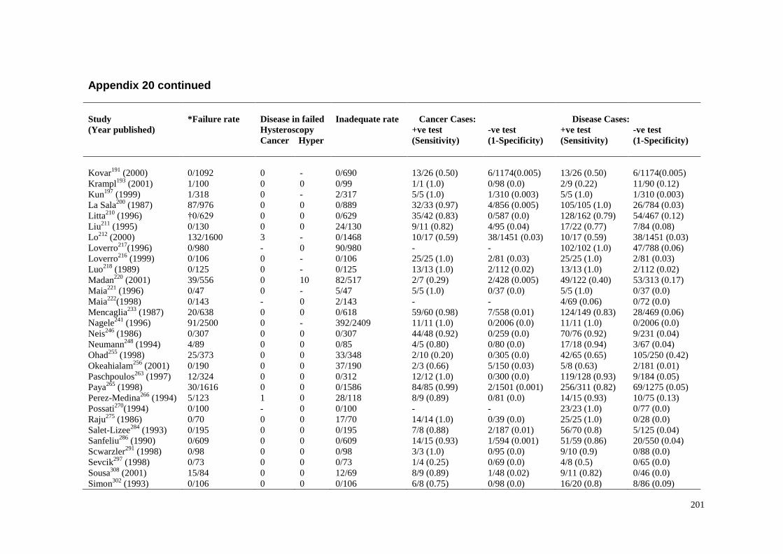
201
Appendix 20 continued
Study *Failure rate Disease in failed Inadequate rate Cancer Cases: Disease Cases:
(Year published) Hysteroscopy
Cancer Hyper
+ve test
(Sensitivity)
-ve test
(1-Specificity)
+ve test
(Sensitivity)
-ve test
(1-Specificity)
Kovar191
(2000) 0/1092 0 - 0/690 13/26 (0.50) 6/1174(0.005) 13/26 (0.50) 6/1174(0.005)
Krampl193
(2001) 1/100 0 0 0/99 1/1 (1.0) 0/98 (0.0) 2/9 (0.22) 11/90 (0.12)
Kun197
(1999) 1/318 0 - 2/317 5/5 (1.0) 1/310 (0.003) 5/5 (1.0) 1/310 (0.003)
La Sala200
(1987) 87/976 0 0 0/889 32/33 (0.97) 4/856 (0.005) 105/105 (1.0) 26/784 (0.03)
Litta210
(1996) †0/629 0 0 0/629 35/42 (0.83) 0/587 (0.0) 128/162 (0.79) 54/467 (0.12)
Liu211
(1995) 0/130 0 0 24/130 9/11 (0.82) 4/95 (0.04) 17/22 (0.77) 7/84 (0.08)
Lo212
(2000) 132/1600 3 - 0/1468 10/17 (0.59) 38/1451 (0.03) 10/17 (0.59) 38/1451 (0.03)
Loverro217
(1996) 0/980 - 0 90/980 - - 102/102 (1.0) 47/788 (0.06)
Loverro216
(1999) 0/106 0 - 0/106 25/25 (1.0) 2/81 (0.03) 25/25 (1.0) 2/81 (0.03)
Luo218
(1989) 0/125 0 - 0/125 13/13 (1.0) 2/112 (0.02) 13/13 (1.0) 2/112 (0.02)
Madan220
(2001) 39/556 0 10 82/517 2/7 (0.29) 2/428 (0.005) 49/122 (0.40) 53/313 (0.17)
Maia221
(1996) 0/47 0 - 5/47 5/5 (1.0) 0/37 (0.0) 5/5 (1.0) 0/37 (0.0)
Maia222
(1998) 0/143 - 0 2/143 - - 4/69 (0.06) 0/72 (0.0)
Mencaglia233
(1987) 20/638 0 0 0/618 59/60 (0.98) 7/558 (0.01) 124/149 (0.83) 28/469 (0.06)
Nagele241
(1996) 91/2500 0 - 392/2409 11/11 (1.0) 0/2006 (0.0) 11/11 (1.0) 0/2006 (0.0)
Neis246
(1986) 0/307 0 0 0/307 44/48 (0.92) 0/259 (0.0) 70/76 (0.92) 9/231 (0.04)
Neumann248
(1994) 4/89 0 0 0/85 4/5 (0.80) 0/80 (0.0) 17/18 (0.94) 3/67 (0.04)
Ohad255
(1998) 25/373 0 0 33/348 2/10 (0.20) 0/305 (0.0) 42/65 (0.65) 105/250 (0.42)
Okeahialam256
(2001) 0/190 0 0 37/190 2/3 (0.66) 5/150 (0.03) 5/8 (0.63) 2/181 (0.01)
Paschpoulos263
(1997) 12/324 0 0 0/312 12/12 (1.0) 0/300 (0.0) 119/128 (0.93) 9/184 (0.05)
Paya265
(1998) 30/1616 0 0 0/1586 84/85 (0.99) 2/1501 (0.001) 256/311 (0.82) 69/1275 (0.05)
Perez-Medina266
(1994) 5/123 1 0 28/118 8/9 (0.89) 0/81 (0.0) 14/15 (0.93) 10/75 (0.13)
Possati270
(1994) 0/100 - 0 0/100 - - 23/23 (1.0) 0/77 (0.0)
Raju275
(1986) 0/70 0 0 17/70 14/14 (1.0) 0/39 (0.0) 25/25 (1.0) 0/28 (0.0)
Salet-Lizee284
(1993) 0/195 0 0 0/195 7/8 (0.88) 2/187 (0.01) 56/70 (0.8) 5/125 (0.04)
Sanfeliu286
(1990) 0/609 0 0 0/609 14/15 (0.93) 1/594 (0.001) 51/59 (0.86) 20/550 (0.04)
Scwarzler291
(1998) 0/98 0 0 0/98 3/3 (1.0) 0/95 (0.0) 9/10 (0.9) 0/88 (0.0)
Sevcik297
(1998) 0/73 0 0 0/73 1/4 (0.25) 0/69 (0.0) 4/8 (0.5) 0/65 (0.0)
Sousa308
(2001) 15/84 0 0 12/69 8/9 (0.89) 1/48 (0.02) 9/11 (0.82) 0/46 (0.0)
Simon302
(1993) 0/106 0 0 0/106 6/8 (0.75) 0/98 (0.0) 16/20 (0.8) 8/86 (0.09)
202
Appendix 20 continued
Study *Failure rate Disease in failed Inadequate rate Cancer Cases: Disease Cases:
(Year published) Hysteroscopy
Cancer Hyper
+ve test
(Sensitivity)
-ve test
(1-Specificity)
+ve test
(Sensitivity)
-ve test
(1-Specificity)
Tahir320
(1999) 7/400 0 - 30/393 8/11 (0.73) 0/352 (0.0) 8/11 (0.73) 0/352 (0.0)
Todorova323
(1998) 0/20 - 0 0/20 - - 4/6 (0.67) 2/14 (0.14)
Uhiara329
(1999) 14/188 0 0 0/174 1/2 (0.50) 0/172 (0.0) 6/10 (0.6) 8/164 (0.05)
Valli330
(1995) 47/933 0 0 18/886 18/18 (1.0) 9/850 (0.01) 95/103 (0.92) 99/765 (0.13)
Vercellini335
(1997) 23/793 0 0 17/770 2/2 (1.0) 0/751 (0.0) 32/68 (0.47) 48/685 (0.07)
Vigada336
(1995) 13/84 0 0 10/71 1/2 (0.5) 0/59 (0.0) 9/9 (1.0) 9/52 (0.17)
Widrich343
(1995) 10/130 0 0 0/120 1/1 (1.0) 0/119 (0.0) 4/8 (0.5) 0/112 (0.0)
Endometrial disease
studies (65)
937/26346
3.6% (3.3-
3.8%)
25/937
2.7% (1.7-3.9%)
1196/25409
4.7% (4.5-5.0%)
- - 2004/2570 900/21643
Endometrial cancer
studies (56)
927/24649
3.8% (3.6-
4.0%)
8/927
0.8% (0.4-1.7%)
1069/23722
4.5% (4.3-4.8%)
768/889 167/21764 - -
* Failed outpatient hysteroscopic procedures included technical aspects (e.g. cervical stenosis, anatomical factors), inadequate visualization (e.g. obscured by bleeding) or patient factor
(e.g. pain)
†Failed outpatient hysteroscopies, which were successfully performed subsequently as an inpatient NOT included in the failure rates
‡ 128 „unsatisfactory hysteroscopies‟ but all included in results
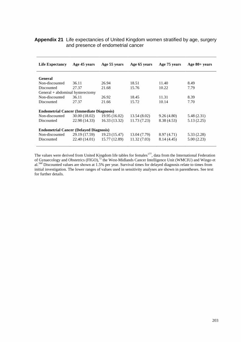
203
Appendix 21 Life expectancies of United Kingdom women stratified by age, surgery and presence of endometrial cancer
Life Expectancy Age 45 years Age 55 years Age 65 years Age 75 years Age 80+ years
General
Non-discounted 36.11 26.94 18.51 11.40 8.49
Discounted 27.37 21.68 15.76 10.22 7.79
General + abdominal hysterectomy
Non-discounted 36.11 26.92 18.45 11.31 8.39
Discounted 27.37 21.66 15.72 10.14 7.70
Endometrial Cancer (Immediate Diagnosis)
Non-discounted 30.00 (18.02) 19.95 (16.02) 13.54 (8.02) 9.26 (4.80) 5.48 (2.31)
Discounted 22.98 (14.33) 16.33 (13.32) 11.73 (7.23) 8.38 (4.53) 5.13 (2.25)
Endometrial Cancer (Delayed Diagnosis)
Non-discounted 29.19 (17.59) 19.23 (15.47) 13.04 (7.79) 8.97 (4.71) 5.33 (2.28)
Discounted 22.40 (14.01) 15.77 (12.89) 11.32 (7.03) 8.14 (4.45) 5.00 (2.23)
The values were derived from United Kingdom life tables for females
177, data from the International Federation
of Gynaecology and Obstetrics (FIGO),72
the West-Midlands Cancer Intelligence Unit (WMCIU) and Wingo et
al.344
Discounted values are shown at 1.5% per year. Survival times for delayed diagnosis relate to times from
initial investigation. The lower ranges of values used in sensitivity analyses are shown in parentheses. See text
for further details.
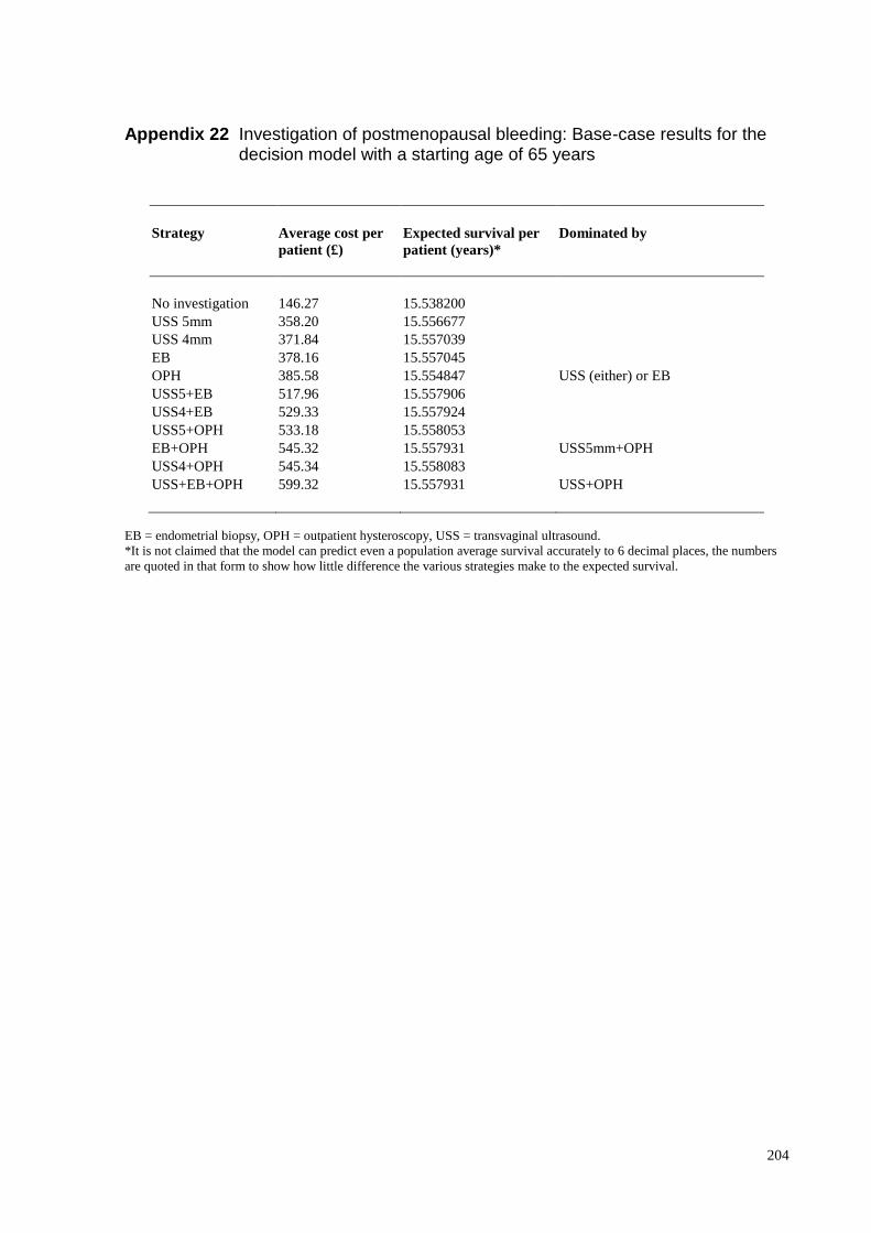
204
Appendix 22 Investigation of postmenopausal bleeding: Base-case results for the decision model with a starting age of 65 years
Strategy Average cost per
patient (£)
Expected survival per
patient (years)*
Dominated by
No investigation 146.27 15.538200
USS 5mm 358.20 15.556677
USS 4mm 371.84 15.557039
EB 378.16 15.557045
OPH 385.58 15.554847 USS (either) or EB
USS5+EB 517.96 15.557906
USS4+EB 529.33 15.557924
USS5+OPH 533.18 15.558053
EB+OPH 545.32 15.557931 USS5mm+OPH
USS4+OPH 545.34 15.558083
USS+EB+OPH 599.32 15.557931 USS+OPH
EB = endometrial biopsy, OPH = outpatient hysteroscopy, USS = transvaginal ultrasound.
*It is not claimed that the model can predict even a population average survival accurately to 6 decimal places, the numbers
are quoted in that form to show how little difference the various strategies make to the expected survival.
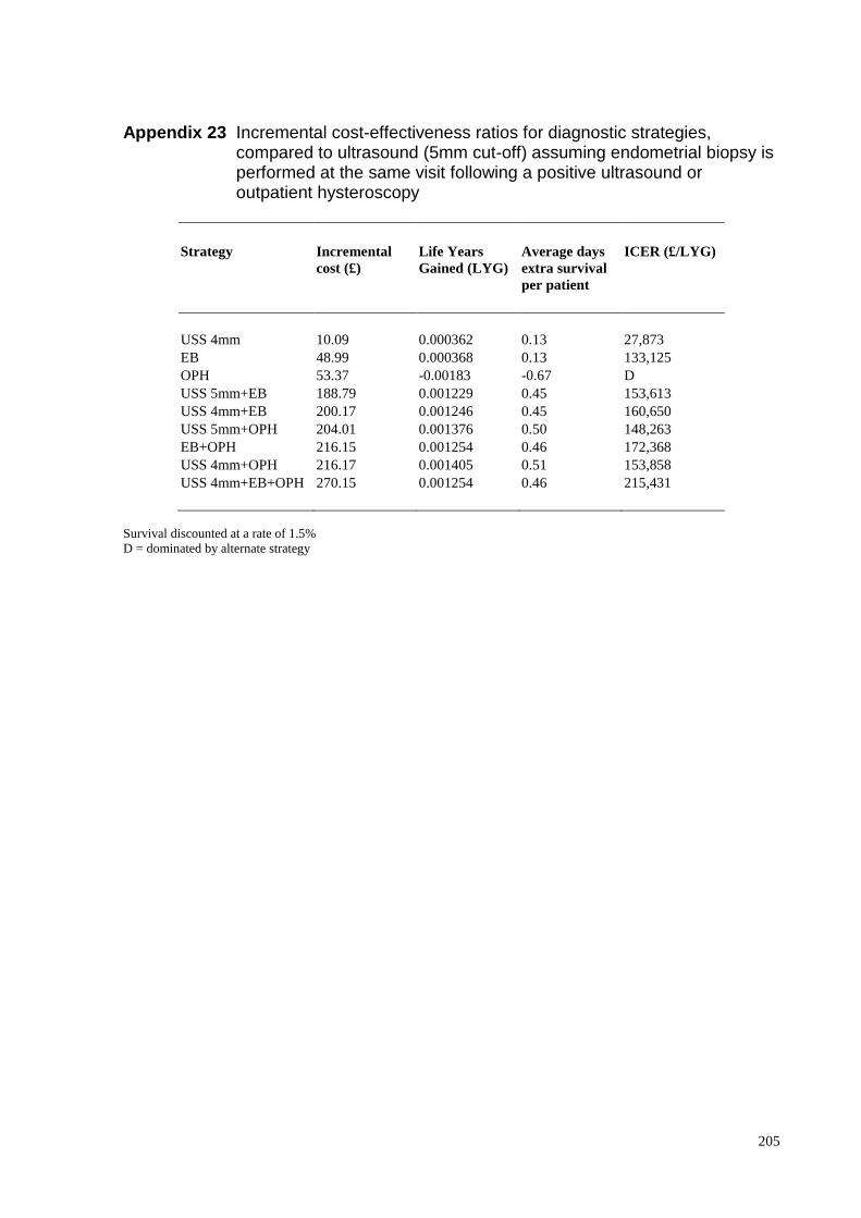
205
Appendix 23 Incremental cost-effectiveness ratios for diagnostic strategies, compared to ultrasound (5mm cut-off) assuming endometrial biopsy is performed at the same visit following a positive ultrasound or outpatient hysteroscopy
Strategy Incremental
cost (£)
Life Years
Gained (LYG)
Average days
extra survival
per patient
ICER (£/LYG)
USS 4mm 10.09 0.000362 0.13 27,873
EB 48.99 0.000368 0.13 133,125
OPH 53.37 -0.00183 -0.67 D
USS 5mm+EB 188.79 0.001229 0.45 153,613
USS 4mm+EB 200.17 0.001246 0.45 160,650
USS 5mm+OPH 204.01 0.001376 0.50 148,263
EB+OPH 216.15 0.001254 0.46 172,368
USS 4mm+OPH 216.17 0.001405 0.51 153,858
USS 4mm+EB+OPH 270.15 0.001254 0.46 215,431
Survival discounted at a rate of 1.5%
D = dominated by alternate strategy
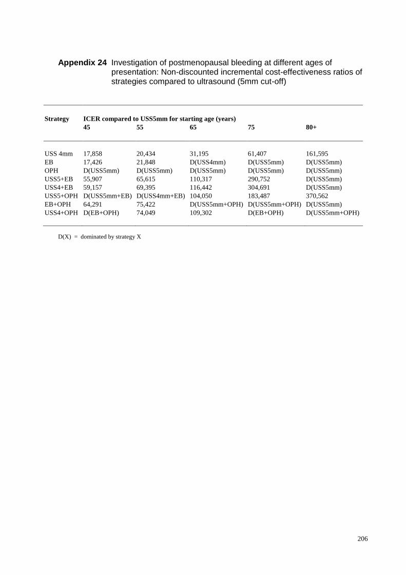
206
Appendix 24 Investigation of postmenopausal bleeding at different ages of presentation: Non-discounted incremental cost-effectiveness ratios of strategies compared to ultrasound (5mm cut-off)
Strategy ICER compared to USS5mm for starting age (years)
45 55 65 75 80+
USS 4mm 17,858 20,434 31,195 61,407 161,595
EB 17,426 21,848 D(USS4mm) D(USS5mm) D(USS5mm)
OPH D(USS5mm) D(USS5mm) D(USS5mm) D(USS5mm) D(USS5mm)
USS5+EB 55,907 65,615 110,317 290,752 D(USS5mm)
USS4+EB 59,157 69,395 116,442 304,691 D(USS5mm)
USS5+OPH D(USS5mm+EB) D(USS4mm+EB) 104,050 183,487 370,562
EB+OPH 64,291 75,422 D(USS5mm+OPH) D(USS5mm+OPH) D(USS5mm)
USS4+OPH D(EB+OPH) 74,049 109,302 D(EB+OPH) D(USS5mm+OPH)
D(X) = dominated by strategy X
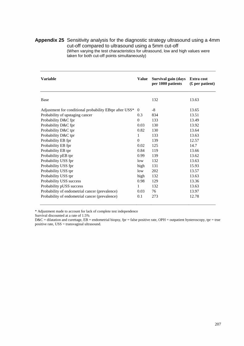
207
Appendix 25 Sensitivity analysis for the diagnostic strategy ultrasound using a 4mm cut-off compared to ultrasound using a 5mm cut-off
(When varying the test characteristics for ultrasound, low and high values were taken for both cut-off points simultaneously)
Variable Value Survival gain (days
per 1000 patients
Extra cost
(£ per patient)
Base 132 13.63
Adjustment for conditional probability EBtpr after USS* 0 -8 13.65
Probability of upstaging cancer 0.3 834 13.51
Probability D&C fpr 0 133 13.49
Probability D&C fpr 0.03 130 13.92
Probability D&C tpr 0.82 130 13.64
Probability D&C tpr 1 133 13.63
Probability EB fpr 0 139 12.57
Probability EB fpr 0.02 125 14.7
Probability EB tpr 0.84 119 13.66
Probability pEB tpr 0.99 139 13.62
Probability USS fpr low 132 13.63
Probability USS fpr high 131 15.93
Probability USS tpr low 202 13.57
Probability USS tpr high 132 13.63
Probability USS success 0.98 129 13.36
Probability pUSS success 1 132 13.63
Probability of endometrial cancer (prevalence) 0.03 76 13.97
Probability of endometrial cancer (prevalence) 0.1 273 12.78
* Adjustment made to account for lack of complete test independence
Survival discounted at a rate of 1.5%
D&C = dilatation and curettage, EB = endometrial biopsy, fpr = false positive rate, OPH = outpatient hysteroscopy, tpr = true
positive rate, USS = transvaginal ultrasound.
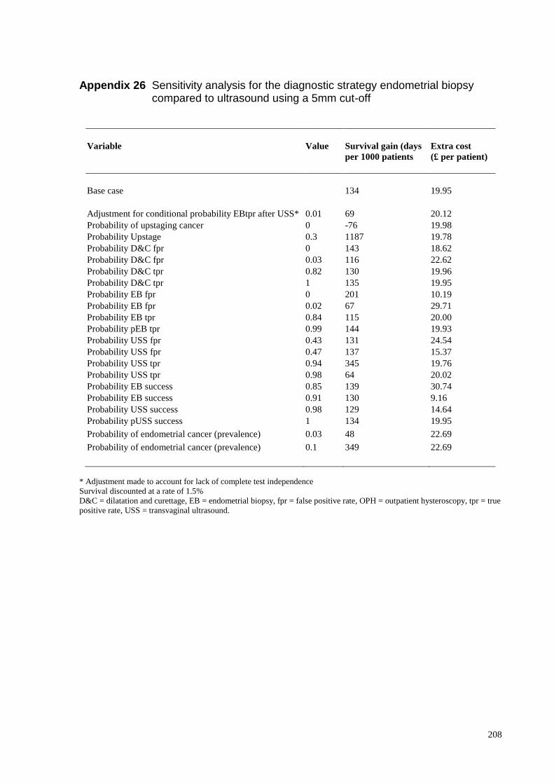
208
Appendix 26 Sensitivity analysis for the diagnostic strategy endometrial biopsy compared to ultrasound using a 5mm cut-off
Variable Value Survival gain (days
per 1000 patients
Extra cost
(£ per patient)
Base case 134 19.95
Adjustment for conditional probability EBtpr after USS* 0.01 69 20.12
Probability of upstaging cancer 0 -76 19.98
Probability Upstage 0.3 1187 19.78
Probability D&C fpr 0 143 18.62
Probability D&C fpr 0.03 116 22.62
Probability D&C tpr 0.82 130 19.96
Probability D&C tpr 1 135 19.95
Probability EB fpr 0 201 10.19
Probability EB fpr 0.02 67 29.71
Probability EB tpr 0.84 115 20.00
Probability pEB tpr 0.99 144 19.93
Probability USS fpr 0.43 131 24.54
Probability USS fpr 0.47 137 15.37
Probability USS tpr 0.94 345 19.76
Probability USS tpr 0.98 64 20.02
Probability EB success 0.85 139 30.74
Probability EB success 0.91 130 9.16
Probability USS success 0.98 129 14.64
Probability pUSS success 1 134 19.95
Probability of endometrial cancer (prevalence) 0.03 48 22.69
Probability of endometrial cancer (prevalence) 0.1 349 22.69
* Adjustment made to account for lack of complete test independence
Survival discounted at a rate of 1.5%
D&C = dilatation and curettage, EB = endometrial biopsy, fpr = false positive rate, OPH = outpatient hysteroscopy, tpr = true
positive rate, USS = transvaginal ultrasound.
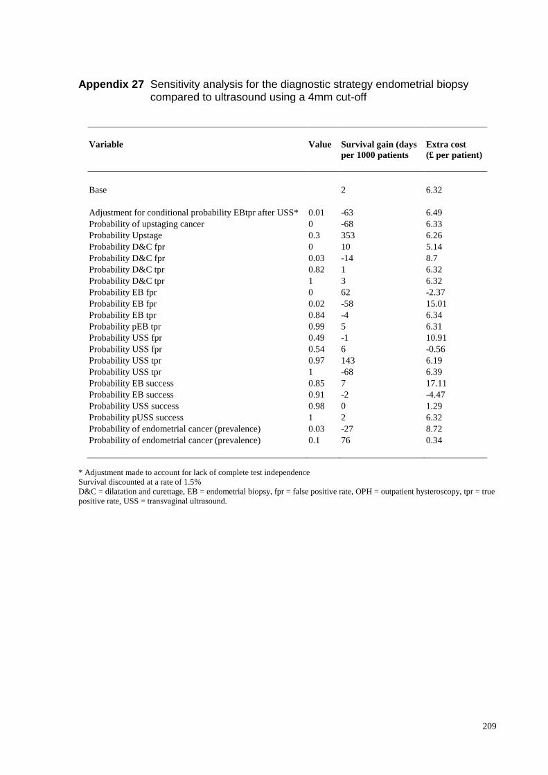
209
Appendix 27 Sensitivity analysis for the diagnostic strategy endometrial biopsy compared to ultrasound using a 4mm cut-off
Variable Value Survival gain (days
per 1000 patients
Extra cost
(£ per patient)
Base 2 6.32
Adjustment for conditional probability EBtpr after USS* 0.01 -63 6.49
Probability of upstaging cancer 0 -68 6.33
Probability Upstage 0.3 353 6.26
Probability D&C fpr 0 10 5.14
Probability D&C fpr 0.03 -14 8.7
Probability D&C tpr 0.82 1 6.32
Probability D&C tpr 1 3 6.32
Probability EB fpr 0 62 -2.37
Probability EB fpr 0.02 -58 15.01
Probability EB tpr 0.84 -4 6.34
Probability pEB tpr 0.99 5 6.31
Probability USS fpr 0.49 -1 10.91
Probability USS fpr 0.54 6 -0.56
Probability USS tpr 0.97 143 6.19
Probability USS tpr 1 -68 6.39
Probability EB success 0.85 7 17.11
Probability EB success 0.91 -2 -4.47
Probability USS success 0.98 0 1.29
Probability pUSS success 1 2 6.32
Probability of endometrial cancer (prevalence) 0.03 -27 8.72
Probability of endometrial cancer (prevalence) 0.1 76 0.34
* Adjustment made to account for lack of complete test independence
Survival discounted at a rate of 1.5%
D&C = dilatation and curettage, EB = endometrial biopsy, fpr = false positive rate, OPH = outpatient hysteroscopy, tpr = true
positive rate, USS = transvaginal ultrasound.
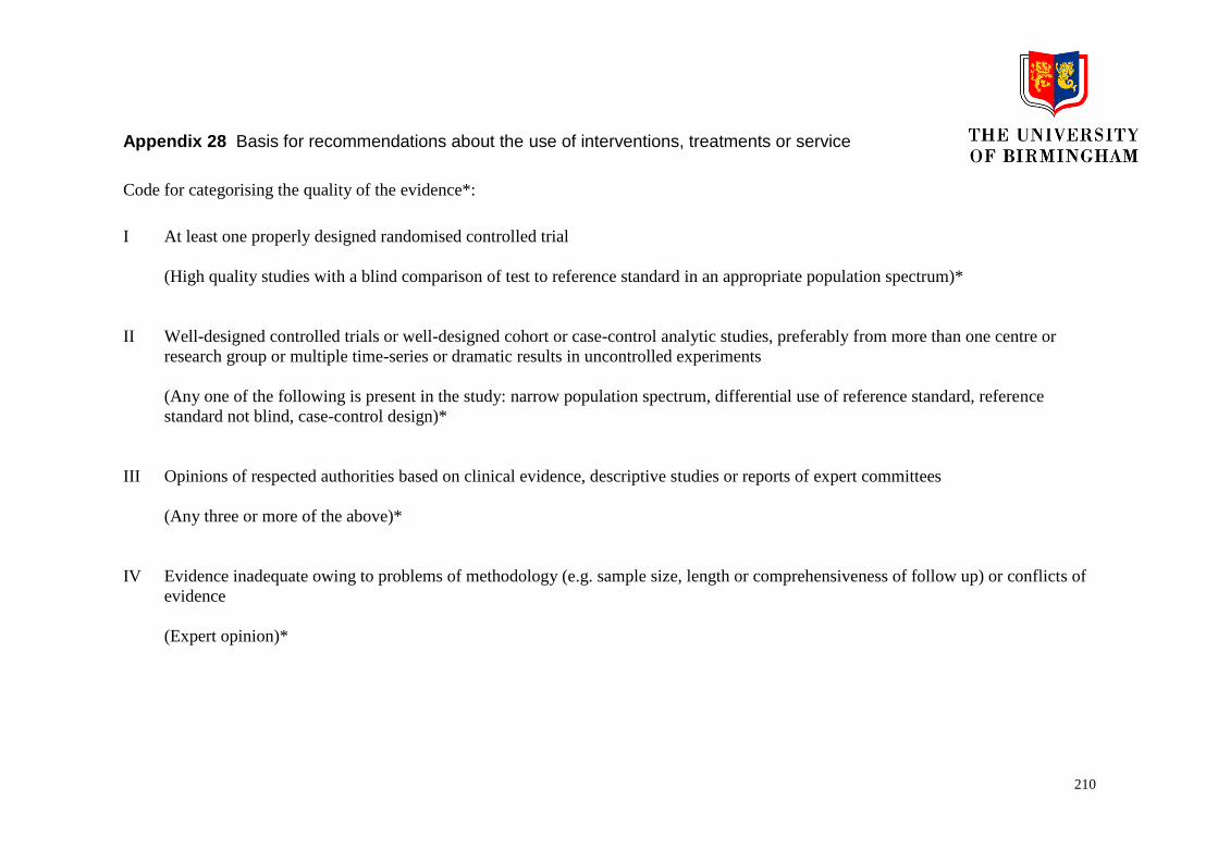
210
Appendix 28 Basis for recommendations about the use of interventions, treatments or service
Code for categorising the quality of the evidence*:
I At least one properly designed randomised controlled trial
(High quality studies with a blind comparison of test to reference standard in an appropriate population spectrum)*
II Well-designed controlled trials or well-designed cohort or case-control analytic studies, preferably from more than one centre or
research group or multiple time-series or dramatic results in uncontrolled experiments
(Any one of the following is present in the study: narrow population spectrum, differential use of reference standard, reference
standard not blind, case-control design)*
III Opinions of respected authorities based on clinical evidence, descriptive studies or reports of expert committees
(Any three or more of the above)*
IV Evidence inadequate owing to problems of methodology (e.g. sample size, length or comprehensiveness of follow up) or conflicts of
evidence
(Expert opinion)*
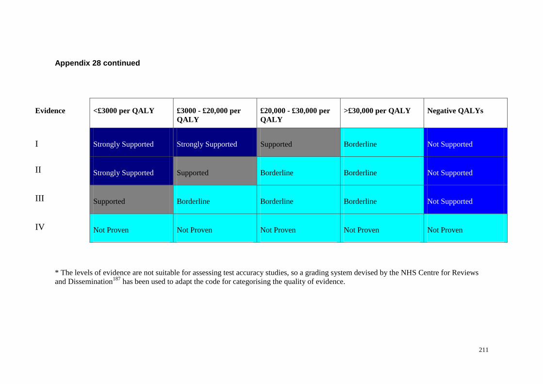
211
Appendix 28 continued
Evidence <£3000 per QALY £3000 - £20,000 per
QALY
£20,000 - £30,000 per
QALY
>£30,000 per QALY Negative QALYs
I Strongly Supported Strongly Supported Supported Borderline Not Supported
II Strongly Supported Supported Borderline Borderline Not Supported
III Supported Borderline Borderline Borderline Not Supported
IV Not Proven Not Proven Not Proven Not Proven Not Proven
* The levels of evidence are not suitable for assessing test accuracy studies, so a grading system devised by the NHS Centre for Reviews
and Dissemination187
has been used to adapt the code for categorising the quality of evidence.
212
Appendix 29 Publications from the thesis
1. Clark TJ, Mann CH, Shah N, Song F, Khan KS, Gupta JK. Accuracy of outpatient
endometrial biopsy in the diagnosis of endometrial hyperplasia: A systematic review. Acta
Obstet Gynecol Scand 2001; 80(9): 784-793.
2. Clark TJ, Mann CH, Shah N, Song F, Khan KS, Gupta JK. Accuracy of outpatient
endometrial biopsy in the diagnosis of endometrial cancer: A systematic review. Br J
Obstet Gynecol 2002; 109: 313-321.
3. Clark TJ, Bakour SH, Khan KS, Gupta JK. Evaluation of outpatient hysteroscopy and
ultrasonography in the diagnosis of endometrial disease. Obstet Gynecol 2002; 99: 1001-
1007.
4. Gupta JK, Chien PFW, Voit D, Clark TJ, Khan KS. Ultrasonographic endometrial
thickness for diagnosing endometrial pathology in women with postmenopausal bleeding:
A meta-analysis. Acta Obstet Gynecol Scand 2002;81:799-816.
5. Clark TJ, Voit D, Gupta JK, Hyde C, Song FS, Khan KS. Accuracy of hysteroscopy in the
diagnosis of endometrial cancer and hyperplasia: a systematic quantitative review. JAMA
2002,288:1610-22.
6. Clark TJ, Khan KS, Gupta JK. The diagnosis of intrauterine pathology in postmenopausal
women: An evidence-based approach. Reviews in Gynaecological Practice 2002; 2 : 109-
116.
7. Clark TJ, Gupta JK. Endometrial sampling of gynaecological pathology. Obstetrician and
Gynaecologist 2002;4:169–174.
8. Clark TJ, Gupta JK. Outpatient Hysteroscopy. Obstetrician and Gynaecologist
2002;4:217-221.
9. Bachmann LM, Clark TJ, ter Riet G, Gupta JK, Khan KS. Probability analysis for
diagnosis of endometrial hyperplasia and cancer in postmenopausal bleeding: An
approach for a rational diagnostic workup. Acta Obstet Gynecol Scand 2003; 82: 1-6.
10. Clark TJ, Barton PM, Gupta JK, Khan KS. Ambulatory diagnosis of endometrial cancer in
women with postmenopausal bleeding. (ISBN) West Midlands Health Technology
Assessment Collaboration Report 2003University of Birmingham [in press, will be
available at http://www.publichealth.bham.ac.uk]
11. Clark TJ, Khan KS, Ter Riet G, Gupta JK. Bias associated with delayed verification in
test accuracy studies: Accuracy of tests for endometrial hyperplasia may be much higher
than we think! BMC Med Res Methodol 2003 (submitted)
213
REFERENCES
1. Key Health Statistics from General Practice: Analyses of morbidity and treatment
data, including time trends, England and Wales. London: National Statistics. Available
at http://www.statistics.gov.uk/downloads/theme_health/Key_Health_Stats_1998.pdf.
Accessed 14th January 2002., 1998.
2. Management of Gynaecological Cancers. Effective Health Care Bulletin
1999;5:[Available at http://www.york.ac.uk/inst/crd/ Accessibility verified September
27, 2002].
3. Existing facilities for investigating postmenopausal bleeding in Scotland. [Available
at http://www.sign.ac.uk/ Accessibility verified January 24th, 2002]. 2002.
4. Abu-Ghazzeh Y, Shakoury WA, Barqawi R. Comparative study of transvaginal
hysterosonography and biopsy for the evaluation of post-menopausal bleeding. Annals
of Saudi Medicine 1999;19:116-9.
5. al Azzawi F. Hysteroscopy or ultrasound?. Curr Opin Obstet Gynecol 1996;8:246-9.
6. Aleem F, Predanic M, Calame R, Moukhtar M, Pennisi J. Transvaginal colour and
pulsed doppler sonography of the endometrium: a possible role in reducing the
number of dilatation and curettage procedures. J Ultrasound Med 1995;14:139-45.
7. Alexopoulos ED, Fay TN, Simonis CD. A review of 2581 out-patient diagnostic
hysteroscopies in the management of abnormal uterine bleeding. Gynae Endosc
1999;8:105-10.
8. Altaras MM, Aviram R, Cohen I, Markov S, Goldberg GL, Beyth Y.
Microhysteroscopy and endometrial biopsy results following failed diagnostic
dilatation and curettage in women with postmenopausal bleeding. Int J Gynaecol
Obstet 1993;42:255-60.
9. Altuncu N, Kal U, Benhabib M, Nurluoglu M. Vaginosonographic measurements of
the postmenopausal endometrial thickness for the early detection of endometrial
carcinoma. Istanbul Tip Fakultesi Mecmuasi 1992;55:445-9.
10. Anastasiadis PG, Skaphida PG, Koutlaki NG, Galazios GC, Tsikouras PN, Liberis
VA. Descriptive epidemiology of endometrial hyperplasia in patients with abnormal
uterine bleeding. Eur.J Gynaecol Oncol. 2000;21:131-4.
11. Anderson JP, Bush JW, Chen M, Dolenc D. Policy space areas and properties of
benefit-cost/utility analysis. JAMA 1986;255:794-5.
214
12. Anonymous. Bioeffects considerations for the safety of diagnostic ultrasound.
American Institute of Ultrasound in Medicine. Bioeffects Committee. J Ultrasound
Med 1988;7:S1-S38.
13. Archer DF, Lobo RA, Land HF, Pickar JH. A comparative study of transvaginal
uterine ultrasound and endometrial biopsy for evaluating the endometrium of
postmenopausal women taking hormone replacement therapy. Menopause
1999;6:201-8.
14. Auslender R. Vaginal ultrasound in patients with postemnopausal bleeding.
Ultrasound Obstet Gynecol 1993;3:426-8.
15. Azzena A, Pellizzari P, Ferrara A. Diagnosis of endometrial pathology: Comparison
between hysterosonography, hysteroscopy and histologic findings. Ital J Obstet
Gynaecol 1999;11:112-5.
16. Bachmann LM, ter Riet G., Clark TJ, Gupta JK, Khan KS. Probability analysis for
diagnosis of endometrial hyperplasia and cancer in postmenopausal bleeding: An
approach for a rational diagnostic workup. Acta Obstetricia et Gynecologica
Scandinavica 2003;82:1-6.
17. Bakour SH, Dwarakanath LS, Khan KS, Newton JR. The diagnostic accuracy of
outpatient miniature hysteroscopy in predicting premalignant and malignant
endometrial lesions. Gynae Endosc 1999;8:143-8.
18. Bakour SH, Dwarakanath LS, Khan KS, Newton JR, Gupta JK. The diagnostic
accuracy of ultrasound scan in predicting endometrial hyperplasia and cancer in
postmenopausal bleeding. Acta Obstet Gynecol Scand 1999;78:447-51.
19. Bakour SH, Khan KS, Gupta JK. Controlled analysis of factors associated with
insufficient sample on outpatient endometrial biopsy. Br J Obstet Gynaecol
2000;107:1312-4.
20. Bakour SH, Khan KS, Gupta JK. The risk of premalignant and malignant pathology in
endometrial polyps. Acta Obstet Gynecol Scand 2002;81:182-3.
21. Bakour S, Khan K, Gupta J. Transvaginal ultrasonography and endometrial histology
in peri and postmenopausal women on hormone replacement therapy. Br J Obstet
Gynecol 2000;107:295.
22. Batool T, Reginald PW, Hughes JH. Outpatient pipelle endometrial biopsy in the
investigation of postmenopausal bleeding. Br J Obstet Gynaecol 1994;101:545-6.
23. Begg C. Operating characteristics of a rank correlation test for publication bias.
Biometric 1994;50:1088-101.
24. Ben-Baruch G, Seidman D, Schiff E, Moran O, Menczer J. Outpatient endometrial
sampling with the pipelle curette. Gynecol Obstet Invest 1994;37:260-2.
215
25. Ben Yehuda OM, Kim YB, Leuchter RS. Does hysteroscopy improve upon the
sensitivity of dilatation and curettage in the diagnosis of endometrial hyperplasia or
carcinoma? Gynecol Oncol 1998;68:4-7.
26. Bennett CC,.Richards DS. Patient acceptance of endovaginal ultrasound. Ultrasound
Obstet Gynecol 2000;15:52-5.
27. Berends MJ, Kleibeuker JH, de Vries EG, Mourits MJ, Hollema H, Pras E et al. The
importance of family history in young patients with endometrial cancer. Eur.J Obstet
Gynecol Reprod.Biol. 1999;82:139-41.
28. Bhan V, Amso N, Whitehead MI, Campbell S, Royston P, Collins WP. Characteristics
of persistent ovarian masses in asymptomatic women. Br.J Obstet Gynaecol
1989;96:1384-91.
29. Bissett D, Davis JA, George WD. Gynaecological monitoring during tamoxifen
therapy. Lancet 1994; 344:1244.
30. Bistoletti P, Hjerpe A, Mollerstrom G. Cytological diagnosis of endometrial cancer
and preinvasive endometrial lesions. A comparison of the Endo-Pap sampler with
fractional curettage. Acta Obstet Gynecol Scand 1988;67:343-5.
31. Black WC. The CE plane: a graphic representation of cost-effectiveness. Med
Decis.Making 1990;10:212-4.
32. Bocanera AR, Roncoroni EC, Schlaen I, Ben J, Monteverde R, Gonzalez et al. An
articulated rotating brush for office endometrial evaluation of climacteric outpatients.
Maturitas 1994;19:67-76.
33. Bonilla-Musoles F, Raga F, Blanes J, Bailao LA, Osborne NG.
Sonohysterosalpingography with transvaginal color Doppler and three- dimensional
ultrasound: State of the art. Journal of Gynecologic Surgery 1996;12:227-40.
34. Bossuyt PM, Reitsma JB, Bruns DE, Gatsonis CA, Glasziou PP, Irwig LM et al.
Towards complete and accurate reporting of studies of diagnostic accuracy: The
STARD Initiative. Ann.Intern Med 2003;138:40-4.
35. Bossuyt PM, Reitsma JB, Bruns DE, Gatsonis CA, Glasziou PP, Irwig LM et al. The
STARD statement for reporting studies of diagnostic accuracy: explanation and
elaboration. Ann.Intern Med 2003;138:W1-12.
36. Botsis D, Kassanos D, Pyrgiotis E, Zourlas PA. Vaginal sonography of the
endometrium in postmenopausal women. Clin Exp Obstet Gynecol 1992;19:189-92.
37. Bradley LD, Falcone T, Magen AB. Radiographic imaging techniques for the
diagnosis of abnormal uterine bleeding. Obstet Gynecol Clin North Am 2000;27:245-
76.
216
38. Bree RL, Bowerman RA, Bohm-Velez M, Benson CB, Doubilet PM, DeDreu S et al.
US evaluation of the uterus in patients with postmenopausal bleeding: A positive
effect on diagnostic decision making. Radiology 2000;216:260-4.
39. Briggs A. Economics notes: handling uncertainty in economic evaluation. BMJ
1999;319 :120.
40. Briggs A,.Gray A. Using cost effectiveness information. BMJ 2000;320:246.
41. Briggs AH,.Gray AM. Handling uncertainty when performing economic evaluation of
healthcare interventions. Health Technol Assess 1999;3:1-134.
42. Briley M,.Lindsell DRM. The role of transvaginal ultrasound in the investigation of
women with post-menopausal bleeding. Clin Radiol 1998;53:502-5.
43. Brolmann HAM, Van Der Linden PJQ, Bongers, MY, Moret E, Meuwissen JHJM.
Ultrasonic diagnosis of endometrial lesions; correlation with the histological findings
in 112 patients. Ned Tijdschr Geneeskd 1993;137:1823-7.
44. Bronz L, Suter T, Rusca T. The value of transvaginal sonography with and without
saline instillation in the diagnosis of uterine pathology in pre- and postmenopausal
women with abnormal bleeding or suspect sonographic findings. Ultrasound Obstet
Gynecol 1997;9:53-8.
45. Buchholz F, Bonatz G, Semm K. Possibilities and limitations in differentiating
postmenopausal bleedings by contact-hysteroscopy. Zentralbl Gynakol 1988;110:884-
9.
46. Buyuk E, Durmusoglu F, Erenus M, Karakoc B. Endometrial disease diagnosed by
transvaginal ultrasound and dilatation and curettage. Acta Obstetricia et Gynecologica
Scandinavica 1999;Vol 78:-422.
47. Byford S,.Raftery J. Perspectives in economic evaluation. BMJ 1998;316:1529-30.
48. Cacciatore B, Ramsay T, Lehtovirta P, Ylostalo P. Transvaginal sonography and
hysteroscopy in postmenopausal bleeding. Acta Obstet Gynecol Scand 1994;73:413-6.
49. Cairns J. Discounting in economic evaluation. In Drummond M, McGuire A, eds.
Economic Evaluation in Health Care, 236-55. Oxford: Oxford University Press, 2001.
50. Cameron ST, Walker J, Chambers S, Critchley H. Comparison of transvaginal
ultrasound, saline infusion sonography and hysteroscopy to investigate
postmenopausal bleeding and unscheduled bleeding on HRT. Aust NZ J Obstet
Gynaecol 2001;41:291-4.
51. Campo R, Van Belle Y, Rombauts L, Brosens I, Gordts S. Office mini-hysteroscopy.
Hum Reprod Update 1999;5:73-81.
217
52. Caserta D, Toro G, Porretta M, Mancini E, Moscarini M. Hysteroscopic vs histologic
diagnosis. Study of 222 cases of abnormal uterine hemorrhage. Minerva Ginecol
1999;51:169-72.
53. Cecchini S, Ciatto S, Bonardi R, Grazzini G, Mazzotta A. Endometrial
ultrasonography - An alternative to invasive assessment in women with
postmenopausal vaginal bleeding. Tumori 1996;82:38-9.
54. Chan FY, Chau M-T, Pun T-C, Lam C, Ngan HYS, Leong L et al. Limitations of
transvaginal sonography and color Doppler imaging in the differentiation of
endometrial carcinoma from benign lesions. J Ultrasound Med 1994;13:623-8.
55. Cicchetti DV,.Fleiss JL. A comparison of the null distributions of weighted kappa and
the cordinal statistic. App Psychol Measurement 1997;1:195-201.
56. Cicinelli E, Comi N, Scorcia P, Petruzzi D, Epifani S. Hysteroscopy for diagnosis and
treatment of endometrial adenocarcinoma precursors: A review of literature. Eur J
Gynaecol Oncol 1993;14:425-36.
57. Clark TJ, Bakour SH, Gupta JK, Khan KS. Evaluation of outpatient hysteroscopy and
ultrasonography in the diagnosis of endometrial disease. Obstet Gynecol
2002;99:1001-7.
58. Clark TJ, Khan KS, Foon R, Pattison HM, Bryan S, Gupta JK. Quality of life
instruments in studies of menorrhagia: a systematic review. Eur.J Obstet Gynecol
Reprod.Biol. 2002;104:96.
59. Clark TJ, Khan KS, Gupta JK. Current practice for the treatment of benign intrauterine
polyps: A national questionnaire survey of consultant gynaecologists in the United
Kingdom. Eur J Obstet Gynecol Reprod Biol 2002;103:65-7.
60. Clark TJ, Khan KS, Gupta JK. The diagnosis of intrauterine pathology in post-
menopasual women: an evidence-based approach. Reviews in Gynaecological practice
2002;6:1-8.
61. Clark TJ, Mann CH, Shah N, Khan KS, Song F, Gupta JK. Accuracy of outpatient
endometrial biopsy in the diagnosis of endometrial hyperplasia. Acta Obstet Gynecol
Scand 2001;80:784-93.
62. Clark TJ, Mann CH, Shah N, Khan KS, Song F, Gupta JK. Accuracy of outpatient
endometrial biopsy in the diagnosis of endometrial cancer: a systematic quantitative
review. Br J Obstet Gynaecol 2002;109:313-21.
63. Clark TJ, Voit D, Gupta JK, Hyde C, Song FS, Khan KS. Accuracy of hysteroscopy in
the diagnosis of endometrial cancer and hyperplasia: a systematic quantitative review .
JAMA 2002;288:1610-22.
64. Clarke M, Oxman A, editors. Cochrane Reviewer's Handbook [updated September
1997]. In: The Cochrane Library, Oxford: Update Software, 2000.
218
65. Claxton K, Sculpher M, Drummond M. A rational framework for decision making by
the National Institute For Clinical Excellence (NICE). Lancet 2002;360:711-5.
66. Cochrane Methods Working Group on Systematic Reviews of Screening and
Diagnostic Tests. Recommended Methods. [Available at:
http://www.cochrane.org/cochrane/sadtdoc1.htm.Accessed September 3rd, 2001
1996].
67. Cohen J. Weighted kappa: nominal scale agreement with provision for scaled
disagreement or partial credit. Psychol Bull 1968;70:213-20.
68. Cohen I, Rosen DJ, Shapira J, Cordoba M, Gilboa S, Altaras MM et al. Endometrial
changes in postmenopausal women treated with tamoxifen for breast cancer. Br.J
Obstet Gynaecol 1993;100:567-70.
69. Cook DJ, Sackett DL, Spitzer WO. Methodologic guidelines for systematic reviews of
randomized control trials in health care from the Potsdam Consultation on Meta-
Analysis. J Clin.Epidemiol. 1995;48:167-71.
70. Cooper JM,.Brady RM. Hysteroscopy in the management of abnormal uterine
bleeding. [Review] [65 refs]. Obstet Gynecol Clin North Am 1999;26:217-36.
71. Coulter A, Kelland A, Long Aea. The management of menorrhagia. Effective Health
Care Bulletin 1995;9.
72. Creasman WT, Odicino F, Maisonneuve P, Beller U, Benedet JL, Heintz AP et al.
Carcinoma of the corpus uteri. J Epidemiol.Biostat. 2001;6:47-86.
73. Cutler D,.Richardson E. The value of health. Am Econ Rev Papers Proc 1998;88:97-
100.
74. Dargent D,.Scasso JC. Hysteroscopy-curettage under local anesthesia in the
exploration of abnormal uterine bleeding. Rev Fr Gynecol Obstet 1984;79:293-6.
75. Davydov SN, Khachatrian AA, Klimenko SA, Danilova EA. The role of echography
and hysteroscopy in detection of the causes of uterine hemorrhage in the
postmenopausal period. Akush Ginekol 1989;35-7.
76. De Jong PR. Outpatient diagnostic hysteroscopy. Gynae Endosc 1993;2:242.
77. De Mendonca R, Kay T, Alves S, Botica M, Dinis M, Cabrai I. Value of hysteroscopy
in the diagnosis of endometrial carcinoma in the post menopausal woman with
metrorrhagia. Int J Gynaecol Obstet 1994;46:80.
78. De Silva BY, Stewart K, Steven JD, Sathanandan M. Transvaginal ultrasound
measurement of endometrial thickness and endometrial pipelle sampling as an
alternative diagnostic procedure to hysteroscopy and dilatation and curettage in the
management of post-menopausal bleeding. J Obstet Gynaecol 1997;17:399-402.
219
79. De Vivo D, Valentini AL, La Vecchia G, Ceccarelli D, Palla G, Vincenzoni et al.
Hysterography and hysteroscopy. Comparative study of 50 patients. Radiol Med
1986;72:305-7.
80. De Vries LD, Dijkhuizen FP, Mol BW, Brolmann HA, Moret E, Heintz AP.
Comparison of transvaginal sonography, saline infusion sonography, and hysteroscopy
in premenopausal women with abnormal uterine bleeding. J Clin.Ultrasound
2000;28:217-23.
81. Decloedt JF,.Fenton DW. Outpatient hysteroscopy: Indications and hysteroscopic
findings in pre- and postmenopausal patients. Gynae Endosc 1999;8:137-41.
82. Deeks, J, Khan, KS, Song, F, Popay, J, Nixon, J, and Kleijnen, J. Data Synthesis. In:
Khan KS, Ter Riet G, Glanville J, Sowden AJ, Kleijnen J (Eds). Undertaking
Systematic Reviews of Research on Effectiveness. CRD's Guidance for Carrying Out
or Commissioning Reviews. 2nd Edition. (ISBN 1900640201) . CRD report no.4.
2001. York: NHS Centre for Reviews and Dissemination (CRD), University of York,
2001. (URL: http://www.york.ac.uk/inst/crd/report4.htm). Ref Type: Report
83. Deeks JJ. Systematic reviews of evaluations of diagnostic and screening tests. In
Egger M, Smith GD, Altman DG, eds. Systematic Reviews in Health Care: Meta-
Analysis in Context., London: BMJ Books, 2001.
84. Deeks JJ,.Morris JM. Evaluating diagnostic tests. Baillieres Clinical Obstetrics and
Gynaecology 1996;10:613-30.
85. Degenhardt F, Bohmer S, Frisch K, Schneider J. Assessment of endometrium in
postmenopausal women via vaginal sonography. Ultraschall Med 1991;12:119-23.
86. Department of Clinical Epidemiology and Biostatistics MU. How to read clinical
journals. II. To learn about a diagnostic test. CMAJ 1981;124:703-10.
87. Department of Clinical Epidemiology and Biostatistics MUHSC. How to read clinical
journals, VII: to understand an economic evaluation. CMAJ 1984;130:1428-34.
88. Department of Health. Policy appraisal and health. 1995. London: Department of
Health. Ref Type: Report
89. Department of Health. Referral Guidelines for Suspected Cancer. [Available at:
http://www.doh.gov.uk/cancer Accessibility verified September 27, 2002]. 1999.
90. Descargues G, Lemercier E, David C, Genevois A, Lemoine JP, Marpeau L. Which
initial tests should be performed to evaluate abnormal uterine bleeding? A comparison
of hysterography, transvaginal sonohysterography and hysteroscopy. J Gynecol Obstet
Biol Reprod 2001;30:59-64.
91. Deville W, Buntinx F. Guidelines for conducting systematic reviews of studies
evaluating the accuracy of diagnostic studies. In Knottnerus JA, ed. The evidence base
of clinical diagnosis, pp 145-65. London: BMJ Books, 2002.
220
92. Dijkhuizen FP, Brolmann HA, Potters AE, Bongers MY, Heinz AP. The accuracy of
transvaginal ultrasonography in the diagnosis of endometrial abnormalities. Obstet
Gynecol 1996;87:345-9.
93. Dijkhuizen FP, Mol BW, Brolmann HA, Heintz AP. The accuracy of endometrial
sampling in the diagnosis of patients with endometrial cancer and hyperplasia: a meta-
analysis. Cancer 2000;89:1765-72.
94. Dijkhuizen FPHL, Brolmann HAM, Potters AE, Bongers MY, Heintz APM. The
accuracy of transvaginal ultrasonography in the diagnosis of endometrial
abnormalities. Obstet Gynecol 1996;87:345-9.
95. Dijkhuizen FPHL, Mol BWJ, Brolmann HAM, Heintz APM. The accuracy of
endometrial sampling in the diagnosis of patients with endometrial carcinoma and
hyperplasia: A meta-analysis. Cancer 2000;89:1765-72.
96. Dorum A, Kristensen GB, Langebrekke A, Sornes T, Skaar, O. Evaluation of
endometrial thickness measured by endovaginal ultrasound in women with
postmenopausal bleeding. Acta Obstet Gynecol Scand 1993;72:116-9.
97. Drummond M, Pang F. Transferability of economic evaluation results. In Drummond
M, McGuire A, eds. Economic Evaluation in Health Care, pp 256-76. Oxford: Oxford
University Press, 2001.
98. Drummond MF,.Jefferson TO. Guidelines for authors and peer reviewers of economic
submissions to the BMJ. The BMJ Economic Evaluation Working Party. BMJ
1996;313:275-83.
99. Drummond MF, Richardson WS, O'Brien BJ, Levine M, Heyland D. Users' guides to
the medical literature. XIII. How to use an article on economic analysis of clinical
practice. A. Are the results of the study valid? Evidence-Based Medicine Working
Group. JAMA 1997;277:1552-7.
100. Dubinsky TJ, Parvey HR, Gormaz G, Curtis M, Maklad N. Transvaginal
hysterosonography: comparison with biopsy in the evaluation of postmenopausal
bleeding. J Ultrasound Med 1995;14:887-93.
101. Dubinsky TJ, Parvey HR, Maklad N. The role of transvaginal sonography and
endometrial biopsy in the evaluation of peri- and postmenopausal bleeding. AJR 1997;
169:145-9.
102. Dueholm M, Jensen ML, Laursen H, Kracht P. Can the endometrial thickness as
measured by trans-vaginal sonography be used to exclude polyps or hyperplasia in
pre-menopausal patients with abnormal uterine bleeding? Acta Obstet Gynecol Scand
2001;80:645-51.
103. Dueholm M, Jensen ML, Laursen H, Kracht P. Can the endometrial thickness as
measured by trans-vaginal sonography be used to exclude polyps or hyperplasia in
pre-menopausal patients with abnormal uterine bleeding? Acta Obstet Gynecol Scand
2001;80:645-51.

221
104. Dueholm M, Lundorf E, Hansen ES, Ledertoug S, Olesen F. Evaluation of the uterine
cavity with magnetic resonance imaging, transvaginal sonography,
hysterosonographic examination, and diagnostic hysteroscopy. Fertil.Steril.
2001;76:350-7.
105. Dueholm M, Lundorf E, Hansen ES, Ledertoug S, Olesen F. Accuracy of magnetic
resonance imaging and transvaginal ultrasonography in the diagnosis, mapping, and
measurement of uterine myomas. Am.J Obstet Gynecol 2002;186:409-15.
106. Dueholm M, Lundorf E, Olesen F. Imaging techniques for evaluation of the uterine
cavity and endometrium in premenopausal patients before minimally invasive surgery.
Obstet Gynecol Surv. 2002;57:388-403.
107. Dunn G, Everitt B. Clinical Biostatistics. An introduction to evidence based medicine,
London: Edward Arnold, 1995.
108. Early Breast Cancer Trialists' Collaborative Group. Tamoxifen for early breast cancer:
an overview of the randomised trials. Lancet 1998;351:1451-7.
109. Eddowes HA, Read MD, Codling BW. Pipelle: a more acceptable technique for
outpatient endometrial biopsy. Br J Obstet Gynaecol 1990;97:961-2.
110. Egger M, Davey Smith G, Schneider M. Systematic Reviews of Observational
Studies. In Egger M, Davey Smith G, Altman D, eds. Systematic Reviews in
Healthcare: Meta-Analysis in Context., pp 211-27. London: BMJ Publishing Group,
2001.
111. Eisenberg JM. Clinical economics. A guide to the economic analysis of clinical
practices. JAMA 1989;262:2879-86.
112. Elewa AM, Abd El Karim MA, Saad SA, Ramadan MA, Abd El Hai MA. Correlation
of vaginal ultrasound and hysteroscopy with endometrial histopathology in
postmenopausal women. Middle East Fertility Society Journal 2001;6:26-33.
113. Elpek G, Uner M, Elpek M, Sedele M, Karaveli S. The diagnostic accuracy of the
Pipelle endometrial sampler in the presence of endometrial polyps. J Obstet Gynaecol
1998;18:274-5.
114. Elwyn G, Edwards A, Eccles M, Rovner D. Decision analysis in patient care. Lancet
2001;358:571-4.
115. Epstein E, Ramirez A, Skoog L, Valentin L. Transvaginal sonography, saline contrast
sonohysterography and hysteroscopy for the investigation of women with
postmenopausal bleeding and endometrium > 5 mm. Ultrasound in Obstetrics &
Gynecology 2001;18:157-62.
116. Epstein E,.Valentin L. Intraobserver and interobserver reproducibility of ultrasound
measurements of endometrial thickness in postmenopausal women. Ultrasound Obstet
Gynecol 2002;20:486-91.
222
117. Etherington I, Harrison K, Read M. A comparison of outpatient endometrialsampling
with hysteroscopy, curettage and cystoscopy in the evaluation of postmenopausal
bleeding. J Obstet Gynaecol 1995;15:259-62.
118. Fagan TJ. Nomogram for Baye's theorem. N Engl J Med 1975;293:257.
119. Feldman S, Berkowitz RS, Tosteson AN. Cost-effectiveness of strategies to evaluate
postmenopausal bleeding. Obstet Gynecol 1993;81:968-75.
120. Ferenczy A,.Gelfand M. The biologic significance of cytologic atypia in progestogen-
treated endometrial hyperplasia. Am.J Obstet Gynecol 1989;160:126-31.
121. Ferry J, Farnsworth A, Webster M, Wren B. The efficacy of the pipelle endometrial
biopsy in detecting endometrial carcinoma. Aust NZ J Obstet Gynaecol 1993;33:76-8.
122. Fistonic I, Hodek B, Klaric P, Jokanovic L, Grubisic G, Ivicevic- et al. Transvaginal
sonographic assessment of premalignant and malignant changes in the endometrium in
postmenopausal bleeding. J Clin Ultrasound 1997;25:431-5.
123. Fleischer AC, Kalemeris GC, Machin JE. Sonographic depiction of normal and
abnormal endometrium with histopathologic correlation. J Ultrasound Med
1986;5:445-52.
124. Fothergill DJ, Brown VA, Hill AS. Histological sampling of the endometrium--a
comparison between formal curettage and the Pipelle sampler. Br.J Obstet Gynaecol
1992;99:779-80.
125. Fox H. Endometrial hyperplasia: A conceptual and practical approach. Gynaecology
Forum 1996;1:7-9.
126. Franchi D, Colombo N, Bocciolone L, Maggioni A, Costa D, Sacchini V. Tamoxifen
and the uterus: Potential uterine risks of anti-oestrogens. The approach of the
European Institute of Oncology. Eur J Cancer 1998;34 Suppl 4:S34-S35.
127. Frei KA,.Kinkel K. Staging endometrial cancer: role of magnetic resonance imaging. J
Magn Reson.Imaging 2001;13:850-5.
128. Freimanis MG,.Jones AF. Transvaginal ultrasonography. Radiol.Clin.North Am.
1992;30 :955-76.
129. Gabrys M, Woyton J, Rabczynski J, Bielanow T. Hysteroscopy and endometrial
histopathology findings. Acta Endosc Pol 1994;4:59-61.
130. Garuti G, Sambruni I, Cellani F, Garzia D, Alleva P, Luerti M. Hysteroscopy and
transvaginal ultrasonography in postmenopausal women with uterine bleeding. Int J
Gynecol Obstet 1999;65:25-33.
131. Garuti G, Sambruni I, Colonnelli M, Luerti M. Accuracy of hysteroscopy in predicting
histopathology of endometrium in 1500 women. Journal of the American Association
of Gynecologic Laparoscopists 2001;8:207-13.
223
132. Gerber B, Krause A, Kuelz T, Quasmeh A, Reimer T, Friese K. The rating of
transvaginal sonography in the evaluation of postmenopausal bleedings. Zentralbl
Gynakol 1999;121:143-8.
133. Giannecopoulos C, Karakitsos P, Stergiou E, Koutroumbi E, Giannikos L, Kyrcou K.
Uterobrush and Pipelle endometrial samplers in diagnosis of endometrial pathology.
Eur J Gynaecol Oncol 1996;17:451-2.
134. Gimpelson RJ,.Rappold HO. A comparative study between panoramic hysteroscopy
with directed biopsies and dilatation and curettage. A review of 276 cases. Am J
Obstet Gynecol 1988;158:489-92.
135. Giusa-Chiferi MG, Goncalves WJ, Baracat EC, Cavalcanti dAN, L, Bortoletto CCR et
al. Transvaginal ultrasound, uterine biopsy and hysteroscopy for postmenopausal
bleeding. Int J Gynecol Obstet 1996;55:39-44.
136. Globocan 2000: Cancer incidence mapw. World Health Organisation.Available at
http://www-dep.iarc.fr/globocan/globocan.html 2000.
137. Gold M, Patrick DL, Torrance GW, Fryback DG, Hadborn DC, Kamlet MS et al.
Identifying and Valuing Outcomes. In Gold MR, Siegel JE, Russell LB, Weinstein
MC, eds. Cost-Effectiveness in Health and Medicine, pp 82-134. New York: Oxford
University Press, 1996.
138. Goldberg GL,.Tsalacopoulos GDDA. A comparison of the Accurette and Vabra
aspirator and uterine curettage. S Afr Med J 1982;61:114-6.
139. Goldchmit R, Katz Z, Blickstein I, Caspi B, Dgani R. The accuracy of endometrial
Pipelle sampling with and without sonographic measurement of endometrial thickness
[see comments]. Obstet Gynecol 1993;82:727-30.
140. Goldstein RB, Bree RL, Benson CB, Benacerraf BR, Bloss JD, Carlos R et al.
Evaluation of the woman with postmenopausal bleeding: Society of radiologists in
ultrasound-sponsored consensus conference statement. J Ultrasound Med
2001;20:1025-36.
141. Goldstein R, Bree R, Benson C, Benacerraf B, Bloss J, Carlos R et al. Evaluation of
the woman with postmenopausal bleeding: Society of Radiologists in Ultrasound-
Sponsored Consensus Conference statement. J Ultrasound Med 2001;20:1025-36.
142. Goldstein SR, Nachtigall M, Snyder JR, Nachtigall L. Endometrial assessment by
vaginal ultrasonography before endometrial sampling in patients with postmenopausal
bleeding. Am J Obstet Gynecol 1990;163:119-23.
143. Goncalves MA, Goncalves WJ, Matias M, Novo NF, Baracat EC, de Lima GR. A
hysteroscopic and anatomicopathological study in women with breast cancer. Minerva
Ginecologica 1998;50:341-6.
224
144. Gorostiaga A, Andia D, Arrizabalaga M, Lobato J-L, Brouard I, Usandizaga et al.
Hysteroscopy: An alternative to dilatation and curettage in the diagnosis of
postmenopausal bleeding. J Obstet Gynaecol 2001;21:67-9.
145. Gosden TB,.Torgerson DJ. Economics notes: Converting international cost
effectiveness data to UK prices. BMJ 2002;325:275-6.
146. Granberg S, Wikland M, Karlsson B, Norstrom A, Friberg, LG. Endometrial thickness
as measured by endovaginal ultrasonography for identifying endometrial abnormality.
Am J Obstet Gynecol 1991;164:47-52.
147. Granberg S, Ylostalo P, Wikland M, Karlsson B. Endometrial sonographic and
histologic findings in women with and without hormonal replacement therapy
suffering from postmenopausal bleeding. Maturitas 1997;27:35-40.
148. Grant JM. Confusion with Doppler, certainty with salt, and more basic science needed
in pre-eclampsia. Br J Obstet Gynaecol 1998;105:v.
149. Gredmark T, Kvint S, Havel G, Mattson LA. Histopathological findings in women
with postmenopausalbleeding. Br J Obstet Gynaecol 1995;102:133-6.
150. Greenhalgh T. How to read a paper. Papers that report diagnostic or screening tests.
BMJ 1997;315:540-3.
151. Grigoriou O, Kalovidouros A, Papadias C, Antoniou G, Antonaki V, Giannikos L.
Transvaginal sonography of the endometrium in women with postmenopausal
bleeding. Maturitas 1996; 23:9-14.
152. Grimes DA. Diagnostic dilation and curettage: a reappraisal. Am J Obstet Gynecol
1982;142:1-6.
153. Grozdanov G. Hysteroscopic assessment of endometrial cancer. Akush Ginekol 1988;
27:76-8.
154. Gruboeck K, Jurkovic D, Lawton F, Savvas M, Tailor A, Campbell S. The diagnostic
value of endometrial thickness and volume measurements by three dimensional
ultrasound in patients with postmenopausal bleeding. Ultrasound Obstet Gynecol
1996;8:272-6.
155. Gu CX, He FF, Xiang H. Differential diagnosis of endometrial abnormality by
transvaginal sonography. Chung Hua Fu Chan Ko Tsa Chih 1994;29:720-3.
156. Gucer F, Arikan MG, Petru E, Mitterdorfer B, Lahousen M, Lax S. Diagnostic value
of combined vaginal ultrasound and hysteroscopy in peri- and postmenopausal
bleeding. Gynakol Geburtshilfliche Rundsch 1996;36:9-13.
157. Guido R, Kanbour-Shakir A, Rulin M, Christopherson W. Pipelle endometrial
sampling: sensitivity in the detection of endometrial cancer. J Reprod Med
1995;40:553-5.
225
158. Gull B, Karlsson B, Milsom I, Granberg S. Can ultrasound replace dilation and
curettage? A longitudinal evaluation of postmenopausal bleeding and transvaginal
sonographic measurement of the endometrium as predictors of endometrial cancer.
Am.J Obstet Gynecol 2003;188:401-8.
159. Guner H, Tiras MB, Karabacak O, Sarikaya H, Erdem M, Yildirim M. Endometrial
assessment by vaginal ultrasonography might reduce endometrial sampling in patients
with postmenopausal bleeding: A prospective study. Aus N Z J Obstet Gynaecol
1996;36:175-8.
160. Gupta JK, Chien PF, Voit D, Clark TJ, Khan KS. Ultrasonographic endometrial
thickness for diagnosing endometrial pathology in women with postmenopausal
bleeding: a meta-analysis. Acta Obstet Gynecol Scand 2002;81:799-816.
161. Gupta JK, Wilson S, Desai P, Hau C. How should we investigate women with
postmenopausal bleeding? Acta Obstet Gynecol Scand 1996;75:475-9.
162. Guyatt GH, Tugwell PX, Feeny DH, Haynes RB, Drummond M. A framework for
clinical evaluation of diagnostic technologies. CMAJ 1986;134:587-94.
163. Guyatt G, Oxman A, Ali M, Willan A, McIlroy W, Patterson C. Laboratory diagnosis
of iron deficiency anemia: an overview. J Gen Intern Med 1992;7:145-53.
164. Haller H, Matejcic N, Rukavina B, Krasevic M, Rupcic S, Mozetic D. Transvaginal
sonography and hysteroscopy in women with postmenopausal bleeding. Int J Gynecol
Obstet 1996;54:155-9.
165. Hanggi W, Brandenberger AW, Ammann M, Laely A, Dietz TU, Herrmann U.
Diagnosis of malignant tumours of the uterus by transvaginal sonography. Ultraschall
Med 1995;16:2-7.
166. Haybittle JL,.Kingsley-Pillers EM. Long-term survival experience of female patients
with genital cancer. Br.J Cancer 1988;57:322-5.
167. Hidlebaugh D. A comparison of clinical outcomes and cost of office versus hospital
hysteroscopy. Journal of the American Association of Gynecologic Laparoscopists
1996;4:39-45.
168. Hricak H, Tscholakoff D, Heinrichs Lea. Uterine leiomyomas: Correlation of MR
histopathologic findings and symptoms. Radiology 1986;158:385-91.
169. Iossa A, Cianferoni L, Ciatto S, Cecchini S, Campatelli C, Lo SF. Hysteroscopy and
endometrial cancer diagnosis: a review of 2007 consecutive examinations in self-
referred patients. Tumori 1991;77:479-83.
170. Irwig L, Tostesen ANA, Gatsonis C, Lau J, Colditz G, Chalmers TC et al. Guidelines
for meta-analyses evaluating diagnostic tests. Ann Intern Med 1994;120:667-76.
171. Itzkowic DJ,.Laverty CR. Office hysteroscopy and curettage--a safe diagnostic
procedure. Aust NZ J Obstet Gynaecol 1990;30:150-3.
226
172. Ivanov S, Kurlov T, Diankova TS, Kurlov A, Katerinski K. The evaluation of the
transvaginal ultrasonography of endometrial thickness in women with postmenopausal
bleeding and suspected endometrial carcinoma. Akush Ginekol 1998;37:23-4.
173. Jaeschke R, Guyatt G, Sackett DL. Users' guides to the medical literature. III. How to
use an article about a diagnostic test. A. Are the results of the study valid? Evidence-
Based Medicine Working Group. JAMA 1994;271:389-91.
174. Jaeschke R, Guyatt G, Sackett DL. Users' guides to the medical literature. III. How to
use an article about a diagnostic test. B. What are the results and will they help me in
caring for my patients? The Evidence-Based Medicine Working Group. JAMA
1994;271:389-91.
175. Jensen HH, Hussain SF, Pedersen PH, Andreasson B. [Atypical endometrial
hyperplasia. Prognosis and course]. Ugeskr.Laeger 2000;162:666-9.
176. Jermy K, Luise C, Bourne T. The characterization of common ovarian cysts in
premenopausal women. Ultrasound Obstet Gynecol 2001;17:140-4.
177. Jobanputra P, Barton P, Bryan S, Burls A. The effectiveness of infliximab and
etanercept for the treatment of rheumatoid arthritis: a systematic review and economic
evaluation. Health Technol Assess 2002;6.
178. Jurkovic D. Three-dimensional ultrasound in gynecology: a critical evaluation.
Ultrasound Obstet Gynecol 2002;19:109-17.
179. Karlsson B, Granberg S, Wikland M, Ryd W, Norstrom A. Endovaginal scanning of
the endometrium compared to cytology and histology in women with postmenopausal
bleeding. Gynecol Oncol 1993;50:173-8.
180. Karlsson B, Granberg S, Wikland M, Ylostalo P, Torvid K, Marsal K et al.
Transvaginal ultrasonography of the endometrium in women with postmenopausal
bleeding--a Nordic multicenter study. Am.J Obstet Gynecol 1995;172:1488-94.
181. Kavak Z, Ceyhan N, Pekin S. Combination of vaginal ultrasonography and pipelle
sampling in the diagnosis of endometrial disease. Aust NZ J Obstet Gynaecol
1996;36:63-6.
182. Kent A, Haines P, Manners BCP. Blind endometrial biopsies: insufficient for
diagnosis in women with intrauterine pathology. Gynaecol Endosc 1998;7:273-8.
183. Khan KS, Dinnes J, Kleijnen J. Systematic reviews to evaluate diagnostic tests. Eur J
Obstet Gynecol Reprod 2001;95:6-11.
184. Khan KS,.Chien PF. Seizure prophylaxis in hypertensive pregnancies: a framework
for making clinical decisions. Br.J Obstet Gynaecol 1997;104:1173-9.
185. Khan KS, Khan SF, Nwosu CR, Chien PFW. Misleading authors' inferences in
obstetric diagnostic test literature. Am J Obstet Gynecol 1999;181:112-5.
227
186. Khan KS, Kunz R, Kleijnen J, Antes G. Systematic Reviews to Support Evidence-
Based Medicine. How to Review and Apply findings of Health Care
Research.[http://www.rsmpress.co.uk/bkkhan.htm] . London: RSM Press, 2003.
187. Khan, K. S., ter Riet, G., and Kleijnen, J. Reporting and Dissemination. In: Khan KS,
Ter Riet G, Glanville J, Sowden AJ, Kleijnen J (Eds). Undertaking Systematic
Reviews of Research on Effectiveness. CRD's Guidance for Carrying Out or
Commissioning Reviews. 2nd Edition. (ISBN 1900640201). CRD Report No. 4. 2003.
York: NHS Centre for Reviews and Dissemination (CRD), University of York, 2001.
(URL: http://www.york.ac.uk/inst/crd/report4.htm). Ref Type: Report
188. Khan KS, Ter Riet G, Kleijnen J. The report and recommendations. In Khan KS, Ter
Riet G, Glanville J, Sowden AJ, Kleijnen J, eds. Undertaking Systematic Reviews of
Research on Effectiveness. CRD's Guidance for Carrying Out or Commissioning
Reviews. CRD Report No. 4. , York: NHS Centre for Reviews and Dissemination
(CRD), University of York, 2001. (URL: http://www.york.ac.uk/inst/crd/report4.htm).
Ref Type: Report
189. Khan, KS, ter Riet, G, Popay, J, Nixon, J, and Kleijnen, J. Study quality assessment.
In: Khan KS, Ter Riet G, Glanville J, Sowden AJ, Kleijnen J (Eds). Undertaking
Systematic Reviews of Research on Effectiveness. CRD's Guidance for Carrying Out
or Commissioning Reviews. 2nd Edition. (ISBN 1900640201) . CRD Report No. 4.
2001. York: NHS Centre for Reviews and Dissemination (CRD), University of York,
2001. (URL: http://www.york.ac.uk/inst/crd/report4.htm). Ref Type: Report
190. Koonings PP, Moyer DL, Grimes DA. A randomized clinical trial comparing Pipelle
and Tis-u-trap for endometrial biopsy [see comments]. Obstetrics & Gynecology
1990;75:293-5.
191. Kovar P, Slonka J, Srubar V. Can hysteroscopy reliably detect malignancy? Analysis
of 1200 hysteroscopy findings. Ceska Gynekologie 2000;65:447-51.
192. Krampl E, Soby B, Istre O. How representative are pipelle endometrial biopsies? A
retrospective analysis of 324 biopsies followed by transcervical resection of the
endometrium or hysterectomy. Gynaecol Endosc 1997;6:277-81.
193. Krampl E, Bourne T, Hurlen-Solbakken H, Istre O. Transvaginal ultrasonography
sonohysterography and operative hysteroscopy for the evaluation of abnormal uterine
bleeding. Acta Obstet Gynecol Scand 2001;80:616-22.
194. Kremer C,.Duffy S. A randomised controlled trial comparing transvaginal ultrasound,
outpatient hysteroscopy and endometrial biopsy with inpatient hysteroscopy and
curettage [letter; comment]. Br J Obstet Gynaecol 2000;107:1058-9.
195. Kremer C, Duffy S, Moroney M. Patient satisfaction with outpatient hysteroscopy
versus day case hysteroscopy: randomised controlled trial. BMJ 2000;320:279-82.
196. Kufahl J, Pederson I, Eriksen PS, Helkjaer PE, Larsen LG, Jensen KL et al.
Transvaginal ultrasound, endometrial cytology sampled by Gynoscann and histology
228
obtained by Uterine Explora Curette compared to the histology of the uterine
specimen. A prospective study in pre- and postmenopasual women undergoing
elective hysterectomy. Acta Obstet Gynecol Scand 1997;76:790-6.
197. Kun K-Y, Lo L, Ho M-W, Tai C-M. A prospective randomized study comparing
hysteroscopy and curettage (H and C) under local anaesthesia (LA) and general
anaesthesia (GA) in Chinese population. J Obstet Gynaecol Res 1999;25:119-27.
198. Kuntz K, Weinstein M. Modelling in economic evaluation. In Drummond M,
McGuire A, eds. Economic Evaluation in Health Care, pp 141-71. Oxford: Oxford
University Press, 2001.
199. Kurman RJ, Kaminski PF, Norris HJ. The behaviour of endometrial hyperplasia: A
long-term study of "untreated" hyperplasia in 170patients. Cancer 1985;56:403-12.
200. La Sala GB, Sacchetti F, Dessanti L. Panoramic diagnostic microhysteroscopy.
Analysis of results obtained from 976 outpatients. Acta Obstet Gynecol Scand
1987;141:1-94.
201. Larson DM, Johnson KK, Broste SK, Krawisz BR, Kresl JJ. Comparison of D&C and
office endometrial biopsy in predicting final histopathologic grade in endometrial
cancer. Obstet Gynecol 1995;86:38-42.
202. Larson DM, Krawisz BR, Johnson KK, Broste SK. Comparison of the Z-sampler and
Novak endometrial biopsy instruments for in-office diagnosis of endometrial cancer.
Gynecol Oncol 1994;54:64-7.
203. Laupacis A, Feeny D, Detsky AS, Tugwell PX. How attractive does a new technology
have to be to warrant adoption and utilization? Tentative guidelines for using clinical
and economic evaluations. CMAJ 1992;146:473-81.
204. Law J. Histological sampling of the endometrium-acomparison between formal
curettage and the pipelle sampler. Br J Obstet Gynaecol 1993;100:503-4.
205. Li S,.Gao S. Diagnostic value of endometrial assessment by transvaginal
ultrasonography in patients with postmenopausal bleeding. Chua Hua Fu Chan Ko
Tsa Chih 1997;32:31-3.
206. Lidor A, Ismajovich B, Confino E, David MP. Histopathological findings in 226
women with post-menopausal uterine bleeding. Acta Obstet Gynecol Scand
1986;65:41-3.
207. Lijmer JG, Mol BW, Heisterkamp S, Bonsel GJ, van der Meulen JHP, Bonsel GJ et al.
Empirical evidence of bias in the evaluation of diagnostic tests. JAMA 1999;282:1061-
6.
208. Lilford RJ, Pauker SG, Braunholtz DA, Chard J. Decision analysis and the
implementation of research findings. BMJ 1998;317:405-9.

229
209. Lipscomb GH, Lopatine SM, Stovall TG, Ling FW. A randomized comparison of the
Pipelle, Accurette, and Explora endometrial sampling devices. Am J Obstet Gynecol
1994;170:591-4.
210. Litta P, Vasile C, Quintieri F, Blandamura S. Correlation between hysteroscopy and
histology in abnormal uterine bleeding. Ital J Obstet Gynaecol 1996;8:22-4.
211. Liu Y, Zhou Y, Wen H. Diagnosis and treatment of postmenopausal uterine bleeding
by hysteroscopy. Chung-Hua Fu Chan Ko Tsa Chih [Chinese Journal of Obstetrics &
Gynecology] 1995;30:732-4.
212. Lo KW,.Yuen PM. The role of outpatient diagnostic hysteroscopy in identifying
anatomic pathology and histopathology in the endometrial cavity. J Am Assoc Gynecol
Laparosc 2000;7:381-5.
213. Loffer FD. Hysteroscopy with selective endometrial sampling compared with D&C
for abnormal uterine bleeding: the value of a negative hysteroscopic view. Obstet
Gynecol 1989;73:16-20.
214. Lofgren O, Alm P, Ionescu A, Skjerris J. Uterine microcurettage with combined
endometrial histopathology and cytology. An alternative to conventional curettage.
Acta Obstet Gynecol Scand 1988;67:401-3.
215. Longacre TA, Kempson RL, Hendrickson MR. Endometrial hyperplasia, metaplasia
and carcinoma. In Fox H, Wells M, eds. Haines and Taylor Obstetrical and
Gynaecological Pathology, pp 421-510. New York: Churchill Livingstone, 1995.
216. Loverro G, Bettocchi S, Cormio G, Nicolardi V, Greco P, Vimercati A et al.
Transvaginal sonography and hysteroscopy in postmenopausal uterine bleeding.
Maturitas 1999;33:139-44.
217. Loverro G, Bettocchi S, Cormio G, Nicolardi V, Porreca MR, Pansini N et al.
Diagnostic accuracy of hysteroscopy in endometrial hyperplasia. Maturitas
1996;25:187-91.
218. Luo QD,.Chen XY. Hysteroscopy in the diagnosis of postmenopausal uterine
bleeding. Chung-Hua Fu Chan Ko Tsa Chih Chinese Journal of Obstetrics &
Gynecology 189; 24:150-2.
219. MacKenzie IZ,.Bibby JG. Critical assessment of dilatation and curettage in 1029
women. Lancet 1978;2:566-8.
220. Madan SM,.Al Jufairi ZA. Abnormal uterine bleeding. Diagnostic value of
hysteroscopy. Saudi Medical Journal 2001;22:153-6.
221. Maia H, Jr., Barbosa IC, Farias JP, Ladipo OA, Coutinho EM. Evaluation of the
endometrial cavity during menopause. Int J Gynaecol Obstet 1996;52:61-6.
230
222. Maia H, Jr., Maltez A, Oliveira M, Calmon LC, Coutinho EM. Diagnosis and
treatment of abnormal uterine bleeding in postmenopausal patients using hormone
replacement therapy. Gynae Endosc 1998;7:319-25.
223. Malinova M,.Pehlivanov B. Transvaginal sonography and endometrial thickness in
patients with postmenopausal uterine bleeding. Eur J Obstet Gynecol Reprod Biol
1995;58:161-5.
224. Malinova M,.Pehlivanov B. Transvaginal sonography and progesterone challenge for
identifying endometrial pathology in postmenopausal women. Int J Gynecol Obstet
1996;52:49-53.
225. Mansley EC,.McKenna MT. Importance of perspective in economic analyses of
cancer screening decisions. Lancet 2001;358:1169-73.
226. Martin-Hirsch PL, Jarvis G, Kitchener H, Lilford R. Progestagens for endometrial
cancer. Cochrane Database.Syst.Rev 2000;CD001040.
227. Mascaretti G, Carta G, Renzi E, Peluzzi C, Moscarini M. Evaluation of the
endometrium by vaginal ultrasonography. Minerva Ginecol 1993;45:1-4.
228. Mason J, Eccles M, Freemantle N, Drummond M. NICEly does it: economic analysis
within evidence -based clinical practice guidelines. 1998. York; University of York
1998;CHE Discussion Paper No.164.[Available from url:
http://www.york.ac.uk/inst/che/DP164.pdf, Accessibility verified 25th April 2003].
229. Mateos F, Zarauz R, Seco C, Rayward JR, del Barrio P, Aguirre J et al. Assessment
with transvaginal ultrasonography of endometrial thickness in women with
postmenopausal bleeding. Eur J Gynaecol Oncol 1997;18:504-7.
230. McAlister F, Straus S, Sackett D. Why we need large, simple studies of the clinical
examination: the problem and a proposed solution. Lancet 1999;354:1721-4.
231. Meltzer MI. Introduction to health economics for physicians. Lancet 2001;358:993-8.
232. Mencaglia L. Hysteroscopy and adenocarcinoma. Obstet Gynecol Clin North Am
1995;22:573-9.
233. Mencaglia L, Perino A, Hamou J. Hysteroscopy in perimenopausal and
postmenopausal women with abnormal uterine bleeding. J Reprod Med 1987;32:577-
82.
234. Merz E, Macchiella D, Mitze M. Die vaginosonographie als nichtivasives Hilfsmittel
bei der Abklärung von Blutungen in der postmenopause. Ultraschall Klin Prax
1990;5:1-7.
235. Moher D, Cook DJ, Eastwood S, Olkin I, Rennie D, Stroup D. Improving the quality
of reports of meta-analyses of randomised controlled trials: the QUOROM statement.
Lancet 1999;354:1896-900.
231
236. Moons, K. G. Diagnostic research: theory and application. 1996. Erasmus University.
Ref Type: Thesis/Dissertation
237. Morales FJ, Dualde D, Marinaro A. Value of vaginal ultrasound in the diagnosis of
postmenopausal metrorrhagia. Radiologia 1998;40:255-62.
238. Mortakis AE,.Mavrelos K. Transvaginal ultrasonography and hysteroscopy in the
diagnosis of endometrial abnormalities. J Am Assoc Gynecol Laparosc 1997;4:449-52.
239. MRC Protocol. ASTEC: A Study in the Treatment of Endometrial Cancer: A
randomised trial of lymphadenecetony and of adjuvant external beam radiotherapy in
the treatment of endometrial cancer. Medical Research Council 1998.
240. Mushlin AI, Ruchlin HS, Callahan MA. Costeffectiveness of diagnostic tests. Lancet
2001;358:1353-5.
241. Nagele F, O'Connor H, Davies A, Badawy A, Mohamed H, Magos A. 2500 Outpatient
diagnostic hysteroscopies. Obstet Gynecol 1996;88:87-92.
242. Nalaboff KM, Pellerito JS, Ben Levi E. Imaging the endometrium: disease and normal
variants. Radiographics 2001;21:1409-24.
243. Nasri MN,.Coast GJ. Correlation of ultrasound findings and endometrial
histopathology in postmenopausal women. Br J Obstet Gynaecol 1989;96:1333-8.
244. Nasri MN, Shepherd JH, Setchell ME, Lowe DG, Chard T. The role of vaginal scan in
measurement of thickness in postmenopausal women. Br J Obstet Gynaecol
1991;98:470-5.
245. National Cancer Institute. Endometrial Cancer. [Available at http://www.cancer.gov
Accessibility verified September 27, 2002] 2002.
246. Neis KJ,.Hepp H. The accuracy of combined hysteroscopy and line biopsy under
ambulatory conditions. Acta Eur Fertil 1986;17:445-8.
247. Netten A, Curtis L. Unit Costs of Health and Social Care (2000), Personal Social
Services Research Unit. University of Kent at Canterbury [Available at:
http://www.ukc.ac.uk/PSSRU/ Accessibility verified September 25, 2002], 2000.
248. Neumann T,.Astudillo J. Hysteroscopic study in patients with abnormal uterine
bleeding. Rev Chil Obstet Ginecol 1994;59:349-52.
249. Newhouse JP. US and UK health economics: two disciplines separated by a common
language? Health Econ 1998;7 Suppl 1:S79-S92.
250. NHS Executive. A policy framework for comissioning cancer services EL(95)51.
Department of health. 1995.
251. NHS Executive. National Cancer Guidance Steering Group. Guidance on
commissioning cancer services: improving outcomes in gynaecological cancers.
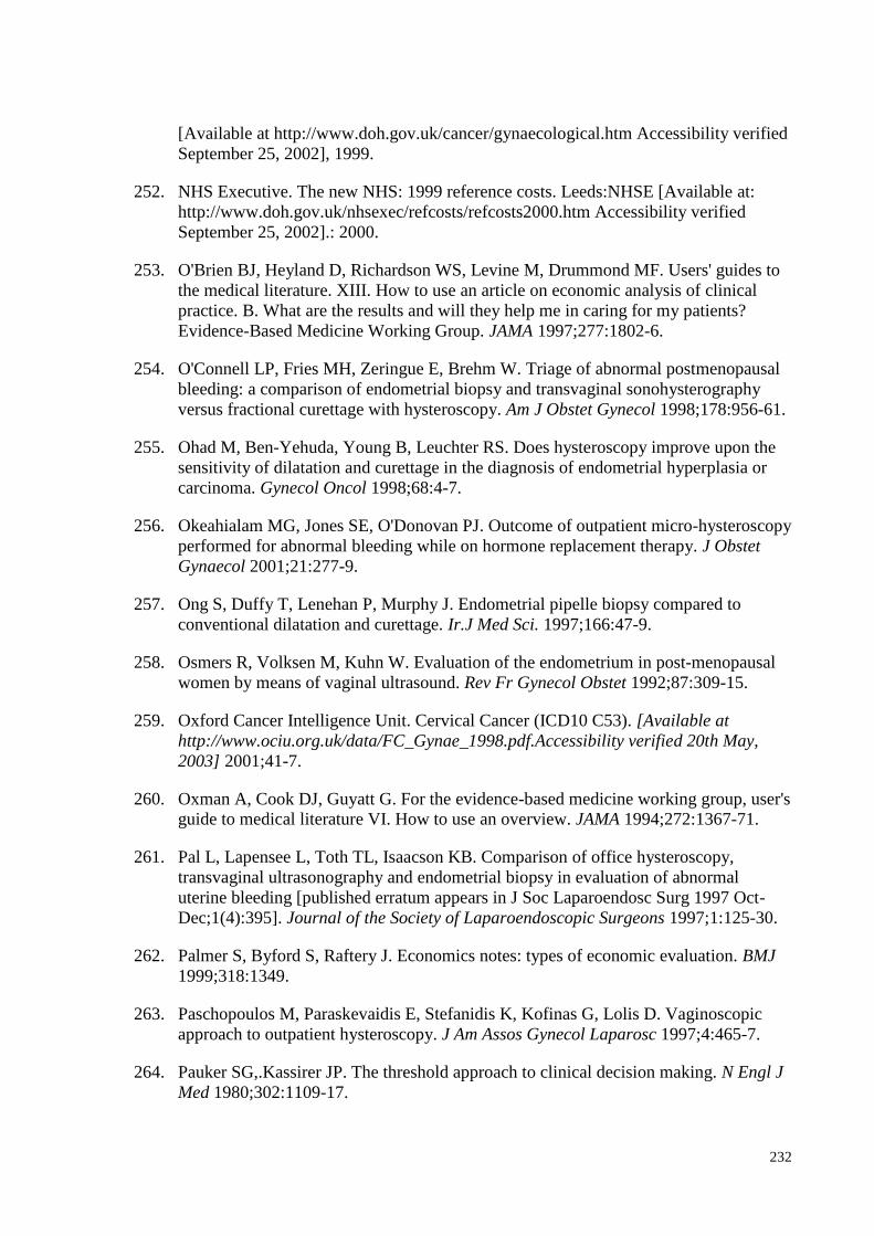
232
[Available at http://www.doh.gov.uk/cancer/gynaecological.htm Accessibility verified
September 25, 2002], 1999.
252. NHS Executive. The new NHS: 1999 reference costs. Leeds:NHSE [Available at:
http://www.doh.gov.uk/nhsexec/refcosts/refcosts2000.htm Accessibility verified
September 25, 2002].: 2000.
253. O'Brien BJ, Heyland D, Richardson WS, Levine M, Drummond MF. Users' guides to
the medical literature. XIII. How to use an article on economic analysis of clinical
practice. B. What are the results and will they help me in caring for my patients?
Evidence-Based Medicine Working Group. JAMA 1997;277:1802-6.
254. O'Connell LP, Fries MH, Zeringue E, Brehm W. Triage of abnormal postmenopausal
bleeding: a comparison of endometrial biopsy and transvaginal sonohysterography
versus fractional curettage with hysteroscopy. Am J Obstet Gynecol 1998;178:956-61.
255. Ohad M, Ben-Yehuda, Young B, Leuchter RS. Does hysteroscopy improve upon the
sensitivity of dilatation and curettage in the diagnosis of endometrial hyperplasia or
carcinoma. Gynecol Oncol 1998;68:4-7.
256. Okeahialam MG, Jones SE, O'Donovan PJ. Outcome of outpatient micro-hysteroscopy
performed for abnormal bleeding while on hormone replacement therapy. J Obstet
Gynaecol 2001;21:277-9.
257. Ong S, Duffy T, Lenehan P, Murphy J. Endometrial pipelle biopsy compared to
conventional dilatation and curettage. Ir.J Med Sci. 1997;166:47-9.
258. Osmers R, Volksen M, Kuhn W. Evaluation of the endometrium in post-menopausal
women by means of vaginal ultrasound. Rev Fr Gynecol Obstet 1992;87:309-15.
259. Oxford Cancer Intelligence Unit. Cervical Cancer (ICD10 C53). [Available at
http://www.ociu.org.uk/data/FC_Gynae_1998.pdf.Accessibility verified 20th May,
2003] 2001;41-7.
260. Oxman A, Cook DJ, Guyatt G. For the evidence-based medicine working group, user's
guide to medical literature VI. How to use an overview. JAMA 1994;272:1367-71.
261. Pal L, Lapensee L, Toth TL, Isaacson KB. Comparison of office hysteroscopy,
transvaginal ultrasonography and endometrial biopsy in evaluation of abnormal
uterine bleeding [published erratum appears in J Soc Laparoendosc Surg 1997 Oct-
Dec;1(4):395]. Journal of the Society of Laparoendoscopic Surgeons 1997;1:125-30.
262. Palmer S, Byford S, Raftery J. Economics notes: types of economic evaluation. BMJ
1999;318:1349.
263. Paschopoulos M, Paraskevaidis E, Stefanidis K, Kofinas G, Lolis D. Vaginoscopic
approach to outpatient hysteroscopy. J Am Assos Gynecol Laparosc 1997;4:465-7.
264. Pauker SG,.Kassirer JP. The threshold approach to clinical decision making. N Engl J
Med 1980;302:1109-17.
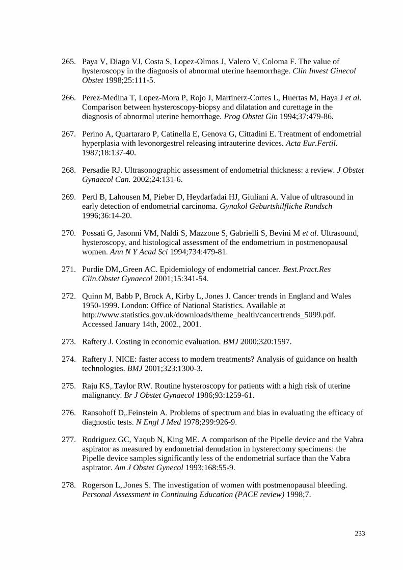
233
265. Paya V, Diago VJ, Costa S, Lopez-Olmos J, Valero V, Coloma F. The value of
hysteroscopy in the diagnosis of abnormal uterine haemorrhage. Clin Invest Ginecol
Obstet 1998;25:111-5.
266. Perez-Medina T, Lopez-Mora P, Rojo J, Martinerz-Cortes L, Huertas M, Haya J et al.
Comparison between hysteroscopy-biopsy and dilatation and curettage in the
diagnosis of abnormal uterine hemorrhage. Prog Obstet Gin 1994;37:479-86.
267. Perino A, Quartararo P, Catinella E, Genova G, Cittadini E. Treatment of endometrial
hyperplasia with levonorgestrel releasing intrauterine devices. Acta Eur.Fertil.
1987;18:137-40.
268. Persadie RJ. Ultrasonographic assessment of endometrial thickness: a review. J Obstet
Gynaecol Can. 2002;24:131-6.
269. Pertl B, Lahousen M, Pieber D, Heydarfadai HJ, Giuliani A. Value of ultrasound in
early detection of endometrial carcinoma. Gynakol Geburtshilfliche Rundsch
1996;36:14-20.
270. Possati G, Jasonni VM, Naldi S, Mazzone S, Gabrielli S, Bevini M et al. Ultrasound,
hysteroscopy, and histological assessment of the endometrium in postmenopausal
women. Ann N Y Acad Sci 1994;734:479-81.
271. Purdie DM,.Green AC. Epidemiology of endometrial cancer. Best.Pract.Res
Clin.Obstet Gynaecol 2001;15:341-54.
272. Quinn M, Babb P, Brock A, Kirby L, Jones J. Cancer trends in England and Wales
1950-1999. London: Office of National Statistics. Available at
http://www.statistics.gov.uk/downloads/theme_health/cancertrends_5099.pdf.
Accessed January 14th, 2002., 2001.
273. Raftery J. Costing in economic evaluation. BMJ 2000;320:1597.
274. Raftery J. NICE: faster access to modern treatments? Analysis of guidance on health
technologies. BMJ 2001;323:1300-3.
275. Raju KS,.Taylor RW. Routine hysteroscopy for patients with a high risk of uterine
malignancy. Br J Obstet Gynaecol 1986;93:1259-61.
276. Ransohoff D,.Feinstein A. Problems of spectrum and bias in evaluating the efficacy of
diagnostic tests. N Engl J Med 1978;299:926-9.
277. Rodriguez GC, Yaqub N, King ME. A comparison of the Pipelle device and the Vabra
aspirator as measured by endometrial denudation in hysterectomy specimens: the
Pipelle device samples significantly less of the endometrial surface than the Vabra
aspirator. Am J Obstet Gynecol 1993;168:55-9.
278. Rogerson L,.Jones S. The investigation of women with postmenopausal bleeding.
Personal Assessment in Continuing Education (PACE review) 1998;7.
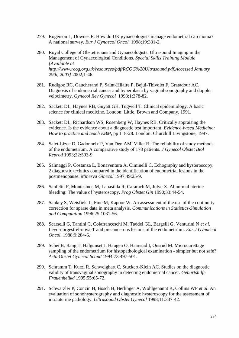
234
279. Rogerson L,.Downes E. How do UK gynaecologists manage endometrial carcinoma?
A national survey. Eur.J Gynaecol Oncol. 1998;19:331-2.
280. Royal College of Obstetricians and Gynaecologists. Ultrasound Imaging in the
Management of Gynaecological Conditions. Special Skills Training Module
[Available at
http://www.rcog.org.uk/resources/pdf/RCOG%20Ultrasound.pdf.Accessed January
29th, 2003] 2002;1-46.
281. Rudigoz RC, Gaucherand P, Saint-Hilaire P, Bejui-Thivolet F, Gratadour AC.
Diagnosis of endometrial cancer and hyperplasia by vaginal sonography and doppler
velocimetry. Gynecol Rev Gynecol 1993;1:378-82.
282. Sackett DL, Haynes RB, Guyatt GH, Tugwell T. Clinical epidemiology. A basic
science for clinical medicine. London: Little, Brown and Company, 1991.
283. Sackett DL, Richardson WS, Rosenberg W, Haynes RB. Critically appraising the
evidence. Is the evidence about a diagnostic test important. Evidence-based Medicine:
How to practice and teach EBM, pp 118-28. London: Churchill Livingstone, 1997.
284. Salet-Lizee D, Gadonneix P, Van Den AM, Villet R. The reliability of study methods
of the endometrium. A comparative study of 178 patients. J Gynecol Obstet Biol
Reprod 1993;22:593-9.
285. Salmaggi P, Costanza L, Bonaventura A, Ciminelli C. Echography and hysteroscopy.
2 diagnostic technics compared in the identification of endometrial lesions in the
postmenopause. Minerva Ginecol 1997;49:25-9.
286. Sanfeliu F, Montesinos M, Labastida R, Cararach M, Julve X. Abnormal uterine
bleeding: The value of hysteroscopy. Prog Obstet Gin 1990;33:44-54.
287. Sankey S, Weisfiels L, Fine M, Kapoor W. An assessment of the use of the continuity
correction for sparse data in meta analysis. Communications in Statistics-Simulation
and Computation 1996;25:1031-56.
288. Scarselli G, Tantini C, Colafranceschi M, Taddei GL, Bargelli G, Venturini N et al.
Levo-norgestrel-nova-T and precancerous lesions of the endometrium. Eur.J Gynaecol
Oncol. 1988;9:284-6.
289. Schei B, Bang T, Halgunset J, Haugen O, Haarstad I, Onsrud M. Microcurettage
sampling of the endometrium for histopathological examination - simpler but not safe?
Acta Obstet Gynecol Scand 1994;73:497-501.
290. Schramm T, Kurzl R, Schweighart C, Stuckert-Klein AC. Studies on the diagnostic
validity of transvaginal sonography in detecting endometrial cancer. Geburtshilfe
Frauenheilkd 1995;55:65-72.
291. Schwarzler P, Concin H, Bosch H, Berlinger A, Wohlgenannt K, Collins WP et al. An
evaluation of sonohysterography and diagnostic hysteroscopy for the assessment of
intrauterine pathology. Ultrasound Obstet Gynecol 1998;11:337-42.
235
292. Scottish Intercollegiate Guidelines Network. SIGN 50: A guideline developer's
handbook. Edinburgh: Edinburgh: Scottish Intercollegiate Guidelines Network, Royal
College of Physicians, Edinburgh, 2002. [Available at
http://www.sign.ac.uk/guidelines/fulltext/50/index.html. Accessibility verified 23
April 2003], 2001.
293. Scottish Intercollegiate Guidelines Network. Investigation of Post-menopasual
Bleeding. 1st Edition. (ISBN 1 899893 13 X). 2002. Edinburgh: Scottish
Intercollegiate Guidelines Network, Royal College of Physicians, Edinburgh, 2002.
(URL: http://www.sign.ac.uk). Ref Type: Report
294. Sculpher M, Fenwick E, Claxton K. Assessing quality in decision analytic cost-
effectiveness models. A suggested framework and example of application.
Pharmacoeconomics. 2000;17:461-77.
295. Seelbach-Gobel B, Rempen A, Kristen P. Transvaginal sonography of postmenopausal
endometrium. Geburtshilfe Frauenheilkd 1995;55:59-64.
296. Serden SP. Diagnostic hysteroscopy to evaluate the cause of abnormal uterine
bleeding. Obstet Gynecol Clin North Am 2000;27:277-86.
297. Sevcik L, Koliba P, Graf P. Diagnosis of endometrial pathology in postmenopausal
women]. Ceska Gynekol 1998;63:95-7.
298. Shapley M,.Redman CW. Endometrial sampling and general practice. Br.J Gen Pract.
1997;47:387-91.
299. Shipley CF, III, Simmons CL, Nelson GH. Comparison of transvaginal sonography
with endometrial biopsy in asymptomatic postmenopausal women. Journal
Ultrasound Med 1994;13:99-104.
300. Siegel JE, Weinstein MC, Russell LB, Gold MR. Recommendations for reporting
cost-effectiveness analyses. Panel on Cost-Effectiveness in Health and Medicine.
JAMA 1996;276:1339-41.
301. Siegel JE, Weinstein MC, Torrance GW. Reporting Cost-Effectiveness Studies and
Results. In Gold MR, Siegel JE, Russell LB, Weinstein MC, eds. Cost-Effectiveness in
Health and Medicine, pp 276-303. New York: Oxford University Press, 1996.
302. Simon P, Hollemaert S, Schwers J. Compared diagnostic effectiveness of
hysterography and hysteroscopy in common uterine diseases. J Gynecol Obstet Biol
Reprod 1993;22:141-4.
303. Smith-Bindman R, Kerlikowske K, Feldstein VA, Subak L, Scheidler J, Segal M et al.
Endovaginal ultrasound to exclude endometrial cancer and other endometrial
abnormalities. JAMA 1998;280:1510-7.
304. Smith P, Bakos O, Heimer G, Ulmsten U. Transvaginal ultrasound for identifying
endometrial abnormality. Acta Obstet Gynecol Scand 1991;70:591-4.
236
305. Song F, Sheldon T, Sutton A, Abrams K, Jones D. Methods for exploring
heterogeneity in meta-analysis. Evaluation & the Health Professions 2001;24:126-51.
306. Song F, Khan KS, Dinnes J, Sutton AJ. Asymmetric funnel plots and publication bias
in meta-analyses of diagnostic accuracy. Int.J Epidemiol. 2002;31:88-95.
307. Sonnendecker EWWSGB, Sevitz H, Hofmeyr GJ. Diagnostic accuracy of the
accurette endometrial sampler. S Afr Med J 1982;61:109-13.
308. Sousa R, Silvestre M, Sousa L, Falcao F, Dias I, Silva T et al. Transvaginal
ultrasonography and hysteroscopy in postmenopausal bleeding: a prospective study.
Acta Obstet Gynecol Scand 2001;80:856-62.
309. Southcott BM. Carcinoma of the endometrium. Drugs 2001;61:1395-405.
310. Spencer CP,.Whitehead MI. Endometrial assessment re-visited. Br J Obstet Gynaecol
1999;106:623-32.
311. Sterne JAC, Egger M, Davey Smith G. Investigating and dealing with publication and
other biases. In Egger M, Davey Smith G, Altman DG, eds. Systematic Reviews in
Health Care: Meta-analysis in Context., pp 189-208. London: BMJ Publishing Group,
2001.
312. Stovall TG, Ling FW, Morgan PL. A prospective, randomized comparison of the
Pipelle endometrial sampling device with the Novak curette. Am J Obstet Gynecol
1991;165:1287-90.
313. Stovall TG, Photopulos GJ, Poston WM, Ling FW, Sandles LG. Pipelle endometrial
sampling in patients with known endometrial carcinoma. Obstet Gynecol
1991;77:954-6.
314. Stovall TG, Solomon SK, Ling FW. Endometrial sampling prior to hysterectomy.
Obstet Gynecol 1989;73:405-9.
315. Suarez RA, Grimes DA, Majmudar B, Benigno BB. Diagnostic endometrial aspiration
with the Karman cannula. J Reprod.Med 1983;28:41-4.
316. Suchocki S, Luczynski K, Szymczyk A, Jastrzebski A, Mowlik R. Evaluation of
endometrial thickness by transvaginal ultrasonography as a screening method in early
diagnosis of endometrial cancer. Ginekol Pol 1998;69:279-82.
317. Sun-Kuie T, Sian-Ann T, Ka-Mui C, Soo-Kim L. The diagnostic value and patient
acceptability of outpatient endometrial sampling with Gynoscann. Aust NZ J Obstet
Gynaecol 1992;32:73-6.
318. Sutton A, Duval S, Tweedie R, Abrams K, Jones D. Empirical assessment of effect of
publication bias on meta-analyses. BMJ 2000;320:1574-7.
319. Tabor A, Watt HC, Wald NJ. Endometrial thickness as a test for endometrial cancer in
women with postmenopausal vaginal bleeding. Obstet Gynecol 2002;99:663-70.
237
320. Tahir MM, Bigrigg MA, Browning JJ, Brookes ST, Smith PA. A randomised
controlled trial comparing transvaginal ultrasound, outpatient hysteroscopy and
endometrial biopsy with inpatient hysteroscopy and curettage. Br J Obstet Gynaecol
1999;106:1259-64.
321. Taviani A, Braccini S, Toniazzi P, Pantani P, Costamagna V, Gambini G et al.
Transvaginal echography in patients with postmenopausal metrorrhagia. Minerva
Ginecol 1995;47:369-72.
322. Thompson SG,.Pocock SJ. Can meta-analyses be trusted? Lancet 1991;338:1127-30.
323. Todorova M, Buzalov S, Atanasov B, Tsaneva M, Gulubova M. Combined diagnostic
methods for women in the pre- and postmenopause. Akush Ginekol 1998;37:33-6.
324. Torgerson D,.Raftery J. Economics notes: measuring outcomes in economic
evaluations. BMJ 1999;318:1413.
325. Torgerson DJ,.Raftery J. Economic notes. Discounting. BMJ 1999;319:914-5.
326. Towbin NA, Gviazda IM, March CM. Office hysteroscopy versus transvaginal
ultrasonography in the evaluation of patients with excessive uterine bleeding [see
comments]. Am J Obstet Gynecol 1996;174:1678-82.
327. Tsuchiya A, Williams A. Welfare economics and economic evaluation. In Drummond
M, McGuire A, eds. Economic Evaluation in Health Care, pp 22-45. Oxford: Oxford
University Press, 2001.
328. Tsuda H, Kawabata M, Kawabata K, Yamamoto K, Umesaki N. Improvement of
diagnostic accuracy of transvaginal ultrasound for identification of endometrial
malignancies by using cutoff level of endometrial thickness based on length of time
since menopause. Gynecol Oncol 1997;64:35-7.
329. Uhiara JE, Dwarakanath LS, Newton JR. Audit of a new one-stop menstrual disorder
service. Gynae Endosc 1999;8:99-104.
330. Valli E,.Zupi E. A new hysteroscopic classification of and nomenclature for
endometrial lesions. J Am Assos Gynecol Laparosc 1995;2:279-83.
331. Van Beek EJ, Schenk BE, Michel BC. The role of plasma D-dimer concentration in
the exclusion of pulmonary embolism. Br J Haematol 1996;92:725-32.
332. van den Bosch T, Vandendael A, van Schoubroeck D, Wranz P, Lombard C.
Combining vaginal ultrasonography and office endometrial sampling in the diagnosis
of endometrial disease in postmenopasual women. Obstet Gynecol 1995;85:349-52.
333. van den Bosch T, Vandendael A, Wranz P, Lombard C. Endopap versus Pipelle
sampling in the diagnosis of postmenopausal endometrial disease. Eur J Obstet
Gynecol Reprod Biol 1996;64:91-4.
238
334. Varner RE, Sparks JM, Cameron CD, Roberts LL, Soong S-J. Transvaginal
sonography of the endometrium in postmenopausal women. Obstet Gynecol
1991;78:195-9.
335. Vercellini P, Cortesi I, Oldani S, Moschetta M, De Giorgi O, Crosignani et al. The
role of transvaginal ultrasonography and outpatient diagnostic hysteroscopy in the
evaluation of patients with menorrhagia. Hum Reprod 1997;12:1768-71.
336. Vigada G,.Malanetto C. Usefulness of hysteroscopy in the management of abnormal
uterine bleeding and intrauterine benign disease. Minerva Ginecol 1995;47:179-82.
337. Weber AM, Belinson JL, Bradley LD, Piedmonte MR. Vaginal ultrasonography
versus endometrial biopsy in women with postmenopausal bleeding. Am J Obstet
Gynecol 1997;177:924-9.
338. Weber G, Merz E, Bahlmann F, Rosch B. Evaluation of different transvaginal
sonographic diagnostic parameters in women with postmenopausal bleeding.
Ultrasound Obstet Gynecol 1998;12:265-70.
339. Wentz B. Progestin therapy in lesions of the endometrium. Seminars in Oncology
1985;12:23-7.
340. West CP. Uterine Fibroids. In Shaw RW, Soutter WP, Stanton SL, eds. Gynaecology,
pp 441-56. London: Chrchill Livingstone, 1997.
341. Whitehead A,.Whitehead J. A general parametric approach to the meta- analysis of
randomised clinical trials. Statist Med 1991;10:1665-77.
342. Whitehead MI,.Fraser D. The effects of estrogens and progestogens on the
endometrium. Modern approach to treatment. Obstet Gynecol Clin North Am
1987;14:299-320.
343. Widrich T, Bradley LD, Mitchinson AR, Collins RL. Comparison of saline infusion
sonography with office hysteroscopy for the evaluation of the endometrium. Am J
Obstet Gynecol 1996;174:1327-34.
344. Wingo PA, Huezo CM, Rubin GL, Ory HW, Peterson HB. The mortality risk
associated with hysterectomy. Am.J Obstet Gynecol 1985;152:803-8.
345. Wolman I, Sagi J, Ginat S, Jaffa AJ, Hartoov J, Jedwab G. The sensitivity and
specificity of vaginal sonography in detecting endometrial abnormalities in women
with postmenopausal bleeding. J Clin Ultrasound 1996;24:79-82.
346. Youssif SN,.McMillan DL. Outpatient endometrial biopsy: the pipelle. Br J Hosp Med
1995;54:198-201.
347. Yusuf S, Wittes J, Probstfield J, Tyroler HA. Analysis and interpretation of treatment
effects in subgroups of patients in randomized clinical trials. JAMA 1991;266:93-8.

239
348. Zannoni E, Radaelli U, Balestri M, Ferrazzi F. L'ecografia transvaginale nella
valutazione dell'endometrio in pazienti con perdite ematiche atipiche in
postmenopausa. In D'Addario V, Cagnasso G, eds. pp 27-30. Italy: CIC Edizioni
Internazionali, 1994.
349. Zorlu CG, Cobanoglu O, Isik AZ, Kutluay L, Kuscu E. Accuracy of pipelle
endometrial sampling in endometrial carcinoma. Gynecol Obstet Invest 1994;38:272-
5.