Embed Size (px)
Citation preview
Airway Management inTrauma
George Kovacs, MD, MHPE, FRCPCa,b,c,d,*, Nicholas Sowers, MD, FRCPCa,d
KEYWORDS
� Airway � Trauma � Airway management
KEY POINTS
� Airway management in trauma presents numerous unique challenges.
� A safe approach to airway management in trauma requires recognition of these anatomicand physiologic challenges.
� An approach to airwaymanagement for these complicated patients is presented based onan assessment of anatomic challenges and optimizing physiologic parameters.
INTRODUCTION
The “ABCs” of trauma resuscitation were born from the assumption that correctinghypoxemia and hypotension reduces morbidity and mortality. Definitive care forseverely injured or polytrauma patients includes the ability to provide advanced airwaymanagement in a variety of settings: in the emergency department, 20% to 30% intu-bations are for trauma.1,2 Airway management in the trauma patient presentsnumerous unique challenges beyond placement of an endotracheal tube (ETT), withoutcomes dependent on the provider’s ability to predict and anticipate difficulty andhave a safe and executable plan.
DOES EARLY DEFINITIVE TRAUMA AIRWAY MANAGEMENT SAVE LIVES?
Despite significant advances in prehospital care, injury prevention, and the develop-ment of trauma systems, early mortality from trauma has essentially remained
Disclosure Statement: The authors have nothing to disclose.a Department of Emergency Medicine, Division of Medical Education, Dalhousie University, 3rdFloor, HI Site, Suite 355, Room 364D, Halifax, Nova Scotia B3H 3A7, Canada; b Department ofAnaesthesia, Division of Medical Education, Dalhousie University, 3rd Floor, HI Site, Suite355, Room 364D, Halifax, Nova Scotia B3H 3A7, Canada; c Department of Medical Neurosci-ences, Division of Medical Education, Dalhousie University, 3rd Floor, HI Site, Suite 355,Room 364D, Halifax, Nova Scotia B3H 3A7, Canada; d Charles V. Keating Trauma & EmergencyCentre, QEII Health Sciences Centre, 1799 Robie Street, Halifax, Nova Scotia B3H 3G1, Canada* Corresponding author. Charles V. Keating Emergency & Trauma Centre, QEII Health SciencesCentre, 1799 Robie Street, Halifax, Nova Scotia B3H 3G1, Canada.E-mail address: [email protected]
Emerg Med Clin N Am 36 (2018) 61–84https://doi.org/10.1016/j.emc.2017.08.006 emed.theclinics.com0733-8627/18/ª 2017 The Authors. Published by Elsevier Inc. This is an open access article underthe CC BY-NC-ND license (http://creativecommons.org/licenses/by-nc-nd/4.0/).
Kovacs & Sowers62
unchanged.3 R. Adams Cowley, founder of Baltimore’s Shock Trauma Institute,defined the “golden hour” as a window to arrest the physiologic consequences of se-vere injury by rapidly transporting trauma patients to definitive care.4,5 The “stay andplay” versus “scoop and run” approach to prehospital trauma care has been a topic ofdebate since the early 1980s.6,7 Specific to airway management, there is evidence tosupport the argument that advanced airway management can be performed in theprehospital setting without delaying transfer to a trauma center.8,9 More recent datasuggest that when performed by skilled emergency medical services (EMS) providers,advanced airway management is associated with a significant decrease in mortal-ity.9,10 In the hospital setting, delayed intubation is associated with increased mortalityin noncritically injured trauma patients.11
Conversely, there is a growing body of evidence that prehospital advanced airwaymanagement may increase mortality for trauma patients in some circum-stances.8,12–14 How does one reconcile this seemingly conflicting data? Is endotra-cheal intubation (ETI) for prehospital trauma patients harmful? The answer is, “itdepends.” The Eastern Association for the Surgery of Trauma (EAST) practice guide-lines on ETI immediately following trauma acknowledged the conflicting prehospitaldata, stating the following:“No conclusion could be reached regarding prehospital intubation for patients with
traumatic brain injury, with or without RSI [rapid sequence intubation]. Diversity of pa-tient population, differing airway algorithms, various experience among emergencymedical service personnel in ETI, and differing reporting make consensus difficult.”15
It may be that the technical, procedure-focused management imperative of “gettingthe tube” is diverting attention away from the physiologic principles of oxygen delivery.Translated physiologically, the ABC priorities of trauma resuscitation are “stop thebleeding, maintain perfusion and oxygenate.” Lifesaving oxygenation maneuversmay include a jaw thrust, temporary bag-mask ventilation (BMV), placement of asupraglottic airway device, or ETI. Advanced does not necessarily mean better.
TRAUMA AND THE DIFFICULT AIRWAY
A “difficult airway” is defined as difficulty with laryngoscopy and intubation, BMV,supraglottic device ventilation, and/or front of neck airway (FONA) access.16,17
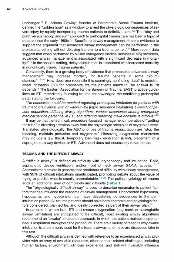
Anatomic markers are in general poor predictors of difficulty with airway management,with 90% of difficult intubations unanticipated, prompting debate about the value oftrying to predict what is usually unpredictable.18–21 The pathophysiology of traumaadds an additional layer of complexity and difficulty (Table 1).The “physiologically difficult airway” is used to describe nonanatomic patient fac-
tors that can influence the outcome of airway management. Uncorrected hypoxemia,hypocapnia, and hypotension can have devastating consequences in the peri-intubation period. All trauma patients should have both anatomic and physiologic fac-tors considered, planned for, and ideally corrected as part of their airway plan.22
In patients in whom both ETI and rescue oxygenation (bag-mask or supraglotticairway ventilation) are anticipated to be difficult, most existing airway algorithmsrecommend an “awake” intubation approach, in which the patient maintains sponta-neous respiration throughout the procedure. There are a variety of reasons why awakeintubation is uncommonly used for the trauma airway, and these are discussed later inthis text.Although the difficult airway is defined with reference to an experienced airway pro-
vider with an array of available recourses, other context-related challenges, includinghuman factors, environment, clinician experience, and skill will invariably influence
Table 1Predictors of difficult airway management in trauma
Difficult Airway Trauma Related Difficulty Approach
Difficult laryngoscopy and intubation
Limited mouth opening/jaw displacement
Collar/improper MILSTrismus
Open collar/ear-muff MILS
Inability to position MILS ELM/bougie/VL
Blood/vomitus Facial injuries/full stomach,delayed gastric emptying
2 suctions/SALAD approachFONA
Penetrating or bluntneck trauma
Disrupted or distorted airway Awake primary FIE; if notfeasible RSI VL-assisted FIE
Difficult BVM
Limited jaw thrust Mandibular fractures Early SGA use
Poor seal Facial injuries with swelling,disruption
Early SGA use
Blood/vomitus Facial injuries/full stomach,delayed gastric emptying
2 suctions/SALAD approachFONA
Penetrating or bluntneck trauma
Distorting subcutaneousemphysema, disruptedairway
Passive oxygen delivery/minimize PPV
Difficult SGA use
Blood/vomitus Facial injuries/full stomach,delayed gastric emptying
2 suctions/SALAD approachFONA
Penetrating or bluntneck trauma
Distorted/disrupted airway Direct visualization FIE/FONA,low tracheotomy
FONA
Penetrating or bluntneck trauma
Distorted/disrupted airwayCTM not accessible or injury
at or below CTM
Low tracheotomy
Abbreviations: BVM, bag-valve-mask; CTM, cricothyroid membrane; ELM, external laryngealmanipulation; FIE, flexible intubating endoscope; FONA, front of neck airway; MILS, manual inlinestabilization; PPV, partial-pressure ventilation; RSI, rapid sequence intubation; SALAD, suction-assisted laryngoscopy airway decontamination; SGA, supraglottic airway; VL, video laryngoscopy.
Trauma Airway 63
outcomes. Understanding when and why trauma patients may encounter difficulty inairway management can help guide the logistical and mental exercise of developingspecific mitigating strategies and contingency planning. A call for help should alwaysbe viewed as a patient-focused measure, not a sign of provider weakness.
AIRWAY MANAGEMENT TRAUMA SCENARIOSThe Head-Injured Patient
Traumatic brain injury (TBI) is the most common cause of mortality in trauma patients.Airway management in this cohort of patients is often performed for airway protection.Given the relatively high incidence of peri-intubation desaturation, hypocapnea, andhypotension in emergency intubations, the benefit of ETI for airway protection to pre-vent aspiration must be weighed against the risk of the occurrence of physiologicadverse events known to increase morbidity and mortality in TBI patients.23–26 If intu-bating for the purpose of airway protection, it is usually less time sensitive and should
Kovacs & Sowers64
not be rushed. Every precaution should be taken to adequately preoxygenate andresuscitate first.Apnea resulting from head injury requires immediate intervention. There are 3 mech-
anisms by which apnea may occur in TBI:
1. Severe or catastrophic brain injury2. Impact brain apnea (IBA)3. Loss of consciousness with resultant functional airway obstruction
Severe or catastrophic brain injury is usually nonsurvivable, and associated withearly death. Predictions of outcome are usually not made until the patient has under-gone a full trauma resuscitation, which often includes ETI. Contrastingly, IBA and func-tional airway obstruction may be correctable with simple airway opening maneuvers,with or without brief ventilation support. IBA from head trauma results in a primary res-piratory arrest without significant parenchymal injury to the brain.27 In contrast to pa-tients with head injury with functional airway obstruction, patients with IBA do notrespond to simple airway opening maneuvers alone, and may require brief ventilationsupport to prevent secondary hypoxic insult. With appropriate treatment, prognosis isgenerally good.Head-injured patients with a decreased level of consciousness frequently receive
prehospital advanced airway management.10 In one series, 30% to 40% of patientsare assessed as having partial or complete airway obstruction on EMS arrival.10 A pro-portion of these patients will respond to basic maneuvers, and those who do not usu-ally have more severe, less survivable injuries. This observation in part explains thecomparatively poor survival rates for trauma patients who are intubated in the preho-spital setting.
Management pearls for the patient with traumatic brain injury (TBI)
� Hypoxemia and hypotension during airway management significantly worsens outcomes inpatients with TBI.
� Airway management for airway protection should proceed only after adequate measureshave been taken to prevent intubation related physiologic disturbances.
� Postintubation hypocapnia is also associated with poor outcomes in patients with TBI andoften the result of adrenaline induced overzealous postintubation ventilation.
� Postinjury apnea requiring ventilation support does not necessarily predict poor outcome.
Airway Management in Patients with Suspected Cervical Spine Injuries
Trauma resuscitations typically proceed under the assumption that the patient has anunstable cervical spine (c-spine) injury until proven otherwise. In the prehospital envi-ronment, trauma patients are often placed in a cervical spine collar and secured to arigid backboard with blocks. Although a long-standing tradition in emergency medi-cine and trauma care, there is very limited published evidence to support the notionthat cervical spine collars and immobilization prevent secondary spinal cordinjury.28,29 Although the incidence of c-spine injuries is relatively low (occurring inapproximately 2% in the general trauma population and 6%–8% in patients withhead and facial trauma), practitioners often operate with deep concern that intubationmay cause secondary spinal cord injury, making it one of the most frequently encoun-tered reasons for difficulty in trauma airway management.30–33
Trauma Airway 65
A frequently studied outcome is the amount of translational or angular movement ofthe cervical spine caused by airway manipulation. Although it appears that spinalmovement occurs to a variable degree depending on the airway technique used, itis unclear whether or not this results in any important differences in clinical out-comes.34 In cadaveric studies of unstable c-spine injuries, movement occurring withboth direct laryngoscopy (DL) and indirect laryngoscopy do not significantly exceedthe physiologic values observed with intact spines.35,36 Despite the need to becautious, even in patients with known cervical spine injuries, secondary neurologicdeterioration is rare, with a reported incidence of 0.03%.33,37–39
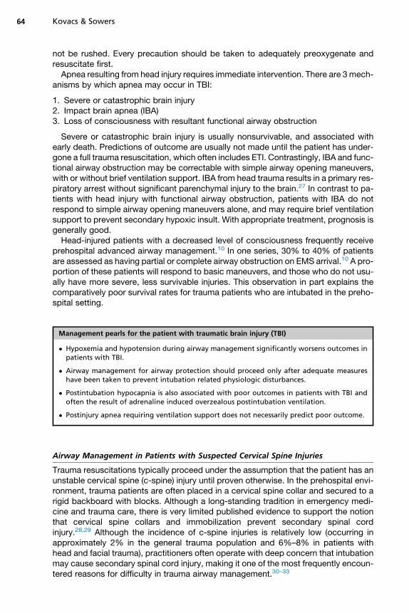
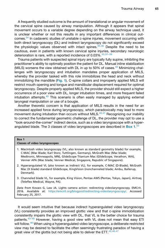
Trauma patients with suspected spinal injury are typically fully supine, inhibiting thepractitioner’s ability to optimally position the patient for DL. Manual inline stabilization(MILS) worsens the view obtained with DL in up to 50% of cases.40 Minimizing chal-lenges with laryngoscopy and intubation mandates proper application of MILS,whereby the provider tasked with this role immobilizes the head and neck withoutimmobilizing the mandible (Fig. 1). C-spine collars and improperly applied MILS willrestrict mouth opening and tongue and mandibular displacement required for optimallaryngoscopy. Despite properly applied MILS, the provider should still expect a higheroccurrence of a poor view with DL, longer intubation times, and more frequent failedintubation attempts.40 This scenario is often easily managed by applying externallaryngeal manipulation or use of a bougie.Another theoretic concern is that application of MILS results in the need for an
increased applied force during laryngoscopy, which paradoxically may lead to moremovement during intubation than occurs without MILS.40,41 Recognizing our inabilityto correct the fundamental geometric challenge of DL, the provider may opt to use a“look-around-the-corner” indirect device, such as a video laryngoscope with a hyper-angulated blade. The 3 classes of video laryngoscopes are described in Box 1.42
Box 1
Classes of video laryngoscopes
1. Macintosh video laryngoscopy (VL; also known as standard geometry blade) for example,C-MAC (Mac Blade; Karl Storz, Tuttlingen, Germany), McGrath Mac (Mac blade;Medtronic, Minneapolis, MN), GlideScope Titanium Mac (GlideScope, Verathon, WA),Venner APA (Mac blade; Venner Medical, Singapore, Republic of Singapore).
2. Hyperangulated VL (also known as indirect VL), for example, C-MAC (D-Blade), McGrathMac (X blade) standard GlideScope, KingVision (nonchanneled blade; Ambu, Ballerup,Denmark).
3. Channeled blade VL, for example, King Vision, Pentax AWS (Pentax, Tokyo, Japan), Airtraq(Teleflex Medical, Wayne, PA).
Data from Kovacs G, Law JA. Lights camera action: redirecting videolaryngoscopy. EMCrit.2016. Available at: https://emcrit.org/blogpost/redirecting-videolaryngoscopy/. AccessedFebruary 25, 2017.
It would seem intuitive that because indirect hyperangulated video laryngoscopy(VL) consistently provides an improved glottic view and that c-spine immobilizationconsistently impairs the glottic view with DL, that VL is the better choice for traumapatients.43–45 However, having a good view with VL does not mean that easy ETIwill follow.46 When using a hyperangulated video laryngoscope, a deliberate restrictedview may be desired to facilitate the often seemingly frustrating paradox of having agreat view of the glottis but not being able to deliver the ETT.42,46,47
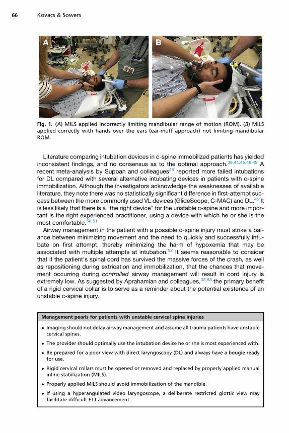
Fig. 1. (A) MILS applied incorrectly limiting mandibular range of motion (ROM). (B) MILSapplied correctly with hands over the ears (ear-muff approach) not limiting mandibularROM.
Kovacs & Sowers66
Literature comparing intubation devices in c-spine immobilized patients has yieldedinconsistent findings, and no consensus as to the optimal approach.38,44,45,48,49 Arecent meta-analysis by Suppan and colleagues45 reported more failed intubationsfor DL compared with several alternative intubating devices in patients with c-spineimmobilization. Although the investigators acknowledge the weaknesses of availableliterature, they note there was no statistically significant difference in first-attempt suc-cess between the more commonly used VL devices (GlideScope, C-MAC) and DL.45 Itis less likely that there is a “the right device” for the unstable c-spine and more impor-tant is the right experienced practitioner, using a device with which he or she is themost comfortable.50,51
Airway management in the patient with a possible c-spine injury must strike a bal-ance between minimizing movement and the need to quickly and successfully intu-bate on first attempt, thereby minimizing the harm of hypoxemia that may beassociated with multiple attempts at intubation.52 It seems reasonable to considerthat if the patient’s spinal cord has survived the massive forces of the crash, as wellas repositioning during extrication and immobilization, that the chances that move-ment occurring during controlled airway management will result in cord injury isextremely low. As suggested by Aprahamian and colleagues,33,53 the primary benefitof a rigid cervical collar is to serve as a reminder about the potential existence of anunstable c-spine injury.
Management pearls for patients with unstable cervical spine injuries
� Imaging should not delay airway management and assume all trauma patients have unstablecervical spines.
� The provider should optimally use the intubation device he or she is most experienced with.
� Be prepared for a poor view with direct laryngoscopy (DL) and always have a bougie readyfor use.
� Rigid cervical collars must be opened or removed and replaced by properly applied manualinline stabilization (MILS).
� Properly applied MILS should avoid immobilization of the mandible.
� If using a hyperangulated video laryngoscope, a deliberate restricted glottic view mayfacilitate difficult ETT advancement.
Trauma Airway 67
The Contaminated Airway
The presence of airway contamination with either blood or vomitus has been shown todecrease the rate of first-attempt intubation success, regardless of the device used.54
Blood and vomit in the airway can lead to early and late complications related to diffi-cult airway management and/or aspiration. The bloody airway is not uncommon intrauma patients with injuries to the face and/or neck and may range in severity fromscant bleeding, which is easily managed, to significant hemorrhage. The combinationof altered levels of consciousness, diminished protective airway reflexes, delayedgastric emptying, and full stomachs place trauma patients at high risk of vomitingand aspiration during airway management. Management of contaminated airwaymust begin with the expectation that the degree of blood, vomit, and secretions appre-ciated externally represents only a fraction of what may be encountered on initiation ofan RSI. As such, providers must ensure that adequate suction is available (at least 2large rigid suction catheters). Consideration must be given to positioning, placingthe patient in reverse Trendelenburg or, if safe to do so, seated upright or even leaningforward to allow drainage of blood and secretions. For c-spine immobilized patients,suction must be immediately within reach, and restraints securing the patient to thebed should be avoided. During the preoxygenation phase, positive-pressure ventila-tion (PPV) should be used only if necessary balanced against the patient’s oxygena-tion status, as ventilatory pressures of 20 cm H2O or more are likely to ventilate thestomach, increasing the risk of regurgitation and aspiration.When blood or vomitus is overwhelming suction capabilities, the provider may place
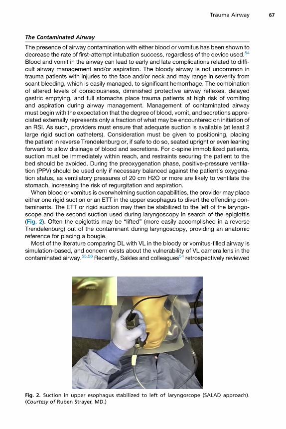
either one rigid suction or an ETT in the upper esophagus to divert the offending con-taminants. The ETT or rigid suction may then be stabilized to the left of the laryngo-scope and the second suction used during laryngoscopy in search of the epiglottis(Fig. 2). Often the epiglottis may be “lifted” (more easily accomplished in a reverseTrendelenburg) out of the contaminant during laryngoscopy, providing an anatomicreference for placing a bougie.Most of the literature comparing DL with VL in the bloody or vomitus-filled airway is
simulation-based, and concern exists about the vulnerability of VL camera lens in thecontaminated airway.55,56 Recently, Sakles and colleagues54 retrospectively reviewed
Fig. 2. Suction in upper esophagus stabilized to left of laryngoscope (SALAD approach).(Courtesy of Ruben Strayer, MD.)
Kovacs & Sowers68
more than 4600 intubations and demonstrated that, although airway contaminationwas associated with a decreased first-attempt success rate, this was irrespective ofthe choice of GlideScope or DL as the first-attempt device used. The use of DL orMacintosh VL, in which a direct approach can be used if the camera is obscuredwith the aid of a bougie, may be preferred approaches.Although not studied in a clinical setting, the Ducanto suction-assisted laryngos-
copy airway decontamination (SALAD) approach has gained acceptance as a methodto manage the soiled airway.57,58 In the uncommon circumstance in which blood orvomit is overwhelming these management strategies, intubation is not possible andthe patient is critically desaturating, rescue oxygenation with a BVM (bag-valve-mask) or SGA (supraglottic airway) is unlikely to work and an FONA approach isindicated.
Ducanto suction-assisted laryngoscopy airway decontamination approach to managingmassive airway contamination
� Use rigid large-bore suction to initially decontaminate
� Perform laryngoscopy keeping blade superior against tongue away from fluid
� Advance suction tip into upper esophagus then wedge in place to left of the laryngoscope
� Use second suction as needed
� Rotate laryngoscope blade 30 degrees to the left to open blade channel
� Place endotracheal tube (ETT), inflate the cuff
Management pearls for the patient with the contaminated airway
� Have at least 2 large-bore rigid suction catheters.
� Consider alternative options for hemorrhage control (sutures, packing, epistaxis kit).
� Minimize positive-pressure ventilation (PPV) and use a monometer for provider feedbackwhen mask ventilation is indicated.
� Look for epiglottis as an important landmark for glottis and have a bougie prepared for usewith DL.
� If a VL is considered the best option, Macintosh VL may be the preferred device, as it may beused directly if contamination obstructs camera.
� Consider esophageal ETT diversion connected to suction.
� Suction-assisted laryngoscopy airway decontamination (SALAD) approach.
� If intubation fails and patient is desaturating, front of neck airway (FONA) rescueoxygenation approach is indicated.
The Uncooperative or Agitated Patient
Uncooperative, violent, or agitated patients can encumber adequate assessment,leading to missed injuries and inadequate resuscitation. Agitation can be multifactorialand may be the result of head injury, hypoperfusion, hypoxemia, or intoxication. It maynot be clear why a patient is agitated and providers must determine if the patient isagitated AND injured or agitated BECAUSE the patient is injured.
Trauma Airway 69
The EAST guidelines recommend that aggressive behavior refractory to initial phar-macologic intervention is a discretionary indication for intubation; specifically that if apatient’s level of agitation prevents assessment and resuscitation, intubation andsedation should follow.15 Sise and colleagues59 reviewed 1078 trauma patients intu-bated for discretionary indications (eg, agitation, alcohol intoxication) and found that62% of patients, once investigated, had a significant head injury. Importantly, therewas no significant difference in complications associated with acute airway manage-ment in patients intubated for discretionary indications, as compared with those intu-bated for higher acuity reasons.59
In severely agitated patients, RSI is at times undertaken before optimal hemody-namic resuscitation and preoxygenation has been achieved. Patients rendered apneicas part of an RSI without adequate preoxygenation are at high risk of desaturation. Theuse of ketamine to facilitate cooperation and allow interventions including preoxyge-nation has been described as “delayed-sequence intubation” by Weingart and col-leagues.60 If given slowly, a dissociative intravenous dose of 1 to 1.5 mg/kg poseslittle risk of respiratory depression. However, the use of any sedative, particularly inthe presence of other intoxicating ingestions, may inhibit airway reflexes. Concernsthat ketamine may raise intracranial pressure and worsen outcomes in TBI is not sup-ported by evidence.61,62
Management pearls in the agitated trauma patient
� Agitation may be a symptom of traumatic pathology.
� Agitated patients may require facilitated cooperation to ensure adequate preoxygenation.
� Ketamine is an appropriate agent to facilitate cooperation in agitated patients inpreparation for airway management.
� Always be prepared to provide definitive airway intervention before administering sedation.
Maxillofacial Injuries
Maxillofacial fractures may present dramatically and affect airway management in oneof several ways.63 Posterior displacement from fractured maxillofacial segments maycause soft tissue collapse and occlude the airway, which may be worsened by thepresence of c-spine collar.64,65 Bleeding may be significant and cause airway man-agement challenges, as previously discussed. In the supine position, the pooling ofblood in the oropharynx may stimulate a gag response or vomiting, which in turnmay worsen bleeding. Although patients with mandibular fractures in 2 or more loca-tions may be easier to intubate due to increased mobility of the mandible and attachedsoft tissues, associated condylar fractures may cause a mechanical obstructionlimitingmouth opening, making laryngoscopy and intubation difficult.64,66 Maxillofacialfractures may also cause trismus that may resolve with the neuromuscular blockade;however, differentiating this from a mechanical obstruction before intubation isrequired and is often difficult.Airway management begins with careful consideration to patient positioning.
Awake, neurologically intact patients without neck pain should be allowed to positionthemselves however they are most comfortable to control tissue obstruction and allowdrainage of blood and secretions. They may be given a rigid suction catheter to usethemselves, which is more often tolerated, effective, and less likely to stimulate agag and resultant vomiting. Adherence to protocols requiring rigid spinal immobiliza-tion and supine positioning may result in catastrophe.
Kovacs & Sowers70
The provider should presume that preoxygenation in patients with facial traumamaybe difficult, and that reoxygenation with mask ventilation during RSI if the first attemptis unsuccessful may be difficult or impossible. Distortion of facial structures may makeobtaining a seal with a BVM device difficult and patients may poorly tolerate PPV, asdisruption of tissues may result in worsening bleeding and in cases of associatedlower airway trauma, significant subcutaneous emphysema. Practitioners must pro-ceed with the assumption that structural collapse of the airway may occur during anRSI.The choice of approach is based on the patient’s ability to maintain a patent airway
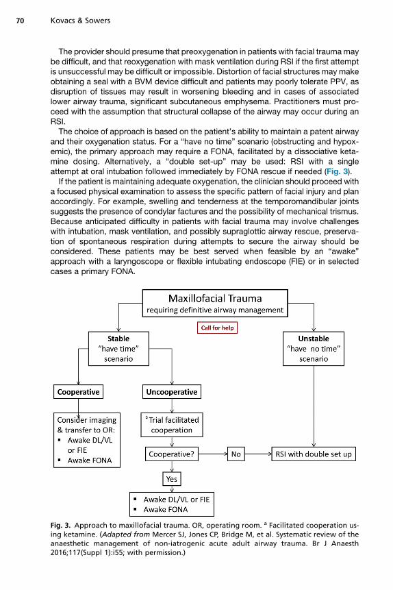
and their oxygenation status. For a “have no time” scenario (obstructing and hypox-emic), the primary approach may require a FONA, facilitated by a dissociative keta-mine dosing. Alternatively, a “double set-up” may be used: RSI with a singleattempt at oral intubation followed immediately by FONA rescue if needed (Fig. 3).If the patient is maintaining adequate oxygenation, the clinician should proceed with
a focused physical examination to assess the specific pattern of facial injury and planaccordingly. For example, swelling and tenderness at the temporomandibular jointssuggests the presence of condylar factures and the possibility of mechanical trismus.Because anticipated difficulty in patients with facial trauma may involve challengeswith intubation, mask ventilation, and possibly supraglottic airway rescue, preserva-tion of spontaneous respiration during attempts to secure the airway should beconsidered. These patients may be best served when feasible by an “awake”approach with a laryngoscope or flexible intubating endoscope (FIE) or in selectedcases a primary FONA.
Fig. 3. Approach to maxillofacial trauma. OR, operating room. a Facilitated cooperation us-ing ketamine. (Adapted from Mercer SJ, Jones CP, Bridge M, et al. Systematic review of theanaesthetic management of non-iatrogenic acute adult airway trauma. Br J Anaesth2016;117(Suppl 1):i55; with permission.)
Management pearls for the patient with facial injuries
� These patients require careful assessment of damaged anatomy recognizing the uniqueairway complications associated with facial fractures.
� Both laryngoscopy and mask ventilation may be challenging and a double set-up should beprepared for when rapid sequence intubation (RSI) is the chosen approach.
� An awake approach, although not always practical, should be considered.
� Management of aggressive bleeding should be anticipated.
� Allow patients to assume a position of comfort when safe to do so.
Trauma Airway 71
The Traumatized Airway
Airway management for the patient with a primary injury to the larynx or trachea is ahigh-stakes scenario, in which the loss of a stable airway can happen rapidly andwith little warning. Suspicion of a traumatized airway should initiate a call for help toan experienced colleague.Primary airway trauma is relatively uncommon in the civilian urban setting, with a re-
ported incidence of less than 1% (0.4% for blunt and 4.5% for penetrating injuries).67
Accordingly, practitioners have infrequent or limited experience in managing these pa-tients and existing management guidelines for care of these patients are mostly basedon expert opinion.68
Clinical findings suggestive of significant laryngotracheal airway injury includedysphagia, hoarseness, stridor, bleeding in the upper airway, subcutaneous emphy-sema, expanding hematoma, or in open penetrating injuries, obvious disruption ofthe larynx or trachea. If airway injury is suspected, aggressive PPV should be avoided.PPV in the setting of airway disruption can create or worsen pneumothorax, pneumo-mediastinum, or subcutaneous emphysema. Massive subcutaneous emphysema candistort airway anatomy, further complicating management. A potentially catastrophiccomplication is the conversion of a partial tracheal transection into a complete tran-section with the force of blindly passing an ETT or a bougie, particularly if relying ondistal “hold-up” to confirm placement.68,69
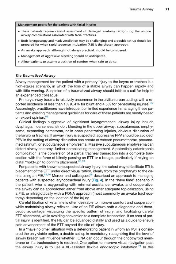
For patients with known or suspected airway injury, the safest way to facilitate ETI isplacement of the ETT under direct visualization, ideally from the oropharynx to the ca-rina using an FIE.68,70 Mercer and colleagues68 described an approach to managingpatients with suspected laryngotracheal injury (Fig. 4). In the “have time” scenario inthe patient who is oxygenating with minimal assistance, awake, and cooperative,the airway can be approached either from above after adequate topicalization, usinga FIE, or infraglottically with a FONA approach (most commonly an awake tracheos-tomy) depending on the location of the injury.Careful titration of ketamine is often desirable to improve comfort and cooperation
while maintaining airway reflexes. Use of an FIE allows both a diagnostic and thera-peutic advantage: visualizing the specific pattern of injury, and facilitating carefulETT placement, while avoiding conversion to a complete transection. If an area of par-tial injury is identified, the FIE can be advanced distally and used as a guide to ensuresafe advancement of the ETT beyond the site of injury.In a “have no time” situation with a deteriorating patient in whom an RSI is consid-
ered the only viable option, a double set-up is mandatory, recognizing that the level ofairway breach will influence whether FONA can occur through the cricothyroid mem-brane or if a tracheostomy is required. One option to improve visual navigation pastthe airway injury is to use a VL-assisted flexible endoscopic intubation.71 In this
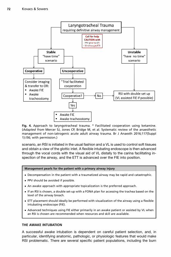
Fig. 4. Approach to laryngotracheal trauma. a Facilitated cooperation using ketamine.(Adapted from Mercer SJ, Jones CP, Bridge M, et al. Systematic review of the anaestheticmanagement of non-iatrogenic acute adult airway trauma. Br J Anaesth 2016;117(Suppl1):i56; with permission.)
Kovacs & Sowers72
scenario, an RSI is initiated in the usual fashion and a VL is used to control soft tissuesand obtain a view of the glottic inlet. A flexible intubating endoscope is then advancedthrough the vocal cords with the visual aid of VL distally to the carina facilitating in-spection of the airway, and the ETT is advanced over the FIE into position.
Management pearls for the patient with a primary airway injury
� Decompensation in the patient with a traumatized airway may be rapid and catastrophic.
� PPV should be avoided if possible.
� An awake approach with appropriate topicalization is the preferred approach.
� If an RSI is chosen, a double set-up with a FONA plan for accessing the trachea based on thelevel of the airway breach.
� ETT placement should ideally be performed with visualization of the airway using a flexibleintubating endoscope (FIE).
� Advanced techniques using FIE either primarily in an awake patient or assisted by VL whenan RSI is chosen are recommended when resources and skill are available.
THE AWAKE INTUBATION
A successful awake intubation is dependent on careful patient selection, and, inparticular, identifying anatomic, pathologic, or physiologic features that would makeRSI problematic. There are several specific patient populations, including the burn
Trauma Airway 73
patient and the patient with penetrating neck injuries, in whom an awake intubationmay be the approach of choice, as a strategy to mitigate both predicted difficultyand anticipated dynamic changes in airway anatomy and physiology.The awake intubation is a “have time” approach, involving placement of an ETT
following adequate topicalization in a patient who is able to maintain spontaneous res-pirations. It is not device-specific and can be performed using DL, VL, or an FIE. Suc-cess with awake intubation is dependent on meticulous airway topicalization, and ingeneral requires an awake and cooperative patient.72,73 The use of sedation is notroutine, and has been associated with increased awake intubation failures.74 Specif-ically, sedation should never be used in place of adequate airway topicalization. A diffi-cult airway paradox exists here: patients identified as difficult are selected to undergo atechnically more challenging awake approach, a procedure that is performed infre-quently bymost emergency physicians. There is no simple answer to this resource, skillavailability dilemma. It is our opinion that physicians who are responsible for acuteairway management should acquire and maintain the skills required for awake intuba-tion, as it can be a lifesaving approach in a specific subset of dynamic airway situations.
RAPID SEQUENCE INTUBATION
RSI involves the rapid administration of an induction agent and a neuromuscularblocking agent in quick succession to facilitate ETT placement in a patient who is pre-sumed to have a full stomach. RSI is the most common approach for airway manage-ment in trauma.75,76 Oxygenation with or without ventilation during the procedure(referred to by some as a “modified” RSI) is considered standard by most acutecare practitioners.77–79 Historically, the application of cricoid pressure (CP) to preventpassive aspiration has been considered an essential component of an RSI. However,its routine use remains controversial, with some evidence suggesting it may makevarious aspects of airway management more challenging. If cricoid pressure is beingapplied and the practitioner experiences difficulty with laryngoscopy, intubation, orventilation, CP should be immediately discontinued.80,81
Hemodynamic instability and hypoxemia must be aggressively managed beforeattempting RSI.22,23 The “rapid” part of an RSI refers to the delivery of the inductiondrug and neuromuscular blocking agent, and is not meant to imply a hurried or rushedprocess. RSI in underresuscitated patients may result in unintended poor outcomes,including critical hypoxemia and circulatory collapse.82,83 The term “resuscitativesequence intubation” has been suggested as a more representative term used todescribe the preparation and optimization of the patient’s physiologic status beforedefinitive airway management.84
Preparation
Numerous preparatory airway acronyms and checklists have been proposed toreduce errors and adverse events associated with RSI.85,86 Although evidence ofoutcome benefit may be lacking, based on an increased understanding of the roleof human factors in contributing to adverse airway outcomes, it seems a reasonablerecommendation that an airway checklist be used in the preparation phase of traumaairway management.87–91 In general, checklists should be simple, use terminologythat is clearly understood by the entire team, and can be performed rapidly (Fig. 5).
Optimizing Hemodynamics
Hemodynamic instability in trauma is most commonly caused by hypovolemia due tohemorrhage. Intravascular depletion shifts the gradient between right atrial pressure
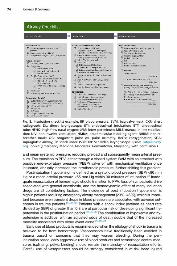
Fig. 5. Intubation checklist example. BP, blood pressure; BVM: bag-valve mask; CXR, chestradiograph; DL: direct laryngoscope; ETI: endotracheal intubation; ETT: endotrachealtube; HFNO: high flow nasal oxygen; LPM: leters per minute; MILS: manual in-line stabiliza-tion; NIV: non-invasive ventilation; NMBA: neuromuscular blocking agent; NRBM: non-re-breather mask; OG: orogastric; pulse ox, pulse oximetry. ReOx: reoxygenation; SGA:supraglottic airway; SI: shock index (SBP/HR); VL: video laryngoscope. (From SaferAirway.org Toolkit (Emergency Medicine Associates, Germantown, Maryland); with permission.)
Kovacs & Sowers74
and mean systemic pressure, reducing preload and subsequently mean arterial pres-sure. The transition to PPV, either through a closed system BVMwith an attached withpositive end-expiratory pressure (PEEP) valve or with mechanical ventilation onceintubated, abruptly increases the intrathoracic pressure, further shifting this gradient.Postintubation hypotension is defined as a systolic blood pressure (SBP) <90 mm
Hg or a mean arterial pressure <65 mm Hg within 30 minutes of intubation.92 Inade-quate resuscitation of hemorrhagic shock, transition to PPV, loss of sympathetic driveassociated with general anesthesia, and the hemodynamic effect of many inductiondrugs are all contributing factors. The incidence of post intubation hypotension ishigh in patients requiring emergency airway management (23%–46%), which is impor-tant because even transient drops in blood pressure are associated with adverse out-comes in trauma patients.23,92–94 Patients with a shock index (defined as heart ratedivided by SBP) of greater than 0.8 are at particular risk of developing significant hy-potension in the postintubation period.82,95,96 The combination of hypoxemia and hy-potension is additive, with an adjusted odds of death double that of the increasedmortality associated with either event alone.23,97,98
Early use of blood products is recommended when the etiology of shock in trauma isbelieved to be from hemorrhage. Vasopressors have traditionally been avoided intrauma based on concerns that they may worsen bleeding. During the peri-intubation phase, early aggressive use of blood products and hemorrhage control mea-sures (splinting, pelvic binding) should remain the mainstay of resuscitation efforts.Careful use of vasopressors should be strongly considered in at-risk head-injured
Trauma Airway 75
patients to prevent or manage postintubation hypotension. Sustained vasopressor useby infusion beyond the immediate peri-intubation period may be required to mitigatethe effects of PPV, postparalysis sedation, and ongoing losses unresponsive to fluidand blood product replacement.All commonly used induction agents will cause hypotension, particularly when a full
dose is administered to a volume-constricted patient. Dosage recommendations forRSI induction agents are largely based on patients without hemodynamic instability.As such, in trauma patients with hypotension or a shock index greater than 0.8, itseems prudent that the dose of the induction agent be reduced by at least50%.83,91,95,99 In North America, etomidate has been the most commonly used induc-tion agent used for RSI. Owing to the association between using etomidate and adre-nal suppression, many institutions have moved to alternative induction agents.1,100
With a favorable hemodynamic profile and strong analgesic effect, ketamine is quicklybecoming the preferred induction agent for trauma patients.100 A study comparingstandard full-dose ketamine with etomidate in trauma patients showed no survivalbenefit of one agent over the other.100 In light of this, it is probably true to suggestthat the dose of the drug is more important than the choice of drug.Paralysis should have no direct effect on the patient’s hemodynamic status, and in
hypoperfused states their dose should be increased. There is some debate regardingwhich neuromuscular blocking agent is superior for an RSI: both succinylcholine androcuronium may be safely used and can provide good intubating conditions.101 Itshould be emphasized, however, that administering too small a dose (<1.0 mg/kg)of rocuronium, particularly in low-flow states, will result in inadequate intubating con-ditions and a larger dose is recommended (1.2–1.6 mg/kg).101,102 Although cliniciansmay be weary of the prolonged effect of high-dose rocuronium in potentially difficultairway cases, extended, deep paralysis is in fact desirable in this circumstance, asit helps to create optimal conditions for laryngoscopy, BVM and SGA ventilation,and FONA. Having the sick trauma patient wake up to a state that he or she will beable rescue his or her own airway is simply not realistic, and will make efforts to securethe airway even more difficult.
Management of peri-intubation hemodynamic instability
� Resuscitation using blood products (packed red blood cells/massive transfusion) should bedone early in the preintubation phase of trauma management.
� In selected scenarios consider the use of vasopressors during the peri-intubation phase.
� Reduce the dose of all induction agents by at least 50% and increase the dose of theparalytic.
Avoiding Hypoxemia
Hypoxemia during emergency department (ED) RSI is common, occurring in morethan one-third of cases.24 Proceeding with an RSI in a patient who is already hypox-emic can result in catastrophic complications. Patients with preintubation oxygen sat-urations of less than 95% are at risk of abrupt desaturation within 90 seconds of theonset of apnea.103 Although recent literature has focused on extending safe apneatime with passive high-flow nasal oxygenation (HFNO) with or without noninvasiveventilation, the most important determinant of time to desaturation remains preoxyge-nation status.104
Although high oxygen saturation is reassuring, it is not a true measurement of oxy-gen reserve. Furthermore, oxygen saturation alone provides little information about
Kovacs & Sowers76
the true safe apnea time, which is defined by the relationship between oxygen reserveand the rate of oxygen consumption. Effective management of the preoxygenationphase requires both increasing the patient’s oxygen reserve while simultaneouslydecreasing the rate of oxygen consumption through aggressive resuscitation.Increasing the oxygen reserve has 3 components: denitrogenation of the lungs,recruitment of alveoli with PEEP, and apneic oxygenation.Denitrogenation is the primary physiologic mechanism of preoxygenation and is
dependent on delivery of a high concentration of oxygen, resulting in a 10-fold in-crease available oxygen. Increased functional residual capacity (FRC) provides anongoing reservoir to keep hemoglobin saturated. Delivery of close to 100% oxygenfor approximately 4 minutes is required to denitrogenate normal lungs and may bemost easily accomplished with high-flow, “flush-rate” (40 LPM) oxygen using a con-ventional non-rebreather mask.105 In patients with a high minute ventilation or shuntphysiology, a closed system BVM with a PEEP valve and a tight-fitting mask is thepreferred preoxygenation technique. If respiratory effort support is required, this isbest achieved using a mechanical (NIV) as opposed to manual ventilator (BVM).With atelectasis, pulmonary contusion, and other lung pathologies, the FRC is
diminished and the resultant shunt physiology renders preoxygenation with a highFiO2 less effective in preventing desaturation.106–108 Alveolar recruitment using an ox-ygen delivery device with PEEP is necessary to help mitigate the negative effects ofshunt physiology. A PEEP valve should be considered standard when using a BVMfor preoxygenation, as it will prevent entrainment in BVMs without a dedicated expira-tory valve. A PEEP valve attached to a BVM and applied over conventional nasalprongs at high flow (�15 LPM) in a spontaneously breathing patient will producecontinuous positive airway pressure–like conditions. Assisted ventilations are bestdelivered using a dedicated noninvasive ventilator; however, can be performed withthe same BVM/PEEP, nasal cannula combination. It is advisable to use a pressuremanometer attached to the BVM when using this combination to minimize high pres-sures that may result in gastric distention and aspiration.Alveolar oxygen delivery that continues without respiratory effort is referred to as
apneic oxygenation (AO). AO is facilitated by the pressure gradient between theoropharynx and the alveoli created by the differential uptake of oxygen and deliveryof CO2 to and from the alveoli, resulting in the passive transfer of oxygen.104,107 Bycontinuously replenishing the FRC oxygen stores, apneic oxygenation using conven-tional or specialized nasal prongs to administer high-flow oxygen (10–70 LPM) mayextend the safe apnea time after an RSI.103,104 Numerous studies have evaluated toeffectiveness of HFNO as an adjunct to preoxygenation for RSI, and the balance of ev-idence suggest the procedure as both safe and effective.104
Of note, the ability to extend the safe apnea time must not allow providers tobecome cavalier and should not encourage prolonged intubation attempts.106,109 Bythe time peripheral oxygen saturation begins to fall, cerebral hypoxemia has alreadyoccurred, a phenomenon known as “pulse-ox lag.”103 Effective situation awarenessis required, even (and perhaps especially) when supporting gas exchange by way ofpassive oxygenation, to stay within the “safe apnea” zone.
Preoxygenation: “the rule of 2s”
� Elevate the head (ear to sternum) and the bed greater than 20� (reverse Trendelenburg).
� Two sources of oxygen for all critically ill patients: high-flow nasal prongs �15 L/min andNRB/bag-mask ventilation �15 L/min.
� Two approaches for obstruction: OPA with a jaw thrust for soft tissue obstruction.
� Two attachments for your BVM: positive end-expiratory pressure valve and pressuremanometer.
� Two hands on all face masks: to ensure closed system oxygenation and ventilation andperform an aggressive jaw thrust.
� Two providers: the most experienced obtaining a tight mask seal and aggressive jaw thrustgiving feedback to the provider squeezing the bag to avoid overventilation andhyperventilation.
Abbreviations: BVM, bag-valve-mask; NRB, non-rebreather mask; OPA, oropharyngeal airway.
Trauma Airway 77
FRONT OF NECK AIRWAY TO SECURE THE AIRWAY
Numerous methods are used to access the trachea infraglottically, and the terminol-ogy describing the procedure is as variable as the techniques available to do so.Perhaps most accurate is the new term “front of neck airway” (FONA), which elimi-nates ambiguous verbiage like “surgical airway” or “airway rescue.”110 Althoughrare (0.05%–1.7% of ED-based intubations), the decision to perform FONA mustbegin during the initial assessment of the patient’s airway, long before the “cannotintubate cannot oxygenate” (CICO) scenario is encountered.111 This begins with apre-procedure briefing with team members to define clear triggers for moving aheadwith the procedure. Palpation of the anterior neck, and perhaps even marking theanticipated location of the cricothyroid membrane should be done routinely for allemergency airway cases and equipment should be both familiar to team membersand immediately available.Cognitive and team-based preparation is vital, as the decision to proceed with a
FONA is often delayed until critical hypoxemia has occurred.111 It is widely speculatedthat the most significant delay is often the result of hesitant decision-making and areluctance to perform a rarely encountered procedure.112 Open discussion of theemergency surgical airway as a potential outcome familiarizes the teamand normalizesthe procedure, shifting from the negative connotation of the “failed airway” to therecognition of the ultimately inevitable surgical airway.113 By normalizing the proced-ure, at least cognitively, we aim to reduce the psychological distress associated with it.No specific oxygen saturation level should be used as a trigger to perform FONA,
recognizing that a failed oxygenation situation is dynamic and characterized by arapidly falling oxygen saturation despite maximal efforts to reoxygenate the patient.111
Failed intubation followed by difficult mask ventilation with falling saturations repre-sents an impending FONA that should be initiated after a single rescue attempt withsupraglottic airway device. In rare situations, FONA may be the first and only invasiveairway technique attempted, even in the setting of normal oxygen saturation; forexample, massive facial trauma disrupting all recognizable airway landmarks. This“surgically inevitable” airway needs to be identified and declared early, such thattime is not wasted on fruitless efforts to intubate “from above.”There are several options for the emergency FONA and there remains some contro-
versy regarding the preferred approach. Historically, methods such as transtrachealjet ventilation and percutaneous cricothyroidotomy with a Seldinger technique, havebeen advocated. However, recent reviews have shown that complications associatedwith jet ventilation are unacceptably high and wire-guided approaches are not as easyor successful as once believed.114–117
There has been a recent push to adopt a modified open technique using a scalpel,finger, and bougie (the “scalpel-bougie” technique). The 2015 Difficult Airway Society
Kovacs & Sowers78
guidelines recommend that all clinicians responsible for airway management be ableto perform a FONA, and that the scalpel-bougie technique is the technique ofchoice.89 The scalpel-bougie technique has several advantages for the emergencyclinician: it relies on gross motor skills (which are more likely to be preserved duringperiods of acute stress), uses familiar equipment (scalpel, bougie, and a #6 ETT),and has a minimal number of steps.The body of evidence for this rarely needed procedure is (and will likely remain)
limited, yet all clinicians need to be mentally and technically prepared to rapidlyperform front of neck access to secure the airway. Success for high-acuity, low-op-portunity events like this requires frequent, deliberate practice using simulation.118
FONA trainers need not be expensive or complicated; motor habit can be developedusing simple models with Venturi oxygen tubing or an empty roll of bathroom tissue.
SUMMARY
Effective trauma care requires a team approach, with resuscitation priorities clearlycommunicated and interventions guided by the physiologic priorities that ensureadequate oxygen delivery. Although ensuring oxygenation and ventilation are prior-ities, airway management as the technical imperative of putting the “tube in thehole” must not overshadow other resuscitative elements.Providing advanced airway management is part of the A and B and C parallel resus-
citative priorities of trauma care. Safely managing both the anticipated and unantici-pated difficult airway requires technical expertise; however, decisions of when andhow to intervene are equally important determinants of outcome. Airway managementin trauma begins as soon as patient contact is made and rarely starts with placementof an ETT. Gaining intravenous access, beginning fluid resuscitation, and applying ox-ygen may be lifesaving and/or bridging interventions that allow for the safe executionof downstream more definitive procedures. Whether the team is a doctor, nurse, andtransporting paramedics or a group of 10, success is dependent on a shared under-standing of the importance of resuscitation before intubation and clear communica-tion of what and when various airway interventions will be performed. Then,securing the airway will have the best chance of making a positive difference in traumapatient outcomes.
REFERENCES
1. Brown CA, Bair AE, Pallin DJ, et al. NEAR III Investigators. Techniques, success,and adverse events of emergency department adult intubations. Ann EmergMed 2015;65(4):363–70.e1.
2. Kerslake D, Oglesby AJ, Di Rollo N, et al. Tracheal intubation in an urban emer-gency department in Scotland: a prospective, observational study of 3738 intu-bations. Resuscitation 2015;89:20–4.
3. Pieters BMA, Wilbers NER, Huijzer M, et al. Comparison of seven videolaryngo-scopes with the Macintosh laryngoscope in manikins by experienced andnovice personnel. Anaesthesia 2016;71(5):556–64.
4. Cowley RA. A total emergency medical system for the State of Maryland. MdState Med J 1975;24(7):37–45.
5. Rogers FB, Rittenhouse KJ, Gross BW. The golden hour in trauma: Dogma ormedical folklore? Injury 2015;46(4):525–7.
6. Border JR, Lewis FR, Aprahamian C, et al. Panel: prehospital trauma care–sta-bilize or scoop and run. J Trauma 1983;23(8):708–11.
Trauma Airway 79
7. Gold CR. Prehospital advanced life support vs “scoop and run” in trauma man-agement. Ann Emerg Med 1987;16(7):797–801.
8. Eckstein M, Chan L, Schneir A, et al. Effect of prehospital advanced life supporton outcomes of major trauma patients. J Trauma 2000;48(4):643–8.
9. Meizoso JP, Valle EJ, Allen CJ, et al. Decreased mortality after prehospital inter-ventions in severely injured trauma patients. J Trauma Acute Care Surg 2015;79(2):227–31.
10. Lockey DJ, Healey B, Crewdson K, et al. Advanced airway management isnecessary in prehospital trauma patients. Br J Anaesth 2014;1–6. https://doi.org/10.1093/bja/aeu412.
11. Miraflor E, Chuang K, Miranda MA, et al. Timing is everything: delayed intuba-tion is associated with increased mortality in initially stable trauma patients.J Surg Res 2011;170(2):286–90.
12. Stockinger ZT, McSwain NE Jr. Prehospital endotracheal intubation for traumadoes not improve survival over bag-valve-mask ventilation. J Trauma 2004;56(3):531–6.
13. Stiell IG, Nesbitt LP, Pickett W, et al. The OPALS Major Trauma Study: impact ofadvanced life-support on survival and morbidity. CMAJ 2008;178(9):1141–52.
14. Lecky F, Bryden D, Little R, et al. Emergency intubation for acutely ill and injuredpatients. Cochrane Database Syst Rev 2008;(2):CD001429.
15. Mayglothling J, Duane TM, Gibbs M, et al. Emergency tracheal intubation imme-diately following traumatic injury: an Eastern Association for the Surgery ofTrauma practice management guideline. J Trauma Acute Care Surg 2012;73(5):S333–40.
16. Murphy M, Hung O, Launcelott G, et al. Predicting the difficult laryngoscopicintubation: are we on the right track? Can J Anaesth 2005;52(3):231–5.
17. Law JA, Broemling N, Cooper RM, et al. The difficult airway with recommenda-tions for management–part 2–the anticipated difficult airway. Can J Anaesth2013;60(11):1119–38.
18. Yentis SM. Predicting difficult intubation–worthwhile exercise or pointless ritual?Anaesthesia 2002;57(2):105–9.
19. Nørskov AK, Wetterslev J, Rosenstock CV, et al. Effects of using the simplifiedairway risk index vs usual airway assessment on unanticipated difficult trachealintubation—a cluster randomized trial with 64,273 participants. Br J Anaesth2016;116(5):680–9.
20. Teoh WH, Kristensen MS. Prediction in airway management: what is worthwhile,what is a waste of time and what about the future? Br J Anaesth 2016;117(1):1–3.
21. Vannucci A, Cavallone LF. Bedside predictors of difficult intubation: a systematicreview. Minerva Anestesiol 2016;82(1):69–83.
22. Mosier JM, Joshi R, Hypes C, et al. The physiologically difficult airway. West JEmerg Med 2015;16(7):1109–17.
23. Spaite DW, Hu C, Bobrow BJ, et al. The effect of combined out-of-hospital hy-potension and hypoxia on mortality in major traumatic brain injury. Ann EmergMed 2017. https://doi.org/10.1016/j.annemergmed.2016.08.007.
24. Bodily JB, Webb HR, Weiss SJ, et al. Incidence and duration of continuouslymeasured oxygen desaturation during emergency department intubation. AnnEmerg Med 2015;1–7. https://doi.org/10.1016/j.annemergmed.2015.06.006.
25. Gebremedhn EG, Mesele D, Aemero D, et al. The incidence of oxygen desatu-ration during rapid sequence induction and intubation. World J Emerg Med2014;5(4):279–85.
Kovacs & Sowers80
26. Heffner AC, Swords DS, Nussbaum ML, et al. Predictors of the complication ofpostintubation hypotension during emergency airway management. J Crit Care2012;27(6):587–93.
27. Wilson MH, Hinds J, Grier G, et al. Impact brain apnoea–a forgotten cause ofcardiovascular collapse in trauma. Resuscitation 2016;105:52–8.
28. Kwan I, Bunn F, Roberts I. Spinal immobilisation for trauma patients. CochraneDatabase Syst Rev 2001;(2):CD002803.
29. Oteir AO, Jennings PA, Smith K, et al. Should suspected cervical spinal cord in-juries be immobilised? A systematic review protocol. Inj Prev 2014;20(3):e5.
30. Mulligan RP, Friedman JA, Mahabir RC. A nationwide review of the associationsamong cervical spine injuries, head injuries, and facial fractures. J Trauma 2010;68(3):587–92.
31. Dupanovic M, Fox H, Kovac A. Management of the airway in multitrauma. CurrOpin Anaesthesiol 2010. https://doi.org/10.1097/ACO.0b013e3283360b4f.
32. Thompson WL, Stiell IG, Clement CM, et al. Association of injury mechanismwith the risk of cervical spine fractures. CJEM 2009;11(1):14–22.
33. Manoach S, Paladino L. Manual in-line stabilization for acute airway manage-ment of suspected cervical spine injury: historical review and current questions.Ann Emerg Med 2007;50(3):236–45.
34. Kill C, Risse J, Wallot P, et al. Videolaryngoscopy with glidescope reduces cer-vical spine movement in patients with unsecured cervical spine. J Emerg Med2013;44(4):750–6.
35. Hindman BJ, Fontes RB, From RP, et al. Intubation biomechanics: laryngoscopeforce and cervical spine motion during intubation in cadavers—effect of severedistractive-flexion injury on C3–4 motion. J Neurosurg Spine 2016;1–11. https://doi.org/10.3171/2016.3.SPINE1640.
36. Hindman BJ, From RP, Fontes RB, et al. Intubation biomechanics: laryngoscopeforce and cervical spine motion during intubation in cadavers—cadavers versuspatients, the effect of repeated intubations, and the effect of type II odontoidfracture on C1-C2 motion. Anesthesiology 2015;123(5):1042–58.
37. Durga P, Sahu BP. Neurological deterioration during intubation in cervical spinedisorders. Indian J Anaesth 2014;58(6):684–92.
38. Crosby ET. Airway management in adults after cervical spine trauma. Anesthe-siology 2006;104(6):1293–318.
39. Farmer J, Vaccaro A, Albert TJ, et al. Neurologic deterioration after cervical spi-nal cord injury. J Spinal Disord 1998;11(3):192–6.
40. Thiboutot F, Nicole PC, Trepanier CA, et al. Effect of manual in-line stabilizationof the cervical spine in adults on the rate of difficult orotracheal intubation bydirect laryngoscopy: a randomized controlled trial. Can J Anaesth 2009;56(6):412–8.
41. LeGrand SA, Hindman BJ, Dexter F, et al. Craniocervical motion during directlaryngoscopy and orotracheal intubation with the Macintosh and Miller blades:an in vivo cinefluoroscopic study. Anesthesiology 2007;107(6):884–91.
42. Kovacs G, Law JA. Lights camera action: redirecting videolaryngoscopy. EMCrit.2016. Available at: https://emcrit.org/blogpost/redirecting-videolaryngoscopy/.Accessed February 25, 2017.
43. Bathory I, Frascarolo P, Kern C, et al. Evaluation of the GlideScope for trachealintubation in patients with cervical spine immobilisation by a semi-rigid collar.Anaesthesia 2009;64(12):1337–41.
Trauma Airway 81
44. Michailidou M, O’Keeffe T, Mosier JM, et al. A comparison of video laryngoscopyto direct laryngoscopy for the emergency intubation of trauma patients. World JSurg 2015;39(3):782–8.
45. Suppan L, Tramer MR, Niquille M, et al. Alternative intubation techniques vsMacintosh laryngoscopy in patients with cervical spine immobilization: system-atic review and meta-analysis of randomized controlled trials. Br J Anaesth2016;116(1):27–36.
46. Gu Y, Robert J, Kovacs G, et al. A deliberately restricted laryngeal view with theGlideScope� video laryngoscope is associated with faster and easier trachealintubation when compared with a full glottic view: a randomized clinical trial.Can J Anaesth 2016;63(8). https://doi.org/10.1007/s12630-016-0654-6.
47. Levitan R. Tips for using a hyperangulated video laryngoscope. ACEP Now 2015.Available at: http://www.acepnow.com/article/tips-for-using-a-hyperangulated-video-laryngoscope/. Accessed February 25, 2017.
48. Kleine-Brueggeney M, Greif R, Schoettker P, et al. Evaluation of six videolar-yngoscopes in 720 patients with a simulated difficult airway: a multicentre ran-domized controlled trial. Br J Anaesth 2016;116(5):670–9.
49. Norris A, Heidegger T. Limitations of videolaryngoscopy. Br J Anaesth 2016.https://doi.org/10.1093/bja/aew122.
50. Vu M, Vu E, Tallon J, et al. Airway management in trauma and the traumatizedairway. In: Kovacs G, Law J, editors. Airway management in emergencies. 2ndedition. Shelton (CT): People’sMedical PublishingHouse-USA; 2011. p. 299–316.
51. Crosby ET. Considerations for airway management for cervical spine surgery inadults. Anesthesiol Clin 2007;25(3):511–33, ix.
52. Duggan LV, Griesdale DEG. Secondary cervical spine injury during airway man-agement: beyonda “one-size-fits-all” approach. Anaesthesia 2015;70(7):769–73.
53. Aprahamian C, Thompson BM, Finger WA, et al. Experimental cervical spineinjury model: evaluation of airway management and splinting techniques. AnnEmerg Med 1984;13(8):584–7.
54. Sakles JC, Corn GJ, Hollinger P, et al. The impact of a soiled airway on intuba-tion success in the Emergency Department when using the GlideScope or thedirect laryngoscope. Acad Emerg Med 2017;38(1):42–9.
55. Ohchi F, Komasawa N, Mihara R, et al. Evaluation of gum-elastic bougiecombined with direct and indirect laryngoscopes in vomitus setting: arandomized simulation trial. Am J Emerg Med 2016. https://doi.org/10.1016/j.ajem.2016.12.032.
56. Mihara R, Komasawa N, Matsunami S, et al. Comparison of direct and indirectlaryngoscopes in vomitus and hematemesis settings: a randomized simulationtrial. Biomed Res Int 2015;806243. https://doi.org/10.1155/2015/806243.
57. DuCanto J, Serrano K, Thompson R. Novel airway training tool that simulatesvomiting: suction-assisted laryngoscopy assisted decontamination (SALAD)system. West J Emerg Med 2017;18(1):117–20.
58. Brainard C, Gerecht R. The art of SUCTIONING. JEMS 2015;40(8):26–30, 32.59. Sise MJ, Shackford SR, Sise CB, et al. Early intubation in the management of
trauma patients: indications and outcomes in 1,000 consecutive patients.J Trauma Inj Infect Crit Care 2009;66(1):32–40.
60. Weingart SD, Trueger NS, Wong N, et al. Delayed sequence intubation: a pro-spective observational study. Ann Emerg Med 2015;65(4):349–55.
61. Cohen L, Athaide V, Wickham ME, et al. The effect of ketamine on intracranialand cerebral perfusion pressure and health outcomes: a systematic review.Ann Emerg Med 2015;65(1):43–51.e2.
Kovacs & Sowers82
62. Zeiler FA, Teitelbaum J, West M, et al. The ketamine effect on ICP in traumaticbrain injury. Neurocrit Care 2014;21(1):163–73.
63. Hutchison I, Lawlor M, Skinner D. ABC of major trauma. Major maxillofacial in-juries. BMJ 1990;301:595–9.
64. Bowman-Howard M. Management of the traumatized airway. In: Hagberg C,editor. Handbook of difficult airway management. Philadelphia: Churchill Liv-ingstone; 2000. p. 199–206.
65. Jose A, Nagori S, Agarwal B, et al. Management of maxillofacial trauma in emer-gency: an update of challenges and controversies. J Emerg Trauma Shock2016;9(2):73.
66. Krausz AA, El-Naaj IA, Barak M. Maxillofacial trauma patient: coping with thedifficult airway. World J Emerg Surg 2009;4:21.
67. Kummer C, Netto FS, Rizoli S, et al. A review of traumatic airway injuries: potentialimplications for airway assessment and management. Injury 2007;38(1):27–33.
68. Mercer SJ, Jones CP, Bridge M, et al. Systematic review of the anaesthetic man-agement of non-iatrogenic acute adult airway trauma. Br J Anaesth 2016;117(suppl 1):i49–59.
69. Jain U, McCunn M, Smith CE, et al. Management of the traumatized airway.Anesthesiology 2016;124(1):199–206.
70. Horton CL, Iii CAB, Raja AS, et al. Trauma reports. J Emerg Med 2014;46(6):814–20.
71. Sowers N, Kovacs G. Use of a flexible intubating scope in combination with achanneled video laryngoscope for managing a difficult airway in the emergencydepartment. J Emerg Med 2016;50(2). https://doi.org/10.1016/j.jemermed.2015.10.010.
72. Higgs A, Cook TM, McGrath BA. Airway management in the critically ill: thesame, but different. Br J Anaesth 2016. https://doi.org/10.1093/bja/aew055.
73. Lapinsky SE. Endotracheal intubation in the ICU. Crit Care 2015;19:258.
74. Cook TM, Woodall N, Harper J, et al. Major complications of airway manage-ment in the UK: results of the Fourth National Audit Project of the Royal Collegeof Anaesthetists and the Difficult Airway Society. Part 2: intensive care and emer-gency departments. Br J Anaesth 2011;106(5):632–42.
75. Dronen S. Rapid-sequence intubation: a safe but ill-defined procedure. AcadEmerg Med 1999;6(1):1–2.
76. Mace SE. Challenges and advances in intubation: rapid sequence intubation.Emerg Med Clin North Am 2008;26(4):1043–68.
77. Salem MR, Clark-Wronski J, Khorasani A, et al. Which is the original and which isthe modified rapid sequence induction and intubation? Let history be the judge!Anesth Analg 2013;116(1):264–5.
78. Tobias JD. Rapid sequence intubation: what does it mean? Does it really matter?Saudi J Anaesth 2014;8(2):153–4.
79. Ehrenfeld JM, Cassedy EA, Forbes VE, et al. Modified rapid sequence inductionand intubation: a survey of United States current practice. Anesth Analg 2012;115(1):95–101.
80. Salem MR, Khorasani A, Zeidan A, et al. Cricoid pressure controversies: narra-tive review. Anesthesiology 2017;9(6):378–91.
81. Algie CM, Mahar RK, Tan HB, et al. Effectiveness and risks of cricoid pressureduring rapid sequence induction for endotracheal intubation. Cochrane Data-base Syst Rev 2015;(4):CD011656.
Trauma Airway 83
82. Heffner AC, Swords DS, Neale MN, et al. Incidence and factors associated withcardiac arrest complicating emergency airway management. Resuscitation2013;84(11):1500–4.
83. Perbet S, De Jong A, Delmas J, et al. Incidence of and risk factors for severecardiovascular collapse after endotracheal intubation in the ICU: a multicenterobservational study. Crit Care 2015;19(1):257.
84. Levitan R. Timing resuscitation sequence intubation for critically ill patients. ACEPNow 2015. Available at: http://www.acepnow.com/article/timing-resuscitation-sequence-intubation-for-critically-ill-patients/.
85. Hardy G, Horner D. BET 2: should real resuscitationists use airway checklists?Emerg Med J 2016;33(6):439–41.
86. Brindley PG, Beed M, Law JA, et al. Airway management outside the operatingroom: how to better prepare. Can J Anaesth 2017. https://doi.org/10.1007/s12630-017-0834-z.
87. Conroy MJ, Weingart GS, Carlson JN. Impact of checklists on peri-intubationcare in ED trauma patients. Am J Emerg Med 2014;32(6):541–4.
88. Smith KA, High K, Collins SP, et al. A preprocedural checklist improves thesafety of emergency department intubation of trauma patients. Acad EmergMed 2015;22(8):989–92. Reardon R, ed.
89. Frerk C, Mitchell VS, Mcnarry AF, et al. Difficult Airway Society 2015 guidelinesfor management of unanticipated difficult intubation in adults. Br J Anaesth2015;115:827–48.
90. Woodall N, Frerk C, Cook TM. Can we make airway management (even) safer?Lessons from national audit. Anaesthesia 2011;66(suppl. 2):27–33.
91. Leeuwenburg T. Airway management of the critically ill patient: modifications oftraditional rapid sequence induction and intubation. Crit Care Horizons 2015;1:1–10.
92. Green RS, Edwards J, Sabri E, et al. Evaluation of the incidence, risk factors,and impact on patient outcomes of postintubation hemodynamic instability.CJEM 2012;14(2):74–82.
93. Green RS, Turgeon AF, McIntyre LA, et al. Postintubation hypotension in inten-sive care unit patients: a multicenter cohort study. J Crit Care 2015. https://doi.org/10.1016/j.jcrc.2015.06.007.
94. Heffner AC, Swords D, Kline JA, et al. The frequency and significance of post-intubation hypotension during emergency airway management. J Crit Care2012;27(4):417.e9-13.
95. Trivedi S, Demirci O, Arteaga G, et al. Evaluation of preintubation shock indexand modified shock index as predictors of postintubation hypotension and othershort-term outcomes. J Crit Care 2015;30(4):861.e1-7.
96. LaiW-H,WuS-C,RauC-S, et al. Systolic bloodpressure lower thanheart rate uponarrival at and departure from the Emergency Department indicates a pooroutcome for adult traumapatients. Int J EnvironResPublicHealth 2016;13(6):528.
97. WangHE,BrownSP,MacDonaldRD, et al.Associationof out-of-hospital advancedairway management with outcomes after traumatic brain injury and hemorrhagicshock in the ROC hypertonic saline trial. Emerg Med J 2014;31(3):186–91.
98. Chou D, Harada MY, Barmparas G, et al. Field intubation in civilian patients withhemorrhagic shock is associated with higher mortality. J Trauma Acute CareSurg 2015;1. https://doi.org/10.1097/TA.0000000000000901.
99. Miller M, Kruit N, Heldreich C, et al. Hemodynamic response after rapidsequence induction with ketamine in out-of-hospital patients at risk of shockas defined by the shock index. Ann Emerg Med 2016;68(2):181–8.e2.
Kovacs & Sowers84
100. Upchurch CP, Grijalva CG, Russ S, et al. Comparison of etomidate and ketaminefor induction during rapid sequence intubation of adult trauma patients. AnnEmerg Med 2017;69(1):24–33.e2.
101. Tran DTT, Newton EK, Mount VAH, et al. Rocuronium versus succinylcholine forrapid sequence induction intubation. Cochrane Database Syst Rev2015;(10):CD002788.
102. Welch JL, Seupaul RA. Update: does rocuronium create better intubating con-ditions than succinylcholine for rapid sequence intubation? Ann Emerg Med2016. https://doi.org/10.1016/j.annemergmed.2016.09.001.
103. Weingart SD, Levitan RM. Preoxygenation and prevention of desaturation duringemergency airway management. Ann Emerg Med 2012;59(3):165–75.
104. Wong DT, Yee AJ, May Leong S, et al. The effectiveness of apneic oxygenationduring tracheal intubation in various clinical settings: a narrative review. Can JAnaesth 2017. https://doi.org/10.1007/s12630-016-0802-z.
105. Driver BE, Prekker ME, Kornas RL, et al. Flush rate oxygen for emergency airwaypreoxygenation. Ann Emerg Med 2017;69(1):1–6.
106. Mosier JM, Hypes CD, Sakles JC. Understanding preoxygenation and apneicoxygenation during intubation in the critically ill. Intensive Care Med 2017;43(2):226–8.
107. Nimmagadda U, Salem MR, Crystal GJ. Preoxygenation: physiologic basis,benefits, and potential risks. Anesth Analg 2017;124(2):507–17.
108. Sirian R, Wills J. Physiology of apnoea and the benefits of preoxygenation. ContEduc Anaesth Crit Care Pain 2009;9(4):105–8.
109. Sakles JC, Mosier J, Patanwala AE, et al. First pass success without hypoxemiais increased with the use of apneic oxygenation during RSI in the EmergencyDepartment. Acad Emerg Med 2016. https://doi.org/10.1111/acem.12931.
110. Pracy JP, Brennan L, Cook TM, et al. Surgical intervention during a can’t intubatecan’t oxygenate ( CICO ) event: emergency front-of-neck airway ( FONA )?. Br JAnaesth 2016;117(4):426–8
111. Law JA, Broemling N, Cooper RM, et al. The difficult airway with recommenda-tions for management—Part 1-Intubation encountered in an unconscious/induced patient. Can J Anaesth 2013;60(11):1089–118.
112. Hamaekers AE, Henderson JJ. Equipment and strategies for emergencytracheal access in the adult patient. Anaesthesia 2011;66(suppl. 2):65–80.
113. Levitan R, Chow Y. Levitan, the laryngeal handshake and the cartilaginous cage.PHARM Prehospital Retr Med 2013. Available at: http://prehospitalmed.com/2013/11/17/levitan-the-laryngeal-handshake-and-the-cartilaginous-cage/.
114. Duggan LV, Ballantyne Scott B, Law JA, et al. Transtracheal jet ventilation in the“can’t intubate can’t oxygenate” emergency: a systematic review. Br J Anaesth2016;117(suppl 1):i28–38.
115. Marshall SD. Evidence is important: safety considerations for emergency cath-eter cricothyroidotomy. Acad Emerg Med 2016;23(9):1074–6.
116. Cook TM, Woodall N, Frerk C. Major complications of airway management in theUK: results of the Fourth National Audit Project of the Royal College of Anaesthe-tists and the Difficult Airway Society. Part 1: anaesthesia. Br J Anaesth 2011;106(5):617–31.
117. Langvad S, Hyldmo PK, Nakstad AR, et al. Emergency cricothyrotomy—a sys-tematic review. Scand J Trauma Resusc Emerg Med 2013;21(1):43.
118. Petrosoniak A, Hicks CM. Beyond crisis resource management. Curr OpinAnaesthesiol 2013;26(6):699–706.