Embed Size (px)
Citation preview

APPROACH TO A CASE OF DIFFICULT AIRWAY WITH SPECIAL REFERANCE TO TRAUMA
Dr Shadab Kamal

• Pre hospital management• Initial evaluation and resuscitation• Airway obstruction and its management• Full stomach consideration• Airway evaluation and intervention• Some definations• Difficult airway management in general• Important consideration in head, eye, major blood
vessel, cervical spine injury and direct airway injury like maxillofacial trauma, neck trauma and tharacic trauma

3
Prehospital Management• Controling airway,
external hemorrhage, rapid transport with supplemental oxygen.
• Immobilization of the C-spine, combination of a hard collar and sandbags on opposite sides of the head.

Initial Evaluation and Resuscitation
Rapid Overview Primary Survey, Secondary Survey
The strategy of initial management will be continuous, priority-driven process of patient assessment, resuscitation, and reassessment. The general approach for evaluation of the acute trauma victim:

Rapid overview takes only a few seconds and is used to determine whether the patient is stable, unstable, dying, or dead.
Primary survey involves rapid evaluation of functions that are crucial to survival. ABCs i.e. airway patency, breathing, and circulation, Brief neurologic examination is performed, Examination for any external injuries.

The secondary survey : More elaborate systematic examination of the entire bodyRadiographic and other diagnostic procedures may also be performed if the stability of the patient permits.
The anesthesiologist: Manage the airway, Contributes for evaluation and resuscitation, Gather information needed for possible future anesthetic management.


Airway Obstruction; Partial or complete
Posteriorly displaced or lacerated pharyngeal soft tissues;
Soft tissue edema of the pharynx
Cervical or mediastinal hematoma; peripharyngeal hematoma
Blood or debris in the oropharynx
Teeth or foreign bodies in the pharynx
Bleeding, secretions, or foreign bodies within the airway
Displaced bone or cartilage fragments
Airway obstruction

Management of Airway obstruction• The initial steps in airway
management are – Chin lift, – Jaw thrust, – Clearing of the oropharynx by
suctioning, – Administration of 100%
oxygen.– Placement of an oro-
pharyngeal, naso-pharyngeal airway or LMA, in inadequately breathing patients, ventilation with a self-inflating bag.


Full stomach• A trauma patient is always considered to
have a full stomach and to be at risk for aspiration, and the urgency of securing the airway often does not permit adequate time for pharmacologic measures to reduce gastric volume and acidity.
• Thus, selection of a safe technique for securing the airway is employed: – Rapid-sequence induction with cricoid pressure for
patients without serious airway problems, – Awake intubation with sedation and topical anesthesia,
for those with anticipated serious airway difficulties.

Airway Evaluation and Intervention
Airway evaluation involves
The diagnosis of any trauma to the airway
or surrounding tissues,
Recognition and anticipation of the
respiratory consequences of these injuries,
Prediction of the potential for
exacerbation of these or other injuries by any
contemplated airway management
maneuvers

• Although nontraumatic causes of airway difficulty, such as pre-existing factors may be present.
• L-E-M-O-N:– Look externally– Evaluate 3-3-2 rule– Mallampati classification– Obstruction– Neck mobility
• ASA difficult airway algorithm can be applied with certain modifications.
• cancellation of airway management when difficulty arises may not be an option.

Difficult airway
According to ASA “The clinlcal
situation in which a conventionally
trained anesthesiologist
experiences difficulty with
mask ventilation, difficulty with
laryngoscopy and intubation, or
both”.
Result of complex interactions between
Patient factors
Clinical setting
Skills & preferences of practitioner

Difficult Mask Ventilation:
• As defined by ASA task force, when it is not possible for the unassisted anesthesiologist to maintain oxygen saturation > 90% using 100% oxygen & positive pressure ventilation in a patient whose oxygen saturation was > 90% before anesthetic intervention.
Difficult Laryngoscopy:
• “It is not possible to visualize any portion of vocal cords with conventional laryngoscope”
• Usually corresponds to Cormack & Lehane’s grade IV view.
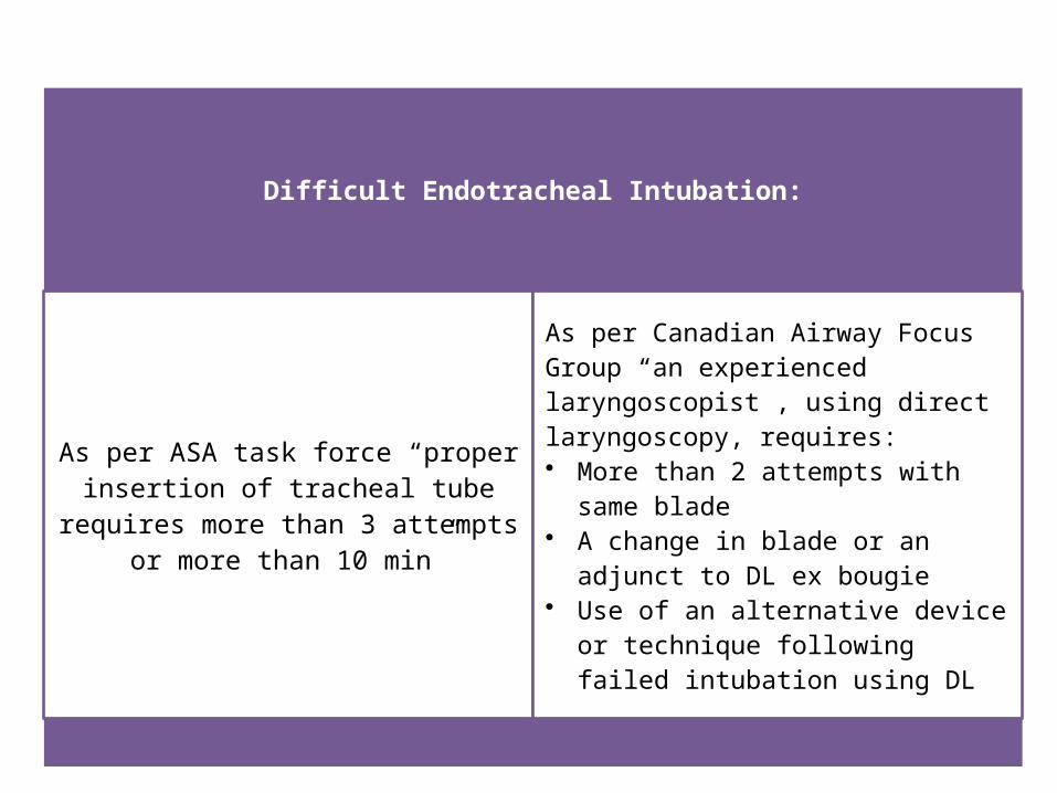
Difficult Endotracheal Intubation:
As per ASA task force “proper insertion of tracheal tube requires more than 3
attempts or more than 10 min”
As per Canadian Airway Focus Group “an experienced laryngoscopist , using direct laryngoscopy, requires:• More than 2 attempts with same blade• A change in blade or an adjunct to DL ex
bougie• Use of an alternative device or
technique following failed intubation using DL

The American Society of Anesthesiologists (ASA) algorithm for management of difficult airways is a useful starting point for the trauma anesthesiologist, whether in the ED or the OR
Emergency airway
management algorithm

Advance Airway Management
Advanced airway management is
indicated if there is
Cardiac arrest,
Apnea,
Persistent obstruction,
Severe head injury,
Maxillofacial trauma,
Penetrating neck injury with an
expanding hematoma, Major chest
injury(flail chest), Inability to maintain SpO2
>90% by facemask

Patient preparation•Monitoring equipments: EtCO2, Pulse-Oximeter, NIBP, ECG•Oxygenation equipments: •Wide bore Suction catheter of appropriate size•I.V. access & premedication•Intubation equipments:•Gum elastic bougie or intubating stylet•Surgical or needle cricothyrodotomy kit:•Equipments for checking tube position:•Prevention of gastric aspiration:•Patient positioning:

The approach depends upon the patient’s injuries, airway status and the care provider’s experience with such equipment & procedures.There is no single universal technique of intubation which may be favorable in all circumstances.Regardless of the associated injuries, the primary means of securing the airway in the vast majority of acutely desaturating patients with maxillofacial trauma is awake orotracheal intubation via direct laryngoscopy.

22
In agitate
d a
nd
unc
ooperative
patie
nts, t
opical a
nest
hesia
of t
he air
way is i
mpossi
ble, se
dative age
nts
may res
ult i
n a
pnea
obstr
ucti
on, as
pirati
on
Ac
ute
obstr
ucti
on fr
om
upper air
way tra
uma
may re
quire e
merge
ncy cric
ot
hyr
ot
omy
or
perc
uta
ne
ous
or s
urgical trac
he
ost
omy .
If facial or
neck i
nj
uries
precl
ude e
ndotrac
heal i
nt
ubati
on, trac
he
ost
omy
under l
ocal a
nest
hesia s
houl
d
be c
onsi
dere
d.
An awake intubation under direct laryngoscopy or
fiberoptic bronchoscopy with topical anesthesia can be attempted if the patient is
cooperative.

Maxillofacial, neck, and chest injuries, as well as cervicofacial burns, are the most common trauma-related causes of difficult tracheal intubation.
Rapid examination of the anterior neck for feasibility of access to the cricothyroid membrane.
Tracheostomy takes longer to perform than a cricothyroidotomy and requires neck extension,
If cricothyroidotomy will be in place for more than 2 to 3 days, Consider for conversion to tracheostomy.
C/I to cricothyroidotomy include: age < 12 years and suspected laryngeal trauma

24
• RSI is reserved for an uncooperative patient• In this technique –100% oxygen for 3 min• Do not bag and mask ventilate for fear of aspiration• Direct Laryngoscopy and orotracheal intubation
with MILS in suspected C-spineis, Avoid Neck hyperextension and excessive axial traction
• Cricoid pressure is applied.• Ketamine for hemodynamically unstable /
thiopentone in stable patients• Succinylcholine 1.0-1.5 mg/kg• ETI and confirmation

25
Oral Vs Nasotracheal Intubation In general, oral intubation is preferable to nasal intubation in the emergency setting because• Epistaxis.• Risk for sinusitis in a patient who will be
mechanically ventilated for > 24 hours.• use of a smaller-diameter tube will also increase the
difficulty of subsequent airway suctioning and fiberoptic bronchoscopy.
• of the risk of injury to the brain in the presence of a basilar skull or cribriform plate fracture.

• However, If nasal intubation is most likely to be successful in a given situation, then prefer nasal, Change to an oral tube once the patient's condition has stabilized.
• ATLS guidelines suggest that airway management provider should proceed with the method of intubation with which they are most proficient.


Head, Open Eye, and Contained Major Vessel Injuries
• Adequate oxygenation and ventilation, deep anesthesia and profound muscle relaxation required.
• This helps prevent hypertension, coughing, bucking, and thereby minimizes ICT, IOP, or BP, which can result in herniation of the brain, extrusion of eye contents, or dislodgment of a hemostatic clot from an injured vessel, respectively.
• Anesthetic sequence preferred includes preoxygenation and opioid loading, followed by relatively large doses of an intravenous anesthetic and muscle relaxant.

• Succinylcholine, may be used with prior administration of defasciculating dose of NDMRs. Alternatively, rocuronium can be used.
• Neither muscle relaxants nor intravenous anesthetics are indicated when initial assessment suggests a difficult airway.
• In any other trauma patient, hypotension dictates either reduced or no intravenous anesthetic administration.

31

Cervical Spine Injury
Initial evaluation:• Emergency airway management may have to be
performed without ruling out C-spine injury while the patients are in a rigid collar and neck stabilizing devices due to lack of time to evaluate the injury, and
• Clearance of the neck at the earliest possible time after airway management should be performed to minimize the complications associated with the collar, such as pressure ulceration, ICP elevation in head injured patients, compromised central venous access, and airway management challenges if reintubation is needed.

According to the American National Emergency X-Radiography Utilization
Study (NEXUS) no need for radiographic evaluation in conscious patient with:
no posterior midline tenderness in the
neck
no focal neurologic deficit with a normal
level of alertness,
no evidence of intoxication,
absence of painful distracting injury

• Canadian rule is more reliable than those for NEXUS in diagnosing C-spine injury in responsive patients.
• Diagnostic capability: Three-view plain films < CT scans < MRI
• CT is not sensitive in picking up soft tissue and ligamentous C-spine injury, and MRI is the gold standard for ruling out C-spine injury. However, it is so sensitive that it can detect subtle injuries that are clinically insignificant.
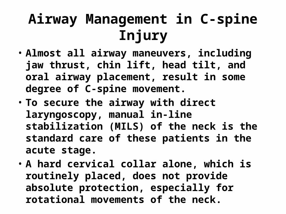
Airway Management in C-spine Injury
• Almost all airway maneuvers, including jaw thrust, chin lift, head tilt, and oral airway placement, result in some degree of C-spine movement.
• To secure the airway with direct laryngoscopy, manual in-line stabilization (MILS) of the neck is the standard care of these patients in the acute stage.
• A hard cervical collar alone, which is routinely placed, does not provide absolute protection, especially for rotational movements of the neck.

• MILS is best accomplished by having two operators in addition to the physician who is actually managing the airway.
• The first operator stabilizes and aligns the head in neutral position without applying cephalad traction. The second operator stabilizes both shoulders by holding them against the table or stretcher.
• The anterior portion of the hard collar, which limits mouth opening, may be removed after immobilization.

• Other measures and techniques, including the McCoy laryngoscope, rigid fiberoptic video laryngoscopes (Glidescope, Airtraq, McGrath, King vision), FOB, lightwand, translaryngeal (retrograde) intubation, and cricothyroidotomy, can be used to secure the airway in patients requiring cervical spine immobilization.

• Supraglottic intubating airways with or without the aid of FOB can be used, but neck movement with these devices appears to be comparable to that produced by conventional laryngoscopes.
• Flexible fiberoptic laryngoscopy, lightwand, and possibly translaryngealguided intubation cause almost no neck movement, but blood or secretions in the airway, a long preparation time, and difficulty in their use in comatose, uncooperative, reduces FOB utility during initial management.

39
DIRECT AIRWAY TRAUMA
• Direct airway damage can occur anywhere between the nasopharynx and the bronchi.
• it can be classified into broad categories of blunt and penetrating trauma, and each of these can be considered in the context of direct injury to the airway itself versus compromise or threat to the airway caused by the proximity of the injury in the neck.

Maxillofacial injuries
• A hematoma or
edema in the face, tongue, or neck may expand during the first several hours after injury and ultimately occlude the airway.
partial or complete airway obstruction
soft tissue edema of the pharynx
peripharyngeal hematoma
blood or debris in the oropharynx
teeth or foreign bodies in the pharynx

41
• Coronal CT is the investigation of choice for facial injuries
• Plain radiograph with waters view and submental-vertical view can also be done
• OrthoPentoGram (OPG) is done for mandibular fractures
• Tracheal intubation or a surgical airway is necessary as an initial measure to avert airway compromise in these circumstances.
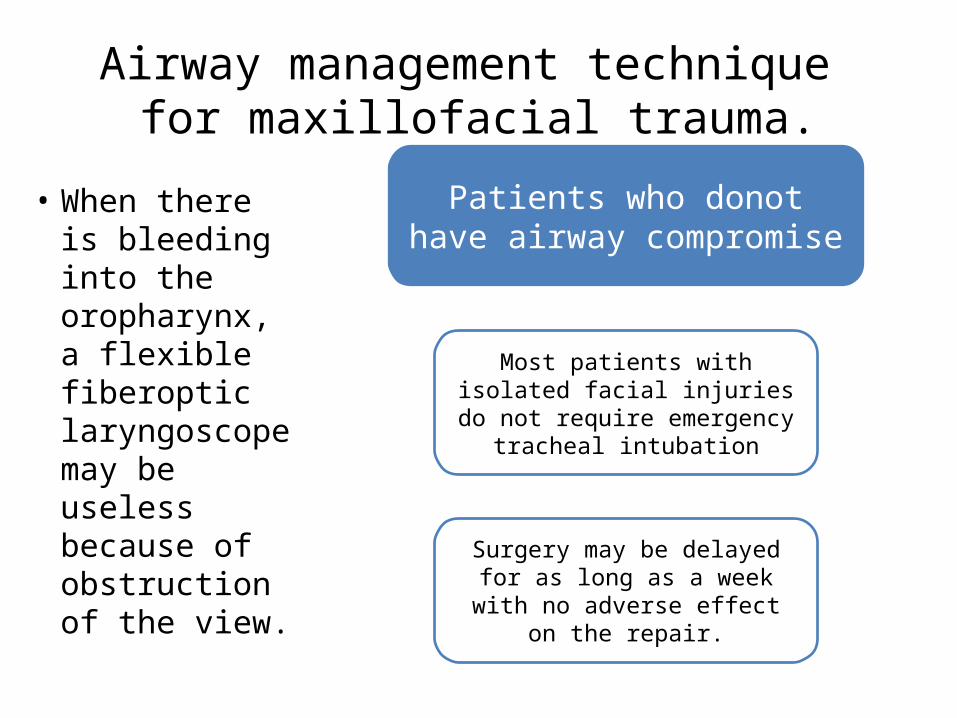
Airway management technique for maxillofacial trauma.
• When there is bleeding into the oropharynx, a flexible fiberoptic laryngoscope may be useless because of obstruction of the view.
Patients who donot have airway compromise
Most patients with isolated facial injuries do not require
emergency tracheal intubation
Surgery may be delayed for as long as a week with no
adverse effect on the repair.


• A retrograde technique, using a wire or epidural catheter passed through a 14-gauge catheter introduced into the trachea through the cricothyroid membrane.
• A surgical airway is indicated in airway compromise, when direct laryngoscopy has failed or is considered impossible
• Tracheostomy may be indicated as an emergency procedure in the emergency room within a few minutes of arrival or as a delayed procedure in the OR for airway control.

• Comminuted mandibular, midfacial bilateral LeFort III, and panfacial fractures are likely to be managed with tracheostomy for definitive surgery.
• To avoid the possible complications of tracheostomy, submental or submandibular intubation, which involves externalizing the proximal end of an orotracheal flexible armored tube through a small submental incision has been performed. Thus, the trachea remains surgically intact.

Penetrating Neck Trauma• Neck is divided into three
zones. Zone 1 extends from the clavicles inferiorly to the level of the cricoid cartilage superiorly. Zone 2 extends from the cricoid cartilage to a line drawn through the angles of the mandible.Zone 3 is the area above the angles of the mandible.
Cervical Airway Injuries

47
Blunt Neck Trauma• Inability to localize injury precisely,
blunt injury is usually more diffuse.
• Initial evaluation:– Asses for any bruising or
ecchymosis related to the external injury.
– Inspection of oropharynx to detect injury to the tongue or dentition.
– The external neck should then be palpated carefully from the mandible to the clavicle.
Palpation
Identification of any swelling, hemorrhage,
or subcutaneous emphysema
Evaluation for tenderness of the neck and in particular of the
airway structures
Evaluation of upper airway anatomy direct airway
injury and landmarks for surgical airway intervention


49
• Infrequently, direct blunt neck trauma can cause laryngeal fracture or tracheal transsection.
• Although a trial of bag-mask ventilation may be tempting, it promptly produces profound subcutaneous emphysema and accelerates the patient's deterioration.
• The best approach is prompt transfer to the operating room for surgical exploration of the anterior neck and establishment of the airway by tracheostomy distal to the transsection.

• The strategy depends on the clinical presentation.• The tracheas of some patients with penetrating
airway injuries, especially stab wounds, may be intubated through the airway defect without the need for anesthetics or optical equipment.
• The presence of cartilaginous fractures or mucosal abnormalities necessitates awake intubation with an FOB or awake tracheostomy.
• Laryngeal damage will make impossible cricothyroidotomy.

Thoracic Airway Injuries
• Occur due to penetrating trauma to intrathoracic region, blunt injury involving membranous trachea, mainstem bronchi and iatrogenic causes such as tracheal intubation.
• In intubated patients without the suspicion of a tracheal injury, difficulty in obtaining a seal around the ETT or the presence on a chest radiograph of a large radiolucent area in the trachea corresponding to the cuff suggests a perforated airway.

• Selective conservative management with an endotracheal tube placed using bronchoscopic guidance distal to the tracheal injury.
• Patients with lesions >4 cm, cartilaginous rather than membranous injuries, concomitant esophageal trauma, progressive subcutaneous emphysema, severe dyspnea requiring intubation and ventilation, difficulty with mechanical ventilation, pneumothorax with an air leak through the chest drains, and mediastinitis are still managed surgically.

• Mental Status– Resolution of intoxication – Able to follow commands – Noncombative – Pain adequately controlled
• Airway Anatomy and Reflexes– Appropriate cough and gag – Ability to protect the
airway from aspiration – No excessive airway edema
or instability
• Respiratory Mechanics– Adequate tidal volume and
respiratory rate – Normal motor strength – Required Fio2 less than 0.50
• Systemic Stability– Adequately resuscitated
(see above) – Small likelihood of urgent
return to the operating room
– Normothermic, without signs of sepsis
Criteria for OR or PACU Extubation of Trauma Patients


• Airway management in the trauma patients is tailored to the type of injury, the nature and degree of airway compromise, and the patient’s hemodynamic and oxygenation status.
Thank you